clinicopathological features of nonalcoholic fatty liver … · · 2015-11-21am deeply grateful...
TRANSCRIPT

Clinicopathological features of nonalcoholic fatty liver disease in
Korean young men
Nu Ri Chon
Department of Medicine
The Graduate School, Yonsei University

Clinicopathological features of nonalcoholic fatty liver disease in
Korean young men
Directed by Professor Kwan Sik Lee, MD
The Master's Thesis submitted to the Department of Medicine, the Graduate School of Yonsei University
in partial fulfillment of the requirements for the degree of Master of Medical Science
Nu Ri Chon December 2008

This certifies that the Master's Thesis of Nu Ri Chon is approved.
------------------------------------
------------------------------------
------------------------------------
The Graduate School Yonsei University
December 2008

ACKNOWLEDGEMENTS
This humble work could not be completed without the
efforts of certain people. As a novice, I am very much short
of writing an article. Without my great masters’ concerns
and contributions, I would never finish my scholarship.
Firstly my deepest gratitude goes to Dr. Kwan Sik Lee, MD,
Ph.D. Through him, I learned many skills and academic
properties. Not only as a teacher in the graduate school, but
as a senior in the hospital , he is a model of my life as a
doctor. My deepest gratitude goes to Dr. Young Nyun Park,
MD, Ph D. and Dong Sup Yoon, MD, Ph D., who advised
and teached me a lot of knowledges for this article. Also I
am deeply grateful to Dr. Yong Han Paik, MD, Ph D. Even
in abroad, he always concerned me and lead me to this
article. Finally, I always thank to my only God and my
parents.

<TABLE OF CONTENTS> ABSTRACT ………………… 1
I. INTRODUCTION ………………… 4
II. MATERIALS AND METHODS ……………………7
1. Subjects and data collections ……………………7
2. Measurements of subjects …………………… 7 3. Serum chemistry …………… ……… 8 4. Pathologic classification and diagnosis of NASH ……… 8 5. Statistical analysis …………………… 12
III. RESULTS ……………… 13
1. Dermographic data and subjects ………………13 2. Comparison of baseline charateristics between three groups
classified by NAS ………………14 3. Pathologic and clinical charateristics of NASH classified by
Schwimmer’s classification ………………18
IV. DISCUSSION ……………………… 24
V. CONCLUSION ……………………… 28
REFERENCES ……………………… 29
ABSTRACT(IN KOREAN) ……………………… 37

<LIST OF FIGURES>
LIST OF FIGURES
Figure 1. Comparison of GOT in three groups classified by NASH activity score · · · · · · · · · · · · · 1 6
Figure 2. Comparison of GTP in three groups classified by
NASH activity score · · · · · · · · · · · · · · 1 6
Figure 3. Correlation between GOT and NASH activity sco re · · · · · · · · · · · · · · 17
Figure 4. Correlation between GPT and NASH activity
score · · · · · · · · · · · · · 1 7 Figure 5. Histologic characteristics of nonalcoholic
steatohepatitis classified by Schwimmer’s c l a s s i f i c a t i o n · · · · · · · · · · · · · 1 9
Figure 6. Comparison of pathological characteristics of
nonalcoholic steatohepatitis classified by Schwimmer’s c lassif icat ion · · · · · · · · · · · · · 21

Figure 7. Composition of the definite nonalcoholic steatohepatitis and borderline nonalcoholic steatohepatitis in three types classified by Schwimmer’s classification ··· · · · · · · · · · · 23
LIST OF TABLES
Table 1. Definition of nonalcoholic steatohepatitis and scoring by NASH activity score ··················· 9
Table 2. Definition of nonalcoholic steatohepatitis type ····· 11
Table 3. Comparison of baseline characteristics between three
groups classified by NASH activity score ··········· 15
Table 4. Pathologic characteristics of nonalcoholic
steatohepatitis classified by Schwimmer ’s
classification · · · · · · · · · · · · · · · · · · · · 20
Table 5. Clinical character is t ics of nonalcoholic
steatohepatit is classif ied by Schwimmer ’s

class i f icat ion · · · · · · · · · · · · · · · 22

1
<ABSTRACT>
Clinicopathological features of nonalcoholic fatty liver disease in Korean young men
Nu Ri Chon
Department of Medicine
The Graduate School, Yonsei University
(Directed by Professor Kwan Sik Lee, MD)
Background: Nonalcoholic fatty liver disease (NAFLD) is a chronic liver
disease occurring without a significant alcohol consumption. Its prevalence is
increasing in Korean society as a dietary pattern and life-style become more
westernized and an obese population increases. Especially nonalcoholic
steatohepatitis (NASH), which has a potential to progress to liver cirrhosis or
even hepatocellular carcinoma, holds considerable clinical significance. In
this study, we examined clinicopathological features of NASH in Korean
young men using a guideline presented by NASH Clinical Research Network
and classifying according to the criteria suggested by Schwimmer et al. to
understand the natures and clinical progression of NAFLD.
Methods: 64 Korean young men under age 30 years (22.2 ± 2.75),
2
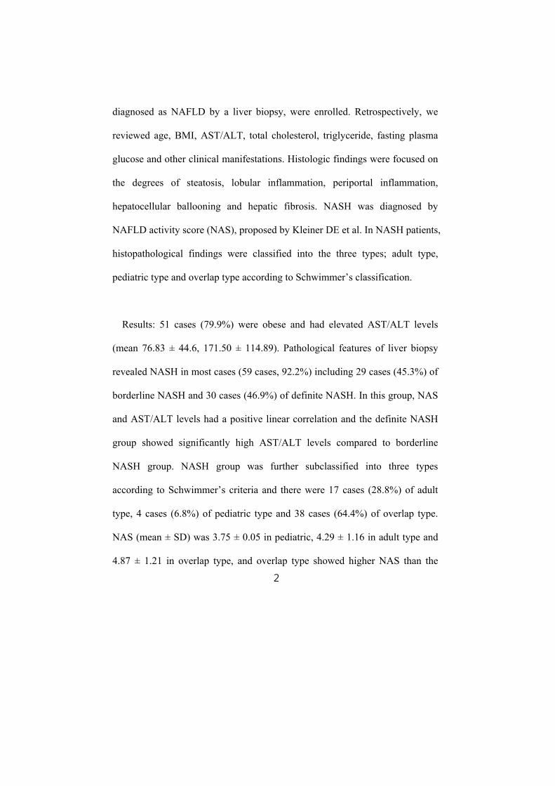
diagnosed as NAFLD by a liver biopsy, were enrolled. Retrospectively, we
reviewed age, BMI, AST/ALT, total cholesterol, triglyceride, fasting plasma
glucose and other clinical manifestations. Histologic findings were focused on
the degrees of steatosis, lobular inflammation, periportal inflammation,
hepatocellular ballooning and hepatic fibrosis. NASH was diagnosed by
NAFLD activity score (NAS), proposed by Kleiner DE et al. In NASH patients,
histopathological findings were classified into the three types; adult type,
pediatric type and overlap type according to Schwimmer’s classification.
Results: 51 cases (79.9%) were obese and had elevated AST/ALT levels
(mean 76.83 ± 44.6, 171.50 ± 114.89). Pathological features of liver biopsy
revealed NASH in most cases (59 cases, 92.2%) including 29 cases (45.3%) of
borderline NASH and 30 cases (46.9%) of definite NASH. In this group, NAS
and AST/ALT levels had a positive linear correlation and the definite NASH
group showed significantly high AST/ALT levels compared to borderline
NASH group. NASH group was further subclassified into three types
according to Schwimmer’s criteria and there were 17 cases (28.8%) of adult
type, 4 cases (6.8%) of pediatric type and 38 cases (64.4%) of overlap type.
NAS (mean ± SD) was 3.75 ± 0.05 in pediatric, 4.29 ± 1.16 in adult type and
4.87 ± 1.21 in overlap type, and overlap type showed higher NAS than the

3
pediatric type (p<0.01). Concerning on fibrosis, 36 cases (94.7%) of overlap
type showed stage 2 and 3, and 4 cases (100%) of pediatric type and 15 cases
(88.2%) of adult type showed stage 1. The fibrosis stage is significantly higher
in the overlap type than the other types (p<0.01). Other pathological features
including steatosis and lobular inflammation showed no significant difference
among three types and clinical features also showed no significant difference.
Conclusion: Majority of Korean young men with nonalcoholic fatty liver
disease (NAFLD) turned out to have borderline or definite NASH based on the
pathological features of liver biopsy. More than half of NASH cases showed
overlap type of pediatric and adult NASH, which showed higher degree of
NAS and fibrosis stage compared to pediatric type. These findings suggest that
overlap type of NASH in Korean young men might be disease progression
starting from pediatric period.
-----------------------------------------------------------------------------------------------Key words : Nonalcoholic fatty liver disease, nonalcoholic steatohepatitis, liver biopsy, Korean young men

4
Clinicopathological features of nonalcoholic fatty liver disease in Korean young men
Nu Ri Chon
Department of Medicine
The Graduate School, Yonsei University
(Directed by Professor Kwan Sik Lee, MD)
I. INTRODUCTION
Nonalcoholic fatty liver disease (NAFLD) is a clinicopathological diagnosis
characterized histologically by macrovesicular fat accumulation in hepatocytes
in nonalcoholic patients with other causes of liver disease excluded.1 It ranges
from simple steatosis to steatosis accompanied by inflammation, fibrosis and
other evidence of cellular injury, so called nonalcoholic steatohepatitis
(NASH). NAFLD is known to be related to the obesity and metabolic
syndrome (hyperinsulinemia, hypertriglyceridemia, peripheral insulin
resistance, type II DM) and can progress to liver cirrhosis and even
hepatocellular carcinoma.2,3,4 In recent, as the obese population increases, the
prevalence of NAFLD increases5 and there is an rising concern about this
category of disease.

5
Whereas laboratory tests (aspartate aminotransferase, alanine aminotransferase
or gamma-glutamyl transferase) and liver imaging (ultrasound or magnetic
resonance imaging or spectroscopy) may suggest NAFLD, histological
evaluation is the most accurate method for diagnosing and assessing the degree
of steatosis, the necroinflammatory changes and fibrosis.2,6,7 Schwimmer et al.
divided NASH into three types according to the histological characteristics.1,10
Type 1 NASH, more common in adults, has been shown to have steatosis with
ballooning degeneration and/or perisinusoidal fibrosis in the absence of portal
features. Whereas type 2 NASH is observed mostly in children and is defined
as the presence of portal inflammation and/or fibrosis in the absence of
ballooning degeneration and perisinusoidal fibrosis.
For evaluating NASH, Brunt et al. proposed semiquantitative system, which
was developed to parallel the concepts and terminology used in chronic
hepatitis, commonly referred to as “grading” and “staging”.8 However this
system was based on the ideas that histological diagnosis of NASH rests on a
constellation of features rather than any individual features. And the system
was developed only for NASH, not for the entire spectrum of NAFLD. In 2005,
Clinical Research Network for NASH proposed a new system, NAFLD
activity score (NAS).2,11 This system comprised of 3 histological features and
evaluated each features semiquantitatively; steatosis (0-3), lobular

6
inflammation (0-3) and hepatocellular ballooning (0-2). According to the
summation of each scores, this system defines definite NASH as the one ≥5,
borderline NASH as the one between 3 to 4, and not-NASH as the one ≤2.
In Korea, as like other countries in Asia, there is an increase in obese
populations and NAFLD patients. However the data are not sufficient to
understand the clinicopathological characteristics and the nature of NAFLD in
Korea. Liver biopsy is necessary to evaluate and diagnose NAFLD, however
the invasiveness of the procedure makes it difficult to be performed,
particularly in children. In Korea, many young men (age under 30) get liver
biopsies for diagnosing NAFLD when they have a health check-up for the
obligatory military service. This study was aimed to histologically classify
NAFLD and to compare pathological characteristics and clinical findings for
understanding natural and clinical progress of NAFLD in Korea.

7
II. MATERIALS AND METHODS
1. Subjects and data collections
We identified subjects under age 30, who admitted for a liver biopsy to
evaluate abnormal liver enzymes in the military health check-up and were
diagnosed as NAFLD at Severance Hospital and Yongdong Severance
Hospital from January 1992 to July 2008. We retrospectively reviewed the
charts of the subjects. All biopsy specimens were examined by one liver
pathology specialist according to the criteria proposed by Brunt et al in 1999.8
In all subjects the diagnosis of NAFLD was made following exclusion of
causes of chronic hepatitis including hepatitis B, hepatitis C, autoimmune
hepatitis, drug toxicity, chronic alcohol intake (≥30g/day) and having history
of resection of small intestine.
2. Measurements of subjects
Charts for all subjects were reviewed for age, sex, weight, height and history
of diabetes. Body mass index (BMI) was calculated as the weight (kg) divided
by the height (m) squared. Obesity status was determined using the
International Obesity Taskforce (IOTF) classification in 200012 and defined as
the one having BMI ≥25 kg/m2.

8
3. Serum chemistry
Results of liver enzyme (serum aspartate aminotransferase and alanine
aminotransferase), fasting plasma glucose, total cholesterol and triglyceride
were recorded at the time of biopsy obtained.
4. Pathological classification and diagnosis of NASH
Among NAFLD patients, NAFLD activity score (NAS)2 was calculated and
NASH was defined using the criteria (Table 1).
9
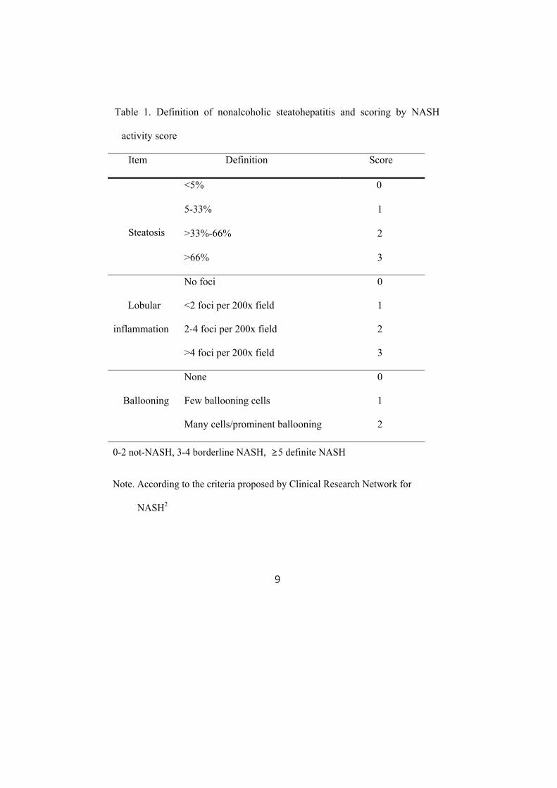
Table 1. Definition of nonalcoholic steatohepatitis and scoring by NASH
activity score
Item Definition Score
Steatosis
<5% 0
5-33% 1
>33%-66% 2
>66% 3
Lobular
inflammation
No foci 0
<2 foci per 200x field 1
2-4 foci per 200x field 2
>4 foci per 200x field 3
Ballooning
None 0
Few ballooning cells 1
Many cells/prominent ballooning 2
0-2 not-NASH, 3-4 borderline NASH, ≥5 definite NASH
Note. According to the criteria proposed by Clinical Research Network for
NASH2

10
For evaluating histological characteristics of NASH patients, we
categorized NASH into three types (adult type, pediatric type and
overlap type) using the classification proposed by Schwimmer et al. in
20051 (Table 2).

11
Table 2. Definition of nonalcoholic steatohepatitis type
Adult type Pediatric type Overlap
Type
Ballooning
degeneration + + -
-
+
Perisinusoidal
fibrosis - + + +
Steatosis +
Portal
inflammation -
+ + - +
Portal fibrosis - + + +
+: feature is present.
-: feature is absent.
¶: Overlap type represents those biopsies that demonstrated steatosis along
with at least one feature from adult type and pediatric type.
Note. According to the classification of Schwimmer et al.1
12
5. Statistical analysis
Means, standard deviations and percentages were reported for various
dermographic and clinical features. Chi-square tests were used to compare
univariate association of histological characteristics with NASH classification
by Schwimmer et al. For comparison of clinical characteristics between three
groups classified by NAS and Schwimmer’s criteria, ANOVA test was used.
Simple Pearson correlation test was used to identify relationships between the
severity of NAS and the clinical features. P-value of 0.05 or less was
considered to indicate statistical significance. All statistical analysis was
performed using SPSS 12.0.

13
III. RESULTS
1. Dermographic data of all subjects
Total 64 patients and specimen were collected. Average age was 22.2 ± 2.75
years old and most of them had abnormal liver enzymes (AST; 76.83 ± 44.64,
ALT; 171.50 ± 114.89). Total cholesterol and triglyceride were 189.34 ± 38.51
(101-350) and 179.52 ± 97.86 (62-583). According to the IOTF classification
in 2000, 51 patients (79.7%) were obese. There were 4 diabetes patients
(6.3%). By NAS criteria, 5 were not-NASH (7.8%), 29 were borderline NASH
(45.3%) and 30 were definite NASH (46.9%).

14
2. Comparison of baseline characteristics between three groups classified by
NAS
Comparing three groups classified by NAS, there was no significant
difference in age, BMI, total cholesterol, triglyceride and fasting plasma
glucose. The prevalence of diabetes was higher in NASH group (n=4, 6.8%)
than in not-NASH group (n=0, 0%) without statistical significance. In NASH
group, AST and ALT were higher than in not-NASH group (Table 3).
Especially definite NASH group showed significantly higher AST and ALT
levels than the other two groups (Figure 1, 2). And there was a linear
correlation between AST or ALT and NAS (Figure 3, 4).

15
Table 3. Comparison of baseline characteristics between three groups
classified by NASH activity score
BMI= Body mass index ; Chol= Total cholesterol ; TG= Triglyceride ;
FPG= Fasting plasma glucose; NAS=NASH activity score
¶: By IOTF classification in 200012, obese patient is defined as the one having
BMI ≥ 25 kg/m2
Not-NASH(n=5)
Borderline
NASH(n=29)
Definite
NASH(n=30) p-value
Age(years) 22.4 ± 3.05 21.93 ± 2.14 22.33 ± 3.26 0.84
AST(IU/L) 64.40 ± 30.24 56.97 ± 29.23 98.10 ± 49.88 < 0.01
ALT(IU/L) 121.80 ± 56.73 120.28 ± 75.17 229.27 ± 127.57 < 0.01
Chol(mg/dL) 189.40 ± 39.73 188.86 ± 42.57 189.82 ± 35.35 0.996
TG(mg/dL) 136.00 ± 42.98 216.53 ± 118.19 160.82 ± 81.78 0.120
FPG(mg/dL) 94.40 ± 6.57 98.79 ± 26.48 106.15 ± 31.57 0.522
BMI(kg/m2) 29.98 ± 4.46 29.67 ± 4.04 29.49 ± 4.80 0.971
Numbers of obese
patients(%)
4 (80%) 26 (89.6%) 21 (70%) 0.281

16
Figure 1. Comparison of AST in three groups classified by NASH activity score
Figure 2. Comparison of ALT in three groups classified by NASH activity score
p <0.01
0
50
100
150
200
250
300
Not-NASH Borderline NASH Definite NASH
ALT (IU/L)
p<0.01
p <0.01 p =ns
0
20
40
60
80
100
120
140
Not-NASH Borderline NASH Definite NASH
AST (IU
/L)
p=0.02
p<0.01 p=ns

17
Figure 3. Correlation between AST and NASH activity score in three groups
Figure 4. Correlation between ALT and NASH activity score in three groups
AALLTT(( IIUU
// LL))
AASSTT(( IIUU
// LL))

18
3. Pathological and clinical characteristics of NASH classified by Schwimmer’s
classification
There were 59 NASH patients who had NAS score over 3. By Schwimmer’s
classification, there were 17 adult type NASH (28.8%), 4 pediatric type NASH
(6.8%) and 38 overlap type NASH (64.4%) (Figure 5). In pathological
characteristics, there was no difference in steatosis (p=0.576) and lobular
inflammation (p=0.477) between three groups (Table 4). About fibrosis, there
were inter-group differences between three groups. In overlap type, stage 2
fibrosis was prominent than in the other two groups (Figure 6).
There was no statistical inter-group difference in clinical characteristics
between three types of NASH (Table 5). However in comparison between
pediatric type NASH group and overlap type NASH group, age and NAS of
pediatric type group were lower than those of overlap type group in statistical
significance (age; p=0.029, NAS; p=0.009). None of pediatric type NASH
patient belonged to the definite NASH and over a half of overlap type NASH
patient (57.9%) was in the definite NASH (Figure 7).

19
Figure 5. Histological characteristics of nonalcoholic steatohepatitis classified
by Schwimmer’s classification
(A) Adult type NASH showing prominent ballooning degeneration with Mallory
bodies, moderate steatosis, perisinusoidal fibrosis in zone 3 (TRC x200)
(B) Pediatric type NASH showing moderate steatosis, portal inflammation and
fibrosis (TRC x100)
(C), (D) Overlap type NASH showing zone 3 injury of adult type (C) and portal
inflammation and fibrosis of pediatric type (D) (TRC x200)

20
Table 4. Pathological characteristics of NASH classified by Schwimmer’s
classification
Adult type
(n=17)
Pediatric
type (n=4)
Overlap type
(n=38) p-value
Steatosis
0 0 (0%) 0 (0%) 0 (0%)
0.576 1 3 (17.6%) 0 (0%) 9 (23.7%)
2 11 (64.7%) 2 (50%) 21 (55.3%)
3 3 (17.6%) 2 (50%) 8 (21.1%)
Fibrosis
0 2 (11.8%) 0 (0%) 0 (0%)
<0.01
1A 12(70.6%) 0 (0%) 0 (0%)
1B 3 (17.6%) 0 (0%) 0 (0%)
1C 0 (0%) 4 (100%) 2 (5.3%)
2 0 (0%) 0 (0%) 36 (94.7%)
3 0 (0%) 0 (0%) 0 (0%)
4 0 (0%) 0 (0%) 0 (0%)
Lobular
inflammation
0 1 (5.9%) 0 (0%) 0 (0%)
0.477 1 9 (52.9%) 3 (75%) 19 (50%)
2 7 (41.2%) 1 (25%) 19 (50%)
3 0 (0%) 0 (0%) 0 (0%)
Portal
inflammation
0 17 (100%) 1 (25%) 9 (23.7%)
<0.01 1 0 (0%) 3 (75%) 29 (76.3%)
Ballooning
degeneration
0 5 (29.4%) 4 (100%) 2 (5.3%)
<0.01 1 8 (47.1%) 0 (0%) 19 (50%)
2 4 (23.5%) 0 (0%) 17 (44.7%)

21
Figure 6. Comparison of pathological characteristics of nonalcoholic
steatohepatitis classified by Schwimmer’s classification
Adulttype
Pediatrictype
Overlaptype
01A
1B1C
23
4
0
5
10
15
20
25
30
35
40
Num
ber
of c
ases
s tage
F i b r o s i s
0 1A 1B 1C 2 3 4
Adulttype Pediatric
type Overlaptype
0
12
3
02468
101214161820
Num
ber
of c
ases
stage
Lobular inflammation
0 1 2 3
Adulttype Pediatric
type Overlaptype
0
12
3
0
5
10
15
20
25
Num
ber
of c
ases
stage
Steatosis
0 1 2 3
Adult typePediatric type
Overlap type
0
10
5
10
15
20
25
30
Num
ber
of c
ases
stage
Portal inflammation
0 1
Adulttype Pediatric
type Overlaptype
0
12
02468
101214161820
Num
ber
of c
ases
stage
Ballooning degeneration
0 1 2

22
Table 5. Clinical characteristics of nonalcoholic steatohepatitis classified by
Schwimmer’s classification
BMI= Body mass index ; Chol= Total cholesterol ; TG= Triglyceride ;
FPG= Fasting plasma glucose ; NAS= NAFLD activity score
Adult type
(n=17)
Pediatric
type(n=4)
Overlap
type(n=38) p-value
Age(years) 21.82 ± 2.90 20.75 ± 0.96 22.4 ± 2.80 0.447
BMI(kg/m2) 28.92 ± 3.68 32.23 ± 1.52 29.61 ± 4.85 0.405
AST(IU/L) 78.82 ± 49.11 70.00 ± 13.19 78.29 ± 46.98 0.939
ALT(IU/L) 161.76 ± 103.07 153.25 ± 70.28 184.29 ± 128.65 0.753
Chol(mg/dL) 185.31 ± 35.54 222.50 ± 30.84 187.44 ± 40.03 0.206
TG(mg/dL) 173.55 ± 76.08 188.75 ± 58.96 189.79 ± 118.57 0.910
FPG(mg/dL) 96.33 ± 12.00 125.75 ± 59.16 102.33 ± 29.45 0.200
NAS 4.29 ± 1.16 3.75 ± 0.50 4.87 ± 1.21 0.082

23
Figure 7. Composition of the definite nonalcoholic steatohepatitis and borderline
nonalcoholic steastohepatitis in three types classified by Schwimmer’s
classification
0
5
10
15
20
25
30
35
40
Adul t t ype Pedi at r i c t ype Over l ap t ype
Border l i ne NASH Def i ni t e NASH
Number of ca
ses

24
IV. DISCUSSION
The aim of this study is to know clinicopathological characteristics and natural
history of NAFLD in Korean young men by using a semiquantitative diagnostic
method and classifying them into three groups (adult type, pediatric type and
overlap type). Recently in Korea, as the obese population increases, the
prevalence of metabolic syndrome increases concurrently. Metabolic syndrome,
especially insulin resistance and visceral obesity, is known to be closely related
to the incidence of NAFLD.13,14,15. In recent studies, NAFLD itself is strongly
related with chronic fatal complications in nonhypertensive and nondiabetic
adults16 and even in children.17 Visceral obesity is thought to cause fatty
accumulation in hepatocytes and this leads hepatic insulin resistance,18,19 and
these changes in turn cause NAFLD or NASH by means of inflammation,
oxidative stress and immunologic alterations.13,20,21,22 Not only in adults but in
children and adolescents, obesity is one of the most related factor causing hepatic
fatty changes. Some studies found that in obese children, 12-25% of them had
elevated serum ALT and 42-77% have abnormal liver ultrasound
findings.10,23,24,25,26,27 In severe obese adolescents (mean BMI 59kg/m2), about 83%
of the subjects had biopsy-proven steatosis.28 But NAFLD and NASH are
difficult to diagnose because a liver biopsy is the only method to diagnose the
disease. Serum chemistry (e.g., alanine aminotransferase) and radiologic

25
modalities (e.g., ultrasonography, computed tomography, magnetic resonance
imaging and magnetic resonance spectroscopy) cannot distinguish a simple
steatosis from NASH1,6,30,31 and can only be used as the screening test, not for the
diagnosis.10 Moreover these methods have no definite quantitative criteria of
assessing the degree of the diseases. NAS (NAFLD activity score), designed for
diagnosing NASH semiquantitatively by scoring pathologic characteristics, can
be used for measuring the activity of NAFLD. In this study, definite NASH
group had higher AST and ALT levels than the others. And statistically NAS had
a linear correlation with AST and ALT (Figure 3, 4). Therefore, not presenting
fibrosis status, NAS can be a good parameter of the acute status of NAFLD
combining with clinical contexts.
In 2005, Schwimmer et al1 reported histopathological characteristics of pediatric
NAFLD. Until then, histologic features were studied mostly in adults and
macrovesicular steatosis, perisinusoidal or pericellular fibrosis, foci of lobular
inflammation, lipid granulomas, Mallory hyaline and megamitochondria were
found in adult type NAFLD.8 The diagnosis of NASH in adults are made by the
combination of macrovesicular steatosis with ballooning changes of hepatocytes
and/or perisinusoidal fibrosis in an appropriate clinical context.29 However the
presence of steatosis along with portal inflammation and/or fibrosis in the
absence of ballooning degeneration and perisinusoidal fibrosis is a common

26
feature of the pediatric NASH in Schwimmer’s study.1 According to the previous
study, NASH can be classified by the histological characteristics. Type 1 NASH
has the adult type histologic feature. Type 2 NASH shows the pediatric type
histologic features. Overlap type NASH has the characteristics of type 1 and type
2. In Schwimmer’s study of 100 children, subjects with simple steatosis were 16
(16%), type 1 NASH were 17 (17%), type 2 NASH were 51 (51%) and overlap
type were 16 (16%). In our study, only 4 cases (6.2%) were pediatric NASH and
over a half (59%) were overlap type NASH. This difference may be due to the
difference of age. In our study, age of subjects was postpuberty, which might
contribute to the difference in the distribution of NASH type. It can be speculated
that as growing older, pediatric type NASH may progress to overlap type NASH
by adding adult type NASH trait.
Our study shows that in the pediatric type NASH group, though not statistically
significant, not only AST or ALT, but NAS is lower than in the overlap type
NASH group (Table 5). As NAS is the one of activity marker of NAFLD,
pediatric type alone is thought to be a milder form than the overlap type. Hence
clinicians should start more aggressive and earlier exercise and dietary therapy
when clinically NASH is suspected in any young men.
Several criticisms of this study are that because of difficulty in diagnosing
NAFLD, only small numbers of subjects were involved and subjects were all

27
men. Especially in children and adolescents, type 1 NASH was predominant in
girls and type 2 NASH in boys.1 This sexual difference may be associated the
difference of the hormonal levels and this difference must be more prominent
after the puberty. So comparison between two sexes after the adolescence is
worth for understanding the hormonal impacts on NAFLD. Secondly, as it is a
cross-sectional study, there was no clinical data representing the severity of the
insulin resistance and oxidative stress. As mentioned above, NAFLD and NASH
are rendered as the consequences of the hepatic insulin resistance and the
hepatocyte fatty accumulation. In the West of Scotland Coronary Prevention
Study,32,33 increased ALT (≥29 U/L) and CRP (≥3 mg/L) was one of the
predicted values for the incident type 2 diabetes mellitus and hepatic fatty
accumulation. In the other study of middle-aged Japanese men,34
gamma-glutamyl transferase was found to be a predictor for developing
metabolic syndrome. To understand interactive effects and possible mechanisms
between NAFLD and metabolic syndrome, further study of other clinical data
and histophathological characteristics will be needed.

28
V. CONCLUSION
- This study was aimed to histologically classify NAFLD and diagnose
NASH in Korean young men, and to compare clinicopathological
characteristics by using new guidelines.
- Approximately over 90% of Korean young men with nonalcoholic fatty
liver disease (NAFLD) turned out to have NASH by applying NAS (92.2%).
Especially in definite NASH group, AST, ALT level was higher than in the
other groups. As NAS had a linear correlations with AST or ALT, it can be a
good parameter of the acute status of NAFLD and NASH in clinical
contexts.
- Histopathological characteristics of NASH patients mostly had
overlapping traits of both adult and pediatric types (64.4%) in contrast to
pediatric type alone (6.8%). And overlap type had higher AST and ALT
levels and NAS than pediatric type though in statistical significance.
- In conclusion, it is possible to speculate that the pattern of NASH in
Korean young men is in the middle of transition from pediatric to adult type
and proper management of pediatric NASH should be of great importance in
preventing disease progression.

29
REFERENCES
1. Jeffrey BS, Cynthia B, Robert N, Reena D, Caroline N, Nicholas JS et
al. Histopathology of pediatric nonalocoholic fatty liver disease.
Hepatology 2005; 42:641-9.
2. David EK, Elizabeth MB, Mark VN, Cynthia B, Melissa JC, Oscar WC
et al. Design and Validation of a histological scoring system for
nonalcoholic fatty liver disease. Hepatology 2005; 41:1313-21.
3. Sanyal AJ, American gastroenterological association. AGA technical
review on nonalcoholic fatty liver disease. Gastroenterol 2002;
123:1705-25.
4. McCullough AJ. Update on nonalcoholic fatty liver disease. J Clin
Gastroenterolo 2002; 34:255-62.
5. Shivakumar C, Geoffrey CF, Etsuko H, Toshiji S, George KL, Jose DS
et al. Non-alcoholic fatty liver disease in the Asia-Pacific region:
Definitions and overview of proposed guidelines. J Gastroenterol

30
Hepatol 2007; 22:778-87.
6. Clark JM. The epidemiology of nonalcoholic fatty liver disease in
adults. J Clin Gastroenterol 2006; 40:S5-S10.
7. Brunt EM. Nonalcoholic steatohepatitis. Semin Liver Dis 2004;
24:3-20.
8. Brunt EM, Janney CG, Di Bisceglie AM, Neuschwander-Tetri BA,
Bacon BR. Nonalcoholic steatohepatitis: a proposal for grading and
staging the histological lesions. Am J Gastroenterol 1999; 94:2467-74.
9. Brunt EM. Grading and staging the histopathological lesions of chronic
hepatitis: the Knodell histology activity index and beyond. Hepatology
2000; 31:241-6.
10. Jeffrey BS. Definitive diagnosis and assessment of risk for
nonalcoholic fatty liver disease in children and adolescents. Semin
Liver Dis 2007; 27:312-8.

31
11. Nonalcoholic steatohepatitis clinical research network. Hepatology
2003; 37:244.
12. Steering Committee. The Asia-Pacific perspective: Redefining obesity
and its treatment. Melbourne: International Diabetes Institute, 2000.
13. Fan JG. Impact of non-alcoholic fatty liver disease on accelerated
metabolic complications. J Dig Dis 2008; 9:63-7.
14. Van der Poorten D, Milner KL, Hui J, Hodge A, Trenell MI, Kench JG
et al. Visceral fat: A key mediator of steatohepatitis in metabolic liver
disease. Hepatology 2008; 48:449-57.
15. McCullough AJ. The clinical features, diagnosis and natural history of
nonalcoholic fatty liver disease. Clin Liver Dis 2004; 8:521-33.
16. Change Y, Ryu S, Sung E, Woo HY, Oh E, Cha K et al. Nonalcoholic
fatty liver disease predicts chronic kidney disease in nonhypertensive
and nondiabetic Korean men. Metabolism 2008: 57:569-76.

32
17. Pacifico L, Cantisani V, Ricci P, Osborn JF, Schiavo E, Anania C et al.
Nonalcoholic fatty liver disease and carotid atherosclerosis in children.
Pediatr Res 2008; 63:423-7.
18. Francanzani AL, Valenti L, Bugianesi E, Andreoletti M, Colli A, Vanni
E et al. Risk of severe liver disease in nonalcoholic fatty liver disease
with normal aminotransferase levels: a role for insulin resistance and
diabetes. Hepatology 2008; 48:792-8.
19. Love-Osborne KA, Nadeau KJ, Sheeder J, Fenton LZ, Zeitler P.
Presence of the metabolic syndrome in obese adolescents predicts
impaired glucose tolerance and nonalcoholic fatty liver disease. J
Adolesc Health 2008; 42:543-8.
20. Bergman RN, Kim SP, Catalano KJ, Hsu IR, Chiu JD, Kabir M et al.
Why visceral fat is bad: Mechanisms of the metabolic syndrome.
Obesity(Silver Spring) 2006; 14:16S-19S.
33
21. Lanfontan M, Berlan M. Do regional differences in adipocyte
biology provide new pathophysiological insights? Trends Pharmacol
Sci. 2003; 24:276-83.
22. Jou J, Choi SS, Diehl AM. Mechanisms of disease progression in
nonalcoholic fatty liver disease. Semin Liver Dis 2008; 28:370-9.
23. Guzzaloni G, Grugni G, Minocci A, Moro D, Morabito F. Liver
steatosis in juvenile obesity: correlations with lipid profile, hepatic
biochemical parameters and glycemic and insulinemic responses to an
oral glucose tolerance test. Int J Obes Relat Metab Disord 2000;
24:772-6.
24. Franzese A, Vajro P, Argeniziano A, Puzziello A, Iannucci MP, Saviano
MC et al. Liver involvement in obese children: ultrasonography and
liver enzyme levels at diagnosis and during follow-up in an Italian
populations. Dig Dis Sci 1997; 42:1428-32.
25. Tazawa Y, Noguchi H, Nishinomiya, Takada G. Serum alanine
aminotransferase activity in obese children. Acta Paediatr 1997;

34
86:238-41.
26. Kinugasa A, Tsunamoto K, Furukawa N, Sawada T, Kusunoki T,
Shimada N. Fatty liver and its fibrous changes found in simple obesity
of children. J Pediatr Gastroenterol Nutr 1983; 3:408-14.
27. Schwimmer JB, Deutsch R, Kahen T, Lavine JE, Stanley C, Behling C.
Prevalence of fatty liver in children and adolescent. Pediatrics 2006;
118:1388-93.
28. Xanthakos S, Miles L, Bucuvalas J, Daniel S, Garcia V, Inge T.
Histologic spectrum of nonalcoholic fatty liver disease in morbidly
obese adolescents. Clin Gastroenterol Hepatol 2006; 4:226-32.
29. Brunt EM. Nonalcoholic steatohepatitis definition and pathology.
Semin Liver Dis 2001; 21:3-16.
30. Saadeh S, Younossi ZM, Remer EM, Gramlich T, Ong JP, Hurley M
et al. The utility of radiological imaging in nonalcoholic fatty liver
disease. Gastroentorol 2002; 123:745-50.
35
31. Ataseven H, Yildirim MH, Yalniz M, Bahcecioglu IH, Celebi S,
Ozercan IH. The value of ultrasonography and computerized
tomography in estimating the histopathological severity of
nonalcoholic steatohepatitis. Acta Gastroenterol Belg 2005; 68:221-5.
32. Sattar N, McConnachie A, Ford I, Gaw A, Cleland SJ, Forouhi NG et al.
Serial metabolic measurements and conversion to type 2 diabetes in the
west of Scotland coronary preventing study: specific elevations in
alanine aminotransferase and triglycerides suggest hepatic fat
accumulation as a potential contributing factor. Diabetes 2007;
56:984-91.
33. Satta N, Scherbakova O, Ford I, O’Reilly DS, Stanley A, Forrest E et al.
Elevated alanine aminotrasferas predicts new-onset type 2 diabetes
independently of classical risk factors, metabolic syndrome and
C-reactive protein in the west of Scotland coronary prevention study.
Diabetes 2004; 53:2855-60.
34. Nakanishi N, Suzuki K, Tatara K. Serum gamma-glutamyl transferase

36
and risk of metabolic syndrome and type 2 diabetes in middle-aged
Japanese men. Diabetes Care 2004; 27:1427-32.

37
< Abstract In Korean>
우리 나라 젊은 성인 남자에서 비알코올성 지방간질환의 임상 병리학적 특징
< 지도교수 이 관 식 >
연세대학교 대학원 의학과
전 누 리
배경: 비알코올성 지방간질환 (NAFLD)은 과도한 알코올의 섭취
없이 나타나는 만성 간질환으로 최근 서구화된 식생활과 그
영향으로 인한 비만인구의 증가로 인해 우리 나라에서도
발병률이 증가하고 있다. 특히 비알코올성 지방간염 (NASH)은
간경변증이나 간암으로도 진행할 수 있어 본 질환의 중요성은
크다. 본 연구는 우리 나라의 젊은 성인 남자에서 NASH Clinical
Research Network 의 진단기준에 의해 NASH로 진단한
환자들의 임상 및 병리학적 특징을 조사하였고 이를
Schwimmer등의 분류 기준에 따라 분류하여 본 질환의 임상
경과와 특성을 알아보고자 하였다.
38
방법: 간생검에서 비알코올성 지방간질환으로 진단받은 30세
이하(16-25세)의 젊은 성인 남자 (n=64)를 대상으로 나이,
체질량지수 (BMI), AST, ALT, 총콜레스테롤, 중성지방, 공복 혈당,
임상적 소견들과 조직병리학적 소견들을 후향적으로 조사하였다.
조직학적 특성은 지방증 정도, 소엽의 염증 정도, 문맥 주위 염증
정도, 간세포의 ballooning degeneration과 간섬유화 정도를
측정하였다. 비알코올성 지방간염 (NASH)는 Kleiner DE 등이
제안한 NAFLD activity score (NAS)를 기준으로 진단하였다.
NASH 환자들은 Schwimmer등의 분류법에 따라 adult type,
pediatric type과 overlap type의 세 가지로 조직학적 분류하였다.
결과: 51명 (79.9%)이 비만하였고 높은 AST, ALT 수치를
나타내었다 (76.83 ± 44.6, 171.50 ± 114.89). 간생검 상 59명
(92.2%)이 NASH소견을 보였으며 29명 (45.3%)은 borderline
NASH였고 30명 (46.9%)은 definite NASH였다. NASH로 진단된
군에서 NAS는 AST 또는 ALT와 양의 선형관계를 나타내었고
definite NASH군은 borderline NASH군에 비해 유의하게 AST 및

39
ALT가 높았다. NASH군은 Schwimmer등의 분류에 따라 3가지
type으로 분류되었고, 17명 (28.8%)이 adult type, 4명 (6.8%)이
pediatric type 그리고 38명 (64.4%)이 overlap type이었다. NAS
(평균 ± 표준편차)는 pediatric type이 3.75 ± 0.05, adult type
4.29 ± 1.16, overlap type 4.87 ± 1.21이었으며 overlap type은
pediatric type에 비해 NAS가 통계학적으로 의미있게 높았다
(p<0.01). 간섬유화의 경우 overlap type 중 36명 (94.7%)이
stage 2의 섬유화를 지녔으며, 4명의 모든 pediatric type (100%)과
adult type 15명 (88.2%)은 stage 1의 섬유화 정도를 보였다.
Overlap type은 다른 type에 비해 유의하게 높은 섬유화 정도를
지녔다 (p<0.01). 지방증 정도와 소엽의 염증 정도를 포함한 다른
병리학적 특성들과 임상적 특징들은 세 군에서 유의한 차이를
보이지 않았다.
결론: 우리 나라 젊은 성인 남자에서 비알코올성 지방간염은
대부분 간생검상 borderline NASH나 definite NASH 소견을
보였다. NASH 중 절반 이상은 pediatric type와 adult type
NASH의 특성을 모두 지닌 overlap type이였으며, 이 type은

40
pediatric type에 비해 높은 NAS와 섬유화 정도를 보였다. 이러한
소견들은 우리 나라 젊은 성인 남자에서 나타나는 overlap type
NASH의 경우 소아 시기에서 발병하여 지속적으로 진행되고
있음을 제시하고 있다.
--------------------------------------------------
핵심되는 말 : 비알코올성 지방간질환, 비알코올성 지방간염, 간생검,
대한민국 젊은 성인 남자