clinico-pathological conference · clinico-pathological conference ......
TRANSCRIPT
CLINICO-PATHOLOGICAL CONFERENCEA case of dilated cardiomyopathy with end-stage heart failure
treated by prolonged continuous hemodiafiltrationSatoshi Ogawa, Shin-ichiro Matsumura, Tsutomu Yoshikawa, Toru Satoh, Hiroo Kumagai, Hideo Mitamura,1
Shiro Iwanaga2 and Akihiro Umezawa3
Departments of Internal Medicine, 1The Suntory Fund for Advanced Cardiac Therapeutics,2Laboratory Medicine, 3Pathology, School of Medicine, Keio University, Tokyo, Japan
(Received for publication on February 27, 2002)
Abstract. A 55-year-old Asian man first visited to our hospital with complaining of exertional dysp-
nea eight years ago, and was diagnosed as having idiopathic dilated cardiomyopathy. One of his sib-
lings also suffered from idiopathic dilated cardiomyopathy. His symptoms became worse gradually, and
he was hospitalized again because of disturbance of consciousness on February 21, 2001. Hemodynamic
monitoring with a Swan-Ganz catheter was started, which revealed that the cardiac index was 1.1 L/
min/BSA, cardiac output 1.8 L/min, and pulmonary artery pressure 43/33 mmHg. The echocardio-
graphic observation showed that the left ventricular ejection fraction was 32%, and serum BNP was
elevated to 5,411 pg/mL. Multi-organ failure including renal and hepatic dysfunction developed be-
cause of the low cardiac output status. Continuous hemodiafiltration (CHDF) was introduced to reduce
the volume overload, improve renal failure, and eliminate adverse cytokines. Although his hemody-
namic status was temporarily improved after starting CHDF, weaning from CHDF was difficult and he
finally died from cardiogenic shock after two month of intensive therapy. The autopsy showed thinning
of the left ventricular wall, and histological examination revealed diffuse fibrous hyperplasia and myo-
cardial fiber deficit in the ventricular myocardium. CHDF was effective in reducing the volume over-
load and improving renal function; however, heart transplantation is inevitable for the patients with
severe heart failure due to dilated cardiomyopathy. (Keio J Med 51 (3): 165–177, September 2002)
Key words: dilated cardiomyopathy, continuous hemodiafiltration, autopsy, heart failure, uncon-
sciousness
Dr. Ogawa (Moderator): I declare the 1042ndclinico-pathological conference (CPC) open. The casefor discussion in today’s CPC is a patient who died fromgradual worsening of his cardiac function over a courseof approximately 8 years. The physician-in-charge, Dr.Matsumura, will first describe the clinical course of thispatient.Dr. Matsumura (Internal Medicine): The patient
presented complaining of exertion-related dyspnea onclimbing stairs or walking up a slope since March 1993.He visited the Cardiopulmonary Division at our hospi-tal in April 1993. After he was admitted to the hospital,cardiac catheterization and left ventriculography wereperformed, and revealed contractile dysfunction of theleft ventricle, which was manifested by diffuse hypo-
kinase and a reduced ejection fraction (EF, 11%). Amyocardial biopsy further revealed hypertrophy ofmyocardial cells and interstitial fibrosis. There was noevident infectious, metabolic, systemic, or hereditaryetiology. The patient was diagnosed as having dilatedcardiomyopathy (DCM). Treatment with an anticoagu-lant (Warfarin1), digitalis glycoside, an angiotensin-converting enzyme (ACE) inhibitor, and furosemidewas started. The patient was then discharged from thehospital and treatment was continued on an outpatientbasis.
In the months of June and October of 1997, the pa-tient developed acute exaggeration of his heart failure,precipitated by a common cold, and he was admitted toour hospital. Oral administration of pimobendan was
This is a record of the 1042nd CPC of Keio University Hospital, held on January 30, 2002.Reprint requests to Dr. Satoshi Ogawa, Department of Internal Medicine, School of Medicine, Keio University, 35 Shinanomachi, Shinjuku-ku,Tokyo 160-8582, Japan
165
started on October. From October 1998, his dyspnea onexertion (DOE) became progressively worse, and inApril 2000, he developed the symptom of chest op-pression at night. Holter ambulatory ECG recordingperformed in January 1999 revealed non-sustainedventricular tachycardia (max. 11 beats in a run). At 5:00a.m. on February 21, 2001, his wife, who was sleepingbeside him, noticed that he was breathing hard. He didnot respond to her calls, and she immediately called foran ambulance. He was taken to Nihon University Su-rugadai Hospital. On initial examination, dyspnea andcyanosis were observed. His consciousness level wasslightly below normal, and systolic blood pressure wasabout 70 mmHg. Following endotracheal intubation,artificial ventilation, and intravenous infusion of dobut-amine and furosemide, the patient’s consciousness im-proved rapidly. He was transferred to our hospital onFebruary 27.Dr. Ogawa: So, the clinical course of the patient’s
illness was as follows: The patient first presented withthe clinical features of heart failure in 1993, and he wasclinically diagnosed as having DCM. Despite intensivetherapy, however, the heart failure became worse grad-ually and progressively. At one point, he was admittedwith loss of consciousness to another hospital. There-after, he was again admitted to our Keio UniversityHospital for thorough examination and treatment.
Echocardiography performed in 1994 revealed end-diastolic and end-systolic left ventricular diameters of5.9 cm and 4.4 cm, respectively, and cardiac pool scin-tigraphy (MUGA) revealed a left ventricular EF ofabout 40%. These dimensions improved slightly to 5.2cm and 4.2 cm, respectively, in the year 1996, but theyincreased again in 1998 and 1999; in particular, the end-systolic diameter increased to 5.2 cm, indicating that thecontractile function of the left ventricle had decreasedconsiderably.
The patient first presented with dyspnea on exertionin March 1993. Are there data from earlier medicalexaminations?Dr. Matsumura: There is one earlier mention of
right axis deviation on ECG; however, no other ECGchanges were recorded. The patient was apparently ingood health in 1982, when he was involved in a trafficaccident in the State of Kuwait and underwent surgeryon his right lung. Subsequently, he developed slightdyspnea on exertion. The symptom worsened rapidly in1993, and he was referred to our hospital.Dr. Ogawa: As for the 11 beat run of non-sustained
ventricular tachycardia revealed by the Holter ECG in1999, was his morbid condition relatively stable duringthe period when the tachycardia was observed? Or wasthe non-sustained ventricular tachycardia associatedwith worsening of his heart failure?Dr. Matsumura: In 1993, his symptoms of heart fail-
ure were relatively mild, classified as New York HeartAssociation (NYHA) functional class II. A little later,he presented with worsening of the heart failure due toa common cold, and he was admitted to our hospital.On admission, the NYHA functional class found tohave worsened from Class II to Class III. When thisimproved again to Class II (IIM), he was dischargedfrom our hospital. The symptoms thereafter graduallyworsened again, and runs of non-sustained ventriculartachycardia were observed on the Holter ECG record-ing associated with worsening of the heart failure.Dr. Ogawa: Was the treatment of the tachycardia
detected by Holter ECG undertaken on an outpatientbasis?Dr. Matsumura: Yes, it was.Dr. Ogawa: If so, can we understand that the Holter
monitoring was not performed specifically during theperiod when his heart failure had become worse?Dr. Matsumura: Yes, we can. Holter ECG monitor-
ing was performed routinely and was not related to theworsening of his heart failure.Dr. Ogawa: We can therefore say that the non-
sustained ventricular tachycardia was incidentally de-tected.
The patient’s chief compliant seems to have beenconsciousness disturbance; when his wife, who was lyingbeside him in bed, called out to him at 5:00 a.m. in themorning, she noticed that he did not respond. Is thatright?Dr. Matsumura: Yes, it is.Dr. Ogawa: I wonder if the ambulance personnel
detected any abnormalities in his vital signs that couldbe responsible for the consciousness disturbance onthat occasion? Is there any record?Dr. Matsumura: His consciousness level was slightly
below normal, but it was apparently returning to nor-mal when he was taken into the ambulance. The ECGrecorded then also did not show any ventricular tachy-cardia or ventricular fibrillation. The possibility of lowcardiac output due to DCM, or transient arrhythmias,such as spontaneously converted ventricular tachy-cardia that terminated spontaneously having caused thesyncope, cannot, however, be ruled out.Dr. Ogawa: How was the control of his coagulation
parameters with the dose of Warfarin1 during this pe-riod? Was he taking an appropriate dose of Warfarin1?Dr. Matsumura: The thrombotest was supported
to 10–20%, which suggests favorable control by theWarfarin1. Nor did echocardiography show any evi-dence of intracardiac thrombus during his course. Thisechocardiographic finding, together with the appropri-ate control by Warfarin1, indicate that it is unlikely thatcerebral embolism may have been responsible for thissyncopal episode. A brain CT was not conducted be-cause the patient was in poor general condition.
166 Ogawa S, et al: A case of DCM treated by prolonged CHDF

Dr. Ogawa: What do you think of this case, Dr.Mitamura?Dr. Mitamura (The Suntory Fund for Advanced
Cardiac Therapeutics): I would like to make a fewpoints. As for the labored breathing and absence of re-sponse to verbal commands, I doubt whether we canrefer to the patient’s state here as ‘‘syncope’’. When weuse the term ‘‘syncope’’, it usually refers to a conditionof complete muscular weakness associated with uncon-sciousness; labored breathing therefore is not charac-teristic of syncope. Syncope in this sense generallyresults from a transient and steep fall of blood pressure.It is also important to know how long the laboredbreathing was sustained in the patient. To sum up,rather than suffering from an episode of ‘‘syncope,’’this patient was probably taken to the hospital in a so-called state of shock with sustained moderate hypo-tension, or the main cause of the patient’s conditionmay have been dysfunction of the central nervous sys-tem (Table 1).
The following possibilities should be considered inthis circumstance, other than DCM: (1) The possibilityof ventricular tachycardia having spontaneously termi-nated when the patient was taken to the hospital shouldbe considered as the most likely; (2) and, despite thefavorable control of warfarinization, there is still thepossibility of cerebral infarction having occurred due toa cerebral embolism derived from an intraventricularthrombus because of poor left ventricular function; (3)there is also the probability of cerebral hemorrhagehaving occurred due to an excess dose of Warfarin1;(4) the possibility of hemorrhagic shock having beencaused by gastrointestinal hemorrhage or hemorrhagefrom other sites; and (5) the possibility of pulmonaryembolism, in which condition the blood pressure ofpatients with very poor cardiac function who are bed-ridden over a long period of time, suddenly declines.Dr. Ogawa: Thank you very much. How about his
family history?Dr. Matsumura: The patient’s younger brother was
diagnosed as having DCM at the age of 45 years.Dr. Tsuji (5th-year medical student): Could the his-
tory of pulmonary contusion on the right side in 1982 be
related to the slight decrease in respiratory sound onthe right side?Dr. Matsumura: As is evident from the chest X-rays,
the patient had developed thoracic deformity and col-lapse of the right lung consequent to the pulmonarycontusion. This is probably the cause for the muted re-spiratory sound on the right side.Dr. Ogawa: What was the nature of this pulmonary
injury? Could you explain it in greater detail?Dr. Matsumura: When he was driving, his car col-
lided head-on with another car. At that time, two otherpeople who were in the car with him died instantly, sothe accident was apparently a major traffic accident.Dr. Ogawa: I wonder how we can correlate his mor-
bid condition, DCM, with the accident. For instance, ithas been reported that myocardial injury of the rightventricle or tricuspid regurgitation can occur followingsteering wheel injury. The patient, however, had notbeen detected as having any significant physical abnor-malities in medical check-ups undertaken subsequent tothe accident. Am I right?Dr. Matsumura: Yes, that’s right.Dr. Ogawa: There is no history to suggest that the
patient was a habitual drinker. The presence of adrinking history is very important in making a differ-ential diagnosis. Now, will you please describe thepatient’s clinical condition on admission to our hospi-tal?Dr. Matsumura: The blood pressure was 110/80
mmHg, and the pulse was 100 beats per minute. Therespiratory rate was 26 per minute. He was fully con-scious. The external jugular vein was distended. In re-gard to the heart sounds, P2 was loud, and 3rd and 4thheart sounds were audible. A systolic murmur could beheard between the cardiac apex and the 4th intercostalspace at the left margin of the sternum. Breath soundswere decreased on the right side of the chest, and therewere no added sounds. The liver was palpable by 3QFB and there was some tenderness below the rightcostal margin. There was no pretibial edema.
Laboratory examination revealed normocytic nor-mochromic anemia and increase in the serum levels offibrinogen and fibrin degradation products. The BUNand CRTNN levels were elevated, which was consid-ered to be related to the administration of diuretics.The serum UA and K levels were also elevated; the se-rum K was 5.6 mEq/L. Furthermore, the serum levels ofLDH, GOT, and GPT were elevated. Serum CPK wasslightly increased. Serum CRP was increased to 4.56mg/dL. Thyroid functions were normal. The serumconcentration of digoxin was slightly high. BNP wasrevealed to be considerably elevated to 5,410.8 pg/mL.Dr. Ogawa: Will you please explain the reason for
the distended jugular vein observed in this patient onadmission?
Table 1 Vital Signs and Acute Clinical Settings
Consciousness Breathing Pulsation
Normal þ þ þPeripheral Artery Occlusion þ þ �Coma � þ þApnea � � þChoke G � þShock G þ GCardiac Arrest � � �
Keio J Med 2002; 51 (3): 165–177 167

Dr. Matsumura: The right jugular vein was found tobe distended up to the level of the mandible, and a vwave was observed.Dr. Ogawa: The marked distension of the jugular
vein indicates that the filling pressure of the right car-diac system was high, doesn’t it? The respiratory ratewas also increased. How about the cardiac murmur?Dr. Matsumura: The murmur was pansystolic. Echo-
cardiography suggested the presence of mitral regurgi-tation (MR) and tricuspid regurgitation (TR), and themurmur was considered to be associated with theseregurgitations.Dr. Ogawa: Do you mean that the signs of right heart
failure were rather significant?Dr. Matsumura: Yes, that’s right.Dr. Ogawa: Will you please present the results of
arterial blood gas analysis?Dr. Matsumura: Under nasal administaration of 2
L/Min oxygen, the pH was 7.44; Pco2, 31.4 Torr; Po2,144.4 Torr; HCO3
�, 24.5 mEq/L; BE 1.5, mEq/L; andthe Sao2, 99.4%. These findings are suggestive of com-pensated respiratory alkalosis from hyperventilation.Dr. Ogawa: How can we correlate the findings of the
morbid condition reported in this conference with theECG findings? What is your opinion, Dr. Mitamura?(Fig. 1)Dr. Mitamura: The ECG fundamentally revealed a
sinus rhythm. An occasional ventricular extrasystolewas observed in the limb leads.
As to the P wave, the negative portion of P wave inlead V1 did not seem to be so clear. There seemed to besome peaking of the first half of the P wave, suggestiveof right atrial overload.
The PQ interval was almost within the normal range.
The QRS axis deviated to the right, and as stated ear-lier, the right axis deviation may be reflective of rightventricular overload. However, the axis might be influ-enced by a right bundle branch block. The first compo-nent in leads V1 and V2 in cases of right bundle branchblock is essentially an upright r, but in this case therewas a negative deflection, a ‘‘q’’ wave, in these leads.This matter weighs on my mind. The presence of the‘‘q’’ wave in these two leads may suggest the possibilityof myocardial necrosis in the ventricular septum. Mildnon specific ST-T changes were observed in the chestleads.
The patient had a thoracic deformity, and there isthe possibility that this also influenced the ECGchanges. It would therefore seem difficult to make adefinitive diagnosis based on the ECG findings alone inthis patient.Dr. Ogawa: The presence of abnormal ‘‘q’’ waves in
leads V1 and V2 in a case with right bundle branchblock should raise the possibility of cardiac sarcoidosis,which selectively damages the interventricular septum.Dr. Matsumura: A chest X-ray taken in the year
2000, when the patient was being followed up as anoutpatient, is shown here. There is thoracic deformityand collapse of the right lung, which are probably con-sequent to the traffic accident. The mediastinum isshifted to the left. There is no obvious evidence ofpulmonary venous congestion, such as the presence ofKerley’s B lines. There is no evidence of pulmonaryvenous re-distribution to the upper lung fields. The tra-cheal bifurcation angle is approximately 90�, whichsuggests the presence of left atrial dilatation.
In the lateral view, narrowing of the retrosternalspace is observed, which suggests the presence of right
Fig. 1 The electrocardiogram has apositive P wave in lead II, and shows a normal sinus rhythm at the rate 60 beats per minute. Although theamplitude of P wave in leads II and III was not high enough to meet the criteria for right atrial overload, peaking of the first half of the P wavein lead VI may be a reflection of the overload of the right atrium. The PQ interval was within the normal range. The right axis deviation andclockwise rotation of the QRS complex may be due to right ventricular overload. The width of the QRS complex was 168 msec, and the rSpattern in V6 with a wide S wave suggests complete right bundle branch block (CRBBB); however, the QRS complex in V1 was not an typicalCRBBB rsR pattern. There was an abnormal Q wave in V1 and V2, which raises the possibility of myocardial necrosis in the ventricular sep-tum. Slight ST depression and biphasic T wave are seen in the left precordial leads.
168 Ogawa S, et al: A case of DCM treated by prolonged CHDF
ventricular dilatation. Dilatation of the left ventricle isalso suspected from the point of intersection betweenthe diaphragm and the inferior vena cava. The costo-phrenic angles are sharp, and there is no evidence ofpleural effusion.
This X-ray taken on admission to our hospital showsopacity of the right lung field. The cardiothoracic ratio(CTR) is slightly high, and no Kerley’s B lines areevident. These findings suggest that there was no sig-nificant pulmonary venous congestion. There is noevidence of pleural effusion. There is also evidencesuggestive of dilatation of the left atrium and rightventricle.Dr. Ogawa: The left ventricular function was sig-
nificantly compromised in this patient, but findings ofright heart failure were more remarkable on admission.There was no evidence of pulmonary congestion, eitherclinically or on chest X-ray. Will you present the echo-cardiographic findings in this patient and explain his
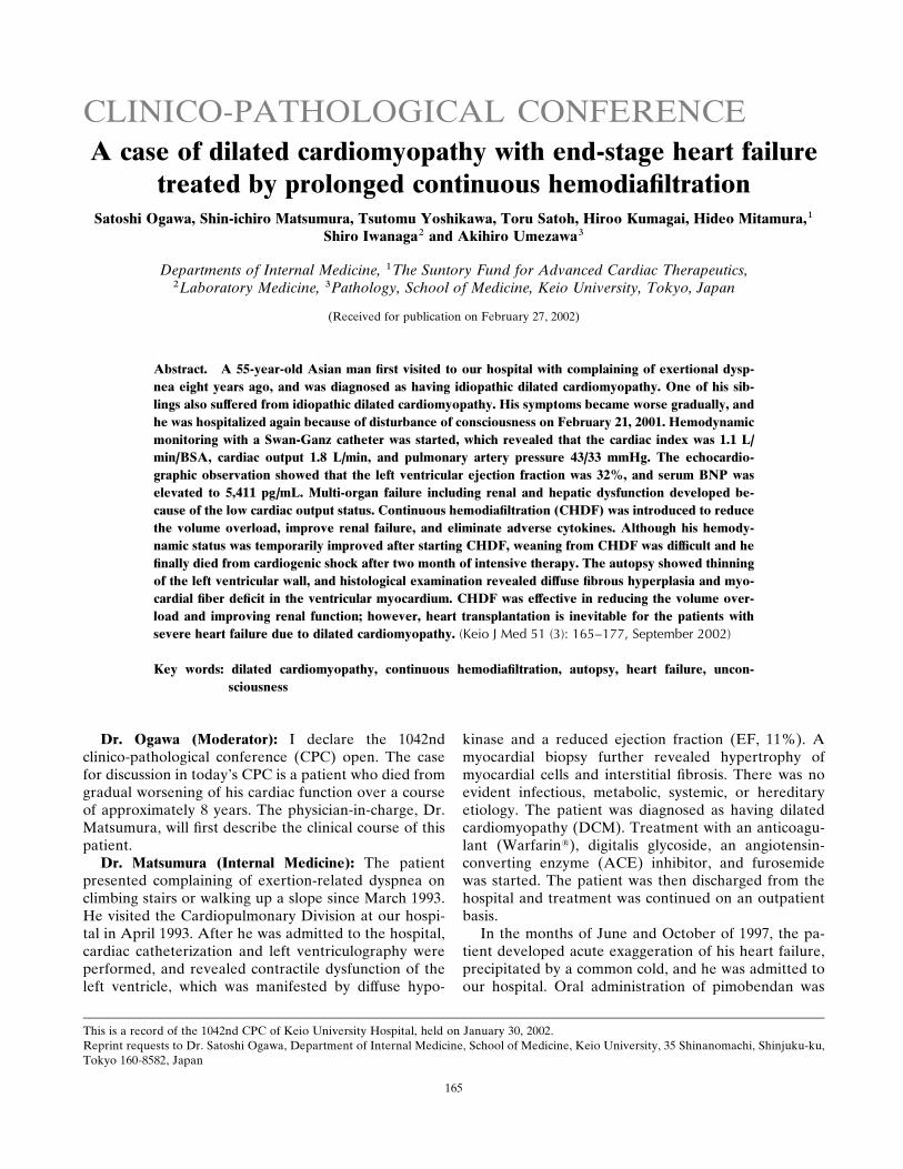
heart condition as evaluated by this investigation, Dr.Iwanaga? (Fig. 2)Dr. Iwanaga (Laboratory Medicine): This videotape
shows the echocardiographic findings of this patientobtained on March 28. The parasternal long-axis viewshows the right and left ventricles, left atrium, aorta,mitral valve, and the aortic valve. The short-axis viewshows the right atrium and right ventricle. We can seea Swan-Ganz catheter inserted. At the level of thepapillary muscle, the interventricular septum is slightlyhyperechoic, which shows little or no wall motion. Themotion of the posterior wall is also poor, and the leftventricle is dilated. As for the valves, moderate to se-vere MR is observed. The aortic valve shows athero-sclerotic degeneration. The most significant findings arethe dilated left ventricle and the poor contraction ofthe interventricular septum and posterior wall. Themitral regurgitation may be due to dilatation of the leftventricle and the mitral annulus. The left ventricular
Fig. 2 The echocardiography was performed on March 28, 2001. Parasternal long axis view on the left upper panel shows dilation of the leftand right ventricles. Thinning of the left ventricular wall and a small amount of pericardial effusion are also observed on the right upper panel,the parasternal short axis view. The two lower panels are M-mode traces at the mid-ventricular level (left) and mitral valve (right). Both tracesrevealed reduced excursion and thinning of the left ventricular wall. The calculated ejection fraction of the left ventricle was 23% by the methodof Pombo, suggesting marked reduction of left ventricular systolic function. B-B0 step of mitral valve on the right lower panel suggests the ele-vation of left ventricular end-diastolic pressure.
Keio J Med 2002; 51 (3): 165–177 169

diameter as measured by M-mode was 6 cm and 5.5 cmat end-diastole and end-systole, respectively, whichreflects significant dilatation. A small amount of peri-cardial effusion is also observed.
The apical four-chamber view shows that the con-traction of the interventricular septum is even worsethan that of the free wall, even though there is signifi-cant thinning of the free wall. The left atrium is dilated.Wall motion of the free wall is relatively sustained, but,on the whole, left ventricular contractile dysfunction isdiffuse. Based on these echocardiographic findings, thediagnosis of DCM is suspected. One problem, however,is localization of the wall thinning; the thinning is dis-tributed in the areas supplied by the right coronary andleft anterior descending arteries. Marked fibrosis isevident in these areas. It is impossible to completelyrule out left ventricular contractile dysfunction due toischemic heart disease from this finding.Dr. Ogawa: What do you think of the possibility of
cardiac sarcoidosis in this case?Dr. Iwanaga: In cardiac sarcoidosis, a relatively
localized contractile dysfunction of the left ventricle isobserved, and wall motion remains relatively normal inother areas. Reports of diffuse hypokinesis of the leftventricle, as observed in the present case, are limited. Acolor Doppler image of the apical four chamber view isshown. Severe TR is evident from this image.Dr. Ogawa: Is there any evidence of an organic le-
gion in the tricuspid valve?Dr. Iwanaga: The TR could be related to the pres-
ence of the Swan-Ganz catheter through the tricuspidvalve and annular dilatation consequent to dilatation ofthe right ventricle. The patient had only mild pulmo-nary hypertension; the pulmonary arterial pressure wasestimated to be 31/21 mmHg. This is reflective of post-capillary pulmonary hypertension due to left heart fail-ure.
From the echocardiographic findings, DCM may besuspected as the first differential diagnosis, and in thepresent case, it was presumably severe and associatedwith hemodynamic decompensation. However, in thepresence of thinning of the ventricular wall in the areasof the right coronary artery and left anterior descendingartery, it is difficult to rule out so-called ischemic car-diomyopathy.Dr. Ogawa: When DCM is suspected, primary DCM
should be differentiated from secondary DCM. Theischemic DCM just mentioned by Dr. Iwanaga cannotbe ruled out, either. While cardiac sarcoidosis may bekept in mind, the possibility is rather remote, becausein this case, not only the interventricular septum, inwhich lesions usually develop, but also the free wall ofthe left ventricular myocardium, showed significant dif-fuse damage. The presence of a positive family historyalso points more towards the possibility of primary or
hereditary DCM.Now, Dr. Matsumura, could you please outline the
treatment initiated in this case?Dr. Matsumura: In 1993, when the patient was first
admitted to our hospital, treatment with digitalis,Warfarin1, an ACE inhibitor, and furosemide wasstarted. Thereafter, the patient was followed up onoutpatient basis. Sometime in mid 1997, the patient wasreadmitted with symptoms of a common cold and wor-sening of his heart failure. On that occasion, adminis-tration of a b-blocker was initiated, and when thepatient showed improvement, he was discharged. How-ever, with the continuation of the b-blocker, the symp-toms of heart failure, including leg edema and dyspneaon exertion (DOE), gradually became worse, and ad-ministration of the b-blocker was discontinued.
Subsequently, when the patient was admitted onceagain with worsening of heart failure, administrationof Acardi1 (pimobendan), a phosphodiesterase (PDE)inhibitor that also enhances the calcium sensitivity ofmyocardial cells, was initiated. In addition, the dose ofthe diuretic was increased and spironolactone wasstarted. The subsequent course of the patient was againmonitored on outpatient basis.Dr. Ogawa: Do you have any comments about the
course of treatment undertaken so far, Dr. Yoshikawa?Dr. Yoshikawa (Internal Medicine): The standard
treatment for all cases ranging from asymptomatic leftventricular dysfunction to severe heart failure includesadministration of ACE inhibitors or angiotensin type Ireceptor antagonists. For mild to moderate heart failureclassified as NYHA functional class II or III, diureticsare usually added to the treatment regimen, and insome patients, also digitalis. Some reports have shownthat the prognosis is improved by the administration ofa b-blocker in such patients. Furthermore, in severeheart failure classified as NYHA functional class III orIV, further improvement of prognosis has been re-ported with the addition of spironolactone to the treat-ment regimen. Although pimobendan does not de-crease the mortality rate, clinical studies in Japan haveshown that the drug decreases the number of patientswho are admitted to hospitals due to worsening heartfailure, i.e., it improves the quality of life. With regardto Warfarin1, there is limited evidence to suggest im-provement in the prognosis of these patients, as therehave been no large-scale studies on the treatment ofheart failure with this drug.Dr. Ogawa: How about digitalis?Dr. Yoshikawa: Some data have shown that digitalis
significantly decreases the mortality associated withworsening heart failure. The overall mortality, how-ever, has been reported to be similar between groupsreceiving digitalis and placebo. The reason for this isunclear, but digitalis probably increases the incidence
170 Ogawa S, et al: A case of DCM treated by prolonged CHDF
of sudden death. Accordingly, there has been a gradualtendency towards reduced usage of this drug.Dr. Ogawa: Dr. Matsumura, could you please de-
scribe the course of this patient after admission?Dr. Matsumura: The blood pressure on admission
was 118/75 mmHg, and frequent non sustained ven-tricular tachycardia were observed. With a Swan-Ganzcatheter, the pulmonary arterial pressure was deter-mined to be 43/33 mmHg; the right atrial pressure, 19mmHg; the cardiac index, 1.1 L/min/m2 BSA; and thecardiac output, 1.8 L/min. Thus, all of these valueswere abnormal, and the pulmonary arterial oxygen sat-uration was markedly decreased to 33%. Intravenousinjections of furosemide and continuous infusion ofdobutamine, which had been initiated for the treatmentof heart failure at the Nihon University Hospital, werecontinued. Administration of amiodarone for the treat-ment of ventricular tachycardia, and also of Adehl1,(colforsin daropate hydrochloride) as an inotropicagent, was started. The hemodynamic status improvedslightly, the cardiac index and right atrial pressureimproving to 1.4 and 16, respectively, however, oli-guria and gradual deterioration of renal function wereobserved.
On March 9, elevation of the BUN/Cr and K levelsto 93.5/6.0 and 6.0, respectively, were observed, andcontinuous hemodiafiltration (CHDF) was initiated.With the start of CHDF, the serum Cr and K levelsdecreased gradually to 2.7 and 3.9, respectively, whilethe heart rate started increasing.
With stabilization of the serum Cr and K levels,CHDF was temporarily discontinued on March 27.However, the patient could not be indefinitely sustainedwithout CHDF, and the procedure was started again onMarch 31. Wide QRS tachycardia began to be observedon this day, and severe hepatic dysfunction was notedon April 3. Hemodialysis (HD) was started to wean thepatient from CHDF, performed once every two days,and the course of the patient was carefully monitored.The wide QRS tachycardia and severe hepatic dysfunc-tion improved slightly, but the patient’s general con-dition continued to deteriorate. On April 14, elevationof the central venous pressure, deterioration of renalfunction, and decreased consciousness level were ob-served, and CHDF was started again.
The consciousness disturbance deteriorated on April15. On April 17, in deference to the family’s wishes,CHDF was discontinued. On the evening of the 18th,the patient started gasping, and developed respiratoryarrest at 18:20 p.m. Thereafter, cardiac arrest occurredat 18:30 p.m., and the patient was declared dead.Dr. Ogawa: The patient, who had DCM, was re-
ferred to our hospital with consciousness disturbance.He had severe cardiac dysfunction. How should thedata collected with the Swan-Ganz catheter be inter-
preted?Dr. Matsumura: According to Forrester’s classifica-
tion of hemodynamic disturbance in acute myocardialinfarction, heart failure is classified as group IV. In thiscategory of patients, not only diuretics, but also an ino-tropic agents are considered to be indicated.Dr. Ogawa: Could you please explain the changes in
the ANP and BNP data in relation to the severity of theheart failure?Dr. Matsumura: The serum level of BNP was 2,356
in 1997, which decreased in subsequent examinationsto 1,557 and 1,217. The level of ANP was 100 to 700,showing no remarkable changes thereafter. On admis-sion, the BNP level was significantly elevated to 5,410,consistent with deterioration of the heart failure. BNP,which is a protein discovered from swine brain, is re-ported to be secreted mainly from the ventricular car-diomyocytes in humans. The normal serum BNP level is15 or less, and the levels are elevated in heart failure.Our patient showed markedly elevated levels of BNP.Dr. Ogawa: Initially, I thought that the present ad-
mission may have been directly related to the ventri-cular arrhythmia. However, judging from the course,the cardiac failure also seems to have worsened con-siderably, and there is the possibility that the nonsus-tained ventricular tachycardia was related to the wor-sening of the cardiac function. Am I right?Dr. Matsumura: Yes, Sir.Dr. Ogawa: What about other possibilities for the
ventricular tachycardia, such as its being a possibleproarrhythmic effect of one of the drugs used?Dr. Matsumura: The possibility of pimobendan, an
inotropic agent, having triggered the ventricular tachy-cardia cannot be ruled out.Dr. Ogawa: What was the blood level of digoxin?Dr. Matsumura: It was slightly elevated to 2.2. The
echocardiographic findings showed that the size of theleft ventricle was almost unchanged, but the short-axisview revealed deterioration of wall motion and thinningof the wall. These observations suggest the possibilitythat the frequent runs of ventricular tachycardia weretriggered by increasing severity of the cardiomyopathyitself.Dr. Ogawa: Cardiomyopathy itself in this case had
progressed considerably, and such progression second-arily induced frequent episodes of tachycardia. Thisalso may have contributed to the worsening of themorbid condition of the patient.
As a last issue regarding the clinical course of thepatient, the serum Cr level in the patient on admissionwas 2.6. Thereafter, the renal function deterioratedrapidly, necessitating the introduction of hemodialysis(HD). I would like to now ask Dr. Kumagai to explainthe patient’s overall pathological findings in relation tothe changes in the renal function and CHDF.
Keio J Med 2002; 51 (3): 165–177 171

Dr. Kumagai (Internal Medicine): On admission,serum Cr level of the patient was 2.6 mg/dl and BUNlevel was approximately 60 ml/dl, but thereafter, therenal function deteriorated gradually. The deteriorationcan be explained by following mechanism: renal bloodflow (RBF) is one-fourth of normal cardiac output de-spite their small size; it is well known that the RBFbecomes extremely reduced in a situation of low cardiacoutput.
The BUN to Cr ratio is usually somewhere between10 : 1 and 20 : 1. Thus given for a serum Cr level of 2.6,the corresponding BUN level should have been 30 to40, whereas it was actually 60 in this patient. To ex-plain this discrepancy, it is important to consider thispatient’s use of a diuretic (furosemide, Lasix1). Diu-retics cause dehydration and vasopressin release, whichmakes water reabsorption at the collecting ducts of thekidneys. Vasopressin causes the reabsorption of ureafrom the tubules. On the other hand, Cr is filtered onlyat the glomeruli and is not reabsorbed in the renaltubules. Therefore, even though vasopressin is released,blood level of Cr does not increase. Since only ureais reabsorbed, the urea nitrogen level is relativelyincreased as compared with serum Cr level when adiuretic is used for the resolution of pulmonary con-gestion and in the presence of dehydration.
Although the cardiac function improved somewhatdue to the use of furosemide, the cardiac index still didnot reach 2, and the patient’s renal function continuedto deteriorate, and eventually CHDF became neces-sary.
In the conventional hemodialysis (HD) shown Fig. 3,the patient’s blood and HD solution are allowed to flowas counter currents, and the excess K, urea, Cr, andwater in the patient’s blood are removed from thepatient’s blood into the HD solution. Conversely, sub-stances that are beneficial for the patient (calcium, bi-carbonate ion), which are contained in the HD solution,enter the patient’s blood.
In CHDF, shown in Fig. 4, the mechanism is basicallythe same as in HD, but since it is a continuous system,the load on the patient’s cardiovascular system is com-paratively lighter. HD is impossible in the presence of alow cardiac output associated with hypotension, as inthis patient. That is the reason why we chose the CHDFfor this patient.
The two merits of CHDF are shown in Table 2. (1)the load on the cardiovascular system is lighter as com-pared to that during HD and hypotension is less com-mon in CHDF. In the healthy humans, approximately 3
Fig. 3 The principle underlying the commonly performed hemodial-ysis (HD).
Fig. 4 A schema of continuous hemodiafiltration (CHDF). As in thecommonly performed HD, the HD solution and the patient’s bloodare allowed to flow in reverse directions and to come in contact witheach other; then a supplementary solution is added to the blood.
172 Ogawa S, et al: A case of DCM treated by prolonged CHDF

L of urine are excreted from the capillaries of the renalglomeruli in 2 days. The same excretion can be ob-tained by CHDF. In contrast, in HD, 3 L of water areremoved within 4 hours. (2) CHDF can remove sub-stances with molecular weights between 1,000 and15,000, which cannot be removed by HD. Cytokinesand some growth factors are involved in the develop-ment of heart failure. As shown Fig. 5, the curve run-ning steeply from the upper left side to the lower rightside illustrates how substances are removed during HD,and the less steep curve alongside it on the right illus-trates how substances are removed during CHDF. Themolecular weights of the substances are plotted alongthe horizontal axis, and the volume (clearance) of thesubstances removed from the patient’s blood is plottedalong the longitudinal axis. Substances, such as urea, Crand uric acid, of which molecular weights are 100 to1,000, are removed efficiently by HD.
However, the substances of which molecular weightsare 1,000 to 10,000 or 15,000, such as TGF-b, IL-8, andIL-1, cannot be removed by HD and can be by CHDF.Since these substances may be involved in the exacer-bation of heart failure, removal of these substances byCHDF is desirable.
On the other hand, CHDF has some demerits; pa-tients are connected to devices with tubes all day long,because it is a continuous process. This is a big disad-vantage for patients. Usually, the procedure is per-formed over 24 hours a day for about 3 days, and whena significant improvement is observed, it is discontinuedfor the subsequent 2 days. CHDF can also be switchedto the conventional HD (conducted for 4–5 hours everyother day or every day). In the present patient, how-ever, CHDF was successful; since the cardiac indexincreased slightly, and the pulmonary arterial wedgepressure decreased. The physician-in-charge wanted forCHDF to be continued. Throughout the treatment, thehighly intelligent patient was conscious and alert. Heand his wife hoped that the CHDF be continued forlonger. Under these circumstances, CHDF was con-tinued for subsequent 20 days, with only a few inter-ruptions, although such long-term continuation of thisprocedure is quite exceptional.
Since the patient is connected to tubes and devicesand the anticoagulant (heparin) is used during CHDF,the risk of hemorrhage should be always borne in mind.Therefore, the patient’s condition has to be closelymonitored. It is usually difficult to continue CHDF for along term, but it was continued in this patient becausehe was extremely cooperative.Dr. Ogawa: Heart failure is always associated with
various degrees of renal dysfunction. While this newtreatment was temporarily efficient, it could not influ-ence the deterioration in cardiac function or improvethe low cardiac output.
Dr. Sato, could you please comment on assisted cir-culation as a possible method of treatment, and alsoother novel methods of treatment for heart failure?Dr. Satoh (Internal Medicine): We had administered
almost all the drugs available for the treatment of heartfailure, including CHDF. We had planned to make thepatient register for a heart transplantation and wait fordonors when his condition had improved a little more.Dr. Ogawa: As for registration for heart transplan-
tation, would this patient have met the criteria for suchregistration? What are the current criteria for registra-tion?Dr. Yoshikawa: There are various criteria: e.g., it is
recommended that the patient be younger than 60 yearsold, that the patient be on a b-blocker, that the pulmo-nary vascular resistance be less than 6 wood unit, andthat there be no serious infectious diseases, such as hu-man immunodeficiency virus infection.
Table 2 Merits and Significance of CHDF
1. It imposes only a slight load on the cardiovascular system andscarcely causes a decrease in blood pressure.HD: 3 L of fluid is forced to be removed (volume normally
removed over 2 days) in 4 hours. Electrolytes, such as Na, Kare rapidly eliminated. Even though the molecular weight ofthese substances is low, a unit of osmotic pressure isdecreased by elimination of one molecule. Consequently,with the decrease in the osmotic pressure, the bloodpressure may decrease. This potential decline is notcompensated for in HD.
Normal kidneys: A volume of 3 L is removed in 2 days.CHDF: The volume of 3 L can be removed in 2 days. When one
molecule is eliminated, the a unit of osmotic pressure isdecreased; however, this decrease is compensated for inCHDF.
2. Substances with molecular weights of 1,000–15,000, which cannotbe eliminated by HD, can be eliminated by CHDF.
Fig. 5 Relationships between the molecular weights of substanceseliminated by HD and CHDF and the elimination rates (clearance).Substances with molecular weights of 1,000 to 10,000 (e.g., TGFb andIL-8) show a high clearance with CHDF.
Keio J Med 2002; 51 (3): 165–177 173
Anyway, our patient had severe heart failure, with aserum BNP level of 5,000 pg/mL, and he would havebeen a good candidate for heart transplantation fromthe point of view of his age as well. We were discussingwhether or not to apply for cardiac transplantation,but it turned out to be too late. If such a patient hadbeen admitted to the institute where transplantation isactively carried out, he might have had a left ventricularassist device (LVAD) implanted and been ranked asstatus I in the transplantation registry, which means anurgent condition.
As a matter of fact, heart transplantation has beenperformed so far in 10 cases in Japan. Approximately168 patients in total have applied to the Japanese Cir-culation Society transplantation sub committee, as ofMarch 2001. Approximately 30 patients have diedduring the waiting period for elective transplantation.Eighty percent of these had DCM. Such patients canthus die without LVAD even while waiting for trans-plantation. In all, about 50% die in a year, and ap-proximately 80% survive even in the presence ofLVAD.
Anyway, the most serious problem is that there arevery few organ donors available. Only a limited patientswho undergo transplantation find themselves in themedia spotlight. In reality, most patients die whilewaiting for transplantation.Dr. Ogawa: As just mentioned, there are many pa-
tients with severe refractory heart failure, who die whilewaiting for cardiac transplantation. Our present patientmight also have been a candidate for another form ofnon-drug therapy, i.e., biventricular pacing. In thisrecently introduced treatment for heart failure, im-provement of the cardiac function is sought by narrow-ing the QRS interval by pacing from both the right andleft ventricles with a pacemaker, particularly in patientswith a very wide QRS interval seen on ECG. What doyou feel, Dr. Mitamura?Dr. Mitamura: The method that Dr. Ogawa just
referred to is called re-synchronization therapy; a lagof synchronism between the left and right ventriclesinduces abnormal motion of the interventricular sep-tum; when the left ventricle contracts, for example, theinterventricular septum protrudes into the right ventri-cle. To avoid this, innovations, such as simultaneousexcitation and contraction of the right and left ven-tricles with a pacemaker have been introduced.
However, the present patient had a right bundlebranch block, so his right ventricle in any event proba-bly contracted later than his left ventricle. So, whilethere is the possibility that he would have felt slight re-lief with pacing of the right ventricle, the probability ofimprovement of his cardiac status is indeed rather lim-ited, even if left ventricular pacing had been performedwith a pacemaker introduced into the coronary sinus.
Dr. Ogawa: In regard to the differential diagnosis,the pathological study would ultimately determinewhether the DCM in this patient was primary or sec-ondary. However, the possibility of primary hereditarycardiomyopathy was clinically considered to be high.Dr. Umezawa, could you please describe the findings inthis patient at autopsy.Dr. Umezawa (Pathology): I shall now describe the
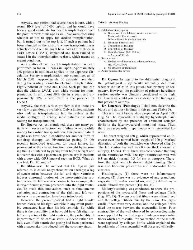
biopsy and autopsy findings in this patient (Table 3).This is a heart biopsy slide prepared in April 1993
(Fig. 6). The myocardium is slightly hypertrophic andcharacterized by the presence of abundant collagenfibrils in the intermyocardial interstitial tissue. Thus,there was myocardial hypertrophy with interstitial fib-rosis.
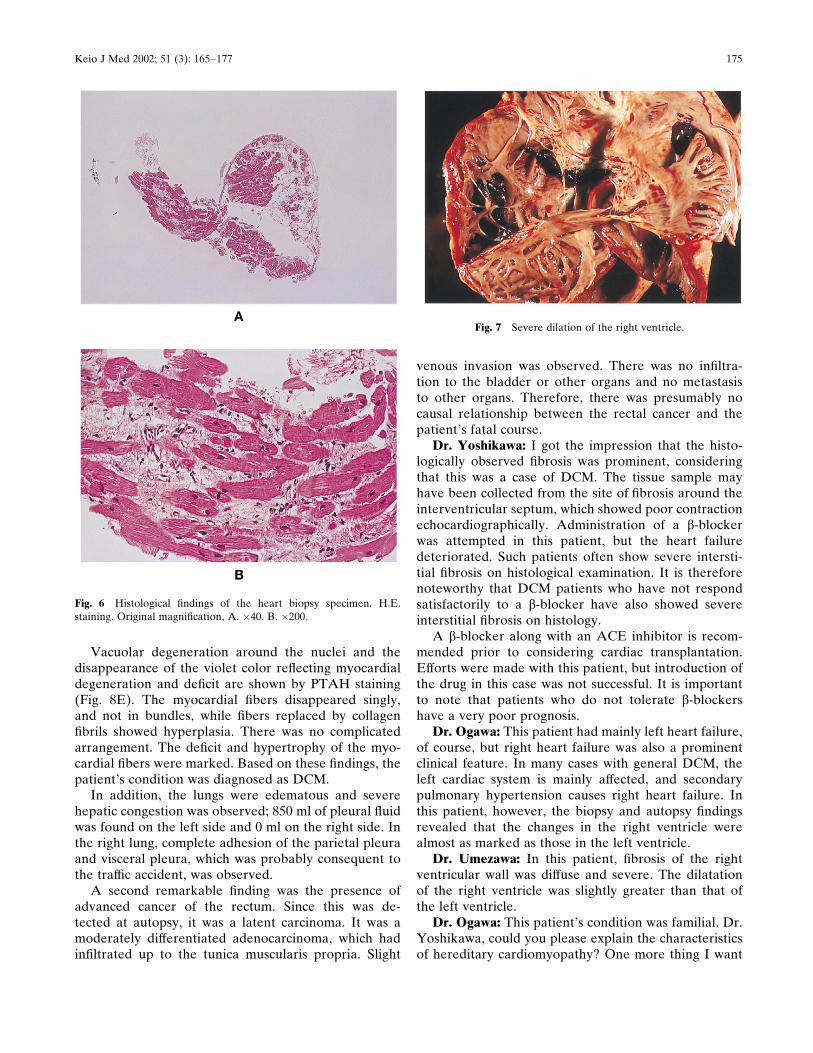
The heart weighed 450 g, which represented an in-crease over the normal heart weight of 300 g. Markeddilatation of both the ventricles was observed (Fig. 7).The left ventricular wall was 0.9 cm thick (normal atautopsy, 1.5 cm). Thus, there was considerable thinningof the ventricular wall. The right ventricular wall was0.3 cm thick (normal, 0.3–0.4 cm at autopsy). There-fore, the right ventricle showed slight thinning. Therewas also fibrinous pericarditis and endocardial fibro-elastosis.
Histologically, (1) there were no inflammatorychanges, (2) there was no evidence of any granulomasuggestive of cardiac sarcoidosis, and (3) diffuse myo-cardial fibrosis was present (Fig. 8A, B).
Mallory’s staining was conducted to show the pro-portions of the myocardial fibers and collagen fibrils(Fig. 8C, D). The myocardial fibers were stained redand the collagen fibrils blue by the stain. The myo-cardial fibers were very coarse, and the collagen fibrilsfilled the spaces between the myocardial fibers. Poorcontractility of the wall observed by echocardiographywas supported by the histological findings – myocardialfibers which are essential for contraction of the musclewere replaced by collagen fibrils, which explained thehypokinesia of the myocardial wall observed clinically.
Table 3 Autopsy Finding
1. Dilated cardiomyopathyA. Dilatation of the bilateral ventricles, severe
Endocardial fibroelastosisDiffuse fibrosis in the left ventricle
B. Fibrinous fibroelastosisC. Congestion of the lungD. Congestion of the liverE. Pleural effusion (left, 850 ml)
þ Ascites (290 ml)2. Rectal cancer
A. Moderately differentiated adenocarcinoma,mp, ly0, v1, INFg
B. No invasion or metastasis3. Acute pancreatitis, severe
174 Ogawa S, et al: A case of DCM treated by prolonged CHDF
Vacuolar degeneration around the nuclei and thedisappearance of the violet color reflecting myocardialdegeneration and deficit are shown by PTAH staining(Fig. 8E). The myocardial fibers disappeared singly,and not in bundles, while fibers replaced by collagenfibrils showed hyperplasia. There was no complicatedarrangement. The deficit and hypertrophy of the myo-cardial fibers were marked. Based on these findings, thepatient’s condition was diagnosed as DCM.
In addition, the lungs were edematous and severehepatic congestion was observed; 850 ml of pleural fluidwas found on the left side and 0 ml on the right side. Inthe right lung, complete adhesion of the parietal pleuraand visceral pleura, which was probably consequent tothe traffic accident, was observed.
A second remarkable finding was the presence ofadvanced cancer of the rectum. Since this was de-tected at autopsy, it was a latent carcinoma. It was amoderately differentiated adenocarcinoma, which hadinfiltrated up to the tunica muscularis propria. Slight
venous invasion was observed. There was no infiltra-tion to the bladder or other organs and no metastasisto other organs. Therefore, there was presumably nocausal relationship between the rectal cancer and thepatient’s fatal course.Dr. Yoshikawa: I got the impression that the histo-
logically observed fibrosis was prominent, consideringthat this was a case of DCM. The tissue sample mayhave been collected from the site of fibrosis around theinterventricular septum, which showed poor contractionechocardiographically. Administration of a b-blockerwas attempted in this patient, but the heart failuredeteriorated. Such patients often show severe intersti-tial fibrosis on histological examination. It is thereforenoteworthy that DCM patients who have not respondsatisfactorily to a b-blocker have also showed severeinterstitial fibrosis on histology.
A b-blocker along with an ACE inhibitor is recom-mended prior to considering cardiac transplantation.Efforts were made with this patient, but introduction ofthe drug in this case was not successful. It is importantto note that patients who do not tolerate b-blockershave a very poor prognosis.Dr. Ogawa: This patient had mainly left heart failure,
of course, but right heart failure was also a prominentclinical feature. In many cases with general DCM, theleft cardiac system is mainly affected, and secondarypulmonary hypertension causes right heart failure. Inthis patient, however, the biopsy and autopsy findingsrevealed that the changes in the right ventricle werealmost as marked as those in the left ventricle.Dr. Umezawa: In this patient, fibrosis of the right
ventricular wall was diffuse and severe. The dilatationof the right ventricle was slightly greater than that ofthe left ventricle.Dr. Ogawa: This patient’s condition was familial. Dr.
Yoshikawa, could you please explain the characteristicsof hereditary cardiomyopathy? One more thing I want
Fig. 6 Histological findings of the heart biopsy specimen. H.E.staining. Original magnification, A. �40. B. �200.
Fig. 7 Severe dilation of the right ventricle.
Keio J Med 2002; 51 (3): 165–177 175

to ask is whether there is any basis for consideringDCM as an autoimmune disease. It is possible thatboth the right and left ventricles are affected whenautoantibodies directed against the myocardium arepresent.Dr. Yoshikawa: No specific description has been
made in terms of the clinical features of hereditary
DCM. Many cases of hereditary hypertrophic car-diomyopathy have been reported, with a variety of genemutations. As for the genetic mechanism responsiblefor the development of DCM, however, only a limitednumber of cases have been reported. Several proteinswhich support the contractile elements, including titin,are responsible for the development of DCM.
Fig. 8 Histological findings of the heart at autopsy. A and B: H.E.staining. C and D: Mallory staining. E: PTAH staining. Original mag-nification. A: �40; B: �100; C: �100; D: �100; E: �400.
176 Ogawa S, et al: A case of DCM treated by prolonged CHDF
Basically, autoimmune cardiomyopathy in animalexperiments shows the following pattern: the rightventricle is dilated and cardiomyopathy mimickingarrhythmogenic right ventricular cardiomyopathy(ARVC) develops. It remains unknown whether or notautoimmunity was involved in the present patient, but itwould have been of interest to study this aspect.Dr. Umezawa: There was no morphological evidence
of viral infection. I wonder whether viruses are relatedto autoimmunity.Dr. Yoshikawa: It has generally been considered that
viral infection causes acute myocarditis, in which cellu-lar immune mechanisms are activated. When the cellu-lar membrane becomes disrupted, various proteins areexposed. If some proteins are presented persistently onantigen presenting cells, some antibodies will be pro-duced, causing chronic and persistent damage to themyocardium.Dr. Ogawa: Could you please give your concluding
comments on the case, Dr. Mitamura?Dr. Mitamura: Of the many patients who die of heart
failure, about half die a sudden death, and the remain-ing die of progressive heart failure. The present patientfollowed a fairly atypical course; the heart function wasconsiderably poor; even as far back as in 1997, the pa-tient had already reached the level where patients aregenerally expected to survive for only 1 or 2 years. Ofthe various treatment methods introduced recently, theuse of a b-blocker was initiated in this patient, butunfortunately, it proved to be ineffective. However,pimobendan, a PDE inhibitor, also known as a calciumsensitizer, exerted some beneficial effect, and the pa-tient himself felt significant improvement in his condi-tion. The heart function was very poor, but it remained
stable for some time. Death in cases of heart failure isfrequently associated with infection, but we often en-counter patients, like the present patient, in whomdeath occurs as a consequence of rapid deterioration ofrenal function.
One remarkable characteristic in this patient wasthat the changes in the right ventricle were alsomarked. The arrhythmia recorded in his case couldeasily explained by this finding. The serum BNP levelwas also markedly increased. These findings suggestwidespread lesions in the myocardium. The elevation ofthe BUN level was believed to be influenced by the useof a diuretic, and it is known to be very difficult to usediuretics in the presence of right heart failure. Thesepatients easily slip into dehydration. This also seems tohave made the treatment of this patient more difficult.
In conclusion, heart transplantation could have beenconsidered, but my impression is that transplantationwould have been a difficult proposition in this case, be-cause his pulmonary function was also compromised asa result of his traffic accident.Dr. Ogawa: Thank you very much for your com-
ments.The patient discussed in today’s CPC had refractory
heart failure. According to the text book, 50% of heartfailure patients die within 5 years of the diagnosis.Thus, it is a very serious disease; patients usually diewithin 2 to 3 years, on average, after the onset of heartfailure. Despite this bleak prognosis, owing to the cur-rently available methods of treatment, our patient livedfor 8 years after the diagnosis of heart failure. If hearttransplantation were introduced more aggressively inclinical cases, it could be lifesaving.
Keio J Med 2002; 51 (3): 165–177 177