clinical trial protocol and ethics application · web viewif it redetects the atrial tachycardia...
TRANSCRIPT
ROYAL ADELAIDE HOSPITAL
DEPARTMENT OF MEDICINE
CENTRE FOR HEART RHYTHM DISORDER
Clinical Trial Protocol and Ethics Application
1. TITLE
Clinical Effectiveness of Atrial Anti-tachycardia Pacing Therapy in Sick Sinus
Syndrome with Previous Atrial Fibrillation Ablation (CEASE-AF)
2. INVESTIGATORS AND QUALIFICATIONS
Professor Prashanthan Sanders, MBBS, PhD, FRACP
Dr. Dennis Lau, MBBS, PhD, FRACP
Dr. Rajiv Mahajan, MD, PhD, FRACP
Dr. Dian Andina Munawar
CONTACT DETAIL
Professor Prashanthan Sanders, MBBS, PhD, FRACP
CVIU, Level 6, Theatre Block, Royal Adelaide Hospital
Dr. Dennis Lau, MBBS, PhD, FRACP
CVIU, Level 6, Theatre Block, Royal Adelaide Hospital
Protocol version number/date : B/26082016

Dr. Rajiv Mahajan, MD, PhD, FRACP
CVIU, Level 6, Theatre Block, Royal Adelaide Hospital
Dr. Dian Andina Munawar, MD.
CVIU, Level 6, Theatre Block, Royal Adelaide Hospital
3. PURPOSE OF THE STUDY
The purpose of the CEASE-AF study is to investigate the impact of anti-
tachycardia pacing (aATP) algorithm with regards to the outcome of atrial
fibrillation (AF) in the population of patients who had AF ablation procedure
prior to this study. The use of aATP has been explored in patients with
pacemakers. The efficacy of aATP therapy is modest with different studies
demonstrating mixed results. Nevertheless, this algorithm appears to be
effective in terminating atrial flutter that potentially degenerates to AF.
Similarly, atrial preferential pacing (APP) had demonstrated promise but
failed to show reduction in AF progression or reduction in AF burden in
randomized control trial. A large proportion of atrial arrhythmias following AF
ablation are re-entrant atrial flutter that may be amenable to APP and aATP
therapy. We hypothesize that patients with pacemakers with previous history
of AF ablation will benefit from APP and aATP therapy.
4. BACKGROUND AND PRELIMINARY STUDIES
Atrial fibrillation (AF) is the most common arrhythmia associated with sinus
node dysfunction (SND). Recent studies suggested that the risk of AF
development was five-fold higher in SND, with the prevalence ranged from
45% to 53%.1-3 In the longer term, individuals with AF showed poor quality of
life indices and associated with 1.5 to 2-fold probability of death.4
The role of cardiac pacing in management of SND has been well established.
Protocol version number/date : B/26082016
However, it is reported that the annual incidence of AF and chronic AF
following pacemaker implantation is at least 5% and 3%, respectively, while
lifetime cumulative incidences is approximately 30 to 40% and 20%,
respectively.5 Over time evidence have emerged that excessive ventricular
pacing is associated with increased risk of developing AF. Pacing algorithms
that minimize ventricular pacing in patients with sinus node disease have
been demonstrated to reduce the risk of AF progression and have become
the standard of care.6
It is proposed that atrial ectopy may trigger AF and atrial preferential pacing
may suppress atrial ectopy and therefore prevent AF episodes. In addition,
atrial antitachycardia pacing (aATP) algorithm has been shown effective for a
termination of atrial arrhythmia such as atrial tachycardia or atrial flutter.7
The aim of this study is to assess the efficacy of aATP algorithm in
termination of atrial tachycardia and prevention of AF progression in patients
with pacemakers and prior AF ablation.
We propose a randomized controlled trial in patients with standard
indication of permanent pacemaker implantation due to sinus node
dysfunction (SND), pacemaker with aATP algorithm and previous history of
AF ablation.
5. PARTICIPANTS – SELECTION AND EXCLUSION CRITERIA
This study will recruit consecutive patients who had dual chamber pacemaker
implanted at EP laboratory of Royal Adelaide Hospital. These patients will be
evaluated for the eligibility to meet the following criteria:
Inclusion criteria
Patients will be considered eligible for this study if they meet the following
criteria:
a. The patient has a dual chamber pacemaker capable of atrial anti-
Protocol version number/date : B/26082016
tachycardia based therapy,
b. Had AF ablation at least 3 months prior to recruitment,
c. AF/atrial arrhythmia burden >0.1% and <30%,
d. ≥ 18 years of age
Exclusion criteria
Patients will be excluded from the study if one of the following criteria is met:
a. Atrioventricular block with ventricular pacing >40 percent,
b. Atrial fibrillation episode >90 days. However, presence of prolonged
episodes of atrial flutter will not be an exclusion corner,
c. Absence of indication for cardiac resynchronization or defibrillator,
d. Had < 12 months of life expectancy,
e. Cardiac surgery in the last six months, or expected during 10 months of
study period,
f. A recent history of (< 1 year) or current malignancy, advanced heart
failure (NYHA class IV), end-stage chronic obstructive pulmonary disease
or other severe life-threatening comorbidities within 3 months of
diagnosis, or
g. Unable to provide consent.
Redo AF ablation will not be performed in the first six month of
randomization. The patient will be censored at the time of AF ablation (if
performed after 6 months of randomization).
The sample size was calculated based on the study that showed a
recurrence rate of 48.6% after AF ablation in patients with paroxysmal AF and
prolonged sinus pauses8 and a 49% relative reduction in the DDDR with all
algorithm ON (MVP+APP+aATP) versus DDDR+MVP group, with a power of
80%, a confidence interval of 95%, and an assumed rate of loss to follow-up
of 10%. Based on the calculation, it is planned to recruit 68 patients in each
arm.
Protocol version number/date : B/26082016
6. STUDY PLAN AND DESIGN
Study design:
Randomized controlled design – two groups (1:1 ratio)
Run-in phase
Following recruitment, patient will commence a 1-month run-in period
during which all pacemakers were programmed with DDDR with MVP
(Managed Ventricular Pacing) algorithm ON only in order to evaluate AF
burden and ventricular pacing percentage. The patients who have more than
30% AF burden will be excluded from the study. Patients with ventricular
pacing >40% on run-in phase will also be excluded. Randomization will be
performed after the end of run-in period.
Randomization Period
Random allocation sequence will be conducted using computer software,
stratified to balance out the presence/absence of AV-block. Concealment of
each patient assignment was performed, and it will not be revealed until all
baseline data had been collected. Eligible patients were randomly assigned in
a 1:1 manner to (i) DDDR with MVP (control group) or (ii) MVP+APP+aATP
group (figure 1).
The patients will be followed for one-year. Assessment will be
undertaken every six-month period. Throughout the study all patients and
the other investigators remained blinded to the programmed pacing mode.
Pacemaker Programming
The enrolled patients will have a Medtronic dual chamber
pacemaker with specific features (i) MVP (atrial pacing with backup
ventricular pacing if AV conduction fails), (ii) APP (three atrial preferential
pacing algorithms to prevent atrial tachyarrhythmias, and (iii) terminating
atrial tachyarrhythmias by overdrive atrial pacing delivered at arrhythmia
onset and during dynamic transition to slower and more organized atrial
Protocol version number/date : B/26082016
tachyarrhythmias (Reactive ATP or aATP). Pacemakers will be programmed
to DDDR with a base rate of 60 bpm and an upper rate dependent on
patient’s age and underlying heart disease.
The algorithms included are as follow:
1. Managed ventricular pacing
MVP is an atrial-based pacing mode that is designed to switch to a
dual-chamber pacing mode in the presence of AV block. Specifically, MVP
provides the following functions:
AAI(R) mode pacing when AV conduction is intact
The ability to switch to DDD(R) pacing during AV block
Periodic conduction checks while operating in DDD(R) mode, with the
ability to switch back to AAI(R) mode when AV conduction resumes
Back-up ventricular support for transient loss of AV conduction
If AV conduction is intact, the device remains in AAIR or AAI mode.
While operating in AAI or AAIR mode, the parameters associated with
single chamber atrial pacing are applicable.
If 2 of the 4 most recent A-A intervals are missing a ventricular
event, the device identifies a loss of AV conduction and switches to the
DDDR or DDD mode. The device provides back-up ventricular pacing in
response to dropped ventricular events until the loss of AV conduction is
identified.
After switching to DDDR or DDD mode, the device periodically
checks AV conduction for an opportunity to return to AAIR or AAI mode.
The first AV conduction check occurs 1 min after switching to DDDR or
DDD mode. During the conduction check, the device switches to AAIR or
AAI pacing mode for one cycle.
If the next A-A interval includes a sensed ventricular beat, the
conduction check succeeds. The device remains in AAIR or AAI pacing
mode.
If the next A-A interval does not include a sensed ventricular beat, the
conduction check fails, and the device switches back to the DDDR or
Protocol version number/date : B/26082016
DDD mode. The time between conduction checks doubles (2, 4, 8 …
min, up to a maximum of 16 hours) with each failed conduction check.
2. Atrial Preference Pacing (APP)
The device includes three atrial preventive pacing algorithms
designed to eliminate some of the onset mechanisms of atrial
tachyarrhythmias and to reduce the incidence of atrial tachyarrhythmias.
These atrial pacing features comprise the atrial pacing preference
algorithm, for maintenance of a pacing rate just above the intrinsic rate,
the atrial rate stabilization algorithm, designed to avoid short-long
intervals following a premature atrial contraction, and the post-mode
switching overdrive pacing algorithm designed to inhibit early re-initiation
of atrial tachyarrhythmia following a mode switching episode.
Atrial Preference Pacing (APP) is available when the device is
operating in the DDDR, DDD, AAIR, AAI, or MVP (AAIR<=>DDDR or
AAI<=>DDD) mode. APP is a programmable feature that is designed to
maximize atrial overdrive pacing when the patient is not experiencing an
atrial tachyarrhythmia. The device responds to changes in the atrial rate
by accelerating the pacing rate until reaching a steady paced rhythm that
is slightly faster than the intrinsic rate. After each non-refractory atrial
sensed event, the device decreases the atrial-pacing interval by the
programmed Interval Decrement value. This progression continues until
the pacing rate exceeds the intrinsic rate, resulting in an atrial-paced
rhythm. It sustains this increased rate for the number of beats
programmed for a Search Beats parameter, then decreases the pacing
rate slightly (by 20 ms) to search for the next intrinsic beat. This results in
a dynamic, controlled, stair-step increase or decrease in the pacing
interval, resulting in a pacing rate slightly above the intrinsic rate. The
Maximum Rate parameter sets an upper rate limit for APP. The Interval
Decrement in CEASE-AF study was set at 50 ms. After 10 Search Beats, i.e.
consecutive atrial paces at the current atrial preference pacing rate, the
pacing rate interval was increased by 20 ms to decelerate the pacing rate.
Protocol version number/date : B/26082016
The Maximum Rate induced by atrial preference pacing was set at 95
bpm.
The atrial rate stabilization algorithm is available when the device is
operating in the DDDR, DDD, AAIR, AAI modes. When it is enabled, at
each atrial event, for example a premature atrial contraction (PAC), the
device calculates a new pacing interval, which is equal to the current
pacing interval increased by the Interval Percentage Increment, and if this
interval ends before the device senses an atrial event, the device delivers
an atrial pace and recalculates its interval using the current atrial interval.
After a PAC, the calculated escape interval stabilizes the atrial rate and
gradually slows it to the intrinsic rate, sensor indicated rate or lower rate.
This prevents the ‘short/long’ sequences of atrial cycle lengths that have
been observed to precede the onset of some spontaneous atrial
tachyarrhythmias. The Interval Percentage Increment, i.e. the pacing
interval increment per beat, measured as a percentage of the preceding
interval was set at 25% in our study. The Maximum Rate induced by atrial
rate stabilization algorithm was set at 95 bpm.
The post-mode switching overdrive pacing algorithm is available
when the device is operating in the DDDR or DDD modes. When it is
enabled, at the termination of a mode switch, i.e. an atrial
tachyarrhythmia episode, post-mode switching overdrive pacing causes
the device to continue to pace in DDIR mode at the higher of the
programmable Overdrive Rate or sensor-activated rate for a
programmable Overdrive Duration. In our study the Overdrive Rate was
set at 80 beats per minute and the Overdrive Duration was required to be
≤ 5 minutes.
3. Reactive atrial Anti-tachycardia pacing
If the device detects an atrial tachyarrhythmia episode, aATP
therapy will be delivered. Treatments for such episodes are intended to
interrupt the atrial tachycardia and restore patient’s normal sinus
Protocol version number/date : B/26082016

rhythm.
The device can deliver up to 3 aATP therapies to treat an AT/AF or
a Fast AT/AF episode. aATP therapies become available when the
duration of sustained atrial tachyarrhythmias exceeds the programmed
value of episode duration before aATP delivery - which in this study will
be set at 0 minute.
When an AT/AF or Fast AT/AF episode is detected, the device
delivers the first sequence of the ATP therapy. After the first ATP
sequence, it continues to monitor for the presence of the atrial
tachycardia episode. If it redetects the atrial tachycardia episode, the
device and repeats this cycle until the episode is terminated or all
sequences in the therapy are exhausted.
In the CEASE-AF study, reactive ATP makes it possible for the device
to repeat programmed sets of atrial ATP therapies in two different
situations. Rhythm Change, one type of Reactive ATP, subdivides the
AT/AF detection zone into smaller regions. This type is enabled to allow
the device to detect changes in the atrial arrhythmia on the basis of both
regularity and cycle length. The atrial tachyarrhythmias zone is subdivided
into a series of narrower regions: specifically the atrial tachyarrhythmia
Interval for regular rhythms is divided into 5 regions of 50 ms length from
100 ms to 350 ms and the atrial tachyarrhythmia interval for irregular
rhythm is divided into 3 regions, the first from 100 ms to 200 ms, the
second from 200 ms to 300 ms and the third from 300 ms to 350 ms. Each
region is supplied with a separate set of the atrial aATP therapies enabled
for atrial tachyarrhythmia episodes. If the rhythm shifts into a different
region because of a change in cycle length or regularity, the device
delivers therapies from those available in the new region. The second
type of reactive ATP is Time Interval feature, which allows to schedule
additional therapies for atrial arrhythmias regardless of rhythm changes. .
In the CEASE-AF the Reactive aATP Time Interval allow to re-arm aATP
sequences when the Sustained Duration value reaches a multiple of 2
hours. The device will be programmed to suspend all atrial therapies if an
Protocol version number/date : B/26082016
atrial episode exceeded the Duration to Stop parameter, which was
programmed at 72 hours.
The aATP therapies that can be delivered by the pacemaker are
Burst+ or Ramp. Burst+ therapy sequences consist of a programmed
number of AOO pulses followed by two premature stimuli that are
delivered at shorter intervals. Ramp therapy sequences consist of a
programmable number of AOO pulses delivered at decreasing intervals.
The Burst+ and Ramp pacing intervals are based on programmed
percentages of the atrial tachycardia cycle length, which is calculated as
the median of the last 12 atrial intervals prior to therapy delivery. The
median atrial tachycardia cycle length can vary from one sequence in a
therapy to the next, and the ATP pacing intervals vary accordingly. The
first pulse of each aATP sequence is delivered at a chosen percentage of
the current atrial tachyarrhythmia cycle length. Subsequent pulses are
delivered at progressively shorter intervals. The A-A Minimum ATP
Interval parameter limits the pacing intervals at which the Burst+ and
Ramp pacing pulses are delivered. If some calculated intervals are
shorter than the programmed A-A Minimum ATP Interval, the pulses are
delivered at the A-A Minimum ATP Interval. If the median of the last 12
A-A intervals is shorter than the programmed A-A Minimum ATP Interval,
the device does not deliver Burst+ or Ramp therapies until the atrial rate
slows.
Parameters for Burst+ therapy were the Number of S1 pulses in
each burst sequence (15), the Pacing interval of the S1 burst pulses, as a
percentage of the pre-therapy atrial cycle length (84%), the Pacing
interval of the S2 stimulus following the burst as a percentage of the pre-
therapy atrial cycle length (81%), the S2-S3 interval equals the S1-S2
interval minus this decrement value (20 ms), the Pacing interval
decrement per sequence (10 ms), the Number of sequences in the Burst+
therapy (10).
The Parameters for Ramp therapy were Number of pulses in the
first Ramp sequence (12), the Pacing interval of the first Ramp pulse as a
Protocol version number/date : B/26082016
percentage of the pre-therapy atrial cycle length (91% in Rx1 and 81% in
Rx2), the Pacing interval decrement per pulse for the remaining Ramp
pulses in each sequence (10 ms), the number of sequences in the Ramp
therapy.
AF detection
The device detects atrial tachyarrhythmias by examining the atrial
rate and the relationship between atrial and ventricular events. The device
confirms initial AT/AF episode detection if the criteria are met (table 2).
After confirmation of detection, the atrial tachyarrhythmia episode is
considered to be sustained; thereafter the device monitors the cardiac
rhythm for changes or episode termination and can respond to detected
episodes by delivering tachyarrhythmia therapies. Once the device detects an
atrial tachyarrhythmia, the episode is considered to be ongoing until episode
termination.
Clinical parameters
A complete medical history will be recorded to obtain co-existing risk factor.
The variables that will be obtained are as follows:
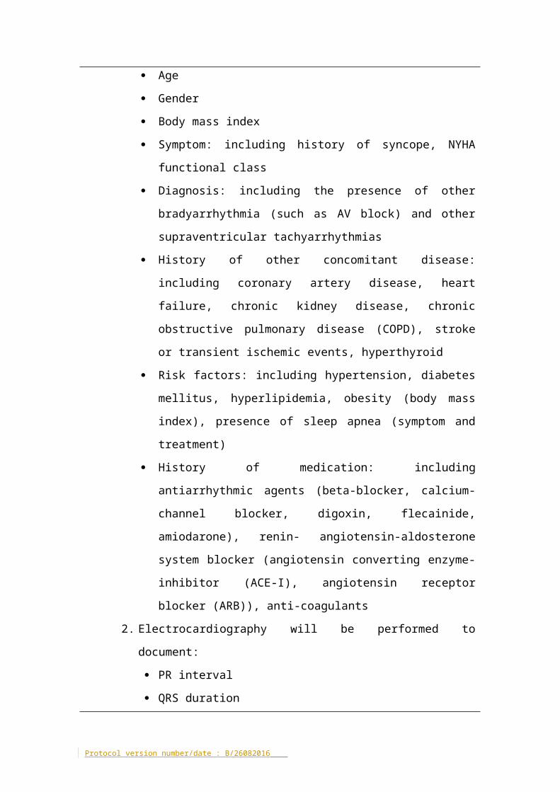
1. Baseline characteristics
Age
Gender
Body mass index
Symptom: including history of syncope, NYHA functional class
Diagnosis: including the presence of other bradyarrhythmia (such as
AV block) and other supraventricular tachyarrhythmias
History of other concomitant disease: including coronary artery
disease, heart failure, chronic kidney disease, chronic obstructive
pulmonary disease (COPD), stroke or transient ischemic events,
hyperthyroid
Risk factors: including hypertension, diabetes mellitus,
hyperlipidemia, obesity (body mass index), presence of sleep apnea
Protocol version number/date : B/26082016
(symptom and treatment)
History of medication: including antiarrhythmic agents (beta-blocker,
calcium-channel blocker, digoxin, flecainide, amiodarone), renin-
angiotensin-aldosterone system blocker (angiotensin converting
enzyme-inhibitor (ACE-I), angiotensin receptor blocker (ARB)), anti-
coagulants
2. Electrocardiography will be performed to document:
PR interval
QRS duration
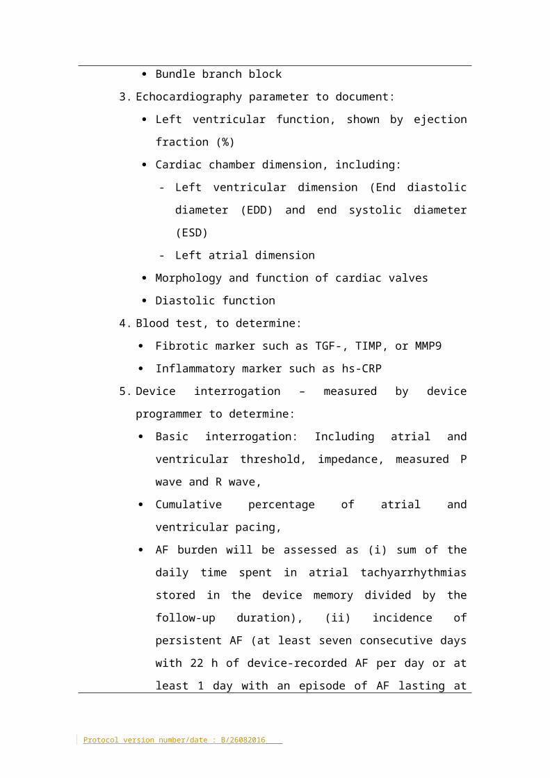
Bundle branch block
3. Echocardiography parameter to document:
Left ventricular function, shown by ejection fraction (%)
Cardiac chamber dimension, including:
- Left ventricular dimension (End diastolic diameter (EDD) and end
systolic diameter (ESD)
- Left atrial dimension
Morphology and function of cardiac valves
Diastolic function
4. Blood test, to determine:
Fibrotic marker such as TGF-, TIMP, or MMP9
Inflammatory marker such as hs-CRP
5. Device interrogation – measured by device programmer to determine:
Basic interrogation: Including atrial and ventricular threshold,
impedance, measured P wave and R wave,
Cumulative percentage of atrial and ventricular pacing,
AF burden will be assessed as (i) sum of the daily time spent in atrial
tachyarrhythmias stored in the device memory divided by the
follow-up duration), (ii) incidence of persistent AF (at least seven
consecutive days with 22 h of device-recorded AF per day or at least
1 day with an episode of AF lasting at least 22 h—and interrupted
with an electrical or chemical cardioversion), and (iii) incidence of AF
with pre-specified daily durations (5 min, 1 h, 6 h, 1 day, 2 days, 7
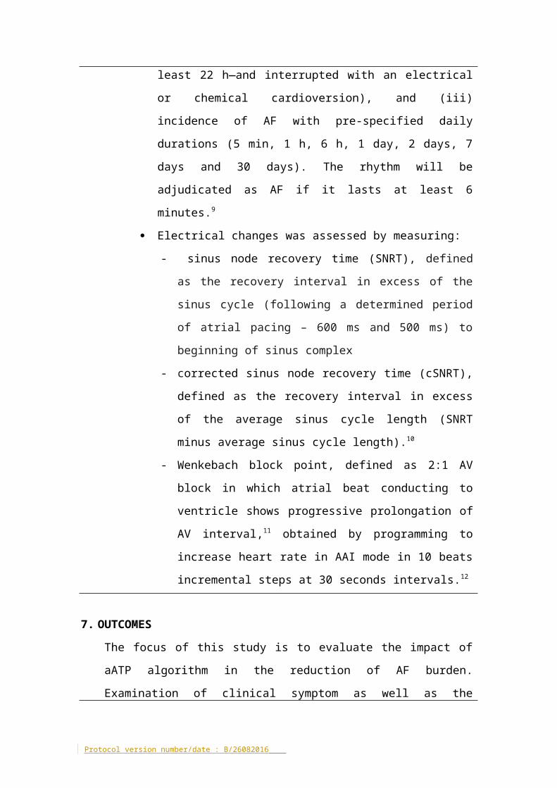
Protocol version number/date : B/26082016
days and 30 days). The rhythm will be adjudicated as AF if it lasts at
least 6 minutes.9
Electrical changes was assessed by measuring:
- sinus node recovery time (SNRT), defined as the recovery
interval in excess of the sinus cycle (following a determined
period of atrial pacing – 600 ms and 500 ms) to beginning of
sinus complex
- corrected sinus node recovery time (cSNRT), defined as the
recovery interval in excess of the average sinus cycle length
(SNRT minus average sinus cycle length).10
- Wenkebach block point, defined as 2:1 AV block in which atrial
beat conducting to ventricle shows progressive prolongation of
AV interval,11 obtained by programming to increase heart rate in
AAI mode in 10 beats incremental steps at 30 seconds
intervals.12
7. OUTCOMES
The focus of this study is to evaluate the impact of aATP algorithm in the
reduction of AF burden. Examination of clinical symptom as well as the
structural and electrical function will be performed after run-in phase (1 month
after pacemaker implantation procedure) and during each follow up visit (every
three months).
1. Primary end point
a. AT/AF burden at 12 month follow up – defined as cumulative number of
hours of AT/AF divided by follow-up period in days (hours/day),
calculated based on data in the device storage over the study terms. The
rhythm will be adjudicated as AF if it lasts at least 6 minutes.9 All
recorded data will be adjudicated manually by a committee of
experienced electrophysiologists.13
2. Secondary endpoint
a. ATP efficacy – defined as the incidence of successful terminations based
Protocol version number/date : B/26082016

on the device counters.
b. Number of subjects who develop:
i. Paroxysmal AF – defined as self-terminating AF, usually within 48 h,
may continue for up to 7 days and spontaneous conversion.9,14
ii. Persistent AF – defined as AF episode either lasts longer than 7 days
or requires termination by cardioversion, either with drugs or by
direct current cardioversion (DCC).9,14
iii. Permanent AF – defined as the presence of the arrhythmia that is
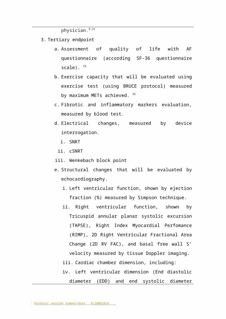
accepted by the patient and physician.9,14
3. Tertiary endpoint
a. Assessment of quality of life with AF questionnaire (according SF-36
questionnaire scale). 15
b. Exercise capacity that will be evaluated using exercise test (using
BRUCE protocol) measured by maximum METs achieved. 16
c. Fibrotic and inflammatory markers evaluation, measured by blood
test.
d. Electrical changes, measured by device interrogation.
i. SNRT
ii. cSNRT
iii. Wenkebach block point
e. Structural changes that will be evaluated by echocardiography.
i. Left ventricular function, shown by ejection fraction (%) measured
by Simpson technique.
ii. Right ventricular function, shown by Tricuspid annular planar
systolic excursion (TAPSE), Right Index Myocardial Perfomance
(RIMP), 2D Right Ventricular Fractional Area Change (2D RV FAC),
and basal free wall S’ velocity measured by tissue Doppler
imaging.
iii. Cardiac chamber dimension, including:
iv. Left ventricular dimension (End diastolic diameter (EDD) and end
systolic diameter (ESD)
Protocol version number/date : B/26082016
v. Right and left atrial dimension
vi. Morphology and function of cardiac valves
The manuscript will be submitted for consideration of publication in a peer-
reviewed journal.
8. ETHICAL CONSIDERATIONS
Managed ventricular pacing (MVP), atrial preferencial (APP) and atrial anti-
tachycardia pacing (aATP) algorithm has been demonstrated to be safe in
several studies.6,7,17
9. SPECIFIC SAFETY
N/A
10. DRUGS/DEVICES
Dual chamber pacemaker has been implanted for the patients based
standard indication according to HRS guideline 2012.18 The subjects will be
recruited after the procedure.
11. ANALYSIS AND REPORTING OF RESULTS
All data will be analyzed according to the intention-to-treat principle. The
nominal data will be presented as number of events (n) and percentage (%).
Normally distributed continuous data will be expressed as mean ± standard
deviation. The difference between groups will be analyzed using chi-squared
comparisons for the nominal data and the normal continuous data tested with
unpaired t-tests between groups. Skewed distributions will be expressed as
Protocol version number/date : B/26082016

median and inter-quartile and means tested using Mann-Whitney U.
Data will be collected from case sheets, echocardiograms and pacemaker
interrogation reports. The data will be stored at the Centre of Heart Rhythm
Disorders. The principal investigators will have access to the research data, and
will collectively own the data and results of the research. Data (whether
positive or negative findings) will be analysed and published. All patient
information will be de-identified.
On completion, the manuscript will be submitted for publication in a pear
review journal
12. REFERENCES
1. Lamas GA, Lee K, Sweeney M, et al. The mode selection trial (MOST) in sinus node dysfunction: design, rationale, and baseline characteristics of the first 1000 patients. American heart journal 2000;140:541-51.
2. Santini M, Alexidou G, Ansalone G, Cacciatore G, Cini R, Turitto G. Relation of prognosis in sick sinus syndrome to age, conduction defects and modes of permanent cardiac pacing. Am J Cardiol 1990;65:729-35.
3. Alonso A, Jensen PN, Lopez FL, et al. Association of sick sinus syndrome with incident cardiovascular disease and mortality: the Atherosclerosis Risk in Communities study and Cardiovascular Health Study. PloS one 2014;9:e109662.
4. Ball J, Carrington MJ, McMurray JJ, Stewart S. Atrial fibrillation: profile and burden of an evolving epidemic in the 21st century. International journal of cardiology 2013;167:1807-24.
5. Nielsen JC. Mortality and incidence of atrial fibrillation in paced patients. J Cardiovasc Electrophysiol 2002;13:S17-22.
6. Boriani G, Tukkie R, Manolis AS, et al. Atrial antitachycardia pacing and managed ventricular pacing in bradycardia patients with paroxysmal or persistent atrial tachyarrhythmias: the MINERVA randomized multicentre international trial. Eur Heart J 2014;35:2352-62.
7. Gillis AM, Koehler J, Morck M, Mehra R, Hettrick DA. High atrial antitachycardia pacing therapy efficacy is associated with a reduction in atrial tachyarrhythmia burden in a subset of patients with sinus node dysfunction and paroxysmal atrial fibrillation. Heart Rhythm 2005;2:791-6.
8. Inada K, Yamane T, Tokutake K, et al. The role of successful catheter ablation in patients with paroxysmal atrial fibrillation and prolonged sinus pauses: outcome during a 5-year follow-up. Europace 2014;16:208-13.
9. European Heart Rhythm A, European Association for Cardio-Thoracic S, Camm AJ, et al. Guidelines for the management of atrial fibrillation: the Task Force for the Management of Atrial Fibrillation of the European Society
Protocol version number/date : B/26082016
of Cardiology (ESC). Europace : European pacing, arrhythmias, and cardiac electrophysiology : journal of the working groups on cardiac pacing, arrhythmias, and cardiac cellular electrophysiology of the European Society of Cardiology 2010;12:1360-420.
10. Narula OS, Samet P, Javier RP. Significance of the sinus-node recovery time. Circulation 1972;45:140-58.
11. Levy S, Roudaut R, Bouvier E, Obel IW, Clementy J, Bricaud H. Alternate ventriculoatrial Wenckebach conduction. Circulation 1980;61:648-52.
12. Haywood GA, Ward J, Ward DE, Camm AJ. Atrioventricular Wenckebach point and progression to atrioventricular block in sinoatrial disease. Pacing and clinical electrophysiology : PACE 1990;13:2054-8.
13. Padeletti L, Purerfellner H, Adler SW, et al. Combined efficacy of atrial septal lead placement and atrial pacing algorithms for prevention of paroxysmal atrial tachyarrhythmia. Journal of cardiovascular electrophysiology 2003;14:1189-95.
14. Wann LS, Curtis AB, January CT, et al. 2011 ACCF/AHA/HRS focused update on the management of patients with atrial fibrillation (Updating the 2006 Guideline): a report of the American College of Cardiology Foundation/American Heart Association Task Force on Practice Guidelines. Heart rhythm : the official journal of the Heart Rhythm Society 2011;8:157-76.
15. Ware JE, Jr., Sherbourne CD. The MOS 36-item short-form health survey (SF-36). I. Conceptual framework and item selection. Med Care 1992;30:473-83.
16. Bruce RA. Exercise testing of patients with coronary heart disease. Principles and normal standards for evaluation. Ann Clin Res 1971;3:323-32.
17. Padeletti L, Purerfellner H, Mont L, et al. New-generation atrial antitachycardia pacing (Reactive ATP) is associated with reduced risk of persistent or permanent atrial fibrillation in patients with bradycardia: Results from the MINERVA randomized multicenter international trial. Heart Rhythm 2015;12:1717-25.
18. Tracy CM, Epstein AE, Darbar D, et al. 2012 ACCF/AHA/HRS Focused Update of the 2008 Guidelines for Device-Based Therapy of Cardiac Rhythm Abnormalities: a report of the American College of Cardiology Foundation/American Heart Association Task Force on Practice Guidelines. Heart Rhythm 2012;9:1737-53.
13. OTHER RELEVANT INFORMATION
Nil
14. OTHER ETHIC COMMITTEES TO WHICH THE PROTOCOL HAS BEEN
SUBMMITTED
Nil
Protocol version number/date : B/26082016
15. DATE OF PROPOSED COMMENCEMENT
July 2016
16. DATE OF EXPECTED COMPLETION
December 2017
17. RESOURCE CONSIDERATION
The staffing and facilities required for the undertaking of this research
protocol will de derived entirely from within the Cardiovascular Investigation
Unit (CVIU) and Centre for Heart Rhythm Disorders (CHRD). In particular, the
senior cardiologists, rostered electrophysiology fellows, technicians, and
staffs involved would all be employees of RAH. Moreover, the principal
investigators will be present during the pacemaker programming, who will
also be responsible for the collection and recording of data. Dr. Munawar will
undertake this project as a part of her PhD studies. The financing of this
research protocol will be initially funded by available research funds. It is
anticipated that after collection of preliminary data, competitive funding will
be sought.
Access to patient medical records will be necessary during the study in
order to assess for cardiovascular risk factors. Such access will be strictly
limited to study personnel, with prior written consent of each patient
involved in the study.
18. FINANCIAL AND INSURANCE ISSUES
There are no financial interests in the outcome of the study project. This is
not a commercially sponsored trial involving a drug or device. Procedures
performed in CVIU will be covered by standard Royal Adelaide Hospital
Protocol version number/date : B/26082016
indemnity agreements.
19. SIGNATURES OF INVESTIGATORS
Professor Prashanthan Sanders, MBBS, PhD, FRACP
Dr. Dennis Lau, MBBS, PhD, FRACP
Dr. Rajiv Mahajan, MD, PhD, FRACP
Dr. Dian Andina Munawar, MD
Dr. Sharath Kumar, MD
Protocol version number/date : B/26082016

Dr. Kashif Khokhar, MD
Mr. Thomas Agbaedeng, BSc
Protocol version number/date : B/26082016
20. APPENDIX
FIGURE 1. Trial Schema
Protocol version number/date : B/26082016
Participant fulfills inclusion / exclusion criteriaIf eligible, participant signs information consent form.
RUN-IN PERIOD (1 MONTH)MVP ON
Participant will be screened prior to randomization
RANDOMISATION
Group A
EnrollmentMVP on
Medical history SF-36/MLWHF Questionnaire ECG Trans Thoracic Echo (TTE) Blood samples Exercise test
Group C
Enrollment MVP + APP + ATP on
Medical history SF-36/MLWHF Questionnaire ECG Trans Thoracic Echo (TTE) Blood samples Exercise test
Clinic visit month 6
SF-36/MLWHF Questionnaire Trans Thoracic Echo (TTE) Device interrogation Blood samples Exercise test
Clinic visit month 6
SF-36/MLWHF Questionnaire Trans Thoracic Echo (TTE) Device interrogation Blood samples Exercise test
Clinic visit month 12
SF-36/MLWHF Questionnaire Trans Thoracic Echo (TTE) Device interrogation Blood samples Exercise test
Clinic visit month 12
SF-36/MLWHF Questionnaire Trans Thoracic Echo (TTE) Device interrogation Blood samples Exercise test
Table 1. The programming of aATP in our study is shown in table 1:
Therapy sequence Rx1 Rx2 Rx3
Therapy Ramp Ramp Burst+
Initial pulse N° 12 12 15
A-S1 91% 81% 84%
S1S2 81%
S2S3 20 ms
Dec Interval 10 ms 10 ms 10 ms
Sequence N° 10 10 10
aATP programmingEpisode duration before aATP: 0 minuteRhythm based rearming: ONTime to stop therapy: 72 hoursaATP output : 4 times atrial threshold (min 3.0 V and max 6.0 V)
Table 2. AF detection criteria and programming
A. AF detection criteriaa. There are at least 2 atrial sensed events per ventricular interval for
a sufficient number of ventricular intervals (which will be set at 32).b. The median of the 12 most recent sensed atrial intervals is shorter
than the programmed AT/AF (or Fast AT/AF) interval. In CEASE-AF study, it will be set at 350 ms (171 bpm)
B. AF detection programmingNumber of detection zones: 1Atrial interval: 350 ms or according to patient characteristics.
Protocol version number/date : B/26082016
Protocol version number/date : B/26082016
1. Lamas GA, Lee K, Sweeney M, et al. The mode selection trial (MOST) in sinus node dysfunction: design, rationale, and baseline characteristics of the first 1000 patients. American heart journal 2000;140:541-51.2. Santini M, Alexidou G, Ansalone G, Cacciatore G, Cini R, Turitto G. Relation of prognosis in sick sinus syndrome to age, conduction defects and modes of permanent cardiac pacing. Am J Cardiol 1990;65:729-35.3. Alonso A, Jensen PN, Lopez FL, et al. Association of sick sinus syndrome with incident cardiovascular disease and mortality: the Atherosclerosis Risk in Communities study and Cardiovascular Health Study. PloS one 2014;9:e109662.4. Ball J, Carrington MJ, McMurray JJ, Stewart S. Atrial fibrillation: profile and burden of an evolving epidemic in the 21st century. International journal of cardiology 2013;167:1807-24.5. Nielsen JC. Mortality and incidence of atrial fibrillation in paced patients. J Cardiovasc Electrophysiol 2002;13:S17-22.6. Boriani G, Tukkie R, Manolis AS, et al. Atrial antitachycardia pacing and managed ventricular pacing in bradycardia patients with paroxysmal or persistent atrial tachyarrhythmias: the MINERVA randomized multicentre international trial. Eur Heart J 2014;35:2352-62.7. Gillis AM, Koehler J, Morck M, Mehra R, Hettrick DA. High atrial antitachycardia pacing therapy efficacy is associated with a reduction in atrial tachyarrhythmia burden in a subset of patients with sinus node dysfunction and paroxysmal atrial fibrillation. Heart Rhythm 2005;2:791-6.8. Inada K, Yamane T, Tokutake K, et al. The role of successful catheter ablation in patients with paroxysmal atrial fibrillation and prolonged sinus pauses: outcome during a 5-year follow-up. Europace 2014;16:208-13.9. European Heart Rhythm A, European Association for Cardio-Thoracic S, Camm AJ, et al. Guidelines for the management of atrial fibrillation: the Task Force for the Management of Atrial Fibrillation of the European Society of Cardiology (ESC). Europace : European pacing, arrhythmias, and cardiac electrophysiology : journal of the working groups on cardiac pacing, arrhythmias, and cardiac cellular electrophysiology of the European Society of Cardiology 2010;12:1360-420.10. Narula OS, Samet P, Javier RP. Significance of the sinus-node recovery time. Circulation 1972;45:140-58.11. Levy S, Roudaut R, Bouvier E, Obel IW, Clementy J, Bricaud H. Alternate ventriculoatrial Wenckebach conduction. Circulation 1980;61:648-52.12. Haywood GA, Ward J, Ward DE, Camm AJ. Atrioventricular Wenckebach point and progression to atrioventricular block in sinoatrial disease. Pacing and clinical electrophysiology : PACE 1990;13:2054-8.
Protocol version number/date : B/26082016
13. Padeletti L, Purerfellner H, Adler SW, et al. Combined efficacy of atrial septal lead placement and atrial pacing algorithms for prevention of paroxysmal atrial tachyarrhythmia. Journal of cardiovascular electrophysiology 2003;14:1189-95.14. Wann LS, Curtis AB, January CT, et al. 2011 ACCF/AHA/HRS focused update on the management of patients with atrial fibrillation (Updating the 2006 Guideline): a report of the American College of Cardiology Foundation/American Heart Association Task Force on Practice Guidelines. Heart rhythm : the official journal of the Heart Rhythm Society 2011;8:157-76.15. Ware JE, Jr., Sherbourne CD. The MOS 36-item short-form health survey (SF-36). I. Conceptual framework and item selection. Med Care 1992;30:473-83.16. Bruce RA. Exercise testing of patients with coronary heart disease. Principles and normal standards for evaluation. Ann Clin Res 1971;3:323-32.17. Padeletti L, Purerfellner H, Mont L, et al. New-generation atrial antitachycardia pacing (Reactive ATP) is associated with reduced risk of persistent or permanent atrial fibrillation in patients with bradycardia: Results from the MINERVA randomized multicenter international trial. Heart Rhythm 2015;12:1717-25.18. Tracy CM, Epstein AE, Darbar D, et al. 2012 ACCF/AHA/HRS Focused Update of the 2008 Guidelines for Device-Based Therapy of Cardiac Rhythm Abnormalities: a report of the American College of Cardiology Foundation/American Heart Association Task Force on Practice Guidelines. Heart Rhythm 2012;9:1737-53.
Protocol version number/date : B/26082016