clinical study long-term observation of triplex surgery...
TRANSCRIPT

Clinical StudyLong-Term Observation of Triplex Surgery for Cataract afterPhakic 6H Implantation for Super High Myopia
Xin Liu,1 Fan Fan,1 Xiaoying Wang,1 Yi Lu,1 Tianyu Zheng,1 Peng Zhou,2
Xingtao Zhou,1 and Yi Luo1
1Department of Ophthalmology, Eye and ENT Hospital, Fudan University, Shanghai 200032, China2Department of Ophthalmology, ParkwayHealth Hong Qiao Medical Center, Shanghai, China
Correspondence should be addressed to Xingtao Zhou; [email protected] and Yi Luo; [email protected]
Received 5 November 2015; Revised 9 February 2016; Accepted 24 February 2016
Academic Editor: Majid M. Moshirfar
Copyright © 2016 Xin Liu et al. This is an open access article distributed under the Creative Commons Attribution License, whichpermits unrestricted use, distribution, and reproduction in any medium, provided the original work is properly cited.
Purpose. To analyze the safety, effectiveness, and stability of triplex surgery for phakic 6H anterior chamber phakic intraocularlens explantation and phacoemulsification with in-the-bag IOL implantation for super high myopia in long-term observations.Methods. This retrospective case series evaluated 16 eyes of 10 patients who underwent triplex surgery. Best corrected visual acuity(BCVA), endothelial cell density (ECD), and associated adverse events were evaluated. Results. The mean follow-up time after thetriplex surgery was 46 ± 14 months. The mean logMAR BCVA was significantly improved after triplex surgery (𝑃 = 0.047). Oneeye developed endophthalmitis five days postoperatively and underwent pars plana vitrectomy (PPV). Five eyes with preoperativesevere endothelial cell loss developed corneal decompensation and underwent keratoplasty at a mean time of 9.4 ± 2.6 monthsafter the triplex surgery. One eye had graft failure and underwent a second keratoplasty. The eye developed rhegmatogenousretinal detachment and underwent PPV with silicone oil 18 months later. ECD before the triplex surgery was not significantlydifferent compared with that at last follow-up (𝑃 = 0.495) apart from these five eyes.Three eyes (18.8%) developed posterior capsuleopacification.Conclusions. Triplex surgerywas safe and effective for phakic 6H related complicated cataracts. Early extraction beforesevere ECD loss is recommended.
1. Introduction
Anterior chamber angle-supported phakic intraocular lenses(AC pIOLs) were once widely accepted as an effective optionfor the correction of high myopia [1]. They offer accurate andstable refraction, preserve the shape of the cornea, preserveaccommodation of the crystalline lens, and are potentiallyreversible compared to laser keratorefractive or refractivelens exchange procedures [2]. The early generation PMMAAC pIOL phakic 6H has a particularly wide optic diameter(6.0mm, 5.5mm in powers greater than−20.0D) and neededa large incision for implantation [3]. Its visual outcomesare encouraging. The best corrected visual acuity (BCVA),refraction, and contrast sensitivity are all improved in theshort-term reports [4–6]. It was used globally since 1990s andwas popular in China since 2002. However, this model wasphased from the market because its use was associated with
progressive ovalization of the pupil, glare, and endothelial cellloss in the long run [2, 7]. InChina, phakic 6Hwaswithdrawnin early 2012 and patients with previous implantation sufferedprolonged endothelial cell loss and pupil ovalization [8].Furthermore, with aging and progression of pathologicalhigh myopia, cataract formation, accompanied by severeproblems involving the iris and cornea, develops and calls forAC pIOL explantation and vision restoration [9].
Surgical outcomes of patients with new-onset AC pIOL-associated cataracts (called bilensectomy surgery, and mostlyfor ZB5M and ZSAL-4 model) are usually successful afterless than 1 year of mean follow-up time [10, 11]. In thepresent study, we applied triplex surgery of phakic 6HAC pIOL explantation and phacoemulsification with in-the-bag intraocular lens (IOL) implantation in order to solvephakic 6H AC pIOL-related complications. We provide thelongest follow-up term after explantation of AC pIOLs and
Hindawi Publishing CorporationJournal of OphthalmologyVolume 2016, Article ID 9569868, 10 pageshttp://dx.doi.org/10.1155/2016/9569868
2 Journal of Ophthalmology
evaluate its safety, effectiveness, and stability, which mightalert ophthalmologists to closer postoperative surveillanceand help surgeons to determine the optimal surgical time.
2. Patients and Methods
2.1. Patients. We retrospectively reviewed 16 eyes of 10patients who were previously implanted with phakic 6HAC pIOL (Ophthalmic Innovations International, Ontario,CA) for the correction of super high myopia and developedlens opacity undergoing triplex cataract surgery from July1, 2008, to June 30, 2012, at Eye & ENT Hospital of FudanUniversity, Shanghai, China. We also included one patient(Case 2) who underwent phakic 6HAC pIOL implantation ata different hospital and developed cataract. This patient wastransferred to our cataract clinic for triplex surgery during theabove period. Written informed consent was obtained fromall patients. This study was carried out with the approval ofthe Institutional Review Board of Eye and ENT Hospital ofFudan University and in accordance with the Declaration ofHelsinki.
2.2. Preoperative Evaluation. The following examinationswere performed preoperatively: best corrected visual acuity(BCVA), slit-lamp biomicroscopy, dilated fundus examina-tion, intraocular pressure (IOP), axial length (AL), andendothelial specularmicroscopy. Cataract type andmorphol-ogy were evaluated using the Lens Opacities ClassificationSystem III (LOCS III) [12]. AL and anterior segment sizewere measured using interferometry (IOL Master, Carl ZeissMeditec AG) or A-scan ultrasonography without any correc-tion for the pIOL in situ. The SRK/T or Holladay 1 formulawas used for posterior chamber IOL calculations and thetargeted refractionwas emmetropic to lowmyopic as directedby the patients.
Triplex surgery was deemed necessary when BCVAdecreased by at least 2 lines from the level achieved afterphakic 6H AC pIOL implantation and was related to cataractor when the endothelial cell count decreased markedly to1500 cells/mm2 in eyes with mild lens sclerosis (C1/N1/P1by LOCS III). When ECD reached 1500 cells/mm2, triplexsurgery was performed in an effort to halt the progressionof ECD loss. If the cornea retains transparency after thetriplex surgery, keratoplasty is not necessary. However, whencorneal decompensation was inevitable, the triplex surgerywas performed followed by keratoplasty.
2.3. Surgical Techniques. All the triplex surgeries were per-formed by 1 of 2 surgeons (Yi Luo and Yi Lu). On the day ofsurgery, the patients were given phenylephrine hydrochloride0.5% and tropicamide 0.5% eye drops to dilate the pupil.After retrobulbar anesthesia, a 3.2mm superior or temporalscleral tunnel incision and a 1.0mm side-port incision weremade. For patients implanted with phakic 6H AC pIOLswho suffered from peripheral anterior synechiae (PAS),goniosynechialysis was performed. The scleral incision wasthen enlarged to 7mm. DisCoVisc (Alcon Laboratories,USA) was injected into the anterior chamber to protect
the endothelium, followed by dislocation of the proximalhaptics of the phakic 6H AC pIOL to the scleral wound bygentle rotation using a Bechert lens manipulator. The ACpIOL was then grasped with forceps and extracted fromthe anterior chamber. The incision was partially closed withtwo 10-0 nylon sutures. After the AC pIOL was explanted,standard bimanual phacoemulsification and placement ofa posterior chamber IOL in the lens capsule were per-formed. One of the following posterior chamber IOLs wasimplanted: AcrySof MA60MA (Alcon Laboratories, USA),Rayner 620H (Buromed, England), or HumanOptics MCX11 ASP (Germany) IOL. Tobramycin-dexamethasone dropswere administered to all patients for 2 weeks and pranoprofenophthalmic solution was used 3 times daily for 1 month.
2.4. Follow-Up. Changes in endothelial cell density (ECD),IOP, BCVA, and intraoperative and postoperative adverseevents were measured. Patients were examined 1 day, 1 week,and 1, 3, and 6 months postoperatively and thereafter every 6months for at least 2 years.
2.5. Statistical Analysis. All statistical analyses were per-formed using SPSS 16.0 (SPSS Inc., Chicago, IL, USA). Visualacuity was determined using Snellen charts, and logMARvalues were used for the calculations. Categorical variableswere expressed as numbers and percentages. Continuousvariables were expressed as the mean ± standard deviation.Categorical variables were compared between groups usingFisher’s test, while numerical variables were compared usingStudent’s t-test or Wilcoxon rank-sum test for independentsamples and paired Student's t-test for paired samples. Thelevel of significance was 0.05.
3. Results
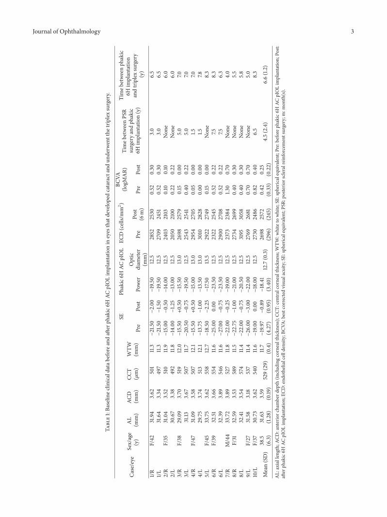
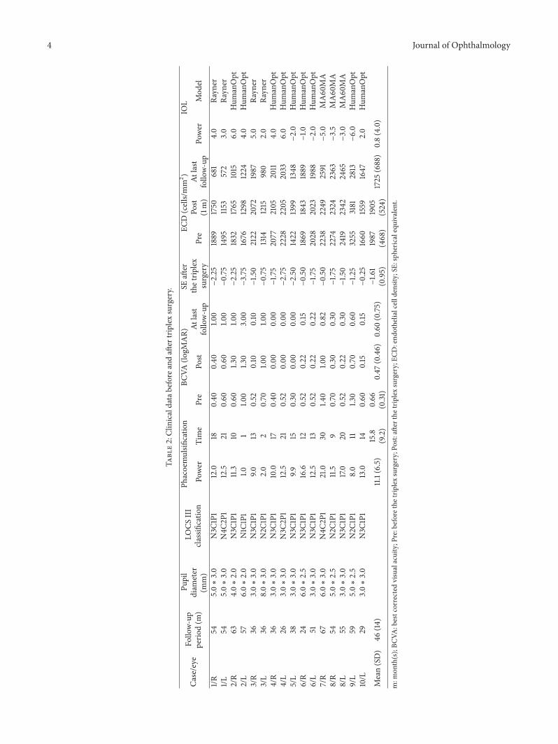
3.1. Baseline Data. Patients had a mean age of 38 ± 6 years(range: 27 to 37 years) at the time of triplex surgery.Themeanfollow-up time after the triplex surgery was 46 ± 14 months(range: 24 to 67 months). Baseline clinical data before andafter phakic 6HAC pIOL implantation in eyes that developedcataract and underwent the triplex surgery are presented inTable 1. Clinical data before and after the triplex surgery arepresented in Table 2.
Nine eyes (56.3%) underwent posterior scleral reinforce-ment surgery before phakic 6H AC pIOL implantation. Onepatient (Case 2) had hyperthyroidism 4 years after phakic6H AC pIOL implantation and developed thyroid eye disease(TED) with signs of bilateral eyelid retraction and mildlagophthalmos. One patient (Case 5) developed diabetesmellitus 3 years after phakic 6H AC pIOL implantation.Problems involving the cornea and iris were common in eyeswith phakic 6H AC pIOL implantation. Four eyes (Cases 1and 2) had mild corneal edema. Nine eyes (56.3%) sufferedpupil ovalization with different degrees of PAS and irisatrophy. Two eyes (right eye of Cases 4 and 5) had mildchronic anterior chamber inflammation. Improper diameterselection led to an undersized (Case 2) pIOL. Rotation anddecreased space between the AC pIOL and endothelium
Journal of Ophthalmology 3
Table1:Ba
selin
eclin
icaldatabefore
andaft
erph
akic6H
ACpIOLim
plantatio
nin
eyes
thatdevelopedcataractandun
derw
entthe
triplexsurgery.
Case/eye
Sex/age
(y)
AL
(mm)
ACD
(mm)
CCT
(𝜇m)
WTW
(mm)
SEPh
akic6H
ACpIOL
ECD(cells/mm2)
BCVA
(logM
AR)
Timeb
etweenPS
Rsurgeryandph
akic
6Him
plantatio
n(y)
Timeb
etweenph
akic
6Him
plantatio
nandtriplexsurgery
(y)
Pre
Post
Power
Optic
diam
eter
(mm)
Pre
Post
(6m)
Pre
Post
1/RF/42
31.94
3.62
501
11.3−21.50−2.00−19.50
12.5
2852
2530
0.52
0.30
3.0
6.5
1/L31.64
3.34
497
11.3−21.50−1.5
0−19.50
12.5
2709
2451
0.52
0.30
3.0
6.5
2/R
F/35
31.04
3.52
510
11.9−15.00−0.50−14.00
12.5
2403
2103
0.10
0.10
Non
e6.0
2/L
30.67
3.38
492
11.8−14.00−1.2
5−13.00
12.5
2050
2100
0.22
0.22
Non
e6.0
3/R
F/38
29.09
3.70
519
12.0−15.50
+0.50−15.50
13.0
2698
2579
0.15
0.00
5.0
7.03/L
31.13
3.67
507
11.7−20.50−0.75−19.50
12.5
2543
2541
0.40
0.22
5.0
7.04/R
F/47
31.09
3.58
507
12.1−15.50
+0.50−15.00
13.0
2954
2705
0.05
0.00
1.57.0
4/L
29.75
3.74
513
12.1−13.75−1.0
0−13.50
13.0
3010
2828
0.00
0.00
1.57.8
5/L
F/45
33.75
3.62
558
12.7−18.50−2.25−17.50
13.5
2922
2749
0.15
0.00
Non
e8.3
6/R
F/39
32.51
3.66
554
11.6−25.00
0.00−23.50
12.5
2322
2545
0.52
0.22
7.58.3
6/L
32.39
3.89
546
11.6−27.00−0.75−23.50
12.5
2900
2708
0.52
0.22
7.56.3
7/R
M/44
33.72
3.89
527
11.8−22.00−0.25−19.00
12.5
2373
2384
1.30
0.70
Non
e4.0
8/R
F/31
32.59
3.53
589
11.5−22.75−1.0
0−21.00
12.5
2734
2699
0.40
0.30
Non
e5.5
8/L
32.41
3.54
574
11.4−22.00−0.75−20.50
12.5
3195
3058
0.40
0.30
Non
e5.8
9/L
F/27
31.58
3.18
537
11.4−26.00−3.00−22.00
12.5
2769
2681
0.70
0.70
Non
e5.0
10/L
F/37
30.73
3.62
540
11.6−19.00
0.00−18.00
12.5
2730
2486
0.82
0.40
6.5
8.3
Mean(SD)
38.5
(6.3)
31.63
(1.28)
3.59
(0.19
)529(29)
11.7
(0.4)−19.97
(4.27)−0.89
(0.95)−18.41
(3.40)
12.7(0.3)
2698
(296)
2572
(245)
0.42
(0.33
)0.25
(0.22)
4.5(2.4)
6.6(1.2)
AL:
axiallength;
ACD:anteriorc
hamberd
epth
(inclu
ding
cornealthickness);CC
T:centralcornealthickn
ess;WTW
:whiteto
white;SE:
spheric
alequivalent;P
re:b
eforeph
akic6H
ACpIOLim
plantatio
n;Po
st:aft
erph
akic6H
ACpIOLim
plantatio
n;EC
D:end
othelialcelld
ensity;BC
VA:bestcorrected
visualacuity;SE:
spheric
alequivalent;P
SR:posterio
rscle
ralreinforcementsurgery;m
:mon
th(s).
4 Journal of Ophthalmology
Table2:Clinicaldatabefore
andaft
ertriplexsurgery.
Case/eye
Follo
w-up
perio
d(m
)
Pupil
diam
eter
(mm)
LOCS
III
classificatio
nPh
acoemulsifi
catio
nBC
VA(lo
gMAR)
SEaft
erthetrip
lex
surgery
ECD(cells/mm2)
IOL
Power
Time
Pre
Post
Atlast
follo
w-up
Pre
Post
(1m)
Atlast
follo
w-up
Power
Mod
el
1/R54
5.0∗3.0
N3C
1P1
12.0
180.40
0.40
1.00
−2.25
1889
1750
681
4.0
Rayn
er1/L
545.0∗3.0
N4C
2P1
12.5
210.60
0.60
1.00
−0.75
1495
1153
572
3.0
Rayn
er2/R
634.0∗2.0
N3C
1P1
11.3
100.60
1.30
1.00
−2.25
1832
1765
1015
6.0
Hum
anOpt
2/L
576.0∗2.0
N1C
1P1
1.01
1.00
1.30
3.00
−3.75
1676
1298
1224
4.0
Hum
anOpt
3/R
363.0∗3.0
N3C
1P1
9.013
0.52
0.10
0.10
−1.5
02122
2072
1987
5.0
Rayn
er3/L
368.0∗3.0
N2C
1P1
2.0
20.70
1.00
1.00
−0.75
1314
1215
980
2.0
Rayn
er4/R
363.0∗3.0
N3C
1P1
10.0
170.40
0.00
0.00
−1.7
52077
2105
2011
4.0
Hum
anOpt
4/L
263.0∗3.0
N3C
2P1
12.5
210.52
0.00
0.00
−2.75
2228
2205
2033
6.0
Hum
anOpt
5/L
383.0∗3.0
N3C
1P1
9.915
0.30
0.00
0.00
−2.50
1422
1399
1348
−2.0
Hum
anOpt
6/R
246.0∗2.5
N3C
1P1
16.6
120.52
0.22
0.15
−0.50
1869
1843
1889
−1.0
Hum
anOpt
6/L
513.0∗3.0
N3C
1P1
12.5
130.52
0.22
0.22
−1.7
52028
2023
1988
−2.0
Hum
anOpt
7/R
676.0∗3.0
N4C
2P1
21.0
301.4
01.0
00.82
−0.50
2238
2249
2591
−5.0
MA60MA
8/R
545.0∗2.5
N2C
1P1
11.5
90.70
0.30
0.30
−1.7
52274
2324
2363
−3.5
MA60MA
8/L
553.0∗3.0
N3C
1P1
17.0
200.52
0.22
0.30
−1.5
02419
2342
2465
−3.0
MA60MA
9/L
595.0∗2.5
N2C
1P1
8.0
111.3
00.70
0.60
−1.2
53255
3181
2813
−6.0
Hum
anOpt
10/L
293.0∗3.0
N3C
1P1
13.0
140.60
0.15
0.15
−0.25
1660
1559
1647
2.0
Hum
anOpt
Mean(SD)
46(14
)11.1(6.5)
15.8
(9.2)
0.66
(0.31
)0.47
(0.46)
0.60
(0.75)−1.6
1(0.95)
1987
(468)
1905
(524)
1725
(688)
0.8(4.0)
m:m
onth(s);BC
VA:bestcorrected
visualacuity;P
re:beforethe
triplexsurgery;Po
st:aft
erthetrip
lexsurgery;EC
D:end
othelialcelld
ensity;SE
:sph
ericalequivalent.
Journal of Ophthalmology 5
threatened the endothelium and caused excessive endothelialcell loss.
3.2. Intraoperative Data. During the triplex surgery, threeeyes (Case 1 and left eye ofCase 3) had severe pupil ovalizationwith severe PAS. The haptics of the phakic 6H AC pIOLstightly adhered to the peripheral iris and anterior chamberangle. Separation and dislocation of the haptics of pIOLswere performed by two IOL manipulators after injection ofDisCoVisc. Mild anterior chamber hemorrhage occurred in1 eye (Case 3) because of injury to the anterior chamberangle. The haptics of AC pIOLs were found to be touchingthe corneal endothelium in two eyes (Case 2). Two eyes hadhard nuclear cataract (N4C2P1 by LOCS III) and the powerof phacoemulsification was 12.5% for 21 seconds (left eye ofCase 1) and 21.0% for 30 seconds (Case 7), respectively. Novitreous loss or capsule rupture occurred in any eye.
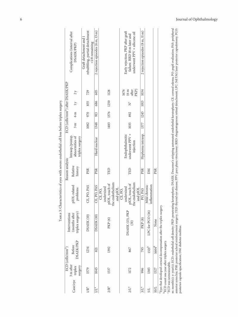
3.3. Postoperative Adverse Events. One eye (Case 2L) pre-sented with ocular pain, loss of visual acuity to hand move-ments, corneal edema, and anterior chamber fibroblasticreaction and was diagnosed with acute endophthalmitis 5days after the triplex surgery. The eye immediately receiveda vitreous tap and intravitreal antibiotics (0.8mg/0.1mLVancomycin and 2.25mg/0.1mL Ceftazidime) injection. Nosign of recovery was observed the following day and the eyeimmediately underwent a pars plana vitrectomy and receivedthe same intravitreal antibiotic injection. Inflammation wasthen resolved. Vitreous culture showed a Staphylococcusauricularis infection. Five eyes (31.3%) of three patients(Cases 1, 2, and 3L) with severe preoperative endothelialcell loss developed corneal decompensation and underwentpenetrating keratoplasty (PKP) or Descemet’s stripping auto-mated endothelial keratoplasty (DSAEK) at a mean time of9.4 ± 2.6 months (range: 6 to 13 months) after the triplexsurgery. The clinical data and characteristics of these eyes arepresented in Table 3. Case 2L experienced early graft rejectionwith corneal edema and steroid-induced IOP elevation afterDSAEK. The endothelial pathology expanded to whole-layerpathology, with subepithelial scarring and stromal opacity.PKP was performed after graft failure. After 18 months, Case2L developed rhegmatogenous retinal detachment (RRD)and underwent PPV with silicone oil. Cases 1L and 3Lboth experienced two late graft rejection episodes. Theselate episodes correspond to reduction of topical steroid usedue to steroid-response glaucoma and poor compliance,respectively. Hourly doses of topical prednisolone acetate 1%while awake and antiglaucoma medications when necessarywere prescribed.The late rejection episodes and elevated IOPwere controlled using only medication. Graft detachmentoccurred in Case 1R and air reinjection was performed 1 weekafter DSAEK. However, peripheral partial detachment < 1/8in the supratemporal region still existed. It did not influencethe visual axis andmostly spontaneously improvedduring thefollow-up period.The above complications were all managedadequately and all corneas maintained transparency at lastfollow-up. Three eyes (18.8%) of 3 patients (Cases 4L, 5L,and 6L) developed PCO and were treated with Nd:YAG laser
posterior capsulotomy at a mean time of 17.0 ± 5.6 months(range: 12 to 23 months) after triplex surgery.
3.4. Visual Outcomes. Themean logMAR BCVA was signifi-cantly improved after phakic 6H AC pIOL implantation (P =0.001) and was significantly reduced after cataract formation(P < 0.001). BCVA was significantly improved after triplexsurgery (P = 0.047). The difference between BCVA at lastfollow-up and that 6 months after phakic 6H AC pIOLimplantation was not significant (P = 0.075). Comparisonbetween eyes that developed corneal decompensation andthose that did not revealed worse BCVA (logMAR) at lastfollow-up (P < 0.0001). BCVA at last follow-up did notsignificantly differ between eyes with PCO and those without(P = 0.057). The mean final manifest spherical equivalentwas −1.61 ± 0.95D (range: −3.75 to −0.25D) after the triplexsurgery. Thirteen eyes (81.3%) were within ±1.0D of theintended correction.
3.5. ECDChange. ECDwas significantly reduced after phakic6H implantation (P = 0.004) and ECD loss significantlyprogressed over time (P < 0.001). The mean percentageof ECD loss was 26.4% over the mean period of 6.6 ±1.2 years (range: 4.0 to 8.3 years) after phakic 6H ACpIOL implantation. A subgroup analysis of eyes with severeendothelial cell loss before the triplex surgery is presented inTable 3. Apart from the five eyes that later developed cornealdecompensation, ECD before the triplex surgery was notsignificantly different compared with ECD at the last follow-up (2144 ± 468 cells/mm2 versus 2103 ± 425 cells/mm2, P =0.495). The triplex surgery effectively stabilized early ECDloss.
Comparison between eyes that developed corneal decom-pensation and those that did not revealed lower ECD afterphakic 6H AC pIOL implantation (P = 0.007), before triplexsurgery (P = 0.041) and at last follow-up (P < 0.0001). Alleyes that developed corneal decompensation suffered pupilovalization. All eyes with corneal edema developed cornealdecompensation.
4. Discussion
The present study showed that triplex surgery of phakic6H AC pIOL explantation and phacoemulsification within-the-bag IOL implantation was an effective means forimprovement of vision and refraction in agreement withprevious studies [9–11]. The current study investigated thesafety, effectiveness, and stability of triplex cataract surgery.To the best of our knowledge, our study provided the longestfollow-up time after AC pIOL explantation for cataract andthe largest case series report for the specific phakic 6Hmodel explantation for cataract. AC pIOL-related severeECD loss leading to corneal decompensation, corneal edemaand pupil ovalization, inflammation, and corneal damageinduced by complications during the perioperative periodand systemic diseases are issues confronted and needed tobe evaluated comprehensively when making the decisionof triplex surgery. PCO was another frequent postoperative
6 Journal of Ophthalmology
Table3:Ch
aracteris
ticso
feyesw
ithsevere
endo
thelialcelllossb
eforetrip
lexsurgery.
Case/eye
ECD(cells/mm2)
Interventio
ns(m
onthsa
fter
triplexsurgery)
Reason
analysis
ECD(cells/mm2)a
fterD
SAEK
/PKP
Com
plications
(intervalaft
erDSA
EK/PKP
)3m
after
triplex
surgery
Before
DSA
EK/PKP
pIOL-related
prob
lems
Relativ
ehisto
ry
Intraop./postop.
abno
rmities
oftriplexsurgery
3m6m
1y2y
1/Ra
1579
1234
DSA
EK(10)
CE,P
O,PAS
PSR
1082
970
835
729
Graftdetachmentand
1rebu
bblin
g,partiald
etachm
ent
<1/8
remaining
1/La
1045
821
DSA
EK(10)
CE,P
O,PAS
PSR
Hardnu
clear
1348
913
686
605
2rejectionepiso
des(6m
,13m
)
2/Ra
1537
1392
PKP(6)
CE,P
O,
undersized
pIOL,touchof
thee
ndotheliu
mandpIOL
TED
1483
1376
1250
1128
2/La
1172
867
DSA
EK(13),P
KP(31)
CE,P
O,
undersized
pIOL,touchof
thee
ndotheliu
mandpIOL
TED
Endo
phthalmitis
underw
entP
PV+
injection
1035
892
Nc
1676
(6m
after
PKP)
Early
rejection,
PKPaft
ergraft
failu
re.R
RD18m
latera
ndun
derw
entP
PV+silicon
eoil
3/La
996
795
PKP(8)
PO,PAS
PSR
Hyphemaintraop.
1377
1245
1013
1034
2rejectionepiso
des(8m
,11m
)
5/L
1385
1321
bLP
CforP
CO(16)
Mild
chronic
inflammation
DM
10/L
1527
1604
bNon
ePS
Ra Eyesthatd
evelo
pedcorneald
ecom
pensationaft
erthetrip
lexsurgery.
b ECD
coun
tone
year
after
triplexsurgery.
c ECD
was
unmeasurable.
m:m
onth(s);y:year(s);EC
D:end
othelialcelld
ensity;PK
P:penetratingkeratoplasty;D
SAEK
:Descemet'sstr
ipping
automated
endo
thelialkeratop
lasty
;CE:
cornealedema;PO
:pup
ilovalization;
PAS:perip
heral
anterio
rsyn
echia;PS
R:po
sterio
rscle
ralreinforcementsurgery;T
ED:thyroid
eyed
isease;PP
V:parsplanav
itrectomy;RR
D:rhegm
atogenou
sretinaldetachment;LP
C:Nd:YA
Glaserp
osterio
rcapsulotomy;PC
O:
poste
riorc
apsuleop
acificatio
n;DM:diabetesm
ellitus.
Journal of Ophthalmology 7
adverse event. Closer surveillance for young patients in long-term follow-up is needed.
Cataract formation is one of the major complications ofpIOLs implantation [13] and is accelerated by super highmyopia [14]. In the current study, the mean age of patientsat the time of triplex surgery was 38.5 years and the meanaxial length was 31.63mm. There is an increased risk ofcataractogenesis afterACpIOL implantation in patients olderthan 40 years that have an axial length longer than 29mm[10]. In our study, themean time between phakic 6HACpIOLimplantation and triplex surgery for cataract was 6.6 ± 1.2years. All the cataracts types found in eyes that underwent thetriplex surgerywere nuclear. Alio et al. reported that themeaninterval between implantation and explantation of AC pIOLs(62 with ZB5M, 1 with ZSAL-4, and 1 with phakic 6H) due tocataract formation was 10.04 ± 3.66 years [11]. In their cases,almost 100% of cataracts detected in the eyes that underwentangle-supported AC pIOL explantation surgery were nuclear[9, 11]. Phakic AC pIOL implantation promotes early changesof the nucleus because of chronically inadequate aqueousperfusion to the lens epithelium, surgical trauma, chronicsubclinical inflammation, and postoperative use of topicalsteroids [13, 15, 16]. Cataract is also themain cause ofACpIOLexplantation andwas reported to represent 51.39%of the casesof explantation [9]. The increase in cataract with aging andprogression of pathogenetic high myopia is a concern andsuggests caution regarding long-term safety.
The triplex surgery was an effective means for visualrestoration. Mean BCVA was significantly improved aftertriplex surgery and was not significantly different from thatafter phakic 6H implantation.Themean final manifest spher-ical equivalent was −1.61 D. Previous study also demonstratedthat final BCVA was not significantly different after cataractsurgery in 9 highly myopic eyes corrected by ZB5M andZSAL-4 AC pIOL from that after AC pIOL implantation. SEwas −0.42 ± 1.94D after cataract surgery [10]. However, theBCVA and SE were significantly different after bilensectomysurgery in a larger case series [9]. Our results were compara-ble to these results.
Corneal endothelial cell loss is the main concern afterAC pIOL implantation [7]. In the current study, eyes sufferedprogressive ECD loss (26.4% over 6.6 years) after phakic6H implantation. Alio et al. found that severe endothelialcell loss leading to AC pIOL explantation was related tothe use of phakic 6H pIOL in the short term (3.22 years,which was much shorter than that for ZB5M cases) [11]. Thereported percentage of ECD loss afterACpIOLs implantationwas 3.8% to 12% in the first year and gradually decreasedby 0.5% to 1.8% per year [7]. The endothelial issues arecontroversial and evoke substantial debate [3]. Damage tothe corneal endothelium mainly results from direct contactbetween AC pIOLs and the inner surface of the cornea andinflammation [7]. Removal of the pIOL based on progressiveloss of endothelial cells or reaching an absolute value (i.e.,1500 cells/mm2), at which point the eye may have decreasedability to sustain other types of surgery, is debatable in theabsence of a clear guideline. Corneal integrity depends onthe absolute number of endothelial cells and their function
and morphology. Apart from the five eyes that later devel-oped corneal decompensation, the triplex surgery effectivelystabilized early ECD loss. When severe ECD loss occurred,especially accompanied by other relative complications orsignificant corneal morphology change, primary keratoplastywith AC pIOL explantation might be a better alternative[17, 18].
Complications during the perioperative period and sys-temic diseases are other core issues that needed to be eval-uated comprehensively when making the decision of triplexsurgery. Pupillary ovalization was detected in 56.3% of casesthat underwent the triplex surgery. Different degrees of PASand iris atrophy were also common.The haptics of AC pIOLsin the sclerocorneal angle lead to mild deformation of the iri-dosclerocorneal architecture, resulting in iris retraction andpupil ovalization [17]. The haptic plate of the pIOL adheredto the tissue in the anterior chamber angle. These problemsincreased the difficulty of dissection and dislocation of thepIOL haptic and elevated the inflammatory reaction, whichmight cause direct toxicity to the endothelium and angle. Asoft-shell technique, higher molecular weight, and viscoelas-tic and other procedures should be used to protect the cornealendothelium during surgery. Furthermore, considering thelong life expectancy of the patients with implanted pIOLs at ayounger age, development of systemic disease should not beignored.
Several reasons, categorized as either “patient-related” or“graft-related” factors, contributed to prolonged low visionof the five eyes that developed corneal decompensation. Themost common patient-related causes were fundus pathologycaused by super high myopic degeneration and uncorrectedrefractive error after keratoplasty. Incomplete visual reha-bilitation was also attributed to problems related to phakic6H, such as severe pupil ovalization and peripheral ante-rior synechiae. Various perioperative complications requiredmultiple surgeries. Case 2L was the extreme case. Multiplesurgical traumas led to repeated inflammation reaction and,to some extent, a change in ocular structure and function.Graft-related factors included complications after cornealintervention of DSAEK/PKP, donor factors, and surgicalexperience. Both early and late graft rejections associatedwith steroid-induced IOP elevation and poor compliance oftopical corticosteroid use resulted in continuous ECD lossand one graft failure. Steroid-induced IOP elevation and glau-coma medications are risk factors for higher graft rejectionand failure rates with higher ECD loss after DSAEK and PKP[19–21]. In developing countries, where there is a perpetualshortage of donor corneal tissue, corneas are often donatedby older people with systemic chronic diseases. The waitingperiod for corneal transplantation is long. Earlier surgery,within a year of disease onset, may produce superior visualoutcomes both in EK and in PKP by limiting the durationof stromal edema and reducing fibrosis [22–24]. Graft-hostinterface irregularity also contributed to incomplete visualrehabilitation after EK [23]. The three eyes that underwentDSAEK were among the first 50 cases of DSAEK in ourhospital. The learning curve for the DSAEK procedure mayinfluence the rate of ECD loss and incidence of graft detach-ment [25]. Factors associated with higher postoperative
8 Journal of Ophthalmology
ECD loss include secondary donor reattachment procedure,episodes of graft rejection, medically treated glaucoma, andolder donor age [19, 26, 27]. Patient- and graft-related factorsand multiple comorbidities resulted in prolonged low visionin these five eyes warranting further intensive follow-up.
The development of PCO is a multifactorial processaffected by patient-related, IOL-related, and surgery-relatedproblems [28]. In our study, 18.8% of eyes developed PCO,which was comparable to other studies [29, 30]. The finalBCVA did not significantly differ between eyes with PCOand those without. High myopia is pathological and isassociated with an increase in certain growth factors in theaqueous humor, which strongly influence the developmentof PCO [31]. Other patient-related factors include a historyof chronic inflammation caused by AC pIOL (Case 4) orrelated to diabetes mellitus (Case 5). Hydrophilic acrylicmaterial offers the advantage of good uveal biocompatibility[32, 33] and is therefore suitable for patients with highmyopiawho undergo the triplex surgery that induces a higherinflammatory response than routine cataract surgery. PCOincidence is primarily influenced by IOL design [28, 34].The PCO score was significantly lower with a sharp opticedge and a capsular bend formation compared to roundedged IOLs [34]. However, the incomplete sharp edge at thebroad optic-haptic junctions represents an Achilles heel andallows migration of lens epithelial cells [35] and is thereforeassociated with a poorer PCO outcome. Thus, in eyes withextreme high myopia that underwent the triplex surgery, anenhanced 360-degree sharp-edged design posterior chamberIOL implantation [35] and manual polishing of the capsuleare recommended and extensive follow-up for the detectionof PCO is needed.
Our study had some limitations. It is a retrospectivestudy, associated with possible selection bias. However, itis difficult to conduct a prospective study because phakic6H AC pIOLs have been phased out of the market. Oncesigns of corneal decompensation occurred, the condition wasextremely complicated and changeable. The patient mightrequire multiple surgeries and might have poor prognosiseven after great effort, as in Case 2. Therefore, before triplexsurgery, a comprehensive and specialist evaluation of risksassociated with AC pIOL involving the iris and cornea, pos-sible complications during the perioperative period, ocularcomorbidities, and systemic diseases is of vital significance.Early extraction of phakic 6H AC pIOL before severe ECDloss is the best option. In case of severe ECD loss, especiallyaccompanied by other related complications or significantcorneal morphological changes, primary keratoplasty com-bined with AC pIOL explantation or triplex surgery mightbe a good alternative [17, 18, 36]. Further investigationscomparing the two procedures are needed to determine theindications and optimal surgical time. Another limitationof our study is that the DSAEKs were among the initial 50cases performed in our hospital. However, in real world,such experience is especially useful due to the limitationsassociated with keratoplasty in China [37]. Such a retro-spective study enables anticipation of the most complicatedproblems of these cases to determine the optimal surgicaltime.
In conclusion, the perspective and information gainedfrom the present study provide some basis for optimism inthe management of complicated problems related to phakic6H AC pIOLs, including cataracts. Corneal decompensationshould be observed closely in eyes with progressive ECDloss presenting with pupil ovalization, corneal edema, orcorneal inflammation. This is especially the case for eyesaccompanying other related ocular or systemic abnormal-ities. The triplex surgery or simple explantation of ACpIOL should be performed early. A 360∘ sharp-edged designposterior chamber IOL implantation and long-term follow-up for PCO development are recommended. Triplex surgeryis safe and effective for phakic 6H complicated cataract andour experience provided comprehensive evaluation of issuesconfronted.
Ethical Approval
The institutional review board of the Eye and ENTHospital ofFudan University, Shanghai, China, approved the study. Thestudy adhered to the tenets of the Declaration of Helsinki andall the laws of the authors’ home country.
Consent
The patients of the study gave informed consent for partici-pation in this study.
Competing Interests
No author has a financial or proprietary interest in anymaterial or method mentioned.
Authors’ Contributions
Xin Liu, Yi Luo, Xingtao Zhou, and Yi Lu contributed tostudy concept and design; Xin Liu, Fan Fan, and XiaoyingWang carried out data collection; Xin Liu, Tianyu Zheng,and PengZhou performed analysis and interpretation of data;Xin Liu, Fan Fan, Tianyu Zheng, and Peng Zhou contributedto drafting of the paper; Yi Luo, Xingtao Zhou, XiaoyingWang, and Yi Lu contributed to critical revision of the paper;Xin Liu, Fan Fan, and Tianyu Zheng provided statisticalexpertise; Yi Luo and Yi Lu contributed to obtaining funding;Yi Luo, Xingtao Zhou, Xiaoying Wang, and Peng Zhouprovided administrative, technical, or material support; YiLuo, Xingtao Zhou, and Yi Lu contributed to supervision ofthe paper. Xin Liu and Fan Fan contributed equally to thiswork and should be considered as equal first authors. XingtaoZhou and Yi Luo contributed equally to this work.
Acknowledgments
The authors would like to acknowledge Rajeev Naidu,BOptom B.S. (UNSW), Sydney, Australia, for his help inediting and proofreading the final paper. This study wassupported by grants from the National Natural ScienceFoundation of China (81371002) and the Municipal Level
Journal of Ophthalmology 9
Hospital Project for Emerging and Frontier Technology ofShanghai (SHDC12012104).
References
[1] J. L. Guell, M. Morral, D. Kook, and T. Kohnen, “Phakicintraocular lenses: Part 1: historical overview, current models,selection criteria, and surgical techniques,” Journal of Cataractand Refractive Surgery, vol. 36, no. 11, pp. 1976–1993, 2010.
[2] L. Espandar, J. J. Meyer, and M. Moshirfar, “Phakic intraocularlenses,” Current Opinion in Ophthalmology, vol. 19, no. 4, pp.349–356, 2008.
[3] C. F. Lovisolo and D. Z. Reinstein, “Phakic intraocular lenses,”Survey of Ophthalmology, vol. 50, no. 6, pp. 549–587, 2005.
[4] A. Yu, Q. Wang, A. Xue et al., “Comparison of contrastsensitivity after angle-supported, iris-fixated and posteriorchamber phakic intraocular lens implantation for highmyopia,”Ophthalmologica, vol. 222, no. 1, pp. 53–57, 2007.
[5] H.-S. Bi, X.-H.Ma,W.-T. Cai, D.-M. Liu, and P. Ji, “Implantationof phakic anterior chamber intraocular lens for the correctionof severe myopia,” Zhonghua Yan Ke Za Zhi, vol. 42, no. 2, pp.145–149, 2006.
[6] S. W. Kwon, H. S. Moon, and K. H. Shyn, “Visual improvementin high myopic amblyopic adult eyes following phakic anteriorchamber intraocular lens implantation,” Korean Journal ofOphthalmology, vol. 20, no. 2, pp. 87–92, 2006.
[7] T. Kohnen, D. Kook, M. Morral, and J. L. Guell, “Phakicintraocular lenses. Part 2. Results and complications,” Journal ofCataract and Refractive Surgery, vol. 36, no. 12, pp. 2168–2194,2010.
[8] Z.-H. Zhang, J.-C. Lian, H.-P. Liao, and S.-S. Zhang, “Threekinds of phakic intraocular lens for correction of high myopia,”Chinese Journal of Ophthalmology, vol. 47, no. 6, pp. 531–538,2011 (Chinese).
[9] J. L. Alio, B. T. Toffaha, P. Pena-Garcia, L. M. Sadaba, and R. I.Barraquer, “Phakic intraocular lens explantation: causes in 240cases,” Journal of Refractive Surgery, vol. 31, no. 1, pp. 30–35, 2015.
[10] J. L. Alio, F. de la Hoz, J. M. Ruiz-Moreno, and T. F. Salem,“Cataract surgery in highly myopic eyes corrected by phakicanterior chamber angle-supported lenses,” Journal of Cataractand Refractive Surgery, vol. 26, no. 9, pp. 1303–1311, 2000.
[11] J. L. Alio, A. M. Abdelrahman, J. Javaloy, M. T. Iradier, and V.Ortuno, “Angle-supported anterior chamber phakic intraocularlens explantation. Causes and outcome,” Ophthalmology, vol.113, no. 12, pp. 2213–2220, 2006.
[12] L. T. Chylack Jr., J. K. Wolfe, D. M. Singer et al., “The lensopacities classification system III,” Archives of Ophthalmology,vol. 111, no. 6, pp. 831–836, 1993.
[13] M. Moshirfar, M. Mifflin, G. Wong, and J. C. Chang, “Cataractsurgery following phakic intraocular lens implantation,” Cur-rent Opinion in Ophthalmology, vol. 21, no. 1, pp. 39–44, 2010.
[14] J. L. Alio, F. de la Hoz, J. J. Perez-Santonja, J. M. Ruiz-Moreno, and J. A. Quesada, “Phakic anterior chamber lensesfor the correction of myopia: a 7-year cumulative analysis ofcomplications in 263 cases,” Ophthalmology, vol. 106, no. 3, pp.458–466, 1999.
[15] J. J. Perez-Santonja, M. T. Iradier, J. M. B. del Castillo, J. M.Serrano, and M. A. Zato, “Chronic subclinical inflammation inphakic eyes with intraocular lenses to correct myopia,” Journalof Cataract and Refractive Surgery, vol. 22, no. 2, pp. 183–187,1996.
[16] J. L. Alio, F. D. L. Hoz, and M. M. Ismail, “Subclinical inflam-matory reaction induced by phakic anterior chamber lensesfor the correction of high myopia,” Ocular Immunology andInflammation, vol. 1, no. 3, pp. 219–224, 1993.
[17] S. R. Patel, D. S. Chu, B. D. Ayres, and P. S. Hersh, “Cornealedema and penetrating keratoplasty after anterior chamberphakic intraocular lens implantation,” Journal of Cataract andRefractive Surgery, vol. 31, no. 11, pp. 2212–2215, 2005.
[18] V. Mittal, R. Mittal, and D. Singh, “Simultaneous bilensec-tomy and endothelial keratoplasty for angle-supported phakicintraocular lens-induced corneal decompensation,” Indian Jour-nal of Ophthalmology, vol. 59, no. 4, pp. 314–317, 2011.
[19] J. Y. Li, M. A. Terry, J. Goshe, N. Shamie, and D. Davis-Boozer, “Graft rejection after Descemet’s stripping automatedendothelial keratoplasty: graft survival and endothelial cell loss,”Ophthalmology, vol. 119, no. 1, pp. 90–94, 2012.
[20] M. O. Price, C. S. Jordan, G. Moore, and F. W. Price Jr., “Graftrejection episodes after Descemet stripping with endothelialkeratoplasty: part two: the statistical analysis of probability andrisk factors,” British Journal of Ophthalmology, vol. 93, no. 3, pp.391–395, 2009.
[21] R. M. K. Stewart, M. N. A. Jones, M. Batterbury, D. Tole, D. F.P. Larkin, and S. B. Kaye, “Effect of glaucoma on corneal graftsurvival according to indication for penetrating keratoplasty,”American Journal of Ophthalmology, vol. 151, no. 2, pp. 257.e1–262.e1, 2011.
[22] N. Morishige, T.-I. Chikama, N. Yamada et al., “Effect ofpreoperative duration of stromal edema in bullous keratopathyon early visual acuity after endothelial keratoplasty,” Journal ofCataract and Refractive Surgery, vol. 38, no. 2, pp. 303–308, 2012.
[23] A. M. Turnbull, M. Tsatsos, P. N. Hossain, and D. F. Anderson,“Determinants of visual quality after endothelial keratoplasty,”Survey of Ophthalmology, 2015.
[24] T. Liu, Y. Xu, D. Sun, and L. Xie, “Histological evaluation ofcorneal scar formation in pseudophakic bullous keratopathy,”PLoS ONE, vol. 7, no. 6, Article ID e39201, 2012.
[25] S. V. Patel, “Graft survival and endothelial outcomes in the newera of endothelial keratoplasty,” Experimental Eye Research, vol.95, no. 1, pp. 40–47, 2012.
[26] S. K. Basak, “Descemet stripping and endothelial keratoplastyin endothelial dysfunctions: three-month results in 75 eyes,”Indian Journal of Ophthalmology, vol. 56, no. 4, pp. 291–296,2008.
[27] W. J. Armitage, M. N. A. Jones, I. Zambrano, F. Carley, and D.M. Tole, “The suitability of corneas stored by organ culture forpenetrating keratoplasty and influence of donor and recipientfactors on 5-year graft survival,” Investigative Ophthalmologyand Visual Science, vol. 55, no. 2, pp. 784–791, 2013.
[28] N. Awasthi, S. Guo, and B. J. Wagner, “Posterior capsular opaci-fication: a problem reduced but not yet eradicated,” Archives ofOphthalmology, vol. 127, no. 4, pp. 555–562, 2009.
[29] W. A. Lyle and G. J. C. Jin, “Phacoemulsification with intraoc-ular lens implantation in high myopia,” Journal of Cataract andRefractive Surgery, vol. 22, no. 2, pp. 238–242, 1996.
[30] J. L. Guell, A. F. Rodriguez-Arenas, O. Gris, F. Malecaze,and F. Velasco, “Phacoemulsification of the crystalline lensand implantation of an intraocular lens for the correction ofmoderate and high myopia: four-year follow-up,” Journal ofCataract and Refractive Surgery, vol. 29, no. 1, pp. 34–38, 2003.
[31] J.-P. Tong, W.-M. Chan, D. T. L. Liu et al., “Aqueous humorlevels of vascular endothelial growth factor and pigment
10 Journal of Ophthalmology
epithelium-derived factor in polypoidal choroidal vasculopathyand choroidal neovascularization,” American Journal of Oph-thalmology, vol. 141, no. 3, pp. 456–462, 2006.
[32] C. Abela-Formanek, M. Amon, G. Schild, J. Schauersberger, G.Heinze, and A. Kruger, “Uveal and capsular biocompatibility ofhydrophilic acrylic, hydrophobic acrylic, and silicone intraoc-ular lenses,” Journal of Cataract and Refractive Surgery, vol. 28,no. 1, pp. 50–61, 2002.
[33] S. N. Arthur, Q. Peng,D. J. Apple et al., “Effect of heparin surfacemodification in reducing silicone oil adherence to variousintraocular lenses,” Journal of Cataract and Refractive Surgery,vol. 27, no. 10, pp. 1662–1669, 2001.
[34] O. Findl, W. Buehl, P. Bauer, and T. Sycha, “Interventions forpreventing posterior capsule opacification,” Cochrane Databaseof Systematic Reviews, no. 2, Article ID CD003738, 2010.
[35] Y.Nishi, T.M. Rabsilber, I.-J. Limberger, A. J. Reuland, andG.U.Auffarth, “Influence of 360-degree enhanced optic edge designof a hydrophilic acrylic intraocular lens on posterior capsuleopacification,” Journal of Cataract and Refractive Surgery, vol.33, no. 2, pp. 227–231, 2007.
[36] S. Sikder, V. Patel, H. A. Holz, M. D. Mifflin, S. Davis, andM. Moshirfar, “Management of corneal endothelial decom-pensation caused by iris-fixated phakic intraocular lenseswith descemet stripping automated endothelial keratoplasty,”Cornea, vol. 30, no. 9, pp. 1045–1047, 2011.
[37] J. Hong, W. Shi, Z. Liu et al., “Limitations of keratoplasty inChina: a survey analysis,” PLoS ONE, vol. 10, no. 7, Article IDe0132268, 2015.

Submit your manuscripts athttp://www.hindawi.com
Stem CellsInternational
Hindawi Publishing Corporationhttp://www.hindawi.com Volume 2014
Hindawi Publishing Corporationhttp://www.hindawi.com Volume 2014
MEDIATORSINFLAMMATION
of
Hindawi Publishing Corporationhttp://www.hindawi.com Volume 2014
Behavioural Neurology
EndocrinologyInternational Journal of
Hindawi Publishing Corporationhttp://www.hindawi.com Volume 2014
Hindawi Publishing Corporationhttp://www.hindawi.com Volume 2014
Disease Markers
Hindawi Publishing Corporationhttp://www.hindawi.com Volume 2014
BioMed Research International
OncologyJournal of
Hindawi Publishing Corporationhttp://www.hindawi.com Volume 2014
Hindawi Publishing Corporationhttp://www.hindawi.com Volume 2014
Oxidative Medicine and Cellular Longevity
Hindawi Publishing Corporationhttp://www.hindawi.com Volume 2014
PPAR Research
The Scientific World JournalHindawi Publishing Corporation http://www.hindawi.com Volume 2014
Immunology ResearchHindawi Publishing Corporationhttp://www.hindawi.com Volume 2014
Journal of
ObesityJournal of
Hindawi Publishing Corporationhttp://www.hindawi.com Volume 2014
Hindawi Publishing Corporationhttp://www.hindawi.com Volume 2014
Computational and Mathematical Methods in Medicine
OphthalmologyJournal of
Hindawi Publishing Corporationhttp://www.hindawi.com Volume 2014
Diabetes ResearchJournal of
Hindawi Publishing Corporationhttp://www.hindawi.com Volume 2014
Hindawi Publishing Corporationhttp://www.hindawi.com Volume 2014
Research and TreatmentAIDS
Hindawi Publishing Corporationhttp://www.hindawi.com Volume 2014
Gastroenterology Research and Practice
Hindawi Publishing Corporationhttp://www.hindawi.com Volume 2014
Parkinson’s Disease
Evidence-Based Complementary and Alternative Medicine
Volume 2014Hindawi Publishing Corporationhttp://www.hindawi.com