cleavage of sea-urchin eggs in colchicine...cleavage of sea-urchin eggs colcjricine in 507 strongly...
TRANSCRIPT
506
CLEAVAGE OF SEA-URCHIN EGGS IN COLCHICINE
BY M. M. SWANN* AND J. M. MITCHISON*
Department of Zoology, University of Cambridge; the Marine Station, Millport;and the Stazione Zoologica, Naples
(With Plates 12 and 13)
(Received 12 December 1952)
INTRODUCTIONWe have recently put forward a theory linking mitosis with cleavage, in which wesuggest that cleavage is caused by an expansion of the cell cortex which starts ateither pole of the cell and later moves round to the region of the furrow (Mitchison,1952). There is evidence that this expansion is brought about by substancesdiffusing from the two groups of daughter chromosomes which, by the end ofanaphase, lie near the poles of the cell (Swann 1951a, b). If this theory is correct,the important structure in mitosis is the spindle which is responsible for separatingthe two groups of chromosomes. The asters, where they exist, should act only aspassive guides for the advancing furrow.
Some theories of cell division, on the other hand, e.g. Gray (1924), Dan (1943),suppose that the spindle and asters have an active function in cleavage, and that thecell surface is pulled passively into the furrow. It should be possible to distinguishbetween these opposing views by suppressing the spindle and asters at a point whenthe chromosomes have already separated, and seeing whether cleavage still continues.The most satisfactory way of doing this is to use colchicine. The action of thispoison in stopping cleavage and suppressing the formation of the spindle withoutaffecting the condensation of the chromosomes, has been known for some time, andthere is a large literature on the subject (Eigsti, 1947). Its action on sea-urchin eggshas also been studied by a number of workers including Nebel (1937), Wilbur (1940)and Beams & Evans (1940). There is general agreement that colchicine, at concen-trations of about 5 x io~* M inhibits mitosis and cleavage in these eggs. It preventsthe formation of the spindle and asters, or, if they are already formed, it causes themto solate and disappear. The normal viscosity rise associated with the growth offibrous material in the mitotic figures is therefore stopped, and viscosity remains low.
In most of the experiments, colchicine has been applied well before mitosis,making it impossible to separate its effects on cleavage from its effects on mitosis.Beams & Evans (1940), however, made the significant observation that the criticalpoint in mitosis for colchicine inhibition is about anaphase. If eggs are treated withcolchicine before anaphase, cleavage is stopped, but if they are treated during orafter anaphase, cleavage takes place, even though the asters appear, by ordinarylight, to have vanished in both fresh and stained material. This suggests rather
• Now at the Department of Zoology, University of Edinburgh.
Cleavage of sea-urchin eggs in colcJricine 507
strongly that the spindle and asters are not required for cleavage. On the otherhand, it is difficult to be certain by ordinary observation that the asters have actuallydisappeared at the moment of cleavage. At this time they are no longer very obviouslyfibrillar in ordinary light, and it would not be impossible to suppose that they haddisappeared, when in fact they were still having a mechanical effect on the cleavageprocess.
There are two ways of confirming the disappearance of the asters. First, the eggscan be observed in polarized light. This is a very sensitive method of detecting thepresence of oriented structures, and in a normal division the asters are in fact readilyvisible throughout cleavage and for a considerable time afterwards. We have there-fore applied colchicine to sea-urchin eggs at different points in mitosis, and observedthe effects under polarized light, using time-lapse photography. Secondly, thereshould be a fall in the viscosity of the egg contents associated with the disappearanceof the asters. We have therefore measured the viscosity of cleaving Arbacia eggs bystratifying the pigment granules with a centrifuge after treatment with colchicine.
We have also attempted to fix more accurately the critical point in mitosis forcolchicine inhibition by applying colchicine at different times and relating chromo-some separation measured on stained material with the percentage cleavage.
It is known that cycles of nuclear activity, corresponding to the condensation ofthe chromosomes at mitosis, continue in many cells after treatment with colchicine,even though mitosis and cleavage have been inhibited (Nebel, 1937). We have founda similar continuance of cycles in sea-urchin eggs (cf. Zeuthen, 1951). On ourtheory this cycle should be paralleled by changes in the state of the cell cortexsimilar to those which take place in normal division; Monroy & Montalenti (1947)have, in fact, found such changes in the cortical birefringence and plasmolysis. Wehave followed this up by investigating cycles of change in the rigidity and light-scattering of the cortex.
EFFECT OF STRONG COLCHICINE ON CLEAVAGE
(a) Observations in polarized light
Although colchicine at 5 x io~* M suppresses the spindle and asters, it only does sorather slowly, and the last traces of birefringence do not disappear for 10-15 min-With stronger concentrations, however, the birefringence vanishes more rapidly.In the experiments described below, colchicine was used at 3-2 x io"3 M; underthese conditions the birefringence disappears completely in 2-3 mm.* In all theexperiments, the fertilization membranes were removed, and the eggs (Psammechinusmiliaris) put in calcium-free sea water to dissolve away the hyaline layer. Roomtemperature was about 180 C.
The normal appearance of mitosis and cleavage in polarized light is shown inPI. 12, figs. 1-8. The interpretation of these birefringence changes has been dealtwith in some detail by Swann (1951a, b). It is only necessary to say here that in
• Inoul (1952) finds that this concentration of colchicine causes the recession of the spindle inChaetopterus eggs in 4 min.

508 M. M. SWANN AND J. M. MlTCHISON
metaphase the spindle and asters are small and brightly birefringent, while duringanaphase they get steadily larger and fainter. The centrosomes also move apart. Atthe end of anaphase the spindle is no longer visible as a separate structure, while theasters become very large and virtually fill the cell but are considerably fainter thanat metaphase. They continue in this condition throughout cleavage. With a littleexperience it is possible to tell the particular stage of the eggs with some accuracyfrom their appearance in polarized light.
The effect of colchicine is shown in PI. 12, figs. 9-12, and PI. 13, figs. 1-16. In eachcase photography was begun 1 min. after the eggs were put in colchicine, the subse-quent photographs being selected at 2 min. intervals. In PI. 12, figs. 9-12, the eggswere put into colchicine during metaphase. The spindle and asters disappearedrapidly, and there were no signs of cleavage. In PI. 13, figs. 1-8, on the other hand,the eggs were not put into colchicine until anaphase was more or less complete.Once again the asters disappeared rapidly, but cleavage continued with no trace ofbirefringence in the cells. The most interesting case is shown in PL 13, figs. 9-16.Here the colchicine was put on when the eggs were in anaphase. The two lower eggswere in mid-anaphase (shown by relatively large and fairly widely spaced asters ofmedium brightness) and presumably developed a little further before they werearrested. Both these eggs completed cleavage, though rather slowly. The centreand top left eggs, on the other hand, were caught quite early in anaphase andpresumably got no further than mid-anaphase. They do, however, show elongationand the beginning of a furrow, though this ultimately retracts. The top right egg wascaught in metaphase and showed no signs of cleavage at all.
An interesting feature of eggs dividing in colchicine, also described by Beams &Evans (1940), is the irregular cleavage shapes which they often show. This can beseen to some extent in PI. 13, figs. 3 and 13, but it is much more striking in the fourselected eggs (in ordinary light) shown in PI. 12, figs. 13-16. A common feature inthese irregular cleavages is for the two sides of the furrow not to lie opposite eachother.
(b) Observations on stained sections
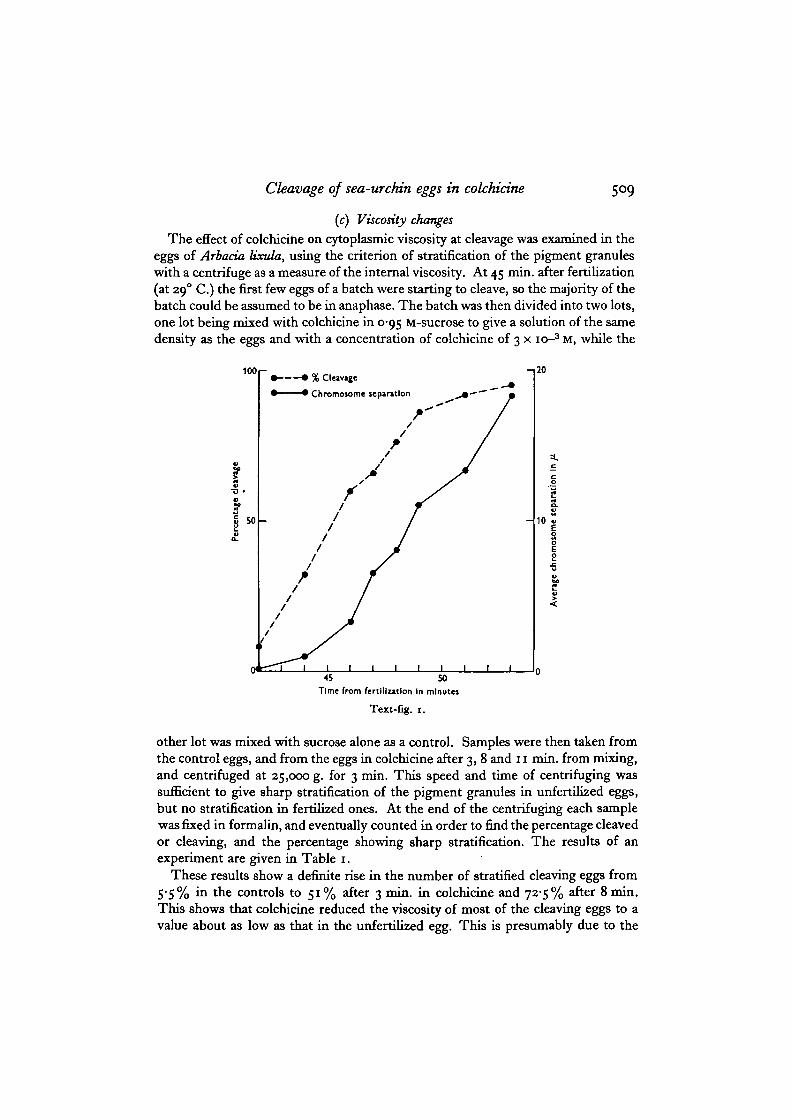
Eggs of P. miliaris were put into 3 x io"3 M colchicine at various times in anaphase.At the same times, small batches of eggs were fixed. The colchicine-treated eggswere later counted for percentage cleavage, and the fixed eggs were sectioned andstained, and measured for chromosome separation. Values for percentage cleavage(based on 200 eggs) and for average chromosome separation (based on fiftymeasurements) are plotted together, against time after fertilization (at 180 C) , inText-fig. 1. Ideally the curve for percentage cleavage should show a suddentransition from o to 100%. This is not, of course, realized in practice, but it will beseen that there is a fairly sharp rise between 42 and 49 min. At these times theaverage chromosome separation is between o and 11 fx. It is evident, therefore, thata chromosome separation of the order of 5 fi is necessary for cleavage to takeplace.
Cleavage of sea-urchin eggs in colchicine 509
(c) Viscosity changes
The effect of colchicine on cytoplasmic viscosity at cleavage was examined in theeggs of Arbada lixula, using the criterion of stratification of the pigment granuleswith a centrifuge as a measure of the internal viscosity. At 45 min. after fertilization(at 290 C.) the first few eggs of a batch were starting to cleave, so the majority of thebatch could be assumed to be in anaphase. The batch was then divided into two lots,one lot being mixed with colchicine in 0-95 M-sucrose to give a solution of the samedensity as the eggs and with a concentration of colchicine of 3 x 10⁻³ M, while the
100
r
• • % Cleavage
• • Chromosome separation
- , 2 0
45 50
Time from fertilization In minutes
Text-fig. 1.
other lot was mixed with sucrose alone as a control. Samples were then taken fromthe control eggs, and from the eggs in colchicine after 3, 8 and 11 min. from mixing,and centrifuged at 25,000 g. for 3 min. This speed and time of centrifuging wassufficient to give sharp stratification of the pigment granules in unfertilized eggs,but no stratification in fertilized ones. At the end of the centrifuging each samplewas fixed in formalin, and eventually counted in order to find the percentage cleavedor cleaving, and the percentage showing sharp stratification. The results of anexperiment are given in Table 1.
These results show a definite rise in the number of stratified cleaving eggs from5*5% in the controls to 51% after 3 min. in colchicine and 725% after 8 min.This shows that colchicine reduced the viscosity of most of the cleaving eggs to avalue about as low as that in the unfertilized egg. This is presumably due to the

5IO M. M. SWANN AND J. M. MlTCHISON
disappearance of the asters. The results are not as clear-cut as might perhaps beexpected: ideally the percentage of stratified eggs should rise from o to ioo. However,the initial value of 5-5% in the control is probably due to the presence of a fewunfertilized or cytolytic eggs, and the final values of 51 and 72̂ 5 % may be under-estimates since a stratified egg viewed from either end (rather than from the side)may appear to be unstratified. The drop in the percentage of cleaving eggs fromsample B to sample C is presumably due to the recession of the furrow which takesplace in the more retarded eggs of a batch.
Table 1
Sample
ControlABC
Time in min. fromaddition of
colchicine to startof centrifuging
3 (No colch.)
811
Cleaving and cleaved eggs
% in sample
725S36347-S
% stratified
5-5Si72-57i
Eggs not cleaved
% in sample
27-5473752-S •
% stratified
S-9516564
Final cleavage in controls, 92 %.
CYCLICAL CHANGES OF CORTEX AND CHROMOSOMESIN WEAK COLCHICINE
(a) Changes in the rigidity of the cortex
We have devised a method of measuring the rigidity of the cortex by using what wehave called a 'cell elastimeter' (Mitchison & Swann, 1954). This method consistsessentially of sucking a small bulge from the egg surface into a micro-pipette. Thesize of the bulge for a given suction is a measure of the rigidity of the cortex. In thenormal developing egg, the rigidity is low in interphase, rises at cleavage, and thenfalls again in the next interphase. This cycle persists in colchicine-treated eggs, eventhough cleavage is inhibited. Text-fig. 2 shows the change of rigidity with time foreggs of Psammechinus militaris placed in 2 x io~* M-colchicine in calcium-free seawater 15 min. after fertilization (at 180 C). The rigidity is given in arbitraryunits which are roughly proportional to the elastic modulus of the cortex. Theperiod of cleavage in untreated controls is also shown. The rise in rigidity at a timeroughly corresponding to the time of cleavage in the controls is very evident, butit should be noted that this rise is only about one-sixth as great as in normal cleavingeggs.
(b) Changes in the light-scattering properties of the cortex
We have described elsewhere (Mitchison & Swann, 1952) the changes in thelight-scattering properties of the cortex that take place during normal division.Under vertical illumination, the brightness of the cortex increases sharply at thestart of anaphase and then diminishes during cleavage. The same cycle occurs in

Cleavage of sea-urchin eggs in colcMcine 511
colchicine-treated eggs, though it is somewhat delayed. Text-fig. 3 shows thechanges in cortical brightness with time for eggs of P. mitiaris treated with2 x io~* M-colchicine at 15 min. after fertilization (at 180 C) . The periods ofcleavage in untreated controls are also shown.
30 r-
20
• 10
CleavageIn controls
10 20 30 40 50 60 70Time from fertilization In minutes
Text-fig, 2.
Oeavage In controls—^
[BrightCortex under!
vertical 1Illumination Dlr|<
i ii ii i
r~ii
i
iii
0 50 100Time from fertilization In minutes
Text-fig. 3.
(c) Changes in the chromosomes
In order to follow the changes in the condensation of the chromosomes, a seriesof stained sections were made of colchicine-treated eggs of P. miliaris. The eggswere placed in 2X io~* M-colchicine at 10 min. after fertilization (at i8° C), andsamples were then taken and fixed every 15 min. for 3 hr. The results are shown inText-fig. 4, together with the cleavage periods in an untreated control. It can beseen that there are cyclical changes in the condensation of the chromosomes (whichcorrespond to the changes in normal division), but these changes are somewhatdelayed relative to the control, as in the previous experiment. This result is similarto that found by Zeuthen (1951) with P. microtuberculatus.

512 M. M. SWANN AND J. M. MlTCHISON
Cleavage j
In controls * l~^~l ' s* ' '^
» \ Nx " - ^Condensedr j — j ~-j " —
DispersedII | I II
.1 I I ! J Li
50 100 150 200Time from fertilization In minutes
Text-fig. 4.
DISCUSSIONIt is clear from these experiments that neither the spindle nor the asters are necessaryfor cleavage. The original observation of Beams & Evans (1940) has been confirmed,both with stained sections in ordinary light, and with the more sensitive method ofpolarized light. These observations show that colchicine destroys both the visiblefibrous structure of an aster and its submicroscopic orientation, while the centrifugingexperiments show that its gel structure is also destroyed. This is not to say that theasters normally play no part in division. Presumably they act as passive guides forthe furrow, and their absence in colchicine-treated dividing eggs may well beresponsible for the irregular cleavage shapes.*
Although colchicine is probably the most convenient way of destroying theasters in a dividing cell, other methods have been used and have yielded similarresults. Chambers (1938) destroyed the asters in a dividing egg by stirring witha microneedle, and found that cleavage continued. We have repeated and confirmedthis observation (Mitchison, 1953). Moreover, Tahmisian (in discussion in Inoue",1952) states that cells will divide when their asters have been destroyed byirradiation.
These observations, we believe, destroy any theory of cell division which involvesthe spindle and asters as necessary agents in the cleavage of the cytoplasm (e.g. Gray,1924; Dan, 1943). They are, however, quite consistent with the 'expanding mem-brane' theory of cell division we have put forward (Mitchison, 1952), where theactive mechanical agent in cleavage is the cell cortex, controlled by the diffusion ofsubstances from the two groups of daughter chromosomes. We should expect onthis theory that cleavage should take place if the chromosome groups are wellseparated, despite the destruction of the asters by colchicine. If, however, thechromosomes are prevented from separating by the destruction of the spindle,cleavage should be stopped. This, in fact, is what appears to happen. Beams &Evans showed that the critical point in mitosis for colchicine inhibition is aboutanaphase; we have found that there must be a separation of about 5 /x between thechromosome groups for cleavage to occur in colchicine.
Since changes in the optical and mechanical properties of the cortex are normallyassociated with the condensation of the chromosomes in mitosis, we should also
• Similar shapes occur in cleaving eggs whose asters have been destroyed by stirring witha microneedle (Mitchison, 1953).

Cleavage of sea-urchin eggs in colchicine 513
expect that similar changes should be associated with chromosome condensation incolchicine-treated eggs, even though the destruction of the spindle has preventedchromosome separation, and therefore cleavage (Swann, 1952). These changeshave, in fact, been shown to take place both in birefringence and plasmolysisby Monroy & Montalenti (1947), and in light-scattering and rigidity in theexperiments described in this paper.
SUMMARYr. Sea-urchin eggs have been treated with strong colchicine during mitosis, and
examined with a variety of techniques.2. Polarized light observations show that cleavage can take place when the asters
and spindle have completely disappeared, provided the cell has reached mid-anaphase, by which time the chromosome groups are well separated.
3. Observations on stained sections have confirmed those with polarized lightand have also shown that the minimum separation of the chromosome groups forsuccessful cleavage is about 5 /x.
4. Viscosity measurements have shown that colchicine destroys the gelatedstructure of the asters, and reduces the viscosity of the cleaving egg to about thesame low value as the unfertilized egg.
5. Cycles of chromosome condensation persist in eggs treated with weakcolchicine, even though cleavage has been prevented by an early application of thecolchicine. These cycles are associated with changes in the rigidity and light-scattering properties of the cortex.
6. The relevance of these observations to some theories of cell division isdiscussed.
We wish to thank the Director and Staff of the Marine Station, Millport, and ofthe Stazione Zoologica, Naples, for their help in this work.
REFERENCESBEAMS, H. W. & EVANS, T. C. (1940). Some effects of colchicine upon the first cleavage in Arbada
punctulata. Biol. Bull., Wood's Hole, 79, 188-98.CHAMBERS, R. (1938). Structural and kinetic aspects of cell division. J. Cell. Comp. Pkysiol. ia,
I49-65-DAN, K. (1943). Behaviour of the cell surface. VI. On the mechanism of cell division, J. Fac. Set.
Tokyo Univ. iv, 6, 323-68.ElGSn, O. J. (1947). Colchicine bibliography. LXoydia, 10, 65-114.GRAY, J. (1924). The mechanism of cell division. PTOC. Camb. Phil. Soc. Biol. Set. 1, 164-88.INOUE, S. (1952). The effect of colchicine on the microscopic and submicroscopic structure of the
mitotic spindle. Exp. Cell. Res. Suppl. a, 305-18.MlTCHiSON, J. M. (1952). Cell membranes and cell division. Symp. Soc. Exp. Biol. 6, 105-27.MTTCHISON, J. M. (1953). Microdissection experiments on sea-urchin eggs at cleavage. J. Exp.
Biol. 30, 515-24-MITCHISON, J. M. & SWANN, M. M. (1952). Optical changes in the membranes of the sea-urchin
egg at fertilization, mitosis and cleavage. J. Exp. Biol. 39, 357-62.MITCHISON, J. M. & SWANN, M. M. (1954). In preparation.MONROY, A. & MONTALENTI, G. (1047). Variations of the submicroscopic structure of the cortical
layer of fertilized and parthenogenetic sea urchin eggs. Biol. Bull., Wood's Hole, 92, 151-61.NEBEL, B. R. (1937). Cytological observations on colchicine. Biol. Bull., Wood's Hole, 73, 351-2.
514 M. M. SWANN AND J. M. MlTCHISONSWANN, M. M. (1951a). Protoplasmic structure and mitosis. I. The birefringence of the metaphase
spindle and asters of the living sea urchin egg. J. Exp. Biol. 28, 417-33.SWANN, M. M. (19516). Protoplasmic structure and mitosis. II. The nature and cause of the
birefringence changes in the sea urchin egg at anaphase. J. Exp. Biol. 38, 434-44.SWANN, M. M. (1953). The nucleus in fertilisation, mitosis and cell division. Symp. Soc. Exp.
Biol. 6, 89-104.WILBUR, K. M. (1940). Effects of colchicine upon viscosity of Arbacia egg. PTOC. SOC. Exp. Biol.,
N.Y., 45, 696-700.ZEUTHEN, E. (1951). Segmentation, nuclear growth and cytoplasmic storage in the eggs of Echino-
derms and Amphibia. Pubbl. Sun, Zool. Napoli, 33 (Suppl.), 47-70.
EXPLANATION OF PLATES ia AND 13
PLATE 12
Fig8. 1-16. PtammecMmu miliaris eggs. Figs. 1-8. Normal cleavage. 2 min. intervals. Polarizedlight, with compensation. Figs. 9-12. After treatment with colchicine »t metaphase. 2 min.intervals. Polarized light, with compensation. Figs. 13-16. Irregular cleavage shapes aftertreatment with colchicine at anaphase. Ordinary light.
PLATE 13
Figs. 1-16. Ptammeckimu miliaris eggs. 2 rain intervals. Polarized light, with compensation.Figs. 1-8. After treatment with colchicine at telophase. FigB. 9-16. After treatment withcolchicine at anaphase.

JOURNAL OF EXPERIMENTAL BIOLOGY, 30, 4 PLATE 12
j . c o , C i ) ;
16
SWANN AND MITCHISON—CLEAVAGE OF SEA-URCHIN EGGS IN COLCHICINE

JOURNAL OF EXPERIMENTAL BIOLOGY, 30, 4 PLATE 11-
5 \ \ 7
16
100/^
SWANN AND MITCH I SON—CLEAVAGE OF SEA-URCHIN EGGS IN COLCHICI?