cleaning and shaping in endodontics
TRANSCRIPT
Chapter 16
Cleaning and Shaping
William T. Johnson D.D.S., M.S.W. Craig Noblett D.D.S., M.S.
1
LEARNING OBJECTIVES
After reading this chapter, the student should be able to:
1 State reasons and describe situations for enlarging the cervical portion of the canal
before performing straight-line access.
2 Define how to determine the appropriate size of the master apical file.
3 Describe objectives for both cleaning and shaping; explain how to determine when
these have been achieved.
4 Diagram “perfect” shapes of flared (step-back) and standardized preparations; draw
these both in longitudinal and cross-sectional diagrams.
5 Diagram probable actual shapes of flared (step-back) and standardized preparations
in curved canals.
6 Describe techniques for shaping canals that are irregular, such as round, oval,
hourglass, bowling-pin, kidney-bean, or ribbon-shaped.
7 Describe techniques, step-by-step, for standardized and flaring (step-back and/or
crown-down) preparations.
8 Distinguish between apical stop, apical seat, and open apex and discuss how to
manage obturation in each.
9 Describe the technique of pulp extirpation.
10 Characterize the difficulties of preparation in the presence of anatomic aberrations
that make complete débridement difficult.
11 List properties of the “ideal” irrigant and identify which irrigant meets most of these
criteria.
12 Describe the needles and techniques that provide the maximal irrigant effect.
2
13 Discuss the properties and role of chelating and decalcifying agents.
14 Explain how to minimize preparation errors in small curved canals.
15 Describe techniques for negotiating severely curved, “blocked,” or constricted
canals.
16 Describe, in general, the principles of application of ultrasonic devices for cleaning
and shaping.
17 Evaluate, in general, alternative means of cleaning and shaping and list their
advantages and disadvantages.
18 Discuss nickel-titanium hand and rotary instruments and how the physical properties
of this metal affect cleaning and shaping.
19 Discuss the properties and role of intracanal, interappointment medicaments.
20. List the principal temporary filling materials; describe techniques for their placement
and removal.
21. Describe temporization of extensively damaged teeth.
22. Outline techniques and materials used for long–term temporization.
3
OUTLINE
INTRODUCTION
Principles of Cleaning
Principles of Shaping
CURRENT CONTROVERSIES IN CLEANING AND SHAPING
Termination of Cleaning and Shaping
Degree of Apical Enlargement
Elimination of Etiology
Apical Patency
PRETREATMENT EVALAUTION
PRINCIPLES OF CLEANING AND SHAPING
IRRIGANTS AND LUBRICANTS
Sodium Hypochlorite
Chlorhexidine
SMEAR LAYER
DECALCIFYING AGENTS
EDTA/Citric Acid
MTAD
TECHNIQUES OF PREPARATION
Watch Winding
Reaming
Filing
Circumferential filing
4
Standardized preparation
Step-back Technique
Canal Bed Enlargement
Reverse Flaring Technique
Anti-Curvature Filing
Balanced Force Technique
Nickel Titanium Rotary Preparation
Apical Clearing
Recapitulation
Combination Technique
General Considerations – A Review
CRITERIA FOR EVALUATING CLEANING AND SHAPING
LUBRICANTS
INTRACANAL MEDICAMENTS
Phenols and aldehydes
Calcium hydroxide
Corticosteroids
Chlorhexidine
Temporary restorations
Objective of temporization
Routine access cavities
Extensive coronal breakdown
5
INTRODUCTION
Successful root canal treatment is based on: establishing an accurate diagnosis and
developing an appropriate treatment plan; applying knowledge of tooth anatomy and
morphology (shape); and performing the debridement, disinfection, and obturation of the
entire root canal system. Initially emphasis was on obturation and sealing the radicular
space. However no technique or material provides a seal that is impervious to moisture
either from the apical or coronal areas. Early prognosis studies indicated failures were
attributed to incomplete obturation.1 This proved fallacious as obturation only reflects
the adequacy of the cleaning and shaping. Canals that are poorly obturated are often
incompletely cleaned and shaped. Adequate cleaning and shaping and establishing a
coronal seal are the essential elements for successful treatment with obturation being less
important for short term success.2 Elimination (or significant reduction) of the inflamed
or necrotic pulp tissue and microorganisms are the most critical factors. The role of
obturation in long term success has not been established but may be significant in
preventing recontamination either from the coronal or apical areas. Sealing the canal
space following cleaning and shaping will entomb any remaining organisms3and, with the
coronal seal, prevent re-contamination of the canal and periradicular tissues.
Principles of Cleaning
Nonsurgical root canal treatment is a predictable method of retaining a tooth that
otherwise would require extraction. Success of root canal treatment in a tooth with a vital
pulp is higher than that of a tooth that is necrotic with periradicular pathosis. The
difference is the persistent irritation of necrotic tissue remnants, and the inability to
remove the microorganisms and their by-products. The most significant factors affecting
6
this process are tooth anatomy and morphology, and the instruments and irrigants
available for treatment. Instruments must contact and plane the canal walls to debride
the canal (Figure 16-1, 16-2, 16-3). Morphologic factors such as lateral (Figures 16-2)
and accessory canals, canal curvatures, canal wall irregularities, fins, cul-de-sacs (Figures
16-1), and ishmuses make total debridement virtually impossible. Therefore the goal of
cleaning not total elimination of the irritants but it is to reduce the irritants.
Currently there are no reliable methods to assess cleaning. The presence of clean
dentinal shavings, the color of the irrigant, and canal enlargement three file sizes beyond
the first instrument to bind have been used to assess the adequacy; however, these do not
correlate well with debridement. Obtaining glassy smooth walls is a preferred indicator.4
The properly prepared canals should feel smooth in all dimensions when the tip of a
small file is pushed against the canal walls. This indicates that files have had contact and
planed all accessible canal walls thereby maximizing debridement (recognizing that total
debridement usually does not occur).
Principles of Shaping
The purpose of shaping is to 1) facilitate cleaning and 2) provide space for placing the
obturating materials. The main objective of shaping is to maintain or develop a
continuously tapering funnel from the canal orifice to the apex. This decreases
procedural errors when cleaning and enlarging apically. The degree of enlargement is
often dictated by the method of obturation. For lateral compaction of gutta percha the
canal should be enlarged sufficiently to permit placement of the spreader to within 1-2
millimeters of the corrected working length. There is a correlation between the depth of
7
spreader penetration and the apical seal.5 For warm vertical compaction techniques the
coronal enlargement must permit the placement of the pluggers to within 3 to 5 mm of
the corrected working length.6
As dentin is removed from the canal walls the root is weakened.7 The degree of shaping
is determined by the preoperative root dimension, the obturation technique, and the
restorative treatment plan. Narrow thin roots such as the mandibular incisors cannot be
enlarged to the same degree as more bulky roots such as the maxillary central incisors.
Post placement is also a determining factor in the amount of coronal dentin removal.
APICAL CANAL PREPARATION
Termination of Cleaning and Shaping
While the concept of cleaning and shaping the root canal space is a simple concept, there
are areas where consensus does not exist. The first is the extent of the apical preparation.
Early studies identified the dentinocemental junction as the area where the pulp ends and
the periodontal ligament begins. Unfortunately, this is a histologic landmark and the
position (which is irregular within the canal) cannot be determined clinically.
Traditionally the apical point of termination has been one millimeter from the
radiographic apex. In a classic study it was noted the apical portion of the canal consisted
of the major diameter of the foramen and the minor diameter of the constriction (Figure
16-4).8 The apical constriction is defined as the narrowest portion of the canal and the
8

average distance from the foramen to the constriction was found to be 0.5 millimeters.
One study found the classic apical constriction to be present in only 46% of the teeth and
when present varied in relation to the apical foramen.9 Variations from the classic
appearance consist of the tapering constriction, the multiple constriction and the parallel
constriction.9 Based on the variations in apical morphology, the term apical constriction
is misnomer. To complicate the issue the foramen is seldom at the apex. Apical anatomy
has also been shown to be quite variable (Figure 16-4). A recent study found no typical
pattern for foraminal openings and that no foramen coincided with the apex of the root. 10
The foramen to apex distance can range from .20 to 3.8 mm.10
It has also been noted that the foramen to constriction distance increases with age8 and
root resorption may destroy the classic anatomical constriction. Resorption is common
with pulp necrosis and apical bone resorption and this can result in loss of the
constriction11 therefore root resorption is an additional factor to consider in length
determination.
In a recent prospective study evaluating prognosis, significant factors influencing success
and failure were perforation, preoperative periradicular disease, and adequate length of
the root canal filling.12 The authors speculated that canals filled more than 2.0 mm short
harbored necrotic tissue, bacteria and irritants that when retreated could be cleaned and
sealed. 12 A meta-analysis evaluation of success/failure indicated a better success rate
when the obturation was confined to the canal space.13 A review of a number of
prognosis studies confirms that extrusion of materials decreases success.14 With pulp
9
necrosis, better success was achieved when the procedures terminated at or within 2 mm
of the radiographic apex. Obturation shorter than 2 mm from the apex or past the apex
resulted in a decreased success rate. In teeth with vital inflamed pulp tissue, termination
between 2-3 mm was acceptable.
While the guideline of 1.0-2.0 mm from the radiographic apex remains rational, the point
of apical termination of the preparation and obturation remains empirical. The need to
compact the gutta-percha and sealer against the apical dentin matrix (constriction of the
canal) is essential for success. The decision of where the minor diameter of the canal lies
is based on knowledge of apical anatomy, tactile sensation, radiographic interpretation,
apex locators, apical bleeding, and the patient’s response. To prevent extrusion, the
cleaning and shaping procedures must be confined to the radicular space. Canals filled to
the radiographic apex are actually overextended.10
Degree of Apical Enlargement
While generalizations can be made regarding tooth anatomy and morphology, each tooth
is unique. Length of canal preparation is often emphasized with little consideration given
to important factors such as canal diameter and shape. Since morphology is variable,
there is no standardized apical canal size. Traditionally preparation techniques were
determined by the desire to limit procedural errors and by the method of obturation.
Small apical preparation limits canal transportation and apical “zipping”, but decreases
the efficacy of the cleaning procedure. It appears that, with traditional hand files, apical
transportation occurs in most curved canals enlarged beyond a size #25 stainless steel
10
file.15 The criteria for cleaning and shaping should be based on the ability to adequately
remove the tissue, necrotic debris, and bacteria and not a specific obturation technique.
Irrigants are unable to reach the apical portion of the root if the canal is not enlarged to a
size #35 or #40 file.16-18 The larger preparation sizes have been shown to provide
adequate irrigation and debris removal as well as significantly decreasing the number of
microorganisms.19-22 Thus there appears to be a relationship between increasing the size
of the apical preparation and canal cleanliness23 and bacterial reduction.24, 25
Instrumentation techniques that advocate minimal apical preparation may be ineffective
at achieving the goal of cleaning and disinfecting the root canal space.26, 23
Bacteria can penetrate the tubules of dentin. These intratubular organisms are protected
from endodontic instruments, the action of irrigants, and intracanal medicaments. Dentin
removal appears to be the primary method for decreasing their numbers. In addition it
may not be possible to remove bacteria that are deep in the tubules regardless of the
technique. There is a correlation between the number of organisms present and the depth
of tubular penetration;27 in teeth with apical periodontitis, bacteria penetrate the tubules to
the periphery of the root.28, 29
Elimination of Etiology
The development of nickel titanium instruments has dramatically changed the techniques
of cleaning and shaping. The primary advantage to using these flexible instruments is
related to shaping. Neither hand instruments nor rotary files have been shown to
completely debride the canal.30-32 Mechanical enlargement of the canal space
11
dramatically decreases the presence of microorganisms present in the canal33 but cannot
render the canal sterile.19 To improve the mechanical preparation techniques
antimicrobial irrigants have been recommended.34 There is no consensus on the most
appropriate irrigant or concentration of solution, although sodium hypochlorite is the
most widely used irrigant.
Common irrigants include sodium hypochlorite and chlorhexidine.35-39 Unfortunately
solutions designed to kill bacteria are often toxic for the host cells,40-43 so extrusion
beyond the canal space therefore is to be avoided.44, 45 A major factor related to
effectiveness is the volume. Increasing the volume produces cleaner preparations.46
Apical Patency
Apical patency has been advocated during cleaning and shaping procedures to ensure
working length is not lost and that the apical portion of the root is not packed with tissue,
dentin debris and bacteria (Figure 16-5). Concerns regarding extrusion of dentinal debris,
bacteria and irrigants have been raised.47 Seeding the periradicular tissues with
microorganisms may occur.48 Studies evaluating treatment failure have noted bacteria
outside the radicular space,49, 50 and bacteria have been shown to exist as plaques or
biofilms on the root external root structure.51
The apical patency concept also has been advocated to facilitate apical preparation.
Extending the file beyond the apex increases the diameter of the canal at working length
consistent with the instrument taper. The value of maintaining patency to prevent
12
transportation is questionable52 and it does not result in bacterial reduction when
compared to not maintaining patency.53 Small files are not effective in debridement
(Figure 16-3).
PRETREATMENT EVALAUTION
Prior to treatment, each case should be evaluated for degree of difficulty. Normal
anatomy as well as anatomic variations are determined as well as variations in canal
morphology (shape).
A parallel preoperative radiograph or image is assessed. The longer a root, the more
difficult it is to treat. Apically, a narrow curved root is susceptible to perforation; in
multi-rooted teeth a narrow area mid root could lead to a lateral stripping perforation.
The degree and location of curvature is determined. Canals are seldom straight and
curvatures in a facial-lingual direction will not be visible on the radiograph. Sharp
curvatures or dilacerations are more difficult to manage than a continuous gentle curve.
Roots with an S-shape or bayonet configuration are difficult to treat. Calcifications will
also complicate treatment. Calcification generally occurs in a coronal to apical direction
(See Chapter 15, Figure 15-14). A large tapering canal may become more cylindrical
with irritation or age. This presents problems when the tapered instruments are used in
the coronal third.
Resorption also will complicate treatment. With internal resorption it is difficult to pass
instruments through the coronal portion of the canal, through the defect and into the
13
apical portion. Also files will not remove tissue, necrotic debris and bacteria from this
inaccessible area. External resorptions may perforate the canal space and present
problems with hemostasis and isolation. Restorations may obstruct access and visibility
as well as change the orientation of the crown in relation to the root.
PRINCIPLES OF CLEANING AND SHAPING
Cleaning and shaping are separate and distinct concepts but are performed concurrently.
The criteria of canal preparation include: developing a continuously tapered funnel,
maintaining the original shape of the canal, maintaining the apical foramen in its original
position, keeping the apical opening as small as possible, and developing glassy smooth
walls6. The cleaning and shaping procedures are designed maintain an apical matrix for
compacting the obturating material regardless of the obturation technique.6
Knowledge of variety of techniques and instruments for treatment of the myriad
variations in canal anatomy is required. There is no consensus on which technique or
instrument is superior.30
Nickel-titanium files have been incorporated into endodontics due to their flexibility and
resistance to and cyclic fatigue.54 The resistance to cyclic fatigue permits the instruments
to be used in a rotary handpiece, an advantage over stainless steel. The instruments are
manufactured in both hand and rotary versions. Both have been demonstrated to produce
superior shaping when compared to stainless steel hand instruments.55, 56
14
The instruments are designed with increased taper when compared to .02 mm
standardized stainless steel files. Common tapers are .04, .06, .08, .10, and .12 and the tip
diameters may or may not conform to the traditional manufacturing specifications. The
file systems can vary the taper while maintaining the same tip diameter or they can
employ varied tapers with ISO standardized tip diameters. They may incorporate cutting
or non-cutting tips.
In general the nickel titanium rotary instruments are not indicated in S-shaped canals,
canals that join within a single root (Type II configuration), in canals with severe
dilacerations, canals in which ledge formation is present, and very large canals where
they fail to contact the canal walls. Straight line access to the canal is essential and the
instruments should be used passively.
Instrument fracture can occur due to torsional forces or cyclic fatigue. Torsional forces
develop due to frictional resistance, therefore as the surface area increases along the
flutes the greater friction and more potential for fracture. Torsional forces may produce
an unraveling of the flutes prior to fracture and inspection of the instruments after each
use is critical. Torsional stress can be reduced by limiting file contact, by using a crown
down preparation technique, and by lubrication. Cyclic fatigue occurs as the file rotates
in a curved canal.57 At the point of curvature the molecules on the outer surface of the
file are under tension while the molecules on the inner surface of the instrument are
compressed. As the instrument rotates the areas of tension and compression alternate and
eventual fracture occurs. There is no visible evidence that fracture is imminent.
15
Therefore it is advised that the use of nickel titanium instruments be monitored58 and
limited to one to five cases. For difficult cases or calcified canals it is recommended the
instruments be used only once.
Ultrasonics
Ultrasonics are used for cleaning and shaping, removal of materials from the canal,
removal of posts and silver cones, thermoplastic obturation, and root end preparation
during surgery.
The main advantage to cleaning and shaping with ultrasonics is acoustic micro
streaming.59 This is described as a complex steady-state streaming patterns in a vortex
like motion or eddy flows formed close to the instrument. Agitation of the irrigant with
an ultrasonically activated file after completion of cleaning and shaping has the benefit of
increasing the effectiveness of the solution.60-63
Initially it was proposed that ultrasonics could clean the canal without procedural errors
such as apical transportation and remove the smear layer.64, 65 However later studies
failed to confirm these results.66-68
IRRIGANTS AND LUBRICANTS
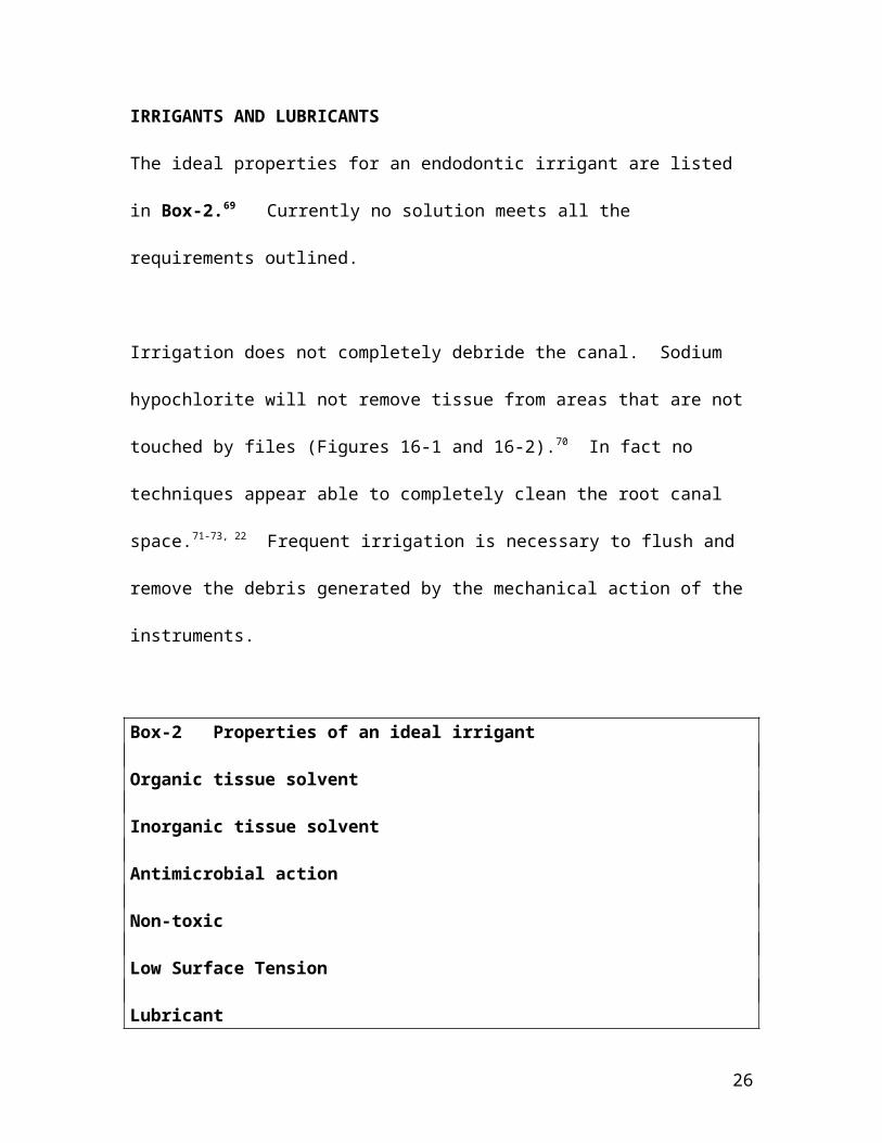
The ideal properties for an endodontic irrigant are listed in Box-2.69 Currently no
solution meets all the requirements outlined.
16
Irrigation does not completely debride the canal. Sodium hypochlorite will not remove
tissue from areas that are not touched by files (Figures 16-1 and 16-2).70 In fact no
techniques appear able to completely clean the root canal space.71-73, 22 Frequent irrigation
is necessary to flush and remove the debris generated by the mechanical action of the
instruments.
Box-2 Properties of an ideal irrigant
Organic tissue solvent
Inorganic tissue solvent
Antimicrobial action
Non-toxic
Low Surface Tension
Lubricant
Antimicrobial action
Sodium Hypochlorite
The most common irrigant is sodium hypochlorite (household bleach). Advantages to
sodium hypochlorite include the mechanical flushing of debris from the canal, the ability
of the solution to dissolve vital74 and necrotic tissue,75 the antimicrobial action of the
solution,32 and the lubricating action.76 In addition it is inexpensive and readily
available.
17
Free chlorine in sodium hypochlorite dissolves necrotic tissue by breaking down proteins
into amino acids. There is no proven appropriate concentration of sodium hypochlorite,
but concentrations ranging form 0.5% to 5.25% have been recommended. A common
concentration is 2.5%; which decreases the potential for toxicity while still maintaining
some tissue dissolving and antimicrobial activity.77, 78 Since the action of the irrigant is
related to the amount of free chlorine, decreasing the concentration can be compensated
by increasing the volume. Warming the solution can also increase effectiveness of the
solution.79, 80
Because of toxicity, extrusion is to be avoided.45, 81, 41 The irrigating needle must be
placed loosely in the canal (Figure 16-6). Insertion to binding and slight withdrawal
minimizes the potential for possible extrusion and a “sodium hypochlorite accident”
(Figure 16-7). Special care should be exercised when irrigating a canal with an open
apex. To control the depth of insertion the needle is bent slightly at the appropriate
length or a rubber stopper placed on the needle.
The irrigant does not move apically more than one millimeter beyond the irrigation tip so
deep placement with small gauge needles enhances irrigation (Figure 16-6).82
Unfortunately the small bore can easily clog, so aspiration after each use is
recommended. During rinsing, the needle is moved up and down constantly to produce
agitation and prevent binding or wedging of the needle.
Chlorhexidine
18
Chlorhexidine possesses a broad spectrum of antimicrobial activity, provides a sustained
action81, 83, and has little toxicity.84-87 Two percent chlorhexadine has similar
antimicrobial action as 5.25% sodium hypochlorite84 and is more effective against
enterococcus faecalis.81 Sodium hypochlorite and chlorhexadine are synergistic in their
ability to eliminate microorganisms. 85 A disadvantage of chlorhexadine is its inability to
dissolve necrotic tissue and remove the smear layer.
LUBRICANTS
Lubricants facilitate file manipulation during cleaning and shaping. They are an aid in
initial canal negotiation especially in small constricted canals without taper. They reduce
torsional forces on the instruments and decrease the potential for fracture.
Glycerin is a mild alcohol that is inexpensive, nontoxic, aseptic, and somewhat soluble.
A small amount can be placed along the shaft of the file or deposited in the canal orifice.
Counterclockwise rotation of the file carries the material apically. The file can then be
worked to place using a watch winding or “twiddling” motion.
Paste lubricants can incorporate chelators. One advantage to paste lubricants is that they
can suspend dentinal debris and prevent apical compaction. One proprietary product
consists of glycol, urea peroxide and ethylenediaminetetraacetic acid (EDTA) in a special
water soluble base. It has been demonstrated to exhibit an antimicrobial action88. Another
type is composed of 19% EDTA in a water soluble viscous solution.
19
A disadvantage to these EDTA compounds appears to be the deactivation of sodium
hypochlorite by reducing the available chlorine89 and potential toxicity90. The addition of
EDTA to the lubricants has not proven to be effective91. In general files remove dentin
faster than the chelators can soften the canal walls. Aqueous solutions such as sodium
hypochlorite should be used instead of paste lubricants when using nickel-titanium rotary
techniques to reduce torque76.
SMEAR LAYER
During the cleaning and shaping, organic pulpal materials and inorganic dentinal debris
accummulates on the radicular canal wall producing a an amorphous irregular smear
layer (Figure 16-8).69 With pulp necrosis, the smear layer may be contaminated with
bacteria and their metabolic by-products. The smear layer is superficial with a thickness
of 1-5 microns and debris can be packed into the dentinal tubules varying distances.92
There does not appear to be a consensus on removing the smear layer prior to obturation.
93, 94, 69 The advantages and disadvantages of the smear layer removal remain
controversial; however, evidence supports removing the smear layer prior to obturation.95,
69 The organic debris present in the smear layer might constitute substrate for bacterial
growth and it has been suggested that the smear layer prohibits sealer contact with the
canal wall and permits leakage. In addition, viable microorganisms in the dentinal tubules
may use the smear layer as a substrate for sustained growth. When the smear layer is not
removed, it may slowly disintegrate with leaking obturation materials, or it may be
removed by acids and enzymes that are produced by viable bacteria left in the tubules or
20
enter via coronal leakage. 96 The presence of a smear layer may also interfere with the
action and effectiveness of root canal irrigants and inter-appointment disinfectants.37
With smear layer removal filling materials adapt better to the canal wall.97, 98 Removal of
the smear layer also enhances the adhesion of sealers to dentin and tubular penetration99,
97, 100, 98 and permits the penetration of all sealers to varying depths.101 Removal of the
smear layer reduces both coronal and apical leakage.102 103
EDTA
Removal of the smear layer is accomplished with acids or other chelating agents such as
ethylenediamine tetracetic acid (EDTA) 104 following cleaning and shaping. Irrigation
with 17% EDTA for one minute followed by a final rinse with sodium hypochlorite105 is a
recommended method. Chelators remove the inorganic components leaving the organic
tissue elements intact. Sodium hypochlorite is then necessary for removal of the
remaining organic components. Citric acid has also been shown to be an effective
method for removing the smear layer106, 107 as has tetracycline. 108, 109
Demineralization results in removal of the smear layer and plugs, and enlargement of the
tubules.110 111 The action is most effective in the coronal and middle thirds of the canal
and reduced apically.104, 112 Reduced activity may be a reflection of canal size62 or
anatomical variations such as irregular or sclerotic tubules.113 The variable structure of
the apical region presents a challenge during endodontic obturation with adhesive
materials.
21
The recommended time for removal of the smear layer with EDTA is 1 minute.114, 104, 115
The small particles of the smear layer are primarily inorganic with a high surface to mass
ratio which facilitates removal by acids and chelators. EDTA exposure over 10 minutes
causes excessive removal of both peritubular and intratubular dentin.116
MTAD
An alternative method for removing the smear layer employs the use of a mixture of a
tetracycline isomer, an acid, and a detergent (MTAD) as a final rise to remove the smear
layer.117 The effectiveness of MTAD to completely remove the smear layer is enhanced
when low concentrations of NaOCl are used as an intracanal irrigant before the use of
MTAD118. A 1.3% concentration is recommended. MTAD may be superior to sodium
hypochlorite in antimicrobial action.119, 120 MTAD has been shown to be effective in
killing E. faecalis, an organism commonly found in failing cases, and may prove
beneficial during retreatment. It is biocompatible121, does not alter the physical properties
of the dentin121 and it enhances bond strength.122
TECHNIQUES OF PREPARATION
Regardless of the technique used in cleaning and shaping, procedural errors can occur.
These included loss of working length, apical transportation, apical perforation, lateral
stripping and instrument fracture.
Loss of working length has several causes. These include failure to have an adequate
reference point from which the corrected working length is determined, packing tissue
and debris in the apical portion of the canal, ledge formation, and inaccurate
22
measurements. Apical transportation and zipping occurs when the restoring force of the
file exceeds the threshold for cutting dentin in cylindrical non-tapering curved canal
(Figures 16-9 and 16-10).123 When this apical transportation continues with larger and
larger files, a “teardrop” shape develops and perforation can occur apically on the lateral
root surface (Figure16-9). Transportation in curved canals begins with a size #25 file15.
Enlargement of curved canals at the corrected working length beyond a size #25 file
should be done only when an adequate coronal flare is developed.
Instrument fracture occurs with torsional and cyclic fatigue. Locking the flutes of a file
in the canal wall while continuing to rotate the coronal portion of the instrument is an
example torsional fatigue (Figure 16-11). Cyclic fatigue results when strain develops in
the metal.
Stripping perforations occur in the furcal region of curved roots, frequently the mesial
roots of maxillary and mandibular molars perforation (Figures 16-12 and 16-13). The
canal in this area is not always centered in the root and prior to preparation the average
distance to the furcal wall (danger zone) is less than the distance to the bulky outer wall
(safety zone). An additional factor is the concavity of the root.
Watch Winding
Watch winding is reciprocating back and forth (clockwise/counterclockwise) rotation of
the instrument in an arch. It is used to negotiate canals and to work files to place. Light
apical pressure is applied to move the file deeper into the canal.
23
Reaming
Reaming is defined as the clockwise, cutting rotation of the file. Generally the
instruments are placed into the canal until binding is encountered. The instrument is then
rotated clockwise 180-360º to plane the walls and enlarge the canal space.
Filing
Filing is defined as placing the file into the canal and pressing it laterally while
withdrawing it along the path of insertion to scrape the wall. There is very little rotation
on the outward cutting stroke. The scraping or rasping action removes the tissue and cuts
superficial dentin from the canal wall. A modification is the turn-pull technique. This
involves placing the file to the point of binding, rotating the instrument 90º and pulling
the instrument along the canal wall.
Circumferential filing
Circumferential filing is used for canals that are larger and or not round. The file is
placed into the canal and withdrawn in a directional manner sequentially against the
mesial, distal, buccal, and lingual walls.
Standardized preparation
After 1961, instruments were manufactured with a standard formula. Clinicians utilized a
preparation technique of sequentially enlarging the canal space with smaller to larger
instruments at the corrected working length.124 In theory this created a standardized
preparation of uniform taper. Unfortunately this does not occur. This technique was
adequate for preparing the apical portion of canals that were relatively straight and
tapered; however in cylindrical and small curved canals procedural errors were identified
with the technique.125
24
Step-back Technique
The step-back technique70, 125 reduces procedural errors and improves debridement. After
coronal flaring and determining the master apical file (initial file that binds slightly at the
corrected working length), the succeeding larger files are shortened by 0.5 or 1.0 m
increments from the previous file length (Figure 16-14 and 16-15). This step-back
process creates a flared, tapering preparation while reducing procedural errors. The step-
back preparation is superior to standardized serial filing and reaming techniques in
debridement and maintaining the canal shape.70 The step-back filing technique results in
more pulpal walls being planed when compared to reaming or filing.
Step-Down Technique
The step down technique is advocated for cleaning and shaping procedures as it removes
coronal interferences and provides coronal taper. Originally advocated for hand file
preparation126 it has been incorporated into techniques employing nickel-titanium files.
With the pulp chamber filled with irrigant or lubricant the canal is explored with a small
instrument to assess patency and morphology (curvature). The working length can be
established at this time. The coronal one third of the canal is then flared with Gates
Glidden drills or rotary files of greater taper (.06, .08, .10,). A large file (such size #70)
is then placed in the canal using a watch winding motion until resistance is
encountered.126 The process is repeated with sequentially smaller files until the apical
portion of the canal is reached. The working length can be determined if this was not
accomplished initially. The apical portion of the canal can now be prepared by enlarging
25
the canal at the corrected working length. Apical taper is accomplished using a step-back
technique.
Passive Step-back
The passive step-back technique is a modification of the incremental step-back
technique.6, 127 After the apical diameter of the canal has been determined, the next higher
instrument is inserted until it first makes contact (binding point). It is then rotated one half
turn and removed (Figure 16-16). The process is repeated with larger and larger instruments
being placed to their binding point. This entire instrument sequence is then repeated. With
each sequence the instruments drop deeper into the canal creating a tapered preparation.
This technique permits the canal morphology to dictate the preparation shape. The
technique does not require arbitrary rigid incremental reductions and forcing files into canals
that cannot accommodate the files. Advantages to the technique include: knowledge of
canal morphology, removal of debris and minor canal obstructions, and a gradual passive
enlargement of the canal in an apical to coronal direction.
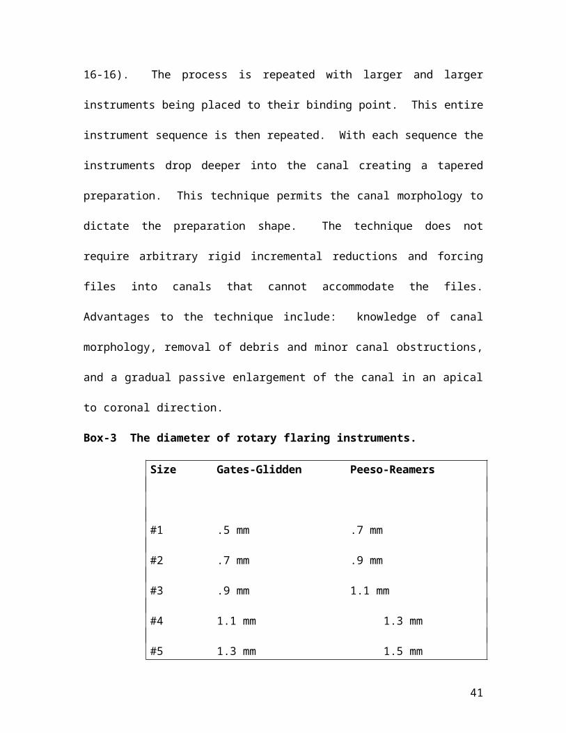
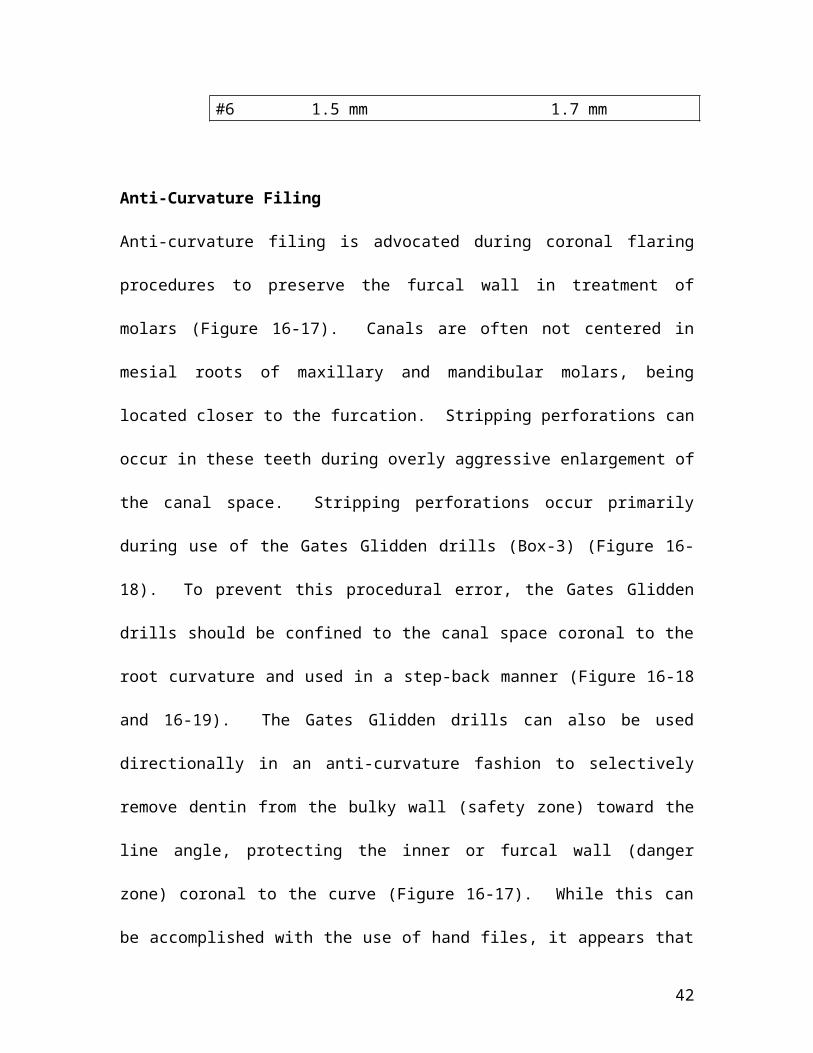
Box-3 The diameter of rotary flaring instruments.
Size Gates-Glidden Peeso-Reamers
#1 .5 mm .7 mm
#2 .7 mm .9 mm
#3 .9 mm 1.1 mm
#4 1.1 mm 1.3 mm
#5 1.3 mm 1.5 mm
26
#6 1.5 mm 1.7 mm
Anti-Curvature Filing
Anti-curvature filing is advocated during coronal flaring procedures to preserve the furcal
wall in treatment of molars (Figure 16-17). Canals are often not centered in mesial roots
of maxillary and mandibular molars, being located closer to the furcation. Stripping
perforations can occur in these teeth during overly aggressive enlargement of the canal
space. Stripping perforations occur primarily during use of the Gates Glidden drills
(Box-3) (Figure 16-18). To prevent this procedural error, the Gates Glidden drills should
be confined to the canal space coronal to the root curvature and used in a step-back
manner (Figure 16-18 and 16-19). The Gates Glidden drills can also be used
directionally in an anti-curvature fashion to selectively remove dentin from the bulky
wall (safety zone) toward the line angle, protecting the inner or furcal wall (danger zone)
coronal to the curve (Figure 16-17). While this can be accomplished with the use of hand
files, it appears that directional forces with Gates Glidden drills is not beneficial.128
Balanced Force Technique
The balanced force technique recognizes the fact that instruments are guided by the canal
walls when rotated.129 Since the files will cut in both a clockwise and counterclockwise
rotation, the balanced force concept of instrumentation consists of placing the file to
length and then a clockwise rotation (less than 180 degrees) engages dentin. This is
followed by a counterclockwise rotation (at least 120 degrees) with apical pressure to cut
and enlarge the canal. The degree of apical pressure varies from light pressure with small
27
instruments to heavy pressure with large instruments. The clockwise rotation pulls the
instrument into the canal in an apical direction. The counterclockwise cutting rotation
forces the file in a coronal direction while cutting circumferentially. Following the
cutting rotation the file is repositioned and the process is repeated until the corrected
working length is reached. At this point a final clockwise rotation is employed to
evacuate the debris.
Nickel Titanium Rotary Preparation
Nickel titanium rotary preparation utilizes a crown-down approach. The specific
technique is based on the instrument system selected. One instrument sequence uses
nickel titanium files with a constant taper and variable ISO tip sizes (Figure 16-20). With
this technique, a .06 taper is selected. Initially a size .06/45 file is used until resistance,
followed by the .06/45, .06/40, .06/35, .06/30, .06/25, and .06/20. In a second technique,
nickel titanium files with a constant tip diameter are used. The initial file is a .10/20
instrument, the second a .08/20, the third a .06/20, and the fourth a .04/20 (Figure 16-21).
For larger canals a sequence of files using ISO standardized tip sizes of 30 or 40 might be
selected. Using the crown down approach creates coronal flare and reduces the contact
area of the file so torsional forces are reduced.
Final Apical Enlargement and Apical Clearing
Apical clearing enhances the preparation of the apical canal, improves debridement, and
produce a more definite apical stop in preparation for obturation.130 Apical clearing is
generally performed when there is an apical stop and the master apical file is less that a
size #40 file. If the apical configuration is open or a seat, apical clearing might make the
28
opening larger and potentiate the possibility of extrusion of the obturation materials.
Apical clearing consists of two distinct steps: final apical enlargement and dry reaming.
Final apical enlargement is performed after the canal has been cleaned and shaped. It
involves enlargement of the apical preparation three to five sizes beyond the master
apical file (Figure 16-22). The degree of enlargement depends on the canal size and root
curvature. In a small curved canal enlargement may only be three sizes to decrease the
potential for transportation. In a straight canal it can be larger without producing a
procedural error. Since the prepared canal exhibits taper, the small files at the corrected
working length can be used to enlarge the canal without transportation. Final apical
enlargement is performed with the irrigant and employs a reaming action at the corrected
working length. The last file used becomes the final apical file. Since the file is only
contacting the apical 1-2 mm the walls of the canal, the technique will result in a less
irregular apical preparation. The canal is then irrigated. The smear layer is removed
with a decalcifying agent and the canal dried with paper points.
After drying the canals, the dry reaming is performed. Dry reaming removes dentin chips
or debris packed apically during drying. The final apical file (or the master apical file in
cases where apical enlargement was not performed) is placed to the corrected working
length and rotated clockwise in a reaming action.
Recapitulation
Recapitulation is important regardless of the technique selected (Figure16-23). This is
accomplished by taking a small file to the corrected working length to loosen
29
accumulated debris and then flushing it with 1-2 ml of irrigant. Recapitulation is
performed between each successive enlarging instrument regardless of the cleaning and
shaping technique.
Combination Technique
This technique combines coronal flaring, nickel titanium rotary preparation, and the
passive step-back technique (BOX-4). Following access, the canal is explored with a #10
or #15 file. If the canal is patent to the estimated working length a working length
radiograph can be obtained and the corrected working length established (Chapter 15,
Figure 15-40). In order to insure an accurate length determination a size #20 file or
larger should be used (Chapter 15, Figures 15-40, 15-41). If a #20 file will not go to the
estimated working length passive step-back instrumentation can be performed by
inserting successively larger files to the point of binding and reaming. This removes
coronal interferences and creates greater coronal taper permitting larger files access to the
apical portion of the root.
After establishing the working length, Gates Glidden drills are used for straight line
access (Figure 16-18). A #2 Gates is used first followed by the #3 and #4. In very
narrow canals a #1 Gates may be needed. It is important to remember the size of the
Gates Glidden drills. If the canal orifice cannot accommodate a size #70 file, passive
step back should be performed to provide adequate initial coronal space. To prevent
stripping perforations, the Gates should not be placed apical to canal curvatures.
Generally the #2-#4 provides adequate coronal enlargement and preserves root structure.
The use of nickel titanium rotary instruments with greater tapers can also be used for this
30
step (.06, .08, and .10 tapers are common). The Gates Glidden drills can be used in either
a crown-down or step-back sequence. Following use, the Gates Glidden drill should be
removed from the handpiece to prevent injury to the clinician, assistant or patient (Figure
16-24).
Master Apical File
Emphasis has traditionally been placed on determining the canal length with little
consideration of the canal diameter in the apical portion of the root. Since every canal is
unique in its morphology the apical canal diameter must be assessed. The size of the
apical portion of the canal is determined by placing successively larger instruments to the
corrected working length until slight binding is encountered (Figure 16-25). Often the
next larger instrument will not go to the corrected working length. If it does go to length
a subjective estimation of the apical diameter must be made depending on the degree of
binding. This file will be the master apical file (initial file to bind). It is defined as the
largest file to bind at the corrected working length following straight line access. This
provides an estimate of the canal diameter before cleaning and shaping and it is the point
where the step-back preparation begins.
Nickel-Titanium Rotary
Once the master apical file is identified, the middle to apical portion of the canal is
prepared using nickel titanium rotary instruments (Figure 16-20 and Figure 16-21)).
Rotary files are used with a crown-down approach to within 3 mm of the corrected
working length. Adequate coronal taper is established when the .06/45 goes to within
3.0 mm of the corrected working length. Using the crown down approach creates coronal
taper and reduces the contact area of the file so torsional forces are reduced.
31
Recapitulation
Recapitulation is accomplished after each instrument used in the canal by taking a small
file to the corrected working length and then flushing the canal with 1-2 ml of irrigant
(Figure 16-23).
Step-Back Apical Preparation
When the body of the canal has been shaped, the apical portion is prepared using
standardized stainless steel or nickel titanium hand files in a step-back process (Figure
16-15). The first instrument selected for this portion of the shaping process is one size
larger that the master apical file (initial file to bind slightly). Larger files are successively
shortened by standardized increments of 0.05 mm or 1.0 mm. Generally sequentially
stepping back to a file size of #60 or #70 will produce adequate flare and blend the apical
and middle thirds of the canal.
Apical Clearing
With a flared preparation from the orifice to the corrected working length, the apical
portion of the canal is enlarged. With a tapered preparation the canal can be enlarged
with a reaming action as the canal walls will keep the instrument centered (Figure 16-25).
Box-4 The Combination Technique Steps
Canal negotiation
Working length determination
Straight line access
Master apical file determination
Rotary preparation of the middle one third of the root
32
Apical step-back preparation
Apical clearing
General Considerations – A Review
The following principles and concepts should be applied regardless of the instruments or
technique selected.
1. Initial canal exploration is always performed with smaller files to gauge
canal size, shape, and configuration.
2. Files are always manipulated in a canal filled with an irrigant or lubricant
present.
3. Copious irrigation is used between each instrument in the canal.
4. Coronal preflaring (passive step-back technique) with hand instruments
will facilitate placing larger working length files (either hand or rotary)
and will reduce procedural errors such as loss of working length and canal
transportation.
5. Apical canal enlargement is gradual, using sequentially larger files from
apical to coronal, regardless of flaring technique.
6. Debris is loosened and dentin is removed from all walls on the outstroke
(circumferential filing) or with a rotating (reaming) action at or close to
working length.
7. Instrument binding or dentin removal on insertion should be avoided. Files
are teased to length using a watch winding or “twiddling” action. This is a
back-and-forth rotating motion of the files (approximately a quarter turn)
33
between the thumb and forefinger, continually working the file apically.
Careful file insertion (twiddling) followed by planing on the outstroke will
help to avoid apical packing of debris and minimize extrusion of debris
into the periradicular tissues.
8. Reaming is defined as the clockwise rotation of the file. Generally the
instruments are placed into the canal until binding is encountered. The
instrument is then rotated clockwise 180-360º to cut and plane the walls.
When withdrawn the instrument tip is pushed alternately against all walls.
The pushing motion is analogous to the action of a paintbrush. Overall,
this is a turn and pull.
9. Filing is defined as placing the file into the canal and withdrawing it along
the path of insertion to scrap the wall. There is very little rotation on the
outward cutting stroke. The scraping or rasping action removes the tissue
and cuts superficial dentin from the canal wall.
10. Turn pull filing involves placing the file into the canal until binding. The
instrument is then rotated to engage the dentin and withdrawn with lateral
pressure against the canal walls.
11. Circumferential filing is used for canals that exhibit cross sectional shapes
that are not round. The file is placed into the canal and withdrawn in a
directional manner against the mesial, distal, buccal, and lingual walls.
12. Regardless of the technique, after each insertion the file is removed and
the flutes are cleaned of debris; the file can then be reinserted into the
34
canal to plane the next wall. Debris is removed from the file by wiping it
with an alcohol-soaked gauze or cotton roll131.
13. The canal is effectively cleaned only where the files actually contact and
plane the walls. Inaccessible regions are poorly cleaned or débrided.
14. Recapitulation is done to loosen debris by rotating the master apical file or
a smaller size at the corrected working length followed by irrigation to
mechanically remove the material. During recapitulation the canal walls
are not planed and the canal should not be enlarged.
15. Small, long, curved, round canals are the most difficult and tedious to
enlarge. They require extra caution during preparation, being the most
prone to loss of length and transportation.
16. Over enlargement of curved canals by files attempting to straighten
themselves will to lead to procedural errors (Figure 16-11).
17. Overpreparation of canal walls toward the furcation may result in a
stripping perforation in the danger zone where root dentin is thinner.
18. It is neither desirable nor necessary to try to remove created steps or other
slight irregularities created during canal preparation.
19. Instruments, irrigants, debris, and obturating materials should be contained
within the canal. These are all known physical or chemical irritants that
will induce periradicular inflammation and may delay or compromise
healing.
20. Creation of an apical stop may be impossible if the apical foramen is
already very large. An apical taper (seat) is attempted, but with care.
35
Overusing large files aggravates the problem by creating an even larger
apical opening.
20. Forcing or locking (binding) files into dentin produces unwanted torsional
force. This tends to untwist, wrap-up, either will weaken, and break the
instrument.
CRITERIA FOR EVALUATING CLEANING AND SHAPING
Following the cleaning and shaping procedures the canal should exhibit “glassy smooth”
walls and there should be no evidence of unclean dentin filings, debris, or irrigant in the
canal. This is determined by pressing the MAF against each wall in an outward stroke.
Shaping is evaluated by assessing the canal taper and identifying the apical configuration.
For obturation with lateral compaction, the finger spreader should go loosely to within
1.0 mm of the corrected working length. For warm vertical compaction the plugger
should reach to within 5 mm of the corrected working length (Figure 16-26).
The apical configuration is identified as an apical stop, apical seat, or open. This is
accomplished by placing the master apical file to the corrected working. If the master
apical file goes past the corrected working length the apical configuration is open. If
master apical file stops at the corrected working length a file one or two sizes smaller is
placed to the corrected working length. If this file stops the apical configuration is a stop.
When the smaller file goes past the corrected working length the apical configuration is a
seat.
36
INTRACANAL MEDICAMENTS
Intracanal medicaments have a long history of use as interim appointment dressings.
They are employed for three purposes: 1) to reduce inter-appointment pain, 2) to decrease
the bacterial count and prevent regrowth, and 3) to render the canal contents inert. Some
common agents are listed in Box 16-5 .
Box 16-5 Groupings of Commonly Used Intracanal Medicaments
Phenolics
Eugenol
Camphorated monoparachlorophenol (CMCP)
Parachlorophenol (PCP)
Camphorated parachlorophenol (CPC)
Metacresylacetate (Cresatin)
Cresol
Creosote (beechwood)
Thymol
Aldehydes
Formocresol
Glutaraldehyde
Halides
Sodium hypochlorite
Iodine-potassium iodide
Steroids
Calcium hydroxide
Antibiotics
37
Combinations
From Walton R: Intracanal medicaments, Dent Clin North Am 28:783, 1984.
Phenols and aldehydes
The majority of the medicaments exhibit non-specific action and can destroy host tissues
as well as microbes132-134. Historically it has been thought that these agents are effective;
their use was based on opinion and empiricism. The phenols and aldehydes are toxic and
the aldehydes are fixative agents135, 136. When placed in the radicular space they have
access to the periradicular tissues and the systemic circulation137, 138 Research has
demonstrated that their clinical use is not justified139-143. Clinical studies assessing the
ability of these agents to prevent or control interappointment pain indicate that they are
not effective.144-147
Calcium hydroxide
One intracanal agent that is effective in inhibiting microbial growth in canals is calcium
hydroxide148. It has antimicrobial action due to the alkaline pH and it may aid in
dissolving necrotic tissue remnants and bacteria and their byproducts149-151.
Interappointment calcium hydroxide in the canal demonstrates no pain reduction
effects152. Calcium hydroxide has been recommended for use in teeth with necrotic pulp
tissue and bacterial contamination. It probably has little benefit with vital pulps. Calcium
hydroxide can be placed as a dry powder, a powder mixed with a liquid such as local
anesthetic solution, saline, water, or glycerin to form a thick paste, or as a proprietary
paste supplied in a syringe (Figure 16-27). A lentulo-spiral is effective and efficient.153-155
38
Spinning the paste into the canal by rotating a file counterclockwise and using an
injection technique is not as effective. It is important to place the material deeply and
densely for maximum effectiveness. To accomplish this straight line access with Gates
Glidden drills or nickel-titanium rotary files should be performed and the apical portion
of the canal prepared to a size #25 file or greater. Removal following placement is
difficult.156 This is especially true in the apical portion of the root.
Corticosteroids
Corticosteroids are anti-inflammatory agents that have been advocated for decreasing
postoperative pain by suppressing inflammation. The use of corticosteroids as
intracanal medicaments may decrease lower levels postoperative pain in certain
situations;157 however, evidence also suggests that they may be ineffective particularly
with greater pain levels147. Cases irreversible pulpitis and cases where the patient is
experiencing acute apical periodontitis are examples where steroid use might be
beneficial158, 159, 157.
Chlorhexidine
Chlorhexidine has recently been advocated as an intracanal medicament.160, 161 A 2% gel
is recommended. It can be used alone in gel form or mixed with calcium hydroxide.
When used with calcium hydroxide the antimicrobial activity is greater than when
calcium hydroxide is mixed with saline162and periradicular healing is enhanced.163 Its
major disadvantages are; it does not affect the smear layer and it is a fixative.
39
TEMPORARY RESTORATIONS (Courtesy of Dr. Harold Messer)
Root canal treatment may involve multiple visits. Also, unless it is limited to a
routine access cavity, the final restoration is usually not completed in the same
appointment as the root canal treatment. A temporary restoration is then required,
normally for 1 to 4 weeks. In special situations when definitive restoration must be
deferred, the temporary must last several months.
Objectives of Temporization
The temporary restoration must
1. Seal coronally, preventing ingress of oral fluids and bacteria and egress of
intracanal medicaments.
2. Enhance isolation during treatment procedures.
3. Protect tooth structure until the final restoration is placed.
4. Allow ease of placement and removal.
5. Satisfy esthetics, but always as a secondary consideration to providing a
seal.
These objectives depend on the intended duration of use. Thus, different materials
are required depending on time, occlusal load and wear, complexity of access, and loss of
tooth structure.
40
Routine Access Cavities
Most access cavities involve only one surface and are surrounded by dentin walls
or by porcelain or metal (if the restoration is retained). The temporary must last from
several days to several weeks. Numerous types are available, including premixed cements
that set on contact with moisture (Cavit), reinforced zinc oxide-eugenol cements (such as
IRM), glass ionomer cements and specially formulated light-polymerized composite
materials (such as TERM®, temporary endodontic restorative material)164. Ease of use and
good sealing ability make Cavit an excellent routine material, but low strength and rapid
occlusal wear limit its use to short-term sealing of simple access cavities. IRM and
TERM provide improved wear resistance, although their sealing ability is probably
marginally less than that of Cavit165, 166. More durable restorative materials, especially
glass ionomer cements, tend to provide the best seal. A double seal of GIC over Cavit
will provide a durable and effective barrier to microbial leakage. It is not known whether
experimental leakage differences based on bacterial leakage or dye penetration are
significant clinically, especially if thermocycling and occlusal loading are not part of the
testing procedure167. Clinically, 4mm of Cavit provided an effective seal against bacterial
penetration for 3 weeks168. Most critical are the thickness and placement of the material.
Techniques of Placement - The quality of the coronal seal depends on the
thickness of the material, how it is compacted into the cavity, and the extent of contact
with sound tooth structure or restoration. A minimum depth of 3 to 4 mm is required
around the periphery, preferably 4 mm or more to allow for wear. In anterior teeth, the
41
access is oblique to the tooth surface; care must be taken to ensure that the material is at
least 3 mm thick in the cingulum area.
Cavit (or a similar material) is placed as follows: Chamber and cavity walls
should be dry. Cavit can be placed directly over the canal orifices, or more commonly a
thin layer of cotton is placed over the canal orifices to prevent canal blockage169. (Figure
16-28) Care must be taken not to incorporate cotton fibers into the restorative material,
which can promote rapid leakage170. Cavit is packed into the access opening with a plastic
instrument in increments from the bottom up and pressed against the cavity walls and into
undercuts (Figure 16-29). Excess is removed, and the surface smoothed with moist
cotton. The patient should avoid chewing on the tooth for at least an hour.
Subsequent removal using a high speed bur requires care to avoid damage to the
access opening. Alternatively, an ultrasonic tip can be used.
Extensive Coronal Breakdown
Teeth without marginal ridges or with undermined cusps require a stronger filling
material (high-strength glass ionomer cement), taking care to ensure an adequate
thickness and good marginal adaptation proximally. The temporary filling material
should extend well into the pulp chamber deep to the proximal margin to ensure a
marginal seal. Reducing the height of undermined cusps well out of occlusion reduces the
risk of fracture. For severely broken-down teeth, a cusp-onlay amalgam or a well-fitting
42
orthodontic band cemented onto the tooth (restored with glass ionomer cement) provides
a durable temporary restoration and strengthens the tooth against fracture171. At the next
appointment, access is prepared through the restoration.
PROVISIONAL POST CROWNS
The use of a provisional crown with an incorporated resin post may be required,
particularly when a cast post and core is being fabricated for a visible tooth with little
remaining coronal tooth structure. However, the use of such a provisional crown retained
with a post (preformed aluminum post, safety pin wire, paper clip, or a sectioned large
endodontic file) has inherent problems. Using the canal space for a provisional post
precludes use of an intracanal medicament, and the coronal seal depends entirely on the
cement. The coronal seal is generally inadequate with a loosely fitting and mobile
provisional post and crown172. However, in spite of these potential difficulties, such
provisional restorations may be required while cast posts and cores are being fabricated.
Due to the potential problems, it is prudent to cement the definitive post as soon as
possible.
When such a provisional crown-post combination is being used, the post should
fit the canal snugly (not binding) and extend apically 4 to 5 mm short of working length
and coronally to within 2 to 3 mm of the incisal edge. A polycarbonate shell is trimmed
to a good fit; autopolymerizing material then is added to the inside of the shell to mold to
the root face and attach to the post. A provisional luting cement (Temp Bond or similar
43
cement) is placed on the coronal 3 to 4 mm of the post and root face, and the unit is
cemented into place. A provisional removable partial overdenture is a useful alternative;
access remains excellent, and there is little chance of disturbing the coronal seal between
appointments.
Long–term Temporary Restorations
Few indications exist to justify delaying the final restoration, and endodontic
procedures (other than trauma management) rarely require prolonged treatment. If a
temporary restoration has to last more than a few weeks, then a durable material such as
amalgam, glass ionomer cement, or acid-etch composite should be used. The pulp
chamber is filled with Cavit to provide a good coronal seal, and covered with a sufficient
thickness of the restorative material to ensure strength and wear resistance. Subsequent
access to the canal space is readily achieved without damage to remaining tooth structure
because the layer of Cavit can be easily removed.
44
Figures
Figure 16-1 Cross-section through a root showing the main canal (C) and a fin (arrow)
and associated cul-de-sac after cleaning and shaping, using files and sodium
hypochlorite. Note the tissue remnants that remain in the fin.
Figure 16-2 The main canal (C) has a lateral canal (arrow) extending to the root surface.
After cleaning and shaping with sodium hypochlorite irrigation, tissue remains in the
lateral canal.
Figure 16–3 A. A size #15 file in the apical canal space. Note the size is inadequate for
planning the walls. B. A size #40 file more closely approximates the canal morphology
(Courtesy of Dr. Randy Madsen).
Figure 16-4 A. The classic apical anatomy consisting of the major diameter of the
foramen and the minor diameter of the constriction. B. An irregular ovoid apical canal
shape and external resorption. C. A bowling pin apical morphology and an accessory
canal. D. Multiple apical foramina.
Figure 16-5 A small file (#10 or #15) is placed beyond the radiographic apex to maintain
patency of the foramen. Note the tip extends beyond the apical foramen (arrow).
Figure 16-6 For effective irrigation the needle must be placed in the apical one-third of
the root and must not bind.
45
Figure 16-7 A sodium hypochlorite accident during treatment of the maxillary left
central incisor. Extensive edema occurred in the upper lip accompanied by severe pain.
Figure 16-8 A. A canal wall with the smear layer present. B. The smear layer removed it
17% EDTA.
Figure 16-9 Procedural errors of canal transportation, zipping and strip perforation occur
during standardized preparation when files remove dentin from the outer canal wall
apical to the curve and from the inner wall coronal to the curve. This is related to the
restoring force (stiffness) of the files. Note in the apical portion the transportation takes
the shape of a tear drop as the larger files are used.
Figure 16-10 The canals have been transported and there is an apical perforation.
Figure 16-11 A. A size #35 file fractured in the mesiobuccal canal. B. SEM
examination reveals torsional fatigue at the point of fracture. Note the tightening of the
flutes near the fracture and the unwinding of the flutes along the shaft.
Figure 16-12 A. The furcal region of molars at the level of the curvature (danger zone)
is a common site for stripping perforation. B. Note the distal concavity (arrows) in the
furcation area of this mandibular molar.
46
Figure 16-13 Straight line access can result in stripping perforations in the furcal areas of
molars. A. The use of large Gates Glidden drills and overpreparation has resulted in the
stripping perforation. B. Note that the perforation is in the concavity of the furcation.
Figure 16-14 The step-back preparation is designed to provide a tapering preparation.
The process begins with one file size larger than the master apical file with incremental
shortening of either .5 or 1.0 mm.
Figure 16-15 As an example of step-back preparation in a moderately curved canal. A.
The size #25 master apical file at the corrected working length of 21.0 mm. B. The step-
back process begins with the #30 file at 20.5 mm. C. #35 at 20.0 mm. D. #40 file at 19.5
mm. E. #45 file at 19.0 mm. F. #50 file at 18.5 mm. G. #55 file at 18.0 mm. H. #60
file at 17.5 mm. I. #70 file at 17.0 mm
Figure 16-16 Passive step-back. Smaller to larger files are inserted to their initial point
of binding and then rotated 180 to 360º and withdrawn. This process creates slight taper
and coronal space. This permits larger instruments to reach the apical one third.
Figure 16-17 The anti-curvature filing technique. Instruments are directed away from the
furcal “danger zone” toward the line angles (safety zone) where the bulk of dentin is
greater.
47
Figure 16-18 Straight line access in a maxillary left first molar with Gates-Glidden drills
used in a slow speed handpiece using a step-back technique. A. The #1 Gates is used
until resistance. B. This is followed by the #2 which should not go past the first
curvature. C. The #3 Gates is used 3-4 mm into the canal. D. Followed by the #4
instrument.
Figure 16-19 A maxillary first molar following straight line access with the Gates
Glidden Drills.
Figure 16-20 The mesiobuccal canal is prepared using nickel-titanium rotary files using
a crown-down technique. In this sequence each instrument exhibits the same .06 taper
with varied ISO standardized tip diameters. Instrument were used to resistance. A. The
process begins with a .06\45 file to resistance at 16.0 mm. B. This is followed by
a .06\40 instrument at 17.0 mm C. The .06\35 file is used to 18.0 mm. D. The .06\30 at
19.0 mm. E. The .06\25 at 20.0 mm. F. The .06\20 file is to the corrected working
length of 21.0mm.
Figure 16-21 Nickel-titanium rotary files with a standardized ISO tip diameter and
variable tapered files can be used in canal preparation. In this sequence, the instruments
have a standardized tip diameter of .20 mm. A. Initially a 10/.0 file is used. B. This is
followed by 08/.20. C. The third instrument is a .06/.20. D. The final instrument is a
04/.20 file to the corrected working length of 21.0 mm.
48
Figure 16-22 Final Apical Enlargement A. The master apical file of size #25 at the
corrected working length of 21.0 mm. B. Enlargement with a #30 file to the corrected
working length of 21.0 mm. C. Further enlargement with a #35 file. D. Final
enlargement to a size #40 file. The final instrument used becomes the Final Apical File.
Figure 16-23 Recapitulation is accomplished between each instrument by reaming with
the Master Apical File or a smaller instrument. This minimizes packing of debris and
loss of length.
Figure 16-24 Following their use, the Gates Glidden drills should be removed from the
handpiece to prevent injury. This #3 drill was accidentally driven into the palm of the
dentist.
Figure 16-25 Following straight line access in this maxillary molar, the Master Apical
File is determined by successively placing small to larger files to the corrected working
length. A. A #15 stainless steel file is placed to 21.0 mm without resistance. B. A #20
is the placed is placed to 21.0 mm without resistance. C. The #25 file reaches 21.0 mm
with slight binding. D. A size #30 file is then placed and does not go the corrected
working length indicating the initial canal size in the apical portion of the canal is a
size#25
Figure 16-26 The coronal taper is assessed using the spreader or plugger depth of
penetration. A. With lateral compaction a finger spreader should fit loosely 1.0 mm from
49
the Corrected Working Length with space adjacent to the spreader. B. For warm vertical
compaction, the plugger should go to within 5.0 mm of the Corrected Working Length.
Figure 16-27 Calcium hydroxide placement. A. Calcium hydroxide mixed with glycerin
to form a thick paste. B. Placement with a lentulo spiral. C. Injection of a proprietary
paste. D. Compaction of calcium hydroxide powder with a plugger.
Figures 16-28 and 16-29 are provided by Dr. Harold Messer
Figure 16-28. Techniques for temporization. On the left are the correct techniques; either
minimal space is occupied by cotton or no cotton pellet is used, particularly if the
proximal is to be restored. It is wrong to pack most of the chamber with cotton, which
leaves inadequate space and strength for the material (3-4 mm are required), and cotton
fibers may promote bacterial leakage. (Courtesy of Dr L Wilcox)
Figure 16-29. Techniques for placing temporary material. A, A single large “blob” placed
in the access opening will not seal the walls. B, The incremental technique, which adds
successive layers, pressing each against the chamber walls, is correct. (Courtesy of Dr L
Wilcox)
50
References
1. Ingle JI, editor. Endodontics. 5th Edition ed ed. Hamilton, London: BC Decker, Inc 2002.2. Sabeti MA, Nekofar M, Motahhary P, Ghandi M, Simon JH. Healing of apical periodontitis after endodontic treatment with and without obturation in dogs. J Endod 32(7):628-33, 2006.3. Delivanis PD, Mattison GD, Mendel RW. The survivability of F43 strain of Streptococcus sanguis in root canals filled with gutta-percha and Procosol cement. J Endod 9(10):407-10, 1983.4. Walton RE. Current concepts of canal preparation. Dental Clinics of North America 36(2):309-26, 1992.5. Allison DA, Weber CR, Walton RE. The influence of the method of canal preparation on the quality of apical and coronal obturation. J Endod 5(10):298-304, 1979.6. Schilder H. Cleaning and shaping the root canal. Dental Clinics of North America 18(2):269-96, 1974.7. Wilcox LR, Roskelley C, Sutton T. The relationship of root canal enlargement to finger-spreader induced vertical root fracture. J Endod 23(8):533-4, 1997.8. Kuttler Y. Microscopic investigation of root apexes. J Am Dent Assoc 50(5):544-52, 1955.9. Dummer PM, McGinn JH, Rees DG. The position and topography of the apical canal constriction and apical foramen. Int Endod J 17(4):192-8, 1984.10. Gutierrez JH, Aguayo P. Apical foraminal openings in human teeth. Number and location. Oral Surg Oral Med Oral Pathol Oral Radiol Endod 79(6):769-77, 1995.11. Malueg LA, Wilcox LR, Johnson W. Examination of external apical root resorption with scanning electron microscopy. Oral Surg Oral Med Oral Pathol Oral Radiol Endod 82(1):89-93, 1996.12. Farzaneh M, Abitbol S, Friedman S. Treatment outcome in endodontics: the Toronto study. Phases I and II: Orthograde retreatment. J Endod 30(9):627-33, 2004.13. Schaeffer MA, White RR, Walton RE. Determining the optimal obturation length: a meta-analysis of literature. J Endod 31(4):271-4, 2005.14. Wu MK, Wesselink PR, Walton RE. Apical terminus location of root canal treatment procedures. Oral Surg Oral Med Oral Pathol Oral Radiol Endod 89(1):99-103, 2000.15. Eldeeb ME, Boraas JC. The effect of different files on the preparation shape of severely curved canals. International Endodontic Journal 18(1):1-7, 1985.16. Chow TW. Mechanical effectiveness of root canal irrigation. Journal of Endodontics 9(11):475-9, 1983.17. Ram Z. Effectiveness of root canal irrigation. Oral Surgery, Oral Medicine, Oral Pathology 44(2):306-12, 1977.18. Salzgeber RM, Brilliant JD. An in vivo evaluation of the penetration of an irrigating solution in root canals. Journal of Endodontics 3(10):394-8, 1977.19. Dalton BC, Orstavik D, Phillips C, Pettiette M, Trope M. Bacterial reduction with nickel-titanium rotary instrumentation. Journal of Endodontics 24(11):763-7, 1998.
51

20. Orstavik D, Kerekes K, Molven O. Effects of extensive apical reaming and calcium hydroxide dressing on bacterial infection during treatment of apical periodontitis: a pilot study. International Endodontic Journal 24(1):1-7, 1991.21. Sjogren U, Figdor D, Spangberg L, Sundqvist G. The antimicrobial effect of calcium hydroxide as a short-term intracanal dressing. International Endodontic Journal 24(3):119-25, 1991.22. Wu YN, Shi JN, Huang LZ, Xu YY. Variables affecting electronic root canal measurement. International Endodontic Journal 25(2):88-92, 1992.23. Usman N, Baumgartner JC, Marshall JG. Influence of instrument size on root canal debridement. Journal of Endodontics 30(2):110-2, 2004.24. Card SJ, Sigurdsson A, Orstavik D, Trope M. The effectiveness of increased apical enlargement in reducing intracanal bacteria. Journal of Endodontics 28(11):779-83, 2002.25. Rollison S, Barnett F, Stevens RH. Efficacy of bacterial removal from instrumented root canals in vitro related to instrumentation technique and size. Oral Surg Oral Med Oral Pathol Oral Radiol Endod 94(3):366-71, 2002.26. Card SJ, Sigurdsson A, Orstavik D, Trope M. The effectiveness of increased apical enlargement in reducing intracanal bacteria. J Endod 28(11):779-83, 2002.27. Akpata ES. Effect of endodontic procedures on the population of viable microorganisms in the infected root canal. Journal of Endodontics 2(12):369-73, 1976.28. Matsuo T, Shirakami T, Ozaki K, Nakanishi T, Yumoto H, Ebisu S. An immunohistological study of the localization of bacteria invading root pulpal walls of teeth with periapical lesions. J Endod 29(3):194-200, 2003.29. Peters LB, Wesselink PR, Buijs JF, van Winkelhoff AJ. Viable bacteria in root dentinal tubules of teeth with apical periodontitis. J Endod 27(2):76-81, 2001.30. Dalton BC, Orstavik D, Phillips C, Pettiette M, Trope M. Bacterial reduction with nickel-titanium rotary instrumentation. J Endod 24(11):763-7, 1998.31. Shuping GB, Orstavik D, Sigurdsson A, Trope M. Reduction of intracanal bacteria using nickel-titanium rotary instrumentation and various medications. J Endod 26(12):751-5, 2000.32. Waltimo T, Trope M, Haapasalo M, Orstavik D. Clinical efficacy of treatment procedures in endodontic infection control and one year follow-up of periapical healing. J Endod 31(12):863-6, 2005.33. Siqueira JF, Jr., Lima KC, Magalhaes FA, Lopes HP, de Uzeda M. Mechanical reduction of the bacterial population in the root canal by three instrumentation techniques. Journal of Endodontics 25(5):332-5, 1999.34. Siqueira JF, Jr., Rjcas IN, Santos SR, Lima KC, Magalhaes FA, de Uzeda M. Efficacy of instrumentation techniques and irrigation regimens in reducing the bacterial population within root canals. Journal of Endodontics 28(3):181-4, 2002.35. Haenni S, Schmidlin PR, Mueller B, Sener B, Zehnder M. Chemical and antimicrobial properties of calcium hydroxide mixed with irrigating solutions. Int Endod J 36(2):100-5, 2003.36. Heling I, Chandler NP. Antimicrobial effect of irrigant combinations within dentinal tubules. Int Endod J 31(1):8-14, 1998.37. Orstavik D, Haapasalo M. Disinfection by endodontic irrigants and dressings of experimentally infected dentinal tubules. Endod Dent Traumatol 6(4):142-9, 1990.
52
38. Siqueira JF, Jr., Rocas IN, Santos SR, Lima KC, Magalhaes FA, de Uzeda M. Efficacy of instrumentation techniques and irrigation regimens in reducing the bacterial population within root canals. Journal of Endodontics 28(3):181-4, 2002.39. Tanomaru Filho M, Leonardo MR, da Silva LA. Effect of irrigating solution and calcium hydroxide root canal dressing on the repair of apical and periapical tissues of teeth with periapical lesion. Journal of Endodontics 28(4):295-9, 2002.40. Gernhardt CR, Eppendorf K, Kozlowski A, Brandt M. Toxicity of concentrated sodium hypochlorite used as an endodontic irrigant. Int Endod J 37(4):272-80, 2004.41. Pashley EL, Birdsong NL, Bowman K, Pashley DH. Cytotoxic effects of NaOCl on vital tissue. J Endod 11(12):525-8, 1985.42. Reeh ES, Messer HH. Long-term paresthesia following inadvertent forcing of sodium hypochlorite through perforation in maxillary incisor. Endod Dent Traumatol 5(4):200-3, 1989.43. Witton R, Brennan PA. Severe tissue damage and neurological deficit following extravasation of sodium hypochlorite solution during routine endodontic treatment. Br Dent J 198(12):749-50, 2005.44. Brown DC, Moore BK, Brown CE, Jr., Newton CW. An in vitro study of apical extrusion of sodium hypochlorite during endodontic canal preparation. Journal of Endodontics 21(12):587-91, 1995.45. Hulsmann M, Hahn W. Complications during root canal irrigation--literature review and case reports. Int Endod J 33(3):186-93, 2000.46. Yamada RS, Armas A, Goldman M, Lin PS. A scanning electron microscopic comparison of a high volume final flush with several irrigating solutions: Part 3. Journal of Endodontics 9(4):137-42, 1983.47. Lambrianidis T, Tosounidou E, Tzoanopoulou M. The effect of maintaining apical patency on periapical extrusion. Journal of Endodontics 27(11):696-8, 2001.48. Debelian GJ, Olsen I, Tronstad L. Bacteremia in conjunction with endodontic therapy. Endod Dent Traumatol 11(3):142-9, 1995.49. Nair PN. On the causes of persistent apical periodontitis: a review. Int Endod J 39(4):249-81, 2006.50. Nair PN, Henry S, Cano V, Vera J. Microbial status of apical root canal system of human mandibular first molars with primary apical periodontitis after "one-visit" endodontic treatment. Oral Surg Oral Med Oral Pathol Oral Radiol Endod 99(2):231-52, 2005.51. Tronstad L, Barnett F, Cervone F. Periapical bacterial plaque in teeth refractory to endodontic treatment. Endod Dent Traumatol 6(2):73-7, 1990.52. Goldberg F, Massone EJ. Patency file and apical transportation: an in vitro study. Journal of Endodontics 28(7):510-1, 2002.53. Coldero LG, McHugh S, MacKenzie D, Saunders WP. Reduction in intracanal bacteria during root canal preparation with and without apical enlargement. Int Endod J 35(5):437-46, 2002.54. Walia HM, Brantley WA, Gerstein H. An initial investigation of the bending and torsional properties of Nitinol root canal files. Journal of Endodontics 14(7):346-51, 1988.
53
55. Gambill JM, Alder M, del Rio CE. Comparison of nickel-titanium and stainless steel hand-file instrumentation using computed tomography. Journal of Endodontics 22(7):369-75, 1996.56. Pettiette MT, Delano EO, Trope M. Evaluation of success rate of endodontic treatment performed by students with stainless-steel K-files and nickel-titanium hand files. J Endod 27(2):124-7, 2001.57. Pruett JP, Clement DJ, Carnes DL, Jr. Cyclic fatigue testing of nickel-titanium endodontic instruments. Journal of Endodontics 23(2):77-85, 1997.58. Zuolo ML, Walton RE. Instrument deterioration with usage: nickel-titanium versus stainless steel. Quintessence Int 28(6):397-402, 1997.59. Ahmad M, Pitt Ford TJ, Crum LA. Ultrasonic debridement of root canals: acoustic streaming and its possible role. Journal of Endodontics 13(10):490-9, 1987.60. Archer R, Reader A, Nist R, Beck M, Meyers WJ. An in vivo evaluation of the efficacy of ultrasound after step-back preparation in mandibular molars. Journal of Endodontics 18(11):549-52, 1992.61. Cameron JA. The use of ultrasonics in the removal of the smear layer: a scanning electron microscope study. Journal of Endodontics 9(7):289-92, 1983.62. Krell KV, Johnson RJ, Madison S. Irrigation patterns during ultrasonic canal instrumentation. Part I. K-type files. Journal of Endodontics 14(2):65-8, 1988.63. Weller RN, Brady JM, Bernier WE. Efficacy of ultrasonic cleaning. Journal of Endodontics 6(9):740-3, 1980.64. Cunningham WT, Martin H. A scanning electron microscope evaluation of root canal debridement with the endosonic ultrasonic synergistic system. Oral Surg Oral Med Oral Pathol 53(5):527-31, 1982.65. Cunningham WT, Martin H, Forrest WR. Evaluation of root canal debridement by the endosonic ultrasonic synergistic system. Oral Surgery, Oral Medicine, Oral Pathology 53(4):401-4, 1982.66. Chenail BL, Teplitsky PE. Endosonics in curved root canals. Part II. Journal of Endodontics 14(5):214-7, 1988.67. Cymerman JJ, Jerome LA, Moodnik RM. A scanning electron microscope study comparing the efficacy of hand instrumentation with ultrasonic instrumentation of the root canal. J Endod 9(8):327-31, 1983.68. Schulz-Bongert U, Weine FS, Schulz-Bongert J. Preparation of curved canals using a combined hand-filing, ultrasonic technique. Compendium of Continuing Education in Dentistry 16(3):270.69. Torabinejad M, Handysides R, Khademi AA, Bakland LK. Clinical implications of the smear layer in endodontics: a review. Oral Surgery Oral Medicine Oral Pathology Oral Radiology & Endodontics 94(6):658-66, 2002.70. Walton RE. Histologic evaluation of different methods of enlarging the pulp canal space. Journal of Endodontics 2(10):304-11, 1976.71. Siqueira JF, Jr., Araujo MC, Garcia PF, Fraga RC, Dantas CJ. Histological evaluation of the effectiveness of five instrumentation techniques for cleaning the apical third of root canals. J Endod 23(8):499-502, 1997.72. Tan BT, Messer HH. The quality of apical canal preparation using hand and rotary instruments with specific criteria for enlargement based on initial apical file size. Journal of Endodontics 28(9):658-64, 2002.
54
73. Wu MK, Wesselink PR. Efficacy of three techniques in cleaning the apical portion of curved root canals. Oral Surg Oral Med Oral Pathol Oral Radiol Endod 79(4):492-6, 1995.74. Rosenfeld EF, James GA, Burch BS. Vital pulp tissue response to sodium hypochlorite. Journal of Endodontics 4(5):140-6, 1978.75. Svec TA, Harrison JW. Chemomechanical removal of pulpal and dentinal debris with sodium hypochlorite and hydrogen peroxide vs normal saline solution. Journal of Endodontics 3(2):49-53, 1977.76. Peters OA, Boessler C, Zehnder M. Effect of liquid and paste-type lubricants on torque values during simulated rotary root canal instrumentation. Int Endod J 38(4):223-9, 2005.77. Zehnder M. Root canal irrigants. J Endod 32(5):389-98, 2006.78. Zehnder M, Kosicki D, Luder H, Sener B, Waltimo T. Tissue-dissolving capacity and antibacterial effect of buffered and unbuffered hypochlorite solutions. Oral Surg Oral Med Oral Pathol Oral Radiol Endod 94(6):756-62, 2002.79. Berutti E, Marini R. A scanning electron microscopic evaluation of the debridement capability of sodium hypochlorite at different temperatures. J Endod 22(9):467-70, 1996.80. Gambarini G, De Luca M, Gerosa R. Chemical stability of heated sodium hypochlorite endodontic irrigants. J Endod 24(6):432-4, 1998.81. Oncag O, Hosgor M, Hilmioglu S, Zekioglu O, Eronat C, Burhanoglu D. Comparison of antibacterial and toxic effects of various root canal irrigants. International Endodontic Journal 36(6):423-32, 2003.82. Abou-Rass M, Piccinino MV. The effectiveness of four clinical irrigation methods on the removal of root canal debris. Oral Surg Oral Med Oral Pathol 54(3):323-8, 1982.83. Rosenthal S, Spangberg L, Safavi K. Chlorhexidine substantivity in root canal dentin. Oral Surg Oral Med Oral Pathol Oral Radiol Endod 98(4):488-92, 2004.84. Jeansonne MJ, White RR. A comparison of 2.0% chlorhexidine gluconate and 5.25% sodium hypochlorite as antimicrobial endodontic irrigants. J Endod 20(6):276-8, 1994.85. Kuruvilla JR, Kamath MP. Antimicrobial activity of 2.5% sodium hypochlorite and 0.2% chlorhexidine gluconate separately and combined, as endodontic irrigants. J Endod 24(7):472-6, 1998.86. Vahdaty A, Pitt Ford TR, Wilson RF. Efficacy of chlorhexidine in disinfecting dentinal tubules in vitro. Endod Dent Traumatol 9(6):243-8, 1993.87. White RR, Hays GL, Janer LR. Residual antimicrobial activity after canal irrigation with chlorhexidine. J Endod 23(4):229-31, 1997.88. Steinberg D, Abid-el-Raziq D, Heling I. In vitro antibacterial effect of RC-Prep components on Streptococcus sobrinus. Endod Dent Traumatol 15(4):171-4, 1999.89. Zehnder M, Schmidlin P, Sener B, Waltimo T. Chelation in root canal therapy reconsidered. J Endod 31(11):817-20, 2005.90. Cehreli ZC, Onur MA, Tasman F, Gumrukcuoglu A, Artuner H. Effects of current and potential dental etchants on nerve compound action potentials. J Endod 28(3):149-51, 2002.
55
91. Goldberg F, Abramovich A. Analysis of the effect of EDTAC on the dentinal walls of the root canal. Journal of Endodontics 3(3):101-5, 1977.92. McComb D, Smith DC. A preliminary scanning electron microscopic study of root canals after endodontic procedures. Journal of Endodontics 1(7):238-42, 1975.93. Chailertvanitkul P, Saunders WP, MacKenzie D. The effect of smear layer on microbial coronal leakage of gutta-percha root fillings. Int Endod J 29(4):242-8, 1996.94. Sen BH, Wesselink PR, Turkun M. The smear layer: a phenomenon in root canal therapy. International Endodontic Journal 28(3):141-8, 1995.95. Clark-Holke D, Drake D, Walton R, Rivera E, Guthmiller JM. Bacterial penetration through canals of endodontically treated teeth in the presence or absence of the smear layer. Journal of Dentistry 31(4):275-81, 2003.96. Delivanis PD, Mattison GD, Mendel RW. The survivability of F43 strain of Streptococcus sanguis in root canals filled with gutta-percha and Procosol cement. Journal of Endodontics 9(10):407-10, 1983.97. Oksan T, Aktener BO, Sen BH, Tezel H. The penetration of root canal sealers into dentinal tubules. A scanning electron microscopic study. Int Endod J 26(5):301-5, 1993.98. Wennberg A, Orstavik D. Adhesion of root canal sealers to bovine dentine and gutta-percha. Int Endod J 23(1):13-9, 1990.99. Leonard JE, Gutmann JL, Guo IY. Apical and coronal seal of roots obturated with a dentine bonding agent and resin. Int Endod J 29(2):76-83, 1996.100. Sen BH, Piskin B, Baran N. The effect of tubular penetration of root canal sealers on dye microleakage. Int Endod J 29(1):23-8, 1996.101. Kokkas AB, Boutsioukis A, Vassiliadis LP, Stavrianos CK. The influence of the smear layer on dentinal tubule penetration depth by three different root canal sealers: an in vitro study. J Endod 30(2):100-2, 2004.102. Cobankara FK, Adanr N, Belli S. Evaluation of the influence of smear layer on the apical and coronal sealing ability of two sealers. J Endod 30(6):406-9, 2004.103. Clark-Holke D, Drake D, Walton R, Rivera E, Guthmiller JM. Bacterial penetration through canals of endodontically treated teeth in the presence or absence of the smear layer. J Dent 31(4):275-81, 2003.104. Hulsmann M, Heckendorff M, Lennon A. Chelating agents in root canal treatment: mode of action and indications for their use. Int Endod J 36(12):810-30, 2003.105. Baumgartner JC, Mader CL. A scanning electron microscopic evaluation of four root canal irrigation regimens. Journal of Endodontics 13(4):147-57, 1987.106. Baumgartner JC, Brown CM, Mader CL, Peters DD, Shulman JD. A scanning electron microscopic evaluation of root canal debridement using saline, sodium hypochlorite, and citric acid. J Endod 10(11):525-31, 1984.107. Haznedaroglu F. Efficacy of various concentrations of citric acid at different pH values for smear layer removal. Oral Surg Oral Med Oral Pathol Oral Radiol Endod 96(3):340-4, 2003.108. Barkhordar RA, Watanabe LG, Marshall GW, Hussain MZ. Removal of intracanal smear by doxycycline in vitro. Oral Surgery Oral Medicine Oral Pathology Oral Radiology & Endodontics 84(4):420-3, 1997.109. Haznedaroglu F, Ersev H. Tetracycline HCl solution as a root canal irrigant. J Endod 27(12):738-40, 2001.
56

110. Guignes P, Faure J, Maurette A. Relationship between endodontic preparations and human dentin permeability measured in situ. Journal of Endodontics 22(2):60-7, 1996.111. Hottel TL, el-Refai NY, Jones JJ. A comparison of the effects of three chelating agents on the root canals of extracted human teeth. J Endod 25(11):716-7, 1999.112. Lim TS, Wee TY, Choi MY, Koh WC, Sae-Lim V. Light and scanning electron microscopic evaluation of Glyde File Prep in smear layer removal. International Endodontic Journal 36(5):336-43, 2003.113. Mjor IA, Smith MR, Ferrari M, Mannocci F. The structure of dentine in the apical region of human teeth. Int Endod J 34(5):346-53, 2001.114. Çalt S, Serper A. Smear layer removal by EGTA. Journal of Endodontics 26:459-61, 2000.115. Scelza MF, Teixeira AM, Scelza P. Decalcifying effect of EDTA-T, 10% citric acid, and 17% EDTA on root canal dentin. Oral Surgery Oral Medicine Oral Pathology Oral Radiology & Endodontics 95(2):234-6, 2003.116. Calt S, Serper A. Smear layer removal by EGTA. Journal of Endodontics 26(8):459-61, 2000.117. Torabinejad M, Khademi AA, Babagoli J, Cho Y, Johnson WB, Bozhilov K, et al. A new solution for the removal of the smear layer. J Endod 29(3):170-5, 2003.118. Torabinejad M, Cho Y, Khademi AA, Bakland LK, Shabahang S. The effect of various concentrations of sodium hypochlorite on the ability of MTAD to remove the smear layer. J Endod 29(4):233-9, 2003.119. Shabahang S, Pouresmail M, Torabinejad M. In vitro antimicrobial efficacy of MTAD and sodium hypochlorite. J Endod 29(7):450-2, 2003.120. Shabahang S, Torabinejad M. Effect of MTAD on Enterococcus faecalis-contaminated root canals of extracted human teeth. J Endod 29(9):576-9, 2003.121. Zhang W, Torabinejad M, Li Y. Evaluation of cytotoxicity of MTAD using the MTT-tetrazolium method. J Endod 29(10):654-7, 2003.122. Machnick TK, Torabinejad M, Munoz CA, Shabahang S. Effect of MTAD on the bond strength to enamel and dentin. J Endod 29(12):818-21, 2003.123. Powell SE, Wong PD, Simon JH. A comparison of the effect of modified and nonmodified instrument tips on apical canal configuration. Part II. J Endod 14(5):224-8, 1988.124. Ingle JI. A standardized endodontic technique utilizing newly designed instruments and filling materials. Oral Surg Oral Med Oral Pathol 14:83-91, 1961.125. Weine FS, Kelly RF, Lio PJ. The effect of preparation procedures on original canal shape and on apical foramen shape. Journal of Endodontics 1(8):255-62, 1975.126. Morgan LF, Montgomery S. An evaluation of the crown-down pressureless technique. J Endod 10(10):491-8, 1984.127. Torabinejad M. Passive step-back technique. Oral Surgery, Oral Medicine, Oral Pathology 77(4):398-401, 1994.128. Wu MK, van der Sluis LW, Wesselink PR. The risk of furcal perforation in mandibular molars using Gates-Glidden drills with anticurvature pressure. Oral Surg Oral Med Oral Pathol Oral Radiol Endod 99(3):378-82, 2005.129. Roane JB, Sabala CL, Duncanson MG, Jr. The "balanced force" concept for instrumentation of curved canals. Journal of Endodontics 11(5):203-11, 1985.
57
130. Parris J, Wilcox L, Walton R. Effectiveness of apical clearing: histological and radiographical evaluation. Journal of Endodontics 20(5):219-24, 1994.131. Ferreira Murgel CA, Walton RE, Rittman B, Pecora JD. A comparison of techniques for cleaning endodontic files after usage: a quantitative scanning electron microscopic study. J Endod 16(5):214-7, 1990.132. Chang YC, Tai KW, Chou LS, Chou MY. Effects of camphorated parachlorophenol on human periodontal ligament cells in vitro. J Endod 25(12):779-81, 1999.133. Spangberg L. Cellular reaction to intracanal medicaments. Trans Int Conf Endod 5(0):108-23, 1973.134. Spangberg L, Rutberg M, Rydinge E. Biologic effects of endodontic antimicrobial agents. J Endod 5(6):166-75, 1979.135. Harrison JW, Bellizzi R, Osetek EM. The clinical toxicity of endodontic medicaments. J Endod 5(2):42-7, 1979.136. Thoden van Velzen SK, Feltkamp-Vroom TM. Immunologic consequences of formaldehyde fixation of autologous tissue implants. J Endod 3(5):179-85, 1977.137. Myers DR, Shoaf HK, Dirksen TR, Pashley DH, Whitford GM, Reynolds KE. Distribution of 14C-formaldehyde after pulpotomy with formocresol. J Am Dent Assoc 96(5):805-13, 1978.138. Walton RE, Langeland K. Migration of materials in the dental pulp of monkeys. J Endod 4(6):167-77, 1978.139. Bystrom A, Claesson R, Sundqvist G. The antibacterial effect of camphorated paramonochlorophenol, camphorated phenol and calcium hydroxide in the treatment of infected root canals. Endod Dent Traumatol 1(5):170-5, 1985.140. Doran MG, Radtke PK. A review of endodontic medicaments. Gen Dent 46(5):484-8; quiz 9-90, 1998.141. Harrison JW, Baumgartner CJ, Zielke DR. Analysis of interappointment pain associated with the combined use of endodontic irrigants and medicaments. J Endod 7(6):272-6, 1981.142. Harrison JW, Gaumgartner JC, Svec TA. Incidence of pain associated with clinical factors during and after root canal therapy. Part 1. Interappointment pain. J Endod 9(9):384-7, 1983.143. Walton RE. Intracanal medicaments. Dent Clin North Am 28(4):783-96, 1984.144. Kleier DJ, Mullaney TP. Effects of formocresol on posttreatment pain of endodontic origin in vital molars. J Endod 6(5):566-9, 1980.145. Maddox D, Walton R, Davis C. Incidence of post-treatment endodontic pain related to medicaments and other factors. Journal of Endodontics 3:447, 1977.146. Torabinejad M, Kettering JD, McGraw JC, Cummings RR, Dwyer TG, Tobias TS. Factors associated with endodontic interappointment emergencies of teeth with necrotic pulps. J Endod 14(5):261-6, 1988.147. Trope M. Relationship of intracanal medicaments to endodontic flare-ups. Endod Dent Traumatol 6(5):226-9, 1990.148. Law A, Messer H. An evidence-based analysis of the antibacterial effectiveness of intracanal medicaments. J Endod 30(10):689-94, 2004.149. Safavi KE, Nichols FC. Alteration of biological properties of bacterial lipopolysaccharide by calcium hydroxide treatment. J Endod 20(3):127-9, 1994.
58
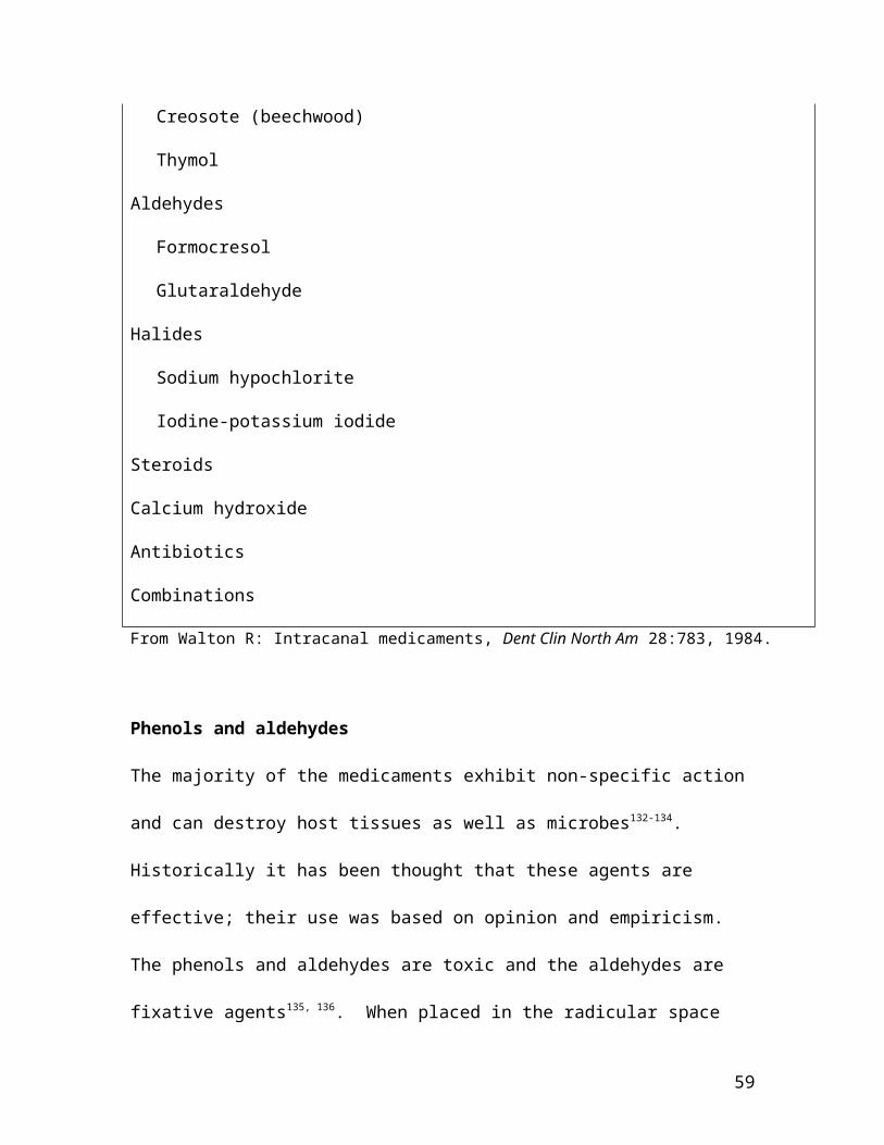
150. Safavi KE, Nichols FC. Effect of calcium hydroxide on bacterial lipopolysaccharide. J Endod 19(2):76-8, 1993.151. Yang SF, Rivera EM, Baumgardner KR, Walton RE, Stanford C. Anaerobic tissue-dissolving abilities of calcium hydroxide and sodium hypochlorite. J Endod 21(12):613-6, 1995.152. Walton RE, Holton IF, Jr., Michelich R. Calcium hydroxide as an intracanal medication: effect on posttreatment pain. J Endod 29(10):627-9, 2003.153. Rivera EM, Williams K. Placement of calcium hydroxide in simulated canals: comparison of glycerin versus water. J Endod 20(9):445-8, 1994.154. Sigurdsson A, Stancill R, Madison S. Intracanal placement of Ca(OH)2: a comparison of techniques. J Endod 18(8):367-70, 1992.155. Torres CP, Apicella MJ, Yancich PP, Parker MH. Intracanal placement of calcium hydroxide: a comparison of techniques, revisited. Journal of Endodontics 30(4):225-7, 2004.156. Lambrianidis T, Kosti E, Boutsioukis C, Mazinis M. Removal efficacy of various calcium hydroxide/chlorhexidine medicaments from the root canal. Int Endod J 39(1):55-61, 2006.157. Ehrmann EH, Messer HH, Adams GG. The relationship of intracanal medicaments to postoperative pain in endodontics. Int Endod J 36(12):868-75, 2003.158. Chance K, Lin L, Shovlin FE, Skribner J. Clinical trial of intracanal corticosteroid in root canal therapy. J Endod 13(9):466-8, 1987.159. Chance KB, Lin L, Skribner JE. Corticosteroid use in acute apical periodontitis: a review with clinical implications. Clin Prev Dent 10(1):7-10, 1988.160. Dametto FR, Ferraz CC, de Almeida Gomes BP, Zaia AA, Teixeira FB, de Souza-Filho FJ. In vitro assessment of the immediate and prolonged antimicrobial action of chlorhexidine gel as an endodontic irrigant against Enterococcus faecalis. Oral Surg Oral Med Oral Pathol Oral Radiol Endod 99(6):768-72, 2005.161. Dammaschke T, Schneider U, Stratmann U, Yoo JM, Schafer E. Effect of root canal dressings on the regeneration of inflamed periapical tissue. Acta Odontol Scand 63(3):143-52, 2005.162. Gomes BP, Vianna ME, Sena NT, Zaia AA, Ferraz CC, de Souza Filho FJ. In vitro evaluation of the antimicrobial activity of calcium hydroxide combined with chlorhexidine gel used as intracanal medicament. Oral Surg Oral Med Oral Pathol Oral Radiol Endod 102(4):544-50, 2006.163. De Rossi A, Silva LA, Leonardo MR, Rocha LB, Rossi MA. Effect of rotary or manual instrumentation, with or without a calcium hydroxide/1% chlorhexidine intracanal dressing, on the healing of experimentally induced chronic periapical lesions. Oral Surg Oral Med Oral Pathol Oral Radiol Endod 99(5):628-36, 2005.164. Naoum HJ, Chandler NP. Temporization for endodontics. International Endodontic Journal 35(12):964-78, 2002.165. Barthel CR, Zimmer S, Wussogk R, Roulet JF. Long-Term bacterial leakage along obturated roots restored with temporary and adhesive fillings. J Endod 27(9):559-62, 2001.166. Zmener O, Banegas G, Pameijer CH. Coronal microleakage of three temporary restorative materials: an in vitro study. J Endod 30(8):582-4, 2004.
59
167. Mayer T, Eickholz P. Microleakage of temporary restorations after thermocycling and mechanical loading. J Endod 23(5):320-2, 1997.168. Beach CW, Calhoun JC, Bramwell JD, Hutter JW, Miller GA. Clinical evaluation of bacterial leakage of endodontic temporary filling materials. J Endod 22(9):459-62, 1996.169. Vail MM, Steffel CL. Preference of temporary restorations and spacers: a survey of Diplomates of the American Board of Endodontists. J Endod 32(6):513-5, 2006.170. Newcomb BE, Clark SJ, Eleazer PD. Degradation of the sealing properties of a zinc oxide-calcium sulfate-based temporary filling material by entrapped cotton fibers. Journal of Endodontics 27(12):789-90, 2001.171. Pane ES, Palamara JE, Messer HH. Stainless steel bands in endodontics: effects on cuspal flexure and fracture resistance. Int Endod J 35(5):467-71, 2002.172. Gutmann JL. The dentin-root complex: anatomic and biologic considerations in restoring endodontically treated teeth. Journal of Prosthetic Dentistry 67(4):458-67, 1992.
60