clinicalstudydownloads.hindawi.com/journals/tswj/2018/6425857.pdf · include renal failure,...
TRANSCRIPT
Clinical StudyA Pilot Study of Short-Duration Hyperbaric OxygenTherapy to Improve HbA1c, Leukocyte, and Serum Creatinine inPatients with Diabetic Foot Ulcer Wagner 3-4
Hendry Irawan ,1 I. Nyoman Semadi,2 and I. Gde RakaWidiana3
1Surgery Department, Faculty of Medicine, Udayana University, Sanglah General Hospital, Denpasar, Indonesia2Thorax and Cardiovascular Surgery Division, Surgery Department, Faculty of Medicine, Udayana University,Sanglah General Hospital, Denpasar, Indonesia3Nephrology and Hypertension Division, Internal Medicine Department, Faculty of Medicine, Udayana University,Sanglah General Hospital, Denpasar, Indonesia
Correspondence should be addressed to Hendry Irawan; hendry [email protected]
Received 10 April 2018; Accepted 9 July 2018; Published 12 August 2018
Academic Editor: Juei-Tang Cheng
Copyright © 2018 Hendry Irawan et al.This is an open access article distributed under the Creative Commons Attribution License,which permits unrestricted use, distribution, and reproduction in any medium, provided the original work is properly cited.
Objective. To evaluate the short-duration hyperbaric oxygen therapy (HBOT) can improve HbA1c levels, leukocyte count, andserum creatinine levels in patients with diabetic foot ulcer (DFU) Wagner 3-4. Methods. Blood samples from all DFU patients atSanglah General Hospital, Denpasar, were taken for HbA1c, leukocyte, and serum creatinine test before debridement procedure,and the patients were then grouped into either standard therapy or standard therapy with HBOT for 10 sessions (combinationtherapy). At the end of therapy, all blood tests were resumed. Results. Each group consisted of 15 patients. Results of laboratoryanalysis before and after treatment were significant regarding decrease of HbA1c levels in standard therapy (10.98 ± 2.37 % to 9.70± 2.46 %; p = 0.006), HbA1c levels in combination therapy (9.42 ± 1.96 % to 7.07 ± 1.16 %; p < 0.001), and leukocyte count incombination therapy (13.97 ± 6.24 x 10³ cells/𝜇L to 8.84 ± 2.88 x 10³ cells/𝜇L; p = 0.009). The HbA1c levels at the end of therapywere significantly different between groups (p = 0.001). Serum creatinine level was decreased only in combination therapy butit was not significant. The effect size of all variables was larger in the combination therapy, but it was not significant (p > 0.05).Conclusion. The use of short-duration HBOT on DFU reduces HbA1c levels, leukocyte count, and serum creatinine levels betterthan standard therapy alone.This protocol would save time and effort in future HBOT implementation.This trial is registered withClinicalTrials.gov Identifier: NCT03615755.
1. Introduction
Diabetes mellitus (DM) is one of the causes of public healthproblems which have a worldwide influence due to its highprevalence and large economic and social consequences. TheInternationalDiabetes Federation (IDF) states thatmore than371 million people in the world between the ages of 20 and79 years have DM, while Indonesia is the seventh countrywith the highest prevalence of DM under China, India, USA,Brazil, Russia, and Mexico [1]. Complications of DM caninclude renal failure, diabetic foot ulcers, and even lowerlimb amputations. Diabetic patients with diabetic foot ulcers(DFUs) require long-termwound care, resulting in social and
economic consequences [2]. Diabetic patients are estimatedto be 15-25 % with DFUs; 40-80 % of DFU patients havean infection risk [3] and 10-20 % of DFU patients requireamputation [4].
The principle purpose of DFUs is wound healing. Themain components of standard therapy are controlling bloodsugar, antibiotic drug, ulcer debridement, wound care,offloading, and improved blood flow/revascularization [5,6].
In addition to standard therapies, there are adjuvant ther-apies such as hyperbaric oxygen therapy (HBOT), maggottherapy, growth factor therapy, collagen products, bioengi-neered tissue, and stem cells, also used in the management of
Hindawie Scientific World JournalVolume 2018, Article ID 6425857, 6 pageshttps://doi.org/10.1155/2018/6425857
2 The Scientific World Journal
DFUs [5, 7–9]. Hyperbaric oxygen therapy is a 100 % oxygendelivery with a pressure of 2 to 3 atmosphere absolute (ATA)in the hyperbaric chamber. The mechanism of HBOT is toincrease tissue oxygen levels resulting in accelerated woundhealing, decreased edema, and killing anaerobic bacteria [10,11].
Based on previous research, we want to learn more aboutHBOT and its role in not only accelerating wound healingas a clinical outcome, but also improving the hematologicaland biochemical conditions in patient with DFU. The aim ofthis study was to evaluate that the short-duration HBOT canimprove glycohemoglobin (HbA1c) levels, leukocyte count,and serum creatinine levels in patients with DFU Wagner 3-4.
2. Methods
This study uses pretest and posttest control group design,to know the role of standard therapy and combinationtherapy (standard therapy with adjuvant HBOT) to decreaseHbA1c levels, leukocyte count, and serum creatinine levelsin DFU patient Wagner 3-4. All DM patients with DFU atSanglah General Hospital, Denpasar, who meet the inclusionand exclusion criteria and willing to follow the researchprocedure were included in the study. All patients aresigning the agreement paper after getting research explana-tion. All patients were briefed on the study research usingHBOT. If they are willing to participate in the study anduse HBOT, they were grouped to combination therapy,and if they are willing to participate in the study but donot want to use HBOT, they were grouped to standardtherapy, but if they are not willing participate, then theywere excluded. The study was approved by InstitutionalReview Board of Medical Faculty of Udayana University andSanglah General Hospital Denpasar with the ethical number580/UN.14.2/KEP/2016.
Thirty diabetic patients with DFU Wagner 3-4 partici-pated in this study. Blood test was taken by all patients forHbA1c levels, leukocyte count, and serum creatinine levelsbefore debridement, and theywere then grouped for standardtherapy or standard therapy with 10 sessions of HBOT. Onesession of HBOT uses oxygen at 2.4 ATA for 90 minutesper day at multiplace hyperbaric chamber. This therapy isgiven as five sessions per week, so it takes two weeks. At theend of therapy, all blood tests were performed again in bothgroups.
The inclusion criteria were patients who had type 2diabetes and DFU Wagner class 3 or 4, aged over 18 years,and underwent debridement with or without toe amputation.The exclusion criteria were patients who had severe organsdysfunction such as heart failure, pulmonary infection,pneumothorax, chronic obstructive pulmonary disease, andstroke.
Statistical analysis using SPSS 17.0 (SPSS Inc., Chicago,Illinois, USA) was conducted. All variables were describedbefore and after treatment. Analysis pretest and posttestvalues on both groups were performed using paired t-test andindependent t-test.The statistical test results are significant ifp < 0.05.
3. Results
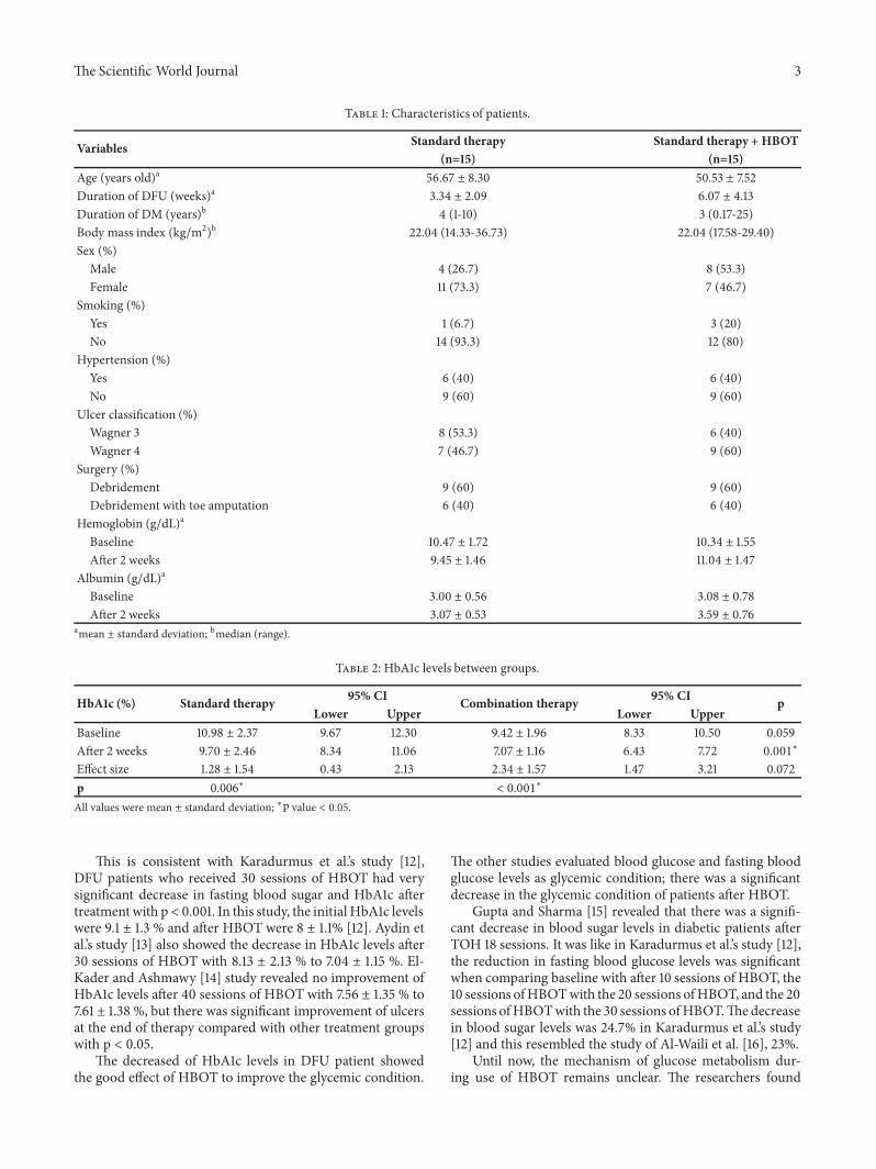
All patients were treated with insulin as antidiabeticand intravenous empiric antibiotics. We used combinationshort-acting and long-acting insulin daily: a dose of short-acting (NovoRapid�, insulin aspart) 8 units every 8 hours andlong-acting (Lantus�, insulin glargine) 10 units at night. Wegrouped the patients into two groups, each group consisted of15 patients (Table 1), with the mean age of standard therapygroup 56.67 ± 8.30 years and combination therapy group50.53 ± 7.52 years. Patients with the combination therapygroup experienced longer DFUs, 6.07 ± 4.13 weeks, thanstandard therapy group, 3.34 ± 2.09 weeks.
Analyses of HbA1c levels, leukocyte count, and serumcreatinine levels were described in Tables 2, 3, and 4.Comparison of baseline HbA1c levels between groups wasnot different with p = 0.059, but after two weeks there wassignificant difference between groups, p = 0.001 (Table 2).Evaluation pre- and posttherapy of HbA1c was significant inboth groups, in standard therapy group (from 10.98 ± 2.37 %to 9.70± 2.46%; p = 0.006) and in combination therapy group(from 9.42 ± 1.96 % to 7.07 ± 1.16 %; p < 0.001).
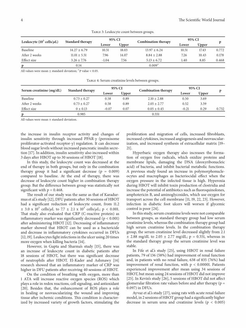
Leukocyte count at baseline and the end of therapybetween groups (Table 3) was not significant (p = 0.772 andp = 0.178, respectively), although leukocyte count after twoweeks in combination therapy group decreased higher thanin standard therapy group. Leukocyte count in combinationtherapy group was significantly decreased from 13.97 ± 6.24x 10³ cells/𝜇L to 8.84 ± 2.88 x 10³ cells/𝜇L; p = 0.009. In thisstudy, the effect size of HbA1c and leukocyte count was notsignificantly different. At the end of therapy, leukocyte countbetween debridement and debridement with amputation wasnot significantly different. In combination therapy, patientswho did debridement had leukocyte count 8.63 ± 3.04 x 10³cells/𝜇L compared with debridement with toe amputation9.17 ± 2.84 x 10³ cells/𝜇L. Compared with standard therapy,the leukocyte count was higher in debridement and debride-ment with amputation (11.2 ± 6.30 x 10³ cells/𝜇L, 10.74 ± 4.62x 10³ cells/𝜇L, respectively).
In Table 4, serum creatinine levels at baseline and aftertwo weeks of were not comparable because, in standardtherapy group, the patients had good renal function, whereas,in combination therapy group, they had impaired renalfunction. In standard therapy group, serum creatinine levelswere stable, but serum creatinine levels were little decreasedin combination therapy group.The effect size was not signifi-cantly decreased in serumcreatinine level (p = 0.732) betweengroups.
4. Discussion
In this study, we found that HbA1c levels were significantlydecreased after therapy in the standard therapy group (p =0.006) and in the combination therapy group (p < 0.001)compared to baseline levels. The HbA1c levels at the endof therapy were significantly different between groups (p =0.001), whereas the difference of HbA1c was greater in thecombination therapy group, but statistically not significant (p= 0.072).
The Scientific World Journal 3
Table 1: Characteristics of patients.
Variables Standard therapy Standard therapy + HBOT(n=15) (n=15)
Age (years old)a 56.67 ± 8.30 50.53 ± 7.52Duration of DFU (weeks)a 3.34 ± 2.09 6.07 ± 4.13Duration of DM (years)b 4 (1-10) 3 (0.17-25)Body mass index (kg/m2)b 22.04 (14.33-36.73) 22.04 (17.58-29.40)Sex (%)
Male 4 (26.7) 8 (53.3)Female 11 (73.3) 7 (46.7)
Smoking (%)Yes 1 (6.7) 3 (20)No 14 (93.3) 12 (80)
Hypertension (%)Yes 6 (40) 6 (40)No 9 (60) 9 (60)
Ulcer classification (%)Wagner 3 8 (53.3) 6 (40)Wagner 4 7 (46.7) 9 (60)
Surgery (%)Debridement 9 (60) 9 (60)Debridement with toe amputation 6 (40) 6 (40)
Hemoglobin (g/dL)a
Baseline 10.47 ± 1.72 10.34 ± 1.55After 2 weeks 9.45 ± 1.46 11.04 ± 1.47
Albumin (g/dL)a
Baseline 3.00 ± 0.56 3.08 ± 0.78After 2 weeks 3.07 ± 0.53 3.59 ± 0.76
amean ± standard deviation; bmedian (range).
Table 2: HbA1c levels between groups.
HbA1c (%) Standard therapy 95% CI Combination therapy 95% CI pLower Upper Lower Upper
Baseline 10.98 ± 2.37 9.67 12.30 9.42 ± 1.96 8.33 10.50 0.059After 2 weeks 9.70 ± 2.46 8.34 11.06 7.07 ± 1.16 6.43 7.72 0.001∗
Effect size 1.28 ± 1.54 0.43 2.13 2.34 ± 1.57 1.47 3.21 0.072p 0.006∗ < 0.001∗
All values were mean ± standard deviation; ∗p value < 0.05.
This is consistent with Karadurmus et al.’s study [12],DFU patients who received 30 sessions of HBOT had verysignificant decrease in fasting blood sugar and HbA1c aftertreatmentwith p < 0.001. In this study, the initial HbA1c levelswere 9.1 ± 1.3 % and after HBOT were 8 ± 1.1% [12]. Aydin etal.’s study [13] also showed the decrease in HbA1c levels after30 sessions of HBOT with 8.13 ± 2.13 % to 7.04 ± 1.15 %. El-Kader and Ashmawy [14] study revealed no improvement ofHbA1c levels after 40 sessions of HBOT with 7.56 ± 1.35 % to7.61 ± 1.38 %, but there was significant improvement of ulcersat the end of therapy compared with other treatment groupswith p < 0.05.
The decreased of HbA1c levels in DFU patient showedthe good effect of HBOT to improve the glycemic condition.
The other studies evaluated blood glucose and fasting bloodglucose levels as glycemic condition; there was a significantdecrease in the glycemic condition of patients after HBOT.
Gupta and Sharma [15] revealed that there was a signifi-cant decrease in blood sugar levels in diabetic patients afterTOH 18 sessions. It was like in Karadurmus et al.’s study [12],the reduction in fasting blood glucose levels was significantwhen comparing baseline with after 10 sessions of HBOT, the10 sessions ofHBOTwith the 20 sessions ofHBOT, and the 20sessions ofHBOTwith the 30 sessions ofHBOT.Thedecreasein blood sugar levels was 24.7% in Karadurmus et al.’s study[12] and this resembled the study of Al-Waili et al. [16], 23%.
Until now, the mechanism of glucose metabolism dur-ing use of HBOT remains unclear. The researchers found
4 The Scientific World Journal
Table 3: Leukocyte count between groups.
Leukocyte (103 cells/𝜇L) Standard therapy 95% CI Combination therapy 95% CI pLower Upper Lower Upper
Baseline 14.27 ± 6.79 10.51 18.03 13.97 ± 6.24 10.51 17.43 0.772After 2 weeks 11.01 ± 5.51 7.96 14.07 8.84 ± 2.88 7.26 10.43 0.178Effect size 3.26 ± 7.76 -1.04 7.56 5.13 ± 6.72 1.40 8.85 0.468p 0.14 0.009∗
All values were mean ± standard deviation; ∗p value < 0.05.
Table 4: Serum creatinine levels between groups.
Serum creatinine (mg/dL) Standard therapy 95% CI Combination therapy 95% CI pLower Upper Lower Upper
Baseline 0.73 ± 0.27 0.58 0.89 2.10 ± 2.88 0.50 3.69After 2 weeks 0.73 ± 0.27 0.58 0.89 2.05 ± 2.77 0.52 3.59Effect size 0 ± 0.13 -0.07 0.07 0.05 ± 0.45 -0.21 0.29 0.732p 0.985 0.551All values were mean ± standard deviation.
the increase in insulin receptor activity and changes ofinsulin sensitivity through increased PPAR-𝛾 (peroxisomeproliferator-activated receptor-𝛾) regulation. It can decreaseblood sugar levels without increased pancreatic insulin secre-tion [17]. In addition, insulin sensitivity also increased within3 days after HBOT up to 30 sessions of HBOT [18].
In this study, the leukocyte count was decreased at theend of therapy in both groups, but only in the combinationtherapy group it had a significant decrease (p = 0.009)compared to baseline. At the end of therapy, there wasdecrease of leukocyte count higher in combination therapygroup. But the difference between group was statistically notsignificant with p = 0.468.
The result of our study was the same as that of Karadur-mus et al.’s study [12], DFUpatients after 30 sessions of HBOThad a significant reduction of leukocyte count, from 11.2± 3.0 x 10³ cells/𝜇L to 7.7 ± 2.1 x 10³ cells/𝜇L; p < 0.001.That study also evaluated that CRP (C-reactive protein) asinflammatory marker was significantly decreased (p < 0.001)after administering HBOT [12]. Decreasing of inflammatorymarker showed that HBOT can be used as a bactericideand decrease in inflammatory cytokines occurred in DFUs[12, 19]. Leukocytes fight infections in the ulcer using 20 timesmore oxygen when killing bacteria [14].
However, in Gupta and Sharma’s study [15], there wasan increase of leukocyte count in diabetic patients after18 sessions of HBOT, but there was significant decreaseof neutrophils after HBOT. El-Kader and Ashmawy [14]research showed that an inflammatory marker of CRP washigher in DFU patients after receiving 40 sessions of HBOT.
On the condition of breathing with oxygen, more than1 ATA will increase reactive oxygen species (ROS) whichplays a role in redox reactions, cell signaling, and antioxidant[20]. Besides that, the enhancement of ROS plays a rolein healing or neovascularizing the wound and improvingtissue after ischemic conditions. This condition is character-ized by increased variety of growth factors, stimulating the
proliferation and migration of cells, increased fibroblasts,increased cytokines, increased angiogenesis and neovascular-ization, and increased synthesis of extracellular matrix [19–21].
Hyperbaric oxygen therapy also increases the forma-tion of oxygen free radicals, which oxidize proteins andmembrane lipids, damaging the DNA (deoxyribonucleicacid) of bacteria, and inhibit bacterial metabolic functions.A previous study found an increase in polymorphonucle-ocytes and macrophages as bacteriocidal effect when theoxygen pressure in the infected tissue is high. Hyperoxiaduring HBOT will inhibit toxin production of clostridia andincrease the potential of antibiotics such as fluoroquinolones,amphotericin B, and aminoglycosides, which use oxygen fortransport across the cell membrane [11, 19, 22, 23]. However,infection in diabetic foot ulcers will worsen if glycemiccontrol is poor [24].
In this study, serum creatinine levels were not comparablebetween groups, as standard therapy group had low serumcreatinine levels, whereas the combination therapy group hadhigh serum creatinine levels. In the combination therapygroup, the serum creatinine level decreased slightly from 2.1± 2.88 mg/dL to 2.05 ± 2.77 mg/dL; p = 0.551, whereas inthe standard therapy group the serum creatinine level wasstable.
In Fife et al.’s study [25], using HBOT in renal failurepatients, 79 of 136 (58%) had improvement of renal functionand, in patients with no renal failure, 638 of 835 (76%) hadimprovement of renal function, with p < 0.00001. Patientsexperienced improvement after mean using 34 sessions ofHBOT, but mean using 24 sessions of HBOTdid not improve[25]. In Kevin’s study [26], 5 sessions of HBOT did not affectglomerular filtration rate values before and after therapy (p =0.097) in DFUs.
Ayvaz et al.’s study [27], using rats with acute renal failuremodel, in 2 sessions ofHBOTgroup had a significantly higherdecrease in serum urea and creatinine levels (p < 0.005)
The Scientific World Journal 5
than the non-HBOT group. In addition, histopathologicalexamination showed that HBOT group decreased necrosiscells, decreased cylinder/cast formation, and decreased apop-totic cells [28].This was similar to Solmazgul et al. [29], usingrats model of renal reperfusion/ischemia, the assessment ofrenal function with serum urea and serum creatinine levelswas significantly higher than in healthy rats (p < 0.05). Ingroups of rats undergoing renal reperfusion/ischemia givenHBOT, therewas a significant decrease in serumurea and cre-atinine levels (p < 0.05) compared to the non-HBOT group.The HBOT effects can reduce the occurrence of damage torenal tissue and reduce infiltration neutrophils that play animportant role in renal injury [29]. In Migita et al. [28], theeffect of HBOT may decrease apoptotic cells by increasingthe proliferative reaction after renal reperfusion/ischemia inrats.
Rubinstein et al.’s study [30] showed an improvement inglomerular filtration rate in the groupwho received 2 sessionsof HBOT in 24 hours after renal ischemia, at 2 hours and 22hours. In renal ischemiawithoutHBOT, therewas decrease inglomerular filtration rate of 94% compared to healthy renal,and in renal ischemia with HBOT there was decrease inglomerular filtration rate of 68% compared to healthy renalwith significant difference (p < 0.05). In this study, HBOTimproved renal vasodilation associated with increased bloodflow to the renal, thus improving renal cortical perfusion[30]. Berkovitch et al.’s study [31], using healthy rats givenHBOT did not cause impaired renal function, which wasassessed with normal serum urea and serum creatinine levels[31].
In conditions of hyperglycemia, glycation of variousproteins occurs and collagen formed advanced glycationend-products (AGEs). This will lead to damage to thebody, such as vascular disorders, platelet activation andaggregation, increased free radicals and inflammation, andimmune system impairment. Increased AGEs will leadto complications of DM patients such as nephropathy[32].
Evaluation of the short-duration HBOT on improvingrenal function in humans has not shown significant results,including in our study. However, in animal experiments,HBOT has an effect of improving renal function. Furtherresearch can be done to evaluate influence of HBOT in DFUpatients with larger sample, control group uniformity andtreatment, and other assessed variables, including biomolec-ular markers.
5. Conclusion
Hyperbaric oxygen therapy of 2.4 ATA for 90 minutes perday for 10 sessions has the benefit of decreasing HbA1clevels and leukocyte count of patients with DFU Wagner3-4. However, there is little effect of HBOT on serumcreatinine levels as a sign of improved kidney function.This shows that HBOT can be used as a therapy that helpsstandard therapy in handling DFU in reducing glycemic andinflammatory levels. The use of short-duration HBOT onDFU would save time and effort in future HBOT implemen-tation.
Data Availability
The data used to support the findings of this study areavailable from the corresponding author upon request.
Conflicts of Interest
All the authors have no conflicts of interest.
Authors’ Contributions
Hendry Irawan contributed to study design, conductingresearch, data analysis, interpretation of findings, and draft-ing of the manuscript. I. Nyoman Semadi contributed tothe study design, surgical debridement with or without toeamputation, interpretation of findings, and drafting of themanuscript. I. Gde Raka Widiana contributed to statisti-cal analysis, interpretation of findings, and drafting of themanuscript.
Acknowledgments
The authors would like to thank the many investigators andpatients for their participation, without which this studywould not have been possible.
References
[1] “Kementerian Kesehatan Republik Indonesia. Diabetes MelitusPenyebab Kematian Nomor 6 di Dunia: Kemenkes TawarkanSolusi Cerdik Melalui Posbind,” 2013, http://www.depkes.go.id/article/print/2383/diabetes-melitus-penyebab-kematian-nom-or-6-di-dunia-kemenkes-tawarkan-solusi-cerdik-melalui-pos-bindu.html.
[2] WorldHealth Organization,Global status report on noncommu-nicable diseases 2010, WHO Press,, World Health, Switzerland,2011.
[3] “Revision de la IVe Conference de consensus en therapeutiqueanti-infectieuse de la Societe de pathologie infectieuse de languefrancaise (SPILF).,”Medecine etMaladies Infectieuses, vol. 31, no.5, pp. 265-266, 2001.
[4] World Health Organization, Global Report on Diabetes, WHOPress, Switzerland, 2016.
[5] R. G. Frykberg, T. Zgonis, D. G. Armstrong et al., “Diabetic footdisorders. A clinical practice guideline (2006 revision),” Journalof Foot and Ankle Surgery, vol. 45, no. 5, pp. S1–S66, 2006.
[6] Wounds International. International Best Practice Guidelines:WoundManagement inDiabetic FootUlcers. London:WoundsInternational A division of Schofield Healthcare Media LimitedEnterprise House; 2013.
[7] L. Kessler, P. Bilbault, F. Ortega et al., “Hyperbaric oxygenationaccelerates the healing rate of nonischemic chronic diabetic footulcers a prospective randomized study,” Diabetes Care, vol. 26,no. 8, pp. 2378–2382, 2003.
[8] D. Waniczek, A. Kozowicz, M. Muc-Wierzgon, T. Kokot, E.Swiętochowska, and E. Nowakowska-Zajdel, “AdjunctMethodsof the Standard Diabetic Foot Ulceration Therapy,” Evidence-Based Complementary and Alternative Medicine, vol. 2013,Article ID 243568, 12 pages, 2013.
6 The Scientific World Journal
[9] W. J. Jeffcoate andF. L. Game, “Evidence for theUse of biologicaltherapies in ulcers of the foot in diabetes,” BioDrugs, vol. 28, no.1, pp. 1–6, 2014.
[10] M. S. Flood, “Hyperbaric Oxygen Therapy for diabetic FootUlcers,”The Journal of Lancaster General Hospital, pp. 140–145,2007.
[11] S. Bhutani and G. Vishwanath, “Hyperbaric oxygen and woundhealing,” Indian Journal of Plastic Surgery, vol. 45, no. 2, pp. 316–324, 2012.
[12] N. Karadurmus, M. Sahin, C. Tasci et al., “Potential benefitsof hyperbaric oxygen therapy on atherosclerosis and glycaemiccontrol in patients with diabetic foot,” Endokrynologia Polska,vol. 61, no. 3, pp. 275–279, 2010.
[13] F. Aydin, A. Kaya, L. Karapinar et al., “IGF-1 increases withhyperbaric oxygen therapy and promotes wound healing indiabetic foot ulcers,” Journal of Diabetes Research, vol. 2013,Article ID 567834, 2013.
[14] S. M. A. El-Kader and E. M. Ashmawy, “Impact of DifferentTherapeuticModalities onHealing ofDiabetic FootUlcers,”EurJ Gen Med, vol. 12, pp. 319–325, 2015.
[15] S. K. Gupta and A. K. Sharma, “Effects of hyperbaric oxygentherapy on haematological and biochemical parameters,” Ind JAerospace Med, vol. 44, pp. 1–5, 2000.
[16] N. S. Al-Waili, G. J. Butler, J. Beale et al., “Influences of hyper-baric oxygen on blood pressure, heart rate and blood glucoselevels in patients with diabetes mellitus and hypertension,”Archives of Medical Research, vol. 37, no. 8, pp. 991–997, 2006.
[17] T. S. Nwafor andN. Collins, “Managing low blood glucose levelsin patients undergoing hyperbaric oxygen therapy,” OstomyWound Management, vol. 60, no. 4, pp. 12–15, 2014.
[18] D. Wilkinson, I. M. Chapman, and L. K. Heilbronn, “Hyper-baric oxygen therapy improves peripheral insulin sensitivity inhumans,” Diabetic Medicine, vol. 29, no. 8, pp. 986–989, 2012.
[19] H. Irawan and Kartika, “Terapi Oksigen Hiperbarik sebagaiTerapi Adjuvan Kaki Diabetik,” Cermin Dunia Kedokteran-245,vol. 43, pp. 782–785, 2016.
[20] S. R. Thom, “Hyperbaric oxygen: its mechanisms and efficacy,”Plastic and Reconstructive Surgery, vol. 127, no. 1, pp. 131S–141S,2011.
[21] K. C. Klein and S. C. Guha, “Cutaneouswound healing: Currentconcepts and advances in wound care,” Indian Journal of PlasticSurgery, vol. 47, no. 3, pp. 303–317, 2014.
[22] D. Mathieu, Handbook on Hyperbaric Medicine, Springer,Berlin, Germany, 2006.
[23] A. Wibowo, “Oksigen Hiperbarik: Terapi Percepatan Penyem-buhan Luka,” JuKe Unila, vol. 5, pp. 124–128, 2015.
[24] G.Uzun,M.Mutluoglu, and O.Uz, “Hyperbaric oxygen therapyin diabetic patients - comments on the paper by Karadurmus etal,” Endokrynologia Polska, vol. 62, pp. 286-287, 2011.
[25] C. E. Fife, C. Buyukcakir, G. Otto, P. Sheffield, T. Love, andR. Warriner III, “Factors influencing the outcome of lower-extremity diabetic ulcers treated with hyperbaric oxygen ther-apy,”Wound Repair and Regeneration, vol. 15, no. 3, pp. 322–331,2007.
[26] T. Kevin, Pengaruh Terapi Oksigen Hiperbarik Terhadap eGFRberdasarkan Formula MDRD pada pasien Luka Kaki Dia-betik (skripsi), Universitas Katolik Widya Mandala, Surabaya,Indonesia, 2015.
[27] S. Ayvaz, B. Aksu, M. Kanter et al., “Preventive effects ofhyperbaric oxygen treatment on glycerol-induced myoglobin-uric acute renal failure in rats,” Journal of Molecular Histology,vol. 43, no. 2, pp. 161–170, 2012.
[28] H. Migita, S. Yoshitake, Y. Tange, N. Choijookhuu, and Y.Hishikawa, “Hyperbaric oxygen therapy suppresses apoptosisand promotes renal tubular regeneration after renal ischemia/reperfusion injury in rats,” Nephro-Urology Monthly, vol. 8, no.1, Article ID e34421, 2016.
[29] E. Solmazgul, G. Uzun, H. Cermik, E. M. Atasoyu, S. Aydinoz,and S. Yildiz, “Hyperbaric oxygen therapy attenuates renalischemia/reperfusion injury in rats,” Urologia Internationalis,vol. 78, no. 1, pp. 82–85, 2007.
[30] I. Rubinstein, Z. Abassi, F. Milman et al., “Hyperbaric oxygentreatment improves GFR in rats with ischaemia/reperfusionrenal injury: a possible role for the antioxidant/oxidant balancein the ischaemic kidney,” Nephrology Dialysis Transplantation ,vol. 24, no. 2, pp. 428–436, 2009.
[31] M.Berkovitch, R. Tsadik, E.Kozer, and I.Abu-Kishk, “The effectof hyperbaric oxygen therapy on kidneys in a rat model,” TheScientific World Journal, vol. 2014, Article ID 105069, 2014.
[32] V. P. Singh, A. Bali, N. Singh, and A. S. Jaggi, “Advancedglycation end products and diabetic complications,” KoreanJournal of Physiology & Pharmacology, vol. 18, no. 1, pp. 1–14,2014.

Hindawiwww.hindawi.com
International Journal of
Volume 2018
Zoology
Hindawiwww.hindawi.com Volume 2018
Anatomy Research International
PeptidesInternational Journal of
Hindawiwww.hindawi.com Volume 2018
Hindawiwww.hindawi.com Volume 2018
Journal of Parasitology Research
GenomicsInternational Journal of
Hindawiwww.hindawi.com Volume 2018
Hindawi Publishing Corporation http://www.hindawi.com Volume 2013Hindawiwww.hindawi.com
The Scientific World Journal
Volume 2018
Hindawiwww.hindawi.com Volume 2018
BioinformaticsAdvances in
Marine BiologyJournal of
Hindawiwww.hindawi.com Volume 2018
Hindawiwww.hindawi.com Volume 2018
Neuroscience Journal
Hindawiwww.hindawi.com Volume 2018
BioMed Research International
Cell BiologyInternational Journal of
Hindawiwww.hindawi.com Volume 2018
Hindawiwww.hindawi.com Volume 2018
Biochemistry Research International
ArchaeaHindawiwww.hindawi.com Volume 2018
Hindawiwww.hindawi.com Volume 2018
Genetics Research International
Hindawiwww.hindawi.com Volume 2018
Advances in
Virolog y Stem Cells International
Hindawiwww.hindawi.com Volume 2018
Hindawiwww.hindawi.com Volume 2018
Enzyme Research
Hindawiwww.hindawi.com Volume 2018
International Journal of
MicrobiologyHindawiwww.hindawi.com
Nucleic AcidsJournal of
Volume 2018
Submit your manuscripts atwww.hindawi.com