circulating tumor cells in newly diagnosed inflammatory ... article open access circulating tumor...
TRANSCRIPT

Mego et al. Breast Cancer Research (2015) 17:2 DOI 10.1186/s13058-014-0507-6
RESEARCH ARTICLE Open Access
Circulating tumor cells in newly diagnosedinflammatory breast cancerMichal Mego1,5,9, Antonio Giordano1, Ugo De Giorgi1,6, Hiroko Masuda2, Limin Hsu2, Mario Giuliano1,7,Tamer M Fouad2, Shaheenah Dawood2,8, Naoto T Ueno2,4, Vicente Valero2,4, Eleni Andreopoulou2,4,Ricardo H Alvarez2,4, Wendy A Woodward3, Gabriel N Hortobagyi2, Massimo Cristofanilli2,9 and James M Reuben1,4*
Abstract
Introduction: Circulating tumor cells (CTCs) are an independent prognostic factor for progression-free survival (PFS)and overall survival (OS) in patients with metastatic breast cancer. Inflammatory breast cancer (IBC) is one of themost aggressive forms of breast cancer. The prognostic value of a CTC count in newly diagnosed IBC has not beenestablished. The aim of this study was to assess the prognostic value of a baseline CTC count in patients with newlydiagnosed IBC.
Methods: This retrospective study included 147 patients with newly diagnosed IBC (77 with locally advanced and70 with metastatic IBC) treated with neoadjuvant therapy or first-line chemotherapy during the period from January2004 through December 2012 at The University of Texas MD Anderson Cancer Center. CTCs were detected andenumerated by using the CellSearch system before patients were started with chemotherapy.
Results: The proportion of patients with ≥1 CTC was lower among patients with stage III than among patients withmetastatic IBC (54.5% versus 84.3%; P = 0.0002); the proportion of patients with ≥5 CTCs was also lower for stage IIIthan for metastatic IBC (19.5% versus 47.1%; P = 0.0004). Patients with fewer than five CTCs had significantly betterprogression-free survival (PFS) (hazard ratio (HR) = 0.60; P = 0.02) and overall survival (HR = 0.59; P = 0.03) than patientswith five or more CTCs. Among patients with stage III IBC, there was a nonsignificant difference in PFS (HR = 0.66; 95%confidence interval (CI), 0.31 to 1.39; P = 0.29) and OS (HR = 0.54; 95% CI, 0.24 to 1.26; P = 0.48) in patients with noCTCs compared with patients with one or more CTCs. In multivariate analysis, CTC was prognostic for PFS and OSindependent of clinical stage.
Conclusions: CTCs can be detected in a large proportion of patients with newly diagnosed IBC and are a strongpredictor of worse prognosis in patients with newly diagnosed IBC.
IntroductionInflammatory breast cancer (IBC) is one of the mostaggressive forms of primary breast cancer, and theincidence of IBC is increasing [1]. The prognosis ofpatients with IBC remains poor: the 10-year disease-freesurvival rate is only 20% to 25%, despite a multimodalitytreatment approach [2-7]. These reports suggest thatcurrent treatment modalities are inadequate and underscorethe need for better understanding of this disease.
* Correspondence: [email protected] of Hematopathology, The University of Texas MD AndersonCancer Center, 1515 Holcombe Blvd, Houston, TX 77030, USA4Morgan Welch Inflammatory Breast Cancer Research Program and Clinic,The University of Texas MD Anderson Cancer Center, Houston, TX, USAFull list of author information is available at the end of the article
© 2015 Mego et al.; licensee BioMed Central.Commons Attribution License (http://creativecreproduction in any medium, provided the orDedication waiver (http://creativecommons.orunless otherwise stated.
IBC is associated with special clinical and biologicalfeatures and a distinctive pattern of recurrence withhigh incidence of visceral metastases (central nervoussystem, lung, and liver) as first site of relapse [3-7]. Itis characterized by a high proliferation rate, frequenthormone-receptor negativity, HER2 overexpression,high grade, and increased tumor angiogenesis [7-11].Studies of several molecular factors in IBC suggestfrequent epidermal growth factor receptor overexpressionand high expression of p53, MUC1, RhoC, E-cadherin,and transcription factors associated with a stem cellphenotype [12-17].In patients with metastatic breast cancer, circulating
tumor cells (CTCs) are an independent predictor ofprogression-free survival (PFS) and overall survival
This is an Open Access article distributed under the terms of the Creativeommons.org/licenses/by/4.0), which permits unrestricted use, distribution, andiginal work is properly credited. The Creative Commons Public Domaing/publicdomain/zero/1.0/) applies to the data made available in this article,

Figure 1 Patients’ flow.
Mego et al. Breast Cancer Research (2015) 17:2 Page 2 of 12
(OS) [18]. In patients with metastatic disease, superiorsurvival was observed among patients with fewer thanfive CTCs per 7.5 ml of peripheral blood regardlessof histologic subtype, hormone receptor and HER2/neustatus, sites of first metastasis, or whether the patient hadrecurrent or de novo metastatic disease [18-21]. Theprognostic value of a CTC count was found to be superiorto that of tumor burden as measured by Swenerton scoreor by serum tumor markers, ascribing a peculiar biologicalvalue to CTCs. These observations also raised the possibil-ity that CTCs might represent a population of tumorigenic
cancer cells with stem cell properties that might play animportant role in tumor dissemination [22,23].Previously, we showed that among patients with
metastatic breast cancer (MBC) treated with first orsubsequent lines of chemotherapy, patients with meta-static IBC had lower CTC counts than did patientswith non-inflammatory metastatic breast cancer (non-IBC)[24]. We also showed, for patients with metastaticIBC, that differences in OS between patients withfewer than five CTCs and others with five or moreCTCs were not statistically significant; hence, the

Table 1 Patient characteristics and prevalence of circulating tumor cells at baseline (n = 147)
Stage III IBC Metastatic IBC
Characteristic N % ≥1 CTCa % ≥5 CTCa % N % ≥1 CTCa % ≥5 CTCa %
All patients 77 100.0 42 54.5 15 19.5 70 100.0 59 84.3 32 45.7
Histology
Infiltrative ductal carcinoma 74 96.1 40 54.1 15 20.3 66 94.3 55 83.3 31 47.0
Other histology 3 3.9 2 66.7 0 0.00 4 5.7 4 100.0 1 25.0
P value 1.00 0.61 0.61 0.62
ER/PR status
Positive for either 43 55.8 19 44.2 5 11.6 40 57.1 33 82.5 20 50.0
Negative for both 34 44.2 23 67.6 10 29.4 30 42.9 26 86.7 12 40.0
P value 0.06 0.08 0.74 0.47
HER-2/neu status
Overexpressed 26 33.8 17 65.4 7 26.9 19 27.1 13 68.4 9 47.4
Negative 51 66.2 25 49.0 8 15.7 51 72.9 46 90.2 23 45.1
P value 0.23 0.36 0.04 1.00
Grade
High grade 55 71.4 32 58.2 11 20.0 53 75.7 43 81.1 24 45.3
Intermediate/low grade 22 28.6 10 45.5 4 18.2 15 21.4 14 93.3 8 53.3
Unknown - - - - - - 2 2.9 2 - 0 -
P value 0.31 1.00 0.43 0.77
ER/PR and HER2/neu status
Triple receptor negative 19 24.7 12 63.2 5 26.3 19 27.1 17 89.5 7 36.8
Not-triple receptor negative 58 75.3 30 51.7 10 17.2 51 72.9 42 82.4 25 49.0
P value 0.44 0.50 0.71 0.43
Sites of metastases
Non-visceral - - - - - - 42 60.0 34 81.0 18 42.9
Visceral - - - - - - 28 40.0 25 89.3 14 50.0
P value 0.50 0.63
Bone metastasis
Present - - - - - - 39 55.7 32 82.1 19 48.7
Absent - - - - - - 31 44.3 27 87.1 13 41.9
P value 0.70 0.63
Number of metastases
1 - - - - - - 29 41.4 22 75.9 13 44.8
≥2 - - - - - - 41 58.6 37 90.2 19 46.3
P value 0.20 1.00
Statin use
No-statins 62 80.5 39 62.9 14 22.6 60 85.7 51 85.0 30 50.0
L-statinsb 7 9.1 1 14.3 1 14.3 5 7.1 5 100.0 1 20.0
H-statinsb 8 10.4 2 25.0 0 0.0 5 7.1 3 60.0 1 20.0
P value 0.01 0.30 0.20 0.21
Menopausal status
Premenopausal 27 35.1 19 70.4 9 33.3 25 35.7 21 84.0 13 52.0
Postmenopausal 50 64.9 23 46.0 6 12.0 45 64.3 38 84.4 19 42.2
P value 0.06 0.03 1.00 0.46
Mego et al. Breast Cancer Research (2015) 17:2 Page 3 of 12
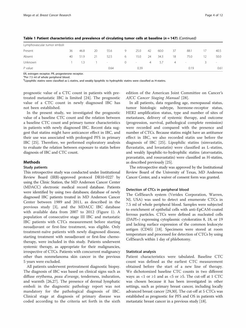
Table 1 Patient characteristics and prevalence of circulating tumor cells at baseline (n = 147) (Continued)
Lymphovascular tumor emboli
Present 36 46.8 20 55.6 9 25.0 42 60.0 37 88.1 17 40.5
Absent 40 51.9 21 52.5 6 15.0 24 34.3 18 75.0 12 50.0
Unknown 1 1.3 1 - 0 - 4 5.7 4 - 3 -
P value 0.64 0.39 0.19 0.61
ER, estrogen receptor; PR, progesterone receptor.aPer 7.5 ml of whole peripheral blood.bLipophilic statins were classified as L-statins, and weakly lipophilic to hydrophilic statins were classified as H-statins.
Mego et al. Breast Cancer Research (2015) 17:2 Page 4 of 12
prognostic value of a CTC count in patients with pre-treated metastatic IBC is limited [24]. The prognosticvalue of a CTC count in newly diagnosed IBC hasnot been established.In the present study, we investigated the prognostic
value of a baseline CTC count and the relation betweena baseline CTC count and primary tumor characteristicsin patients with newly diagnosed IBC. Recent data sug-gest that statins might have anticancer effect in IBC, andtheir use was associated with prolonged PFS in primaryIBC [25]. Therefore, we performed exploratory analysisto evaluate the relation between exposure to statin beforediagnosis of IBC and CTC count.
MethodsStudy patientsThis retrospective study was conducted under InstitutionalReview Board (IRB)-approved protocol DR10-0227 byusing the Clinic Station, the MD Anderson Cancer Center(MDACC) electronic medical record database. Patientswere identified by using two databases; database of newlydiagnosed IBC patients treated in MD Anderson CancerCenter between 1989 and 2011, as described in theprevious study [3], and the MDACC IBC databasewith available data from 2007 to 2012 (Figure 1). Apopulation of consecutive stage III IBC and metastaticIBC patients with CTCs measurement before startingneoadjuvant or first-line treatment, was eligible. Onlytreatment-naïve patients with newly diagnosed disease,starting treatment with neoadjuvant or first-line chemo-therapy, were included in this study. Patients underwentsystemic therapy, as appropriate for their malignancies,irrespective of CTCs. Patients with concurrent malignancyother than nonmelanoma skin cancer in the previous5 years were excluded.All patients underwent pretreatment diagnostic biopsy.
The diagnosis of IBC was based on clinical signs such asdiffuse erythema, peau d'orange, tenderness, induration,and warmth [26,27]. The presence of dermal lymphaticemboli in the diagnostic pathology report was notmandatory for the pathological diagnosis of IBC.Clinical stage at diagnosis of primary disease wascoded according to the criteria set forth in the sixth
edition of the American Joint Committee on Cancer’sAJCC Cancer Staging Manual [28].In all patients, data regarding age, menopausal status,
tumor histologic subtype, hormone-receptor status,HER2 amplification status, type and number of sites ofmetastases, delivery of systemic therapy, and outcome(progression, survival, pathological complete remission)were recorded and compared with the presence andnumber of CTCs. Because statins might have an antitumoreffect in IBC, we also recorded statin use before thediagnosis of IBC [25]. Lipophilic statins (simvastatin,fluvastatin, and lovastatin) were classified as L-statins,and weakly lipophilic-to-hydrophilic statins (atorvastatin,pravastatin, and rosuvastatin) were classified as H-statins,as described previously [25].The retrospective study was approved by the Institutional
Review Board of the University of Texas, MD AndersonCancer Center, and a waiver of consent form was granted.
Detection of CTCs in peripheral bloodThe CellSearch system (Veridex Corporation, Warren,NJ, USA) was used to detect and enumerate CTCs in7.5 ml of whole peripheral blood. Samples were subjectedto enrichment of epithelial cells with anti-EpCAM-coatedferrous particles. CTCs were defined as nucleated cells(DAPI+) expressing cytoplasmic cytokeratins 8, 18, or 19and lacking surface expression of the common leukocyteantigen (CD45) [18]. Specimens were stored at roomtemperature and processed for detection of CTCs by usingCellSearch within 1 day of phlebotomy.
Statistical analysisPatient characteristics were tabulated. Baseline CTCcount was defined as the earliest CTC measurementobtained before the start of a new line of therapy.We dichotomized baseline CTC counts in two differentways: as <1 or ≥1 and as <5 or ≥5. The cut-off at 1 CTCwas chosen because it has been investigated in othersettings, such as primary breast cancer, including locallyadvanced breast cancer [29,30]. The cut-off at 5 CTCs wasestablished as prognostic for PFS and OS in patients withmetastatic breast cancer in a previous study [18].

Figure 2 Kaplan-Meier estimates of probabilities of progression-free survival, according to baseline circulating tumor cell count inpatients with newly diagnosed inflammatory breast cancer.
Mego et al. Breast Cancer Research (2015) 17:2 Page 5 of 12
In an exploratory analysis, we correlated baseline CTCcounts with PFS and OS. Median follow-up period wascalculated as a median observation time among allpatients and among those still alive at the time oftheir last follow-up. PFS was calculated from the dateof baseline CTC enumeration to the date of progressionor death or the date of last adequate follow-up. OS wascalculated from the date of baseline CTC enumeration tothe date of death or last follow-up. PFS and OS wereestimated by using the Kaplan-Meier product-limitmethod and compared between groups by using thelog-rank test. Univariate analyses were performed witheither χ2 or Fisher Exact test, as appropriate. A multivari-ate Cox proportional hazards model for PFS and OS wasused to assess differences in outcome on the basis ofbaseline CTC counts, hormone-receptor status (positive foreither or negative for both), HER-2 status (overexpressed ornegative), stage (stage III versus metastatic IBC), site ofmetastasis (visceral versus non-visceral), and number ofmetastatic sites. Visceral metastases were defined here aslung, liver, adrenal gland, brain, kidney, pancreas, and/orperitoneal involvement with or without ascites and/orpleural effusions. Nonvisceral metastases were defined asinvolvement of any of the following sites without visceralmetastases: breast, lymph nodes, chest wall, bone, skin,and/or abdomen. Step-wise regression techniques were
used to build multivariate models by using a significancelevel of 0.10 to remain in the model. Wilcoxon matched-pairs signed-ranks test was used to compare baseline CTCcounts with CTC counts at the time of progression, andMann–Whitney U test was used to compare CTC countsbetween patients with stage III IBC and metastaticIBC. All statistical tests were two-sided, and P values <0.05were considered statistically significant.
ResultsPatient characteristicsA total of 147 patients with newly diagnosed IBC thatmatched the study eligibility criteria was included in thisanalysis. Of these 147 patients, 77 had locally advanced(stage IIIB and IIIC) and 70 had metastatic IBC. Thirteenpatients (8.7%) with newly diagnosed metastatic IBC(mIBC) in this analysis were also included in a previousreport [24]. The median age of the subjects was 54 years(range, 23 to 82 years). One hundred forty patients (95.2%)had invasive ductal carcinoma. Patients’ characteristics andthe prevalence of baseline CTCs are shown in Table 1.
Role of CTCs in IBCMedian baseline CTC count among the 147 patientsassessed for the presence of CTCs was 2 (range, 0 to 249)per 7.5 ml of peripheral blood (PB). Among the 147

Mego et al. Breast Cancer Research (2015) 17:2 Page 6 of 12
patients, a subset of 101 patients (68.7%) had at least oneCTC, whereas 48 patients (32.7%) had at least five CTCs.The median CTC counts in patients with stage III
and mIBC were 1 (range, 0 to 179) and 3 (range, 0to 249) (P < 0.0001), respectively. The proportion ofpatients with one or more CTCs was lower in patientswith stage III than in patients with mIBC (54.5% versus84.3%; P = 0.0002); the proportion of patients with five ormore CTCs was also lower for stage III than for mIBC(19.5% versus 47.1%; P = 0.0004). The proportion ofpatients with one or more and five or more CTCs washigher in premenopausal than in postmenopausal womenin stage III IBC but not in mIBC patients (Table 1), andthere was a trend to detect CTCs more often in patientswith than in patients without lymphovascular tumoremboli (74.4% versus 59.4%; P = 0.07).At a median follow-up time of 26.3 months (range, 1.0
to 92.4 months), 81 patients (55.1%) had experienceddisease progression, and 66 patients (44.9%) had died.Median follow-up of patients still alive was 35.6 (range,8.2 to 92.4 months). Patients with fewer than five CTCshad a significantly better PFS than patients with five ormore CTCs (median PFS, 26.4 versus 10.5 months;hazard ratio [HR] = 0.60; 95% CI, 0.37 to 0.98; P = 0.02)(Figure 2). Furthermore, patients with fewer than fiveCTCs had a significantly better OS than patients with five
Figure 3 Kaplan-Meier estimates of probabilities of overall survival, anewly diagnosed inflammatory breast cancer.
or more CTCs (median OS, 56.9 versus 32.7 months; HR= 0.59; 95% CI, 0.35 to 1.00; P = 0.03) (Figure 3). Similarly,with a cut-off of one CTC, patients with fewer than oneCTCs had a significantly better PFS (HR = 0.42; 95%CI, 0.27 to0.66; P = 0.001) and OS (HR = 0.35; 95% CI,0.21 to 0.58; P = 0.001) compared with patients withone or more 1CTCs.Tables 2 and 3 and Figures 4 and 5 summarize the
Kaplan-Meier PFS and OS estimates by CTC countand patient tumor characteristics for stage III IBCand metastatic IBC patients. CTC prognostic valuewas observed only in metastatic IBC patients, with athreshold of one CTC (Table 3). In multivariate analysisbaseline CTCs, HER2 status and stage of disease wereindependent prognostic factors for PFS, whereas base-line CTCs, hormone-receptor status, HER2 status,and visceral metastases were independent prognosticfactors for OS (Table 4). Same results were obtainedby using a cut-off of one or more CTCs (data notshown).
Relation of Statin and CTCs in IBCWe identified 25 patients (17%) that used statins beforethe diagnosis of IBC (Table 1). Interestingly, an inverseassociation was noted between the use of statins and thepresence of CTCs. The proportion of patients with ≥1
ccording to baseline circulating tumor cells count in patients with

Table 2 Kaplan-Meier progression-free survival (PFS) and overall survival estimates stratified by CTC level and patientand tumor characteristics in patients with newly diagnosed stage III inflammatory breast cancer
Progression-free survival Overall survival
Variable Hazard ratio Lower 95% CI Upper 95% CI P value Hazard ratio Lower 95% CI Upper 95% CI P value
All patients
<1 CTC versus ≥1 CTC 0.66 0.31 1.39 0.29 0.57 0.25 1.31 0.21
<5 CTC versus ≥5 CTC 0.69 0.26 1.78 0.39 0.72 0.26 2.00 0.48
ER/PR positive for either
<1 CTC versus ≥1 CTC 0.59 0.23 1.53 0.28 0.59 0.18 1.94 0.40
<5 CTC versus ≥5 CTC 0.33 0.06 1.70 0.04 0.33 0.05 2.18 0.08
ER/PR negative for both
<1 CTC versus ≥1 CTC 0.76 0.22 2.69 0.69 1.14 0.55 2.39 0.70
<5 CTC versus ≥5 CTC 1.14 0.31 4.14 0.85 1.05 0.28 3.91 0.94
HER-2/neu positive
<1 CTC versus ≥1 CTC 0.64 0.15 2.77 0.58 0.37 0.07 1.97 0.34
<5 CTC versus ≥5 CTC 1.25 0.27 5.71 0.79 0.97 0.18 5.33 0.97
HER-2/neu negative
<1 CTC versus ≥1 CTC 0.62 0.26 1.49 0.29 0.51 0.19 1.37 0.18
<5 CTC versus ≥5 CTC 0.42 0.11 1.59 0.08 0.51 0.13 2.05 0.23
Triple negative
<1 CTC versus ≥1 CTC 0.40 0.11 1.51 0.24 0.31 0.08 1.14 0.11
<5 CTC versus ≥5 CTC 0.56 0.11 2.71 0.39 0.60 0.13 2.82 0.45
High grade
<1 CTC versus ≥1 CTC 0.65 0.28 1.47 0.31 0.42 0.17 1.06 0.09
<5 CTC versus ≥5 CTC 0.56 0.19 1.69 0.22 0.44 0.13 1.46 0.09
Low/intermediate grade
<1 CTC versus ≥1 CTC 0.87 0.15 5.08 0.87 1.34 0.18 9.70 0.76
<5 CTC versus ≥5 CTC 1.44 0.20 10.50 0.74 NAa NAa NAa 0.19
ER, estrogen receptor; NR, not reached; PR, progesterone receptor; NA, not applicable.aNo events in patients with five or more CTCs.
Mego et al. Breast Cancer Research (2015) 17:2 Page 7 of 12
CTCs was lower in patients taking statins than in thosenot taking statins (44.0% versus 73.8%; P = 0.005); theproportion of patients with ≥5 CTCs was also lower inpatients taking statins (12.0% versus 36.9%; P = 0.02).This effect was more striking for patients using H-statinsthan for patients using L-statins.
Role of CTCs in Primary IBCIn nonmetastatic breast cancer patients, a cut-off at oneCTC is established based on previous trials [29-31],therefore, we analyzed the prognostic value of CTCs byusing this cut-off in stage III IBC patients as well.Among patients with stage III IBC, non-significantdifferences occurred in PFS (HR = 0.66; 95% CI, 0.31 to1.39; P = 0.29) and OS (HR = 0.54; 95% CI, 0.24 to 1.26;P = 0.48) in patients with no CTCs compared with thoseof patients with one or more CTCs (Figure 4). Inexploratory analysis, we evaluated the prognosticvalue of CTCs by using a cut-off at 5. Similarly to
these data, patients with fewer than 5 CTCs had anonsignificantly better PFS (HR = 0.69; 95% CI, 0.26to 1.78; P = 0.38) and OS (HR = 0.75; 95% CI, 0.27 to2.06; P = 0.53) than patients with five or more CTCs.Of the 77 patients with stage III IBC, 15 (19.2%)
achieved a pathologic complete response (pCR) aftertreatment with neoadjuvant chemotherapy; however,no correlation was found between baseline CTCs countand pCR. Table 5 shows association between CTCs andpCR in stage III IBC patients.
DiscussionTo our knowledge, this is the first study to assessthe prognostic value of CTCs in patients with newlydiagnosed IBC. This study indicates that baselineCTCs, as enumerated by the CellSearch technology,are prognostic for PFS and OS in patients with newlydiagnosed mIBC. The proportion of patients with abaseline CTC count of ≥1 in stage III and in mIBC

Table 3 Kaplan-Meier progression-free survival (PFS) and overall survival (OS) estimates stratified by CTC level and pa-tient and tumor characteristics in patients with newly diagnosed metastatic IBC
Progression-free survival Overall survival
Variable Hazard ratio Lower 95% CI Upper 95% CI P value Hazard ratio Lower 95% CI Upper 95% CI P value
All patients
<1 CTC vs. ≥ 1 CTC 0.40 0.21 0.75 0.03 0.29 0.14 0.62 0.03
<5 CTC vs. ≥ 5 CTC 0.76 0.43 1.31 0.30 0.65 0.35 1.19 0.15
ER/PR positive for either
<1 CTC vs. ≥ 1 CTC 0.30 0.13 0.67 0.03 0.14 0.05 0.37 0.02
<5 CTC vs. ≥ 5 CTC 0.57 0.27 1.20 0.13 0.54 0.24 1.22 0.14
ER/PR negative for both
<1 CTC vs. ≥ 1 CTC 0.69 0.24 2.00 0.54 0.64 0.19 2.19 0.54
<5 CTC vs. ≥ 5 CTC 0.97 0.41 2.27 0.94 0.67 0.26 1.73 0.37
HER-2/neu positive
<1 CTC vs. ≥ 1 CTC 0.22 0.05 0.90 0.11 NA a NA a NA a 0.11
<5 CTC vs. ≥ 5 CTC 0.82 0.20 3.32 0.78 0.48 0.10 2.36 0.38
HER-2/neu negative
<1 CTC vs. ≥ 1 CTC 0.75 0.32 1.72 0.53 0.52 0.21 1.29 0.26
<5 CTC vs. ≥ 5 CTC 0.66 0.36 1.22 0.16 0.58 0.30 1.14 0.09
Triple negative
<1 CTC vs. ≥ 1 CTC 0.92 0.22 3.79 0.91 0.79 0.20 3.04 0.74
<5 CTC vs. ≥ 5 CTC 0.34 0.10 1.14 0.01 0.35 0.10 1.28 0.02
High Grade
<1 CTC vs. ≥ 1 CTC 0.38 0.19 0.77 0.03 0.23 0.10 0.54 0.03
<5 CTC vs. ≥ 5 CTC 0.61 0.31 1.18 0.11 0.54 0.26 1.12 0.08
Low/intermediate grade
<1 CTC vs. ≥ 1 CTC 0.75 0.12 4.54 0.77 1.05 0.13 8.45 0.96
<5 CTC vs. ≥ 5 CTC 1.30 0.44 3.87 0.61 0.87 0.28 2.71 0.81
Visceral metastases
<1 CTC vs. ≥ 1 CTC 0.13 0.05 0.32 0.01 0.00 0.00 0.00 0.03
<5 CTC vs. ≥ 5 CTC 0.66 0.30 1.45 0.28 0.49 0.21 1.16 0.07
Non-visceral metastases
<1 CTC vs. ≥ 1 CTC 0.66 0.28 1.56 0.39 0.62 0.22 1.77 0.44
<5 CTC vs. ≥ 5 CTC 0.87 0.40 1.88 0.71 0.78 0.33 1.83 0.55
Bone metastasis
<1 CTC vs. ≥ 1 CTC 0.29 0.13 0.66 0.03 0.18 0.06 0.50 0.06
<5 CTC vs. ≥ 5 CTC 0.67 0.31 1.43 0.28 0.51 0.22 1.20 0.12
No-bone metastasis
<1 CTC vs. ≥ 1 CTC 0.56 0.21 1.51 0.33 0.47 0.16 1.41 0.29
<5 CTC vs. ≥ 5 CTC 0.78 0.34 1.79 0.55 0.78 0.32 1.87 0.56
ER, estrogen receptor; NR, not reached; PR, progesterone receptor; NA, not applicable.aNo events in patients with fewer than one CTCs.
Mego et al. Breast Cancer Research (2015) 17:2 Page 8 of 12
was 54.5% and 84.3%, respectively; that is muchhigher than the proportions previously reported inpatients with non-IBC, even for those with metastaticdisease. In contrast, the proportion of patients withfive or more CTCs in metastatic IBC was 45.7%,
within the range previously observed in patients withmetastatic breast cancer [18-20]. We also confirmedthe findings of previous reports that tumors in patientswith IBC frequently were hormone-receptor negative,were of high grade, and overexpressed HER2 [7-9,11].

Figure 4 Kaplan-Meier estimates of probabilities of progression-free survival (A, cut-off 1 CTC; C, cut-off 5 CTCs), and overall survival(B, cut-off 1 CTC; C, cut-off 5 CTCs cut-off 1 CTC; D, cut-off 5 CTCs) according to baseline circulating tumor cell count in patients withnewly diagnosed stage III inflammatory breast cancer.
Mego et al. Breast Cancer Research (2015) 17:2 Page 9 of 12
We also found that the proportion of patients withCTCs was lower in patients with nonmetastatic IBCthan in those with metastatic IBC (54.5% versus 84.3%;P = 0.0002). The previously reported prevalence of CTCsamong patients with early primary non-IBC is lower(range, 21% to 38%) [29,30,32-36] than that of CTCsamong patients with stage III IBC in this study (55.5%).In a phase II study of HER2-positive primary inflammatorybreast cancer patients, the prevalences of patients with oneor more CTCs and five or more CTCs were 35% and 13%,respectively, and lower compared with our study, whichincludes HER2-negative patients as well [36]; unfortunately,we are not aware of any data on CTC prevalence in patientswith newly diagnosed locally advanced non-IBC, a moreappropriate group for comparison with our patients withstage III IBC.
In our study, we did not observe a correlation betweenpathologic complete remission and patients’ outcome,and similar to previous reports, we observed a lack ofcorrelation between baseline CTC count and pathologiccomplete remission [32,33,36].We observed that IBC patients with CTC counts of
less than five had significantly better outcomes than IBCpatients with CTC counts of five or more. In a previousstudy of patients with metastatic IBC, we found theCTC count to be of limited prognostic value; differencesin OS between patients with CTC counts of less thanfive and CTC counts of five or more were not significant[24]. In that study, we observed a lower prevalence ofCTCs and fewer CTCs in patients with metastaticIBC than in patients with metastatic non-IBC [24].However, the vast majority of those patients had received
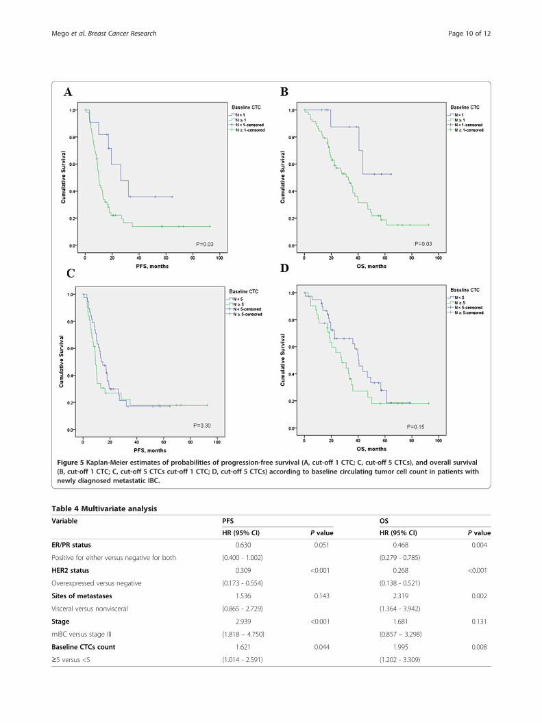
Figure 5 Kaplan-Meier estimates of probabilities of progression-free survival (A, cut-off 1 CTC; C, cut-off 5 CTCs), and overall survival(B, cut-off 1 CTC; C, cut-off 5 CTCs cut-off 1 CTC; D, cut-off 5 CTCs) according to baseline circulating tumor cell count in patients withnewly diagnosed metastatic IBC.
Table 4 Multivariate analysis
Variable PFS OS
HR (95% CI) P value HR (95% CI) P value
ER/PR status 0.630 0.051 0.468 0.004
Positive for either versus negative for both (0.400 - 1.002) (0.279 - 0.785)
HER2 status 0.309 <0.001 0.268 <0.001
Overexpressed versus negative (0.173 - 0.554) (0.138 - 0.521)
Sites of metastases 1.536 0.143 2.319 0.002
Visceral versus nonvisceral (0.865 - 2.729) (1.364 - 3.942)
Stage 2.939 <0.001 1.681 0.131
mIBC versus stage III (1.818 – 4.750) (0.857 – 3.298)
Baseline CTCs count 1.621 0.044 1.995 0.008
≥5 versus <5 (1.014 - 2.591) (1.202 - 3.309)
Mego et al. Breast Cancer Research (2015) 17:2 Page 10 of 12

Table 5 Association between baseline CTCs andpathologic complete remission (pCR) in stage III IBC
non-pCR (%) pCR (%) P value
CTC < 1 30 (85.7) 5 (14.3) 0.39
CTC ≥ 1 32 (76.2) 10 (23.8)
CTC < 5 53 (85.5) 9 (14.5) 0.06
CTC≥ 5 9 (60.0) 6 (40.0)
Mego et al. Breast Cancer Research (2015) 17:2 Page 11 of 12
neoadjuvant or first-line therapy. In our current study,we observed that CTC counts at the time of diseaseprogression after chemotherapy were lower than CTCcounts at baseline, and that the proportions of patientswith CTC counts of five or more or with one or moreCTCs were significantly lower at the time of diseaseprogression than at baseline, which is consistent withour earlier report [24].Emerging data suggest that statins, in addition to
their known antiinflammatory effects, might also havean antiproliferative effect on breast cancer cells; however,available evidence on breast cancer risk is conflicting[37,38]. Statins are usually well tolerated, but their adminis-tration is associated with some important side effects,including myositis, rhabdomyolysis, hepatotoxicity, anddiarrhea. A recent cohort study showed that use of weaklylipophilic to hydrophilic statins (H-statins) is associatedwith significantly improved PFS compared with no statinuse in patients with IBC [25]. In our study, we observedthat patients who took statins before the diagnosis of IBChad significantly lower baseline CTC counts than patientsnot taking statins. Consistent with previous observations,patients who used H-statins had lower baseline CTCcounts compared with patients without statins. Thesedata, even though hypothesis generating, further addcredence to the earlier observation that statins mighthave an anticancer effect, especially in IBC, and warrantadditional study.
ConclusionIn conclusion, this retrospective study suggests a prognosticvalue of CTCs in patients with newly diagnosed IBC. Weobserved that patients with nonmetastatic IBC had a lowerCTC prevalence and lower CTC counts at progression thandid patients with metastatic IBC. Further research shouldfocus on characterization of CTCs in IBC patients and theimplications for treatment decisions.
Ethics approvalInstitutional Review Board, The University of Texas, MDAnderson Cancer Center, protocol DR10-0227.
AbbreviationsCD45+: CD45 positive; CTC: circulating tumor cell; DAPI: 4′,6-diamidino-2-phenylindole; IBC: inflammatory breast cancer; H-statins: hydrophilic statins;L-statins: lipophilic statins; MBC: metastatic breast cancer; mIBC: metastatic
IBC; MDACC: The MD Anderson Cancer Centre; non-IBC: non-inflammatorymetastatic breast cancer; OS: overall survival; pCR: pathologic completeresponse; PFS: progression-free survival.
Competing interestsThe authors declare that they have no competing interests.
Authors’ contributionsMM, AG, and UDG conceived of and designed the study, collected clinicaldata, performed data analysis and interpretation, and drafted the manuscript.HM, MG, TF, and SD collected clinical data, analyzed the data, and reviewedthe manuscript. LH performed data analysis and reviewed the manuscript.NTU, VV, EA, RA, and WW provided clinical assessment of patients andreviewed the manuscript. GNH conceived of and designed the study, andreviewed the manuscript. JMR and MC conceived of and designed the study,performed data analysis and interpretation, and reviewed the manuscript.All authors read and approved the final version of the manuscript.
AcknowledgementsWe thank Stephanie P. Deming for discussions and critical reading andediting of the manuscript. This work was supported by a grant from theState of Texas Rare and Aggressive Breast Cancer Research Program, whichsupports the Morgan Welch Inflammatory Breast Cancer Research Programand Clinic; a UICC American Cancer Society International Fellowship forBeginning Investigators Award (ACS/08/006 to M.M.); a grant from the SlovakGrant Agency (VEGA 1/0724/11 to M.M.); and a grant from the NationalCancer Institute, National Institutes of Health (CA138239-02 to M.C., W.A.W.,and J.M.R.).
Author details1Department of Hematopathology, The University of Texas MD AndersonCancer Center, 1515 Holcombe Blvd, Houston, TX 77030, USA. 2BreastMedical Oncology, The University of Texas MD Anderson Cancer Center,Houston, TX, USA. 3Radiation Oncology, The University of Texas MDAnderson Cancer Center, Houston, TX, USA. 4Morgan Welch InflammatoryBreast Cancer Research Program and Clinic, The University of Texas MDAnderson Cancer Center, Houston, TX, USA. 5Department of MedicalOncology, Comenius University, School of Medicine, Bratislava, Slovakia.6Medical Oncology, Istituto Scientifico Romagnolo per lo Studio e la Cura deiTumori (IRST) – IRCCS, Meldola, FC, Italy. 7Department of Clinical Medicineand Surgery, University Federico II, Naples, Italy. 8Medical OncologyDepartment, Dubai Hospital, Dubai, UAE. 9Present affiliation: Breast Center,Thomas Jefferson University-Kimmel Cancer Center, Philadelphia, PA, USA.
Received: 6 August 2014 Accepted: 17 December 2014
References1. Walshe J, Swain S. Clinical aspects of inflammatory breast cancer. Breast Dis.
2005–2006;22:35–44.2. Ueno NT, Buzdar AU, Singletary SE, Ames FC, McNeese MD, Holmes FA, et al.
Combined-modality treatment of inflammatory breast carcinoma: twenty yearsof experience at M.D. Anderson Cancer Center. CancerChemother Pharmacol. 1997;40:321–9.
3. Masuda H, Brewer TM, Liu DD, Iwamoto T, Shen Y, Hsu L, et al. Long-termtreatment efficacy in primary inflammatory breast cancer by hormonalreceptor- and HER2-defined subtypes. Ann Oncol. 2014;25:384–91.
4. Fouad TM, Kogawa T, Liu DD, Shen Y, Masuda H, El-Zein R, et al. Survivaldifferences between patients with metastatic inflammatory and non-inflammatorybreast cancer. Cancer Res. 2013;73:Abstract nr P6-12-02.
5. Cristofanilli M, Buzdar AU, Hortobagyi GN. Update on the management ofinflammatory breast cancer. Oncologist. 2003;8:141–8.
6. Cristofanilli M, Valero V, Buzdar AU, Kau SW, Broglio KR, Gonzalez-AnguloAM, et al. Inflammatory breast cancer (IBC) and patterns of recurrence:understanding the biology of a unique disease. Cancer. 2007;110:1436–44.
7. Robertson FM, Bondy M, Yang W, Yamauchi H, Wiggins S, Kamrudin S, et al.Inflammatory breast cancer: the disease, the biology, the treatment.CA Cancer J Clin. 2010;60:351–75.
8. Kleer C, Van Golen K, Merajver S. Molecular biology of breast cancermetastasis: inflammatory breast cancer: clinical syndrome and moleculardeterminants. Breast Cancer Res. 2000;2:423–9.
Mego et al. Breast Cancer Research (2015) 17:2 Page 12 of 12
9. Wu M, Merajver S. Molecular biology of inflammatory breast cancer:applications to diagnosis, prognosis, and therapy. Breast Dis.2005–2006;22:25–34.
10. Van der Auwera I, Van Laere SJ, Van den Eynden GG, Benoy I, van Dam P,Colpaert CG, et al. Increased angiogenesis and lymphangiogenesis ininflammatory versus noninflammatory breast cancer by real-time reversetranscriptase-PCR gene expression quantification. Clin Cancer Res.2004;10:7965–71.
11. Parton M, Dowsett M, Ashley S, Hills M, Lowe F, Smith IE. High incidence ofHER2 positivity in inflammatory breast cancer. Breast. 2004;13:97–103.
12. Resetkova E, Gonzalez-Angulo AM, Sneige N, Mcdonnell TJ, Buzdar AU,Kau SW, et al. Prognostic value of p53, MDM-2, and MUC-1 for patients withinflammatory breast carcinoma. Cancer. 2004;101:913–7.
13. van Golen KL, Wu ZF, Qiao XT, Bao LW, Merajver SD. RhoC GTPase, a noveltransforming oncogene for human mammary epithelial cells that partiallyrecapitulates the inflammatory breast cancer phenotype. Cancer Res.2000;60:5832–8.
14. Yamauchi H, Cristofanilli M, Nakamura S, Hortobagyi GN, Ueno NT.Molecular targets for treatment of inflammatory breast cancer. Nat Rev ClinOncol. 2009;6:387–94.
15. Woodward WA, Krishnamurthy S, Yamauchi H, El-Zein R, Ogura D, Kitadai E,et al. Genomic and expression analysis of microdissected inflammatorybreast cancer. Breast Cancer Res Treat. 2013;138:761–72.
16. Colpaert CG, Vermeulen PB, Benoy I, Soubry A, van Roy F, van Beest P, et al.Inflammatory breast cancer shows angiogenesis with high endothelialproliferation rate and strong E-cadherin expression. Br J Cancer.2003;88:718–25.
17. Xiao Y, Ye Y, Yearsley K, Jones S, Barsky SH. The lymphovascular embolus ofinflammatory breast cancer expresses a stem cell-like phenotype. Am JPathol. 2008;173:561–74.
18. Cristofanilli M, Budd GT, Ellis MJ, Stopeck A, Matera J, Miller MC, et al.Circulating tumor cells, disease progression, and survival in metastaticbreast cancer. N Engl J Med. 2004;351:781–91.
19. Dawood S, Broglio K, Valero V, Reuben J, Handy B, Islam R, et al. Circulatingtumor cells in metastatic breast cancer: from prognostic stratification tomodification of the staging system? Cancer. 2008;113:2422–30.
20. Giuliano M, Giordano A, Jackson S, Hess KR, De Giorgi U, Mego M, et al.Circulating tumor cells as prognostic and predictive markers in metastaticbreast cancer patients receiving first-line systemic treatment. Breast CancerRes. 2011;13:R67.
21. De Giorgi U, Valero V, Rohren E, Dawood S, Ueno NT, Miller MC, et al.Circulating tumor cells and [18 F]fluorodeoxyglucose positron emissiontomography/computed tomography for outcome prediction in metastaticbreast cancer. J Clin Oncol. 2009;27:3303–11.
22. Cristofanilli M, Broglio KR, Guarneri V, Jackson S, Fritsche HA, Islam R, et al.Circulating tumor cells in metastatic breast cancer: biologic staging beyondtumor burden. Clin Breast Cancer. 2007;7:471–9.
23. Reuben JM, Lee BN, Li C, Gao H, Broglio KR, Valero V, et al. Circulatingtumor cells and biomarkers: implications for personalized targetedtreatments for metastatic breast cancer. Breast J. 2010;16:327–30.
24. Mego M, De Giorgi U, Hsu L, Ueno NT, Valero V, Jackson S, et al. Circulatingtumor cells in metastatic inflammatory breast cancer. Ann Oncol.2009;20:1824–8.
25. Brewer TM, Masuda H, Liu DD, Shen Y, Liu P, Iwamoto T, et al. Statin use inprimary inflammatory breast cancer: a cohort study. Br J Cancer.2013;109:318–24.
26. Greene FL, Page DL, Fleming ID, Fritz AG, Balch CHM, Haller DG, et al.AJCC Cancer Staging Handbook. 6th ed. New York, NY: Springer; 2002.
27. Dawood S, Merajver SD, Viens P, Vermeulen PB, Swain SM, Buchholz TA,et al. International expert panel on inflammatory breast cancer: consensusstatement for standardized diagnosis and treatment. Ann Oncol.2011;22:515–23.
28. Singletary SE, Allred C, Ashley P, Bassett LW, Berry D, Bland KI, et al. Stagingsystem for breast cancer: revisions for the 6th edition of the AJCC CancerStaging Manual. Surg Clin North Am. 2003;83:803–19.
29. Bidard FC, Mathiot C, Delaloge S, Brain E, Giachetti S, de Cremoux P, et al.Single circulating tumor cell detection and overall survival in nonmetastaticbreast cancer. Ann Oncol. 2010;21:729–33.
30. Lang JE, Mosalpuria K, Cristofanilli M, Krishnamurthy S, Reuben J, Singh B,et al. HER2 status predicts the presence of circulating tumor cells in patientswith operable breast cancer. Breast Cancer Res Treat. 2009;113:501–17.
31. Bidard FC, Peeters DJ, Fehm T, Nolé F, Gisbert-Criado R, Mavroudis D, et al.Clinical validity of circulating tumour cells in patients with metastatic breastcancer: a pooled analysis of individual patient data. Lancet Oncol.2014;15:406–14.
32. Pierga JY, Bidard FC, Mathiot C, Brain E, Delaloge S, Giachetti S, et al.Circulating tumor cell detection predicts early metastatic relapse afterneoadjuvant chemotherapy in large operable and locally advanced breastcancer in a phase II randomized trial. Clin Cancer Res. 2008;14:7004–10.
33. Riethdorf S, Müller V, Zhang L, Rau T, Loibl S, Komor M, et al. Detection andHER2 expression of circulating tumor cells: prospective monitoring in breastcancer patients treated in the neoadjuvant GeparQuattro trial. Clin CancerRes. 2010;16:2634–45.
34. Lucci A, Hall CS, Lodhi AK, Bhattacharyya A, Anderson AE, Xiao L, et al.Circulating tumour cells in non-metastatic breast cancer: a prospectivestudy. Lancet Oncol. 2012;13:688–95.
35. Rack B, Schindlbeck C, Jückstock J, Andergassen U, Hepp P, Zwingers T,et al. Circulating tumor cells predict survival in early average-to-high riskbreast cancer patients. J Natl Cancer Inst. 2014;106. doi:10.1093/jnci/dju066.
36. Pierga JY, Petit T, Delozier T, Ferrero JM, Campone M, Gligorov J, et al.Neoadjuvant bevacizumab, trastuzumab, and chemotherapy for primaryinflammatory HER2-positive breast cancer (BEVERLY-2): an open-label,single-arm phase 2 study. Lancet Oncol. 2012;13:375–84.
37. Bjarnadottir O, Romero Q, Bendahl PO, Jirström K, Rydén L, Loman N, et al.Targeting HMG-CoA reductase with statins in a window-of-opportunitybreast cancer trial. Breast Cancer Res Treat. 2013;138:499–508.
38. Undela K, Srikanth V, Bansal D. Statin use and risk of breast cancer: ameta-analysis of observational studies. Breast Cancer Res Treat.2012;135:261–9.
Submit your next manuscript to BioMed Centraland take full advantage of:
• Convenient online submission
• Thorough peer review
• No space constraints or color figure charges
• Immediate publication on acceptance
• Inclusion in PubMed, CAS, Scopus and Google Scholar
• Research which is freely available for redistribution
Submit your manuscript at www.biomedcentral.com/submit