chronic ear disease akira ishiyama, m.d.. contemporary approach dipolar restriction: largely...
TRANSCRIPT
Chronic Ear DiseaseChronic Ear Disease
Akira Ishiyama, M.D.
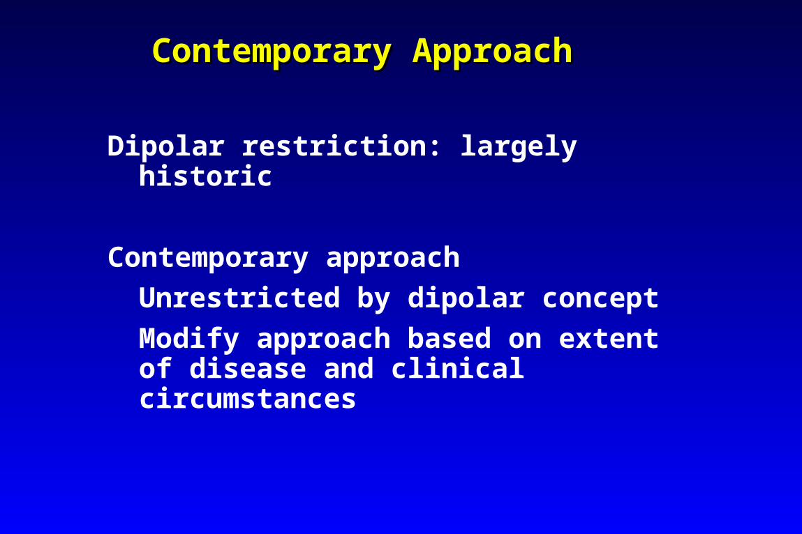
Contemporary ApproachContemporary Approach
Dipolar restriction: largely historic
Contemporary approach
Unrestricted by dipolar concept
Modify approach based on extent of disease and clinical circumstances
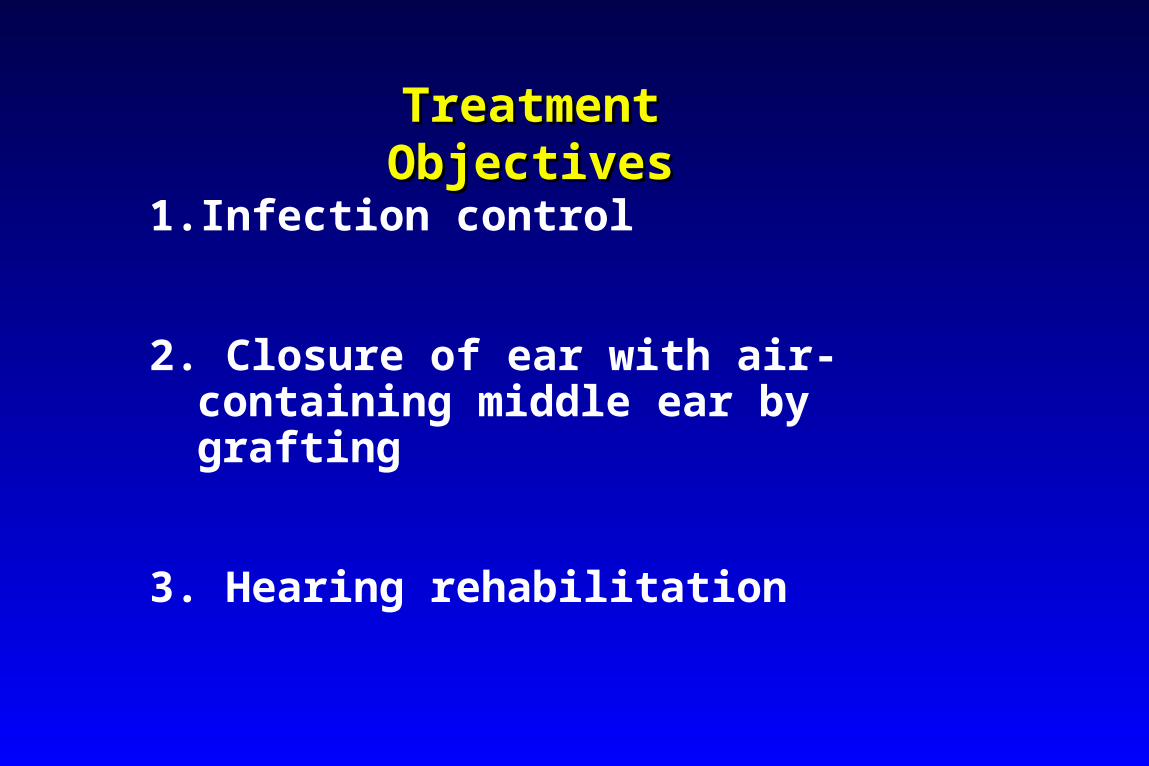
Treatment Treatment ObjectivesObjectives
1. Infection control
2. Closure of ear with air-containing middle ear by grafting
3. Hearing rehabilitation
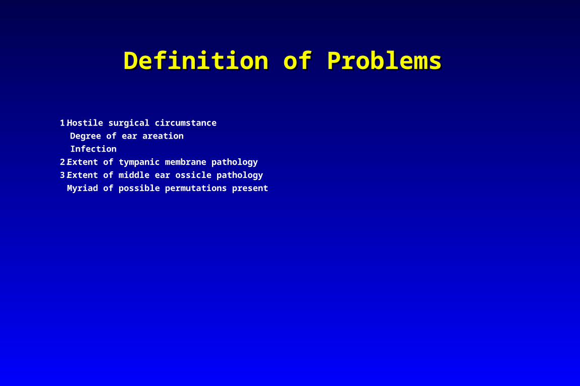
Definition of ProblemsDefinition of Problems
1.Hostile surgical circumstance
Degree of ear areation
Infection
2.Extent of tympanic membrane pathology
3.Extent of middle ear ossicle pathology
Myriad of possible permutations present
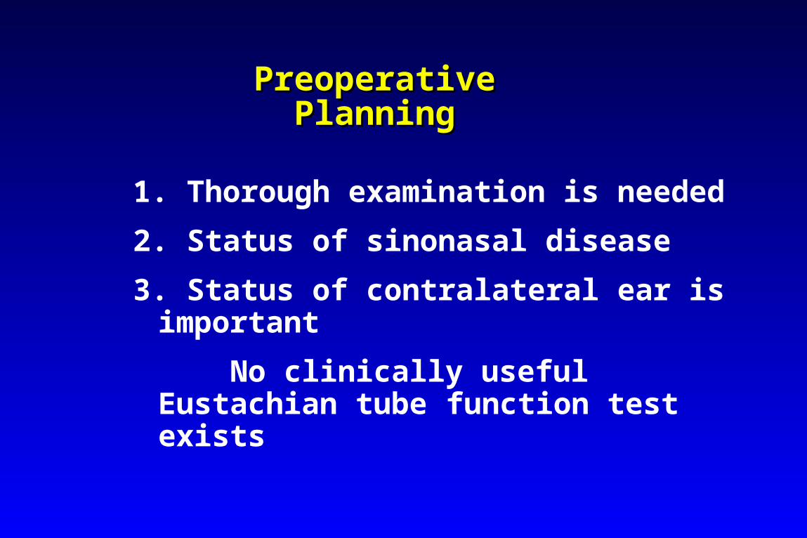
Preoperative PlanningPreoperative Planning
1. Thorough examination is needed
2. Status of sinonasal disease
3. Status of contralateral ear is important
No clinically useful Eustachian tube function test exists
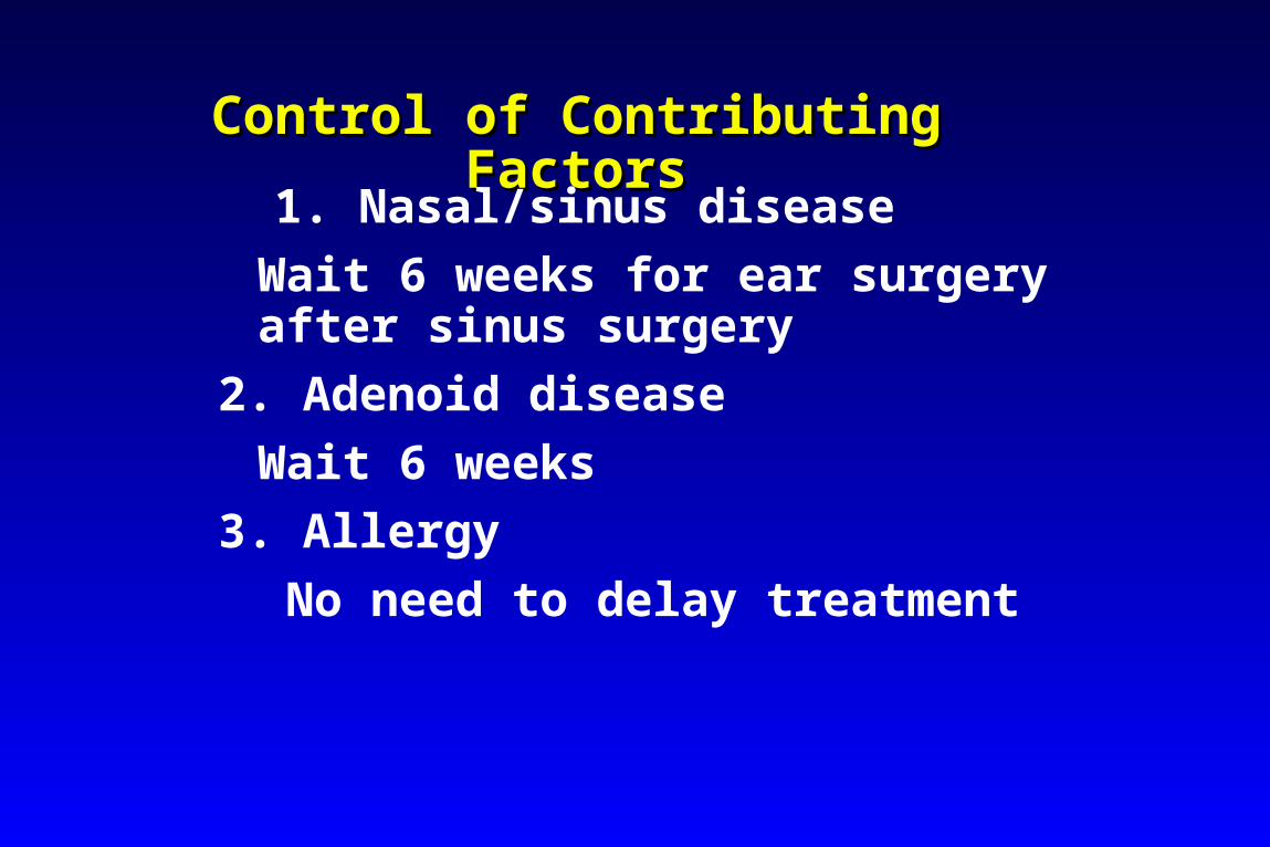
Control of Contributing FactorsControl of Contributing Factors
1. Nasal/sinus disease
Wait 6 weeks for ear surgery after sinus surgery
2. Adenoid disease
Wait 6 weeks
3. Allergy
No need to delay treatment
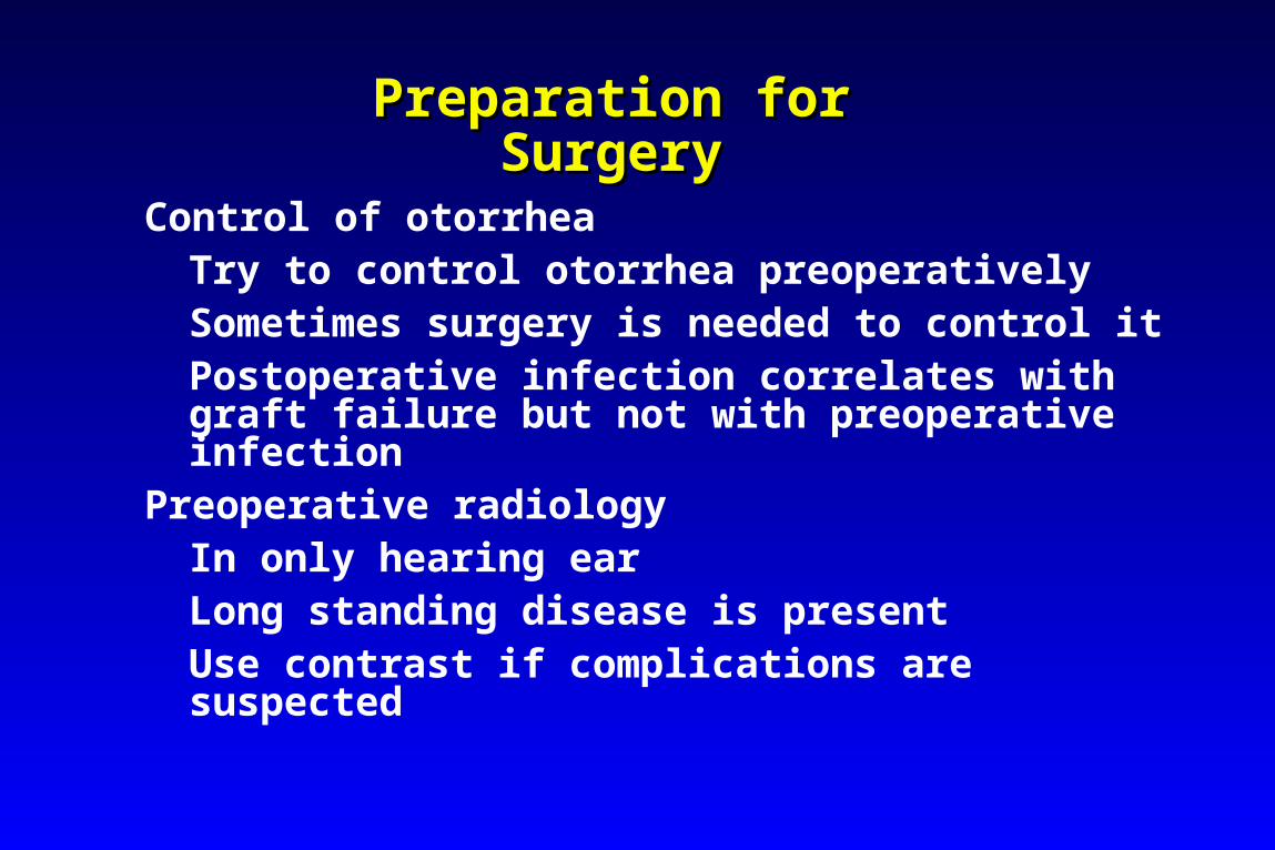
Preparation for Preparation for SurgerySurgery
Control of otorrheaTry to control otorrhea preoperativelySometimes surgery is needed to control itPostoperative infection correlates with graft failure but not with preoperative infection
Preoperative radiologyIn only hearing earLong standing disease is presentUse contrast if complications are suspected
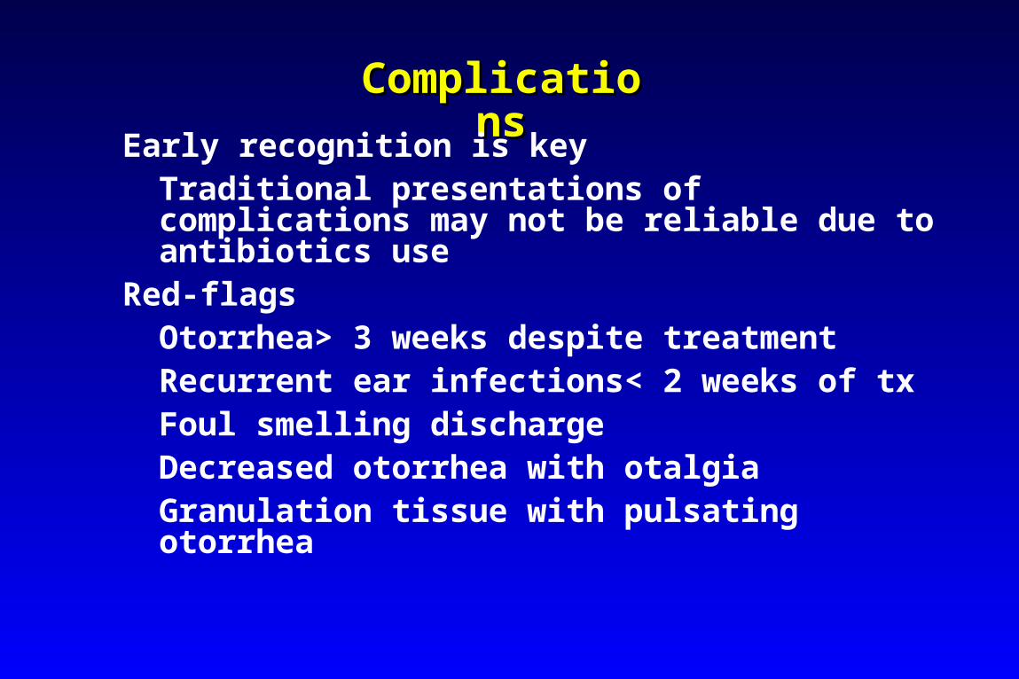
ComplicationsComplications
Early recognition is keyTraditional presentations of complications may not be reliable due to antibiotics use
Red-flagsOtorrhea> 3 weeks despite treatmentRecurrent ear infections< 2 weeks of txFoul smelling dischargeDecreased otorrhea with otalgiaGranulation tissue with pulsating otorrhea
Patient InformationPatient Information
Provide details of surgery
Potential risksComplicationsReasonable expectations
Informed consentMake sure your patients understand what you are planning to do
Surgical Technique
Basic principlesInfection controlExposureHemostasisGrafting techniqueMastoidectomyStagingOssicular reconstructionTympanosclerosisAtelectatic ear
Infection Control
1. Adhere to sterile technique
2. Irrigate the ear with Betadine
Harmless for 10-15 minutes
Copious saline irrigation
3. Use of prophylactic antibiotics
Controversial
ExposureExposurePostauricualr approach
Better illuminationBetter access to all regions of TM and middle ear
Graft placement successMost effective when you can clearly see where the graft is
Complete mastoidectomyInfection control techniqueExposure device
HemostasisHemostasis
Meticulous hemostasis is critical for successful surgeryPostauricualr injectionCanal injection at the beginning
Small syringe and a 27G needleIntraoperative hemostasis
Bone waxEpinephrine 1:1000 soaked GelfoamBipolar cautery
Grafting TechniqueGrafting Technique
Whatever works for you is fine
Superficial temporal areolar fascia
Temporalis fascia
Tragal perichondrium
Periosteum
Vein
MastoidectomyMastoidectomy
Relative indications for mastoidectomy
1. Revision surgery after failed multiple tympanoplasties
2. In some children where areation is a problem
3. Recent and prolonged otorrhea recurrent after, or unresponsive to tx
4. Extreme epitympanic ossicular fixation pathologies
StagingStaging
Staging is still valuable
When to consider staging1. TM pathologies coexist with ossicular problems2. Very poor mucosal status in the middle ear3. High chance of recurrent cholesteatoma4. Disease on the stapes suprastructure and footpalte
Ossicular ReconstructionsOssicular Reconstructions
Points to consider
Know your prosthesis well
Use cartilage/tissue graft
Stage less optimal ears
Place the prosthesis vertical
TympanosclerosisTympanosclerosis
Immunobiological nature and consequences poorly understood
Limited tympanosclerosis should be removedStapes fixation is very problematic
Minor oval window tympanosclerosis can be repaired with stapedectomyExtensive disease: use hearing aids
Atelectatic EarAtelectatic Ear
Adynamic monomeric TMCholeateatoma, ossicular erosion
Atelectatic ear looks terrible but functions wellBetter to fix itIt will progress and better to fix earlyRule out nasopharyngeal pathology
Problem CavityProblem Cavity
Poor prior CWD technique
Otorrhea with odor
Debris accumulation
Hearing loss
Requires constant medical attention
Patient is a water cripple
Problem Problem CavityCavity
Morphologic findingsA small meatusA high facial ridgeIncomplete degrees of EAC wall removalA deep, dependent mastoid tip cavityA TM perforation with weeping, diseased mucosaImpacted debrisMucopurulent otorrhea
Absolute Indications for Absolute Indications for CWDCWD
Unresectable diseaseDefinition of unresectability varies
Impossibility of follow up surveillanceUnwillingness of patientsInability of compliance
Posterior canal wall eroded by the prior surgery or disease process
Relative Indications for Relative Indications for CWDCWD
1. Disease in the only hearing ear2. Disease in the dead ear3. When disease in the medically informed
precludes staging or multiple anesthetics4. Disease complicated by intracranial or
intratemporal problems. 5. When neoplasms such as glomus tumors or
primary adenomas require CWD exposure