chronic akinetic mutism after mesencephalic-diencephalic infarction: remediated with dopaminergic...
TRANSCRIPT
http://nnr.sagepub.com/Repair
Neurorehabilitation and Neural
http://nnr.sagepub.com/content/15/2/151The online version of this article can be found at:
DOI: 10.1177/154596830101500208
2001 15: 151Neurorehabil Neural RepairMichael P. Alexander
Dopaminergic MedicationsChronic Akinetic Mutism After Mesencephalic-Diencephalic Infarction: Remediated with
Published by:
http://www.sagepublications.com
On behalf of:
American Society of Neurorehabilitation
can be found at:Neurorehabilitation and Neural RepairAdditional services and information for
http://nnr.sagepub.com/cgi/alertsEmail Alerts:
http://nnr.sagepub.com/subscriptionsSubscriptions:
http://www.sagepub.com/journalsReprints.navReprints:
http://www.sagepub.com/journalsPermissions.navPermissions:
http://nnr.sagepub.com/content/15/2/151.refs.htmlCitations:
What is This?
- Mar 1, 2001Version of Record >>
at Utah State University on September 27, 2014nnr.sagepub.comDownloaded from at Utah State University on September 27, 2014nnr.sagepub.comDownloaded from

151
Case Report
Chronic Akinetic Mutism After Mesencephalic-DiencephalicInfarction: Remediated with Dopaminergic Medications
Michael P Alexander
Objective: Akinetic mutism (AKM) is an uncommon disorder with a complexneuropathology. There is no generally accepted treatment, and it is not known iflate treatments are effective. The relationship between AKM and abulia is uncertain.Methods: The effects of dopaminergic treatment of a patient with chronic AKM afterdiscrete bilateral infarctions of the mesencephalic ventral tegmental area and the lat-eral hypothalamus were studied with motor measures, the Functional IndependenceMeasure (FIM), and neuropsychological tests. Results: Treatment with a combinationof carbidopa/levodopa and pergolide produced prompt amelioration of AKM with dra-matic and rapid improvement in FIM. An apathetic, amotivational state persisted de-spite resolution of akinesia and normal frontal executive functions. Conclusions: AKM
may respond to dopaminergic treatment even after months of severe akinesia. Themechanism of abulia is more complex than simply a partial dopaminergic deficiencystate and may persist even when AKM is treated and frontal cognitive functions arenormal. Key Words: Akinetic mutism—Dopamine—Abulia.
Neurorehabilitation and Neural Repair 2001;15:151-156© 2001 Demos Medical Publishing
Akinetic mutism (AKM) is an uncommon neuro-logic condition characterized by paucity and slowness ofmovement and speech despite retamed capacity to moveand to speak (1). Although the pathologic anatomy ofAKM has been identified, many diseases that may causeit are known, and some treatments have been reported,several aspects of AKM are not optimally clarified (2).The clinical recognition of AKM is not always straight-forward. When there is also paresis, the contribution of
From Memory Disorders Research Center, Boston University De-
partment of Neurology, and Departments of Neurology, Beth Israel Dea-coness Medical Center and Harvard Medical School, Boston, Massa-chusetts
Presented m part at the American Academy of Neurology, Seattle,May 1995.
Address correspondence and reprmt requests to Michael P Alexan-der, M D., Behavioral Neurology Unit, Beth Israel Deaconess MedicalCenter, 330 Brooklme Avenue, Boston, MA 02215, U.S.A.
akmesia to disability may not be recognized. Potentialmarkers for specific treatments have not been established,nor is it clear whether chronic AKM responds to treat-ment. It has been suggested that the amotivational state(abulia) that sometimes accompanies frontal lobe dam-age is a lesser form of AKM (2), but it is unknownwhether they necessarily have the same pathophysiology.If abulia represents lesser injury to the same neural sys-tem as AKM, they might have a common treatment.
Case Report
This 36-year-old, right-handed, college-educatedwoman had a stepwise neurologic deficit that progressedover 15 days. History is significant only for infrequentcommon migrame headaches and two normal term preg-nancies. Her only medication was an oral contraceptive
at Utah State University on September 27, 2014nnr.sagepub.comDownloaded from

152
that she had been using for 2 years. After 3 days of se-vere headache, she collapsed at home, was bmefly unre-sponse, and was admitted to the hospital. She was alert,cognitively mtact, but had left lower facial weakness.EEG, unenhanced computed tomography (CT), and lum-bar puncture were normal. The next day she was less re-sponsive but otherwise unchanged. Magnetic resonanceimaging (MRI) demonstrated bilateral paramedian mid,brain-thalamic infarcts. Magnetic resonance angtogra-phy (MRA) suggested a narrowed distal basilar artery.Complete coagulation profile, transthoracic echocardio-gram, chest radiograph, mammogram, and pelvic ultra-sound were normal. Over the next 2 days, she becamedrowsier, and vertical gaze paresis and left-sided weaknessdeveloped. Conventional arteriography demonstratednarrowing of the midbasilar and no filling m the distalbasilar. Heparin was begun. For 6 days, she showed rapidimprovement. On day 14, she again became lethargicwith worsened left hemiparesis. MRI demonstrated a new,left pontme infarct. Repeated conventional arteriogra-phy showed increased narrowing of the basilar artery. Sheremamed minimally responsive. On day 32, a right tem-poral bram/meningeal biopsy was negative. A feedingtube was placed, and she was discharged to a rehabilita-tion hospital 40 days after the initial symptoms.
On admission to rehabilitation, she was described asmute, lying in bed m essentially decorticate posture, withno movement. She was bed-bound and dependent m allcare. She underwent 3.5 months of daily mtense multi-disciplinary therapy. Relaxation, neuromuscular reeduca-tion, passive ranging, serial casting to all four limbs, re-peated phenol blocks, and trials of diazepam (up to 15 mg,t.i.d.), dantrolene (up to 125 mg, qd), and baclofen (Li-oresal ; up to 30 mg, t.i.d.) produced little change m func-tion. Vigorous attention to positioning improved swal-lowmg, and the g-tube was removed after 3 months. Atdischarge (5 months after onset), she still had minimalspeech and movement. Medications were dantrolene, 25mg qd; baclofen, 20 mg, b.i.d.; and oxycodone with acet-aminophen (Percocet; 1-4 qd) for limb pam.
At home she required daily services for self-care, sus-pension transfers, and positioning. Six days weekly, shehad 3 h of outpatient therapy. Treatment contmued tofocus on relaxation, maintenance of range, positioning,and strengthening. She had botulmum treatments at theleft hand and wrist and left ankle with modest results.Dantrolene and baclofen were increased with no im-
provement but with decreased responsmeness.She was admitted to a second rehabilitation hospital
m hope of identifymg interventions that would allow lessmtense home services. She had almost no spontaneousspeech and very terse responses to questions, with responselatencies routinely >20 s. Speech was aprosodic and hy-
pophonic. Allowing for her extremely slow responses, lan-guage and memory seemed normal. Visual fields and pupil-lary size and responses were normal. All pursmt and horn-zontal saccadic eye movements were normal, but she couldproduce no vertical saccadic movements. Vertical doll’s-eyes responses were present. She had moderate lid retrac-tion, very reduced blink rate (<1/mm), and very exagger-ated glabellar response. She had masked facies and leftlower facial weakness to command. There was low-ampli-tude titubation. She was generally akmetic, particularly onthe left, and tended to rest in bed or chair m a near-decor-ticate position. When prompted, she had good power onthe right, although all requested movements were delayedand had reduced amplitude, with a coarse proximal tremor.She required 29 min to do 10 fmger-to-nose circuits at adistance of 30 cm. She could isolate all small-finger move-ments, although she required much prompting to continue.Power m the left leg was good, although agam delayed andalso with a significant action dystoma. The left arm wasfisted m decorticate position with decreased passive rangeat all omts. Sensation was normal. Tendon reflexes werebrisk and equal; both toes went down.
Her functional status on admission after 6.5 monthsof intense therapy (-600-700 h) was still limited. Shetransferred from bed to chair with maximal assistance.She sat with maximal assistance for support and stoodwith moderate assistance using the left ankle-foot or-thosis (AFO), but she had no postural reflexes and couldnot ambulate or advance her wheelchair. She could do
feeding, grooming, and oral hygiene with maximal assis-tance ; all other self-care was dependent. Speech mtelli-gibility was poor: 32% at sentence level. Her overallFunctional Independence Measure (FIM) was 42 (3).
Standard rehabilitation mterventions were contmued,and carbidopa/levodopa was begun at 10/100 b.i.d. Withindays her movement and speech began to improve. She wasmcreased to a daily dose of 60/600 over 2 weeks. Pergolidewas begun 0.05 mg, b.i.d.. Dantrolene was stopped width-out any clmcal change. Speech latencies were < 10 s; m-
telligibility at sentence level had improved to 55%. Titu-bation and right arm tremor were reduced. Speed ofmovement increased; 10 cycles of finger-to-nose, as pre-viously described, required 16 s (45% faster). She sat m-dependently and stood with supervision, no AFO required,although postural stability was still poor. She advanced herwheelchair mdependently and walked 150 feet with arolling walker with minimal assistance. Her upper bodyself-care required at most supervision, and lower body,moderate assistance. Her overall FIM was now 87. She wastransferred to a subacute facility for further therapy.
Four weeks later, taking carbidopa/levodopa 60/600daily m five doses, and pergolide, 0.10 t.i.d., spontaneousmovement and speech were much more frequent. Re-
at Utah State University on September 27, 2014nnr.sagepub.comDownloaded from
153
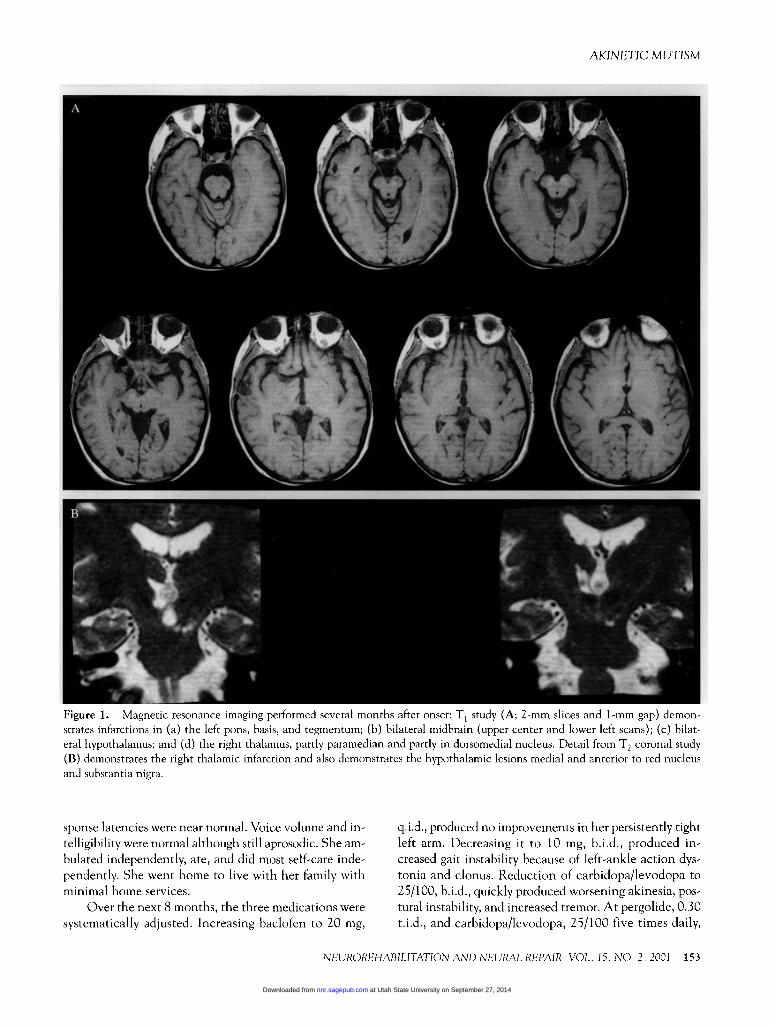
Figure 1. Magnetic resonance imaging performed several months after onset: T, study (A; 2-mm slices and 1-mm gap) demon-strates mfarctions in (a) the left pons, basis, and tegmentum; (b) bilateral midbram (upper center and lower left scans); (c) bilat-eral hypothalamus; and (d) the right thalamus, partly paramedian and partly in dorsomedial nucleus. Detail from T2 coronal study(B) demonstrates the right thalamic infarction and also demonstrates the hypothalamic lesions medial and anterior to red nucleusand substantia nigra.
sponse latencies were near normal. Voice volume and m-
telligibility were normal although still aprosodic. She am-bulated mdependently, ate, and did most self-care mde-pendently. She went home to live with her family withminimal home services.
Over the next 8 months, the three medications weresystematically adjusted. Increasing baclofen to 20 mg,
q.i.d., produced no improvements m her persistently tightleft arm. Decreasing it to 10 mg, b.i.d., produced m-creased gait mstability because of left-ankle action dys-tonia and clonus. Reduction of carbidopa/levodopa to25/100, b.i.d., quickly produced worsening akmesia, pos-tural mstability, and increased tremor. At pergolide, 0.30t.i.d., and carbidopa/levodopa, 25/100 five times daily,
at Utah State University on September 27, 2014nnr.sagepub.comDownloaded from

154
she ambulated completely independently without any as-sistive device. She was essentially mdependent m self-care, more talkatme, and doing some household chores.Her family noted that she appeared unmotivated to m-crease her independence.
She was lost to follow-up for 11 months. Her familybrought her for reevaluation because of her apathy andpoor motivation. Dosages of medications had not beenchanged. Examination was unchanged. Increasing eitherpergolide or carbidopa/levodopa caused orthostatic hy-potension. When either was reduced, tremors and bal-ance worsened. Fluoxetme was added at 10 mg qd withno effect. After it was mcreased to 20 mg qd, her moti-vation to do home activities seemed better, but she alsohad a marked increase m irritability. The dose was re-duced to 10 mg qd. At last follow-up, she had not at-tempted any return to employment. She expressed littlemterest m household activities, in mcreasmg her role mher family, doing the home activities that she is able todo, or pursuing any leisure activities. She often simply sitsfor hours.
Neuropsychological Assessments
At 7 months after onset, she was onented, had nor-mal nammg and repetition, normal praxis, and normal
delayed recall of a four-word list. Performance was qmtedelayed, and she often required multiple prompts to pro-duce any response. Because of severe akmesia and
mutism, formal testmg was not possible before treatment.Her first comprehensive evaluation was 13 months afteronset. Language, fund of knowledge, verbal subtests ofthe WAIS-R, Wisconsin Card Sort, Raven’s ProgressiveMatrices, and verbal fluency were entirely normal.
Follow-up evaluation (28 months after onset)demonstrated little change, although some time-de-pendent tasks were faster. The Stroop test was normal.Word-list generation (FAS) was average. List learningwas mildly impaired (7-11-14-14-13), but semantic clus-tering was used as a learning strategy. Delayed recall(12) was slightly low, but recognition was normal (16 of16).
At the final neuropsychological evaluation at 39months after onset, results were essentially normal. Sheremained slow on some tasks, especially perceptual-motortasks. She had material-specific memory deficits for visualnonverbal material. All executive cognitive tasks werenormal. The patient was, however, quite aware of, and herfamily complamed of, her lack of mterests and poor mo-tivation. She demed any depression, and there were novegetative symptoms of depression. Her manner was joc-ular and breezy.
Discussion
AKM is a disorder m activation to move and to
speak despite conscious awareness, perhaps even the &dquo;de-sire&dquo; to speak or move, and no impairment m power, co-ordination, or tone that restricts movement (1). AKMhas been reported as a consequence of lesions m numer-ous bram regions: (a) bilateral medial frontal (4-7), evenwhen restricted to anterior cmgulate gyn (ACG) (8-10);(b) the anterior hypothalamus (11,12) (usually tumors);(c) with acquired hydrocephalus (13-17); (d) severe dif-fuse axonal injury of trauma (durmg recovery) (18,19);(e) bilateral globus pallidus (20-22); (f) the paramedianmesencephalic-diencephalic region (usually infarctions)(23-28).
Lesions in any of these areas can damage part of aneural system critical for activating movement (includingspeech) and probably cogmtion without affectmg the pat-terns or content of either. Damage directly to dopamm-ergic neurons m the midbram or to their ascendmg pro-jections may be the most common pathophysiology ofAKM. There is substantial overlap in the midbram of thedopammergic neurons projecting to stnatum and thoseprojecting to cortex (29), but the cortical projections aremore medial and dorsal than the stnatal; that is, they arecentered m the ventral tegmental area (VTA) (30). Thismesocortical system projects through the medial forebrambundle (MFB) (31 ). The MFB runs through the lateralhypothalamus into the septal area and then into the in-fraventricular white matter before projecting to the ACGand other, predommantly medial frontal structures (32).MFB pathways are strictly uncrossed (29). Lesions m thenonnigral ventral tegmental area (VTA, region A10)cause dopamine depletion m frontal cortex only, and dam-age to the MFB produces akinesia (31 ).
The terminations of the mesocortical pathway mfrontal lobes are pnmanly the supplementary motor area(SMA) and the ACG, and bilateral damage to the ACGand SMA produces AKM. ACG and SMA share manyconnectivity properties (33, 34). In addition to the
dopaminergic afferents from the VTA (29), both have bi-lateral efferents to frontal and premotor cortex and majorprojections to the stnatum (33, 34) running in theperiventricular white matter (35). Damage to medialfrontal cortex or to its deep projections produces impairedactivation. Unless very extensive, even bilateral SMA andACG damage does not typically cause severe global aki-nesia, but m some cases, the clmcal presentation can bequite similar clinically to that of the midbrain lesions (36).
Dopaminergic inputs to the stnatum arise from morelateral midbram structures, primarily the zona compactaof the substantia nigra (29, 37), and pass through the ven-tral posterior limb mternal capsule (30). Damage re-
at Utah State University on September 27, 2014nnr.sagepub.comDownloaded from

155
stncted to the cells of origin produces profound akinesia(37). The stnatum projects to the globus pallidus. Dam-age to the globus pallidus produces severe akinesia (38),and stimulation of globus pallidus can elimmate akme-sia (39). Much of the mternal segment of the globus pal-lidus output is to the thalamus: VA, VL, and CM nuclei,and the IL region. Damage to the pallidothalamic path-ways as they cross the ventral mternal capsule or m themtramedullary thalamus may also produce akinesia (40).
The ascending systems are strictly uncrossed. Thecortical efferents are bilaterally distributed. The pallidalefferents are uncrossed. Complete AKM requires bilat-eral lesions, although they need not be symmetrical. Inthis case, damage was asymmetrical. The precise basis isuncertam but mcludes at least the right VTA, ascend-mg dopammergic pathways m the left lateral hypothala-mus, and the medial pallidothalamic pathways.
That AKM responds to vanous medications has alsobeen known for years (41). L-DOPA, bromocriptme,lisuride, metoprolol, methylphenidate, and amphetaminehave all been used, but the best agents, optimal doses,and duration of treatment are unknown. Successful treat-ment has been reported m TBI (18, 42), herpes simplexencephalitis (43), hydrocephalus after ventricular shuntfailures ( 13, 14, 16, 17 ), basal tumors (12), hypoxia (42),and various causes of chronic hepatic encephalopathy(44). Bromocnptme is the most commonly reportedagent. Once successfully treated, sensitivity to dose re-duction has been variables. Given the remarkable num-ber of possible lesions within a very small anatomic re-gion that can cause AKM, it is not surprising that drugtreatment has had variable results. Damage to the affer-ent (dopammergic) system should be preferentially re-sponsive to dopa agents.
In this case, AKM was not clmically recogmzed for8 months after onset, perhaps because left hemiparesis de-veloped at the same time. Two features of the examina-tion might have suggested coexistent AKM. First, preser-vation of isolated finger movements, even with long delayafter requested movement, is atypical m corticospmalpattern paresis. Second, loss of vertical gaze pomts to aparamedian injury that often produces AKM. Althoughthe initial dose was low and peripheral dopamme me-tabolism was surely not blocked at the starting doses, astriking clmcal response was seen within days of start-ing carbidopa/levodopa after months of unchangmgdeficit. Additional benefit was seen with pergolide. Inthis case, treatment will probably be permanently re-quired, as attempts to decrease either drug caused wors-ened deficits within days. Dopaminergic treatment shouldbe considered even in very chronic cases of AKM.
Despite excellent motor recovery with treatment andno evidence of executive cognitive deficits, she has re-
mained apathetic and emotionally mdifferent (i.e., abu-lic). The lack of response of the motivational deficitsunder dopammergic treatment suggests that abulia neednot be simply a milder form of AKM. It is still possiblethat dopamine deficiency does underlie both disorders,but that the motor-response impairment (AKM) is moredirectly mediated by dopamine and, thus, more directlytreated with dopammergic agents. A similar observationof differential response to treatment has been made in
cognitive functions (45). In a group of patients with cog-nitive impairments, but not AKM, after traumatic braminjury, bromocnptme improved a subset of executivefunctions but did not affect working memory, an allegedlydopamme-dependent operation (46). Various cognitive,motor, and emotional capacities may share a requirementfor ascending dopammergic input but remain anatomi-cally segregated, use different dopamme receptor systems,or differ m the extent of direct modulation by dopamme.In this patient, it can be concluded that abulia is not dueto a simple dopamme deficiency and is not just part ofAKM, as the latter improved so dramatically, and the for-mer not at all. It is also not due to a general executive-system impairment, as standard assessments of executivecognitive functions are mtact. Apathy is likely a verycomplex disorder with cognitive, emotional, visceral, andactivational components (9). If they have an effect,dopammergic agents may treat only the activationalcomponent. Perhaps in this patient, the lesion in theright antenor thalamus has disrupted the emotional com-ponents and is the source of the persistent apathy.
AcknowledgmentSupported in part by program project grant NS
26985 to the Boston University School of Medicme,Memory Disorders Research Center.
References
1 Alexander MP Disturbances in language initiation mutism andits lesser forms In Young R, Josephs AB, eds Movement disordersm neurology and neuropsychiatry Oxford. Blackwell, 1992.389-96.
2 Fisher CM Honored guest presentation abulia minor vs agitatedbehavior Clin Neurosurg 1985;31.9-31.
3 Stmeman MG, Escarce JJ, Gom JE, Hamilton BB, Granger CV,Williams SV. A case mix classification system for medical reha-bilitation Med Care 1994;32:366-79.
4 Ferbert A, Thron A Bilateral anterior cerebral atery territory m-farction in the differential diagnosis of basilar artery occlusion JNeurol 1992;239:162-4.
5 Laplane D, Talaraich J, Meininger V, Bancaud J, Orgogozo JMClinical consenquences of coticectomeis involving the supple-mentary motor area in man. J Neurol Sci 1977;34 301-14.
6 Lechevalier B, Bertran F, Busson P, Chapon F, Raoul G, DeLa Sayette V (Akmetic mutism with right hemiplegia caused by
at Utah State University on September 27, 2014nnr.sagepub.comDownloaded from
156
infarction m the territory of the left anterior cerebral artery). RevNeurol (Pans), 1996,152.181-9.
7. Minagar A, David NJ. Bilateral infarction m the terntory of theanterior cerebral arteries (see comments). Neurology 1999,52:886-8.
8. Barns RW, Schuman HR. Bilateral anterior cingulate lesions syn-drome of the anterior cingulate gyn Neurology 1953,3 44-52.
9 Devinsky O, Morrell MJ, Vogt BA. Contributions of the anteriorcingulate cortex to behavior Brain 1995,118.279-306.
10. Nielsen JM, Jacobs LL Bilateral lesions of the anterior cingulategyri. Bull LA Neurol Soc 1951;6:231-4
11 Cairns H,Oldfield RC, Pennybacker JB, Whitteridge D. Akineticmutism with an epidermoid cyst of the third ventncle Brain
1941;64:273-90.12. Ross ED, Stewart RM. Akmetic mutism from hypothalamic dam-
age. successful treatment with dopamine agonists Neurology1981,31.1435-9
13. Anderson B. Relief of akmetic mutism from obstructive hydro-cephalus using bromocriptine and ephedrine: case report J Neuro-surg 1992;76.152-5
14 Berger L, Gauthier S, Leblanc R Akinetic mutism and parkin-sonism associated with obstructive hydrocephalus. Can J Neurosci1985,12 255-8
15. Moser A, Freyberger HJ, Bruckmann H, Kompf D. Akinetischermutismus bei dekompensiertem triventrikularen hudrozephalus.Fortschr Neurol Psycbaitr 1995,63.248-51
16. Aidi S, Elalaom-Faris M, Benabdeljlil M, Benomar A, ChaouiM, Chkili T Akinetic mutism and progressive supranuclear palsy-like syndrome after the shunt of an obstructive hydrocephalus:successful treatment with bromocriptine 2 cases. Rev Neurol(Pans) 2000;56. 380-3.
17. Caner H, Altmors N, Benli S, Calisaneller T, Albayrak A. Aki-netic mutism after fourth ventncle choroid plexus papilloma:treatment with a dopamine agonist. Surg Neurol 1999;51:181-4.
18. Crismon ML, Childs A, Wilcox RE, Barrow N The effect ofbromocriptine on speech dysfunction m patients with diffuse braininjury (akinetic mutism). Clin Neuropharmacol 1988,11.462-6.
19 Levm HS, Madison CF, Bailey CB, Meyers CA, Eisenberg HM,Gumto FC. Mutism after closed head injury Arch Neurol 1983;40.601-6
20. Laplane D, Baulac M, Widlocher D, Dubois B. Pure psychic aki-nesia with bilateral lesions of basal ganglia. J Neurol NeurosurgPsychiatry 1984,47:377-85
21. Mega MS, Cohenour RC. Akinetic mutism: disconnection offrontal-subcortical circuits. Neuropsychiatry Neuropsychol BehavNeurol 1997,10.254-9.
22. Ure J,Faccio E, Videla H, et al Akinetic mutism: a report of threecases. Acta Neurol Scand 1998,98.439-44.
23. Botez MI, Barbeau A Role of subcortical structures, and partic-ularly of the thalamus, m the mechanisms of speech and languagea review. Int J Neurol 1971 8.300-20
24 Castaigne P, Lhermitte F, Buge A, Escourolle R, Hauw JJ, Lyon-Caen O. Paramedian thalamic and midbram infarct. clinical and
neuropathological study Ann Neurol, 1981;10.127-48.25. Guberman A, Stuss D. The syndrome of bilateral paramedian
thalamic infarction. Neurology 1983;33.540-6.26. Katz DI, Alexander MP, Mandell AM Dementia following strokes
in the mesencephalon and diencephalon Arch Neurol 1987,441127-33
27. Robles A, Aldrey JM, Rodriguez Fernandez RM, et al. (Parame-dian bithalamic infarct syndrome report of five new cases). RevNeurol, 1995,23 276-84.
28. Segarra JM. Cerebral vascular disease and behavior, I. the syn-drome of the mesencephalic artery (basilar artery bifurcation)Arch Neurol 1970,22:408-18.
29 Fallon JH. Topographic organization of ascending dopaminergicprojections Ann N Y Acad Sci 1988;537:1-9.
30. Ungerstedt U Stereotaxic mapping of the monoamine pathwaysm the rat brain. Acta Physiol Scand Suppl 1971,367.1-48
31. Lmdvall O, Bjorklund A, Moore RY, Stenevi U. Mesencephalicdopamine neurons projecting to neocortex Brain Res 1974,81325-31.
32 Nauta WHJ, Feirtag M Fundamental neuroanatomy. New YorkWH Freeman, 1986
33 Baleydier C, Mauguiere F The duality of the cingulate gyrus inmonkey. Neuroanatomical study and functional hypothesis Bram1980,103:525-54
34. Jurgens U. The efferent and afferent connections of the supple-mentary motor area. Bram Res 1984;300.63-81
35. Yakolev PL, Locke S. Limbic nuclei of the thalamus and con-nections of limbic cortex. Arch Neurol 1961;5.364-400
36. Borggreve F, De Deyn PP, Marien P, Cras P, Dierckx RA Bilat-eral infarction in the anterior cerebral artery vascular territory dueto an unusual anomaly of the circle of Willis (see comments)Stroke 1994;25:1279-81.
37. Burns RS, Chiueh CC, Markey SP, Ebert MH, Jacobowitz DM,Kopm IJ. A primate model of parkinsonism: selective destruc-tion of dopaminergic neurons in the pars compacta of the sub-stantia nigra by N-methyl-4- phenyl-1,2,3,6-tetrahydropyridineProc Natl Acad Sci U S A 1983;80.4546-50
38. Horak FB, Anderson ME. Influence of globus pallidus on armmovements in monkeys, I effects of kainic acid-induced lesions
J Neurophysiol 1984,52:290-304.39. Iacono RP, Lonser RR, Mandybur G, Yamada S. Stimulation of
the globus pallidus in Parkinson’s disease. Br J Neurosurg1995,9.505-10
40. Kuo JS, Carpenter MB Organization of pallidothalamic projec-tions in the rhesus monkey. J Comp Neurol 1973,151 201-36.
41. Daley DD, Love JG. Akinetic mutism Neurology 1958,8:238-42.42 Muller U, von Cramon DY The therapeutic potential of
bromocriptine in neuropsychological rehabilitation of patientswith acquired brain damage. Prog Neuropsychopharmacol Biol Psy-chiatry 1994,18 1103-20.
43. Milhaud D, Bernardin G, Roger PM, Deloffre P, Corcelle P, Mat-tei M. Treatment of akinetic mutism with bromocriptine [Let-ter]. Presse Med 1993,22.688.
44 Barrett K Treating organic abulia with bromocriptine and lisuride:four case studies. J Neurol Neurosurg Psychiatry 1991,54. 718-21.
45. McDowell S, Whyte J, D’Esposito M. Differential effects of adopaminergic agonist on prefrontal function in traumatic braininjury patients. Brain 1998; 121.1155-64.
46. Sawaguchi T, Goldman-Rakic PS. D1 dopamine receptors in pre-frontal cortex: involvement in working memory Science 1991,251:947-50
at Utah State University on September 27, 2014nnr.sagepub.comDownloaded from