chemical biology methods for investigating g protein-coupled receptor signaling
TRANSCRIPT
Chemistry & Biology
Review
Chemical Biology Methods for InvestigatingG Protein-Coupled Receptor Signaling
Thomas Huber1,* and Thomas P. Sakmar1,2,*1Laboratory of Chemical Biology & Signal Transduction, The Rockefeller University, New York, NY 10065, USA2Department of Neurobiology, Care Sciences and Society, Division for Neurogeriatrics, Center for Alzheimer Research, Karolinska Institutet,141 57 Huddinge, Sweden*Correspondence: [email protected] (T.H.), [email protected] (T.P.S.)http://dx.doi.org/10.1016/j.chembiol.2014.08.009
G protein-coupled receptors (GPCRs) are targets for a quarter of prescription drugs. Despite recent prog-ress in structural biology of GPCRs, only few key conformational states in the signal transduction processhave been elucidated. Agonist ligands frequently display functional selectivity where activated receptors arebiased to either G protein- or arrestin-mediated downstream signaling pathways. Selective manipulation ofindividual steps in the GPCR activation scheme requires precise information about the kinetics of ligandbinding and the dynamics of downstream signaling. One approach is to obtain time-resolved informationusing receptors tagged with fluorescent or structural probes. Recent advances allow for site-specificintroduction of genetically encoded unnatural amino acids into expressed GPCRs. We describe howbioorthogonal functional groups on GPCRs enable the mapping of receptor-ligand interactions andhow bioorthogonal chemical reactions can be used to introduce fluorescent labels for single-molecule fluo-rescence applications to study the kinetics and conformational dynamics of GPCR signaling complexes(‘‘signalosomes’’).
Seven-transmembrane helical G protein-coupled receptors
(GPCRs) are highly dynamic membrane proteins that transmit
extracellular signals across the membrane to activate and
mediate several signaling pathways (Huber et al., 2008; Menon
et al., 2001). GPCRs play fundamental roles in a wide spectrum
of physiological and pathophysiological processes. In the
classical paradigm of GPCR signaling, an extracellular ligand
triggers ligand-induced conformational changes (Ye et al.,
2009, 2010) in the receptor that leads to recruitment of cyto-
solic heterotrimeric G proteins, which in turn activate enzyme
effectors to amplify the signal by generating a large number
of cellular second messengers (Katritch and Abagyan, 2011;
Steyaert and Kobilka, 2011). Recent advances in crystallog-
raphy have provided high-resolution structures of a growing
number of GPCRs in multiple conformational states, including
a G protein-bound complex (Hollenstein et al., 2014; Kobilka
and Schertler, 2008; Rasmussen et al., 2011), but there is
now a pressing need for additional biophysical and biochemical
techniques to achieve a precise understanding of the chemical
basis and structural dynamics of receptor activation and allo-
steric mechanism.
One very active area of research involves the biology of GPCR
oligomerization in cell membranes. In cell-based systems, GPCR
oligomerization has been studied using resonance energy
transfer (RET) assays that measure the proximity of donor and
acceptor probes (fluorescent and/or bioluminescent) attached
to target receptors (Ciruela et al., 2010). Although RET assays
have provided extensive data concerning GPCR oligomeriza-
tion (Pfleger and Eidne, 2005) and ligand-dependent receptor
coupling to cellular proteins, there are a number of inherent lim-
itations in these approaches (Bouvier et al., 2007; James et al.,
2006; Salahpour and Masri, 2007). Single-molecule detection
(SMD) fluorescence is another approach to visualize membrane
1224 Chemistry & Biology 21, September 18, 2014 ª2014 Elsevier Lt
protein oligomers in membrane bilayers (Calebiro et al., 2013;
Hern et al., 2010; Kasai et al., 2011; Lin et al., 2014). In principle,
single molecule tracking can detect the movement of individual
oligomers and monitor association and dissociation events in
real time. The recent applications of single-molecule tracking
to study GPCRs were transformative because they provided ev-
idence for the existence of oligomers in living cells at essentially
physiological expression levels (Calebiro et al., 2013; Hern et al.,
2010; Prummer et al., 2006; Snaar-Jagalska et al., 2013; Suzuki
et al., 2005). However, the precise functional role of oligomeri-
zation, the structural basis for receptor association, and the
contributions of membrane lipids to the process remain ill
defined in part because of a lack of flexible and robust methods
to tag receptors in biological membranes.
Fluorescence techniques are indispensable for monitoring the
conformational states of purified receptors (Bockenhauer et al.,
2011; Dunham and Farrens, 1999; Gether et al., 1997; Peleg
et al., 2001) and receptors in live cells (Calebiro et al., 2013;
Hern et al., 2010; Kasai et al., 2011; Kusumi et al., 2010; Lohse
et al., 2012; Maurel et al., 2008; Vilardaga et al., 2009). In studies
of enzyme catalysis, multicolor SMD fluorescence techniques
allow the correlation of conformational dynamics of doubly
labeled enzymes to the binding of their labeled substrates
(Tinnefeld and Sauer, 2005). SMD approaches can in principle
detect both the function and stoichiometry of discrete bimolec-
ular complexes (Ha, 2013).
Transmembrane signaling is an allosteric process because
the conformational change induced in the receptor upon ligand
binding must be transmitted to the binding site of a cellular
transducer protein, such as a G protein or b arrestin, on the
opposite side of the membrane (Christopoulos and Kenakin,
2002). Different agonist ligands for the same receptor might
be biased for a particular signaling pathway, leading to the
d All rights reserved
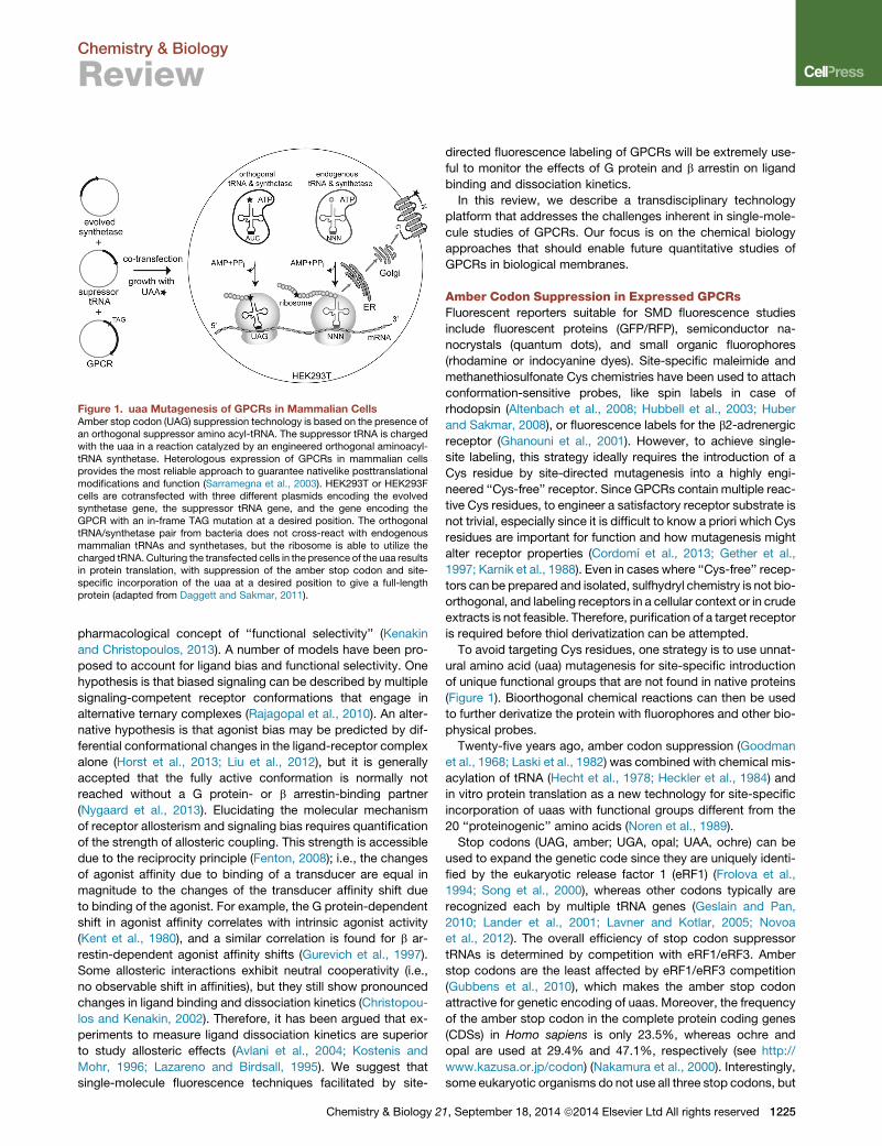
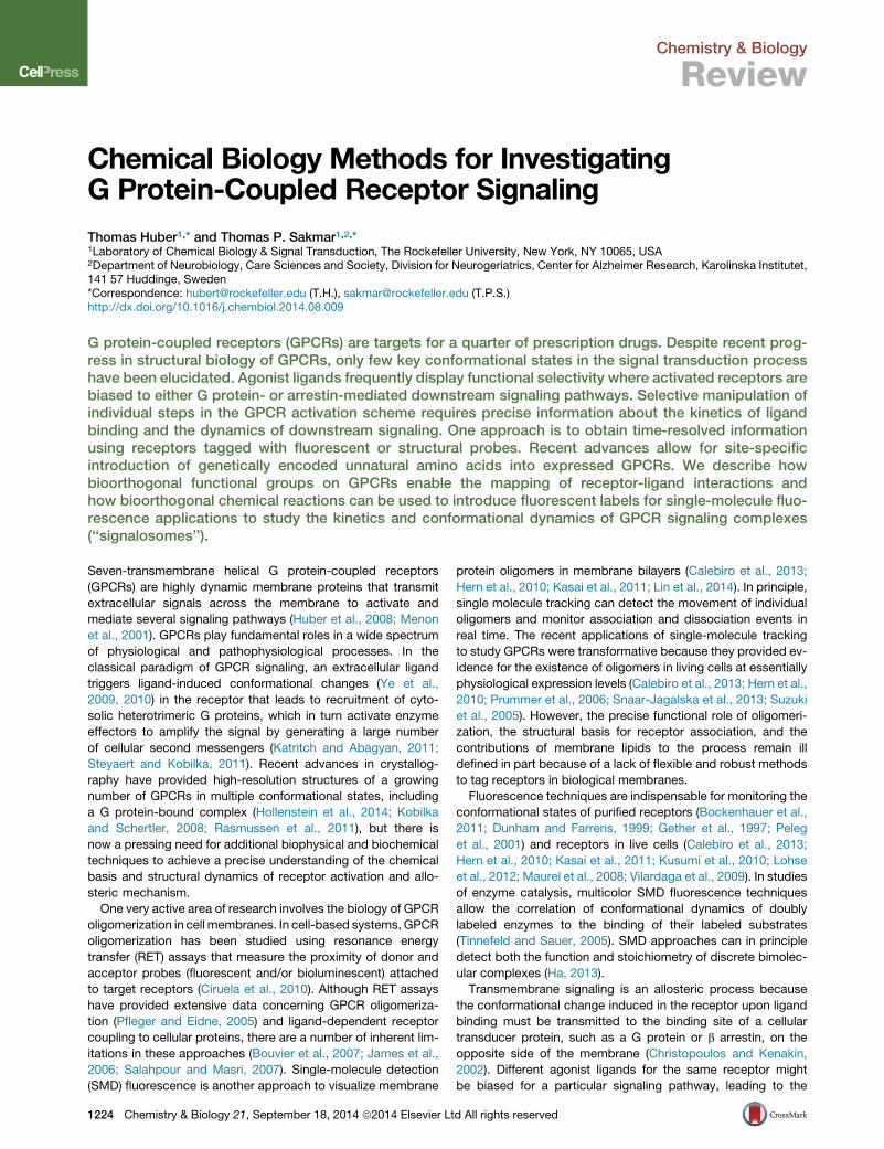
Figure 1. uaa Mutagenesis of GPCRs in Mammalian CellsAmber stop codon (UAG) suppression technology is based on the presence ofan orthogonal suppressor amino acyl-tRNA. The suppressor tRNA is chargedwith the uaa in a reaction catalyzed by an engineered orthogonal aminoacyl-tRNA synthetase. Heterologous expression of GPCRs in mammalian cellsprovides the most reliable approach to guarantee nativelike posttranslationalmodifications and function (Sarramegna et al., 2003). HEK293T or HEK293Fcells are cotransfected with three different plasmids encoding the evolvedsynthetase gene, the suppressor tRNA gene, and the gene encoding theGPCR with an in-frame TAG mutation at a desired position. The orthogonaltRNA/synthetase pair from bacteria does not cross-react with endogenousmammalian tRNAs and synthetases, but the ribosome is able to utilize thecharged tRNA. Culturing the transfected cells in the presence of the uaa resultsin protein translation, with suppression of the amber stop codon and site-specific incorporation of the uaa at a desired position to give a full-lengthprotein (adapted from Daggett and Sakmar, 2011).
Chemistry & Biology
Review
pharmacological concept of ‘‘functional selectivity’’ (Kenakin
and Christopoulos, 2013). A number of models have been pro-
posed to account for ligand bias and functional selectivity. One
hypothesis is that biased signaling can be described by multiple
signaling-competent receptor conformations that engage in
alternative ternary complexes (Rajagopal et al., 2010). An alter-
native hypothesis is that agonist bias may be predicted by dif-
ferential conformational changes in the ligand-receptor complex
alone (Horst et al., 2013; Liu et al., 2012), but it is generally
accepted that the fully active conformation is normally not
reached without a G protein- or b arrestin-binding partner
(Nygaard et al., 2013). Elucidating the molecular mechanism
of receptor allosterism and signaling bias requires quantification
of the strength of allosteric coupling. This strength is accessible
due to the reciprocity principle (Fenton, 2008); i.e., the changes
of agonist affinity due to binding of a transducer are equal in
magnitude to the changes of the transducer affinity shift due
to binding of the agonist. For example, the G protein-dependent
shift in agonist affinity correlates with intrinsic agonist activity
(Kent et al., 1980), and a similar correlation is found for b ar-
restin-dependent agonist affinity shifts (Gurevich et al., 1997).
Some allosteric interactions exhibit neutral cooperativity (i.e.,
no observable shift in affinities), but they still show pronounced
changes in ligand binding and dissociation kinetics (Christopou-
los and Kenakin, 2002). Therefore, it has been argued that ex-
periments to measure ligand dissociation kinetics are superior
to study allosteric effects (Avlani et al., 2004; Kostenis and
Mohr, 1996; Lazareno and Birdsall, 1995). We suggest that
single-molecule fluorescence techniques facilitated by site-
Chemistry & Biology 2
directed fluorescence labeling of GPCRs will be extremely use-
ful to monitor the effects of G protein and b arrestin on ligand
binding and dissociation kinetics.
In this review, we describe a transdisciplinary technology
platform that addresses the challenges inherent in single-mole-
cule studies of GPCRs. Our focus is on the chemical biology
approaches that should enable future quantitative studies of
GPCRs in biological membranes.
Amber Codon Suppression in Expressed GPCRsFluorescent reporters suitable for SMD fluorescence studies
include fluorescent proteins (GFP/RFP), semiconductor na-
nocrystals (quantum dots), and small organic fluorophores
(rhodamine or indocyanine dyes). Site-specific maleimide and
methanethiosulfonate Cys chemistries have been used to attach
conformation-sensitive probes, like spin labels in case of
rhodopsin (Altenbach et al., 2008; Hubbell et al., 2003; Huber
and Sakmar, 2008), or fluorescence labels for the b2-adrenergic
receptor (Ghanouni et al., 2001). However, to achieve single-
site labeling, this strategy ideally requires the introduction of a
Cys residue by site-directed mutagenesis into a highly engi-
neered ‘‘Cys-free’’ receptor. Since GPCRs contain multiple reac-
tive Cys residues, to engineer a satisfactory receptor substrate is
not trivial, especially since it is difficult to know a priori which Cys
residues are important for function and how mutagenesis might
alter receptor properties (Cordomı et al., 2013; Gether et al.,
1997; Karnik et al., 1988). Even in cases where ‘‘Cys-free’’ recep-
tors can be prepared and isolated, sulfhydryl chemistry is not bio-
orthogonal, and labeling receptors in a cellular context or in crude
extracts is not feasible. Therefore, purification of a target receptor
is required before thiol derivatization can be attempted.
To avoid targeting Cys residues, one strategy is to use unnat-
ural amino acid (uaa) mutagenesis for site-specific introduction
of unique functional groups that are not found in native proteins
(Figure 1). Bioorthogonal chemical reactions can then be used
to further derivatize the protein with fluorophores and other bio-
physical probes.
Twenty-five years ago, amber codon suppression (Goodman
et al., 1968; Laski et al., 1982) was combined with chemical mis-
acylation of tRNA (Hecht et al., 1978; Heckler et al., 1984) and
in vitro protein translation as a new technology for site-specific
incorporation of uaas with functional groups different from the
20 ‘‘proteinogenic’’ amino acids (Noren et al., 1989).
Stop codons (UAG, amber; UGA, opal; UAA, ochre) can be
used to expand the genetic code since they are uniquely identi-
fied by the eukaryotic release factor 1 (eRF1) (Frolova et al.,
1994; Song et al., 2000), whereas other codons typically are
recognized each by multiple tRNA genes (Geslain and Pan,
2010; Lander et al., 2001; Lavner and Kotlar, 2005; Novoa
et al., 2012). The overall efficiency of stop codon suppressor
tRNAs is determined by competition with eRF1/eRF3. Amber
stop codons are the least affected by eRF1/eRF3 competition
(Gubbens et al., 2010), which makes the amber stop codon
attractive for genetic encoding of uaas. Moreover, the frequency
of the amber stop codon in the complete protein coding genes
(CDSs) in Homo sapiens is only 23.5%, whereas ochre and
opal are used at 29.4% and 47.1%, respectively (see http://
www.kazusa.or.jp/codon) (Nakamura et al., 2000). Interestingly,
some eukaryotic organisms do not use all three stop codons, but
1, September 18, 2014 ª2014 Elsevier Ltd All rights reserved 1225
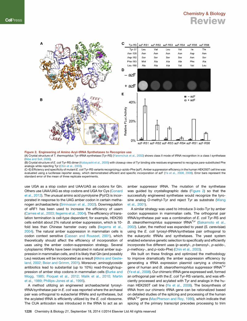
Figure 2. Engineering of Amino Acyl-tRNA Synthetases to Recognize uaa(A) Crystal structure of T. thermophilus Tyr-tRNA synthetase (Tyr-RS) (Yaremchuk et al., 2002) shows class II mode of tRNA recognition in a class I synthetase(Ibba and Soll, 2000).(B) Crystal structure of E. coli Tyr-RS dimer (Kobayashi et al., 2005) with closeup view of Tyr binding site residues engineered to recognize para-substituted Pheanalogs while rejecting Tyr (Chin et al., 2003).(C–E) Efficiency and specificity of mutant E. coli Tyr-RS variants recognizing p-azido-Phe (azF). Amber suppression efficiency in the human HEK293T cell line wasevaluated using a luciferase reporter assay, which demonstrated efficient and specific incorporation of azF (Ye et al., 2008, 2009). Error bars represent thestandard error of the mean of three replicate experiments.
Chemistry & Biology
Review
use UGA as a stop codon and UAA/UAG as codons for Gln.
Others use UAA/UAG as stop codons and UGA for Cys (Conard
et al., 2012). The unusual amino acid pyrrolysine (Pyl/O) is incor-
porated in response to the UAG amber codon in certain metha-
nogen archaebacteria (Srinivasan et al., 2002). Downregulation
of eRF1 has been used to increase the efficiency of uaam
(Carnes et al., 2003; Ilegems et al., 2004). The efficiency of trans-
lation termination is cell-type dependent; for example, HEK293
cells exhibit about 2% natural amber suppression, which is 10-
fold less than Chinese hamster ovary cells (Ilegems et al.,
2004). The natural amber suppression in mammalian cells is
codon context sensitive (Cassan and Rousset, 2001), which
theoretically should affect the efficiency of incorporation of
uaas using the amber codon-suppression strategy. Several
cytoplasmic tRNAs have been implicated in natural amber sup-
pression inmammalian cells, and it is likely that Gln (and possibly
Leu) residues will be incorporated as a result (Atkins and Geste-
land, 2002; Beier and Grimm, 2001). Moreover, aminoglycoside
antibiotics lead to substantial (up to 10%) read-through/sup-
pression of amber stop codons in mammalian cells (Burke and
Mogg, 1985; Floquet et al., 2012; Malik et al., 2010; Martin
et al., 1989; Phillips-Jones et al., 1995).
A method utilizing an engineered archaebacterial tyrosyl-
tRNA/synthetase pair in E. coli was reported where the archaeal
pair was orthogonal to eubacterial tRNAs and synthetases, but
the acylated tRNA is efficiently utilized by the E. coli ribosome.
The CUA anticodon was introduced in the tRNA to act as an
1226 Chemistry & Biology 21, September 18, 2014 ª2014 Elsevier Lt
amber suppressor tRNA. The mutation of the synthetase
was guided by crystallographic data (Figure 2) so that the
successfully engineered synthetase would recognize the tyro-
sine analog O-methyl-Tyr and reject Tyr as substrate (Wang
et al., 2001).
A similar strategy was used to introduce 3-iodo-Tyr by amber
codon suppression in mammalian cells. The orthogonal pair
tRNA/synthetase pair was a combination of E. coli Tyr-RS and
B. stearothermophilus suppressor tRNATyr (Sakamoto et al.,
2002). Later, the method was expanded to yeast (S. cerevisiae)
using the E. coli tyrosyl-tRNA/synthetase pair orthogonal to
the eukaryotic tRNAs and synthetases. The yeast system
enabled extensive genetic selection to specifically and efficiently
incorporate five different uaas (p-acetyl-, p-benzoyl-, p-azido-,
p-methoxy-, and p-iodo-Phe) (Chin et al., 2003).
We built on these findings and optimized the methodology
to improve dramatically the amber suppression efficiency by
generating a tRNA expression plasmid carrying a chimeric
gene of human and B. stearothermophilus suppressor tRNATyr
(Ye et al., 2008). Our chimeric tRNA gene expressed well, formed
an orthogonal pair with the E. coli Tyr-RS variants, and was effi-
ciently processed and acylated with Tyr and analogs in the hu-
man HEK293T cell line (Ye et al., 2008). The biosynthesis of
tRNA from our chimeric tRNA gene can be rationalized based
on detailed studies of the splicing and processing of the human
tRNATyr gene (MacPherson and Roy, 1986), which indicate that
spicing of the primary transcript precedes processing to trim
d All rights reserved
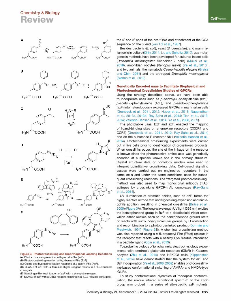
Figure 3. Photocrosslinking and Bioorthogonal Labeling Reactions(A) Photocrosslinking reaction with p-azido-Phe (azF).(B) Photocrosslinking reaction with p-benzoyl-Phe (BzF).(C) Oxime and hydrazone ligation reactions of p-acetyl-Phe (AcF).(D) CuAAC of azF with a terminal alkyne reagent results in a 1,2,3-triazoleconjugate.(E) Staudinger-Bertozzi ligation of azF with a phosphine reagent.(F) SpAAC of azF with a DIBO reagent resulting in a 1,2,3-triazole conjugate.
Chemistry & Biology 2
Chemistry & Biology
Review
the 50 and 30 ends of the pre-tRNA and attachment of the CCA
sequence on the 30 end (van Tol et al., 1987).
Besides bacteria (E. coli), yeast (S. cerevisiae), and mamma-
lian cells in culture (Chin, 2014; Liu and Schultz, 2010), uaamuta-
genesis methods have been developed for cultured insect cells
(Drosophila melanogaster Schneider 2 cells) (Mukai et al.,
2010), amphibian oocytes (Xenopus laevis) (Ye et al., 2013),
and two animals, the nematode Caenorhabditis elegans (Greiss
and Chin, 2011) and the arthropod Drosophila melanogaster
(Bianco et al., 2012).
Genetically Encoded uaas to Facilitate Biophysical andPhotochemical Crosslinking Studies of GPCRsUsing the strategy described above, we have been able
to incorporate uaas such as p-benzoyl-L-phenylalanine (BzF),
p-acetyl-L-phenylalanine (AcF), and p-azido-L-phenylalanine
(azF) into heterologously expressed GPCRs in mammalian cells
(Grunbeck et al., 2011, 2012; Huber et al., 2013; Naganathan
et al., 2013a, 2013b; Ray-Saha et al., 2014; Tian et al., 2013,
2014; Valentin-Hansen et al., 2014; Ye et al., 2008, 2009).
The photolabile uaas, BzF and azF, enabled the mapping
of ligand-binding sites on chemokine receptors (CXCR4 and
CCR5) (Grunbeck et al., 2011, 2012; Ray-Saha et al., 2014)
and on the substance P receptor NK1 (Valentin-Hansen et al.,
2014). Photochemical crosslinking experiments were carried
out in live cells prior to identification of crosslinked products.
When crosslinks occur, the site of the linkage on the receptor
is known since the photoreactive amino acid was genetically
encoded at a specific known site in the primary structure.
Crystal structure data or homology models were used to
interpret quantitative crosslinking data. Cell-based signaling
assays were carried out on engineered receptors in the
same cells and under the same conditions used for subse-
quent crosslinking reactions. The ‘‘targeted photocrosslinking’’
method was also used to map monoclonal antibody (mAb)
epitopes by crosslinking GPCR-mAb complexes (Ray-Saha
et al., 2014).
UV illumination of aromatic azides, such as azF, forms the
highly reactive nitrone that undergoes ring expansion and nucle-
ophile addition, resulting in chemical crosslinks (Brase et al.,
2005) (Figure 3A). The long-wavelength UV light (365 nm) excites
the benzophenone group in BzF to a diradicaloid triplet state,
which either relaxes back to the benzophenone ground state
or reacts with surrounding molecular groups by H abstraction
and recombination to a photocrosslinked product (Dorman and
Prestwich, 1994) (Figure 3B). A chemical crosslinking method
was also reported using a p-fluoroacetyl-Phe (Ffact) residue in
the receptor that reacts with a nearby Cys residue introduced
in a peptide ligand (Coin et al., 2013).
To probe the biology of ion channels, electrophysiology exper-
iments with ionotropic glutamate receptors (iGluR) in Xenopus
oocytes (Zhu et al., 2014) and HEK293 cells (Klippenstein
et al., 2014) have demonstrated that the system for azF and
BzF incorporation (Ye et al., 2008, 2009) enables photocrosslink-
ing-based conformational switching of AMPA- and NMDA-type
iGluRs.
To study conformational dynamics of rhodopsin photoacti-
vation, the unique infrared vibrational spectrum of the azido
group was probed in a series of site-specific azF mutants.
1, September 18, 2014 ª2014 Elsevier Ltd All rights reserved 1227
Chemistry & Biology
Review
Fourier-transform infrared (FTIR)-difference spectra were
measured to show that the electrostatic environments of some
residues change very early in the activation process (Ye et al.,
2009, 2010). These types of receptor dynamics experiments
can be correlated with high-resolution structures of receptor
activation intermediates to gain significant insights into the
biology of receptor signaling.
Bioorthogonal Labeling StrategiesDifferent bioorthogonal labeling strategies using AcF or azF have
been compared (Huber et al., 2013; Naganathan et al., 2013a,
2013b; Tian et al., 2013, 2014; Ye et al., 2008). Ketone groups
incorporated into a variety of proteins and other macromolecules
have facilitated novel ligation strategies such as hydrazone
and oxime ligation reactions that utilize aminooxy/hydroxylamine
(R = –O–R’) or hydrazide (R = –NH–CO–R’’) reagents (Figure 3C).
Under approximately physiological conditions, ketones and al-
dehydes react specifically with hydrazide (or aminooxy) reagents
through hydrazone (or oxime) ligation (Bayer et al., 1988; Wang
et al., 2003). The observation that aminooxy derivatives have
faster labeling kinetics at close to neutral pH as compared with
hydrazides is advantageous for protein labeling. These reactions
have been used to label proteins at genetically encoded
N-terminal aldehyde tags (Shi et al., 2012), enzymatically
coupled chemical handles (Chen et al., 2005), engineered cell
surface oligosaccharides (Hang and Bertozzi, 2001; Mahal
et al., 1997), and with more flexibility at site-specifically incorpo-
rated uaas (Cornish et al., 1996; de Graaf et al., 2009; Fleissner
et al., 2009; Wang and Schultz, 2004; Wang et al., 2003; Ye
et al., 2008; Zhang et al., 2003).
We observed substantial covalent modification of wild-type
rhodopsin and other control proteins that did not contain AcF
(Huber et al., 2013; Tian et al., 2014; Ye et al., 2008). This unde-
sired reactivity was caused by cellular oxidation processes
that convert some amino acid residues (Arg, Cys, His, Lys,
and Pro) to carbonyl derivatives that are reactive toward hy-
drazide and aminooxy reagents (Ahn et al., 1987; Stadtman
and Levine, 2003). This ‘‘protein carbonylation’’ phenomenon
severely limits the applicability of ketone-selective bio-
orthogonal labeling reactions (Grimsrud et al., 2008; Stadtman,
1993). The degree of target-specific background labeling due to
protein carbonylation will be roughly proportional to the pro-
tein’s molecular mass, with about one reactive carbonyl per
each thousand amino acid residues (Huber et al., 2013; Ye
et al., 2008).
Advantages of Bioorthogonal Azido TagsThe azido group has been reported to be a superior handle for
bioconjugation reactions (Brase et al., 2005; Debets et al.,
2010b; Schilling et al., 2011; van Berkel et al., 2011). Azide pro-
vides an alternative bioorthogonal handle that can be incorpo-
rated in proteins by uaa mutagenesis. Azides react specifically
with phosphines through the Staudinger-Bertozzi ligation (see
below) (Kiick et al., 2002; Saxon and Bertozzi, 2000), with alkyne
reagents through Cu(I)-catalyzed [3+2] azide-alkyne cycload-
dition (CuAAC) (Meldal and Tornøe, 2008; Rostovtsev et al.,
2002; Tornøe et al., 2002), with oxanorbornadienes through
tandem [3+2] cycloaddition-retro-Diels-Alder (tandem crDA)
(van Berkel et al., 2007), or with cyclooctynes through strain-pro-
1228 Chemistry & Biology 21, September 18, 2014 ª2014 Elsevier Lt
moted azide-alkyne cycloaddition (SpAAC) (Agard et al., 2004;
Debets et al., 2011; Link et al., 2006; Sletten and Bertozzi, 2011).
The discovery of a cell-compatible and selective ligation re-
action modeled after the classical Staudinger reaction between
an azide and a phosphine inspired the nascent field of bio-
orthogonal chemical reactions. This reaction, sometimes called
Staudinger-Bertozzi ligation, is a highly selective reaction
between modified triarylphosphines and azides to form an
aza-ylide intermediate that undergoes cyclization followed by
hydrolysis to form a stable amide-linked adduct (Kohn and
Breinbauer, 2004; Saxon and Bertozzi, 2000; Saxon et al.,
2002; Tsao et al., 2005). It has been used to modify cell surface
glycans with haptenes, epitope tags, and fluorescent probes
(Chang et al., 2007; Cohen et al., 2010; Hangauer and Bertozzi,
2008; Kiick et al., 2002; Laughlin and Bertozzi, 2007; Lemieux
et al., 2003; Saxon and Bertozzi, 2000). It has also been used
to label azido amino acids in proteins in residue-specific (Kiick
et al., 2002) or site-specific (Mukai et al., 2010; Tsao et al.,
2005) manners. In another study, FLAG phosphine (FLAG is
a peptide epitope for a useful well-characterized mAb) was
used to label azido-bearing biotin. The FLAG biotin was then
used to tag an endogenous biotin acceptor protein (Slavoff
et al., 2008).
Despite the susceptibility of phosphines used for the Stau-
dinger ligation to air oxidation (Agard et al., 2004) and their sub-
stoichiometric modification of azF in recombinant proteins
(Huber et al., 2013; Yanagisawa et al., 2008) and azido sugars
(Agard et al., 2006), their compatibility to cell-based applica-
tions (Prescher et al., 2004) makes them attractive candidates
for further study.
In studies on GPCR labeling, we found that the reaction
yields essentially background-free product, consistent with
a truly bioorthogonal reaction (Huber et al., 2013). However,
despite reports of stoichiometric conjugation of azF in other
recombinant proteins with fluorescein-phosphine (Tsao et al.,
2005), we found that substoichiometric conjugation to azF
rhodopsin is not the result of its relatively slow reaction kinetics,
but possibly due to a classical Staudinger reaction instead
of the desired Staudinger-Bertozzi ligation. Consistent with
our findings, others have reported incomplete modification
of azF and a longer azido-containing pyrrolysine analog by
Staudinger ligation, which was attributed to the shorter linker
length between the backbone and the azide (Yanagisawa
et al., 2008).
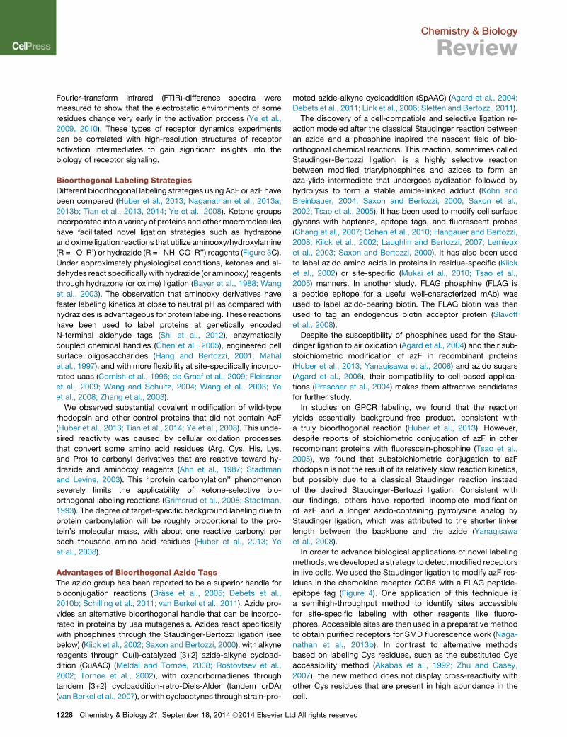
In order to advance biological applications of novel labeling
methods, we developed a strategy to detect modified receptors
in live cells. We used the Staudinger ligation to modify azF res-
idues in the chemokine receptor CCR5 with a FLAG peptide-
epitope tag (Figure 4). One application of this technique is
a semihigh-throughput method to identify sites accessible
for site-specific labeling with other reagents like fluoro-
phores. Accessible sites are then used in a preparative method
to obtain purified receptors for SMD fluorescence work (Naga-
nathan et al., 2013b). In contrast to alternative methods
based on labeling Cys residues, such as the substituted Cys
accessibility method (Akabas et al., 1992; Zhu and Casey,
2007), the new method does not display cross-reactivity with
other Cys residues that are present in high abundance in the
cell.
d All rights reserved
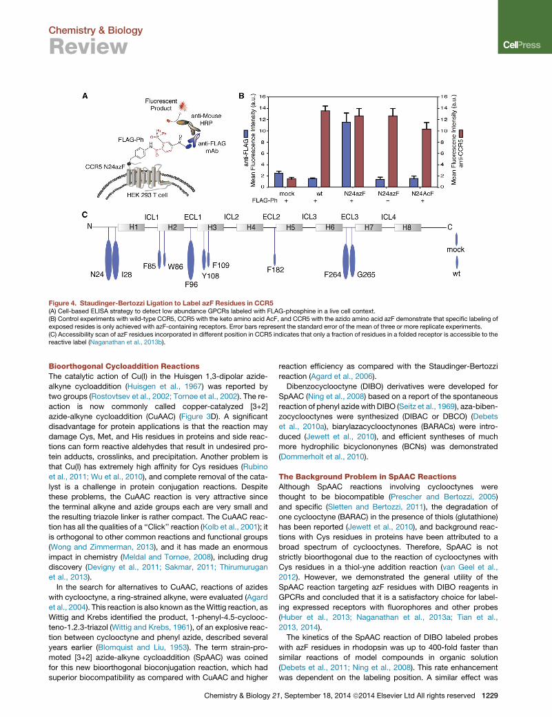
Figure 4. Staudinger-Bertozzi Ligation to Label azF Residues in CCR5(A) Cell-based ELISA strategy to detect low abundance GPCRs labeled with FLAG-phosphine in a live cell context.(B) Control experiments with wild-type CCR5, CCR5 with the keto amino acid AcF, and CCR5 with the azido amino acid azF demonstrate that specific labeling ofexposed resides is only achieved with azF-containing receptors. Error bars represent the standard error of the mean of three or more replicate experiments.(C) Accessibility scan of azF residues incorporated in different position in CCR5 indicates that only a fraction of residues in a folded receptor is accessible to thereactive label (Naganathan et al., 2013b).
Chemistry & Biology
Review
Bioorthogonal Cycloaddition ReactionsThe catalytic action of Cu(I) in the Huisgen 1,3-dipolar azide-
alkyne cycloaddition (Huisgen et al., 1967) was reported by
two groups (Rostovtsev et al., 2002; Tornøe et al., 2002). The re-
action is now commonly called copper-catalyzed [3+2]
azide-alkyne cycloaddition (CuAAC) (Figure 3D). A significant
disadvantage for protein applications is that the reaction may
damage Cys, Met, and His residues in proteins and side reac-
tions can form reactive aldehydes that result in undesired pro-
tein adducts, crosslinks, and precipitation. Another problem is
that Cu(I) has extremely high affinity for Cys residues (Rubino
et al., 2011; Wu et al., 2010), and complete removal of the cata-
lyst is a challenge in protein conjugation reactions. Despite
these problems, the CuAAC reaction is very attractive since
the terminal alkyne and azide groups each are very small and
the resulting triazole linker is rather compact. The CuAAC reac-
tion has all the qualities of a ‘‘Click’’ reaction (Kolb et al., 2001); it
is orthogonal to other common reactions and functional groups
(Wong and Zimmerman, 2013), and it has made an enormous
impact in chemistry (Meldal and Tornøe, 2008), including drug
discovery (Devigny et al., 2011; Sakmar, 2011; Thirumurugan
et al., 2013).
In the search for alternatives to CuAAC, reactions of azides
with cyclooctyne, a ring-strained alkyne, were evaluated (Agard
et al., 2004). This reaction is also known as theWittig reaction, as
Wittig and Krebs identified the product, 1-phenyl-4.5-cyclooc-
teno-1.2.3-triazol (Wittig and Krebs, 1961), of an explosive reac-
tion between cyclooctyne and phenyl azide, described several
years earlier (Blomquist and Liu, 1953). The term strain-pro-
moted [3+2] azide-alkyne cycloaddition (SpAAC) was coined
for this new bioorthogonal bioconjugation reaction, which had
superior biocompatibility as compared with CuAAC and higher
Chemistry & Biology 2
reaction efficiency as compared with the Staudinger-Bertozzi
reaction (Agard et al., 2006).
Dibenzocyclooctyne (DIBO) derivatives were developed for
SpAAC (Ning et al., 2008) based on a report of the spontaneous
reaction of phenyl azide with DIBO (Seitz et al., 1969), aza-biben-
zocyclooctynes were synthesized (DIBAC or DBCO) (Debets
et al., 2010a), biarylazacyclooctynones (BARACs) were intro-
duced (Jewett et al., 2010), and efficient syntheses of much
more hydrophilic bicyclononynes (BCNs) was demonstrated
(Dommerholt et al., 2010).
The Background Problem in SpAAC ReactionsAlthough SpAAC reactions involving cyclooctynes were
thought to be biocompatible (Prescher and Bertozzi, 2005)
and specific (Sletten and Bertozzi, 2011), the degradation of
one cyclooctyne (BARAC) in the presence of thiols (glutathione)
has been reported (Jewett et al., 2010), and background reac-
tions with Cys residues in proteins have been attributed to a
broad spectrum of cyclooctynes. Therefore, SpAAC is not
strictly bioorthogonal due to the reaction of cyclooctynes with
Cys residues in a thiol-yne addition reaction (van Geel et al.,
2012). However, we demonstrated the general utility of the
SpAAC reaction targeting azF residues with DIBO reagents in
GPCRs and concluded that it is a satisfactory choice for label-
ing expressed receptors with fluorophores and other probes
(Huber et al., 2013; Naganathan et al., 2013a; Tian et al.,
2013, 2014).
The kinetics of the SpAAC reaction of DIBO labeled probes
with azF residues in rhodopsin was up to 400-fold faster than
similar reactions of model compounds in organic solution
(Debets et al., 2011; Ning et al., 2008). This rate enhancement
was dependent on the labeling position. A similar effect was
1, September 18, 2014 ª2014 Elsevier Ltd All rights reserved 1229
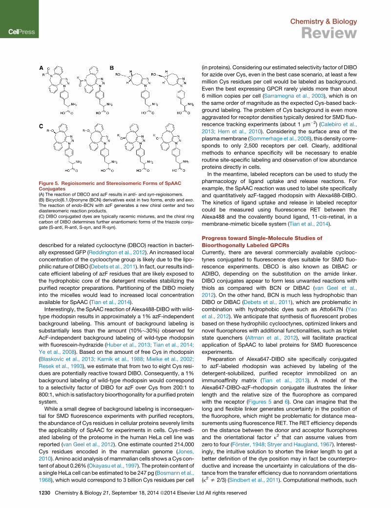
Figure 5. Regioisomeric and Stereoisomeric Forms of SpAACConjugates(A) The reaction of DBCO and azF results in anti- and syn-regioisomers.(B) Bicyclo[6.1.0]nonyne (BCN) derivatives exist in two forms, endo and exo.The reaction of endo-BCN with azF generates a new chiral center and twodiastereomeric reaction products.(C) DIBO conjugated dyes are typically racemic mixtures, and the chiral ringcarbon of DIBO determines further enantiomeric forms of the triazole conju-gate (S-anti, R-anti, S-syn, and R-syn).
Chemistry & Biology
Review
described for a related cyclooctyne (DBCO) reaction in bacteri-
ally expressed GFP (Reddington et al., 2012). An increased local
concentration of the cyclooctyne group is likely due to the lipo-
philic nature of DIBO (Debets et al., 2011). In fact, our results indi-
cate efficient labeling of azF residues that are likely exposed to
the hydrophobic core of the detergent micelles stabilizing the
purified receptor preparations. Partitioning of the DIBO moiety
into the micelles would lead to increased local concentration
available for SpAAC (Tian et al., 2014).
Interestingly, the SpAAC reaction of Alexa488-DIBO with wild-
type rhodopsin results in approximately a 1% azF-independent
background labeling. This amount of background labeling is
substantially less than the amount (10%�30%) observed for
AcF-independent background labeling of wild-type rhodopsin
with fluorescein-hydrazide (Huber et al., 2013; Tian et al., 2014;
Ye et al., 2008). Based on the amount of free Cys in rhodopsin
(Blaskovic et al., 2013; Karnik et al., 1988; Mielke et al., 2002;
Resek et al., 1993), we estimate that from two to eight Cys resi-
dues are potentially reactive toward DIBO. Consequently, a 1%
background labeling of wild-type rhodopsin would correspond
to a selectivity factor of DIBO for azF over Cys from 200:1 to
800:1, which is satisfactory bioorthogonality for a purified protein
system.
While a small degree of background labeling is inconsequen-
tial for SMD fluorescence experiments with purified receptors,
the abundance of Cys residues in cellular proteins severely limits
the applicability of SpAAC for experiments in cells. Cys-medi-
ated labeling of the proteome in the human HeLa cell line was
reported (van Geel et al., 2012). One estimate counted 214,000
Cys residues encoded in the mammalian genome (Jones,
2010). Amino acid analysis of mammalian cells shows aCys con-
tent of about 0.26% (Okayasu et al., 1997). The protein content of
a single HeLa cell can be estimated to be 247 pg (Bosmann et al.,
1968), which would correspond to 3 billion Cys residues per cell
1230 Chemistry & Biology 21, September 18, 2014 ª2014 Elsevier Lt
(in proteins). Considering our estimated selectivity factor of DIBO
for azide over Cys, even in the best case scenario, at least a few
million Cys residues per cell would be labeled as background.
Even the best expressing GPCR rarely yields more than about
6 million copies per cell (Sarramegna et al., 2003), which is on
the same order of magnitude as the expected Cys-based back-
ground labeling. The problem of Cys background is even more
aggravated for receptor densities typically desired for SMD fluo-
rescence tracking experiments (about 1 mm�2) (Calebiro et al.,
2013; Hern et al., 2010). Considering the surface area of the
plasmamembrane (Sommerhage et al., 2008), this density corre-
sponds to only 2,500 receptors per cell. Clearly, additional
methods to enhance specificity will be necessary to enable
routine site-specific labeling and observation of low abundance
proteins directly in cells.
In the meantime, labeled receptors can be used to study the
pharmacology of ligand uptake and release reactions. For
example, the SpAAC reaction was used to label site specifically
and quantitatively azF-tagged rhodopsin with Alexa488-DIBO.
The kinetics of ligand uptake and release in labeled receptor
could be measured using fluorescence RET between the
Alexa488 and the covalently bound ligand, 11-cis-retinal, in a
membrane-mimetic bicelle system (Tian et al., 2014).
Progress toward Single-Molecule Studies ofBioorthogonally Labeled GPCRsCurrently, there are several commercially available cyclooc-
tynes conjugated to fluorescence dyes suitable for SMD fluo-
rescence experiments. DBCO is also known as DIBAC or
ADIBO, depending on the substitution on the amide linker.
DIBO conjugates appear to form less unwanted reactions with
thiols as compared with BCN or DIBAC (van Geel et al.,
2012). On the other hand, BCN is much less hydrophobic than
DIBO or DIBAC (Debets et al., 2011), which are problematic in
combination with hydrophobic dyes such as Atto647N (Yao
et al., 2012). We anticipate that synthesis of fluorescent probes
based on these hydrophilic cyclooctynes, optimized linkers and
novel fluorophores with additional functionalities, such as triplet
state quenchers (Altman et al., 2012), will facilitate practical
application of SpAAC to label proteins for SMD fluorescence
experiments.
Preparation of Alexa647-DIBO site specifically conjugated
to azF-labeled rhodopsin was achieved by labeling of the
detergent-solubilized, purified receptor immobilized on an
immunoaffinity matrix (Tian et al., 2013). A model of the
Alexa647-DIBO-azF-rhodopsin conjugate illustrates the linker
length and the relative size of the fluorophore as compared
with the receptor (Figures 5 and 6). One can imagine that the
long and flexible linker generates uncertainty in the position of
the fluorophore, which might be problematic for distance mea-
surements using fluorescence RET. The RET efficiency depends
on the distance between the donor and acceptor fluorophores
and the orientational factor k2 that can assume values from
zero to four (Forster, 1948; Stryer and Haugland, 1967). Interest-
ingly, the intuitive solution to shorten the linker length to get a
better definition of the dye position may in fact be counterpro-
ductive and increase the uncertainty in calculations of the dis-
tance from the transfer efficiency due to nonrandom orientations
(k2 s 2/3) (Sindbert et al., 2011). Computational methods, such
d All rights reserved
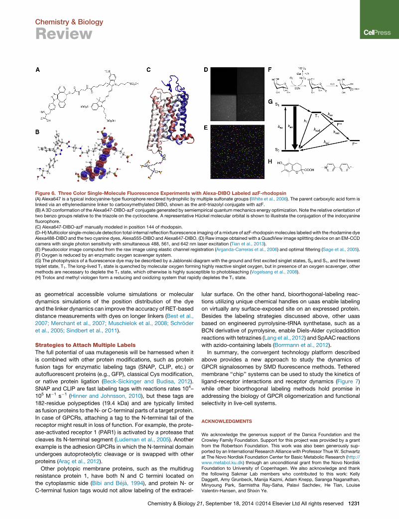
Figure 6. Three Color Single-Molecule Fluorescence Experiments with Alexa-DIBO Labeled azF-rhodopsin(A) Alexa647 is a typical indocyanine-type fluorophore rendered hydrophilic by multiple sulfonate groups (White et al., 2006). The parent carboxylic acid form islinked via an ethylenediamine linker to carboxymethylated DIBO, shown as the anti-triazolyl conjugate with azF.(B) A 3D conformation of the Alexa647-DIBO-azF conjugate generated by semiempirical quantummechanics energy optimization. Note the relative orientation oftwo benzo groups relative to the triazole on the cyclooctene. A representative Huckel molecular orbital is shown to illustrate the conjugation of the indocyaninefluorophore.(C) Alexa647-DIBO-azF manually modeled in position 144 of rhodopsin.(D–H) Multicolor single-molecule detection total-internal reflection fluorescence imaging of amixture of azF-rhodopsin molecules labeled with the rhodamine dyeAlexa488-DIBO and the two cyanine dyes, Alexa555-DIBO and Alexa647-DIBO. (D) Raw image obtained with a QuadView image splitting device on an EM-CCDcamera with single photon sensitivity with simultaneous 488, 561, and 642 nm laser excitation (Tian et al., 2013).(E) Pseudocolor image computed from the raw image using elastic channel registration (Arganda-Carreras et al., 2006) and optimal filtering (Sage et al., 2005).(F) Oxygen is reduced by an enzymatic oxygen scavenger system.(G) The photophysics of a fluorescence dye may be described by a Jablonski diagram with the ground and first excited singlet states, S0 and S1, and the lowesttriplet state, T1. The long-lived T1 state is quenched by molecular oxygen forming highly reactive singlet oxygen, but in presence of an oxygen scavenger, othermethods are necessary to deplete the T1 state, which otherwise is highly susceptible to photobleaching (Vogelsang et al., 2008).(H) Trolox and methyl viologen form a reducing and oxidizing system that rapidly depletes the T1 state.
Chemistry & Biology
Review
as geometrical accessible volume simulations or molecular
dynamics simulations of the position distribution of the dye
and the linker dynamics can improve the accuracy of RET-based
distance measurements with dyes on longer linkers (Best et al.,
2007; Merchant et al., 2007; Muschielok et al., 2008; Schroder
et al., 2005; Sindbert et al., 2011).
Strategies to Attach Multiple LabelsThe full potential of uaa mutagenesis will be harnessed when it
is combined with other protein modifications, such as protein
fusion tags for enzymatic labeling tags (SNAP, CLIP, etc.) or
autofluorescent proteins (e.g., GFP), classical Cys modification,
or native protein ligation (Beck-Sickinger and Budisa, 2012).
SNAP and CLIP are fast labeling tags with reactions rates 104–
105 M�1 s�1 (Hinner and Johnsson, 2010), but these tags are
182-residue polypeptides (19.4 kDa) and are typically limited
as fusion proteins to theN- or C-terminal parts of a target protein.
In case of GPCRs, attaching a tag to the N-terminal tail of the
receptor might result in loss of function. For example, the prote-
ase-activated receptor 1 (PAR1) is activated by a protease that
cleaves its N-terminal segment (Ludeman et al., 2005). Another
example is the adhesion GPCRs in which the N-terminal domain
undergoes autoproteolytic cleavage or is swapped with other
proteins (Arac et al., 2012).
Other polytopic membrane proteins, such as the multidrug
resistance protein 1, have both N and C termini located on
the cytoplasmic side (Bibi and Beja, 1994), and protein N- or
C-terminal fusion tags would not allow labeling of the extracel-
Chemistry & Biology 2
lular surface. On the other hand, bioorthogonal-labeling reac-
tions utilizing unique chemical handles on uaas enable labeling
on virtually any surface-exposed site on an expressed protein.
Besides the labeling strategies discussed above, other uaas
based on engineered pyrrolysine-tRNA synthetase, such as a
BCN derivative of pyrrolysine, enable Diels-Alder cycloaddition
reactions with tetrazines (Lang et al., 2012) and SpAAC reactions
with azido-containing labels (Borrmann et al., 2012).
In summary, the convergent technology platform described
above provides a new approach to study the dynamics of
GPCR signalosomes by SMD fluorescence methods. Tethered
membrane ‘‘chip’’ systems can be used to study the kinetics of
ligand-receptor interactions and receptor dynamics (Figure 7)
while other bioorthogonal labeling methods hold promise in
addressing the biology of GPCR oligomerization and functional
selectivity in live-cell systems.
ACKNOWLEDGMENTS
We acknowledge the generous support of the Danica Foundation and theCrowley Family Foundation. Support for this project was provided by a grantfrom the Robertson Foundation. This work was also been generously sup-ported by an International Research Alliance with Professor Thue W. Schwartzat The Novo Nordisk Foundation Center for Basic Metabolic Research (http://www.metabol.ku.dk) through an unconditional grant from the Novo NordiskFoundation to University of Copenhagen. We also acknowledge and thankthe following Sakmar Lab members who contributed to this work: KellyDaggett, Amy Grunbeck, Manija Kazmi, Adam Knepp, Saranga Naganathan,Minyoung Park, Sarmistha Ray-Saha, Palavi Sachdev, He Tian, LouiseValentin-Hansen, and Shixin Ye.
1, September 18, 2014 ª2014 Elsevier Ltd All rights reserved 1231
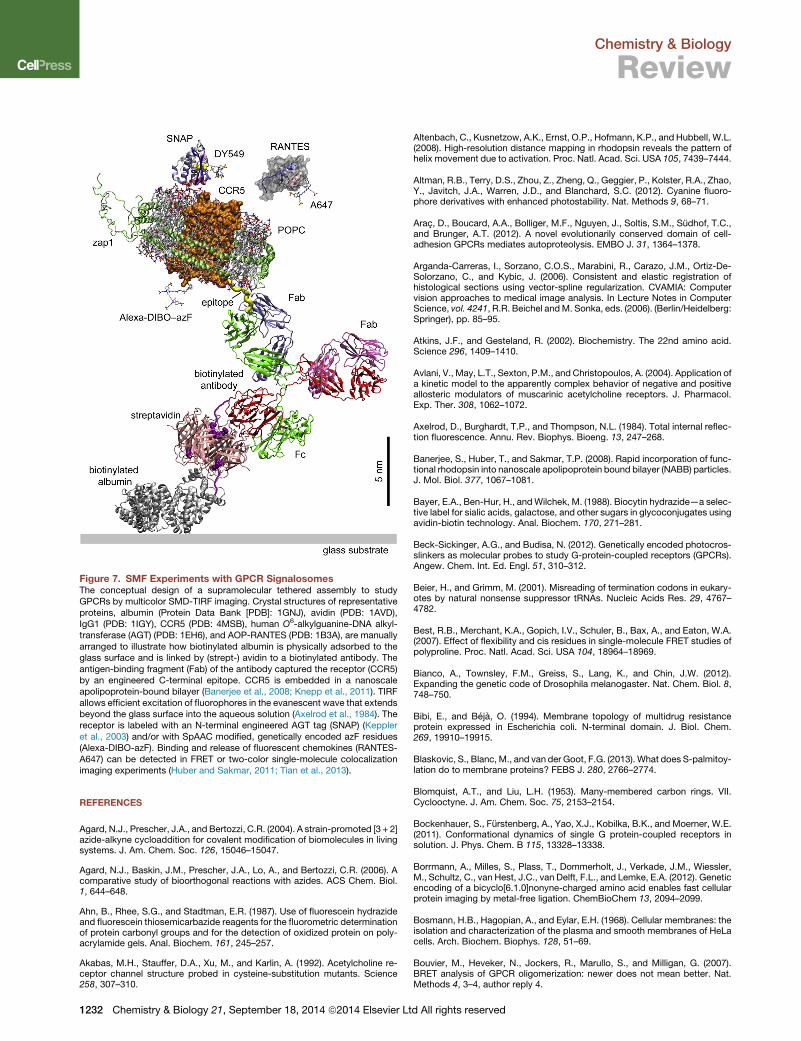
Figure 7. SMF Experiments with GPCR SignalosomesThe conceptual design of a supramolecular tethered assembly to studyGPCRs by multicolor SMD-TIRF imaging. Crystal structures of representativeproteins, albumin (Protein Data Bank [PDB]: 1GNJ), avidin (PDB: 1AVD),IgG1 (PDB: 1IGY), CCR5 (PDB: 4MSB), human O6-alkylguanine-DNA alkyl-transferase (AGT) (PDB: 1EH6), and AOP-RANTES (PDB: 1B3A), are manuallyarranged to illustrate how biotinylated albumin is physically adsorbed to theglass surface and is linked by (strept-) avidin to a biotinylated antibody. Theantigen-binding fragment (Fab) of the antibody captured the receptor (CCR5)by an engineered C-terminal epitope. CCR5 is embedded in a nanoscaleapolipoprotein-bound bilayer (Banerjee et al., 2008; Knepp et al., 2011). TIRFallows efficient excitation of fluorophores in the evanescent wave that extendsbeyond the glass surface into the aqueous solution (Axelrod et al., 1984). Thereceptor is labeled with an N-terminal engineered AGT tag (SNAP) (Keppleret al., 2003) and/or with SpAAC modified, genetically encoded azF residues(Alexa-DIBO-azF). Binding and release of fluorescent chemokines (RANTES-A647) can be detected in FRET or two-color single-molecule colocalizationimaging experiments (Huber and Sakmar, 2011; Tian et al., 2013).
Chemistry & Biology
Review
REFERENCES
Agard, N.J., Prescher, J.A., and Bertozzi, C.R. (2004). A strain-promoted [3 + 2]azide-alkyne cycloaddition for covalent modification of biomolecules in livingsystems. J. Am. Chem. Soc. 126, 15046–15047.
Agard, N.J., Baskin, J.M., Prescher, J.A., Lo, A., and Bertozzi, C.R. (2006). Acomparative study of bioorthogonal reactions with azides. ACS Chem. Biol.1, 644–648.
Ahn, B., Rhee, S.G., and Stadtman, E.R. (1987). Use of fluorescein hydrazideand fluorescein thiosemicarbazide reagents for the fluorometric determinationof protein carbonyl groups and for the detection of oxidized protein on poly-acrylamide gels. Anal. Biochem. 161, 245–257.
Akabas, M.H., Stauffer, D.A., Xu, M., and Karlin, A. (1992). Acetylcholine re-ceptor channel structure probed in cysteine-substitution mutants. Science258, 307–310.
1232 Chemistry & Biology 21, September 18, 2014 ª2014 Elsevier Lt
Altenbach, C., Kusnetzow, A.K., Ernst, O.P., Hofmann, K.P., and Hubbell, W.L.(2008). High-resolution distance mapping in rhodopsin reveals the pattern ofhelix movement due to activation. Proc. Natl. Acad. Sci. USA 105, 7439–7444.
Altman, R.B., Terry, D.S., Zhou, Z., Zheng, Q., Geggier, P., Kolster, R.A., Zhao,Y., Javitch, J.A., Warren, J.D., and Blanchard, S.C. (2012). Cyanine fluoro-phore derivatives with enhanced photostability. Nat. Methods 9, 68–71.
Arac, D., Boucard, A.A., Bolliger, M.F., Nguyen, J., Soltis, S.M., Sudhof, T.C.,and Brunger, A.T. (2012). A novel evolutionarily conserved domain of cell-adhesion GPCRs mediates autoproteolysis. EMBO J. 31, 1364–1378.
Arganda-Carreras, I., Sorzano, C.O.S., Marabini, R., Carazo, J.M., Ortiz-De-Solorzano, C., and Kybic, J. (2006). Consistent and elastic registration ofhistological sections using vector-spline regularization. CVAMIA: Computervision approaches to medical image analysis. In Lecture Notes in ComputerScience, vol. 4241, R.R. Beichel andM. Sonka, eds. (2006). (Berlin/Heidelberg:Springer), pp. 85–95.
Atkins, J.F., and Gesteland, R. (2002). Biochemistry. The 22nd amino acid.Science 296, 1409–1410.
Avlani, V., May, L.T., Sexton, P.M., and Christopoulos, A. (2004). Application ofa kinetic model to the apparently complex behavior of negative and positiveallosteric modulators of muscarinic acetylcholine receptors. J. Pharmacol.Exp. Ther. 308, 1062–1072.
Axelrod, D., Burghardt, T.P., and Thompson, N.L. (1984). Total internal reflec-tion fluorescence. Annu. Rev. Biophys. Bioeng. 13, 247–268.
Banerjee, S., Huber, T., and Sakmar, T.P. (2008). Rapid incorporation of func-tional rhodopsin into nanoscale apolipoprotein bound bilayer (NABB) particles.J. Mol. Biol. 377, 1067–1081.
Bayer, E.A., Ben-Hur, H., andWilchek, M. (1988). Biocytin hydrazide—a selec-tive label for sialic acids, galactose, and other sugars in glycoconjugates usingavidin-biotin technology. Anal. Biochem. 170, 271–281.
Beck-Sickinger, A.G., and Budisa, N. (2012). Genetically encoded photocros-slinkers as molecular probes to study G-protein-coupled receptors (GPCRs).Angew. Chem. Int. Ed. Engl. 51, 310–312.
Beier, H., and Grimm, M. (2001). Misreading of termination codons in eukary-otes by natural nonsense suppressor tRNAs. Nucleic Acids Res. 29, 4767–4782.
Best, R.B., Merchant, K.A., Gopich, I.V., Schuler, B., Bax, A., and Eaton, W.A.(2007). Effect of flexibility and cis residues in single-molecule FRET studies ofpolyproline. Proc. Natl. Acad. Sci. USA 104, 18964–18969.
Bianco, A., Townsley, F.M., Greiss, S., Lang, K., and Chin, J.W. (2012).Expanding the genetic code of Drosophila melanogaster. Nat. Chem. Biol. 8,748–750.
Bibi, E., and Beja, O. (1994). Membrane topology of multidrug resistanceprotein expressed in Escherichia coli. N-terminal domain. J. Biol. Chem.269, 19910–19915.
Blaskovic, S., Blanc, M., and van der Goot, F.G. (2013). What does S-palmitoy-lation do to membrane proteins? FEBS J. 280, 2766–2774.
Blomquist, A.T., and Liu, L.H. (1953). Many-membered carbon rings. VII.Cyclooctyne. J. Am. Chem. Soc. 75, 2153–2154.
Bockenhauer, S., Furstenberg, A., Yao, X.J., Kobilka, B.K., and Moerner, W.E.(2011). Conformational dynamics of single G protein-coupled receptors insolution. J. Phys. Chem. B 115, 13328–13338.
Borrmann, A., Milles, S., Plass, T., Dommerholt, J., Verkade, J.M., Wiessler,M., Schultz, C., van Hest, J.C., van Delft, F.L., and Lemke, E.A. (2012). Geneticencoding of a bicyclo[6.1.0]nonyne-charged amino acid enables fast cellularprotein imaging by metal-free ligation. ChemBioChem 13, 2094–2099.
Bosmann, H.B., Hagopian, A., and Eylar, E.H. (1968). Cellular membranes: theisolation and characterization of the plasma and smooth membranes of HeLacells. Arch. Biochem. Biophys. 128, 51–69.
Bouvier, M., Heveker, N., Jockers, R., Marullo, S., and Milligan, G. (2007).BRET analysis of GPCR oligomerization: newer does not mean better. Nat.Methods 4, 3–4, author reply 4.
d All rights reserved
Chemistry & Biology
Review
Brase, S., Gil, C., Knepper, K., and Zimmermann, V. (2005). Organic azides: anexploding diversity of a unique class of compounds. Angew. Chem. Int. Ed.Engl. 44, 5188–5240.
Burke, J.F., and Mogg, A.E. (1985). Suppression of a nonsense mutation inmammalian cells in vivo by the aminoglycoside antibiotics G-418 and paromo-mycin. Nucleic Acids Res. 13, 6265–6272.
Calebiro, D., Rieken, F., Wagner, J., Sungkaworn, T., Zabel, U., Borzi, A., Co-cucci, E., Zurn, A., and Lohse, M.J. (2013). Single-molecule analysis of fluores-cently labeled G-protein-coupled receptors reveals complexes with distinctdynamics and organization. Proc. Natl. Acad. Sci. USA 110, 743–748.
Carnes, J., Jacobson,M., Leinwand, L., and Yarus,M. (2003). Stop codon sup-pression via inhibition of eRF1 expression. RNA 9, 648–653.
Cassan, M., and Rousset, J.P. (2001). UAG readthrough in mammalian cells:effect of upstream and downstream stop codon contexts reveal different sig-nals. BMC Mol. Biol. 2, 3.
Chang, P.V., Prescher, J.A., Hangauer, M.J., and Bertozzi, C.R. (2007). Imag-ing cell surface glycans with bioorthogonal chemical reporters. J. Am. Chem.Soc. 129, 8400–8401.
Chen, I., Howarth, M., Lin, W., and Ting, A.Y. (2005). Site-specific labeling ofcell surface proteins with biophysical probes using biotin ligase. Nat. Methods2, 99–104.
Chin, J.W. (2014). Expanding and reprogramming the genetic code of cells andanimals. Annu. Rev. Biochem. 83, 379–408.
Chin, J.W., Cropp, T.A., Anderson, J.C., Mukherji, M., Zhang, Z., and Schultz,P.G. (2003). An expanded eukaryotic genetic code. Science 301, 964–967.
Christopoulos, A., and Kenakin, T. (2002). G protein-coupled receptor alloster-ism and complexing. Pharmacol. Rev. 54, 323–374.
Ciruela, F., Vilardaga, J.P., and Fernandez-Duenas, V. (2010). Lighting up mul-tiprotein complexes: lessons from GPCR oligomerization. Trends Biotechnol.28, 407–415.
Cohen, A.S., Dubikovskaya, E.A., Rush, J.S., and Bertozzi, C.R. (2010). Real-time bioluminescence imaging of glycans on live cells. J. Am. Chem. Soc. 132,8563–8565.
Coin, I., Katritch, V., Sun, T., Xiang, Z., Siu, F.Y., Beyermann, M., Stevens,R.C., and Wang, L. (2013). Genetically encoded chemical probes in cellsreveal the binding path of urocortin-I to CRF class B GPCR. Cell 155,1258–1269.
Conard, S.E., Buckley, J., Dang, M., Bedwell, G.J., Carter, R.L., Khass,M., andBedwell, D.M. (2012). Identification of eRF1 residues that play critical andcomplementary roles in stop codon recognition. RNA 18, 1210–1221.
Cordomı, A., Gomez-Tamayo, J.C., Gigoux, V., and Fourmy, D. (2013). Sulfur-containing amino acids in 7TMRs: molecular gears for pharmacology andfunction. Trends Pharmacol. Sci. 34, 320–331.
Cornish, V.W., Hahn, K.M., and Schultz, P.G. (1996). Site-specific proteinmodification using a ketone handle. J. Am. Chem. Soc. 118, 8150–8151.
Daggett, K.A., and Sakmar, T.P. (2011). Site-specific in vitro and in vivo incor-poration of molecular probes to study G-protein-coupled receptors. Curr.Opin. Chem. Biol. 15, 392–398.
de Graaf, A.J., Kooijman, M., Hennink, W.E., and Mastrobattista, E. (2009).Nonnatural amino acids for site-specific protein conjugation. Bioconjug.Chem. 20, 1281–1295.
Debets, M.F., van Berkel, S.S., Schoffelen, S., Rutjes, F.P., van Hest, J.C., andvan Delft, F.L. (2010a). Aza-dibenzocyclooctynes for fast and efficient enzymePEGylation via copper-free (3+2) cycloaddition. Chem. Commun. (Camb.) 46,97–99.
Debets, M.F., van der Doelen, C.W.J., Rutjes, F.P.J.T., and van Delft, F.L.(2010b). Azide: a unique dipole for metal-free bioorthogonal ligations. Chem-BioChem 11, 1168–1184.
Debets, M.F., van Berkel, S.S., Dommerholt, J., Dirks, A.T.J., Rutjes, F.P.J.T.,and van Delft, F.L. (2011). Bioconjugation with strained alkenes and alkynes.Acc. Chem. Res. 44, 805–815.
Chemistry & Biology 2
Devigny, C., Perez-Balderas, F., Hoogeland, B., Cuboni, S., Wachtel, R.,Mauch, C.P., Webb, K.J., Deussing, J.M., and Hausch, F. (2011). Biomimeticscreening of class-B G protein-coupled receptors. J. Am. Chem. Soc. 133,8927–8933.
Dommerholt, J., Schmidt, S., Temming, R., Hendriks, L.J.A., Rutjes,F.P.J.T., van Hest, J.C.M., Lefeber, D.J., Friedl, P., and van Delft, F.L.(2010). Readily accessible bicyclononynes for bioorthogonal labeling andthree-dimensional imaging of living cells. Angew. Chem. Int. Ed. Engl. 49,9422–9425.
Dorman, G., and Prestwich, G.D. (1994). Benzophenone photophores inbiochemistry. Biochemistry 33, 5661–5673.
Dunham, T.D., and Farrens, D.L. (1999). Conformational changes in rhodopsin.Movement of helix f detected by site-specific chemical labeling and fluores-cence spectroscopy. J. Biol. Chem. 274, 1683–1690.
Fenton, A.W. (2008). Allostery: an illustrated definition for the ‘second secret oflife’. Trends Biochem. Sci. 33, 420–425.
Fleissner, M.R., Brustad, E.M., Kalai, T., Altenbach, C., Cascio, D., Peters,F.B., Hideg, K., Peuker, S., Schultz, P.G., and Hubbell, W.L. (2009). Site-directed spin labeling of a genetically encoded unnatural amino acid. Proc.Natl. Acad. Sci. USA 106, 21637–21642.
Floquet, C., Hatin, I., Rousset, J.P., and Bidou, L. (2012). Statistical analysis ofreadthrough levels for nonsensemutations in mammalian cells reveals a majordeterminant of response to gentamicin. PLoS Genet. 8, e1002608.
Forster, T. (1948). *Zwischenmolekulare Energiewanderung und Fluoreszenz.Annalen Der Physik 2, 55–75.
Frolova, L., Le Goff, X., Rasmussen, H.H., Cheperegin, S., Drugeon, G., Kress,M., Arman, I., Haenni, A.L., Celis, J.E., Philippe, M., et al. (1994). A highlyconserved eukaryotic protein family possessing properties of polypeptidechain release factor. Nature 372, 701–703.
Geslain, R., and Pan, T. (2010). Functional analysis of human tRNA isode-coders. J. Mol. Biol. 396, 821–831.
Gether, U., Lin, S., Ghanouni, P., Ballesteros, J.A., Weinstein, H., andKobilka, B.K. (1997). Agonists induce conformational changes in trans-membrane domains III and VI of the beta2 adrenoceptor. EMBO J. 16,6737–6747.
Ghanouni, P., Steenhuis, J.J., Farrens, D.L., and Kobilka, B.K. (2001). Agonist-induced conformational changes in the G-protein-coupling domain of the beta2 adrenergic receptor. Proc. Natl. Acad. Sci. USA 98, 5997–6002.
Goodman, H.M., Abelson, J., Landy, A., Brenner, S., and Smith, J.D. (1968).Amber suppression: a nucleotide change in the anticodon of a tyrosine transferRNA. Nature 217, 1019–1024.
Greiss, S., and Chin, J.W. (2011). Expanding the genetic code of an animal.J. Am. Chem. Soc. 133, 14196–14199.
Grimsrud, P.A., Xie, H., Griffin, T.J., and Bernlohr, D.A. (2008). Oxidative stressand covalent modification of protein with bioactive aldehydes. J. Biol. Chem.283, 21837–21841.
Grunbeck, A., Huber, T., Sachdev, P., and Sakmar, T.P. (2011). Mapping theligand-binding site on a G protein-coupled receptor (GPCR) using geneticallyencoded photocrosslinkers. Biochemistry 50, 3411–3413.
Grunbeck, A., Huber, T., Abrol, R., Trzaskowski, B., Goddard, W.A., 3rd, andSakmar, T.P. (2012). Genetically encoded photo-cross-linkers map the bind-ing site of an allosteric drug on a G protein-coupled receptor. ACS Chem.Biol. 7, 967–972.
Gubbens, J., Kim, S.J., Yang, Z., Johnson, A.E., and Skach, W.R. (2010).In vitro incorporation of nonnatural amino acids into protein usingtRNA(Cys)-derived opal, ochre, and amber suppressor tRNAs. RNA 16,1660–1672.
Gurevich, V.V., Pals-Rylaarsdam, R., Benovic, J.L., Hosey, M.M., and Onor-ato, J.J. (1997). Agonist-receptor-arrestin, an alternative ternary complexwith high agonist affinity. J. Biol. Chem. 272, 28849–28852.
Ha, T. (2013). Single-molecule approaches embrace molecular cohorts. Cell154, 723–726.
1, September 18, 2014 ª2014 Elsevier Ltd All rights reserved 1233
Chemistry & Biology
Review
Hang, H.C., and Bertozzi, C.R. (2001). Chemoselective approaches to glyco-protein assembly. Acc. Chem. Res. 34, 727–736.
Hangauer, M.J., and Bertozzi, C.R. (2008). A FRET-based fluorogenic phos-phine for live-cell imaging with the Staudinger ligation. Angew. Chem. Int.Ed. Engl. 47, 2394–2397.
Hecht, S.M., Alford, B.L., Kuroda, Y., and Kitano, S. (1978). ‘‘Chemical amino-acylation’’ of tRNA’s. J. Biol. Chem. 253, 4517–4520.
Heckler, T.G., Chang, L.H., Zama, Y., Naka, T., Chorghade, M.S., and Hecht,S.M. (1984). T4 RNA ligase mediated preparation of novel ‘‘chemically misacy-lated’’ tRNAPheS. Biochemistry 23, 1468–1473.
Hern, J.A., Baig, A.H., Mashanov, G.I., Birdsall, B., Corrie, J.E., Lazareno,S., Molloy, J.E., and Birdsall, N.J. (2010). Formation and dissociation ofM1 muscarinic receptor dimers seen by total internal reflection fluores-cence imaging of single molecules. Proc. Natl. Acad. Sci. USA 107,2693–2698.
Hinner, M.J., and Johnsson, K. (2010). How to obtain labeled proteins andwhat to do with them. Curr. Opin. Biotechnol. 21, 766–776.
Hollenstein, K., de Graaf, C., Bortolato, A., Wang, M.W., Marshall, F.H., andStevens, R.C. (2014). Insights into the structure of class B GPCRs. TrendsPharmacol. Sci. 35, 12–22.
Horst, R., Liu, J.J., Stevens, R.C., and Wuthrich, K. (2013). b2-adrenergicreceptor activation by agonists studied with 19F NMR spectroscopy. Angew.Chem. Int. Ed. Engl. 52, 10762–10765.
Hubbell, W.L., Altenbach, C., Hubbell, C.M., and Khorana, H.G. (2003).Rhodopsin structure, dynamics, and activation: a perspective from crystallog-raphy, site-directed spin labeling, sulfhydryl reactivity, and disulfide cross-link-ing. Adv. Protein Chem. 63, 243–290.
Huber, T., and Sakmar, T.P. (2008). Rhodopsin’s active state is frozen like aDEER in the headlights. Proc. Natl. Acad. Sci. USA 105, 7343–7344.
Huber, T., and Sakmar, T.P. (2011). Escaping the flatlands: new approachesfor studying the dynamic assembly and activation of GPCR signaling com-plexes. Trends Pharmacol. Sci. 32, 410–419.
Huber, T., Menon, S., and Sakmar, T.P. (2008). Structural basis for ligand bind-ing and specificity in adrenergic receptors: implications for GPCR-targeteddrug discovery. Biochemistry 47, 11013–11023.
Huber, T., Naganathan, S., Tian, H., Ye, S., and Sakmar, T.P. (2013). Unnaturalamino acid mutagenesis of GPCRs using amber codon suppression and bio-orthogonal labeling. Methods Enzymol. 520, 281–305.
Huisgen, R., Szeimies, G., and Mobius, L. (1967). 1.3-Dipolare Cycloaddi-tionen 0.32. Kinetik Der Additionen Organischer Azide an CC-Mehrfachbin-dungen. Chemische Berichte-Recueil 100, 2494–2507.
Ibba, M., and Soll, D. (2000). Aminoacyl-tRNA synthesis. Annu. Rev. Biochem.69, 617–650.
Ilegems, E., Pick, H.M., and Vogel, H. (2004). Downregulation of eRF1 by RNAinterference increases mis-acylated tRNA suppression efficiency in humancells. Protein Eng. Des. Sel. 17, 821–827.
James, J.R., Oliveira, M.I., Carmo, A.M., Iaboni, A., and Davis, S.J. (2006). Arigorous experimental framework for detecting protein oligomerization usingbioluminescence resonance energy transfer. Nat. Methods 3, 1001–1006.
Jewett, J.C., Sletten, E.M., and Bertozzi, C.R. (2010). Rapid Cu-free clickchemistry with readily synthesized biarylazacyclooctynones. J. Am. Chem.Soc. 132, 3688–3690.
Jones, D.P. (2010). Redox sensing: orthogonal control in cell cycle andapoptosis signalling. J. Intern. Med. 268, 432–448.
Karnik, S.S., Sakmar, T.P., Chen, H.B., and Khorana, H.G. (1988). Cysteineresidues 110 and 187 are essential for the formation of correct structure inbovine rhodopsin. Proc. Natl. Acad. Sci. USA 85, 8459–8463.
Kasai, R.S., Suzuki, K.G.N., Prossnitz, E.R., Koyama-Honda, I., Nakada, C.,Fujiwara, T.K., and Kusumi, A. (2011). Full characterization of GPCR mono-mer-dimer dynamic equilibrium by single molecule imaging. J. Cell Biol. 192,463–480.
1234 Chemistry & Biology 21, September 18, 2014 ª2014 Elsevier Lt
Katritch, V., and Abagyan, R. (2011). GPCR agonist binding revealed bymodeling and crystallography. Trends Pharmacol. Sci. 32, 637–643.
Kenakin, T., and Christopoulos, A. (2013). Signalling bias in new drug discov-ery: detection, quantification and therapeutic impact. Nat. Rev. Drug Discov.12, 205–216.
Kent, R.S., De Lean, A., and Lefkowitz, R.J. (1980). A quantitative analysis ofbeta-adrenergic receptor interactions: resolution of high and low affinity statesof the receptor by computer modeling of ligand binding data. Mol. Pharmacol.17, 14–23.
Keppler, A., Gendreizig, S., Gronemeyer, T., Pick, H., Vogel, H., and Johnsson,K. (2003). A general method for the covalent labeling of fusion proteins withsmall molecules in vivo. Nat. Biotechnol. 21, 86–89.
Kiick, K.L., Saxon, E., Tirrell, D.A., and Bertozzi, C.R. (2002). Incorporation ofazides into recombinant proteins for chemoselective modification by the Stau-dinger ligation. Proc. Natl. Acad. Sci. USA 99, 19–24.
Klippenstein, V., Ghisi, V., Wietstruk, M., and Plested, A.J.R. (2014). Photoi-nactivation of glutamate receptors by genetically encoded unnatural aminoacids. J. Neurosci. 34, 980–991.
Knepp, A.M., Grunbeck, A., Banerjee, S., Sakmar, T.P., and Huber, T. (2011).Direct measurement of thermal stability of expressed CCR5 and stabilizationby small molecule ligands. Biochemistry 50, 502–511.
Kobayashi, T., Sakamoto, K., Takimura, T., Sekine, R., Kelly, V.P., Kamata, K.,Nishimura, S., and Yokoyama, S. (2005). Structural basis of nonnatural aminoacid recognition by an engineered aminoacyl-tRNA synthetase for geneticcode expansion. Proc. Natl. Acad. Sci. USA 102, 1366–1371.
Kobilka, B., and Schertler, G.F.X. (2008). New G-protein-coupled receptorcrystal structures: insights and limitations. Trends Pharmacol. Sci. 29, 79–83.
Kohn, M., and Breinbauer, R. (2004). The Staudinger ligation-a gift to chemicalbiology. Angew. Chem. Int. Ed. Engl. 43, 3106–3116.
Kolb, H.C., Finn, M.G., and Sharpless, K.B. (2001). Click chemistry: diversechemical function from a few good reactions. Angew. Chem. Int. Ed. Engl.40, 2004–2021.
Kostenis, E., and Mohr, K. (1996). Two-point kinetic experiments to quantifyallosteric effects on radioligand dissociation. Trends Pharmacol. Sci. 17,280–283.
Kusumi, A., Shirai, Y.M., Koyama-Honda, I., Suzuki, K.G.N., and Fujiwara, T.K.(2010). Hierarchical organization of the plasma membrane: investigations bysingle-molecule tracking vs. fluorescence correlation spectroscopy. FEBSLett. 584, 1814–1823.
Lander, E.S., Linton, L.M., Birren, B., Nusbaum, C., Zody, M.C., Baldwin, J.,Devon, K., Dewar, K., Doyle, M., FitzHugh, W., et al.; International HumanGenome Sequencing Consortium (2001). Initial sequencing and analysis ofthe human genome. Nature 409, 860–921.
Lang, K., Davis, L., Torres-Kolbus, J., Chou, C., Deiters, A., and Chin, J.W.(2012). Genetically encoded norbornene directs site-specific cellular proteinlabelling via a rapid bioorthogonal reaction. Nat. Chem. 4, 298–304.
Laski, F.A., Belagaje, R., RajBhandary, U.L., and Sharp, P.A. (1982). An ambersuppressor tRNA gene derived by site-specific mutagenesis: cloning andfunction in mammalian cells. Proc. Natl. Acad. Sci. USA 79, 5813–5817.
Laughlin, S.T., and Bertozzi, C.R. (2007). Metabolic labeling of glycans withazido sugars and subsequent glycan-profiling and visualization via Staudingerligation. Nat. Protoc. 2, 2930–2944.
Lavner, Y., and Kotlar, D. (2005). Codon bias as a factor in regulating expres-sion via translation rate in the human genome. Gene 345, 127–138.
Lazareno, S., and Birdsall, N.J.M. (1995). Detection, quantitation, and verifica-tion of allosteric interactions of agents with labeled and unlabeled ligands at Gprotein-coupled receptors: interactions of strychnine and acetylcholine atmuscarinic receptors. Mol. Pharmacol. 48, 362–378.
Lemieux, G.A., De Graffenried, C.L., and Bertozzi, C.R. (2003). A fluorogenicdye activated by the staudinger ligation. J. Am. Chem. Soc. 125, 4708–4709.
Lin, W.C., Iversen, L., Tu, H.L., Rhodes, C., Christensen, S.M., Iwig, J.S., Han-sen, S.D., Huang, W.Y.C., and Groves, J.T. (2014). H-Ras forms dimers on
d All rights reserved
Chemistry & Biology
Review
membrane surfaces via a protein-protein interface. Proc. Natl. Acad. Sci. USA111, 2996–3001.
Link, A.J., Vink, M.K.S., Agard, N.J., Prescher, J.A., Bertozzi, C.R., and Tirrell,D.A. (2006). Discovery of aminoacyl-tRNA synthetase activity through cell-sur-face display of noncanonical amino acids. Proc. Natl. Acad. Sci. USA 103,10180–10185.
Liu, C.C., and Schultz, P.G. (2010). Adding new chemistries to the geneticcode. Annu. Rev. Biochem. 79, 413–444.
Liu, J.J., Horst, R., Katritch, V., Stevens, R.C., and Wuthrich, K. (2012). Biasedsignaling pathways in b2-adrenergic receptor characterized by 19F-NMR. Sci-ence 335, 1106–1110.
Lohse, M.J., Nuber, S., and Hoffmann, C. (2012). Fluorescence/biolumines-cence resonance energy transfer techniques to study G-protein-coupled re-ceptor activation and signaling. Pharmacol. Rev. 64, 299–336.
Ludeman, M.J., Kataoka, H., Srinivasan, Y., Esmon, N.L., Esmon, C.T., andCoughlin, S.R. (2005). PAR1 cleavage and signaling in response to activatedprotein C and thrombin. J. Biol. Chem. 280, 13122–13128.
MacPherson, J.M., and Roy, K.L. (1986). Two human tyrosine tRNA genescontain introns. Gene 42, 101–106.
Mahal, L.K., Yarema, K.J., and Bertozzi, C.R. (1997). Engineering chemicalreactivity on cell surfaces through oligosaccharide biosynthesis. Science276, 1125–1128.
Malik, V., Rodino-Klapac, L.R., Viollet, L., and Mendell, J.R. (2010). Aminogly-coside-induced mutation suppression (stop codon readthrough) as a thera-peutic strategy for Duchenne muscular dystrophy. Ther. Adv. Neurol. Disord.3, 379–389.
Martin, R., Mogg, A.E., Heywood, L.A., Nitschke, L., and Burke, J.F. (1989).Aminoglycoside suppression at UAG, UAA and UGA codons in Escherichiacoli and human tissue culture cells. Mol. Gen. Genet. 217, 411–418.
Maurel, D., Comps-Agrar, L., Brock, C., Rives, M.L., Bourrier, E., Ayoub,M.A., Bazin, H., Tinel, N., Durroux, T., Prezeau, L., et al. (2008). Cell-surface protein-protein interaction analysis with time-resolved FRET andsnap-tag technologies: application to GPCR oligomerization. Nat. Methods5, 561–567.
Meldal, M., and Tornøe, C.W. (2008). Cu-catalyzed azide-alkyne cycloaddi-tion. Chem. Rev. 108, 2952–3015.
Menon, S.T., Han, M., and Sakmar, T.P. (2001). Rhodopsin: structural basis ofmolecular physiology. Physiol. Rev. 81, 1659–1688.
Merchant, K.A., Best, R.B., Louis, J.M., Gopich, I.V., and Eaton, W.A. (2007).Characterizing the unfolded states of proteins using single-molecule FRETspectroscopy and molecular simulations. Proc. Natl. Acad. Sci. USA 104,1528–1533.
Mielke, T., Alexiev, U., Glasel, M., Otto, H., and Heyn, M.P. (2002). Light-induced changes in the structure and accessibility of the cytoplasmic loopsof rhodopsin in the activated MII state. Biochemistry 41, 7875–7884.
Mukai, T., Wakiyama, M., Sakamoto, K., and Yokoyama, S. (2010). Geneticencoding of non-natural amino acids in Drosophila melanogaster Schneider2 cells. Protein Sci. 19, 440–448.
Muschielok, A., Andrecka, J., Jawhari, A., Bruckner, F., Cramer, P., andMichaelis, J. (2008). A nano-positioning system for macromolecular structuralanalysis. Nat. Methods 5, 965–971.
Naganathan, S., Grunbeck, A., Tian, H., Huber, T., and Sakmar, T.P. (2013a).Genetically-encoded molecular probes to study G protein-coupled receptors.J. Vis. Exp. (79), e50588.
Naganathan, S., Ye, S., Sakmar, T.P., and Huber, T. (2013b). Site-specificepitope tagging of G protein-coupled receptors by bioorthogonal modificationof a genetically encoded unnatural amino acid. Biochemistry 52, 1028–1036.
Nakamura, Y., Gojobori, T., and Ikemura, T. (2000). Codon usage tabulatedfrom international DNA sequence databases: status for the year 2000. NucleicAcids Res. 28, 292.
Ning, X., Guo, J., Wolfert, M.A., and Boons, G.J. (2008). Visualizing metaboli-cally labeled glycoconjugates of living cells by copper-free and fast huisgencycloadditions. Angew. Chem. Int. Ed. Engl. 47, 2253–2255.
Chemistry & Biology 2
Noren, C.J., Anthony-Cahill, S.J., Griffith, M.C., and Schultz, P.G. (1989). Ageneral method for site-specific incorporation of unnatural amino acids intoproteins. Science 244, 182–188.
Novoa, E.M., Pavon-Eternod, M., Pan, T., and Ribas de Pouplana, L. (2012). Arole for tRNA modifications in genome structure and codon usage. Cell 149,202–213.
Nygaard, R., Zou, Y., Dror, R.O., Mildorf, T.J., Arlow, D.H., Manglik, A., Pan,A.C., Liu, C.W., Fung, J.J., Bokoch, M.P., et al. (2013). The dynamic processof b(2)-adrenergic receptor activation. Cell 152, 532–542.
Okayasu, T., Ikeda, M., Akimoto, K., and Sorimachi, K. (1997). The amino acidcomposition of mammalian and bacterial cells. Amino Acids 13, 379–391.
Peleg, G., Ghanouni, P., Kobilka, B.K., and Zare, R.N. (2001). Single-moleculespectroscopy of the beta(2) adrenergic receptor: observation of conforma-tional substates in a membrane protein. Proc. Natl. Acad. Sci. USA 98,8469–8474.
Pfleger, K.D.G., and Eidne, K.A. (2005). Monitoring the formation of dynamicG-protein-coupled receptor-protein complexes in living cells. Biochem. J.385, 625–637.
Phillips-Jones, M.K., Hill, L.S.J., Atkinson, J., and Martin, R. (1995). Contexteffects on misreading and suppression at UAG codons in human cells. Mol.Cell. Biol. 15, 6593–6600.
Prescher, J.A., and Bertozzi, C.R. (2005). Chemistry in living systems. Nat.Chem. Biol. 1, 13–21.
Prescher, J.A., Dube, D.H., and Bertozzi, C.R. (2004). Chemical remodelling ofcell surfaces in living animals. Nature 430, 873–877.
Prummer, M., Meyer, B.H., Franzini, R., Segura, J.M., George, N., Johnsson,K., and Vogel, H. (2006). Post-translational covalent labeling reveals heteroge-neous mobility of individual G protein-coupled receptors in living cells. Chem-BioChem 7, 908–911.
Rajagopal, S., Rajagopal, K., and Lefkowitz, R.J. (2010). Teaching old recep-tors new tricks: biasing seven-transmembrane receptors. Nat. Rev. DrugDiscov. 9, 373–386.
Rasmussen, S.G., DeVree, B.T., Zou, Y., Kruse, A.C., Chung, K.Y., Kobilka,T.S., Thian, F.S., Chae, P.S., Pardon, E., Calinski, D., et al. (2011). Crystalstructure of the b2 adrenergic receptor-Gs protein complex. Nature 477,549–555.
Ray-Saha, S., Huber, T., and Sakmar, T.P. (2014). Antibody epitopes on g pro-tein-coupled receptors mapped with genetically encoded photoactivatablecross-linkers. Biochemistry 53, 1302–1310.
Reddington, S.C., Tippmann, E.M., and Jones, D.D. (2012). Residue choicedefines efficiency and influence of bioorthogonal protein modification viagenetically encoded strain promoted Click chemistry. Chem. Commun.(Camb.) 48, 8419–8421.
Resek, J.F., Farahbakhsh, Z.T., Hubbell, W.L., and Khorana, H.G. (1993). For-mation of the meta II photointermediate is accompanied by conformationalchanges in the cytoplasmic surface of rhodopsin. Biochemistry 32, 12025–12032.
Rostovtsev, V.V., Green, L.G., Fokin, V.V., and Sharpless, K.B. (2002). Astepwise huisgen cycloaddition process: copper(I)-catalyzed regioselective‘‘ligation’’ of azides and terminal alkynes. Angew. Chem. Int. Ed. Engl. 41,2596–2599.
Rubino, J.T., Chenkin, M.P., Keller, M., Riggs-Gelasco, P., and Franz, K.J.(2011). A comparison of methionine, histidine and cysteine in copper(I)-bindingpeptides reveals differences relevant to copper uptake by organisms in diverseenvironments. Metallomics 3, 61–73.
Sage, D., Neumann, F.R., Hediger, F., Gasser, S.M., and Unser, M. (2005).Automatic tracking of individual fluorescence particles: application to thestudy of chromosome dynamics. IEEE Trans. Image Process. 14, 1372–1383.
Sakamoto, K., Hayashi, A., Sakamoto, A., Kiga, D., Nakayama, H., Soma, A.,Kobayashi, T., Kitabatake, M., Takio, K., Saito, K., et al. (2002). Site-specificincorporation of an unnatural amino acid into proteins in mammalian cells.Nucleic Acids Res. 30, 4692–4699.
Sakmar, T.P. (2011). Receptors: clicking class B GPCR ligands. Nat. Chem.Biol. 7, 500–501.
1, September 18, 2014 ª2014 Elsevier Ltd All rights reserved 1235
Chemistry & Biology
Review
Salahpour, A., and Masri, B. (2007). Experimental challenge to a ‘rigorous’BRET analysis of GPCR oligomerization. Nat. Methods 4, 599–600, authorreply 601.
Sarramegna, V., Talmont, F., Demange, P., and Milon, A. (2003). Heterologousexpression of G-protein-coupled receptors: comparison of expression sys-tems from the standpoint of large-scale production and purification. Cell.Mol. Life Sci. 60, 1529–1546.
Saxon, E., and Bertozzi, C.R. (2000). Cell surface engineering by a modifiedStaudinger reaction. Science 287, 2007–2010.
Saxon, E., Luchansky, S.J., Hang, H.C., Yu, C., Lee, S.C., and Bertozzi, C.R.(2002). Investigating cellular metabolism of synthetic azidosugars with theStaudinger ligation. J. Am. Chem. Soc. 124, 14893–14902.
Schilling, C.I., Jung, N., Biskup, M., Schepers, U., and Brase, S. (2011). Bio-conjugation via azide-Staudinger ligation: an overview. Chem. Soc. Rev. 40,4840–4871.
Schroder, G.F., Alexiev, U., and Grubmuller, H. (2005). Simulation of fluores-cence anisotropy experiments: probing protein dynamics. Biophys. J. 89,3757–3770.
Seitz, G., Pohl, L., and Pohlke, R. (1969). 5,6-Didehydro-11,12-dihydrodibenzo[a,E]dyclooctene. Angew. Chem. Int. Ed. 8, 447–448.
Shi, X., Jung, Y., Lin, L.-J., Liu, C., Wu, C., Cann, I.K.O., and Ha, T. (2012).Quantitative fluorescence labeling of aldehyde-tagged proteins for single-molecule imaging. Nat. Methods 9, 499–503.
Sindbert, S., Kalinin, S., Nguyen, H., Kienzler, A., Clima, L., Bannwarth, W.,Appel, B., Muller, S., and Seidel, C.A. (2011). Accurate distance determinationof nucleic acids via Forster resonance energy transfer: implications of dyelinker length and rigidity. J. Am. Chem. Soc. 133, 2463–2480.
Slavoff, S.A., Chen, I., Choi, Y.A., and Ting, A.Y. (2008). Expanding the sub-strate tolerance of biotin ligase through exploration of enzymes from diversespecies. J. Am. Chem. Soc. 130, 1160–1162.
Sletten, E.M., and Bertozzi, C.R. (2011). From mechanism to mouse: a tale oftwo bioorthogonal reactions. Acc. Chem. Res. 44, 666–676.
Snaar-Jagalska, B.E., Cambi, A., Schmidt, T., and de Keijzer, S. (2013). Single-molecule imaging technique to study the dynamic regulation of GPCR functionat the plasma membrane. Methods Enzymol. 521, 47–67.
Sommerhage, F., Helpenstein, R., Rauf, A., Wrobel, G., Offenhausser, A., andIngebrandt, S. (2008). Membrane allocation profiling: a method to characterizethree-dimensional cell shape and attachment based on surface reconstruc-tion. Biomaterials 29, 3927–3935.
Song, H., Mugnier, P., Das, A.K., Webb, H.M., Evans, D.R., Tuite, M.F., Hem-mings, B.A., and Barford, D. (2000). The crystal structure of human eukaryoticrelease factor eRF1—mechanism of stop codon recognition and peptidyl-tRNA hydrolysis. Cell 100, 311–321.
Srinivasan, G., James, C.M., and Krzycki, J.A. (2002). Pyrrolysine encoded byUAG in Archaea: charging of a UAG-decoding specialized tRNA. Science 296,1459–1462.
Stadtman, E.R. (1993). Oxidation of free amino acids and amino acid residuesin proteins by radiolysis and by metal-catalyzed reactions. Annu. Rev. Bio-chem. 62, 797–821.
Stadtman, E.R., and Levine, R.L. (2003). Free radical-mediated oxidation offree amino acids and amino acid residues in proteins. Amino Acids 25,207–218.
Steyaert, J., and Kobilka, B.K. (2011). Nanobody stabilization of G protein-coupled receptor conformational states. Curr. Opin. Struct. Biol. 21, 567–572.
Stryer, L., and Haugland, R.P. (1967). Energy transfer: a spectroscopic ruler.Proc. Natl. Acad. Sci. USA 58, 719–726.
Suzuki, K., Ritchie, K., Kajikawa, E., Fujiwara, T., and Kusumi, A. (2005). Rapidhop diffusion of a G-protein-coupled receptor in the plasma membrane asrevealed by single-molecule techniques. Biophys. J. 88, 3659–3680.
Thirumurugan, P., Matosiuk, D., and Jozwiak, K. (2013). Click chemistry fordrug development and diverse chemical-biology applications. Chem. Rev.113, 4905–4979.
1236 Chemistry & Biology 21, September 18, 2014 ª2014 Elsevier Lt
Tian, H., Sakmar, T.P., and Huber, T. (2013). Site-specific labeling ofgenetically encoded azido groups for multicolor, single-molecule fluorescenceimaging of GPCRs. Methods Cell Biol. 117, 267–303.
Tian, H., Naganathan, S., Kazmi, M.A., Schwartz, T.W., Sakmar, T.P., andHuber, T. (2014). Bioorthogonal fluorescent labeling of functional g-protein-coupled receptors. ChemBioChem 15, 1820–1829.
Tinnefeld, P., and Sauer, M. (2005). Branching out of single-molecule fluores-cence spectroscopy: challenges for chemistry and influence on biology.Angew. Chem. Int. Ed. Engl. 44, 2642–2671.
Tornøe, C.W., Christensen, C., and Meldal, M. (2002). Peptidotriazoles onsolid phase: [1,2,3]-triazoles by regiospecific copper(i)-catalyzed 1,3-dipolar cycloadditions of terminal alkynes to azides. J. Org. Chem. 67,3057–3064.
Tsao, M.-L., Tian, F., and Schultz, P.G. (2005). Selective Staudinger modifica-tion of proteins containing p-azidophenylalanine. ChemBioChem 6, 2147–2149.
Valentin-Hansen, L., Park, M., Huber, T., Grunbeck, A., Naganathan, S.,Schwartz, T.W., and Sakmar, T.P. (2014). Mapping substance p binding siteson the neurokinin-1 receptor using genetic incorporation of a photoreactiveamino acid. J. Biol. Chem. 289, 18045–18054.
van Berkel, S.S., Dirks, A.T.J., Debets, M.F., van Delft, F.L., Cornelissen,J.J.L.M., Nolte, R.J.M., and Rutjes, F.P.J.T. (2007). Metal-free triazole forma-tion as a tool for bioconjugation. ChemBioChem 8, 1504–1508.
van Berkel, S.S., van Eldijk, M.B., and van Hest, J.C.M. (2011). Staudingerligation as a method for bioconjugation. Angew. Chem. Int. Ed. Engl. 50,8806–8827.
van Geel, R., Pruijn, G.J.M., van Delft, F.L., and Boelens, W.C. (2012). Prevent-ing thiol-yne addition improves the specificity of strain-promoted azide-alkynecycloaddition. Bioconjug. Chem. 23, 392–398.
van Tol, H., Stange, N., Gross, H.J., and Beier, H. (1987). A human and a plantintron-containing tRNATyr gene are both transcribed in a HeLa cell extract butspliced along different pathways. EMBO J. 6, 35–41.
Vilardaga, J.-P., Bunemann, M., Feinstein, T.N., Lambert, N., Nikolaev,V.O., Engelhardt, S., Lohse, M.J., and Hoffmann, C. (2009). GPCR andG proteins: drug efficacy and activation in live cells. Mol. Endocrinol. 23,590–599.
Vogelsang, J., Kasper, R., Steinhauer, C., Person, B., Heilemann, M., Sauer,M., and Tinnefeld, P. (2008). A reducing and oxidizing systemminimizes photo-bleaching and blinking of fluorescent dyes. Angew. Chem. Int. Ed. Engl. 47,5465–5469.
Wang, L., Brock, A., Herberich, B., and Schultz, P.G. (2001). Expanding the ge-netic code of Escherichia coli. Science 292, 498–500.
Wang, L., and Schultz, P.G. (2004). Expanding the genetic code. Angew.Chem. Int. Ed. Engl. 44, 34–66.
Wang, L., Zhang, Z., Brock, A., and Schultz, P.G. (2003). Addition of the ketofunctional group to the genetic code of Escherichia coli. Proc. Natl. Acad. Sci.USA 100, 56–61.
White, S.S., Li, H., Marsh, R.J., Piper, J.D., Leonczek, N.D., Nicolaou, N., Bain,A.J., Ying, L., and Klenerman, D. (2006). Characterization of a single moleculeDNA switch in free solution. J. Am. Chem. Soc. 128, 11423–11432.
Wittig, G., and Krebs, A. (1961). Zur existenz niedergliedriger cycloalkine 0.1.Chemische Berichte-Recueil 94, 3260–3275.
Wong, C.H., and Zimmerman, S.C. (2013). Orthogonality in organic, polymer,and supramolecular chemistry: from Merrifield to click chemistry. Chem.Commun. (Camb.) 49, 1679–1695.
Wu, Z., Fernandez-Lima, F.A., and Russell, D.H. (2010). Amino acid influenceon copper binding to peptides: cysteine versus arginine. J. Am. Soc. MassSpectrom. 21, 522–533.
Yanagisawa, T., Ishii, R., Fukunaga, R., Kobayashi, T., Sakamoto, K., andYokoyama, S. (2008). Multistep engineering of pyrrolysyl-tRNA synthetaseto genetically encode N(epsilon)-(o-azidobenzyloxycarbonyl) lysine for site-specific protein modification. Chem. Biol. 15, 1187–1197.
d All rights reserved
Chemistry & Biology
Review
Yao, J.Z., Uttamapinant, C., Poloukhtine, A., Baskin, J.M., Codelli, J.A.,Sletten, E.M., Bertozzi, C.R., Popik, V.V., and Ting, A.Y. (2012).Fluorophore targeting to cellular proteins via enzyme-mediated azideligation and strain-promoted cycloaddition. J. Am. Chem. Soc. 134,3720–3728.
Yaremchuk, A., Kriklivyi, I., Tukalo, M., and Cusack, S. (2002). Class I tyrosyl-tRNA synthetase has a class II mode of cognate tRNA recognition. EMBO J.21, 3829–3840.
Ye, S., Kohrer, C., Huber, T., Kazmi, M., Sachdev, P., Yan, E.C., Bhagat,A., RajBhandary, U.L., and Sakmar, T.P. (2008). Site-specific in-corporation of keto amino acids into functional G protein-coupled re-ceptors using unnatural amino acid mutagenesis. J. Biol. Chem. 283,1525–1533.
Ye, S., Huber, T., Vogel, R., and Sakmar, T.P. (2009). FTIR analysis of GPCRactivation using azido probes. Nat. Chem. Biol. 5, 397–399.
Chemistry & Biology 2
Ye, S., Zaitseva, E., Caltabiano, G., Schertler, G.F.X., Sakmar, T.P., Deupi, X.,and Vogel, R. (2010). Tracking G-protein-coupled receptor activation usinggenetically encoded infrared probes. Nature 464, 1386–1389.
Ye, S., Riou, M., Carvalho, S., and Paoletti, P. (2013). Expanding the geneticcode in Xenopus laevis oocytes. ChemBioChem 14, 230–235.
Zhang, Z., Smith, B.A.C., Wang, L., Brock, A., Cho, C., and Schultz, P.G.(2003). A new strategy for the site-specific modification of proteins in vivo.Biochemistry 42, 6735–6746.
Zhu, Q., andCasey, J.R. (2007). Topology of transmembrane proteins by scan-ning cysteine accessibility mutagenesis methodology. Methods 41, 439–450.
Zhu, S., Riou, M., Yao, C.A., Carvalho, S., Rodriguez, P.C., Bensaude, O., Pao-letti, P., and Ye, S. (2014). Genetically encoding a light switch in an ionotropicglutamate receptor reveals subunit-specific interfaces. Proc. Natl. Acad. Sci.USA 111, 6081–6086.
1, September 18, 2014 ª2014 Elsevier Ltd All rights reserved 1237