chemactivity l4: proton (1h) nmr - drew university · chemactivity l4: proton (1h) nmr 299...
TRANSCRIPT
ChemActivity L4: Proton (1H) NMR
(What can a 1H NMR spectrum tell you about the structure of a molecule?)
This activity is designed to be completed in a 1 ½ hour laboratory session or two classroom sessions.
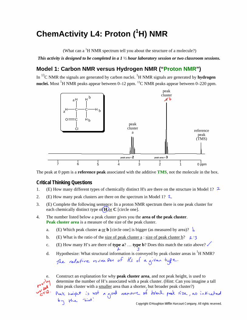
Model 1: Carbon NMR versus Hydrogen NMR (“Proton NMR”) In 13C NMR the signals are generated by carbon nuclei. 1H NMR signals are generated by hydrogen nuclei. Most 1H NMR peaks appear between 0–12 ppm. 13C NMR peaks appear between 0–220 ppm.
CC
H
H
H
H
C
H
a
a
b
b
b
0 ppm1234567
peak cluster
a
O
Cl
peak cluster
a
reference peak
(TMS)
peak area = 2 peak area = 3
The peak at 0 ppm is a reference peak associated with the additive TMS, not the molecule in the box.
Critical Thinking Questions 1. (E) How many different types of chemically distinct H's are there on the structure in Model 1?
2. (E) How many peak clusters are there on the spectrum in Model 1?
3. (E) Complete the following sentence: In a proton NMR spectrum there is one peak cluster for each chemically distinct type of H or C [circle one].
4. The number listed below a peak cluster gives you the area of the peak cluster. Peak cluster area is a measure of the size of the peak cluster.
a. (E) Which peak cluster a or b [circle one] is bigger (as measured by area)?
b. (E) What is the ratio of the size of peak cluster a : size of peak cluster b?
c. (E) How many H’s are there of type a? … type b? Does this match the ratio above?
d. Hypothesize: What structural information is conveyed by peak cluster areas in 1H NMR?
e. Construct an explanation for why peak cluster area, and not peak height, is used to determine the number of H’s associated with a peak cluster. (Hint: Can you imagine a tall thin peak cluster with a smaller area than a shorter, but broader peak cluster?)
Copyright © Houghton Mifflin Harcourt Company. All rights reserved.
298 ChemActivity L4: Proton (1H) NMR
Memorization Task L4.1: Peak area tells you number of H’s associated with peak cluster Peak area is calculated (by the instrument) using a mathematical technique called integration. For this reason peak cluster area is often referred to as a peak’s integral, integration, or integrated area.
The area of each peak cluster is often represented by a number written above or below a peak cluster.
This integration number tells you the relative number of H’s associated with each peak cluster. Because it is a relative number, the ratio 2:3 would also apply to a molecule with [4 & 6 H’s] or [6 & 9 H’s], etc. Be careful! Peak area (not peak height) tells you the number of H’s associated with a peak cluster. That is, sometimes a tall, thin peak will have a smaller area than a short, broad peak.
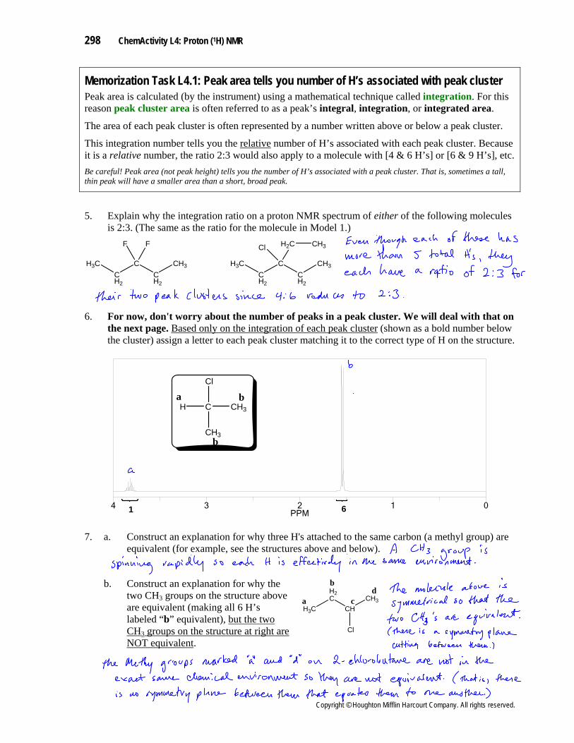
5. Explain why the integration ratio on a proton NMR spectrum of either of the following molecules is 2:3. (The same as the ratio for the molecule in Model 1.)
CH2
CCH2
CH3
F
H3C
F
CH2
CCH2
CH3
H2C
H3C
Cl CH3
6. For now, don't worry about the number of peaks in a peak cluster. We will deal with that on the next page. Based only on the integration of each peak cluster (shown as a bold number below the cluster) assign a letter to each peak cluster matching it to the correct type of H on the structure.
01234PPM
CH3C
Cl
CH3
H
1 6
a b
b
7. a. Construct an explanation for why three H's attached to the same carbon (a methyl group) are
equivalent (for example, see the structures above and below).
b. Construct an explanation for why the two CH3 groups on the structure above are equivalent (making all 6 H’s labeled “b” equivalent), but the two CH3 groups on the structure at right are NOT equivalent.
H3C
H2C
CHCH3
Cl
a
b
cd
Copyright © Houghton Mifflin Harcourt Company. All rights reserved.
ChemActivity L4: Proton (1H) NMR 299
Memorization Task L4.2: Finding NMR Equivalent H’s using Symmetry Our rules for finding equivalent carbons also apply to finding equivalent hydrogens. These rules state that two hydrogens are equivalent if any of the following hold true. The hydrogens…
• are directly across a mirror plane (symmetry plane) from one another.
• can be rotated into one another without changing the overall representation of the molecule.
• can be rotated into one another via a rotational symmetry axis.
For those who have completed ChemActivity 12…
Interestingly, it turns out that H's on different molecules are NMR equivalent if there is an external mirror plane that links them. This means that a molecule and its enantiomer will produce the exact same NMR spectrum. That is, enantiomers are indistinguishable from one another by NMR.
CH2CH3
CH3C Br
H
CH2CH3
CCH3Br
HS R
external mirror plane
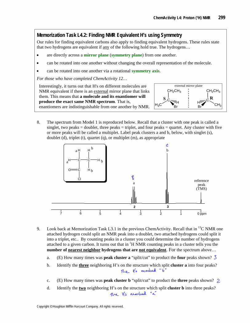
8. The spectrum from Model 1 is reproduced below. Recall that a cluster with one peak is called a singlet, two peaks = doublet, three peaks = triplet, and four peaks = quartet. Any cluster with five or more peaks will be called a multiplet. Label peak clusters a and b, below, with singlet (s), doublet (d), triplet (t), quartet (q), or multiplet (m), as appropriate
CC
H
H
H
H
C
H
a
a
b
b
b
0 ppm1234567
O
Cl reference peak
(TMS)
2 3
a
b
9. Look back at Memorization Task L3.1 in the previous ChemActivity. Recall that in 13C NMR one attached hydrogen could split an NMR peak into a doublet, two attached hydrogens could split it into a triplet, etc.. By counting peaks in a cluster you could determine the number of hydrogens attached to a given carbon. It turns out that in 1H NMR counting peaks in a cluster tells you the number of nearest neighbor hydrogens that are not equivalent. For the spectrum above…
a. (E) How many times was peak cluster a “split/cut” to product the four peaks shown?
b. Identify the three neighboring H’s on the structure which split cluster a into four peaks?
c. (E) How many times was peak cluster b “split/cut” to product the three peaks shown?
d. Identify the two neighboring H’s on the structure which split cluster b into three peaks?
Copyright © Houghton Mifflin Harcourt Company. All rights reserved.
300 ChemActivity L4: Proton (1H) NMR
Memorization Task L4.3: Multiplicity tells you the number of “foreign” H’s within 3 bondsIn 1H NMR, H's split the signals of foreign (= non-equivalent) hydrogens within three bonds.
This means chemically equivalent H’s DO NOT split each other. (Review) In 13C NMR, H's split the signal of carbons one bond away.
10. Confirm that Memorization Task L4.3 explains the splitting patterns of the spectra in CTQ’s 6 & 8.
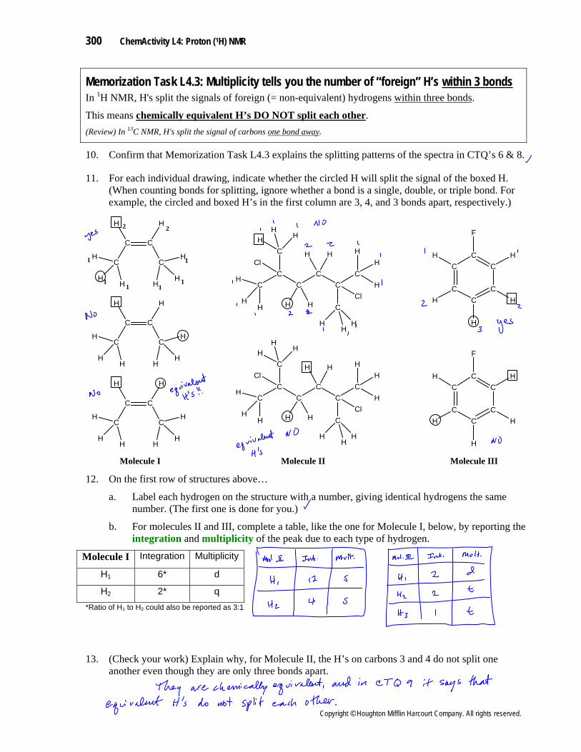
11. For each individual drawing, indicate whether the circled H will split the signal of the boxed H. (When counting bonds for splitting, ignore whether a bond is a single, double, or triple bond. For example, the circled and boxed H’s in the first column are 3, 4, and 3 bonds apart, respectively.)
CC
CC
H
C
H
HH
C
C
CCl
HH
H
HHH
Cl
H
H
H
HH
H
C
CC
C
CC
F
H
H
H
H
HC C
CC
HH
H
HHH
H
H
11
1
11
1
22
CC
CC
H
C
H
HH
C
C
CCl
HH
H
HHH
Cl
H
H
H
HH
H
C
CC
C
CC
F
H
H
H
H
H
C C
CC
HH
H
HHH
H
H
Molecule I Molecule II Molecule III
C C
CC
HH
H
HHH
H
H
12. On the first row of structures above…
a. Label each hydrogen on the structure with a number, giving identical hydrogens the same number. (The first one is done for you.)
b. For molecules II and III, complete a table, like the one for Molecule I, below, by reporting the integration and multiplicity of the peak due to each type of hydrogen.
Molecule I Integration Multiplicity
H1 6* d
H2 2* q *Ratio of H1 to H2 could also be reported as 3:1
13. (Check your work) Explain why, for Molecule II, the H’s on carbons 3 and 4 do not split one another even though they are only three bonds apart.
Copyright © Houghton Mifflin Harcourt Company. All rights reserved.
ChemActivity L4: Proton (1H) NMR 301
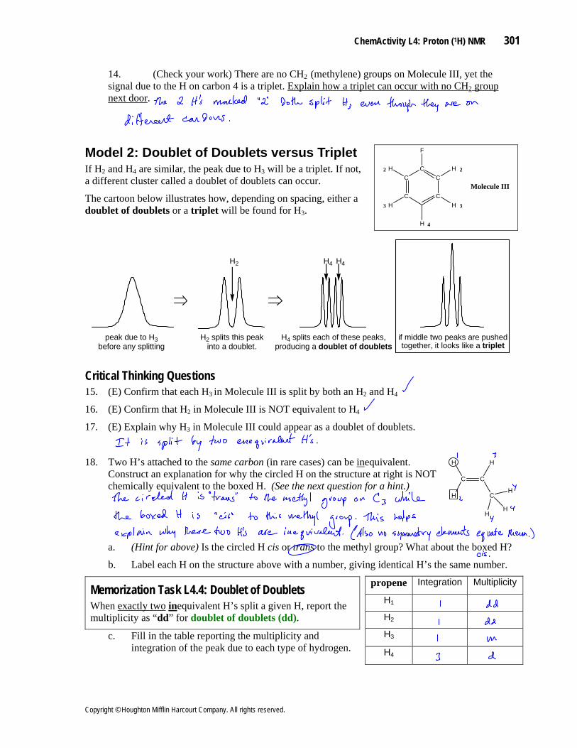
14. (Check your work) There are no CH2 (methylene) groups on Molecule III, yet the signal due to the H on carbon 4 is a triplet. Explain how a triplet can occur with no CH2 group next door.
Model 2: Doublet of Doublets versus Triplet If H2 and H4 are similar, the peak due to H3 will be a triplet. If not, a different cluster called a doublet of doublets can occur.
The cartoon below illustrates how, depending on spacing, either a doublet of doublets or a triplet will be found for H3.
C
C C
CC
F
H
H
H
H
H
Molecule III
22
33
4
peak due to H3 before any splitting
H2
H2 splits this peak into a doublet.
H4 H4
H4 splits each of these peaks, producing a doublet of doublets
if middle two peaks are pushed together, it looks like a triplet
Critical Thinking Questions 15. (E) Confirm that each H3 in Molecule III is split by both an H2 and H4
16. (E) Confirm that H2 in Molecule III is NOT equivalent to H4
17. (E) Explain why H3 in Molecule III could appear as a doublet of doublets.
18. Two H’s attached to the same carbon (in rare cases) can be inequivalent. Construct an explanation for why the circled H on the structure at right is NOT chemically equivalent to the boxed H. (See the next question for a hint.)
C C
CH
HH
H
HH
a. (Hint for above) Is the circled H cis or trans to the methyl group? What about the boxed H?
b. Label each H on the structure above with a number, giving identical H’s the same number.
propene Integration Multiplicity Memorization Task L4.4: Doublet of Doublets
H1 inWhen exactly two equivalent H’s split a given H, report the multiplicity as “dd” for doublet of doublets (dd). H 2
H3c. Fill in the table reporting the multiplicity and integration of the peak due to each type of hydrogen. H4
Copyright © Houghton Mifflin Harcourt Company. All rights reserved.
302 ChemActivity L4: Proton (1H) NMR
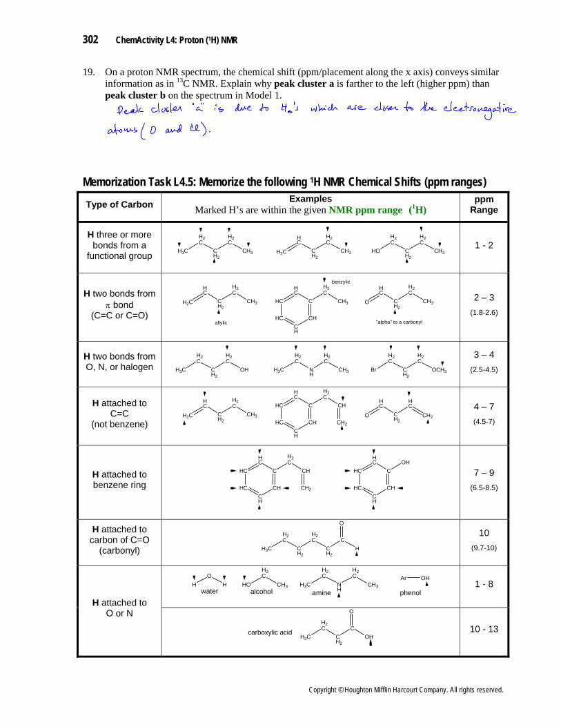
19. On a proton NMR spectrum, the chemical shift (ppm/placement along the x axis) conveys similar information as in 13C NMR. Explain why peak cluster a is farther to the left (higher ppm) than peak cluster b on the spectrum in Model 1.
Memorization Task L4.5: Memorize the following 1H NMR Chemical Shifts (ppm ranges) Examples
Marked H’s are within the given ppm
Range Type of Carbon NMR ppm ranges ( H)1
H3C
H2C
CH2
H2C
CH3 H2C
HC
CH2
H2C
CH3 HO
H2C
CH2
H2C
CH3
H three or more bonds from a
functional group 1 - 2
HC
HC
C
H2C
CH3H2C
HC
CH2
H2C
CH3
HCCH
CH
O
HC
CH2
H2C
CH3
allylic
benzylic
"alpha" to a carbonyl
H two bonds from π bond
(C=C or C=O)
2 – 3 (1.8-2.6)
H3C
H2C
CH2
H2C
OH H3C
H2C
NH
H2C
CH3 Br
H2C
CH2
H2C
OCH3
3 – 4 H two bonds from O, N, or halogen (2.5-4.5)
HC
HC
C
H2C
CH
H2C
HC
CH2
H2C
CH3HC
CH
CHO
HC
CH2
HC
CH2CH2
H attached to C=C
(not benzene)
4 – 7 (4.5-7)
HC
HC
C
H2C
CH
HCCH
CH CH2
HC
HC
COH
HCCH
CH
7 – 9 H attached to benzene ring (6.5-8.5)
H3C
H2C
CH2
H2C
CH2
C
O
H
H attached to carbon of C=O
(carbonyl)
10 (9.7-10)
CH3
H2C
HO H3C
H2C
NH
H2C
CH3alcohol amine
HO
Hwater
Ar OH
phenol1 - 8
H attached to O or N
H3C
H2C
CH2
C
O
OHcarboxylic acid 10 - 13
Copyright © Houghton Mifflin Harcourt Company. All rights reserved.
ChemActivity L4: Proton (1H) NMR 303
a
b
OC
C
H H
HH
HH
b
c
cc
TMS
1 2 3relative peak areas
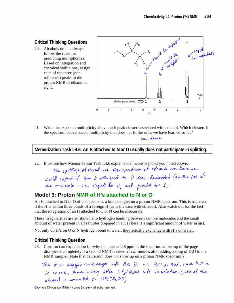
Critical Thinking Questions 20. Alcohols do not always
follow the rules for predicting multiplicities. Based on integration and chemical shift alone, assign each of the three (non-reference) peaks in the proton NMR of ethanol at right.
21. Write the expected multiplicity above each peak cluster associated with ethanol. Which clusters in the spectrum above have a multiplicity that does not fit the rules we have learned so far?
Memorization Task L4.6: An H attached to N or O usually does not participate in splitting.
22. Illustrate how Memorization Task L4.6 explains the inconsistencies you noted above.
Model 3: Proton NMR of H’s attached to N or O An H attached to N or O often appears as a broad singlet on a proton NMR spectrum. This is true even if the H is within three bonds of a foreign H (as is the case with ethanol). Also watch out for the fact that the integration of an H attached to O or N can be inaccurate.
These irregularities are attributable to hydrogen bonding between sample molecules and the small amount of water present in all samples prepared in air. (There is a significant amount of water in air).
Not only do H’s on O or N hydrogen bond to water, they actually exchange with H’s on water.
Critical Thinking Question 23. Construct an explanation for why the peak at 4.8 ppm in the spectrum at the top of the page
disappears completely if a second NMR is taken a few minutes after adding a drop of D2O to the NMR sample. (Note that deuterium does not show up on a proton NMR spectrum.)
Copyright © Houghton Mifflin Harcourt Company. All rights reserved.
304 ChemActivity L4: Proton (1H) NMR
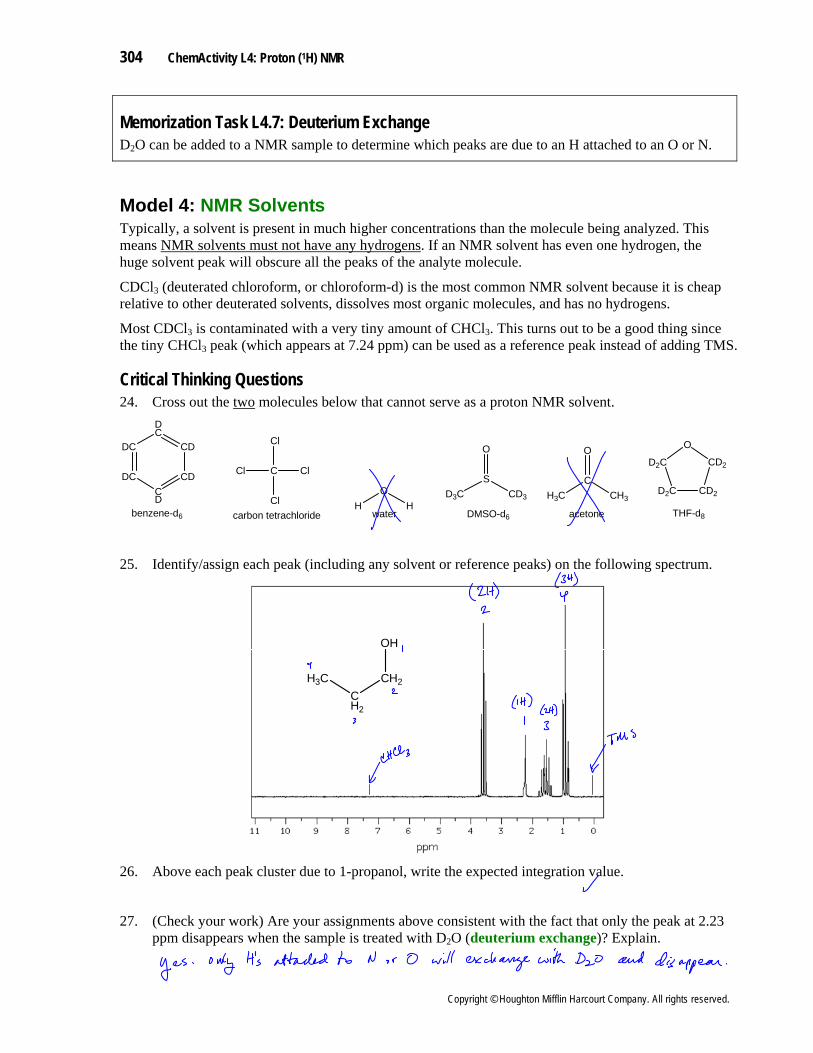
Memorization Task L4.7: Deuterium Exchange D O can be added to a NMR sample to determine which peaks are due to an H attached to an O or N. 2
Model 4: NMR SolventsTypically, a solvent is present in much higher concentrations than the molecule being analyzed. This means NMR solvents must not have any hydrogens. If an NMR solvent has even one hydrogen, the huge solvent peak will obscure all the peaks of the analyte molecule.
CDCl3 (deuterated chloroform, or chloroform-d) is the most common NMR solvent because it is cheap relative to other deuterated solvents, dissolves most organic molecules, and has no hydrogens.
is contaminated with a very tiny amount of CHClMost CDCl3 3. This turns out to be a good thing since the tiny CHCl3 peak (which appears at 7.24 ppm) can be used as a reference peak instead of adding TMS.
Critical Thinking Questions 24. Cross out the two molecules below that cannot serve as a proton NMR solvent.
DC
DCCD
CD
CD
DC
C
Cl
Cl
ClCl
HO
H
C
O
CH3H3CS
O
CD3D3C
benzene-d6 DMSO-d6watercarbon tetrachloride acetone
D2C
D2C CD2
CD2
O
THF-d8
25. Identify/assign each peak (including any solvent or reference peaks) on the following spectrum.
H3CCH2
CH2
OH
26. Above each peak cluster due to 1-propanol, write the expected integration value.
27. (Check your work) Are your assignments above consistent with the fact that only the peak at 2.23 ppm disappears when the sample is treated with D deuterium exchange2O ( )? Explain.
Copyright © Houghton Mifflin Harcourt Company. All rights reserved.
ChemActivity L4: Proton (1H) NMR 305
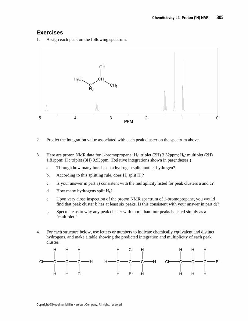
Exercises 1. Assign each peak on the following spectrum.
H3CCH2
CHCH3
OH
012345PPM
2. Predict the integration value associated with each peak cluster on the spectrum above.
3. Here are proton NMR data for 1-bromopropane: Ha: triplet (2H) 3.32ppm; Hb: multiplet (2H) 1.81ppm; H : triplet (3H) 0.93ppm. (Relative integrations shown in parentheses.) c
a. Through how many bonds can a hydrogen split another hydrogen?
b. According to this splitting rule, does Ha split Hc?
c. Is your answer in part a) consistent with the multiplicity listed for peak clusters a and c?
d. How many hydrogens split Hb?
e. Upon very close inspection of the proton NMR spectrum of 1-bromopropane, you would find that peak cluster b has at least six peaks. Is this consistent with your answer in part d)?
f. Speculate as to why any peak cluster with more than four peaks is listed simply as a "multiplet."
4. For each structure below, use letters or numbers to indicate chemically equivalent and distinct hydrogens, and make a table showing the predicted integration and multiplicity of each peak cluster.
Cl C C C H
H
Cl
H
H
H
H
H C C C H
H
H
Cl
Br
H
H
Cl C C C Br
H
H
H
H
H
H
Copyright © Houghton Mifflin Harcourt Company. All rights reserved.
306 ChemActivity L4: Proton (1H) NMR
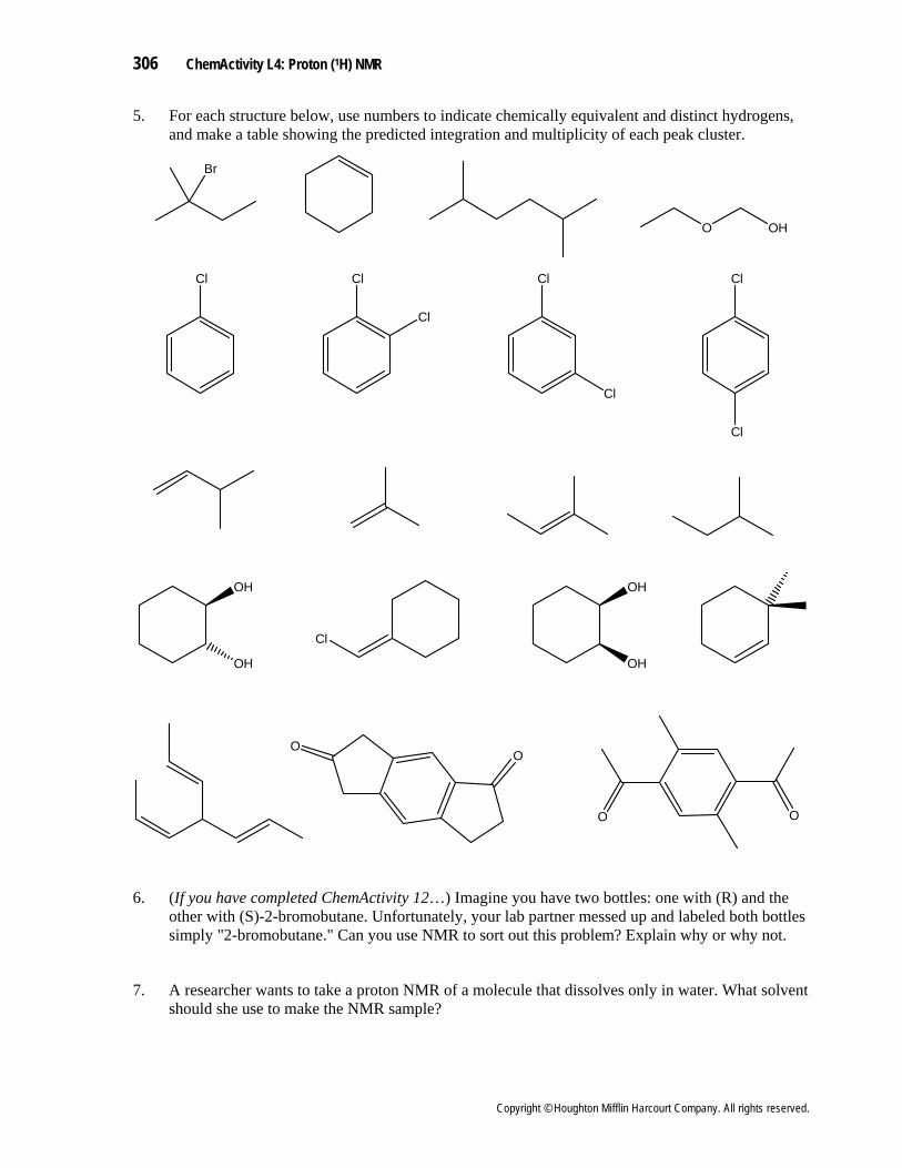
5. For each structure below, use numbers to indicate chemically equivalent and distinct hydrogens, and make a table showing the predicted integration and multiplicity of each peak cluster.
Br
Cl Cl Cl
Cl
Cl
Cl
Cl
OO
OO
OH
OH
Cl
OH
OH
O OH
6. (If you have completed ChemActivity 12…) Imagine you have two bottles: one with (R) and the other with (S)-2-bromobutane. Unfortunately, your lab partner messed up and labeled both bottles simply "2-bromobutane." Can you use NMR to sort out this problem? Explain why or why not.
7. A researcher wants to take a proton NMR of a molecule that dissolves only in water. What solvent should she use to make the NMR sample?
Copyright © Houghton Mifflin Harcourt Company. All rights reserved.
ChemActivity L4: Proton (1H) NMR 307
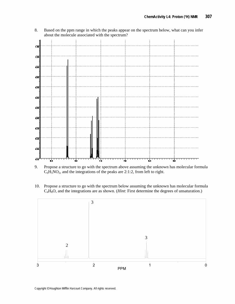
8. Based on the ppm range in which the peaks appear on the spectrum below, what can you infer about the molecule associated with the spectrum?
9. Propose a structure to go with the spectrum above assuming the unknown has molecular formula
C H6 5NO , and the integrations of the peaks are 2:1:2, from left to right. 2
10. Propose a structure to go with the spectrum below assuming the unknown has molecular formula C4H8O, and the integrations are as shown. (Hint: First determine the degrees of unsaturation.)
0123PPM
2
3
3
Copyright © Houghton Mifflin Harcourt Company. All rights reserved.
308 ChemActivity L4: Proton (1H) NMR
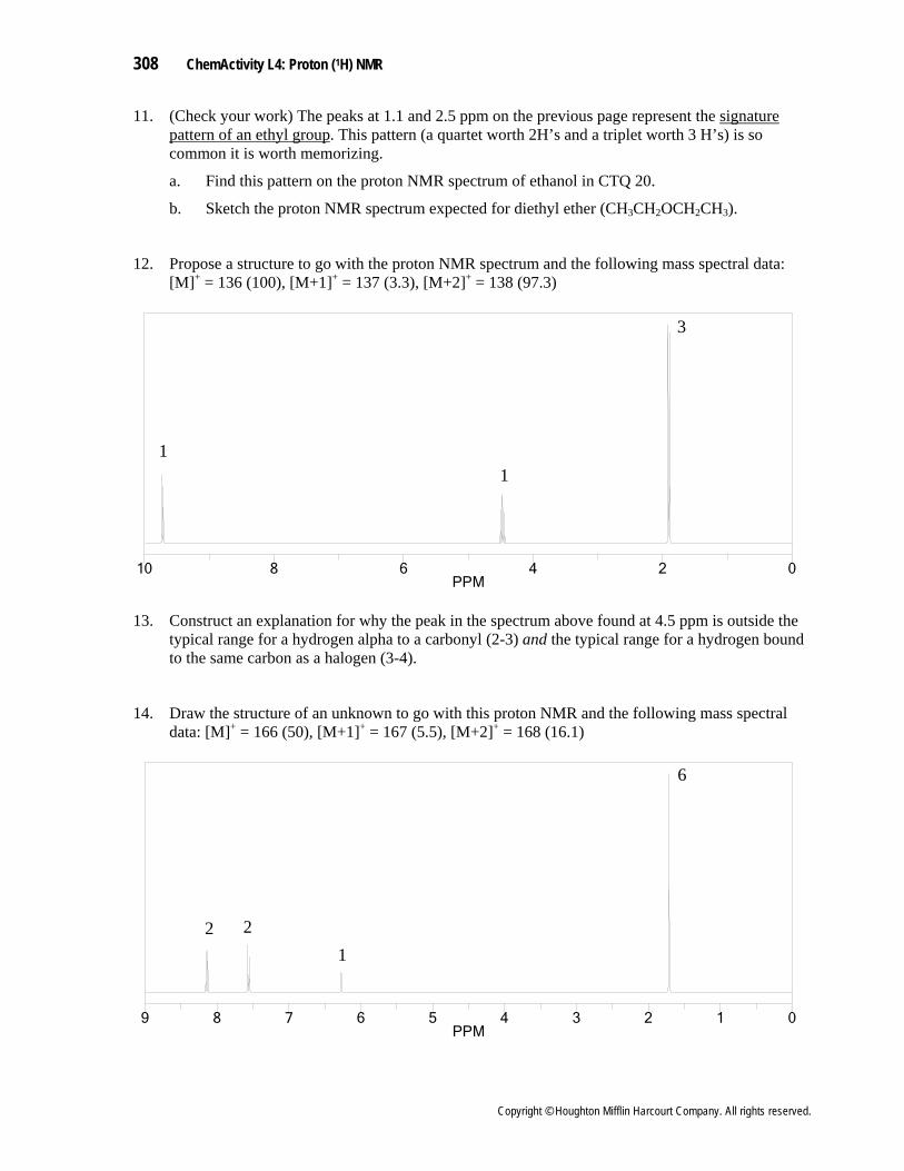
11. (Check your work) The peaks at 1.1 and 2.5 ppm on the previous page represent the signature pattern of an ethyl group. This pattern (a quartet worth 2H’s and a triplet worth 3 H’s) is so common it is worth memorizing.
a. Find this pattern on the proton NMR spectrum of ethanol in CTQ 20.
CH OCH CH ). b. Sketch the proton NMR spectrum expected for diethyl ether (CH3 2 2 3
12. Propose a structure to go with the proton NMR spectrum and the following mass spectral data: [M]+ = 136 (100), [M+1]+ = 137 (3.3), [M+2]+ = 138 (97.3)
0246810PPM
11
3
13. Construct an explanation for why the peak in the spectrum above found at 4.5 ppm is outside the
typical range for a hydrogen alpha to a carbonyl (2-3) and the typical range for a hydrogen bound to the same carbon as a halogen (3-4).
14. Draw the structure of an unknown to go with this proton NMR and the following mass spectral data: [M]+ = 166 (50), [M+1]+ = 167 (5.5), [M+2]+ = 168 (16.1)
0123456789PPM
2 21
6
Copyright © Houghton Mifflin Harcourt Company. All rights reserved.
ChemActivity L4: Proton (1H) NMR 309
15. Peak clusters in the 7-9 ppm region are almost always indicative of H’s attached to a benzene ring. (As we will learn later, the region from 7-9 ppm is called the aromatic region, and molecules containing a benzene ring are by far the most common type of aromatic molecule).
Peak clusters in the aromatic region often overlap one another. For example, the mass of peaks between 7.3 and 7.4 ppm on the spectrum below is actually two overlapping peak clusters: a doublet of doublets worth 2H’s overlapping with a triplet worth 1H.
Draw one structure that goes with both spectra on this page.
012345678PPM
3 2
1 1
3
020406080100120140PPM
16. (Check your work) How can you tell the top spectrum on this page is a proton NMR spectrum and
the bottom spectrum is a carbon NMR spectrum?
17. (Check your work) According to the CMR spectrum, how many unique C’s are there?
18. (Check your work) Explain why, on the proton NMR spectrum, the peak at 6.1 is expected to be a doublet of quartets.
Copyright © Houghton Mifflin Harcourt Company. All rights reserved.
310 ChemActivity L4: Proton (1H) NMR
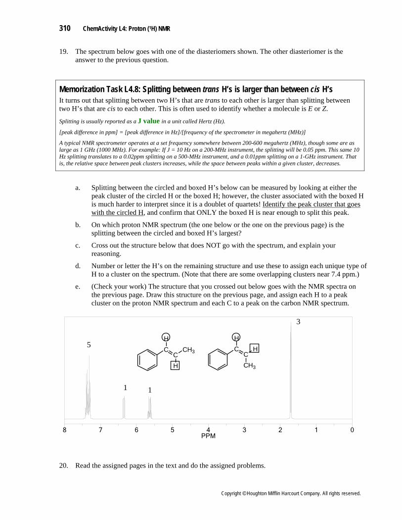
19. The spectrum below goes with one of the diasteriomers shown. The other diasteriomer is the answer to the previous question.
Memorization Task L4.8: Splitting between trans H’s is larger than between cis H’s It turns out that splitting between two H’s that are trans to each other is larger than splitting between two H’s that are cis to each other. This is often used to identify whether a molecule is E or Z.
Splitting is usually reported as a J value in a unit called Hertz (Hz).
[peak difference in ppm] = [peak difference in Hz]/[frequency of the spectrometer in megahertz (MHz)] A typical NMR spectrometer operates at a set frequency somewhere between 200-600 megahertz (MHz), though some are as large as 1 GHz (1000 MHz). For example: If J = 10 Hz on a 200-MHz instrument, the splitting will be 0.05 ppm. This same 10 Hz splitting translates to a 0.02ppm splitting on a 500-MHz instrument, and a 0.01ppm splitting on a 1-GHz instrument. That is, the relative space between peak clusters increases, while the space between peaks within a given cluster, decreases.
a. Splitting between the circled and boxed H’s below can be measured by looking at either the peak cluster of the circled H or the boxed H; however, the cluster associated with the boxed H is much harder to interpret since it is a doublet of quartets! Identify the peak cluster that goes with the circled H, and confirm that ONLY the boxed H is near enough to split this peak.
b. On which proton NMR spectrum (the one below or the one on the previous page) is the splitting between the circled and boxed H’s largest?
c. Cross out the structure below that does NOT go with the spectrum, and explain your reasoning.
d. Number or letter the H’s on the remaining structure and use these to assign each unique type of H to a cluster on the spectrum. (Note that there are some overlapping clusters near 7.4 ppm.)
e. (Check your work) The structure that you crossed out below goes with the NMR spectra on the previous page. Draw this structure on the previous page, and assign each H to a peak cluster on the proton NMR spectrum and each C to a peak on the carbon NMR spectrum.
012345678PPM
CC
CH3 CCCH3
H
H
HH
5
1 1
3
20. Read the assigned pages in the text and do the assigned problems.
Copyright © Houghton Mifflin Harcourt Company. All rights reserved.
ChemActivity L4: Proton (1H) NMR 311
The Big Picture Proton NMR works in much the same way as carbon NMR, but again interpretation of the spectra is a much more important skill for an organic chemist than understanding the complex physics behind the instrument. The key elements of a proton NMR spectrum are reviewed below.
Common Points of Confusion The following is a summary of the key elements of proton NMR:
ppm chemical shift or (given by location along the x axis) tells you the amount of electron density around an H. The closer the H is to an electronegative element, the more “
• deshielded” it
is and therefore the higher the ppm number of its peak cluster (farther left on the spectrum). Multiple bonds also cause the signal of nearby H's to be shifted to the left. Memorization Task L4.5 gives "chemical shifts" for common functional groups. Each chemically distinct H should have a unique chemical shift, though in practice different peak clusters sometimes overlap just by coincidence. This is a bigger problem in proton NMR than in carbon NMR (especially in the so-called “aromatic region” from 7-9 ppm), since most proton NMR peaks are squeezed into just an 8 ppm range (1-9 ppm).
• Peak area, not peak height, tells you the relative number of H’s associated with a peak cluster.
Integration peak cluster area or (given by a number above or below a peak cluster, or by a line stepping up from left to right called the integration line) tells you the relative area of each peak and therefore the relative number of equivalent H’s represented by each peak. Note that integration gives you only a ratio of peak areas, making it impossible to tell the difference between a 1H to 3H ratio and a 2H to 6H ratio.
•
• Splitting in carbon NMR tells you the number of H’s attached to a given carbon. For this reason, students incorrectly assume that splitting in proton NMR tells you the number of H’s associated with a peak cluster. In fact, it is a bit more complicated (see next bullet).
Multiplicity is the number of peaks in a peak cluster (also called splitting or proton-proton coupling
• ). It tells you the number of nonequivalent neighbor H’s within three bonds. For
example, a doublet (two peaks) tells you there is exactly one non-equivalent H within three bonds of the H responsible for this signal.
Equivalent H’s do NOT split each other• .
Copyright © Houghton Mifflin Harcourt Company. All rights reserved.