characterization of yolk sac proteins of bos indicus...
TRANSCRIPT
©FUNPEC-RP www.funpecrp.com.brGenetics and Molecular Research 11 (4): 3942-3954 (2012)
Characterization of yolk sac proteins of Bos indicus cattle embryos
F.S. Matsumoto1, V.C. Oliveira1, C.A.F. Mançanares2, C.E. Ambrosio2 and M.A. Miglino1
1Laboratório de Anatomia de Animais Domésticos e Silvestres, Departamento de Cirurgia, Faculdade de Medicina Veterinária e Saúde Animal, Universidade de São Paulo, São Paulo, SP, Brasil2Laboratório de Morfofisiologia Molecular e Desenvolvimento, Departamento de Ciências Básicas, Faculdade de Zootecnia e Engenharia de Alimentos, Universidade de São Paulo, Pirassununga, SP, Brasil
Corresponding author: C.E. AmbrósioE-mail: [email protected]
Genet. Mol. Res. 11 (4): 3942-3954 (2012)Received June 11, 2012Accepted July 26, 2012Published November 14, 2012DOI http://dx.doi.org/10.4238/2012.November.14.1
ABSTRACT. The yolk sac is an embryonic membrane that is essential for the embryo’s initial survival in many mammals. It also plays an important role in the production of proteins necessary for development. We studied proteins of the yolk sac in bovine embryos at up to 40 days of gestation. We examined the yolk sac of 17 bovine embryos at different gestational periods, measuring α-fetoprotein, α-1-antitrypsin, and transferrin. This experiment was carried out by Western blot technique, associated with electrophoresis on a 6% sodium dodecyl sulfate polyacrylamide gel. Mouse monoclonal antibody anti-human-α-fetoprotein, mouse antibody anti-human-transferrin and rabbit polyclonal anti-human-α-1-antitrypsin were used as primary antibodies, and conjugated peroxidase as a secondary antibody. We detected the three proteins in some of the yolk sac samples; however, the bands in some specimens (samples) were weak, maybe a result of poor antigen-antibody reaction, since the antibodies used in this study were not
3943
©FUNPEC-RP www.funpecrp.com.brGenetics and Molecular Research 11 (4): 3942-3954 (2012)
Bovine yolk sac proteins
specific to bovine proteins. The fact that weak bands appeared might be due to a weak cross-reaction.
Key words: Yolk sac; Western blotting; Proteins; Alpha-fetoprotein; Alpha 1-antitrypsin; Transferring
INTRODUCTION
Bos indicus is considered a different species in comparison to Bos taurus due to its morphology, physiological peculiarities and ecological characteristics; it is considered the world’s largest breed, with about 200 million in India and showing a continuous expansion in tropical countries. Nowadays, the Zebu breed is considered an icon in tropical cattle investiga-tion due to its considerable economical production (Bezerra, 1976), and in vitro embryo pro-duction and transfer techniques have been increasing and improving more and more, although the culture medium used in this technique usually affects embryo development after fertiliza-tion and interferes with metabolism and important gene expression (Alves et al., 2003). The extra-embryonic membranes (allantois, amnion, chorion, and yolk sac) are responsible for the fetal-maternal communication and placental formation (Wolf et al., 2003). The yolk sac is responsible for early embryo survival in many mammal species, producing important proteins for their development. The proteins are polymers that are involved in numerous biological functions and also determine cell structure and shape. Besides, the proteins catalyze many chemical reactions and control membrane permeability, to regulate metabolite concentration and to control genetic function (Zaha et al., 2003). Specialized proteins can play antibody, toxin, hormone, anticoagulant, and elastic fiber functions as well (Alberts et al., 2006).
The fetal membranes, chorion, allantois, amnion, and yolk sac, represent the mem-branous structures involved in placental development (Bowen and Burghardt, 2000). These membranes can establish intrinsic connections during placental development, which could be transient or permanent, depending on the animal species (Assis Neto, 2010). The placenta represents a tissue apposition or fusion, responsible for fetal-maternal nutrition and residual excretion. The morphological efficiency of the fetal membrane is related to the large superfi-cial apposition area of layers, and even though the maternal and fetal blood are close, they are morphologically separated by the fetal membrane cell layers (Björkman, 1973).
The yolk sac plays a major role in embryonic development in mammals. The extra-embryonic portion is responsible for initial hematopoiesis. In rodents, it is the main fetal-maternal membrane for exchanges during gestation. This capacity to synthesize and secrete proteins in the embryonic and extra-embryonic spaces is frequently used in experimental em-bryology and reproduction toxicology (Gulbis et al., 1998). Salisbury and Vandermark (1964) affirm that the young embryo could persist for a short time due to the nutritive absorption of the yolk sac and uterine milk, and as long as development continues, a well-developed nutri-tive system is required. The three embryonic layers derived from diverse embryonic tissues, as well as the protective layers involved, have a nutritive function. The blood vessels of the yolk sac are responsible for transporting the nutrients from the uterine milk to the embryo. The functional activity of the yolk sac is short-term, whose function is developed by the allantoids. Barone (1976) affirms that the yolk sac undergoes an early involution and degeneration in ruminants and no vestige remains after the second week to the end of pregnancy. It has been observed that in domestic ruminants, such as cattle, sheep and goats, proteins produced by
F.S. Matsumoto et al. 3944
©FUNPEC-RP www.funpecrp.com.brGenetics and Molecular Research 11 (4): 3942-3954 (2012)
extra-embryonic membranes have a mediator function in some fetal-maternal interactions for the establishment and development of gestation. The bovine yolk sac is well developed by the 20th day of gestation, and afterwards, its regression begins (Godkin et al., 1988). The yolk sac of domestic mammals is initially large but involutes with placental progression. Despite being a transitory structure, the yolk sac shows important functions, such as permanent pla-cental nutrition until its complete formation, especially in equine and carnivores. Some of the yolk blood vessels are incorporated by the embryo forming the adult visceral blood vessels. Its endodermic portion originates the primordial germ cells (primary sex cells), which migrate to the gonads and also show characteristics similar to the steroidal hormone-producing cells (Latshaw, 1987). The yolk sac could have functions of the liver in embryonic development before fetal liver maturation and produce a variety of fetal liver proteins (Gulbis et al., 1998).
MATERIAL AND METHODS
After the evisceration of the animals in the abattoir, the uterus were identified and incised. Afterwards, the embryo and its membranes were collected and placed in plastic con-tainers that were kept in a box with ice. The yolk sac was characterized macroscopically, col-lected and frozen at -20°C or placed in physiological solution for protein extraction. The liver was also collected, sampled and its fragments (0.5 cm) were placed in 1.5-mL conical tubes for protein extraction.
Estimation of fetal period
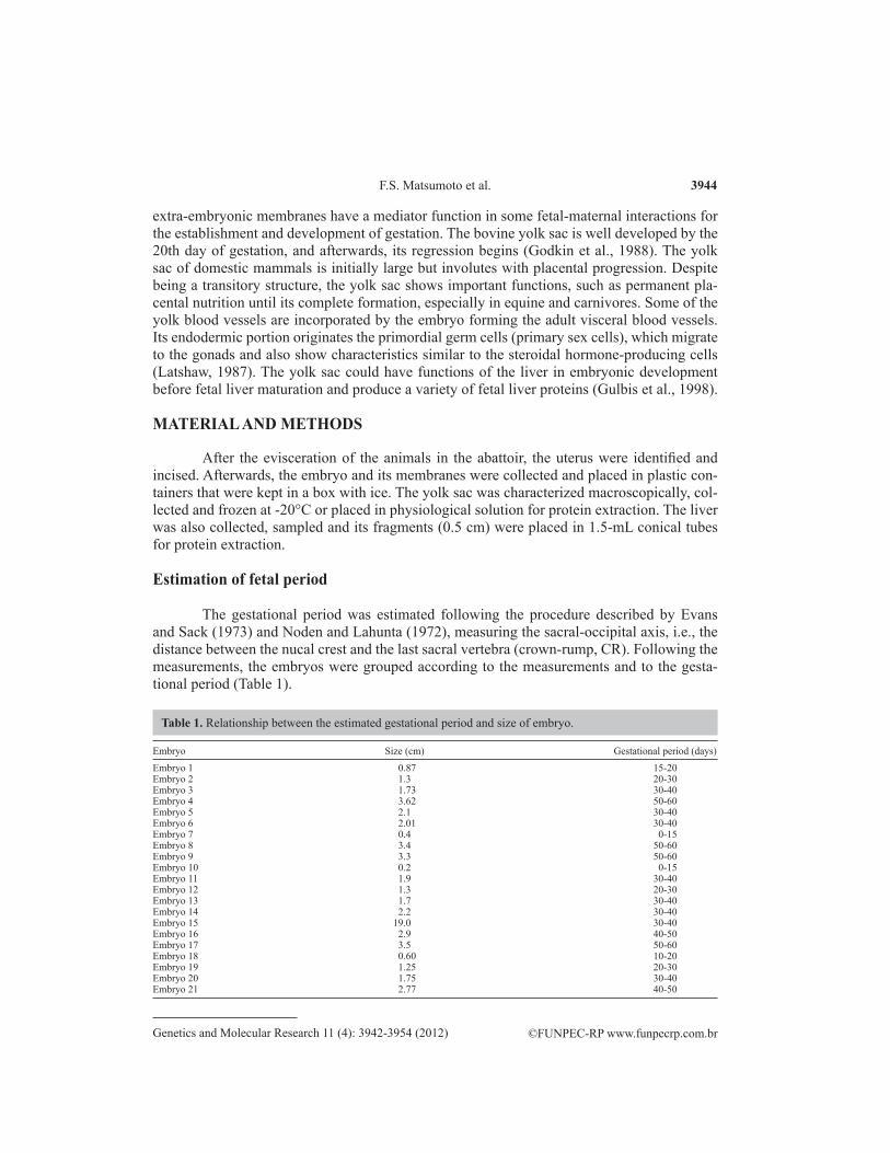
The gestational period was estimated following the procedure described by Evans and Sack (1973) and Noden and Lahunta (1972), measuring the sacral-occipital axis, i.e., the distance between the nucal crest and the last sacral vertebra (crown-rump, CR). Following the measurements, the embryos were grouped according to the measurements and to the gesta-tional period (Table 1).
Embryo Size (cm) Gestational period (days)
Embryo 1 0.87 15-20Embryo 2 1.3 20-30Embryo 3 1.73 30-40Embryo 4 3.62 50-60Embryo 5 2.1 30-40Embryo 6 2.01 30-40Embryo 7 0.4 0-15Embryo 8 3.4 50-60Embryo 9 3.3 50-60Embryo 10 0.2 0-15Embryo 11 1.9 30-40Embryo 12 1.3 20-30Embryo 13 1.7 30-40Embryo 14 2.2 30-40Embryo 15 19.0 30-40Embryo 16 2.9 40-50Embryo 17 3.5 50-60Embryo 18 0.60 10-20Embryo 19 1.25 20-30Embryo 20 1.75 30-40Embryo 21 2.77 40-50
Table 1. Relationship between the estimated gestational period and size of embryo.
3945
©FUNPEC-RP www.funpecrp.com.brGenetics and Molecular Research 11 (4): 3942-3954 (2012)
Bovine yolk sac proteins
Protein extraction
The fragments of yolk sac and liver were boiled in a solution composed of 1 g SDS, 0.3362 g EDTA and 1.21 g Tris in 100 mL distilled water, after adding 500 µM DDT, for 20 min. In these cases, a persistence of fragments was observed, and a maceration technique was applied to dissolve the remaining fragments; afterwards, the suspension was centrifuged for 15 min at 13,500 rpm. The resulting supernatant was transferred to other tubes, identified and considered to be the pure total protein extracted from the samplings. For Western blotting, a 1:1 mixture of the protein extract and sample buffer dye was used. This dilution was separated and frozen at -20°C.
Protein quantification
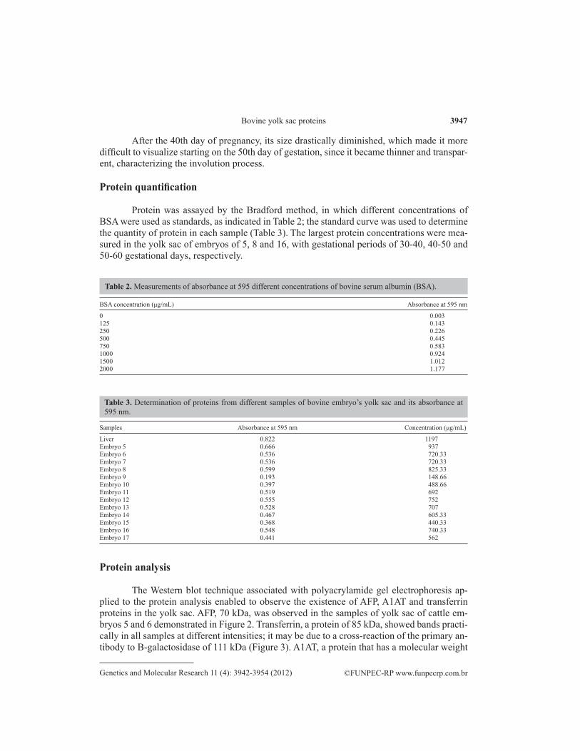
Protein was determined with the Quick Start Bradford Protein Assay kit (BioRad). Concentrations of bovine serum albumin (BSA; 2.0, 1.5, 1.0, 0.75, 0.5, 0.25, and 0.125 mg/mL) were used for the standard curve. The analysis was performed with the aid of a spectro-photometer at 595 nm.
Polyacrylamide gel electrophoresis
The method applied here followed the descriptions of Laemmli (1970). Polyacryl-amide gel electrophoresis was performed on 6 and 7.5% gels. The 6% separation gel was pre-pared with 5.425 mL water, 2.5 mL buffer, 2.0 mL 30% acrylamide, 2.5 µL TEMED, and 75 µL 10% APS, and the 7.5% gel (for fetuin protein) was prepared with 4.925 mL water, 2.5 mL buffer, 2.5 mL 30% acrylamide, 2.5 µL TEMED, and 75 µL 10% APS. Stacking 4% gel was made with 1.585 mL water, 625 µL buffer, 287.5 µL 30% acrylamide, 2.5 µL TEMED, and 31 µL 10% APS. Aliquots of 20 µL sample and 5 µL molecular weight standard were loaded, according to Bio-Rad (161-0324) instructions, i.e., 5 to 10 µL molecular weight standard for 4 to 20% gels. During the initial part of electrophoresis (10-20 min), gels were run at 70 V for protein compaction, and the voltage was then increased to 100 V and in the last 10 min to 120 V. The gel was stained with Coomassie blue (250 mL methanol, 100 mL acetic acid, 0.5 g Coomassie blue R-250 in 11 mL distilled water) for 3 x 30 min up to 100 min.
Transfer to nitrocellulose membrane
Before the antibody reaction, the membrane was blocked in 5% milk. The primary antibody was diluted in TTBS and applied to the membrane overnight. The secondary an-tibody was also diluted in TTBS and applied to the membrane for 1 h. Mouse anti-human α-fetoprotein monoclonal antibody (AFP; Serotec, MCA1863HT), mouse anti-human trans-ferrin antibody (BD, 612124) and rabbit anti-human α-1-antitrypsin polyclonal antibody (A1AT; Abcan, AB922) were applied as primary antibodies, and anti-mouse- and anti-rabbit-peroxidase conjugate as secondary antibody (GE, NIF825).
RESULTS
A total of 21 embryos were collected and grouped in accordance with the size and ges-
F.S. Matsumoto et al. 3946
©FUNPEC-RP www.funpecrp.com.brGenetics and Molecular Research 11 (4): 3942-3954 (2012)
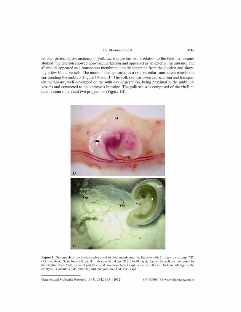
tational period. Gross anatomy of yolk sac was performed in relation to the fetal membranes studied; the chorion showed non-vascularization and appeared as an external membrane. The allantoids appeared as a transparent membrane, totally separated from the chorion and show-ing a few blood vessels. The amnion also appeared as a non-vascular transparent membrane surrounding the embryo (Figure 1A and B). The yolk sac was observed as a thin and transpar-ent membrane, well developed on the 40th day of gestation, being proximal to the umbilical vessels and connected to the embryo’s intestine. The yolk sac was composed of the vitelline duct, a central part and two projections (Figure 1B).
Figure 1. Photograph of the bovine embryo and its fetal membranes. A. Embryo with 2.1 cm crown-rump (CR) (35 to 40 days). Scale bar = 2.0 cm. B. Embryo with 0.4 cm CR (15 to 20 days); observe the yolk sac composed by the vitelline duct (Ysd), a central part (Ysc) and two projections (Ysp). Scale bar = 0.2 cm. Note in both figures the embryo (E), allantois (Al), amnion (Am) and yolk sac (Ysd, Ysc, Ysp).
3947
©FUNPEC-RP www.funpecrp.com.brGenetics and Molecular Research 11 (4): 3942-3954 (2012)
Bovine yolk sac proteins
After the 40th day of pregnancy, its size drastically diminished, which made it more difficult to visualize starting on the 50th day of gestation, since it became thinner and transpar-ent, characterizing the involution process.
Protein quantification
Protein was assayed by the Bradford method, in which different concentrations of BSA were used as standards, as indicated in Table 2; the standard curve was used to determine the quantity of protein in each sample (Table 3). The largest protein concentrations were mea-sured in the yolk sac of embryos of 5, 8 and 16, with gestational periods of 30-40, 40-50 and 50-60 gestational days, respectively.
BSA concentration (μg/mL) Absorbance at 595 nm
0 0.003125 0.143250 0.226500 0.445750 0.5831000 0.9241500 1.0122000 1.177
Table 2. Measurements of absorbance at 595 different concentrations of bovine serum albumin (BSA).
Samples Absorbance at 595 nm Concentration (μg/mL)
Liver 0.822 1197Embryo 5 0.666 937Embryo 6 0.536 720.33Embryo 7 0.536 720.33Embryo 8 0.599 825.33Embryo 9 0.193 148.66Embryo 10 0.397 488.66Embryo 11 0.519 692Embryo 12 0.555 752Embryo 13 0.528 707Embryo 14 0.467 605.33Embryo 15 0.368 440.33Embryo 16 0.548 740.33Embryo 17 0.441 562
Table 3. Determination of proteins from different samples of bovine embryo’s yolk sac and its absorbance at 595 nm.
Protein analysis
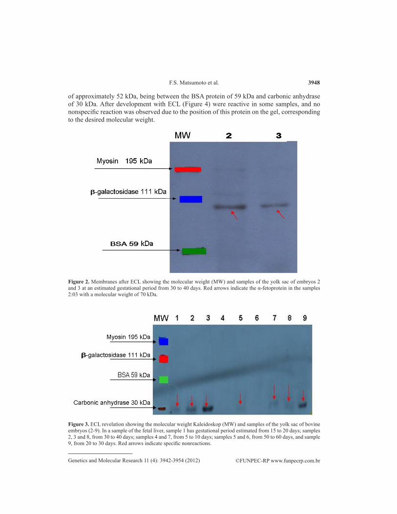
The Western blot technique associated with polyacrylamide gel electrophoresis ap-plied to the protein analysis enabled to observe the existence of AFP, A1AT and transferrin proteins in the yolk sac. AFP, 70 kDa, was observed in the samples of yolk sac of cattle em-bryos 5 and 6 demonstrated in Figure 2. Transferrin, a protein of 85 kDa, showed bands practi-cally in all samples at different intensities; it may be due to a cross-reaction of the primary an-tibody to B-galactosidase of 111 kDa (Figure 3). A1AT, a protein that has a molecular weight
F.S. Matsumoto et al. 3948
©FUNPEC-RP www.funpecrp.com.brGenetics and Molecular Research 11 (4): 3942-3954 (2012)
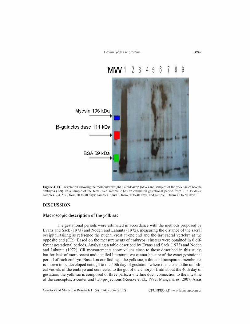
of approximately 52 kDa, being between the BSA protein of 59 kDa and carbonic anhydrase of 30 kDa. After development with ECL (Figure 4) were reactive in some samples, and no nonspecific reaction was observed due to the position of this protein on the gel, corresponding to the desired molecular weight.
Figure 2. Membranes after ECL showing the molecular weight (MW) and samples of the yolk sac of embryos 2 and 3 at an estimated gestational period from 30 to 40 days. Red arrows indicate the α-fetoprotein in the samples 2:03 with a molecular weight of 70 kDa.
Figure 3. ECL revelation showing the molecular weight Kaleidoskop (MW) and samples of the yolk sac of bovine embryos (2-9). In a sample of the fetal liver, sample 1 has gestational period estimated from 15 to 20 days; samples 2, 3 and 8, from 30 to 40 days; samples 4 and 7, from 5 to 10 days; samples 5 and 6, from 50 to 60 days, and sample 9, from 20 to 30 days. Red arrows indicate specific nonreactions.
3949
©FUNPEC-RP www.funpecrp.com.brGenetics and Molecular Research 11 (4): 3942-3954 (2012)
Bovine yolk sac proteins
DISCUSSION
Macroscopic description of the yolk sac
The gestational periods were estimated in accordance with the methods proposed by Evans and Sack (1973) and Noden and Lahunta (1972), measuring the distance of the sacral occipital, taking as reference the nuchal crest at one end and the last sacral vertebra at the opposite end (CR). Based on the measurements of embryos, clusters were obtained in 6 dif-ferent gestational periods. Analyzing a table described by Evans and Sack (1973) and Noden and Lahunta (1972), CR measurements show values close to those described in this study, but for lack of more recent and detailed literature, we cannot be sure of the exact gestational period of each embryo. Based on our findings, the yolk sac, a thin and transparent membrane, is shown to be developed enough to the 40th day of gestation, where it is close to the umbili-cal vessels of the embryo and connected to the gut of the embryo. Until about the 40th day of gestation, the yolk sac is composed of three parts: a vitelline duct, connection to the intestine of the conceptus, a center and two projections (Ruesse et al., 1992; Mançanares, 2007; Assis
Figure 4. ECL revelation showing the molecular weight Kaleidoskop (MW) and samples of the yolk sac of bovine embryos (1-9). In a sample of the fetal liver, sample 2 has an estimated gestational period from 0 to 15 days; samples 3, 4, 5, 6, from 20 to 30 days; samples 7 and 8, from 30 to 40 days, and sample 9, from 40 to 50 days.
F.S. Matsumoto et al. 3950
©FUNPEC-RP www.funpecrp.com.brGenetics and Molecular Research 11 (4): 3942-3954 (2012)
Neto, 2010). Its size decreases dramatically, which makes it difficult to view from 50 days of gestation, where the process of devolution is well characterized, making it into a very thin and transparent membrane.
According to studies by Barone (1976), the yolk sac regresses and degenerates very early in ruminants, and as of the second week their remains are not found until the end of preg-nancy. Latshaw (1987) describes that the yolk sac of domestic mammals is initially large, but it subsides when the placenta permanently develops. Although this is a transitional structure, the yolk sac has several important functions, such as to nourish the permanent placenta until it is formed, where it is especially important in horses and carnivores. Initially, the yolk sac is large, vascular, where it is completely surrounded by the coelom, and completely separated from the vascularized chorion around the 20th day of gestation in the cow, with only a transi-tory existence, since it is displaced by the early rapid growth of the allantoic sac.
The same observations were made by Bryden et al. (1972) and Noden and Lahunta (1972), who noted that in ruminants the yolk sac is large and vascularized and is completely separate from the chorion around the 20th day of pregnancy in the cow. Around the 25th day of pregnancy, it is reduced to a solid structure like a string. In domestic mammals, the yolk sac is transient, but is of great importance to the embryonic and maternal placenta exchange process before the maternal placenta becomes permanent. As the cavity expands, the effective transfer area of the vitelline placenta decreases, and when the formation of the extra embryonic cavity is complete, the separation of mesoderm at the point of attachment between the chorion and the yolk sac is destroyed. At this time, the vitelline placenta no longer exists.
Protein analysis
Little information was found in the literature searched regarding the presence of these proteins in the yolk sac of the bovine embryo. The descriptions are more related to the pro-teins produced by human and mouse yolk sac. Liu et al. (1991) found that the yolk sac of mice produces specific proteins identical to the bovine fetal serum proteins, such as fetal bovine transferrin AFP, A1AT, α1-acid glycoprotein. According to Thomas et al. (1990), the visceral yolk sac and fetal liver of mice synthesize proteins such as transthyretin, transferrin and AFP, but a larger amount of these proteins is produced by the yolk sac in early pregnancy. Studies by Gitlin and Perricelli (1970) demonstrated that the yolk sac of rabbits produces proteins such as prealbumin, albumin, transferrin, AFP, and A1AT.
When grown, the yolk sac of human embryos with 5.5 weeks of gestation possesses large quantities of the proteins: albumin, prealbumin, AFP, A1AT, and transferrin. Slightly smaller amounts are present in the cultured embryonic yolk sac with 8.5 weeks of gestation, and cultures of embryos of 11.5 weeks contain prealbumin and a small amount of albumin, AFP and A1AT. Before 4 weeks of gestation, the human yolk sac appears as a well-defined blad-der and liver by the hepatic ducts and buds that synthesize prealbumin, albumin, AFP, A1AT, transferrin, and other proteins. Little is known about the proteins synthesized by the yolk sac of bovine animals. Therefore, we used the above cited researchs related to the proteins produced by the yolk sac of mouse and human as a reference to select the proteins under study.
The yolk sac has been recognized for a long time as having the fetal liver for hema-topoiesis and production of protein during early embryonic development (Liu et al., 1991). Farrington et al. (1997) describe some vital functions of the visceral yolk sac for embryonic
3951
©FUNPEC-RP www.funpecrp.com.brGenetics and Molecular Research 11 (4): 3942-3954 (2012)
Bovine yolk sac proteins
growth and differentiation, such as digestion and absorption of nutrients, maternal protein synthesis, and formation of the vitelline circulation and intravascular secretion of nutrients. During our studies, we observed the presence of some proteins in some samples of the bovine embryo yolk sac.
In our results, AFP has a molecular weight close to 111 kDa, which is the molecular weight of the protein β-galactosidase, being close to the molecular weight studied by several authors ranging from 69 to 72 kDa, which is close to the molecular weight determined by some authors, ranging from 69 to 72 kDa. Gulbis et al. (1998) describe this as a human protein having a molecular weight of 70 kDa, which is close to that found by Milunsky (1992) of 69 kDa and by Alpert et al. (1972) of 72 kDa. Regarding the test done with the primary monoclo-nal antibody anti-mouse for human AFP (SERO-MCA1863HT - 1 mL), with no reaction with the other samples, we can assume that in the other samples, there was a lack of this protein or that the primary antibody had spoiled, which was unlikely to happen.
Previous tests with the other primary anti-mouse monoclonal antibodies for human AFP (Vector laboratories, VP-A104), also showed no reaction. Since this antibody had not been tested in Western blotting, we cannot guarantee that it worked in our samples. Accord-ing to Gulbis et al. (1998), AFP is an embryonic glycoprotein produced in the second month of pregnancy, where it is the main plasma glycoprotein synthesized by hepatocytes during development (Hau et al., 1981). Studies by Milusnsky (1992) showed that this protein is syn-thesized by the gestational sac (yolk sac), gastrointestinal tract and mainly the fetal liver, but small amounts can be produced by the kidneys and placenta. According to Zizkovsky (1975), AFP is a protein synthesized by the yolk sac and fetal liver of most mammals, with still un-known function.
The biological function of AFP in the fetus remains unknown. It was believed to have only immune function in preventing rejection of the fetus by the mother (Daffos and Forestier, 1988). Yachnin et al. (1980) conclude that the AFP is immunosuppressive. Gorin et al. (1981) studied fetuses and concluded that this protein may be comparable to serum albumin in adults, the major protein for the developing fetus, which is synthesized by the liver and embryonic yolk sac. After birth, AFP concentrations decrease drastically to levels that are barely detect-able in adult non-pregnant women. The decrease in this protein results in a gradual decrease of its rate synthesized by the liver and, in the case of rodents, the loss of the yolk sac. However, the synthesis of AFP in adult liver is resumed during its degeneration, and in specific tumors, such as hepatomas and teratocarcinomas. It has also been reported that there are structural and functional similarities between AFP and albumin, which led to the suggestion that AFP serves as a fetal albumin.
High concentrations of AFP and albumin in plasma help control plasma osmolality and intravenous fluid. AFP is also related to the mother’s immune response and protection of the fetus in rodents, and to the effects of maternal estrogens. Lowseth et al. (1991) comments that this protein acts in the protection of neural cells from high levels of estrogen during fetal development. They further noticed that the level of AFP is significantly increased in hepato-cellular carcinoma and embryonic tumoral cells in humans, which help in the diagnosis and monitoring of response and therapies. Samples of the yolk sac of the bovine embryo showed the presence of transferrin, which is close to β-galactosidase with a molecular weight of 111 kDa. Because the antibody is not specific it can be difficult because it probably cross-reacts weakly with bovine protein. In fact, nonspecific bands appeared when the membrane reacted
F.S. Matsumoto et al. 3952
©FUNPEC-RP www.funpecrp.com.brGenetics and Molecular Research 11 (4): 3942-3954 (2012)
with the antibody, which may be explained by the blocking time. According to Brock et al. (1976), this protein has a molecular weight of 85 kDa and is mainly synthesized by the liver; it is involved in iron transport and shows a bacteriostatic effect in vitro. Also, in vivo experi-ments suggest that this mechanism may contribute to defense against pathogenic bacteria, in-dicating thus another possible physiological function of this protein. According to Streu et al. (2000), transferrin, a glycoprotein present in high concentrations in amniotic fluid, is involved in iron transport during pregnancy, meeting increased fetal iron demand.
It also suggests that this glycoprotein plays a role in the control of the progesterone production, modulating the endocrine function of trophoblasts. According to Carlson (1994), the placental surface has specific receptors for this protein. Apparently, the iron moves from transferrin on the placental surface, and is then actively transported to the fetal tissues.
We noted that in most samples (samples of yolk sac of bovine embryos 5, 6, 7, 9, 11, 12, 14, 15, and 16) there was human antibody reaction with bovine protein, and that this reac-tion appeared to be stronger in samples 3 and 9, representing the embryos with gestational periods of 0-15 days and 20-30 days, respectively. This protein has a molecular weight close to that of carbonic anhydrase, 30 kDa. According to Aires (1999), A1AT, a glycoprotein with a molecular weight of 52 kDa, is mainly produced by hepatocytes, which release into the cir-culation about 2 g protein daily. The main function of A1AT is to inhibit neutrophil elastase, a serine protease that has the ability to hydrolyze elastin fibers in the lung. This enzyme inhibits the action of other enzymes that break down proteins. The deficiency of A1AT allows the action of other enzymes affecting the lung tissue. According Takashina et al. (2004), A1AT is a protease inhibitor produced by epithelial cells of the amnion during pregnancy and the hepatocytes throughout the life of the individual. Vatne et al. (1996) proved that A1AT acts as an inhibitor of elastase and trypsin, but at different binding sites. Its importance in inflamma-tory pulmonary diseases has also been noted, where it has the capacity to inhibit the migra-tion of macrophages. A deficiency of this protein, with an unknown etiology, triggers clinical manifestations of different grades, such as liver cirrhosis and chronic obstructive pulmonary emphysema, and even panniculitis. This deficiency can be diagnosed in the gestational period in humans, by assessing the level of this protein (Richmond and Zellner, 2005). We cannot be sure about the size of the proteins under study. Bands were revealed, which may represent some other specificity, with a different molecular weight, since the primary antibodies used were not specific antibodies, and thus, the bands observed could be dimers or chains of the proteins studied.
ACKNOWLEDGMENTS
Research supported by Faculdade de Medicina Veterinária e Saúde Animal, Universi-dade de São Paulo and FAPESP.
REFERENCES
Aires MM (1999). Fisiologia. 2ª ed. Guanabara Koogan, Rio de Janeiro.Alberts B, Bray D, Karen H, Jonshon A, et al. (2006). Fundamentos da Biologia Celular. 2ª ed. Artmed, Porto Alegre.Alpert P, Drysidale JW, Isselbacher KJ and Schur PH (1972). Human a-fetoprotein: Isolation, characterization, and
demonstration of microheterogeneity. J. Biol. Chem. 247: 3792-3798.Alves DF, Rauber LP, Rubin FB, Bernardi ML, et al. (2003). Desenvolvimento embrionário in vitro de oócitos bovinos
3953
©FUNPEC-RP www.funpecrp.com.brGenetics and Molecular Research 11 (4): 3942-3954 (2012)
Bovine yolk sac proteins
mantidos em líquido folicular ou TCM-hepes. Braz. J. Vet. Res. Anim. Sci. 40: 279-286.Assis Neto AC, Pereira FT, Santos TC, Ambrosio CE, et al. (2010). Morpho-physical recording of bovine conceptus (Bos
indicus) and placenta from days 20 to 70 of pregnancy. Reprod. Domest. Anim. 45: 760-772.Barone R (1976). Anatomie Comparèe dês Mammifères Domestiques. Splanchnologie. Vigot Frères, Paris, 579-605.Bezerra LR (1976). Biblioteca Virtual de Tropicologia. Pecuária e Trópico 2: 606-624. Available at [www.tropicologia.
org.br]. Accessed February 2005.Björkman N (1973). Fine structure of the fetal-maternal area of change in the epithelial and endothelial types of
placentation. Acta Anat. 86: 1-22.Bowen JA and Burghardt RC (2000). Cellular mechanisms of implantation in domestic farm animals. Semin. Cell Dev.
Biol. 11: 93-104.Brock JH, Arzabe F, Lampreave F and Pineiro A (1976). The effect of trypsin on bovine transferrin and lactoferrin.
Biochim. Biophys. Acta 446: 214-225.Bryden MM, Evans HE and Binns W (1972). Embryology of the sheep. I. Extraembryonic membranes and the development
of body form. J. Morphol. 138: 169-185.Carlson BM (1994). Embriologia Humana e Biologia do Desenvolvimento. Guanabara, Koogan, Rio de Janeiro.Daffos F and Forestier F (1988). Biologie du Sang Foetal. In: Médicine et Biologie du Foetus Humain (Daffos F and
Forestier, eds.). Maloine, Paris, 79-123.Evans HE and Sack WO (1973). Prenatal development of domestic and laboratory mammals: growth curves, external
features and selected references. Zentralbl. Veterinarmed. C. 2: 11-45.Farrington SM, Belaoussoff M and Baron MH (1997). Winged-helix, Hedgehog and Bmp genes are differentially
expressed in distinct cell layers of the murine yolk sac. Mech. Dev. 62: 197-211.Gitlin D and Perricelli A (1970). Synthesis of serum albumin, prealbumin, µ-foetoprotein, µ1-antitrypsin and transferrin
by the human yolk sac. Nature 228: 995-997.Godkin JD, Lifsey BJ Jr and Baumbach GA (1988). Characterization of protein production by bovine chorionic and
allantoic membranes. Biol. Reprod. 39: 195-204.Gorin MB, Cooper DL, Eiferman F, van de Rijn P, et al. (1981). The evolution of alpha-fetoprotein and albumin. I. A
comparison of the primary amino acid sequences of mammalian alpha-fetoprotein and albumin. J. Biol. Chem. 256: 1954-1959.
Gulbis B, Jauniaux E, Cotton F and Stordeur P (1998). Protein and enzyme patterns in the fluid cavities of the first trimester gestational sac: relevance to the absorptive role of secondary yolk sac. Mol. Hum. Reprod. 4: 857-862.
Hau J, Svendsen P, Teisner B, Pederson GT, et al. (1981). Correlation between fetal weight and maternal serum levels of murine µ-fetoprotein and quantitation of four molecular forms. Biol. Reprod. 24: 683-689.
Laemmli UK (1970). Cleavage of structural proteins during the assembly of the head of bacteriophage T4. Nature 227: 680-685.
Latshaw WK (1987). Veterinary Developmental Anatomy: A Clinical Oriented Approach. Decker, Toronto, 49-79.Liu KH, Brewton RG, Baumbach GA and Godkin JD (1991). Characterization of protein production by ovine placental
membranes: identification of a placental retinol-binding protein. Endocrinology 129: 126-132.Lowseth LA, Gillett NA, Chang IY, Muggenburg BA, et al. (1991). Detection of serum alpha-fetoprotein in dogs with
hepatic tumors. J. Am. Vet. Med. Assoc. 199: 735-741.Mançanares CAF (2007). Análise Morfológica da Área de Transição do Intestino Primitivo para o Saco Vitelino em
Embriões e Fetos Bovinos (24 a 50 Dias de Gestação). Doctoral thesis, Faculdade de Medicina Veterinária e Zootecnia, Universidade de São Paulo, São Paulo.
Milunsky A (1992). Maternal Serum Screening for Neural Tube and Other Defects. In: Fetus: Diagnosis, Prevention and Treatment (Milunsky A, ed.). 3rd edn. The Johns Hopkins University Press, Baltimore, 507-563.
Noden DM and Lahunta A (1972). Embriologia de los Animales Domésticos Mecanismos de Desarrollo y Malformaciones. Acribia, Zaragoza.
Richmond RJ and Zellner KM (2005). a1-Antitrypsin Deficiency: Incidence and Implications. Dimens. Crit. Care Nurs. 39: 255-256.
Rüsse I, Sinowatz F, Richter L, Lehmann M, et al. (1992). The development of the yolk sac in ruminants (sheep and cattle). Anat. Histol. Embryol. 21: 324-347.
Salisbury G and Vandermark N (1964). Fisiologia de la Reproducción y de Inseminación Artificial de los Bovidos. Acribia, Zaragosa.
Streu A, Jeschke U, Richter DU, Muller H, et al. (2000). Human amniotic fluid transferrin stimulates progesterone production by human trophoblast cells in vitro. Zentralbl. Gynakol. 122: 407-412.
Takashima S, Ise H, Zhao P, Akaike T, et al. (2004). Human amniotic epithelial cells possess hepatocyte-like characteristics and functions. Cell Struct. Funct. 29: 73-84.
F.S. Matsumoto et al. 3954
©FUNPEC-RP www.funpecrp.com.brGenetics and Molecular Research 11 (4): 3942-3954 (2012)
Thomas T, Southwell BR, Schreiber G and Jaworowski A (1990). Plasma protein synthesis and secretion in the visceral yolk sac of the fetal rat: gene expression, protein synthesis and secretion. Placenta 11: 413-430.
Vatne M, Andersson M, Sevelius E and Jonsson L (1996). Elastase inhibitory capacity of purified canine a-1-antitrypsin. J. Comp. Pathol. 114: 211-219.
Wolf E, Arnold GJ, Bauersachs S, Beier HM, et al. (2003). Embryo-maternal communication in bovine - strategies for deciphering a complex cross-talk. Reprod. Domest. Anim. 38: 276-289.
Yachnin S, Soltani K and Lester EP (1980). Further studies on the mechanism of suppression of human lymphocyte transformation by human alpha fetoprotein. J. Allergy Clin. Immunol. 65: 127-135.
Zaha A, Ferreira HB and Passaglia LMP (2003). Biologia Molecular Básica. Mercado Aberto, Porto Alegre.Zizkovsky V (1975). Specific fetal serum proteins of thirteen mammalian species. Comp. Biochem. Physiol. B 51: 87-91.