characterization of small intestinal submucosa regenerated canine detrusor: assessment of...
TRANSCRIPT
0022-5347/96/1562-O599$03 .OO/O THE JOURNAL OF UROLOGY Copyright 0 1996 by AMERICAN UROLOCICAL ASSOCIATION, INC.
Vol. 156,599-607, August 1996 Printed in U S A .
-
Augmentation/Diversion
CHARACTERIZATION OF SMALL INTESTINAL SUBMUCOSA REGENERATED CANINE DETRUSOR: ASSESSMENT OF
REINNERVATION, IN VITRO COMPLIANCE AND CONTRACTILITY BRADLEY P. KROPP,* BARRY D. SAWYER, HARLON E. SHANNON, MARIAN K. RIPPY,
STEPHEN F. BADYLAK, MARK C. ADAMS, MICHAEL A. KEATING, RICHARD C. RINK AND KARL B. THOR
From the Department of Urology, Riley Children's Hospital, Indiana University School of Medicine and Division of CNSIGIIGU Research, Eli Lilly and Company, Indianapolis and William A. Hillenbrand Center for Biomedical Engineering, Purdue University, West
Lafayette, Indiana
ABSTRACT
Purpose: We characterized small intestinal submucosa regenerated canine bladder. Materials and Methods: We subjected 15-month small intestinal submucosa regenerated
canine bladder strips to in vitro muscle bath compliance, contractility testing and immunohis- tochemical staining.
Results: Compliance studies demonstrated no significant difference between small intestinal submucosa regenerated and control bladders, which were 30-fold more compliant t h a n native small intestinal submucosal graft material. Contractility studies demonstrated contractile re- sponses and innervation similar to those of normal canine bladder. Afferent nerves were dem- onstrated through immunohistochemical techniques.
Conclusions: These characteristics further support the regenerative capacity of small intesti- nal submucosa and its potential use as a bladder augmentation material.
KEY WORDS: bladder; muscle, smooth; innervation; regeneration; biocompatible materials
Small intestinal submucosa, a xenogenic, biodegradable, collagen based graft material derived primarily from the submucosal layer of porcine small intestine, induces regen- eration of vascular and connective tissues.'-6 In addition, it has been shown to promote regeneration of all 3 layers of the bladder (urothelium, smooth muscle and serosa) in rat and canine models.7. * Small intestinal submucosa regenerated rat detrusor smooth muscle exhibited contractile properties and innervation similar to those of normal rat detrusor smooth muscle.9 However, because the rat is not a good augmentation model, studies were repeated in a larger ca- nine model. In a partial cystectomy canine model small in- testinal submucosa regenerated bladders demonstrated nor- mal in vivo function and compliance.7 To characterize further canine small intestinal submucosa regenerated detrusor smooth muscle and to determine whether regenerated blad- der is actively contributing to normal in vivo function, a series of in vitro compliance and contractility studies were performed on long-term (15 months aRer augmentation) ca- nine regenerated bladders. In addition, regenerated bladders were examined for afferent and efferent innervation using immunohistochemical and electrical stimulation techniques, respectively.
MATERIAL AND METHODS
Preparation of small intestinal submucosal grafts. Sections of porcine jejunum were harvested from 400 to 600 pound SOWS within 10 minutes of sacrifice and prepared according to
*Requests for re rink: Department of Urology, University of Ouahoma Medical center, P. 0. Box 2690, 920 Stanton L. Young Blvd., 55P330, Oklahoma City, Oklahoma 73190.
the methods of Badylak et a1.l Mesenteric tissue was re- moved and the intestine was everted. The mucosal epithe- lium and lamina propria were removed by gentle abrasion. The segment was then everted to normal orientation, and the tunica serosa and tunica muscularis externa were also me- chanically removed, leaving a 0.1 mm. white tube of small intestinal submucosa comprising tunica submucosa with at- tached stratum compactum and muscularis mucosa of the tunica mucosa. Subsequently the graft was sterilized with 0.1% peracetic acid and stored in sterile water for up to 3 months.
Surgical procedure. Eight male beagle dogs (10 to 13 kg.) were anesthetized with 4% isoflurane. Bladders were ex- posed through a midline abdominal incision and the thin attachments of the perivesical fat were sharply freed from the bladder, exposing the bladder neck. The entire ventral surface of the bladder (35 to 45% of the detrusor) was re- sected, weighed and discarded. A single layer small intestinal submucosal graR, slightly larger than the resected bladder and oriented with the stratum compactum surface (the side where mucosa previously was present) facing the bladder lumen, was sewn into place with mild chromic suture in a watertight fashion. The average small intestinal submucosal patch was 5 x 4 cm. Four permanent marking sutures were placed at the 3 , 6 , 9 and 12 o'clock positions around the small intestinal submucosal augmentation graft for future refer- ence. Adjacent perivesical fat or a small piece of omentum was used to cover the graft and the abdomen was then closed in layers with absorbable suture. All dogs received antibiotics (cephalothin) for 5 days. Urine was diverted with a transure-
599

600 SMALL INTESTINAL SUBMUCOSA REGENERATED CANINE DETRUSOR
thral catheter for 3 days and then the dogs were allowed to void spontaneously. In uitro compliance studies. Bladders were removed 15
months after augmentation with the dogs under isoflurane anesthesia. Full thickness regenerated longitudinal bladder strips (8 x 3 mm.) were cut from within the area outlined by the permanent marking sutures and f~-ZerO cotton ties were placed on both ends of the bladder strips, so that the distance between the ties was 5.0 mm. Small intestinal submucosa regenerated bladder strips were then suspended between special tissue supports in an organ bath and attached to the transducer. The strips were placed in modified Krebs solu- tion (134 mmol. sodium chloride, 3.4 mmol. potassium chlo- ride, 2.8 mmol. calcium chloride, 1.3 mmol. potassium phos- phate monobasic, 16 mmol. sodium bicarbonate, 0.6 mmol. magnesium sulfate and 7.7 mmol. glucose. Modified Krebs solution pH was maintained at 7.4 during all experiments by constant bubbling with 95% oxygen/5% carbon dioxide and the temperature was maintained at 37C with an external heating plate. A force required to increase the initial slack length approximately 100% was applied 1 time and the tissue was allowed to equilibrate for 20 minutes. Control longitudi- nal bladder strips from the anterior dome of normal canine bladder (nonoperated, age matched control dogs) were treated in an identical manner.
The compliance protocol included 5 stressktrain pulls to 190% of resting length (Lo = 5 mm.). Each pull was per- formed for a 5-minute period. Curves were plotted with stress on the ordinate and strain ((L - Lo)/Lo) on the abscissa. The average slope of each curve was calculated using a computer line fit program included in a software package. The average slope of each curve was then used to calculate Young's elastic modulus, which allowed comparison of the relative elasticity of small intestinal submucosa regenerated and normal blad- ders.10 Mean Young's elastic modulus was obtained from 4 dogs with 3 or 4 bladder strips harvested per dog.
In uitro Contractility studies. Small intestinal submucosa regenerated bladder strips were obtained only from within the area outlined by the permanent marking sutures as de- scribed. Control bladder strips were obtained from the same anterior portion of the bladder from nonoperated sex and aged matched beagles. Full thickness control and small in- testinal submucosa regenerated longitudinal bladder strips (18 X 4 mm.) were suspended within platinudiridium ring electrodes in 10 ml. organ baths containing modified Krebs solution as described. Strips were attached to Grass FT 0.03
Carbachol(1 m) + A.
Atropine (1 pM) E:S EFS
followed by Carbachol 1aM
Atropine (1 pM) + a, 8-methylene ATP desensitization
followed by EFS
Tetrodotoxin (3 pM)+
force transducers. A passive force of approximately 8 gm. was applied 3 times at 15-minute intervals. This tension stretched the strips to approximately 2.5 times slack length, defined by Zderic et al to provide an acceptable length-tension relationship.11 The strips were allowed to equilibrate for a t least 60 minutes before the start of an experiment. Organ baths were maintained at 37C. Changes in isometric tension were recorded and analyzed with a signal processing center using software and a computer. Data were expressed as means plus or minus standard error of mean. Activation of intrinsic nerves was achieved by electrical field stimulation with the following parameters: 100 volts, 1.0 msec. duration and 5-second trains at 5 , 10, 20, 30, 40 and 50 Hz. with approximately 5 minutes between each stimulation.
The drug administration and electrical stimulation proto- cols were designed to maximize the data obtained from each piece of tissue and to demonstrate functional contractility, muscarinic, purinergic, a and P-adrenergic receptors, and innervation (fig. 1). Tissues were washed a minimum of 3 times with a minimum 15-minute equilibration period be- tween each drug administration or series of electrical field stimulation. All tension recordings represent peak responses to a pharmacological challenge or electrical field stimulation. Preliminary studies performed in our laboratory demon- strated that bladder strips remain viable with no significant change in response to electrical field stimulation for 6 hours (data not shown).
Drug concentrations were based on our previous studies in the rat bladder,g preliminary studies in the normal dog blad- der and previously reported concentrations. 12, l3 Drugs in- cluded carbamylcholine chloride (carbachol), atropine sulfate, tetrodotoxin, phenylephrine hydrochloride, phentolamine hy- drochloride, isoproterenol, propranolol and a,P-methylene adenosine 5'-triphosphate dilithium salt.
Immunohistochemistry techniques. Small intestinal sub- mucosa regenerated bladders were removed from deeply anesthetized dogs and placed in 4% paraformaldehyde in a partially distended state for 2 hours. The area outlined by the permanent marking sutures was then separated from the remaining bladder, and both sets of tissue were placed in 30% sucrose overnight and frozen in embedding medium. Frozen sections (20 pm.) were cut on a cryostat at -2OC and mounted alternately on a series of 10 chromium-aluminum coated slides stored at -8OC until processed. The slides were thawed and processed with rabbit antisera against calcitonin gene-related peptide (1:750 dilution), substance P (1:1,000
B. Carbachol(1 pM) + E:S
Phenylephrine Dose Reponse Curve (0.3-1OOpM) + + 4
EFS
Phentolamine (1 OpM)
Maintenance of 15.0 gms passive tension (60 min) EFS
+ Repeat Phenylephrine (30pM)
+ lsoproterenol
FIG. 1. Drug administration and electrical field stimulation (EFS) protocol for in vitro comparison of small intestinal submucosa regenerated and normal canine bladders. A, muscarinic acid and P-adrenergic protocol. B, a-adrenergic protocol.

SMALL INTESTINAL SUBMUCOSA REGENERATED CANINE DETRUSOR 601
A 6.26-
s 5 . 5 2 - T
4.78-
= 4.94- s s 3.39-
2 . 5 6 -
dilution) and fluorescein isothiocyanate conjugated second- ary goat-anti-rabbit antibodies ( 1 : l O O dilution) as previously described.'? Slides were then examined under an epifluores- cent microscope using the I2 filter combination for visualiza- tion of fluorescein isothiocyanate. Control sections were pro- cessed in the same manner using antisera that was preabsorbed by addition of 50 pg./ml. of the respective pep- tide or by omission of the primary antibody. These controls gave uniformly negative results.
First Pull
RESULTS
Eight dogs underwent the small intestinal submucosal augmentation procedure as described. All dogs survived the intended survival period of 15 months without voiding diffi- culties, urinary tract infections or bladder stone formation.
In vitro compliance. Figure 2, A shows a typical set of stresdstrain compliance curves for small intestinal submu- cosa regenerated bladder. The first pull represents the first phase of the viscoelastic properties and the remaining pulls represent the second phase.15 There was no significant dif- ference in Young's elastic modulus in small intestinal sub- mucosa regenerated or normal bladder strips for the first or second phases of the compliance curves (fig. 2, B and C). Native small intestinal submucosal graft material had a 30-
fold greater Young's elastic modulus for both phases of the compliance curves compared to that of regenerated or normal bladder strips. There was no significant difference between average strip weights of the normal or regenerated bladder strips (8.62 t 0.589 and 8.24 5 0.505 mg., respectively).
In vitro contractility. Bladder strips were weighed after each experiment, and there was no significant difference between normal and small intestinal submucosa regenerated strips (34.1 t 1.6 and 32.7 t 2.0 mg., respectively).
Agonist-Induced Contraction: The muscarinic cholinergic receptor agonist carbachol(1 pM.) induced tonic contractions in small intestinal submucosa regenerated and normal blad- der strips. The magnitude of the response in regenerated bladder strips was 43 ? 6% of that in normal bladder strips (p <0.05). In the presence of the muscarinic cholinergic re- ceptor antagonist atropine (1 pM.) the response to carbachol was completely abolished in normal and regenerated bladder strips (fig. 3, A).
The purinergic receptor agonist a,&methylene adenosine triphosphate (1 and 100 pM.) also produced contractions in regenerated and normal bladder strips. Absolute responses (that is gm. tension) to 1 and 100 pM. a,P-methylene aden- osine triphosphate in regenerated bladder strips were 204 t 98 and 72 ? 16% of normal bladder strips, respectively (p
= 1.82 a
1.98
9.34 .3.4,5
49.99 80.99 129.99 169.99 290.09 -9.494
9.99
STRnIN Z
10 B.
v) a 'El
- a 1
P 0
v) Q Lu v) cn c 0.1 3 0 >.
.- c -
0.01
C. W SIS-regenerated bladder 0 Normal bladder 19 Native SIS grafl *
T
f
T
FIG. 2. Stress/strain compliance measurements in small intestinal submucosa ( S I S ) regenerated bladder, normal bladder and native small intestinal submucosal grafts, A typical stresdstrain compliance curve from 15-month small intestinal submucosa regenerated bladder strip. Note that first pull of CompliLnce Curve has greater slope than remaining 4 pulls. This pattern occurred in normal canine bladder. Shsdstrain relationship is defined by Young's elastic modulus with small value representing greater tissue compliance. B , compliance during first phase. c compliance during second phase. Note that regenerated and normal bladders are 30-fold more compliant than native small intestinal subhucosal graft material at first and second phases of stresdstrain compliance curves as defined by Young's elastic modulus. Bars represent means PIUS or minus standard error of mean of 4 dogs (3 or 4 bladder strips per dog). *, p <0.05.

602 SMALL INTESTINAL SUBMUCOSA REGENERATED CANINE DETRUSOR
A. 25
20 n
2 15
E 0
E .CI v)
7
B. 25 -
20.
15
10
5
- a Carb (1pM) ATR (1pM)
9 501
100 pM a, 0-methylsne ATP
SIS-regenerated Bladder Normal Bladder
1 pM 100 pM 1 pM 100 pM
Alpha, Beta, Methylene ATP
FIG. 3. Contractile effects of carbachol (Carb) and a$-methylene adenosine triphosphatase (ATP) on small intestinal submucosa (SIS) regenerated and normal bladder strips. A, effects of carbachol in presence of atropine (ATR) on regenerated (solid bars) and normal bladder strips (striped bars). Note that absolute magnitude of contractions to carbachol of normal bladder strips is significantly greater fp <0.05) than regenerated bladder strips. Response to carbachol in presence of atropine was completely abolished for both tissues. B , effects of 1 and 100 pM. u,p-methylene adenosine triphosphatase and repeat doses of 1 and 100 pM. a$-methylene adenosine triphosphatase. Note that responses of small intestinal submucosa regenerated and normal bladder strips were not significantly different when expressed as gm. tension. Inset, when normalized to 1 pM. carbachol, response of regenerated bladder strips is significantly greater (p <0.01) than normal bladder strips. Response to repeat doses of 1 and 100 pM. u,p-methylene adenosine triphosphatase demonstrates desensitization. Bars represent means plus or minus standard error of mean of 4 to 6 dogs (3 or 4 bladder strips per dog). *, p 10.05.
4
h z 3-
s 2-
'3, C 0 v) .-
I-
1-
-7 -6 -5 -4 log [Phenylephrine], M
-I)- SIS-regenerated Bladder
-0- Normal Bladder
1 9 -
-6 -5 -4 log [Phenylephrine], M
FIG. 4. Phenylephrine (0.3 to 100 N.) dose response curves for small intestinal submucosa (SZS) regenerated (0) and normal (0) bladder #trips. A, re enerated and normal bladder strips have similar responses when data are expressed in gm. tension. B, when data are normalized to 1 pM. ca%achol. regenerated bladder strips have significantly eater (p <0.05) res onse to phenylephrine than normal bladder strips at all but lowest dose. Curves represent means plus or minus s t a n g r d error of mean o f 4 to 6 dogs (3 or 4 bladder strips per dog).
SMALL INTESTINAL SUBMUCOSA REGENERATED CANINE DETRUSOR 603
>0.05). Responses of regenerated bladder strips to 1 and 100 pM. a$-methylene adenosine triphosphate, when normal- ized and expressed as a percent of the response to 1 pM. carbachol, were 366 -C 88 and 154 5 5% of normal bladder strips, respectively (p <0.05). After the response to 100 pM. &methylene adenosine triphosphate returned to baseline a second addition of 1 and 100 pM. a,P-methylene adenosine triphosphate failed to contract the regenerated or normal bladder strips, demonstrating the well documented desensi- tization of purinergic receptors (fig. 3, B).16
The al-adrenergic receptor agonist phenylephrine (0.3 to 100 pM.) produced concentration dependent contractions in the regenerated and normal bladder strips (fig. 4, A). There was no significant difference between the 2 dose response curves when expressed in gm. tension. Importantly when data are expressed as a percent of the response to carbachol, regenerated bladder strips had a significantly greater re- sponse (p <0.05) to phenylephrine than normal bladder strips (fig. 4, B). In the presence of the al-adrenergic receptor antagonist phentolamine (10 pM.) the response to 30 pM. phenylephrine was completely abolished in regenerated and normal bladder strips (data not shown).
Agonist-Induced Relaxation: After extensive washing a passive force of approximately 15 gm. was applied to strips 3 times a t 15-minute intervals and tissues were allowed to equilibrate for 90 minutes. Increasing passive tension caused a slowly declining baseline and increased spontaneous activ- ity in regenerated and normal bladder strips. Spontaneous activity in the normal bladder was usually of high frequency and short duration. However, in 4 of the 16 regenerated bladder strips spontaneous activity was of low frequency and long duration complexes. Similar spontaneous activity in re- generated bladder strips occurred on rare occasions (1 of 16) before the increase to 15 gm. tension. These differences in spontaneous activity were not pursued in this study.
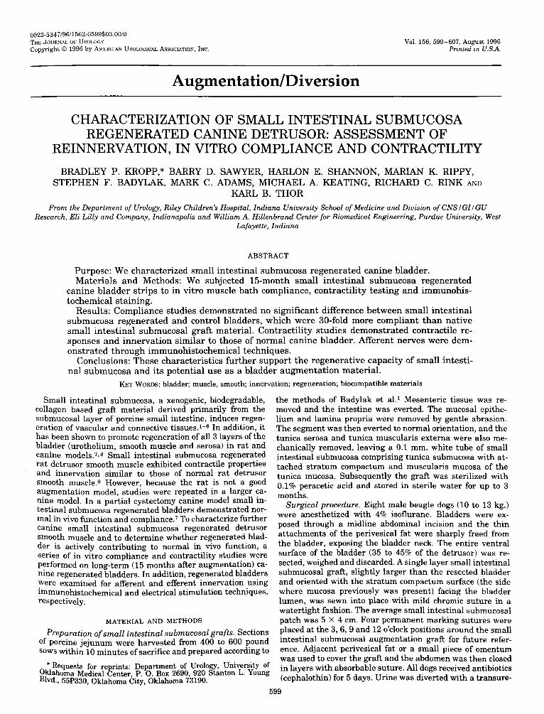
Administration of the p-adrenergic receptor agonist isopro- terenol (10 pM.) caused relaxation and elimination of spon- taneous activity in small intestinal submucosa regenerated and normal bladder strips (fig. 5). Because of the continuous decline in baseline tension, reliable quantification of relax- ation was not possible. The p-adrenergic receptor antagonist propranolol(100 pM.) reversed the effects of isoproterenol in regenerated and normal bladder strips (data not shown).
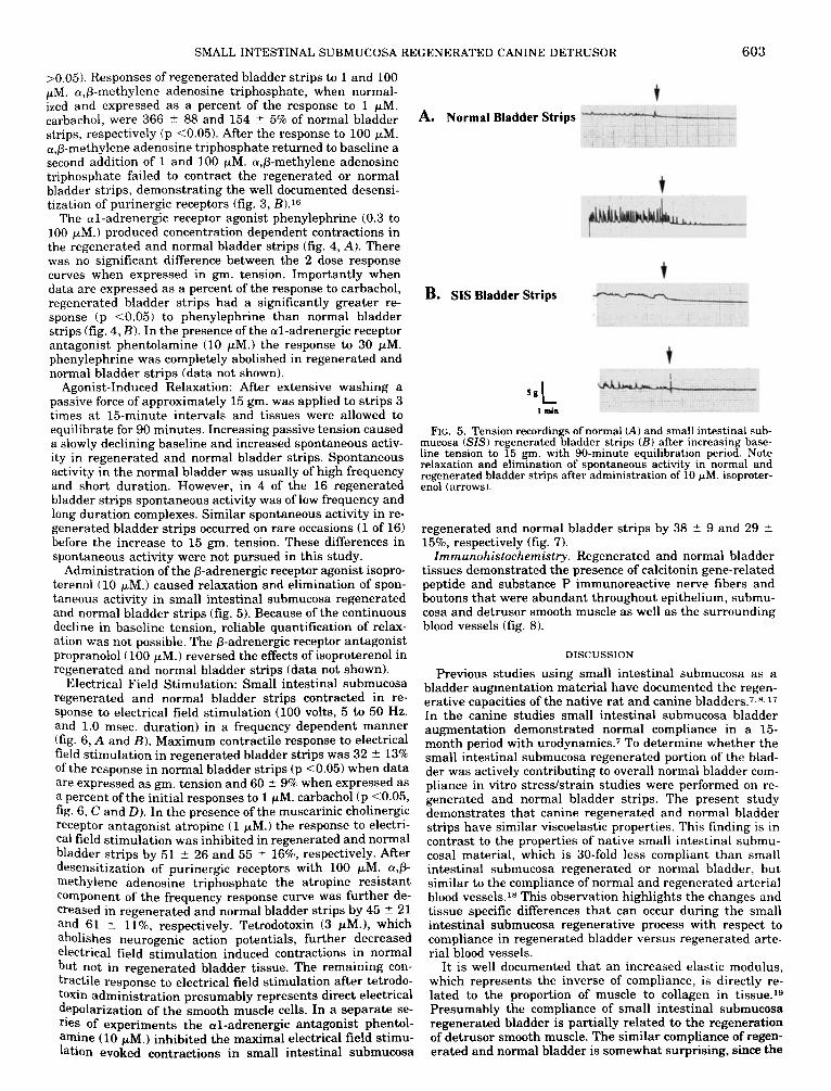
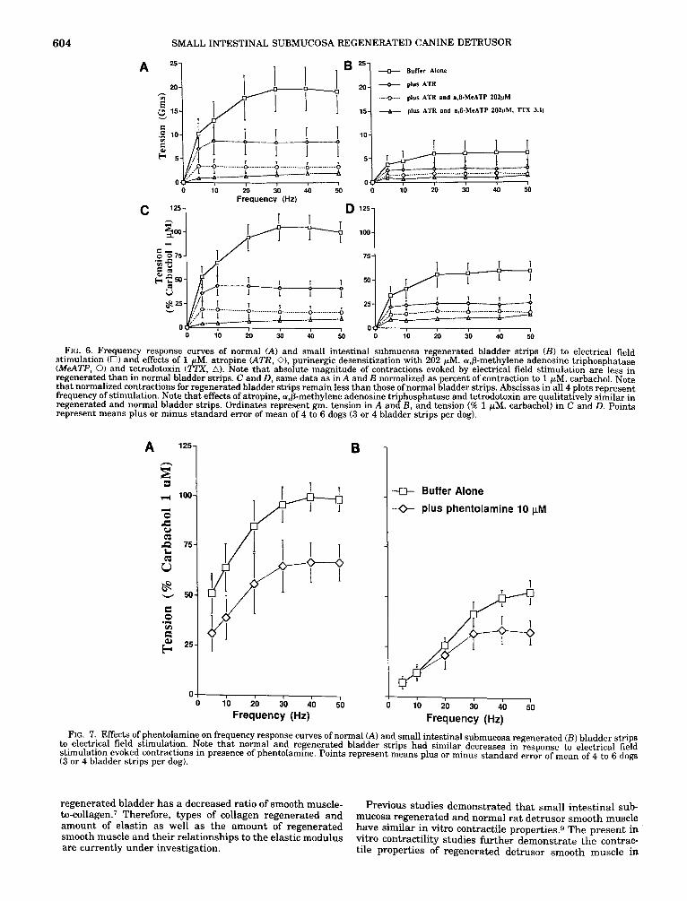
Electrical Field Stimulation: Small intestinal submucosa regenerated and normal bladder strips contracted in re- sponse to electrical field stimulation (100 volts, 5 to 50 Hz. and 1.0 msec. duration) in a frequency dependent manner (fig. 6, A and B). Maximum contractile response to electrical field stimulation in regenerated bladder strips was 32 2 13% of the response in normal bladder strips (p <0.05) when data are expressed as gm. tension and 60 -C 9% when expressed as a percent of the initial responses to 1 pM. carbachol (p <0.05, fig. 6, C and D). In the presence of the muscarinic cholinergic receptor antagonist atropine (1 pM.) the response to electri- cal field stimulation was inhibited in regenerated and normal bladder strips by 51 ? 26 and 55 5 1676, respectively. After desensitization of purinergic receptors with 100 pM. a$- methylene adenosine triphosphate the atropine resistant component of the frequency response curve was further de- creased in regenerated and normal bladder strips by 45 T 21 and 61 ? 1196, respectively. Tetrodotoxin (3 pM.), which abolishes neurogenic action potentials, further decreased electrical field stimulation induced contractions in normal but not in regenerated bladder tissue. The remaining con- tractile response to electrical field stimulation after tetrodo- toxin administration presumably represents direct electrical depolarization of the smooth muscle cells. In a separate se- ries of experiments the al-adrenergic antagonist phentol- a m h e (10 pM.) inhibited the maximal electrical field stimu- lation evoked contractions in small intestinal submucosa
A. Normal Bladder Strips
B. SIS Bladder Strips
5gL I min
FIG. 5. Tension recordings of normal (A) and small intestinal sub- mucosa (SIS) regenerated bladder strips ( B ) after increasing base- line tension to 15 gm. with 90-minute equilibration period. Note relaxation and elimination of spontaneous activity in normal and regenerated bladder strips after administration of 10 pM. isoproter- enol (arrows).
regenerated and normal bladder strips by 38 2 9 and 29 2 15%, respectively (fig. 7).
Zmmunohistochemistry. Regenerated and normal bladder tissues demonstrated the presence of calcitonin gene-related peptide and substance P immunoreactive nerve fibers and boutons that were abundant throughout epithelium, submu- cosa and detrusor smooth muscle as well as the surrounding blood vessels (fig. 8).
DISCUSSION
Previous studies using small intestinal submucosa as a bladder augmentation material have documented the regen- erative capacities of the native rat and canine bladders.7,". 17
In the canine studies small intestinal submucosa bladder augmentation demonstrated normal compliance in a 15- month period with u r~dynamics .~ To determine whether the small intestinal submucosa regenerated portion of the blad- der was actively contributing to overall normal bladder com- pliance in vitro stresdstrain studies were performed on re- generated and normal bladder strips. The present study demonstrates that canine regenerated and normal bladder strips have similar viscoelastic properties. This finding is in contrast to the properties of native small intestinal submu- cosal material, which is 30-fold less compliant than small intestinal submucosa regenerated or normal bladder, but similar to the compliance of normal and regenerated arterial blood vessels.'* This observation highlights the changes and tissue specific differences that can occur during the small intestinal submucosa regenerative process with respect to compliance in regenerated bladder versus regenerated arte- rial blood vessels.
I t is well documented that an increased elastic modulus, which represents the inverse of compliance, is directly re- lated to the proportion of muscle to collagen in tissue.lg Presumably the compliance of small intestinal submucosa regenerated bladder is partially related to the regeneration of detrusor smooth muscle. The similar compliance of regen- erated and normal bladder is somewhat surprising, since the
604 SMALL INTESTINAL SUBMUCOSA REGENERATED CANINE DETRUSOR
A " 25 + BuNer Alone
.---o.-- plus ATR and a,R-MeATP 202pM
d- plus ATR and a$-MeATP 202pM. 7 T X 3.11 I 1 L / I ,
i D .... 0 ............ B ........... 6 ............ ............ 6 2,. I 1 I --+---A
0 .' 0 10 30 40 50
Freauency (Hz) 1 D
0 1 0 2 0 3 0 4 0 M
10 1 I 1 I I 1 5j&qp3q _.. ..... ...................................
0 ' 0 1 0 2 0 3 0 4 0 5 0
125
75
50
25 .... ............ ........... ............
0 0 I 0 2 0 3 0 4 0 5 0
FIG. 6. Frequency response curves of normal (A) and small intestinal submucosa regenerated bladder strips ( B ) to electrical field stimulation (0) and effects of 1 pM. atropine (ATR, O), purinergic desensitization with 202 pM. a$-methylene adenosine triphosphatase (MeATP, 0) and tetrodotoxin (WX, A). Note that absolute magnitude of contractions evoked by electrical field stimulation are less in regenerated than in normal bladder strips. C and D, same data as in A and B normalized as percent of contraction to 1 pM. carbachol. Note that normalized contractions for regenerated bladder strips remain less than those of normal bladder strips. Abscissas in all 4 plots represent frequency of stimulation. Note that effects of atropine, a$-methylene adenosine tri hosphatase and tetrodotoxin are qualitatively similar in regenerated and normal bladder stri s Ordinates represent gm. tension in A anfB, and tension (% 1 pM. carbachol) in C and D. Points represent means plus or minus stanfard error of mean of 4 to 6 dogs (3 or 4 bladder strips per dog).
A 125
E 3 r( 100
E 0
2 25
.I cn
0
B
1 10 20 30 40 50
Frequency (Hz)
-11 Buffer Alone
-Q- plus phentolamine 10 pM
10 20 30 40 50 I
Frequency (Hz) FIG. 7. Effects of phentolamine on frequency response curves of normal (A) and small intestinal submucosa regenerated (B) bladder strips
to electrical field stimulation. Note that normal and regenerated bladder strips had similar decreases in response to electrical field stimulation evoked contractions in presence of phentolamine. Points represent means plus or minus standard error of mean of 4 to 6 dogs (3 or 4 bladder strips per dog).
regenerated bladder has a decreased ratio of smooth muscle- to-~ol lagen.~ Therefore, types of collagen regenerated and amount of elastin as well as the amount of regenerated smooth muscle and their relationships to the elastic modulus are currently under investigation.
Previous studies demonstrated that small intestinal sub- mucosa regenerated and normal rat detrusor smooth muscle have similar in vitro contractile properties.9 The present in vitro contractility studies further demonstrate the contrac- tile properties of regenerated detrusor smooth muscle in i

SMALL INTESTINAL SUBMUCOSA REGENERATED CANINE DETRUSOR 605
FIG. 8. Fluorescein isothiocyanate immunofluorescent staining for substance P (A to D and G ) and calcitonin gene-related peptide (E, F and E.n in sections of small intestinal submucosa regenerated bladder. A and B, substance P immunoreactive nerve fibers and boutons (arrows) in submucosal layer. B , note close proximity of labeled fibers (arrows) to epithelial cell layer (li ht green layer a t top). C and D, substance P immunoreactive fibers and boutons (arrows) in smooth muscle layer. Note large bundle of sutstance P immunoreactive fibers (D, arrow). E, calcitonin gene-related peptide immunoreactive fibers (arrows) in submucosal layer. F, calcitonin gene-related peptide immunoreactive fibers in smooth muscle layer (arrows). G and H, substance P and calcitonin gene-related peptide immunoreactive boutons (arrows) surround blood vessels (*) in regenerated bladder. Calibration bar equals 100 pm. in A, C and F, 40 pm. in B, D G and H, and 200 Pm. in E.
larger grafts of canine bladder. Based on the results of the Present study we conclude that canine small intestinal sub- mucosa regenerated detrusor smooth muscle expresses mus- carinic cholinergic, purinergic, and a and p-adrenergc recep- tors, and it is capable of generating neurotransmitter mediated contraction and relaxation as in normal tis- Sue.12, 13,20-22 There are several differences between regener- ated a d normal detrusor smooth muscles, such as a decrease 1n the maximal contractile response to carbachol. A posslble
for the decreased response may be related to the decreased ratio of muscle-to-collagen in regenerated blad- der.7 In other words, there are less muscle fibers available for
contraction in regenerated than in normal bladder. Also, contractile responses for a,&methylene adenosine triphos- phate and phenylephrine were significantly greater in the small intestinal submucosa regenerated bladder when ex- pressed as a percent of the carbachol response. The increased response suggests supersensitivity for the purinergic and a1-adrenergic receptors in regenerated detrusor smooth muscle. Other possible explanations for differences in agonist induced contractile responses include changes in relative receptor density and/or differences in relative efficiencies of receptor-contraction coupling in regenerated smooth mus- cle. Additional studies are required to differentiate the exact

606 SMALL INTESTINAL SUBMUCOSA REGENERATED CANINE DETRUSOR
cause for relative agonist induced differences in the contrac- tile responses.
In addition to the agonist induced contractile responses, small intestinal submucosa regenerated bladder also demon- strated frequency dependent contractile responses to electri- cal field stimulation. Since these contractions were partially antagonized by atropine, a$-methylene adenosine triphos- phate and phentolamine, it appears that the electrical field stimulation evoked contractions are mediated by cholinergic, purinergic and adrenergic mechanisms, respectively. This indicates that efferent parasympathetic, purinergic and sym- pathetic nerves reinnervated the regenerated smooth muscle cells.
Similar to agonist induced contractions, the magnitude of electrical field stimulation evoked contractions was less in the regenerated than normal bladder, whether expressed in gm. tension or normalized to a percent of the carbachol in- duced contraction. Importantly the phentolamine sensitive component of the electrical field stimulation evoked contrac- tions (that is al-adrenergic component) was larger in the regenerated than normal bladder. Thus, it is tempting to speculate that there is a greater ratio of adrenergic-to- cholinergic innervation in regenerated tissue. Previous stud- ies on the reinnervation of regenerated minced smooth mus- cle cells in the vas deferens demonstrated an increased ratio of adrenergic-to-cholinergic nerves in regenerating smooth muscle.‘:’ Further work on the quantification of the nerves is required to answer this and other questions related to rein- nervation of small intestinal submucosa regenerated smooth muscle.
Substance P and calcitonin gene-related peptide are thought to be neurotransmitters of primary afferent nerve fibers in the bladder.24.25 In the present study substance P and calcitonin gene-related peptide immunoreactive nerve fibers were identified within the submucosa and muscularis in regenerated and normal bladders. Although a formal quantitative analysis was not performed, it appeared that the quantity and distribution of the nerve fibers were similar in regenerated and normal bladder. The presence of calcito- nin gene-related peptide and substance P immunoreactive nerves and boutons may suggest that primary afferent fibers reinnervated the regenerated bladder. However, further studies to determine the function capacities of the afferent nerves are required.
CONCLUSIONS
Previous studies have shown that small intestinal submu- cosa acts as a biodegradable scaffold for the regeneration of all 3 layers of the normal canine bladder (transitional epi- thelium, smooth muscle and serosa). The present study dem- onstrates that small intestinal submucosa regenerated blad- ders have viscoelastic properties similar to those of the normal bladder. Furthermore, small intestinal submucosa regenerated detrusor smooth muscle is capable of receptor mediated contraction and relaxation similar to that in nor- mal bladders. In addition, regenerated detrusor smooth mus- cle is reinnervated by afferent and efferent nerve fibers. It appears that small intestinal submucosa promotes regener- ation of the bladder and small intestinal submucosa regen- erated bladder actively contributes to normal in vivo bladder dynamics. Therefore, this study further supports the poten- tial use of small intestinal submucosal grafts for clinical bladder augmentation.
Carbachol, atropine sulfate, tetrodotoxin, phenylephrine hydrochloride, phentolamine hydrochloride, isoprotenol and propranolol were provided by Sigma, St. Louis, Missouri, and a,@methylene adenosine 5’-triphosphate dilithium salt was provided by Research Biochemical International, Natick, Massachusetts.
REFERENCES
1. Badylak, S. F., Lantz, G. C., Coffey, A. and Geddes, L. A.: Small intestinal submucosa as a large diameter vascular graft in the dog. J. Surg. Res., 47: 74, 1989.
2. Badylak, S. F.: Personal communication, 1994. 3. Lantz, G. C., Badylak, S. F., Coffey, A. C., Geddes, L. A. and
Blevins, W. E.: Small intestinal submucosa as a small- diameter arterial graft in the dog. J . Invest. Surg., 3: 217, 1990.
4. Lantz, G. C., Badylak, S. F., Coffey, A. C., Geddes, L. A. and Sandusky, G. E.: Small intestinal submucosa as a superior vena cava graft in the dog. J. Surg. Res., 53: 175, 1992.
5. Lantz, G. C., Badylak, S. F., Hiles, M. C., Coffey, A. C., Geddes, L. A,, Kokini, K., Sandusky, G. E. and Morff, R. J.: Small intestinal submucosa as a vascular graft: a review. J. Invest. Surg., 6 297, 1993.
6. Sandusky, G. J., Badylak, S. F., Morff, R. J., Johnson, W. D. and Lantz, G.: Histologic findings after in vivo placement of small intestine submucosal vascular grafts and saphenous vein grafts in the carotid artery in dogs. Amer. J . Path., 140: 317, 1992.
7. Kropp, B. P., Rippy, M. K., Badylak, S. F., Adams, M. C., Keating, M. A,, Rink, R. C. and Thor, K. B.: Regenerative urinary bladder augmentation using small intestinal submu- cosa: urodynamic and histopathologic assessment in long-term canine bladder augmentations. J. Urol., 1 5 5 2098, 1996.
8. Kropp, B. P., Eppley, B. L., Prevel, C. D., Rippy, M. K., Harruff, R. C., Badylak, S. F., Adams, M. C., Rink, R. C. and Keating, M. A.: Experimental assessment of small intestine submucosa as a bladder wall substitute. Urology, 4 6 396, 1995.
9. Vaught, J. D., Kropp, B. P., Sawyer, B. D., Rippy, M. K., Badylak, S. F., Shannon, H. E. and Thor, K. B.: Detrusor smooth muscle regeneration in the rat using porcine small intestinal submucosal grafts: functional innervation and re- ceptor expression. J. Urol., 1 5 5 374, 1996.
10. Minns, R. J., Soden, P. D. and Jackson, D. S.: The role of the fibrous components and ground substance in the mechanical properties of biological tissues: a preliminary investigation. J. Biomechanics, 6 153, 1973.
11. Zderic, S. A., Liu, G. H., Haab, J. P., Monson, F. C., Gong, C. and Levin, R. M.: What is the most accurate way to study the active properties of bladder smooth muscle? Pharmacology, 4 8 380, 1994.
12. Rohner, T. J., Jr., Hannigan, J . D. and Sanford, E. J.: Altered in vitro adrenergic responses of dog detrusor muscle after chronic bladder outlet obstruction. Urology, 11: 357, 1978.
13. Leoni, J. V., Wein, A. J., Raezer, D. M. and Schoenberg, H. W.: The effect of adrenergic stimulation on the contractile re- sponse of canine detrusor muscle. Invest. Urol., 10: 419, 1973.
14. Thor, K. B. and Helke, C. J.: Serotonin- and substance p-containing projections to the nucleus tractus solitarii of the rat. J . Comp. Neurol., 265 275, 1987.
15. Susset, J. G. and Regnier, C. H.: Viscoelastic properties of blad- der strips: standardization of a technique. Invest. Urol., 1 8 445, 1981.
16. Burnstock, G., Dumsday, B. and Smythe, A,: Atropine-resistant excitation of the urinary bladder: the possibility of transmis- sion via nerves releasine a Durine nucleotide. Brit. J . Pharma- - . col., 44: 451, 1972.
17. KnaDD. P. M.. Lineeman. J. E.. Sieeel. Y. I.. Badvlak. S. F. and . - . Y . I - I
Demeter, R. J.: Biocompatibility of small-intestinal submucosa in urinary tract as augmentation cystoplasty graft and inject- able suspension. J . Endourol., 8: 125, 1994.
18. Herbert, S. T., Badylak, S. F., Geddes, L. A,, Hillberry, B., Lantz, G. C. and Kokini, K.: Elastic modulus of prepared canine jejunum, a new vascular graft material. Ann. Biomed. Eng., 21: 727, 1993.
19. Kondo, A. and Susset, J. G.: Viscoelastic properties of bladder. 11. Comparative studies in normal and pathologic dogs. Invest. Urol., 11: 459, 1974.
20. Burnstock, G., Cocks, T., Kasakov, L. and Wong, H. K.: Direct evidence for ATP release from non-adrenergic, non-cholinergic (purinergic) nerves in the guinea-pig taenia coli and bladder. Eur. J. Pharmacol., 4 9 145, 1978.
21. Brading, A. F. and Williams, J . H.: Contractile responses of smooth muscle strips from rat and guinea-pig urinary bladder to transmural stimulation: effects of atropine and a,P- methylene ATP. Brit. J. Pharmacol., 99: 493, 1990.

SMALL INTESTINAL SUBMUCOSA REGENERATED CANINE DETRUSOR 607 22. Kasakov, L. and Burnstock, G.: The use of the slowly degradable
analog, n,p-methylene ATP, to produce desensitization of the Pa-purinoceptor: effect on non-adrenergic, non-cholinergic re- sponses of the guinea-pig urinary bladder. Eur. J . Pharmacol., 86 291,1982.
23. Burnstock, G., Yokota, R. and Jones, R.: Reinnervation of regen- erating smooth muscle cells in minced vas deferens of the guinea-pig. Cell. Tissue Res., 190: 495, 1978.
24. de Groat, W. C. and Kawatani, M.: Neural control of the urinary bladder: possible relationship between peptidergic inhibitory mechanisms and detrusor instability. Neurourol. Urodynam., 4: 285, 1985.
25. Maggi, C. A,, Giuliani, S., Del Bianco, E., Geppetti, P., Theodorsson, E. and Santicioli, P.: Calcitonin gene-related peptide in the regulation of urinary tract motility. Ann. N. Y. Acad. Sci., 657: 328, 1992.