characterization and biological control of bovine of...
TRANSCRIPT
i
Characterization and biological control of bovine of Streptococcus uberis strains associated with
bovine mastitis
A thesis submitted in fulfilment of the requirements for the degree of Doctor of Philosophy
Nalin Wongkattiya M.Sc. (Biology)
School of Applied Sciences Sciences, Engineering and Technology Portfolio
RMIT University March 2008
ii
Declaration
I certify that except where due acknowledgement has been made, the work is that of
the author alone; the work has not been submitted previously, in whole or in part, to
qualify for any other academic award; the content of the thesis is the result of work
which has been carried out since the official commencement date of the approved
research program; and any editorial work, paid or unpaid, carried out by a third party
is acknowledged.
Nalin Wongkattiya
27 March 2008

iii
Acknowledgements
I would like to express my deep and sincere gratitude to my supervisors, Associate
Professor Margaret Deighton and Prudence Bramwell. Their wide knowledge and
logical way of thinking have been of great value to me. Their understanding,
encouraging and personal guidance have provided a good basis for the present thesis.
I would also like to thank Royal Thai Government for the financial support for my
studies.
I am grateful to all the farmers and veterinarians for their time and enthusiasm in
supplying samples for this project.
I am indebted to Associate Professor Ann Lawrie for her kind induction in electron
microscopy work. I also wish to thank Kylie White and Phil Francis for their essential
assistance in electron microscopy work. I wish to thank Dr. John Fecondo and They
Ng for their advice in protein work. My thanks to Associate Professor Peter Cullis for
his guidance in mass spectrometry work and Dr. Danilla Grando for her advice in
PFGE work.
I would also like to thank all the support staff and friends of the Department of
Biotechnology and Environmental Biology for their assistance and friendship.
I am especially grateful to Nicole Grech, Carmine D’Errico, Somprasong Laingam,
Dennis O’Donelle and Dr. Chareeporn Akekawatchai for all the emotional support,
friendship, entertainment, and caring they provided.
Lastly, and most importantly, I wish to thank Ian Fraser, my sisters and my parents on
whose constant encouragement and love I have relied throughout my time at the
University.
iv
Contents Summary ………...…………………………………………………………………………1
Chapter 1 General introduction .......................................................................................... 3
1.1 Bovine mastitis.......................................................................................................... 3
1.2 Streptococcus uberis ................................................................................................. 9
1.3 Host-parasite interactions in Streptococcus uberis infection .................................. 12
1.3.1 Host .......................................................................................................... 12
1.3.2 Environment ............................................................................................. 13
1.3.3 Streptococcus uberis strain....................................................................... 14
1.4 Control of mastitis caused by Streptococcus uberis................................................ 14
1.4.1 Antibiotic treatment.................................................................................. 14
1.4.2 Reduction of pathogen exposure .............................................................. 16
1.4.3 Interaction of bovine streptococci and minor mastitis pathogens ............ 20
1.4.4 Cows immune system............................................................................... 21
1.5 Bacteriocins............................................................................................................. 24
1.5.1 General properties of bacteriocins from staphylococci............................ 27
1.5.2 Assay of bacteriocin ................................................................................. 29
1.5.3 Exclusion of non-bacteriocin inhibitory agents ....................................... 32
1.5.4 Production of bacteriocins........................................................................ 33
1.5.5 Estimation of bacteriocin molecular mass ............................................... 37
1.5.6 Bacteriocin extraction .............................................................................. 38
1.5.7 Purification of bacteriocins ...................................................................... 39
1.6 Aims of this study ................................................................................................... 41
Chapter 2 Isolation and identifications of Streptococcus uberis by conventional
methods ................................................................................................................................ 42
2.1 Introduction ............................................................................................................. 42
2.2 Materials and methods ............................................................................................ 44
2.2.1 Milk samples collection ......................................................................... 44
2.2.2 Bacterial reference strains ………………………………………………45
2.2.3 Streptococcus uberis isolation and identification..................................... 45
2.2.4 Pulse field gel electrophoresis (PFGE) of Streptococcus parauberis ...... 51
2.3 Results ..................................................................................................................... 53
2.4 Discussion ............................................................................................................... 59
v
Chapter 3 Determination of Streptococcus uberis antibacterial susceptibility patterns64
3.1 Introduction ............................................................................................................. 64
3.2 Material and methods .............................................................................................. 65
3.2.1 Bacterial strains ........................................................................................ 66
3.2.2 Calibration of turbidity............................................................................. 66
3.2.3 Antibiotic compounds .............................................................................. 67
3.2.4 Antibiotic susceptibility tests ................................................................... 67
3.2.5 Erythromycin resistance gene PCR.......................................................... 70
3.2.6 Transmission electron microscopy........................................................... 71
3.3 Results ..................................................................................................................... 71
3.4 Discussion ............................................................................................................... 76
Chapter 4 Bacteriocins isolation and production............................................................. 80
4.1 Introduction ............................................................................................................. 80
4.2 Materials and methods ............................................................................................ 81
4.2.1 Bacterial isolates and control strains........................................................ 81
4.2.2 Identification procedures of coagulase negative staphylococci ............... 82
4.2.3 Detection and isolation of bacteriocin-producing strains......................... 83
4.2.4 Confirmation that observed activity is due to a bacteriocin..................... 85
4.2.5 Study of Bacteriocin production .............................................................. 86
4.3 Results ..................................................................................................................... 90
4.3.1 Isolation of coagulase negative Staphylococcus and the bacteriocin-
producing strain................................................................................................ 90
4.3.2 Characterization of antibiotic substance .................................................. 95
4.3.3 Bacteriocin production ............................................................................. 97
4.4 Discussion ............................................................................................................. 105
Chapter 5 Bacteriocin characterization and purification ............................................. 109
5.1 Introduction ........................................................................................................... 109
5.2 Material and methods ............................................................................................ 109
5.2.1 Bacteriocin characterization................................................................... 109
5.2.2 Electron microscopy............................................................................... 111
5.2.3 Bacteriocin purification.......................................................................... 113
5.3 Results................................................................................................................... 127
vi
5.3.1 Bacteriocin characterization................................................................... 127
5.3.2 Electron microscopy............................................................................... 133
5.3.3 Bacteriocin purification.......................................................................... 136
5.3.4 Mass spectrometry.................................................................................. 149
5.4 Discussion ............................................................................................................. 153
Final discussion ……...…………………………………………………………….. ……159
Appendix A ………………………...…………………………….……………………... 162
Appendix B ………………………………………………………….…………………... 167
Appendix C ………………………...………………………………….………………... 188
References ……………………………………………………………..…………….….. 189
vii
List of Tables Table 1.1 Bacteriocins from the genus Staphylococcus .............................................. 28
Table 2.1 Biochemical test patterns of esculin-degrading streptococci isolated from bovine
milk samples.................................................................................................................55
Table 2.2 Association between disease status and qualitative growth of Streptococcus uberis
......................................................................................................................................56
Table 3.1 Minimal inhibitory concentration breakpoints for veterinary streptococci..69
Table 3.2 Distribution of Minimal Inhibitory Concentration (MIC) of 68 isolates of
Streptococcus uberis ....................................................................................................72
Table 3.3 Activity of antibiotic agent for Streptococcus uberis ..................................78
Table 4.1 Factors affecting antibioticl activity of Macrococcus caseolyticus
2042-4 ……………………………………………………………………………….96
Table 4.2 Effect of media on bacteriocin activity ........................................................98
Table 4.3 Growth of Macrococcus caseolyticus 2042-4 in BHB ..............................100
Table 4.4 Minimal inhibitory concentration of a range of antibiotic agents for Macrococcus
caseolyticus 2042-4 ....................................................................................................102
Table 5.1 Protein standard preparation ......................................................................117
Table 5.2 The composition of the acrylamide mixtures to make two mini SDS-PAGE gels
....................................................................................................................................122
Table 5.3 Bacteriocin activity in semi-solid BHI agar...............................................137
Table 5.4 The bacteriocin activity of M. caseolyticus 2042-4 extract obtained by ammonium
sulphate precipitation .................................................................................................139
Table 5.5 Purification scheme of the bacteriocin ………………………….……….148
viii
List of Figures Figure 2.1 PFGE of 10 isolates of S. parauberis from milk samples. ........................ 58
Figure 3.1 PCR of ermA gene erythromycin-resistant isolate......................................74
Figure 3.2 Streptococcus uberis 796-2 treated with erythromycin.. ............................75
Figure 4.1 Inhibitory effect of the strain 2042-4..........................................................91
Figure 4.2 Multiple alignments of 16S rRNA gene partial sequencing of the strain 2042-4
and Macrococcus caseolyticus accession numbers AY126154, DQ518592, EF204307,and
EU048336.....................................................................................................................94
Figure 4.3 Inhibitory effect of crude bacteriocin extracted from semi-solid agar by agar
diffusion method ........................................................................................................104
Figure 5.1 Purification scheme of the bacteriocin......................................................114
Figure 5.2 Bio-Rad Mini-Protean apparatus ..............................................................123
Figure 5.3 Effect of temperatures on the bacteriocin activity ....................................128
Figure 5.4 Effect of pH on the bacteriocin inhibitory activity ...................................130
Figure 5.5 Effect of ammonium sulphate precipitate bacteriocin of Macrococcus
caseolyticus 2042-4 on cells of Streptococcus uberis 796-2......................................132
Figure 5.6 Scanning electron microscopy of S. uberis in presence of bacteriocin. ...134
Figure 5.7 Transmission electron microsopy of S. uberis treated with bacteriocin ...135
Figure 5.8 Elution profile of the bacteriocin activity on gel filtration. ......................141
Figure 5.9 Coomassie blue stained SDS-PAGE gel of bacteriocin purification…... .142
Figure 5.10 Elution profile of the active fractions from gel filtration to cation exchange
chromatography at pH 5.5. .........................................................................................144
Figure 5.11 PR-HPLC chromatogram of the bacteriocin component I from cation exchange
chromatography, C4 column. .....................................................................................146
Figure 5.12 PR-HPLC chromatogram of the bacteriocin component II from cation exchange
chromatography, C4 column. ...................................................................................147
Figure 5.13 Mass spectrometric analysis of purified the bacteriocin component I....150
Figure 5.14 Mass spectrometric analysis of purified the bacteriocin component II. .151
Figure 5.15 Summary of purification of the bacteriocins from Macrococcus caseolyticus
2042-4.........................................................................................................................152
ix
Lists of Abbreviations and Symbols $ Dollar % percent & and £ Pound ® registered sign ≤ less-than or equal to ≥ greater-than or equal to °C degree Celsius µg microgram µl microlitre µm micrometer € Euro 16S 16 subunit 23S 23 subunit 3’ three prime 5’ five prime A600 absorbance at 600 nanometre AU Arbitrary unit bp base pairs CaCO3 calcium carbonate CFU colony forming unit cm centimetre CO2 carbon dioxide Da Dalton DNA deoxyribonucleic acid dNTP deoxyribonucleotide triphosphate EDTA ethylenediaminetetraacetic acid et al. Et ali, and others g relative centrifugation force g gram h hour H2O2 hydrogen peroxide k kilo kb kilobase L litre M molarity mg milligram MgCl2 magnesium chloride min minute mL millilitre mm millimetre mM millimole NaCl sodium chloride NaOH sodium hydroxide No. number OD optical density PBS phosphate buffer saline
x
PCR polymerase chain reaction pH potential of hydrogen RNA ribonucleic acid (rRNA-ribosomal RNA) s second TBE Tris Boric Acid EDTA Tris-HCl Tris hydrochloride Tris Tris (hydroxymethyl) amino methane ™ trade mark sign U unit UV ultra violet V volt v/v volume per volume w/v weight per volume X times x by β beta
1
Summary
Bovine mastitis still remains a major problem to the dairy industry world wide although
there are mastitis control strategies in place. In the last few decades there has been a
change in the predominant pathogens causing mastitis. Streptococcus uberis is now the
leading cause of bovine mastitis. Accurate and cost effective methods for identification of
S. uberis by diagnostic laboratories would assist farm management to reduce the infection.
One common practice to control mastitis is the use of antimicrobial agents. Long term
antibiotic use can cause antibiotic resistance and there is concern that resistant bacteria
could be transferred to humans. Knowledge of current antibiotic susceptibility test patterns
for mastitis pathogens could improve therapy protocols for particular dairy farms. An
alternative way to eliminate mastitis pathogens is biological control. Recent evidence has
shown that non-pathogenic coagulase-negative staphylococci have the ability to produce
antibacterial proteins (bacteriocins) that are active against some mastitis pathogens.
One hundred and fifteen isolates of esculin hydrolyzing streptococci from intramammary
infections in dairy cows, which are commonly categorized as S. uberis, were further
identified using four additional biochemical tests (hippurate hydrolysis, fermentation of
inulin and mannitol and growth in 6.5% NaCl). 16S rRNA sequencing was used as a gold
standard for species identification. The additional biochemical tests generated seven
different patterns. All isolates belonging to pattern 1 (hippurate hydrolysis positive, inulin
and mannitol fermentation positive and negative for growth in 6.5% NaCl) were identified
by 16S rRNA sequencing as S. uberis (91%) or S. parauberis (9%). Half of the isolates
displaying pattern 2 which differed from pattern 1 only in inulin fermentation (negative) were
also identified as S. uberis (33%) or S. parauberis (17%) by 16S rRNA sequencing.
Differentiation of S. uberis and S. parauberis required genotypic examination. Use of these
additional biochemical tests (hippurate hydrolysis, inulin and mannitol fermentation and
growth in 6.5% NaCl) resulted in relatively low numbers of misidentification (8.6%). These
2
tests represent an affordable cost and can therefore be considered an adequate method for
veterinary diagnostic laboratories for identifying S. uberis isolated from bovine mastitis.
The susceptibility of S. uberis to nine antimicrobial agents was determined by the broth
dilution method. The minimal inhibitory concentrations (MICs) at which 90% of isolates
were inhibited (MIC90s) for each of ampicillin, penicillin, oxacillin, erythromycin, tetracycline,
novobiocin, cephalothin, vancomycin and gentamicin was 1.0, 0.06, 0.25, 0.5, 0.5, 4.0,
0.25, 0.5 and 8.0 µg/mL respectively. No isolates showed resistance to those antibiotics.
However the continuous monitoring of antibiotic resistance is essential, as the emergence
of resistance strains has become a concern.
A bacteriocin-producing bacterium, Macrococcus caseolyticus (formerly Staphylococcus
caseolyticus) was isolated from a cow’s milk sample in Victoria from a farm with a “uberis”
problem; i.e. an unacceptable prevalence of clinical and sibclinical cases of mastitis. The
bacteriocin was heat stable and remained active over a wide range of pH, with highly
specific activity against the genus Streptococcus. Purification of the bacteriocin, included
ammonium sulphate precipitation, gel filtration, cation exchange chromatography,
extraction from polyacrylamide gel and reverse phase high performance liquid
chromatography. The results showed two components which were independently active
and the mass spectrometry showed the mass of both was less than 3000 Da. Scanning
and transmission electron microscopy showed damage of target cells. It is suggested that
the use of bacteriocins could provide effective control and prevention of bovine mastitis
caused by S. uberis.
3
Chapter 1 General introduction
1.1 Bovine mastitis
Bovine mastitis is an inflammatory condition of the mammary gland in dairy cattle and is
associated with chemical, physical and usually bacteriological changes in milk (Radostits,
et al., 1994). Clinical findings in mastitis are classified according to severity of disease.
Clinical mastitis is defined as obvious inflammation of the udder (Smith, et al., 1985b,
Radostits, et al., 1994). The symptoms include redness, swelling, heat and pain. There is
decreased milk production, changes in composition of the milk and changes in milk
appearance, such as discoloration, the presence of flakes, clots or a watery secretion and
the presence of numerous leukocytes. Subclinical mastitis is defined as an inflammation of
the mammary gland that is not visible and requires diagnostic tests for detection (Radostits,
et al., 1994). Subclinical mastitis is the most prevalent type of inflammation of the
mammary gland in dairy cows. During subclinical mastitis, microorganisms are present in
milk, the leukocyte content is elevated and milk quality is reduced. This type of mastitis
causes the greatest economic loss (Smith, et al., 1985b, Radostits, et al., 1994). Chronic
mastitis may persist in subclinical form for months or years (Radostits, et al., 1994).
Diagnosis of clinical mastitis is based on abnormal appearance of the udder or milk, which
can be determined by farmers or veterinarians. Milk may present colour changes from
slightly yellow, yellow to bloody, bloody or have the appearance of serum (watery).
Abnormal milk may also contain a varying amount of clots. Signs of udder abnormalities
such as swelling, severity of pain and the overall appearance indicate the severity of
infection and serve as a guide for the course of treatment (Radostits, et al., 1994).
Diagnosis of subclinical infection is more problematic since the milk appears normal.
Analysis of subclinical mastitis is usually based upon some indicators of inflammation
which can be made in several ways, including direct measurement of the somatic cell count
4
(SCC) level or indirect methods by performing a California Mastitis Test (CMT) on
suspected quarters or measurement related to increased vascular permeability which
results in alterations in the chemistry of the milk. For routine milk analysis a combination of
the above methods should be an ongoing part of any mastitis control program. Milk culture
will definitively identify the presence of mastitis pathogens.
The cells found in milk (somatic cells) consist of white blood cells or leukocytes and
epithelial cells (Radostits, et al., 1994). Leukocyte numbers increase in response to
bacterial infection. The epithelial cells originate from udder secretory tissue and increase as
a result of injury or infection. An increase in somatic cell numbers is largely a result of an
increase in the number of leucocytes. The concentration of somatic cells serves as a
measurement of the level of infection in the cow’s mammary gland.
Individual cow somatic cell count (SCC) will provide a determination of the prevalence of
infection within the herd. Bulk tank somatic cell counts (BTSCC) are performed routinely as
a indication of milk quality but the BTSCC can also be used to monitor the level of udder
health by monitoring the bulk tank scores over time (Radostits, et al., 1994). Normal
quarters show less than 150,000 cells/mL while quarters with SCC over 250,000 cells/mL
are considered to be infected (Brightling, et al., 1998, Leigh, 1999). The Australian
guidelines (Countdown Downunder) suggest Australian dairy farms supply milk with a cell
count of less than 250,000 cells/mL to ensure the production of high-quality milk (Brightling,
et al., 1998). Most farmers are willing to act on farm management practices (five-point plan
and Countdown Downunder) to obtain a low SCC (Brightling, et al., 1998).
The SCC is determined in a mastitis examination usually by electronic cell-counting
machines (Miller, et al., 1986, Radostits, et al., 1994). An indirect measurement of somatic
cell count in milk could be determined by California Mastitis Test (CMT) (Dingwell, et al.,
2003). The test is based upon the amount of cellular nuclear protein present in the milk
sample. CMT is a simple, inexpensive, rapid screening test for mastitis and can be
performed by milkers. The test is appropriate for cow-side evaluation of udder health.
5
Electrical conductivity of milk increases during mastitis due to increases in Na+ and Cl- and
decreases in K+ and lactose. Changes in conductivity can be easily detected by hand-held
devices (Seguya & Mansell, 2000). However, treatment of mastitis works best if there is
some information on the particular bacterium causing the problem. Therefore,
bacteriological culturing of milk samples is essential (Radostits, et al., 1994).
Due to the successful control of mastitis by modern dairy management, bulk milk somatic
cell counts have been reduced but mastitis still remain in herds with low BTSCC. In nine
well managed herds in the USA with low SCCs, 82.3% of clinical cases were caused by
coliforms and environmental streptococci which were difficult to control (Hogan, et al.,
1989b). A situation in three dairy herds from UK showed that either very low (less than
21,000 cells/mL) or high quarter SCCs (greater than 200,000 cells/mL) have been
associated with an increase in the incidence of clinical mastitis incidence which was mainly
caused by environmental pathogens (Peeler, et al., 2003).
Bovine mastitis causes a major economic loss in the dairy industry worldwide (Beck, et al.,
1992, Seegers, et al., 2003, Petrovski, et al., 2006). The dairy industry is Australia’s third
largest rural industry, with a farmgate value of $3.17 billion in 2004/05. It is the largest
value-added food industry, increasing value more than three-fold through processing and
marketing to contribute $9 billion to the nation’s economy. Dairy exports amount to about
one million tonnes of product to 130 countries and generates an estimated $3 billion in
export income each year. Victoria is a state that contributes most to Australian milk
production (65.7%) (Dairy Australia, 2007). Reduction in milk production and milk quality,
drugs, discarded milk, veterinarian expenses, labour cost, antibiotic uses, and culling
chronically infected cows contribute to the economic loss from mastititis (Østerås, 2000). The
costs of mastitis control are not difficult to calculate but loss from reduced production and
quality are more difficult to evaluate accurately. The average cost per case of clinical
mastitis is estimated to be €277 in The Netherlands and £203 in the UK (Østerås, 2000). The
annual cost is estimated to be £300 million in the UK (Hillerton & Berry, 2005).
6
After a calf is born, the cow will continue to produce milk for approximately 300 days. This
stage is called the lactation period. Subsequently, cessation of regular milking (dry period)
is recommended for at least 2 months prior to calving again (Dais & Allaire, 1982). The dry
period benefits the dairy cow in several ways. The change in diet to lower energy and
higher fibre content allows liver lesions and rumen ulcers to heal and increases rumen
muscle tone. Replenishment of the energy reverses of cows during the dry period by
increasing the dietary energy density might increase milk production. Also during this time,
the secretory cells in the mammary gland involute, regress, and then multiply again. A short
or absent dry period greatly reduces the numbers of secretory cells which are a major
factor affecting milk yield (Beever, 2006). In addition, cows are most susceptible to
environmental mastitis infections during the dry period (Eberhart, 1986). To reduce the risk
of new infections and cure existing cases, all quarters of all cows are treated with an
approved long-lasting antibiotic product at drying-off (Hillerton & Kliem, 2002, Hillerton &
Berry, 2005).
Mastitis is almost always caused by bacterial infection (Radostits, et al., 1994). Mastitis can
be classified according to the source of infectious organisms, either contagious mastitis or
environmental mastitis. Causes of contagious mastitis include Staphylococcus aureus,
Streptococcus dysgalactiae and Streptococcus agalactiae (Radostits, et al., 1994, Leigh,
1999). Contagious mastitis indicates that the infection is spread from cow to cow. The
bacteria can be transmitted during the milking process by milking hands, milking equipment
and other objects. Organisms causing environmental mastitis include Streptococcus uberis
and coliform bacteria (Smith, et al., 1985b, Radostits, et al., 1994). The primary source of
environmental pathogens is the surroundings in which a cow lives. Teats of cows can also
be colonized by harmless bacteria such as Corynebacterium sp., Bacillus sp., Aerococcus
sp., Micrococcus sp. and coagulase negative staphylococci (CoNS) (Woodward, et al.,
1988, White, et al., 1989, Vliegher, et al., 2004).
7
Over the past 30 years a great reduction of mastitis, particularly in the UK and USA has
been achieved due to the five-point plan (Leigh, 1999). The five-point plan for controlling
mastitis was launched by The National Institute for Research in Dairying (NIRD) and the
Central Veterinary Laboratory, UK. The plan aimed at reducing exposure, duration and
transmission of intramammary infections which consists of:
1. Keeping well designed milking machines maintained and functioning properly
2. Cleaning and drying teats before milking and using a teat dip afterwards
3. Dry cow treatment of every quarter of every cow with a long lasting antibiotic
4. Recognizing clinical mastitis cases early and treating them promptly
5. Culling chronically infected cows
The Countdown Downunder program for mastitis control in Australia is another consistent
set of "best practice" mastitis control measures and milk quality guidelines (Brightling, et al.,
1998). The guidelines cover periods of calving, lactation, late lactation, drying-off and the
dry period. For each period of the cow’s milking year, Countdown Downunder provides a
set of guidelines about what has to be done, why it should be done, how to do it and how to
check that it has been achieved. The goal of the Countdown Downunder program is to
reduce the BTSCC of at least 90% of Australian dairy farms supplying milk to less than
250,000 cells/mL and to reduce the cell count of 100% of Australian dairy farms supplying
milk to a cell count of less than 400,000 cells/mL in all milk supply periods. In 2005, about
400 dairy herds across Australia had an average BTSCC exceeding 400,000 cells/mL, but
another 1000 herds had levels between 300,000 and 400,000 cells/mL (DairyAustralia,
2007).
Since good management of mastitis has been used, the species of bacteria causing
mastitis has changed. The reduction of contagious mastitis has been remarkably
successful but there has been little effect on the incidence of mastitis due to bacteria which
infect the gland from environmental sources (Leigh, 1999, Bradley, 2002). Environmental
8
mastitis pathogens have become a major problem in dairy herds worldwide. Fifty dairy
herds in Ohio, USA reported that the most common clinical isolates were CoNS (14.6%)
and E. coli (14.6%) (Bartlett, et al., 1992). A one year study on three dairy herds in the UK
reported clinical mastitis incidence rates of 25.4, 55.2 and 67.6 quarter-cases per 100 cows
per year respectively. S. uberis and E. coli caused approximately 50% of clinical cases
(Peeler, et al., 2003). A study of five herds in New Zealand reported that S. uberis (63.2%)
was the most prevalent pathogen isolated from clinical mastitis cases followed by CoNS
(11.5%) (McDougall, et al., 2004). Moreover, the majority of S. uberis infections are
subclinical (Zadoks, et al., 2003). In subclinical mastitis, decreased milk production results
in the greatest loss, representing about 75% of the total loss (DeGraves & Fetrow, 1993). It
is likely that inadequacies of current mastitis control strategies will lead to a changing
aetiology of mastitis. The search for additional understanding and procedures to control the
environmental bacteria is essential.
1.2 Streptococcus uberis
Bergey’s manual of systemic bacteriology lists seven genera of facultative anaerobic Gram-
positive coccci (GPC); Aerococcus, Leuconostoc, Micrococcus, Pediococcus,
Staphylococcus, Streptococcus and Stomatococcus (Schleifer, 1986). A review written in
1995 listed additional new genera which included Globicatella, Enterococcus, Lactococcus,
Vagococcus, Allolococcus, Gemella, Tetragenococcus, and Helcococcus (Facklam &
Elliott, 1995). Streptococci are non-motile Gram-positive cocci, spherical or ovoid in shape,
which are arranged in pairs or chains. The genus Streptococcus consists of non-sporing,
catalase negative facultative anaerobic bacteria. Streptococci have complex nutritional
requirements and form mainly lactic acid or lactic, acetic and formic acids and ethanol and
CO2 from carbohydrates (Holt, et al., 1994). They are important in the dairy industry and as
human and animal pathogens (Jones, 1978). Jones (1978) reviewed several classifications
of streptococci. Most notable classifications are based on cultural characteristics (such as
hemolytic reaction when cultured on blood containing agar), and use of carbohydrate
fermentation reactions together with physiological and morphological tests. Serological
9
grouping, which was established by Lancefield in 1933, has played an important role in the
identification of streptococci (Lancefield, 1933). Serological or Lancefield grouping
differentiates streptococci according to antigenic characteristics of the group specific
carbohydrate reactions using the precipitation technique.
Identification of S. uberis is important to therapeutic decision-making and development of
methods to control mastitis. According to Jones (1978) and Holt, et al. (1994) the taxonomy
of S. uberis appears to be unsatisfactory. The species is biochemically and serologically
heterogeneous (Lammler, 1991, Khan, et al., 2003, Odierno, et al., 2006). One of the most
widely used methods for streptococci identification are commercial kits such as API 20
Strep (Odierno, et al., 2006), however, newly recognized species such as S. parauberis is
not included in the identification database. It is now becoming clear that identification of S.
uberis requires the use of both conventional and molecular methods. Current classification
of S. uberis relies on genotypic strategies. On the basis of DNA-DNA hybridization, Gravie
and Bramly (1979) demonstrated genetic heterogeneity of S. uberis and differentiated two
genotypes, designated as types I and II. After comparison of 16S rRNA nucleotide
sequences of S. uberis types I and II, Williams and Collins (1990) suggested type II as a
new species “Streptococcus parauberis”.
Streptococcus uberis is commonly described as an environmental mastitis pathogen
because it has an ability to survive and multiply in extramammary sites. It has been isolated
from various anatomical sites of cows and from the cow’s environment. These include
bovine lips, skin, udder surface, belly, teats, urogenital tract, tonsil, rectum, rumen, nostrils,
eyes and vulva (Cullen, 1966, Mundt, 1982, Bramley, et al., 1984). In addition, the
organism can be isolated from water, soil, plant matter, bedding materials, flies, fecal
samples and hay (Bramley, et al., 1984, Hogan, et al., 1989a, Hogan et al., 1990, Zadoks,
et al., 2005). With the increasing focus on the control of environmental bacteria, particularly
S. uberis, it is necessary to understand the epidemiology of the pathogen.
10
The most widely used method for sub-species differentiation is pulse-field gel
electrophoresis (PFGE). DNA macrorestriction analysis by PFGE appears to be the most
promising, useful, and reproducible typing system for epidemiological investigation which
makes it useful for comparison of strains between laboratories (Smith & Cantor, 1987,
Baseggio, et al., 1997, Wang, et al., 1999, Phuektes, et al., 2001, Khan, et al., 2003).
PFGE has been used successfully to investigate genomic diversity among strains of S.
uberis. Epidemiological studies of S. uberis with differing PFGE patterns indicated that the
main route of transmission appeared to be from environmental sources (Baseggio, et al.,
1997, Phuektes, et al., 2001, Khan, et al., 2003). For example, three studies of
epidemiological typing of bovine S. uberis in Victoria, Australia showed (i) 74 distinct PFGE
profiles from 130 isolates from 73 cows on three farms (Wang, et al., 1999), (ii) 17 different
PFGE patterns from 21 isolates from ten herds (Baseggio, et al., 1997) and (iii) 62 different
strains amonst 138 isolates from four herds (Phuektes, et al., 2001). The epidemiological
study of 69 S. uberis isolates from 57 cows on 26 farms in Germany also showed 55
different PFGE patterns (Khan, et al., 2003). However, the above epidemiological studies
have also showed some identical PFGE patterns indicating cow to cow transmission of S.
uberis could occur (Baseggio, et al., 1997, Wang, et al., 1999, Phuektes, et al., 2001,
Khan, et al., 2003). For example, Wang et al. (1999) found five strains of the same PFGE
pattern of S. uberis isolated from different cows on the same farm. An identical strain of S.
uberis (C2) was the most prevalent in one herd, accounting for 40% of isolates. (Phuektes,
et al., 2001). In an earlier study by the same group, identical isolates of S. uberis (isolates
P3 and P9, R2 and R3) were found from different cows within the same herds (Baseggio,
et al., 1997).
Streptococcus uberis infects cows through the teat canal. During infection of mammary
glands, the tissue damage could be caused by the bacteria or their products, including cell
wall material, toxins and other extracellular products. The organism attaches, proliferates
and induces an influx of neutrophils into the secretory acini of the teat canal within 24
hours. Subsequently septal edema, vacuolation of secretory cells, necrosis of alveoli and
11
infiltration of septa by lymphocytes is observed. As the disease progresses, there is
hypertrophy of ductular epithelium, involution of glandular tissue, and early stage fibrosis.
S. uberis is found within macrophages and neutrophils, alveolar lumina, lymphatic vessels,
lymph nodes and attached to ductular epithelium (Thomas, et al., 1994, Pedersen, et al.,
2003). Mammary tissue damage during bovine mastitis reduces the number and activity of
epithelial cells and could lead to death of these cells (Zhao & Lacasse, 2007). Some strains
of S. uberis can survive and persist within mammary epithelial cells without affecting the
host cell viability (Almeida, et al., 2005). Polymerase chain reaction-based DNA
fingerprinting showed that infection with S. uberis persists in dairy cows (Oliver, et al.,
1998). Persistently infected cows are at greater risk of being culled in the next lactation
(McDougall, et al., 2004, Green, et al., 2005). All of these events contribute to a decrease
in milk production and account for approximately 70% of the total cost of mastitis (Zhao &
Lacasse, 2007).
1.3 Host-parasite interactions in Streptococcus uberis infection
Bovine mastitis caused by S. uberis is influenced by the host status, the environment and
the pathogen itself.
1.3.1 Host
Infection with S. uberis may occur in lactating and non-lactating cows (Todhunter, et al.,
1995, Dingwell, et al., 2002, McDougall, et al., 2004). Both contagious and environmental
mastitis pathogens cause new dry period infection. Exposure to contagious pathogens
probably decreases with cessation of regular milking but exposure to environmental
pathogens continues throughout the dry period (Eberhart, 1986). Therefore, many studies
have reported a high rate of environmental streptococcal infection in the dry period (Smith,
et al., 1985a Todhunter, et al., 1995, Jayarao, et al., 1999, McDougall, et al., 2004, Green,
et al., 2005). Studies of bovine mastitis in England and Wales demonstrated that S. uberis
is a predominant cause of clinical mastitis in the late dry period (Francis, et al., 1986).
Recent studies indicate that the clinical cases of streptococcal mastitis that occur during
12
the subsequent lactation were caused by infections acquired during the preceding dry
period (McDougall, et al., 2004, Green, et al., 2005).
The incidence of intramammary infection caused by streptococci (including S. uberis) is
influenced by the age of cows. The rate of infection is higher in older cows (Smith, et al.,
1985a, Todhunter, et al., 1995, Jayarao, et al., 1999, Busato, et al., 2000). Moreover,
McDougall et al. (2004) showed that cows over two years of age had a longer duration of
infection than younger cows. The importance of leukocytes has been established in
defense against E. coli (Hill, et al., 1978, Burvenich, et al., 2004) and S. aureus infection of
the bovine mastitis glands (Schalm, et al., 1976) but the pathogenesis of S. uberis infection
remains unclear. Postmilking teat disinfection in low BTSCC herds (less than 150000
cells/mL) can increase the risk of E. coli mastitis (Schukken, et al., 1990, Peeler, et al.,
2000). A similar occurrence was reported in low cell count herds in the UK where 50% of
clinical cases were caused by E. coli and S. uberis (Peeler, et al., 2003). The authors
suggested that removal of minor pathogens such as Corynebacterium spp. and CoNS
caused the removal of a protective effect that these bacteria may provide. No information
exists on the role of leukocytes in controlling the incidence of S. uberis, however, after
challenging with S. uberis, an increase in milk leukocyte recruitment into mammary glands
was prompt and significant (Rambeaud, et al., 2003).
1.3.2 Environment
The rate of mastitis is also influenced by season, with most streptococcal infections
occurring during summer (Smith, et al., 1985a). The authors suggested that the
environmental conditions during summer such as humidity and high temperature favor
bacterial growth. The farm environment was reported to be a major source of S. uberis
infection (Zadoks, et al., 2005). Streptococcus uberis can also survive and multiply in
environmental sites such as fecal and soil samples which appear to be a natural niche for
the bacteria (Zadoks, et al., 2005). A finding that faecal carriage of S. uberis was highest
during summer was consistent with the increase in S. uberis infections in summer (Zadoks,
13
et al., 2005). The finding that no S. uberis was isolated from non-farm soil indicated that the
presence of cattle may be important for maintenance of S. uberis in the dairy environment.
1.3.3 Streptococcus uberis strain
It is possible that emergence of strains of S. uberis of certain genotypes are associated with
increased virulence. A report on strain variation showed that one PFGE pattern of S. uberis
was more commonly associated with clinical mastitis than other PFGE types (Phuektes, et
al., 2001). It was suggested that the predominant strain had enhanced virulence. Multilocus
Sequence Typing (MLST) is another approach to study bacterial epidemiology. MLST is an
unambiguous procedure for characterising isolates of bacterial species using the sequences
of internal fragments of seven house-keeping genes. Approximately 450-500 bp internal
fragments of each gene are used, as these can be accurately sequenced on both strands using
an automated DNA sequencer. For each house-keeping gene, the different sequences present
within a bacterial species are assigned as distinct alleles and, for each isolate, the alleles at
each of the seven loci define the allelic profile or sequence type (ST). STs are grouped into
clonal complexes (CC) by similarity to a central allelic profile (genotype). A recent
publication from our laboratory on S. uberis MLST which compared STs and CCs of
isolates from different countries (Australia, New Zealand and UK) indicated that specific
groups of genotypes (GCC ST5 and ST143) were found predominantly in clinical and
subclinical mastitis, suggesting that these types could have enhanced virulence (Tomita, et
al., 2008).
1.4 Control of mastitis caused by Streptococcus uberis
1.4.1 Antibiotic treatment
Intramammary infection caused by S. uberis can occur during the lactating and non-
lactating period. The successful control of S. uberis mastitis relies on antibiotic treatment,
that is practical, prompt and effective (Hillerton & Kliem, 2002, Hillerton & Berry, 2005). Dry
14
cow therapy with antibiotics given by the intramammary route is now a part of dairy farm
management systems (Five-point plan and Countdown Downunder) recommended to
reduce the level of intramammary infection. The aim of antibiotic treatment is not only to
eliminate present infection but to reduce new intramammary infection. Antibiotic treatment
remains necessary to cure the intramammary infection caused by S. uberis. A UK study of
54 lactating cows reported that antibiotic treatment was successful in an experimental herd
(Hillerton & Kliem, 2002). Intramammary antibiotic treatment with Leo Yellow (Leo Animal
Health, Princes Risborough, UK which contains penethemate hydroiodide,
dihydrostreptomycin sulphate, framycetin sulphate, and prednisolone) achieved 70%
clinical cure in three days and 100% cure within 6 days (Hillerton & Kliem, 2002). A study
was conducted in four British herds (two herds at the Institute for Animal Health (IAH) and
two commercial herds undergoing conversion to organic status) on the effect of antibiotic
dry cow treatment with Cepravin Dry Cow intramammary infusion (contains cephalonium,
first generation of cephalosporin) and Orbenin Extra (contains cloxacillin) (Berry &
Hillerton, 2002b, Hillerton & Kliem, 2002). No cases of clinical mastitis were detected
during the dry period in the treated quarters in any of the herds (443 quarters from IAH
herds and 144 quarters from organic herds). Significantly more quarters in the untreated
groups were found to have clinical mastitis in the dry period in both the IAH and the organic
herds (7 of 499 quarters, P = 0.012 and 7 out of 172 quarters, P = 0.05, respectively). Dry
cow therapy also remains important in controlling infection by S. uberis at calving. There
were significantly more new intramammary infections in the untreated (58 of 433 quarters)
than treated groups (19 of 424 quarters) for the IAH herds (P < 0.001). A similar situation,
of more new intramammary infection at calving in the untreated quarters (38 of 134) but no
new infection in the treated group (144 quarters) was reported for the organic herds (P <
0.0001). Dry cow therapy appeared to be advantageous for control of environmental
streptococcal infection during the early dry period but not late dry period (Smith, et al.,
1985). Moreover, another advance has been the demonstration experimentally that earlier
antibiotic treatment before visible signs of disease was effective against bovine mastitis
caused by S. uberis (Milner, et al., 1997).
15
There has been criticism of excessive use of antibiotics in animals, including dairy cattle
(Joint Expert Advisory Committee on Antibiotic Resistance, 1999, Gustafson & Bowen,
1997). Also there is greater concern about use of antibiotics, related to increased trends of
organic food consumption (National Mastitis Council, 2004) and the increasing concern
about the presence of antibiotic residues in milk (McEwen, et al., 1992, Cui, et al., 2007). In
the case of regular antibiotic use in animal production, it is of great importance to
administer antibiotics with appropriate regimes to prevent increases in the incidence of
resistance genes. This is particularly for the case for classes of antibiotic that could result in
transmission of antibiotic-resistant strains of bacteria to humans (McEwen, et al., 1992,
Gustafson & Bowen, 1997, National Mastitis Council, 2004). Therefore, to assist in solving
these problems and for improved surveillance of the prudent use of antibiotics,
understanding the current prevalence of antibiotic-resistance in microorganisms isolated
from infected cows in particular geographic areas is required. The antibiotics used in
veterinary therapy are usually based on previous information on antibiotic susceptibility
trends. It becomes necessary at regular intervals to monitor mastitis pathogens for changes
in antibiotic resistance patterns. In addition, antibiotic treatment of S. uberis to control
mastitis has not been completely effective. Smith, et al. (1985b) reported that dry cow
therapy reduced the rate of streptococcal infection during the early dry period but was
without effect during the prepartum period. Therefore, additional methods and antibiotic
agents to control this infection are required.
1.4.2 Reduction of pathogen exposure
An alternative method of control of environmental mastitis could be achieved by decreasing
exposure of teat ends to the pathogen. The procedures could involve reduction of the
pathogen load in the environment, use of teat disinfectants, and prevention of exposure of
teats by the use of barriers.
16
In the USA and the UK where cows are kept in housing systems during winter, a high
incidence of S. uberis mastitis has often been associated with the use of organic bedding
material, particularly straw, which is a good substrate for S. uberis replication (Rendos, et
al., 1975, Ward, et al., 2002). These authors recommended choosing appropriate bedding
material and frequent replacement of bedding to reduce the exposure of the bacteria to the
cow teats. There is no report on contaminated bedding materials in Australia since cows
live outdoors at all times. Cleaning the farm environment, such as the calving area, was
suggested to reduce the prevalence of S. uberis infection (Brightling, et al., 1998) because
the pathogen is able to infect via the teat canal after parturition (McDougall, et al., 2004).
Iodine teat dips are the most common form of teat disinfection in Australia and other places
such as in the USA (Galton, et al., 1986, Brightling, et al., 1998). The Countdown
Downunder guideline recommends the use of post-milking teat disinfection to reduce
contamination during lactation (Brightling, et al., 1998). Postmilking teat dip is not highly
effective in preventing new S. uberis infection; for example, chlorous acid and chlorine
dioxide reduced only 27.8% of S. uberis infections (Oliver, et al., 1989). Predipping and
postdipping with a mixture of sodium chlorite and lactic acid, (4XLA®; Alcide Corp.,
Norwalk, CT) resulted in a significant reduction in new intramammary infection by S. uberis
and S. aureus compared with quarters with teats postdipped only (Oliver, et al., 1993). The
advantage of predipping is to reduce the incidence and chance of bacteria entering the bulk
milk tank. No chapping or irritation of teats was observed in this study. A nisin (a
bacteriocin of Lactococcus lactis)-based germicidal formulation was evaluated in vivo as a
teat dip (Sears, et al., 1992). The bactericidal activity of nisin (AMBICIN N®) against five
mastitis pathogens (S. aureus, S. agalactiae. S. uberis, K. pneumoniae and E. coli)
suggested the product could be used for prevention of mastitis. This product caused
greater than three logs reduction in viable count of these pathogens after only one min
exposure. The inhibitory activity was similar to that exhibited by 1% iodophor teat dip but
greater than diluted iodophor or 0.5% chlohexidine. The teat dip containing nisin showed a
lower skin irritation compared to iodophor and chlorine. This indicated that it should be
17
evaluated further for efficacy as a teat sanitizer to reduce new intramammary infection.
Nisin is known as a broad spectrum agent with activity against Gram-positve bacteria.
Other ingredients present in AMBICIN N® could contribute to the spectrum of activity
(Stevens et al., 1991, Stevens et al., 1992). AMBICIN N® contains nisin in combination with
a chelator, disodium EDTA, facilitated inhibitory activity against Gram-negative bacteria
(several Salmonella species, Enterobacter aerogenes, Shigella flexneri, Citrobacter freundii
and Escherichia coli). These authors suggested that the chelator bound magnesium ions in
the lipopolysaccharide layer of Gram-negative bacteria, producing cells with increased
susceptibility to antibiotics. Therefore, their results indicated that the inhibitory activity of
nisin can be extended to Gram-negative bacteria when the outer cell membrane is altered
with the chelator.
A physical teat barrier could protect intramammary infection by limiting exposure of the
teats to bacterial invasion. It has been suggested to use teat seals in the non-lactating
period (Ryan, et al., 1998, Ryan et al., 1999, Berry & Hillerton, 2002a). Teat Seal (Osmond
Teat Seal, Cross Vetpharm Group Ltd., Dublin, Ireland) is an inert internal teat barrier
made from bismuth sub-nitrate (an inert salt) in a paraffin base (Berry & Hillerton, 2002a).
Teat Seal can be used alone or combined with an antibacterial substance such as an
antibiotic or bacteriocin. Teat Seal in combination with cloxacillin is available in Ireland
(Berry & Hillerton, 2002a). A formulation of Teat Seal without antibiotics is available in New
Zealand (Berry & Hillerton, 2002a). Many studies reported success in reducing
intramammary infection using Teat Seal. A study of seven dairy herds in the UK reported
no cases of clinical mastitis observed in cows (n = 197) treated with Teat Seal plus
cloxacillin, while 204 cows with clinical mastitis were observed in the untreated group
during dry period (Berry & Hillerton, 2002a). This study also found that Teat Seal reduced
cases of new infection caused by S. aureus (70%) and S. uberis (80%) at calving and
reduced subsequencet cases of clinical mastitis compared with no treatment (Berry &
Hillerton, 2002a). In addition, the use of teat sealant plus antibiotic was reported to have a
better effect on mastitis control than antibiotic alone. Reduction in the incidence of new
18
intramammary infection caused by S. uberis in the early dry period, late dry period and at
calving was greater for internal teat sealant combined with long-acting antibiotic
(cefalonium) compared to the same antibiotic alone (Berry & Hillerton, 2007). Cows
receiving teat seal plus antibiotic were less likely to become infected with S. uberis or
CoNS than cows receiving antibiotic alone (Berry & Hillerton, 2007). Teat seal can also be
used in combination with a bacteriocin. Ryan et al. (1998) conducted a study in which a
bacteriocin from Lactococcus lactis, lacticin 3147, produced by a food grade organism, was
incorporated into a commercial teat seal (antibiotic free). Lacticin 3147 is a broad spectrum
bacteriocin with bactericidal activity against Streptococcus and Staphylococcus. Using the
agar diffusion assay, the teat seal containing lacticin 3147 showed zones of inhibition
against, S. agalactiae, S. dysgalactiae, S. uberis and S. aureus, whereas no zones were
produced by teat seal alone. The teat seal combination was not irritating to cows. Later, the
same group of researchers conducted an in vivo study using a teat seal containing lacticin
3147. In an experiment in which cows were challenged with S. dysgalactiae (Ryan, et al.,
1999) or S. aureus (Twomey, et al., 2000), a significant reduction in clinical mastitis due to
both pathogens was shown. These studies showed that the teat seal containing the
bacteriocin not only provides a physical barrier but the bacteriocin can also inhibit the
growth of pathogens that have potential to invade the teat. Therefore, this non-antibiotic
teat sealant could be used as an alternative treatment of dry cow therapy. It would be
possible to use teat sealants with other bacteriocins that have an inhibitory effect on other
mastitis pathogens.
1.4.3 Interaction of bovine streptococci and minor mastitis pathogens
The presence of minor pathogens or normal teat flora was reported to influence the
incidence of streptococcal mastitis. A French study showed that colonization of the teat
canal by Corynebacterium bovis may prevent infection with major pathogens (S. aureus
and Streptococcus) (Rainard & Poutrel, 1988). A recent study conducted in six well
managed commercial dairy herds in the UK, similarly showed a significant reduction in the
number of S. uberis in milk samples in the presence of Corynebacterium species (normal
19
teat flora) (Green, et al., 2005). The mechanism of the effect of naturally occurring teat
microflora on the inhibition of S. uberis is not clearly understood. However, a number of
contributing effects have been postulated including immune stimulation, increased somatic
cell count (Peeler, et al., 2000), competition for nutrients or attachment sites (Rainard &
Poutrel, 1988) or the production of an inhibitory substance by the minor bacteria that are
active against pathogens (Vliegher, et al., 2004, Nascimento, et al., 2005).
The use of minor pathogens, such as CoNS, that have an inhibitory reaction against the
major mastitis pathogens also shows promise as a method of environmental mastitis
control. Using an in vitro cross-streaking method, 25% of teat skin normal flora showed
inhibition against the growth of mastitis pathogens (Woodward, et al., 1987). Species of
normal flora or contaminants that were able to inhibit Gram-positive mastitis pathogens
were Corynebacterium spp., Bacillus spp., Aerococcus spp. and Staphylococcus spp.
Among those bacteria, S. hominis was a predominant species that was able to colonize
newborn calves and dry cow teats longer than other bacteria and the use of S. hominis was
suggested as a competitive culture for controlling mastitis (Woodward, et al., 1988).
Another similar study showed that Staphylococcus chromogenes was predominant in pre-
partum dairy heifers (Devriese, et al., 2002). The bacteria were not associated with
intramammary infection but protected quarters against elevated SCC in the early post-
partum period (Vliegher, et al., 2003). Two out of ten isolates of S. chromogenes showed
an in vitro inhibitory effect against S. aureus, S. dysgalactiae and S. uberis but not E. coli
(Vliegher, et al., 2004). A recent study from Brazil (Nascimento, et al., 2005) showed 6.4%
of CoNS involved in bovine mastitis presented in vitro antagonistic activity against Listeria
monocytogenes (food-borne pathogen) and several stains of S. agalactiae associated with
bovine mastitis. The CoNS species that showed this activity were S. epidermidis, S.
simulans, S. saprophyticus, S. hominis and S. arlettae. The inhibitory substances were
later demonstrated to be bacteriocins. In addition, three strains (one strain of S. arlettae
20
and two strains of S. saprophyticus) were shown to produce a bacteriocin identical to
aureocin A70, a bacteriocin isolated from S. aureus.
Elevated SCCs and the presence of a large number of bacteria in milk is not a desirable
outcome for the dairy industry. The use of normal flora as a competitor to pathogens or a
stimulator of the immune response in cows is a complicated procedure to ensure effective
control. In addition, Corynebacterium spp. and CoNS are considered to be minor bovine
mastitis pathogens (Radostits, et al., 1994, Haltia, et al., 2006). For these reasons, the use
of an inhibitory substance like a bacteriocin is a better alternative for mastitis control than
colonizing teats with harmless bacteria or low-grade pathogens.
1.4.4 Cows immune system
Another approach to control mastitis is to increase resistance of cows to infection by
increasing immunity by vaccination, altering the diet or breeding.
Studies on live vaccines against S. uberis have been conducted in the UK with some
success. In 1988, 17 lactating cows which had experienced a previous intramammary
infection with S. uberis 0140J demonstrated a reduction (from 87 to 32%) in clinical mastitis
following subsequent experimental challenge (Hill, 1988). Later, a study in which cows
were immunized with a subcutaneous preparation of live S. uberis (0140J) plus an
intramammary booster with bacterial surface extract derived from the same strain, showed
that protection could be achieved against experimental challenge with the same strain (only
12.5% of challenged quarters developed clinical mastitis) (Hill, et al., 1994). In addition, a
study of a killed S. uberis product showed protection from clinical mastitis by the same
strain (Finch, et al., 1994). However, the live vaccine did not protect against challenge with
a different strain, S. uberis C221 (Finch, et al., 1997).
21
Recent advances in molecular biology have lead to progress in more efficient vaccines
against S. uberis. New vaccines are based on the use of a bacterial extract which contains
antigen from the most common bacterial serotypes or from purified antigens which may or
may not be conjugated to carriers in order to increase immunological efficiency. One
possible method of protection from S. uberis infection is to reduce the rate of bacterial
growth in the mammary gland. Plasminogen activator, PauA, converts plasminogen in
bovine milk to the serine protease, plasmin which is required for growth of S. uberis (Ward
& Leigh, 2004). A concentrated culture filtrate containing PauA and purified PauA were
combined with either Freund's incomplete adjuvant (FIA) or a veterinary adjuvant, SB62,
and used to vaccinate dairy cows by the subcutaneous route (Leigh, et al., 1999).
Immunisation of four animals with purified PauA combined with FIA conferred no protection
to mastitis following intramammary challenge with S. uberis 0140J. However, immunisation
of two groups of four animals with the total antigen (PauA) combined with either FIA or
SB62 induced protection in 3 out of 8 and 5 out of 8 similarly challenged quarters,
respectively. Vaccinated, protected cattle generated serum antibody responses that
inhibited plasminogen activation by PauA, which was important in preventing colonisation
of the infection caused by S. uberis. This vaccine has not yet lead to commercial
production but further study on this efficacious vaccine is continuing (McVey, et al., 2005).
Dietary control can have a beneficial effect on the incidence of clinical mastitis. Vitamin E is
a fat-soluble vitamin which is located in membranes. Dietary levels of vitamin E may be
reduced during the winter housing period or in zero-grazing systems when the cows are fed
mainly on stored feedstuffs which may have a low vitamin E content (Ndiweni & Finch,
1995). Selenium is a co-factor of the cytosolic enzyme glutathione peroxidase, which is
essential for the detoxification of hydrogen and lipid peroxides. The selenium status of dairy
cows may be compromised in animals reared in areas in which soil is defecient in selenium
content and fed mainly on locally grown feedstuffs (Ndiweni & Finch, 1995). A low vitamin
E/selenium status has been associated with increased bovine mastitis (Atroshi, et al.,
1987). A study in the USA on supplementation with vitamin E showed 37% reduction of
22
clinical mastitis and reduction in the duration of clinical signs in a selenium-injected group
(Smith, et al., 1984). Since leucocytes provide the major cellular defense mechanism within
the mammary gland, the effect of in vitro supplementation with vitamin E and selenium on
the function of these cells was investigated. In vitro supplementation with vitamin E and
selenium was found to stimulate the number and function of bovine mammary gland
macrophages and lymphocyte (Ndiweni & Finch, 1995), enhance migration of
polymorphonuclear leukocytes and enhance the phagocytosis of opsonised S. aureus by
polymorphonuclear leukocytes (Ndiweni & Finch, 1996).
Breeding for resistance to mastitis pathogens is another approach for sustainable mastitis
control (Detilleux, 2002). There are many criteria for resistance trait selection, such as low
susceptiblility to pathogens, adequate somatic cell count or udder and teat conformation.
Selection of trait resistance can be conducted by phenotypic and genotypic strategies.
Information can be collected on clinical cases of the number and function of leukocytes
from milk and presence of particular genes affecting resistance relating to immune function
such as CD18 and tumor necrosis factor-α (TNF-α) (Detilleux, 2002). Cows with a suitable
combination of characteristics can be selected and used as parents for the next generation.
Breeding improvement is not designed for curing the infection at present; it does not
guarantee resistance in all environments and is very complex, time consuming and could
take several generations to show any effect.
1.5 Bacteriocins
Bacteriocins are ribosomally synthesized bactericidal proteins produced by many species
of bacteria. These proteins demonstrate a quite narrow spectrum of antibiotic activity,
generally against bacteria closely related to the producer strain (Tagg, et al., 1976). They
include a diversity of proteins in terms of size, microbial targets, modes of action and
immunity mechanisms. Many bacteriocins have successfully achieved widespread
application, mainly in food preservation (Coventry, et al., 1997, Wan, et al., 1997,
Aymerich, et al., 2000, Ayad, et al., 2002). As the overuse of antibiotics in human and
23
veterinary medical practice has led to the development of antibiotic-resistant strains, there
is increased interest in the application of bacteriocins to the management of bacterial
infections of humans and animals (Grave, et al., 1999, Erskine, et al., 2002, Catry, et al.,
2003). Introducing benign bacteriocin-producing bacteria into the indigenous microbial
population to control the emergence of pathogens has been suggested by some authors
(Woodward, et al., 1987, Oliveira, et al., 1988, Woodward, et al., 1988, Vliegher, et al.,
2003, Vliegher, et al., 2004).
Tagg et al. (1976) reviewed the discovery of the first bacteriocin, colicin, which was found
in a strain of Escherichia coli. The term bacteriocin usually refers to colicins, which are
generally restricted to strains of Enterobacteriaceae. The authors summarized six criteria in
defining bacteriocins “(i) a narrow inhibitory spectrum of activity centered about the
homologous species; (ii) the presence of an essential, biologically active protein moiety; (iii)
a bactericidal mode of action; (iv) attachment to specific cell receptors; (v) plasmid-borne
genetic determinants of bacteriocin production of host cell bacteriocin immunity; (vi)
production by lethal biosynthesis.” However, most of the bacteriocins produced by Gram-
positive bacteria do not fit into this definition, meeting only criteria (i) and (iii). Moreover,
there is no universally accepted definition of bacteriocins and many bacteriocins have not
been sufficiently characterized (Cladera-Olivera, et al., 2004). Some researchers prefer to
use the term “bacteriocin-like substances” (Tagg, et al., 1976).
Many of the genetic determinants of bacteriocins are encoded in plasmids. Plasmid-
encoded bacteriocins include aureocin from S. aureus A70 (Nascimento, et al., 2002),
aureocin A53 from S. aureus A53 (Netz, et al., 2002a) and nukacin ISK-1 from S. warneri
ISK-1 (Aso, et al., 2005b). Transposons are mobile genetic elements with the capacity to
jump to new target DNA (McGregor, 2003). Nisin from Lactococcus lactis was shown to be
transposon associated (Li & O’Sullivan, 2002, Stentz, et al., 2004).
24
The mode of action of many bacteriocins has not been completely investigated; however,
colicins have been investigated in great detail. Some reviewers (Riley, 1993, Daw &
Falkiner, 1996) have classified the four killing mechanisms of colicins: destruction of cell
permeability by damaging the cytoplasmic membrane, degradation of DNA, inhibition of
protein synthesis and cell lysis by inhibition of peptidoglycan synthesis. The mode of action
of bacteriocins may vary from one type to another. A bacteriocin produced by S.
epidermidis 1580, staphylococcin 1580, inhibited simultaneously the syntheses of DNA,
ribonucleic acid, and protein. (Jetten & Vogels, 1972a). A bacteriocin from S. aureus,
aureocin A53, with bactericidal activity against S. simulans 22 stopped biosynthesis of
DNA, polysaccharides, and protein (Netz, et al., 2002b). Lysostaphin from S. simulans
cleaved peptidoglycans of S. aureus target cells (Baba & Schneewind, 1996). Lactocin 27
produced by a Lactobacillus helveticus strain had a bacteriostatic effect on the indicator,
Lactobacillus helveticus strain LS18. It inhibited primarily protein synthesis without affecting
DNA and RNA synthesis or adenosine 5′-triphosphate levels (Upreti & Hinsdill, 1975).
Most work on bacteriocin production by Gram-positive bacteria has focused on lactic acid
bacteria in fermented food (Klaenhammer, 1993) and in food preservation (Coventry, et al.,
1997, Wan, et al., 1997, Aymerich, et al., 2000, Ayad, et al., 2002). Klaenhammer (1993)
classified the bacteriocins produced by lactic acid bacteria into four distinct classes. Class I
bacteriocins, lantibiotics, are small peptides with a molecular weight of less than five kDa,
containing the unusual amino acid, lanthionine. Class II bacteriocins are non-lanthionine
containing, small (less than 10 kDa), heat-stable peptides that act by membrane
permeabilization of susceptible microorganisms. Class II bacteriocins are further divided
into three subgroups: subclass IIa are Listeria-active peptides, subclass IIb contains two
peptides that act synergically and subclass IIc are thiol-activated peptides. Class III
bacteriocins are large (more than 30 kDa) heat-labile proteins (Nilsen, et al., 2003). Class
IV bacteriocins are complex, consisting of proteins associated with a lipid or carbohydrate
moiety that is required for activity.
25
1.5.1 General properties of bacteriocins from staphylococci
Most bacteriocins are released into surrounding media and therefore they can be detected
in the supernate (Hale & Hinsdill, 1973, Nakamura, et al., 1983, Sahl, 1994, Netz, et al.,
2002a). In cases where no bacteriocin was released into broth media, it may be possible to
extract the bacteriocin from an agar medium (Tagg, et al., 1973, Villani, et al., 2001). Some
bacteriocins remain associated with producer cells and the bacteriocins can be extracted
from the cell pellets (Yang, et al., 1992, Mota-Meira, et al., 1997).
Bacteriocins are proteinaceous compounds of varying amino acid sequences. Many of the
bacteriocins belong to class I and II which are small peptides (Klaenhammer, 1993).
Bacteriocins produced from staphylococci (Table1.1) are normally less than 10 kDa in size
with some exceptions, such as Bac 201 (41000 Da). The proteineacious nature of
bacteriocins can be confirmed by proteolytic enzyme digestion (Coventry, et al., 1996,
Villani, et al., 2001, Hechard, et al., 2005, Nascimento, et al., 2005).
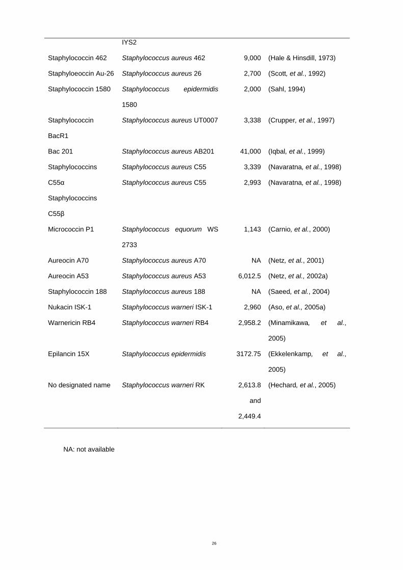
Table 1.1 Bacteriocins from the genus Staphylococcus
Bacteriocin Organism Molecular
weight
(Da)
Reference
No designated name Staphylococcus aureus strain 5,000 (Nakamura, et al., 1983)
26
IYS2
Staphylococcin 462 Staphylococcus aureus 462 9,000 (Hale & Hinsdill, 1973)
Staphyloeoccin Au-26 Staphylococcus aureus 26 2,700 (Scott, et al., 1992)
Staphylococcin 1580 Staphylococcus epidermidis
1580
2,000 (Sahl, 1994)
Staphylococcin
BacR1
Staphylococcus aureus UT0007 3,338 (Crupper, et al., 1997)
Bac 201 Staphylococcus aureus AB201 41,000 (Iqbal, et al., 1999)
Staphylococcins
C55α
Staphylococcins
C55β
Staphylococcus aureus C55
Staphylococcus aureus C55
3,339
2,993
(Navaratna, et al., 1998)
(Navaratna, et al., 1998)
Micrococcin P1 Staphylococcus equorum WS
2733
1,143 (Carnio, et al., 2000)
Aureocin A70 Staphylococcus aureus A70 NA (Netz, et al., 2001)
Aureocin A53 Staphylococcus aureus A53 6,012.5 (Netz, et al., 2002a)
Staphylococcin 188 Staphylococcus aureus 188 NA (Saeed, et al., 2004)
Nukacin ISK-1 Staphylococcus warneri ISK-1 2,960 (Aso, et al., 2005a)
Warnericin RB4 Staphylococcus warneri RB4 2,958.2 (Minamikawa, et al.,
2005)
Epilancin 15X Staphylococcus epidermidis 3172.75 (Ekkelenkamp, et al.,
2005)
No designated name Staphylococcus warneri RK 2,613.8
and
2,449.4
(Hechard, et al., 2005)
NA: not available
27
A number of bacteriocins from Gram positive bacteria have a relatively wide spectrum
compared to bacteriocins from Gram negative bacteria, for example, colicins (Jack, et al.,
1995). Some inhibitory peptides from Staphylococcus spp. are active against a wide range
of Gram positive bacteria and interestingly those bacteriocins also have a broad spectrum
of activity against Gram-negative organisms. For example, bacteriocins from
Staphylococcus aureus; Staphylococcin 462 (Hale & Hinsdill, 1975), Bac 1829 (Crupper &
Iandolo, 1996), Staphylococcin BacR1 (Crupper, et al., 1997) and Bac201 (Iqbal, et al.,
1999) exhibited broad spectrum activity against a variety of species of bacteria. In contrast,
some bacteriocins from Staphylococcus spp. have a spectrum that is very specific to Gram
negative bacteria. For example, an antibacterial peptide from Staphylococcus warneri has
a spectrum of activity directed toward the genus Legionella (Hechard et al., 2005). Another
strain of Staphylococcus warneri RB4 produced an anti-Alicyclobacillus bacteriocin,
warnericin RB4 (Minamikawa, et al., 2005).
1.5.2 Assay of bacteriocins
1.5.2.1 Detection of bacteriocin producer strains
The most commonly used procedure for detecting bacteriocin-producer strains is the spot
on lawn method. Pure cultures of the test organisms are spotted on an appropriate agar
medium and the plate is incubated for sufficient time to develop inhibitory activity. The plate
is then overlaid with a semisolid agar seeded with an indicator strain. Inhibition zones
around the culture after incubation of the overlaid culture indicate the presence of inhibitory
activity (Harting, et al., 1972)
A slightly modified spot on lawn assay has also been described as suitable for screening
mixed cultures for bacteriocin production. A study of antagonistic activity of oral bacteria
28
towards the periodontopathic bacterium, Treponema denticola, used a modified method
(Grenier, 1996). Subgingival plaque samples were plated directly on oral spirochete
medium, which had been spread with the indicator strain. The putative bacteriocin producer
strains were observed and isolated from the original plate. An Australian study for detection
of bacteriocin producers from food was performed by a variation of the spot on lawn
method in which samples contained mixed cultures (Coventry, et al., 1997). Food samples,
including meats, chicken, milk, milk products, vegetables and seafood, were plated in de
Man, Rogosa and Sharpe (MRS) agar and yeast extract lactate agar. After incubation
under anaerobic conditions, the agar plates were overlaid with indicator strains. Clearing
zones of growth inhibition were then observed and selected colonies suspected to be
bacteriocin producers were isolated from within the agar. Villani and colleagues (2001)
grew test organisms from fresh raw cow’s milk samples without isolating individual strains,
before examining the activity against a variety of indicator strains including Listeria
monocytogenes. After incubation of milk samples, the plates were overlaid with soft
medium seeded with indicator strains.
Another method, the agar inversion method, consists of streaking a culture of test strains
across a plate in parallel lines, then incubating the plate to allow bacteriocin to be
produced. Next the agar is inverted and the indicator strain is cross streaked on the
inverted surface. Inhibition zones are observed after a standardized incubation time. This
method has the benefit of distinguishing bacteriophage activity from bacteriocins since
bacteriocins are able to diffuse through the agar, whereas phage cannot (Vliegher, et al.,
2004).
1.5.2.2 Assay of bacteriocin activity
The activity of bacteriocins from broth cultures is commonly measured by the diffusion
method on agar plates with three alternative protocols in which the culture supernate is
delivered in a well cut in the agar, spotted onto the surface or incorporated into a disc. Of
these methods, the agar diffusion assay is the most common (Nakamura, et al., 1983,
29
Crupper & Iandolo, 1996, Crupper, et al., 1997, Crupper & Iandolo, 1997, Iqbal, et al.,
1999, Hechard, et al., 2005, Minamikawa, et al., 2005). The method involves cutting holes
in agar plates seeded with indicator strains, and placing the culture supernate in the holes.
After incubation, the inhibition zones are measured. For the spot protocol, a drop of cell-
free bacteriocin-containing solution is spotted on an agar plate. After the drop is allowed to
dry, the plate is overlaid with a indicator strain and the inhibition zone measured after
incubation for an appropriate period (Hale & Hinsdill, 1973, Wirawan, et al., 2006). For the
disc diffusion method, a solution containing the bacteriocin is applied to a blank paper disc
which is then placed on an agar medium containing the indicator strain. The size of the
inhibition zone is observed after incubation (Bizani & Brandelli, 2002, Delgado, et al.,
2005). The agar diffusion assay is time consuming and laborious but it is simple,
reproducible and no complicated equipment is required (Turcotte, et al., 2004). This assay
is still the most widely used quantitative technique to measure antibiotic activity of
bacteriocins.
Bacteriocin activity can also be quantified by a liquid assay using photometric or turbidity
measurement in a tube or microtiter plate assay (Parente, et al., 1995, Turcotte, et al.,
2004). Briefly, a bacteriocin in liquid form and a standardized indicator strain are incubated
together in suitable conditions. The bacteriocin activity is measured by the reduction of
turbidity of the indicator strain compared with a control sample without bacteriocin. The
method requires optimization, depending on the type of bacteriocin and the sensitivity of
the indicator organism. The limitations of this procedure are low reproducibility, since the
assay is critically dependent on incubation time. Precise measurement of indicator strain
concentration and growth phase are also critical in this assay. The method nevertheless is
a rapid technique (Turcotte, et al., 2004).
1.5.3 Exclusion of non-bacteriocin inhibitory agents
30
The screening methods for bacteriocin producers described in the previous sections give
preliminary information. However, other causes of inhibitory activity, (bacteriophage,
hydrogen peroxide and organic acids) need to be eliminated (Jack, et al., 1995). The
inhibitory effect from bacteriophage can be ruled out by using the agar inversion method
because bacteriophages are not able to pass through agar (Vliegher, et al., 2004). Ultra
structure of bacteriophage visualized using electronmicroscopy can also distinguish phage
from bacteriocin (Zink, et al., 1995). Increasing dilution of culture supernate is another
method used to distinguish bacteriocin and bacteriophage. On dilution, bacteriophage
shows a decreasing number of discrete phage plaques on agar plates seeded with an
indicator strain but bacteriocin shows a diffuse thinning of growth becoming less marked
with increasing dilution of the supernate (Mayr-Harting, et al., 1972). By-products of
metabolism, such as hydrogen peroxide, are usually produced by Streptococcus sp. (Tagg,
et al., 1976). Hydrogen peroxide production may be misinterpreted as an inhibitory effect
caused by bacteriocins. The inhibitory activity of hydrogen peroxide can be eliminated by
incubation of the bacteriocin producer strains on medium supplemented with catalase
(Tagg, et al., 1976). In another method, peroxide production was ruled out by
demonstration of a reduction in inhibitory activity after catalase was added to the crude
bacteriocin solution to destroy peroxide in the solution (Ogunbanwo, et al., 2003, Aslim, et
al., 2005). The inhibitory effect by organic acids was removed by culturing test strains in
media with calcium carbonate as a buffer (Minamikawa, et al., 2005, Wirawan, et al., 2006)
or by neutralizing the inhibitory substances with NaOH to pH 7.0 prior to examining for the
bacteriocin activity (Ogunbanwo, et al., 2003, Aslim, et al., 2005, Nascimento, et al., 2005).
1.5.4 Production of bacteriocins
31
Factors affecting bacteriocin production include both producer cells and environmental
factors. Each bacteriocin is unique in its optimal conditions for production. Therefore, it is
necessary to design experimental conditions specific for each bacteriocin-producing strain.
1.5.4.1 Growth phase
Bacteriocin production depends on the producer’s growth phase. Most bacteriocins are
produced during the exponential growth phase with greatest production occurring during
the beginning of the stationary phase (Iqbal, et al., 1999, Abriouel, et al., 2003, He, et al.,
2006). Bacteriocin levels decrease at the end of stationary phase due to degradation by
proteases (Kim, et al., 2006) or binding of bacteriocins to producer cells (Houlihan &
Russell, 2006). This feature of the bacteriocin production profile is common to almost all
lactic acid bacteria and other organisms that produce bacteriocins, including
Staphylococcus sp. For example, garviecin L1-5, produced by Lactococcus garvieae L1-5,
was produced in the log phase of growth and maximum activity was reached at late log to
early stationary phase of growth (Villani, et al., 2001). Activity of acidocin D20079, a
bacteriocin produced by Lactobacillus acidophilus DSM20079, was observed during the
transition from the exponential to the stationary phase. The activity were highest for a very
short period at 11 to 13 h of incubation and then sharply decreased (Deraz, et al., 2005).
Production of Bac1829 from Staphylococcus aureus KSI1829 began in the stationary
phase of growth (Crupper & Iandolo, 1996). Staphylococcus aureus AB201 produced Bac
201 during exponential phase growth (Iqbal, et al., 1999). Warnericin RB4 by
Staphylococcus warneri RB4 was produced in the exponential phase; the yield reached a
maximum (1,280 AU/mL) at the end of the exponential growth phase (Minamikawa, et al.,
2005).
1.5.4.2 Growth medium
32
Optimization of growth conditions and medium composition is required to obtain the
maximum yields of bacteriocin to facilitate further investigations. Production of a bacteriocin
by Lactobacillus brevis OG1 increased from 3200 to 6400 AU/mL with the addition of yeast
extract (3.0%), NaCl (1.0-2.0%), glucose (1.0%) or Tween 80 (0.5%) to MRS medium
(Ogunbanwo, et al., 2003). Methods for maximum production of four mutacins (A-CM7, B-
Ny266, C-7A and H-29B) by S. mutans were optimized (Nicolas, et al., 2004). Buffering
substances, organic carbon sources and organic nitrogen sources were tested in order to
improve bacteriocin production. The results showed that culture medium consisting of
cheese whey permeate (6%), yeast extract (2%) and CaCO3 (1%) was both inexpensive
and provided optimal growth conditions for production of the four mutacins.
Supplementation of milk medium with yeast extract (0.3-1.0%) enhanced production of
bacteriocins by seven Lactobacillus strains (Avonts, et al., 2004). The yeast extract used in
microbiological media has very reliable composition, however, that for industrial formulation
may vary somewhat more. See John’s comments at the edge of the page.Defined medium
may not assist in production of bacteriocins. Fifteen strains of S. mutans showed
bacteriocin production with basal trypticase medium and brain heart infusion but no
production was detected in a defined medium, M3, by the spot on lawn method (Rogers,
1972). Production of bacteriocin from Bacteroides strain T1-1 showed a bacteriocin titer of
32 to 64 in complex medium (Brain Heart Infusion) but only trace amounts were produced
in a defined medium (Booth, et al., 1977).
It has been reported that production of bacteriocins by lactic acid bacteria was enhanced
by osmotic stress induced by increasing the NaCl concentration in the culture medium.
Investigations into the production of amylovorin L471 from Lactobacillus amylovorus DCE
471, isolated from Sudanese Kisra fermentations of sorghum flour, showed that addition of
10 g/l of NaCl in a sourdough simulation medium gave maximum bacteriocin activity. NaCl
also played an important role on the production of nukacin ISK-1 from Staphylococcus
warneri ISK-1 isolated from fermented rice bran (Sashihara, et al., 2001). It was found that
33
not only addition of NaCl, but also KCl and sorbitol increased the production of this
bacteriocin.
A surfactant, Tween 80, added to culture medium was shown to promote the production of
bacteriocins (Keren, et al., 2004). Moreover, a bacteriocin from S. bovis HC5, Bovicin HC5,
was detected in higher concentration from medium containing Tween 80 than
unsupplemented medium. Cell free culture supernate with Tween 80 added also showed
increased bacteriocin activity against Clostridium sticklandii SR. The authors concluded
that Tween 80 enhanced bacteriocin activity by aiding insertion of bacteriocin into the target
cells rather than by enhancing the release from producer cells. In contrast, Houlihan and
Russell (2006) reported that Tween 80 did not increase production of bacteriocins.
1.5.4.3 pH
Several studies on bacteriocin production by lactic acid bacteria have shown that
maintaining a constant pH, usually around 5.0-7.0, enhances bacteriocin production.
Maximum activity of a bacteriocin produced by Lactobacillus acidophilus DSM 20079,
acidocin D20079, was obtained from a pH-controlled reactor with MRS broth at pH 6.0
(Deraz, et al., 2005). In another study, the effect of pH on bacteriocin production from
Lactococcus sp. GM005 was evaluated in pH-uncontrolled and pH-controlled cultures
(Onda, et al., 2003). The pH of uncontrolled cultures dramatically changed from 7.0 to 4.4
after 12 h of incubation with the maximum inhibitory activity at 1280 AU/mL. The
antibacterial activity was eight times greater at 10,240 AU/mL with pH-controlled cultures at
pH 6.0. In a similar study, enhanced production of enterocin AS-48 by Enterococcus
faecalis A-48-32 was obtained when pH was regulated. The pH was maintained above 5.8
and the maximum inhibitory activity was four times greater than uncontrolled pH culture
(Abriouel, et al., 2003).
1.5.4.4 Temperature
34
Optimal growth temperature (generaaly at 30-37°C) usually result in optimal bacteriocin
production (Kim, et al., 2006). However, a few studies have reported maximal yield of
bacteriocins at suboptimal growth temperatures. Production of brevicin 286 by
Lactobacillus brevis VB286 was optimal at 20°C while optimal cell growth occurred
between 25 and 37°C (Coventry, et al., 1996). For Lacticin RM, maximal activity was
obtained at 10°C, whereas the optimal growth temperature of the producer, Lactococcus
lactis subsp. lactis EZ26, was 30°C (Keren, et al., 2004).
1.5.4.5 Agitation
The effect of agitation was examined in a few reports. Of the four mutacins (A-CM7, B-
Ny266, C-7A and H-29B) produced by four strains of S. mutans, agitation only reduced the
titre of mutacin A-CM7 (Nicolas, et al., 2004). Production of brevicin 286 by Lactobacillus
brevis VB286 was similar when grown under agitation or non-agitation (Coventry, et al.,
1996). The reduction in bacteriocin activity was thought to be due to degradation by
protease contained in the culture (Nicolas, et al. 2004).
1.5.4.6 Other factors
Recently, an investigation of the influence of sub-inhibitory concentrations of an antibiotic,
ciprofloxacin, on bacteriocin production in Escherichia coli was reported (Jerman, et al.,
2005). The results of the agar diffusion assay showed that medium containing sub-lethal
concentrations of ciprofloxacin yielded more colicin (more than 30 fold increase) than
medium without ciprofloxacin. It was suggested that the antibiotic interferes with DNA
replication as well as cell wall synthesis, and induced an SOS response which induced
bacteriocin synthesis.
1.5.5 Estimation of bacteriocin molecular mass
Molecular mass can be estimated by different methods, including gel electrophoresis and
mass spectrometry. Gel electrophoresis is a common procedure to estimate protein size in
the course of purification of bacteriocins. Mass spectrometry is able to present more
accurate results from a purified sample.
35
Sodium dodecyl sulphate polyacrylamide gel electrophoresis (SDS-PAGE) is a method to
separate proteins by size (Bollag & Edelstein, 1991). SDS associates with the proteins and
denatures them and gives an overall negative charge enabling separation based on
molecular weight (Hames, 1998). SDS-PAGE is also a cheap, reproducible and rapid
method for characterizing proteins. After crude protein separation, the gel is left in a
mixture of isopropanol and acetic acid to fix the separated proteins and then soaked with
distilled water. A suitable indicator strain is overlaid on the washed gel, and any zone of
inhibition is measured after incubation. The position of the inhibition zone is used to
determine the molecular weight of the bacteriocin (Crupper, et al., 1997, Iqbal, et al.,
1999).
Mass spectrometry (MS) is an analytical tool used for measuring the molecular mass of a
sample (Hames, 1998). It is known as an accurate technique for protein size determination.
The instrument used for MS is divided into three parts; ionization source, analyzer and
detector. The sample is introduced into the ionization source and the sample molecules are
ionized. These ions are sent to the analyzer where they are separated according to mass
(m) - to - charge (z) ratio (m/z). The separated ions are detected and this signal is sent to a
data system which provides a printout showing molecular weight, and relative amounts of
the protein(s) in the sample (Hames, 1998).
1.5.6 Bacteriocin extraction
Bacteriocins can be extracellular, cell associated or intracellular peptides. Most published
data on the bacteriocins isolated from Staphylococcus sp. shows extracellular bacteriocins
obtained from cell free culture supernate (Crupper & Iandolo, 1996, Crupper, et al., 1997,
Crupper & Iandolo, 1997, Iqbal, et al., 1999, Hechard, et al., 2005, Minamikawa, et al.,
2005).
36
Some studies of bacteriocins have been hampered by the difficulties in obtaining active
liquid preparations of the substances. Some studies described bacteriocins that could be
produced on agar but not when grown in broth. A study from Norway reported that six
Carnobacterium strains produced bacteriocins on agar but only Carnobacterium 377
produced the bacteriocin both on agar plates and in broth (Blom, et al., 2001). It is difficult
to study bacteriocins attached to a solid substrate. For that reason, only the bacteriocin
produced by Carnobacterium 377 was selected for characterization and purification. In
another example, a study of a bacteriocins isolated from oral streptococci showed that the
bacteriocins were labile in liquid media but could be stabilized by increasing the viscosity of
the media (Kelstrup & Gibbons, 1969). The authors suggested the effective stabilizers such
as agar, agarose, starch, dextran and glycerol could be used to generate a similar
environment and possibly similar bacteriocin activity to the matrix of dental plaque
containing a dextran-like polymer.
Another problem encountered by some investigators is the fact that the bacteriocin
sometimes remains attached to the producer cells. Release of cell associated bacteriocin
has been achieved by treatment with saline at acid pH. Bovicin HC5, a bacteriocin from S.
bovis HC5 and nisin variants produced by S. uberis were extracted by this method
(Houlihan & Russell, 2006, Wirawan, et al., 2006). Mutacin B-Ny266 produced by S.
mutans was extracted from cell pellets by a mixture of HCl and ethanol at pH 2.0 (Mota-
Meira, et al., 1997). The suspensions were then heated at 70°C for 40 min and cooled on
ice and a cell free solution was obtained by centrifugation. The supernate was evaporated
at 37°C to obtain the bacteriocin-containing solution. The extract from this cell pellet
showed six times higher specific activity than the culture supernate, which implied the
higher purity of the bacteriocin.
1.5.7 Purification of bacteriocins
37
After obtaining the crude bacteriocin in soluble form, recovery of bacteriocin can be
achieved in many different ways. Purification methods that have been used for bacteriocins
from staphylococci are varied. More than one purification step is necessary to reach the
desired purity in order to identify the protein of interest. The methods most frequently used
for isolation, concentration and purification involve salt precipitation followed by a variety of
chromatography methods such as gel filtration, ion exchange chromatography and
reversed-phase high performance liquid chromatography (RP-HPLC).
Because most bacteriocins are secreted into the culture medium, purification begins with a
step to concentrate bacteriocins from culture supernates. Ammonium sulphate precipitation
(salting out) is frequently used as preliminary step. A high concentration of the salt causes
proteins to aggregate and precipitate out of solution (Bollag & Edelstein, 1991). After
protein precipitation, the residue of ammonium sulphate is generally eliminated by dialysis
(Bollag & Edelstein, 1991). Staphylococcin BacR1, a bacteriocin produced by
Staphylococcus aureus UT0007 was precipitated from culture supernates by saturation
with 60% ammonium sulphate (Crupper, et al., 1997). A 8.3-fold purification was achieved
by this step. The same group of researchers initially purified Bac 1829 from S. aureus
KSI1829 using saturation with 80% ammonium sulphate, achieving a purification of 1.5
fold. The advantage of ammonium sulphate precipitation is not only to concentrate the
protein but also minimize the volume of the sample before proceeding to further purification
steps. After precipitation with ammonium sulphate, the volume of culture supernate
containing Warnericin RB4 produced by Staphylococcus warneri RB4 was reduced from
1000 mL to 100 mL. Ammonium sulphate precipitation does not provide a high degree of
purification, however.
Further purification techniques differ depending on protein properties such as, charge,
hydrophobicity and molecular weight (size). Ion exchange chromatography separates
proteins based on charge. Gel filtration chromatography separates proteins according to
size. Hydrophobic interaction chromatography or reversed phase HPLC distinguishes
38
proteins by their hydrophobic properties (Harris & Angal, 1990). Each step causes some
loss of product. Therefore, selection of appropriate purification techniques is extremely
important.
Affinity chromatography can be very useful for a well-studied bacteriocin such as a
bacteriocin derived from recombinant producer cells. In a study by Moon et al. (2006), the
expressed bacteriocin was designated suitable for affinity chromatography. A plasmid
encoding pediocin PA-1 fused with hexahistidine was introduced to E. coli. The fusion
protein was expressed and purified by nikel-nitrilotriacetic acid (Ni-NTA) metal affinity
chromatography, resulting in a high yield of protein of high purity. This method could be
practical for mass production of bacteriocins (Moon, et al., 2006). A similar method was
used to purify a recombinant bacteriocin from S. dysgalactiae subsp. equisimilis
(dysgalacticin), expressed in E. coli (Heng, et al., 2006).
The final step of purification to obtain the highest purity bacteriocin is generally HPLC
(Muriana & Klaenhammer, 1991, Jimenez-Diaz, et al., 1995, Chumchalova, et al., 2004,
Ghrairi, et al., 2005). A purified sample may require repeated steps of purification. Crupper
and coworkers (1997) achieved the final removal of trace elements by two cycles of RP-
HPLC. Fractions containing bacteriocin were obtained by a second cycle of RP-HPLC with
a shallower gradient of eluent solution.
1.6 Aims of this study
Streptococcus uberis has become an important cause of intramammary infection in dairy
cows. Based on the above background, the first aim of this project was to isolate and
identify S. uberis from bovine milk in Victoria using a minimum group of conventional tests
suitable for use in diagnostic veterinary laboratories. 16S rRNA sequencing was used as a
gold standard for bacterial identification. Since treatment of bovine mastitis requires the use
of antibiotics, the second aim of this project was to determine the antibiotic susceptibility of

39
the isolates. The third aim was to isolate, purify and characterize a bacteriocin from a
member of the normal teat flora of cows.
40
Chapter 2 Isolation and identification of Streptococcus
uberis by conventional methods
2.1 Introduction
Streptococcus uberis is a worldwide environmental mastitis pathogen. Epidemiologic
studies and control of S. uberis have been limited by lack of accurate and convenient
methods for routine species identification. Accurate and cost effective methods of mastitis
pathogen identification are important to diagnostic laboratories. Suitable methods for
identifying this mastitis pathogen would facilitate designing a management strategy to
reduce cases and improve prognosis for infected cows.
Specific identification of S. uberis is difficult as it has variable reactions on culture media, in
biochemical tests and by serotyping. The results may vary from one laboratory to another
depending on the procedures used. Gram-positive cocci causing bovine mastitis are mainly
Staphylococcus spp. or Streptococcus spp. The catalase test helps differentiate between
these two genera. In many diagnostic laboratories, Gram-positive cocci, isolated from
clinical and subclinical mastitis, that are catalase-negative and hydrolyze esculin are
identified as S. uberis (Anonymous, 1990, Devriese, et al., 1999, Odierno, et al., 2006),
however, this group of bacteria contains other species such as Aerococccus spp.,
Enterococcus spp., Lactococcus spp., S. bovis, S. dysgalactiae, S. pluranimalium and S.
gallolyticus (Devriese, et al., 1999, Fortin, et al., 2003).
Generally, differentiation of the genus Streptococcus causing bovine mastitis is undertaken
by observing the hemolytic reaction on blood-containing media, carbohydrate antigen
grouping (Lancefield grouping) and phenotypic tests. Esculin is incorporated into the
primary isolation media (Edwards medium) for isolating streptococci mastitis pathogens by
most veterinary laboratories. In addition to S. uberis, S. bovis, S. equinus, E. faecalis, E.
faecium and E. saccharolyticus are esculin hydrolyzing bacteria and S. dysgalactiae and S.
41
equi as esculin-variable species (Anonymous, 1990). Some authors state that the 6.5%
sodium chloride growth tolerance test must be a part of the S. uberis identification scheme
to exclude enterococci (Watts, 1988, Anonymous, 1990, Cowan, et al., 1993). Fortin et al.
(2003) suggested that hippurate hydrolysis and acid production from inulin assisted
differentiation of S. uberis from catalase negative, esculin-hydrolysing Gram-positive cocci.
Acid production from mannitol has also been used to differentiate S. uberis from other
streptococci (Cowan, et al., 1993). Lancefield grouping is a useful assay to differentiate
mastitis streptococci since S. agalactiae, S. dysgalactiae and enterococci belong to
Lancefield group B, C and D respectively (Cowan, et al., 1993). However, grouping is not a
useful discriminatory test for S. uberis since strains fall into at least six serological groups,
all of which include representatives of a variety of other species. Streptococcus uberis
could belong to group A, E, G, P, U or more than one group or be non-groupable (Lammler,
1991, Khan, et al., 2003). The CAMP test (a test for production enhanced haemolysis,
designed by Christie, Atkins, Munch and Petersen) was considered to assist in
identification of S. agalactiae but some cultures of S. uberis demonstrated CAMP-like
synergistic hemolytic activity on sheep blood agar (Lammler, 1991, Khan, et al., 2003,
McDonald, et al., 2005). Thus, the CAMP test was not recommended in identifying S.
uberis. Several attempts have been made to develop a simple yet accurate scheme for
identification of S. uberis (Fortin, et al., 2003, Khan, et al., 2003, Odierno, et al., 2006),
however, most schemes require a large number of tests or are complicated to use, which
makes them unsuitable for routine application. A commercial kit which contains several
biochemical reactions such as API 20 Strep may be useful to confirm the identity of an
isolate obtained from a cow with mastitis when routine tests give doubtful identification
(Odierno, et al., 2006).
A survey of methods used by veterinary laboratories in Australia and New Zealand to
identify mastitis pathogens was undertaken in 2003 by a former PhD student (W.
McDonald, PhD thesis, RMIT University, 2003). The results showed that of the 17
diagnostic veterinary laboratories surveyed, including commercial, government and clinical
42
laboratories, each used a range of different biochemical and serological tests to identify
mastitis pathogens, but did not use genotypic methods. Species-specific PCR assays are
available for S. uberis. In addition, 16S rRNA gene sequencing is a very accurate, but
expensive method for classification of streptococci, including S. uberis (Facklam, 2002).
The main objective of this section was to improve the identification procedures for S. uberis
from milk by developing a scheme with the required level of accuracy while using a
minimum number of conventional methods suitable for diagnostic veterinary laboratories.
Catalase-negative, esculin-hydrolyzing Gram-positive cocci isolated from infected bovine
milk were identified using four additional tests (hippurate hydrolysis, acid production from
inulin and mannitol and growth in 6.5% NaCl). The API 20 Strep system was used to
confirm the species identity. The method of 16S rRNA gene sequencing was used as the
gold standard for evaluating the results of biochemical testing. Since very little information
is available on the molecular epidemiology of S. parauberis, subspecies identification of
isolates identified a S. parauberis was performed by PFGE.
2.2 Materials and methods
2.2.1 Milk sample collection
Milk samples from each quarter were collected aseptically following Countdown
Downunder instructions (Brightling, et al., 1998). Briefly, gloves were worn throughout the
procedure and were changed after collection from each cow. A paper towel or a cotton ball
with 70% ethanol was used for wiping teats individually. After cleaning teats, the first squirt
of milk was discarded, then one to five mL was collected into a sterile container, transported
on ice and cultured immediately on arrival in the laboratory.
43
Cows with clots in their milk or visible signs of teat inflammation were defined as having
clinical mastitis. Subclinical mastitis was defined if the somatic cell count (SCC) was
greater than 250,000 cells/mL when performed by a herd testing service within two weeks
of milk collection for culture. Cows with milk SCCs less than 250,000 cells/mL were
defined as having a low cell count.
Chi-square analysis was used to examine the association between disease status and
qualitative growth of S. uberis. The significance level was set at 0.05.
2.2.2 Bacterial reference strains
The bacterial reference strain from the American Type Culture Collection (ATCC) used in
this study was S. uberis ATCC 700407. The strain was stored at -80°C in Protect Bacterial
Preservers. The reference strain was cultured from storage at -80°C by aseptically
transferring the bacterial preserver beads onto horse blood agar (HBA, Appendix C) or brain
heart infusion agar (BHA, Appendix C), streaking and incubating for 24 hours for further
testing.
2.2.3 Streptococcus uberis isolation and identification
Milk samples were mixed by swirling, then 50 µl aliquots were plated onto HBA and
Edwards medium (Appendix C) and incubated at 37°C for 48 hours. A single colony
resembling S. uberis was examined macroscopically and microscopically then subcultured
onto another HBA or BHA to obtain a pure isolate for further identification. Isolate
designation was set up as follows: cow’s number, dash (-) and isolate number.
2.2.3.1 Phenotypic identification
Streptococcus uberis ATCC 700407 was used as a control.
44
Macroscopic examinations
Colonial morphology on HBA and Edwards agar was observed. Bacterial growth of 1 to 10,
11 to 50 and more than 50 colonies were defined as +, ++ and +++ respectively.
Microscopic examinations
A gram stain of the isolates was examined. The cell morphology and cell arrangement were
observed under a microscope.
Physiological and biochemical examinations
Physiological and biochemical tests performed on bacterial isolates that were Gram
positive and negative for catalase production were set up for esculin hydrolysis, hippurate
hydrolysis, inulin fermentation, mannitol fermentation and tolerance to 6.5% sodium
chloride.
• Catalase test (Isenberg, 1992)
The catalase test was performed by transferring a bacterial colony with a sterile tooth pick
onto a coverslip and placing a drop of 3% H2O2 on top of the cells. Production of gas
bubbles immediately indicated a positive reaction. Any colony that showed a positive
reaction was discarded.
• Esculin hydrolysis test (Isenberg, 1992).
Esculin broth consisted of 25 g nutrient broth No. 2, 3 g yeast extract, 1 g esculin, 0.5 g
ferric citrate and made up to 1000 mL with distilled water. The ingredients were mixed to
dissolve, adjusted to pH 7.3 to 7.5, and 3 mL aliquots were dispensed into 16 × 110 mm
tubes and sterilized by autoclaving at 110°C for 25 min. After inoculation with the test
strains, broths were incubated at 37°C for 24 h. A positive result showed black coloration
due to hydrolysis of esculin to esculitin, which forms a black complex with ferric ions.
• Hippurate hydrolysis test (Cowan, et al., 1993)
45
Hippurate broth consisted of 10 g sodium hippurate and 1,000 mL distilled water. The
ingredients were mixed to dissolve and adjusted pH 7.1, dispensed 2 mL in 16 × 110 mm
test tubes and sterilized by autoclaving at 121°C for 15 min. A heavy loopful of culture was
aseptically inoculated into the test tube and incubated for 2 h at 37°C. One mL of ninhydrin
reagent was added to a test tube, mixed and incubated for 15 min at 37°C. Ninhydrin
reagent consisted of 3.5 g ninhydrin, 50 mL acetone and 50 mL butanol. The ingredients
were mixed freshly before use. A blue to purple color indicated hydrolysis of the hippurate
to benzoate and glycine and the formation of a purple coloured compound after the addition
of ninhydrin.
• Inulin or mannitol broth (Isenberg, 1992)
Inulin or mannitol broths consisted of 10 g bacteriological peptone, 5 g sodium chloride, 1
mL bromcresol purple (2% solution in ethanol), 10 g inulin or mannitol made up to 1,000
mL with distilled water. The ingredients were mixed and adjusted to pH 7.2-7.3. The
medium was dispensed 3 mL in 16 mm × 110 mm test tubes and sterilized by autoclaving at
110°C for 25 min. The culture was aseptically inoculated into the test tube and incubated for
24 h at 37°C. Acid production was indicated by a color change from purple to yellow.
• 6.5% sodium chloride broth (Isenberg, 1992)
The broth consisted of 25 g nutrient broth No.2, 65 g sodium chloride and 1,000 mL
distilled water. The ingredients were mixed, 3 mL was dispensed in 16 × 110 mm test tubes
and sterilized by autoclaving at 121°C for 15 min. The culture was aseptically inoculated
and incubated for 24 hours at 37°C. Any growth indicates tolerance to sodium chloride,
which is typical of enterococci.
Commercial kit identification– API 20 Strep
46
Isolates that gave equivocal results with the conventional tests were further analyzed using
the API 20 Strep test, following the manufacturer’s instructions. The dehydrated substrates
(n = 20) were rehydrated by adding a bacterial suspension. The inoculum was prepared with
freshly grown bacteria at 37°C overnight on BHA, adjusted with 2 mL sterilized distilled
water to at least McFarland No.4 turbidity standard. Following recommendations of the
manufacturer the test strip was inoculated and incubated. After incubation color changes
were read directly or after addition of reagents. Isolates were identified by referring to the
identification tables provided by the manufacturer. S. uberis ATCC 700407 was used as a
control strain.
2.2.3.2 Genotypic identification
16S rRNA gene sequencing was used to estimate the accuracy of the phenotypic
identification. Another PhD. Student (Takehiro Tomita) performed 16S and 23S species-
specific PCRs on all isolates. Since sequencing is expensive, I set up the 16S rRNA
sequencing reactions only for isolates that were negative for 16S or 23S species-specific
PCR. Takehiro Tomita performed the 16S rRNA sequencing when 16S and 23S species-
specific PCR for S. uberis showed positive results.
General techniques for molecular work
• DNA extraction from cultures by the boiling method
Fresh cultures from BHA were transferred to a 1.5 mL sterile screw cap tube containing 400
µl of molecular grade water using sterile swabs. The tubes were incubated in boiling water
for 10 min, mixed by a vortex mixer for 10 s, and then centrifuged at 13000 x g for 5 min.
The supernates containing DNA were transferred to sterile eppendorf tubes and stored at -
20°C until required.
• Master mix preparation
47
To prevent contamination in PCR reactions, master mixes were prepared in a DNA-free area
(biohazard cabinet sterilized with UV light before use) using DNA-free pipettes (pipettes for
master mix use only). The master mix contained PCR buffer (including MgCl2), dNTPs,
primer and Taq polymerase. The master mix was prepared and aliquoted to each tube of
PCR reaction and stored at -20°C. DNA template was transferred to the tube before starting
the PCR reaction.
• Agarose gel electrophoresis
The agarose gel was prepared by microwaving 1% DNA grade agarose in 1 x Tris acetate
EDTA (TAE) buffer (50X TAE buffer consisted of 2M Tris, 1 M acetic acid and 0.1 M
EDTA) and was cooled for 10 min before pouring. The gel was allowed to set for 30 min. A
mixture of five microliters of PCR product and one microliter of 6 X loading dye was
loaded into wells. Electrophoresis was conducted at 100 V for 45 min in TAE buffer. The
gel was stained in ethidium bromide (0.5 µg/mL) for 10 min then destained in running tap
water for 10 min and the DNA fragments were visualized using a Gel Doc Imaging system
(Bio-Rad).
16S rRNA gene sequencing
The 16S rRNA genes were amplified by PCR in 200 µl PCR tubes. A 50 µl PCR reaction
mix contained five microliters of 10 X PCR reaction buffer, 0.25 µl of Taq DNA
polymerase 5 U/µl, 0.1 µl of 100 mM dNTPs, 50 pmol of primer SEQ27f (5’-AGA GTT
TGA TCA TGG CTC AG-3’), 50 pmol of SEQ1492r (5’-GGC TAC CTT GTT ACG ACT
T-3’) (Pullinger et al., 2006), five microliters of DNA template and molecular grade water
to 50 µl. The PCR cycle consisted of 94°C for five min then 35 cycles of 94°C for 30 s,
56°C for one minute and 72°C for 1.5 min followed by a final extension at 72°C for six
minutes and was conducted in a PCR Thermal Cycler (Corbett Research GP001, Sydney,
Australia). To confirm the size of the PCR product, five microliters was examined by
agarose gel electrophoresis as previously described in section of agarose gel electrophoresis.
48
Two volumes of 20% polyethylene glycol (PEG) in 2.5 M NaCl were added to one volume
of each PCR product and transferred to a new 1.5 mL eppendrof tube. The tubes were
mixed and incubated at 37°C for 15 min. After centrifugation at 13000 x g for 10 min, the
supernate was carefully removed. The pellet was washed with 400 µl of 80% ethanol and
centrifuged at 13000 x g for 5 min. The supernate was then removed and the pellet was
dried in a heat box at 73°C for 5 min. The dried pellet was resuspended to the original
volume with molecular grade water.
The PCR products were sequenced by BigDye™ Terminator system in 200 µl PCR tubes.
The BigDye reaction consisted of 2 µl of 0.8 pmol/µl primer (using forward or reverse
primer in single tubes), 1 µl of BigDye, 1 µl of 10 X PCR buffer and 1 µl of DNA template
(PCR product described above). The PCR cycle consisted of 94°C for 5 min, 35 cycles of
94°C for 30 s, 56°C for 60 s and 72°C for 90 s and followed by final extension at 72°C for 6
min.
After the BigDye reaction, fifteen microliters of molecular grade water was added to the
reaction tubes and transferred to 1.5 mL eppendorf tubes containing 80 µl of 75%
isopropanol to precipitate the DNA. The tubes were mixed and incubated at room
temperature for 15 min then centrifuged at 13000 x g for 20 min. Precipitated DNA was
washed three times in 250 mL 75% (v/v) isopropanol using centrifugation at 13000 x g for
10 min room temperature. The samples were dried in a heat box at 73°C for 5 min and
forwarded to the Microbiological Diagnostic Unit, University of Melbourne for DNA
sequence determination using an ABI 3100 automated sequencer and the resulting
chromatograms were analyzed using the ChromasPro software package (Technelysium
Pty Ltd, Tewantin QLD, Aus). Sequence data for comparison was obtained from NCBI.
Sequence alignment was performed using ClustalW (www.ncbi.nlm.nih.gov).
2.2.4 Pulse field gel electrophoresis (PFGE) of Streptococcus parauberis
49
2.2.4.1 Preparation of genomic DNA in agarose plugs
Genomic DNA was prepared in agarose plugs according to the methods of Smith and
Cantor (1987), with some modifications. Cells from each S. parauberis isolate were grown
on BHA at 37°C for approximately 18 hours. An isolated colony was inoculated into 10 mL
THB and incubated at 37°C with gentle shaking to reach an optical density (A600) between
0.4 and 0.45. The cells were collected by centrifugation at 3,000 x g for 15 min and
resuspened in 10 mL of Pett IV buffer (10 mM Tris-HCl pH 7.6 and 1 M NaCl). Following a
second wash in Pett IV buffer, cells were resuspended in 500 µl of the same buffer. Equal
volumes of cell suspension and molten 2.4% low-melting temperature agarose (Progen)
were mixed and dispensed into 100 µl plug moulds and allowed to set at 4°C for 20 min.
Once solidified, the agarose plugs were incubated in EC lysis buffer (6mM Tris HCl pH7.6,
1M NaCl, 100 mM EDTA pH 7.6, 0.2% sodium deoxycholate, 0.5% Sarkosyl and 1 mg/mL
lysozyme added prior to use) at 37°C overnight. The EC lysis buffer allowed DNA to be
released from the cells. The DNA was then purified by incubating agarose plugs with EPS
buffer (0.5 M EDTA pH 8.6, 1% Sakosyl and 1mg/mL of proteinase K added prior to use) at
50°C for 24 h. The EPS buffer was replaced with fresh ESP buffer, and then agarose plugs
were further incubated at 50°C for 24 h. The proteinase K activity was then inactivated by
incubating the agarose plugs at 37°C for 2 h with TE buffer (10 mM Tris-HCl pH 7.6 and 1
mM EDTA pH 8.0) containing 0.0175 mg/mL PMSF in isopropanol.
2.2.4.2 Restriction of endonuclease digestion of genomic DNA
Prior to digestion, the agarose plugs were equilibrated with TE buffer for 2 h, followed by
four additional 20 min washes in the same buffer to remove proteins and proteinase K. The
agarose plugs were cut to a size of 1 to 2 mm in thickness with a clean blade. The plug
slices were washed twice by incubating in restriction buffer (1 X Tango buffer, Fermentas)
for 30 min. Plug slices were next transferred to the restriction buffer containing 50 units of
the restriction endonuclease SmaI (Fermentas) in 200 µl of restriction buffer and incubated
at 30°C overnight. The SmaI was inactivated by incubating with 1 mL of TE buffer at 37°C
50
for 1 h. The slices were placed at 4°C for 15 min prior to loading into wells of a 1% agarose
gel.
2.2.4.3 Pulsed-field gel electrophoresis
Separation of DNA fragments was achieved by using a CHEF-DR II (BioRad). Digested
samples were electrophoresed through 1.5% agarose gel in 0.5 X TBE buffer (1 X TBE
consisted of 89 mM Tris, 89 mM boric acid, 2 mM EDTA, pH 8.3). Electrophoresis was
carried out at 6 V/cm at 14 °C for 20 h with the pulse time increasingly linearly from 1 to 20
s. The gel was stained with ethidium bromide (0.5 µg/mL) for 20 min and destained in
running water for 20 min. The DNA restriction patterns were viewed on a Gel Doc (BioRad).
A CHEF DNA size standard (Lambda ladder, molecular weight range 48.5 to 970 kbp,
BioRad) was used as a molecular size marker.
Strains were defined using criteria proposed by Tenover et al. (1995). Isolates were
designated different strains if the PFGE patterns differed by more than three bands,
possibly related if patterns differed by ≤ 3 bands and closely related if patterns differed by
one band. Isolates that had similar PFGE patterns were considered to be genetically
related isolates and to be derived from a common parent.
2.3 Results
Three hundred and twelve bovine milk samples from 114 cows were collected from two
farms in western and eastern Victoria (Timboon and Newry), which were reported to have
S. uberis problems.
Reference strain, S. uberis ATCC 700407, was confirmed to the species level by the API
20 Strep system. In addition, the strain showed hydrolysis of esculin and hippurate, acid
production from inulin and mannitol and no growth in 6.5% NaCl.
51
Colonies obtained from milk samples cultured on Edwards medium (dark colonies) or HBA
were selected and further tested if they were alpha or non-hemolytic, circular, translucent,
white to yellowish and approximately one mm in diameter. One hundred and fifteen isolates
that were Gram positive cocci arranged in chains, that did not produce catalase and
hydrolyzed esculin were presumptively identified as S. uberis. Of these, 28 were from
quarters with clinical mastitis, 48 from subclinical mastitis and 39 from cows with low cell
counts (Appendix B, Table 1). An additional four tests (hippurate hydrolysis, acid
production in inulin and mannitol and growth in 6.5% NaCl) performed on the isolates (n =
115), provided seven different patterns (Table 2.1 or more detail in Appendix B, Table 1).
Most isolates (n = 70, 61%) belonged to pattern 1 (hippurate hydrolysis positive, inulin and
mannitol fermentation positive and no growth in 6.5% NaCl). Selected isolates belonging to
pattern 1 were confirmed as S. uberis by the API 20 Strep test. All isolates belonging to
pattern 1 were identified by 16S rRNA sequencing as S. uberis (64, 91%) or S. parauberis
(6, 9%). Isolates showing pattern 2 (differing from pattern 1 only in the inability to ferment
inulin) were identified by API 20 Strep test as S. uberis (n = 4) and Aerococcus viridans (n
= 8). The 16S rRNA sequencing confirmed that the four isolates were indeed S. uberis, but
of the other eight isolates, two were S. parauberis and the remaining six were A. viridans.
Pattern 3 consisted of 13 isolates which were identified by API 20 Strep as A. viridans (11
isolates) and S. bovis (2 isolates). Two isolates identified as A. viridans by phenotypic
methods were identified as S. parauberis by the molecular methods. Isolates belonging to
pattern 4 showed growth tolerance in 6.5% NaCl and the one isolate was identified as
Enterococcus spp. by both the API 20 Strep and 16S rRNA sequencing. Isolates with
patterns 5, 6 and 7 (esculin positive but hippurate negative) were selected and identified as
S. bovis by API 20 Strep. Some of these isolates were randomly selected and sequenced,
and identified as S. bovis.
Chi-square analysis of the association between disease status (clinical, subclinical compared
with cows producing milk with a low somatic cell countcell count) and the qualitative
growth of S. uberis (+++, ++ and +) was statistically significant (P = 0.0003) (Table 2.2).
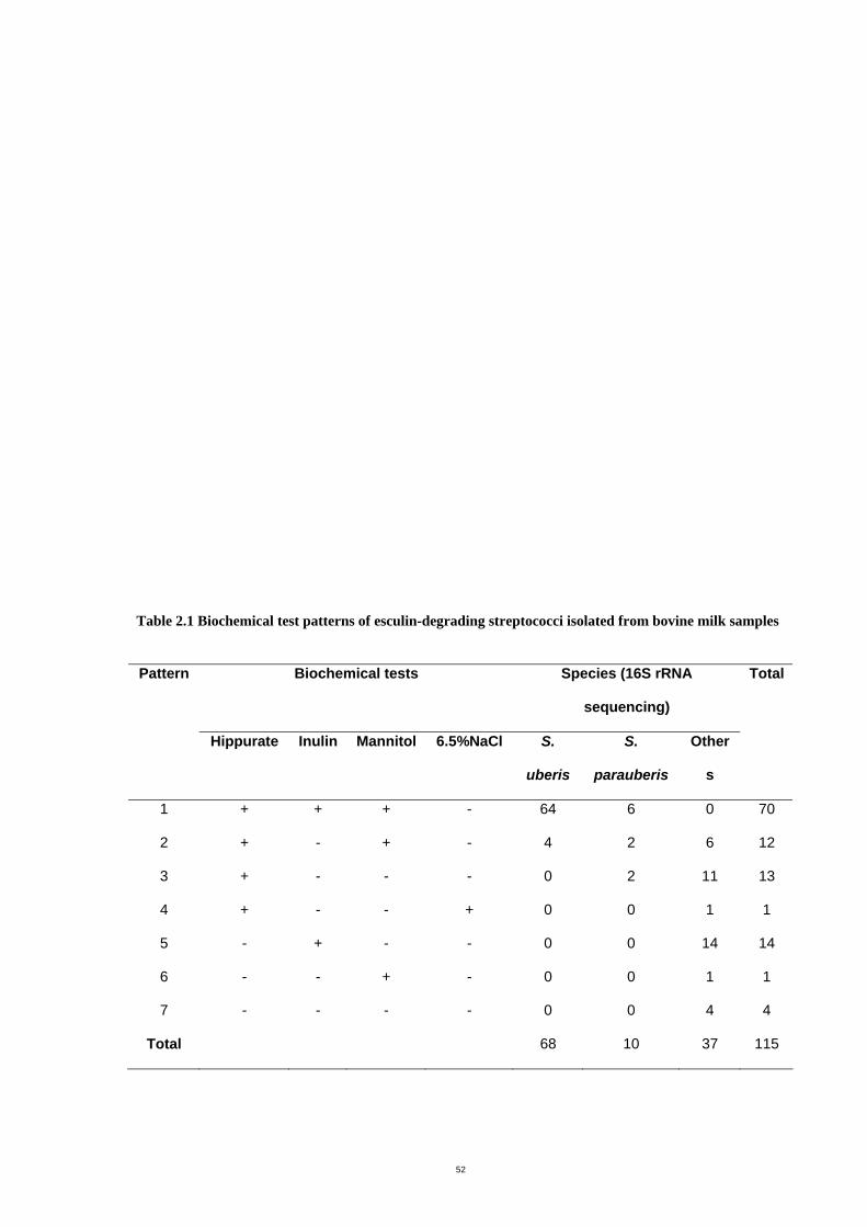
52
Table 2.1 Biochemical test patterns of esculin-degrading streptococci isolated from bovine milk samples
Pattern Biochemical tests Species (16S rRNA
sequencing)
Total
Hippurate Inulin Mannitol 6.5%NaCl S.
uberis
S.
parauberis
Other
s
1 + + + - 64 6 0 70
2 + - + - 4 2 6 12
3 + - - - 0 2 11 13
4 + - - + 0 0 1 1
5 - + - - 0 0 14 14
6 - - + - 0 0 1 1
7 - - - - 0 0 4 4
Total 68 10 37 115
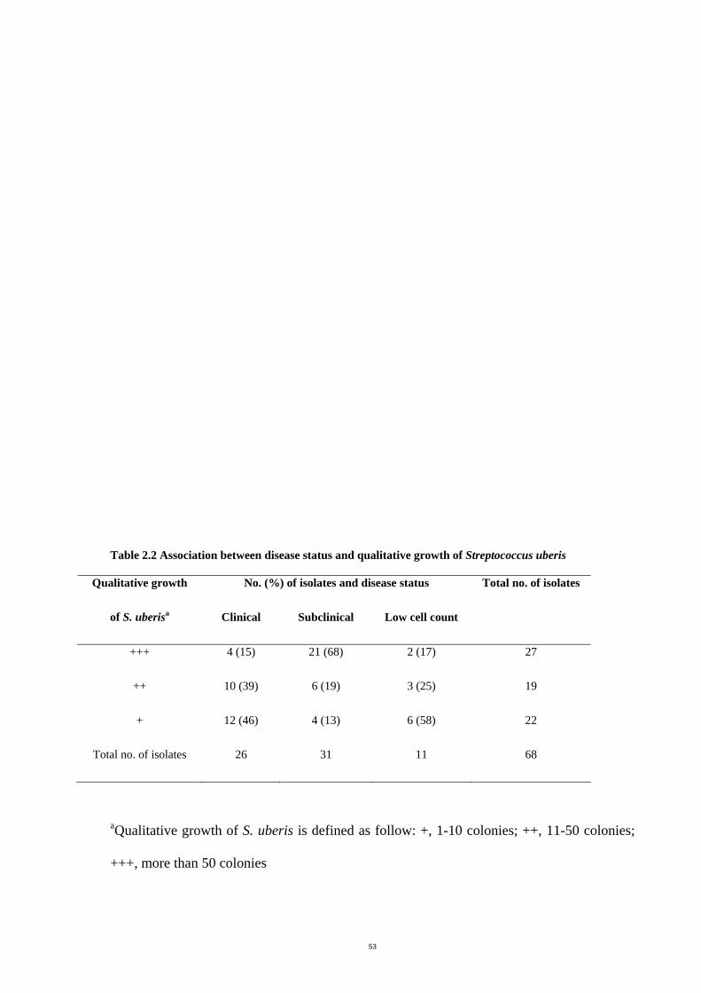
53
Table 2.2 Association between disease status and qualitative growth of Streptococcus uberis
No. (%) of isolates and disease status Qualitative growth
of S. uberisa Clinical Subclinical Low cell count
Total no. of isolates
+++ 4 (15) 21 (68) 2 (17) 27
++ 10 (39) 6 (19) 3 (25) 19
+ 12 (46) 4 (13) 6 (58) 22
Total no. of isolates 26 31 11 68
aQualitative growth of S. uberis is defined as follow: +, 1-10 colonies; ++, 11-50 colonies;
+++, more than 50 colonies
54
Analysis with the chi-square test showed statistical significance (P = 0.0003) in the
association between disease status and the qualitative growth of S. uberis

55
The restriction patterns of chromosomal DNA of ten S. parauberis isolates digested with
SmaI showed from 8 to 16 fragments which allowed the clear differentiation of the isolates
(Figure 2.1). Using the criteria suggested by Tenover et al. (1995) for interpretation of
PFGE patterns, no identical strains were found. All seven isolates from the Timboon farm
(lane 1-7) showed different patterns from each other. Isolates 3218-1, 3218-9 and 3218-10
(lanes 8, 9 and 10) were from the same cow on the farm at Newry. Isolates 3218-9 and
3218-10, from the same quarter, were closely related but not identical (arrows indicate
different bands); isolate 3218-1 was a different strain.
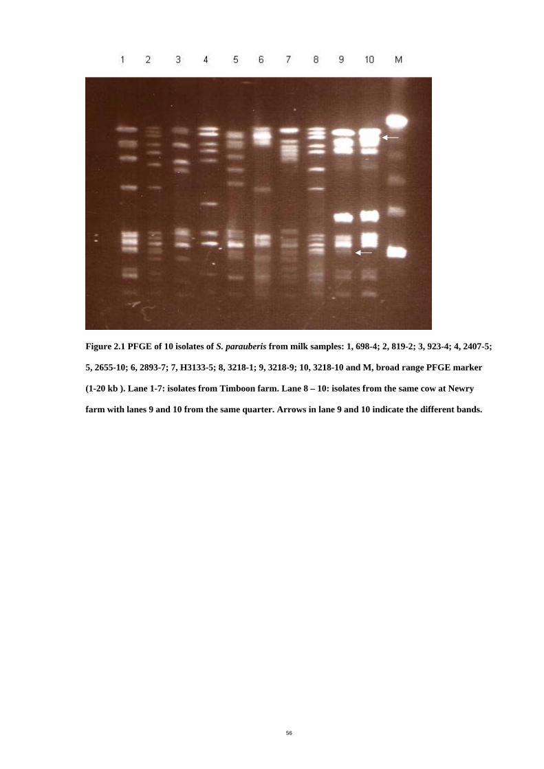
56
Figure 2.1 PFGE of 10 isolates of S. parauberis from milk samples: 1, 698-4; 2, 819-2; 3, 923-4; 4, 2407-5;
5, 2655-10; 6, 2893-7; 7, H3133-5; 8, 3218-1; 9, 3218-9; 10, 3218-10 and M, broad range PFGE marker
(1-20 kb ). Lane 1-7: isolates from Timboon farm. Lane 8 – 10: isolates from the same cow at Newry
farm with lanes 9 and 10 from the same quarter. Arrows in lane 9 and 10 indicate the different bands.
57
2.4 Discussion
The presence of potential pathogens in milk indicates a problem with udder health and milk
quality. Accuracy of isolation and identification could provide information that would
improve treatment procedures, health status, milk quality and farm management.
Esculin degrading streptococci isolated from clinical and subclinical mastitis cases in dairy
cows are commonly categorized as S. uberis in routine diagnostic laboratories. Genotypic
tests were not used in veterinary diagnostic laboratories surveyed in Australia and New
Zealand (W. McDonald, PhD Thesis, RMIT, 2003). The esculin positive streptococci (n =
115) would be identified as S. uberis using the routine classification, but only 59% (68 of
115) of isolates were correctly identified when compared to the 16S rRNA sequencing data.
In this study, esculin degrading streptococci (n = 115) presented seven patterns when
further identified with hippurate hydrolysis, inulin and mannitol fermentation and growth
tolerance in 6.5% NaCl. Most S. uberis isolates in this study belonged to pattern 1 (60.9%,
70 of 115) and the biochemical tests resulted in accuracy of identification of 91.4% (64 of
70). One species in this group that was incorrectly identified was S. parauberis (8.6%, 6
isolates from 70) therefore identification of S. parauberis was included in this study.
Streptococcus parauberis required molecular procedures for accurate identification. Pattern
1 was able to differentiate S. uberis from other streptococci and enterococci. Of the milk
samples from which incorrect identification was obtained, S. parauberis could be found in
cases of clinical mastitis, subclinical mastitis and low cell count milk (Appendix B, Table 1).
The amount of S. parauberis growth varied from slight growth (+) to heavy growth (+++).
Some milk samples contained both S. parauberis and S. aureus or S. uberis. As S.
parauberis is not frequently isolated from milk samples, the proposed tests (hippurate
hydrolysis, inulin and mannitol fermentation and groth in 6.5% NaCl) are acceptable for S.
uberis identification.
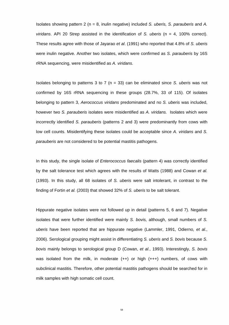
58
Isolates showing pattern 2 (n = 8, inulin negative) included S. uberis, S. parauberis and A.
viridans. API 20 Strep assisted in the identification of S. uberis (n = 4, 100% correct).
These results agree with those of Jayarao et al. (1991) who reported that 4.8% of S. uberis
were inulin negative. Another two isolates, which were confirmed as S. parauberis by 16S
rRNA sequencing, were misidentified as A. viridans.
Isolates belonging to patterns 3 to 7 (n = 33) can be eliminated since S. uberis was not
confirmed by 16S rRNA sequencing in these groups (28.7%, 33 of 115). Of isolates
belonging to pattern 3, Aerococcus viridans predominated and no S. uberis was included,
however two S. parauberis isolates were misidentified as A. viridans. Isolates which were
incorrectly identified S. parauberis (patterns 2 and 3) were predominantly from cows with
low cell counts. Misidentifying these isolates could be acceptable since A. viridans and S.
parauberis are not considered to be potential mastitis pathogens.
In this study, the single isolate of Enterococcus faecalis (pattern 4) was correctly identified
by the salt tolerance test which agrees with the results of Watts (1988) and Cowan et al.
(1993). In this study, all 68 isolates of S. uberis were salt intolerant, in contrast to the
finding of Fortin et al. (2003) that showed 32% of S. uberis to be salt tolerant.
Hippurate negative isolates were not followed up in detail (patterns 5, 6 and 7). Negative
isolates that were further identified were mainly S. bovis, although, small numbers of S.
uberis have been reported that are hippurate negative (Lammler, 1991, Odierno, et al.,
2006). Serological grouping might assist in differentiating S. uberis and S. bovis because S.
bovis mainly belongs to serological group D (Cowan, et al., 1993). Interestingly, S. bovis
was isolated from the milk, in moderate (++) or high (+++) numbers, of cows with
subclinical mastitis. Therefore, other potential mastitis pathogens should be searched for in
milk samples with high somatic cell count.
59
Mannitol negative S. uberis have been found to be rare (2%) (Odierno, et al., 2006). There
were no mannitol negative isolates in this study.
Full identification can be made with commercial biochemical kits as suitable alternatives for
identification of streptococci by conventional methods. However, the cost of the kits may
limit their use by veterinary laboratories. The results from this study suggest that using the
API 20 Strep test to identify S. uberis obtained from mastitis cases where the results of
conventional tests are inconclusive, would be useful (i.e. when the results obtained follow
pattern 2). However, difficulties remain with the use of API 20 Strep for the identification of
streptococcal mastitis pathogens. In this study, there were misidentifications of S.
parauberis as S. uberis (6 isolates, pattern 1) and A. viridans (4 isolates, patterns 2 and 3).
The current database provided with the commercial kit system is incomplete, as it does not
include some of the less well known organisms of veterinary importance; for example, S.
parauberis and S. iniae. Testing for of the enzyme β-D-glucuronidase has been suggested as
phenotypic test that is able to differentiate S. uberis and S. parauberis (Khan, et al., 2003);
however, some isolates of S. uberis have been shown to be β-D-glucuronidase negative
(Schaufuss, et al., 1986). Therefore genotypic examinations such as DNA/DNA
hybridization, restriction fragment length polymorphism analysis of 16S rDNA, species-
specific PCR or 16S rRNA sequencing are required to differentiate between S. uberis and S.
parauberis and other streptococci (Garvie & Bramley, 1979, Jayaro, et al., 1991, Hassan, et
al., 2001, Khan, et al., 2003, Alber, et al., 2004, McDonald, et al., 2005). Of these, PCR is
the most suitable testing platform for diagnostic laboratories.
In summary, the phenotypic identification set proposed tn the current study is convenient for
use in veterinary diagnostic laboratories and would increase the accuracy of routine S.
uberis identification compared with using the results of esculin hydrolysis alone or the
60
combined results of esculin and hippurate hydrolysis. The significance or pathogenicity of
S. parauberis is not known at present. Some isolates were found in infected udders with
cell counts greater than 250,000 cells/mL in cases where no other pathogens were isolated.
Streptococcus parauberis isolates from two dairy herds showed a diverse range of PFGE
patterns. Seven isolates of S. parauberis from the Timboon farm showed different patterns,
indicating that the organism was not likely to have been transmitted from cow to cow but
probably was acquired from the environment or from the endogenous faecal flora. Three
isolates of S. parauberis were from the same cow on another farm (Newry). Isolate 3218-1
showed dissimilar pattern from isolates 3218-9 and 3218-10 which were from the same
quarter of this same cow and were considered to be closely related strains. It is likely these
minor differences in banding patterns were caused by a single genetic event such as a point
mutation or an insertion or deletion of DNA (Tenover, et al., 1995). Similar variations were
observed in other species, on repeated subculture or from the same patient over a period of
time (Tenover, et al., 1995).
The occurrence of S. parauberis as a mastitis pathogen appears to be rare, however it has
been isolated all over the world. For example, it was reported from British dairy herds
(Garvie & Bramley, 1979), the USA (Jayaro, et al., 1991), Europe (Hassan, et al., 2001,
Khan, et al., 2003) and Australia (Baseggio, et al., 1997, Phuektes, et al., 2001, McDonald,
et al., 2005). Similar to reports of S. uberis (Baseggio, et al., 1997, Phuektes, et al., 2001,
Khan, et al., 2003), in this study S. parauberis showed great genetic diversity. At this stage,
studies of the epidemiology of S. parauberis using PFGE fingerprinting have been limited
and no reports have been published so far. This has resulted in lack of information
concerning bacterial reservoirs responsible for new infections and limits our understanding
of transmission patterns. A larger number of S. parauberis isolates from intramammary and
61
extramammary sources is required for examination in order to understand the epidemiology
and pathogenic potential of this species.

62
Chapter 3 Determination of Streptococcus uberis
antibacterial susceptibility patterns
3.1 Introduction
Antibiotic therapy is an important aspect of herd management practice to control mastitis
infections. Understanding the prevalence of resistance and antibiotic susceptibility patterns
of the microorganisms isolated from milk of cows with the infection can improve the
treatment of mastitis. As a matter of practical management on many dairy farms, antibiotic
intramammary infection therapy is initiated prior to microbiological culturing (Guterbock, et
al., 1993, Milner, et al., 1997). The treatment strategies for the environmental streptococci
are often based on the assumption that the antibiotic susceptibility of this group is
homogenous. However, a previous study (Brown and Scasserra, 1990) has reported higher
antibiotic resistance levels in strains of S. uberis than in strains of S. dysgalactiae and S.
agalactiae. Thus, information on the antibiotic susceptibility of the various organisms
categorized as environmental streptococci would guide veterinarians and dairy farmers
towards the most prudent selection of an antibiotic for treatment of mastitis. In addition,
knowledge of antibiotic susceptibility patterns helps in developing therapy protocols for
particular dairy farms and there is need for effective monitoring.
Several reports of antibiotic susceptibility of S. uberis isolated from bovine mastitis have
been published (Brown & Scasserra, 1990, Salmon, et al., 1998, Rossitto, et al., 2002).
However, there is no recent data available in the literature on the susceptibility of S. uberis
to antibacterial agents isolated in Australia.
63
The mechanisms of erythromycin resistance are target site modification, enzymatic
inactivation and active efflux (Leven, 2005). In streptococci, target site modification is a
common resistance mechanism. The mechanism involves production of a methylase which
is encoded by erythromycin resistance methylase genes (ermA or ermB) (Seppala, et al.,
2003). Methylases cause conformational change in the prokaryotic ribosome leading to
reduced binding of the macrolides, lincosomides and streptogramin B (MLSB) group of
antibiotics. Initial results of erythromycin susceptibility testing showed high Minimal
Inhibitory Concentration (MIC) values and identified some isolates likely to be resistant to
this group of antibiotics.
The objective of this study was therefore to determine the antibiotic susceptibility patterns
of S. uberis isolates from the milk of cows in Victoria, to penicillin, ampicillin, oxacillin,
cephalothin, erythromycin, tetracycline, gentamicin, vancomycin and novobiocin. Antibiotic
agents were selected according to those used for routine testing by veterinary laboratories
(NCCLS, 2002). Although, vancomycin is not approved for mastitis treatment, it was
included in this study as representative of the glycopeptide antibiotic family. In addition,
detection of ermA by PCR was performed.
3.2 Material and methods
The determination of antibacterial susceptibility is usually made by exposing
microorganisms to different concentrations of antibacterial agents and evaluating whether
the microorganism is inhibited or killed. The tests were performed according to the recent
recommendations of the National Committee for Clinical Laboratory Standards (NCCLS,
now Clinical Laboratory Standards Institute, CLSI) M31-A2 (NCCLS, 2002) for determining
antibiotic susceptibility of bacteria isolated from animals together with interpretive criteria.
64
Mimimum inhibitory concentrations (MIC) and where indicated minimal bactericidal
concentrations (MBC) were performed.
3.2.1 Bacterial strains
Streptococcus uberis isolates used in this study were the same as these identified from
Chapter 2 and are listed in Appendix B, Table 2. The bacterial reference strain used in this
study as a control was Staphylococcus aureus ATCC 29213. An erythromycin resistance
strain, S. aureus 344/2-7, was obtained from the Microbiology Laboratory Culture
Collection, RMIT University. The bacteria were stored at -80°C in Protect Bacterial
Preservers. They were cultured from the storage at -80°C by aseptically plating the
bacterial preserver beads into HBA or BHA (Appendix C), streaking and incubating for 24
hours for further testing.
3.2.2 Calibration of turbidity
McFarland turbidity standards were used as a reference to adjust the turbidity of bacterial
suspensions so that the numbers of bacteria were estimated within a given range. A
McFarland 0.5 standard suspension was made up by mixing 0.5 mL of 0.048 M BaCl2 and
99.5 mL of 1% H2SO4. The absorbance of the standard at 625 nm was confirmed to be
0.08 to 0.10 (NCCLS, 2002). The standard was transferred to a screw cap tube of the
same size as those used in preparing the culture inoculum and stored in a dark place at
room temperature. Cultures adjusted to this standard turbidity are equivalent to a
concentration of approximately 108 colony forming unit (CFU) per milliliter (mL).
3.2.3 Antibiotic compounds
Stock solutions containing 1280 µg/mL of antibiotic agent were prepared according to
NCCLS guideline (2002). Stock solutions were prepared using the formula (1000/P) × V ×
C = W, where P is potency of the antibiotic, V is volume in mL, C is final concentration and
W is weight of the antibiotic to be dissolved in V. Solvents and diluents for preparation of
stock solutions of antibiotic agents were suggested by NCCLS (2002). Ampicillin was
65
dissolved in 0.1 M phosphate buffer, pH 8.0 then 0.1 M phosphate buffer, pH 6.0 was used
as a diluent. Cephalothin was dissolved in 0.1M phosphate buffer, pH 6.0 then water was
used as a diluent. Erythromycin was dissolved in 95% ethanol then water was used as a
diluent. Gentamicin, oxacillin, penicillin, tetracycline, vancomycin and novobiocin were
dissolved and diluted in sterile distilled water. The stock solutions were stored at -20°C for
a maximum of 1 month.
3.2.4 Antibiotic susceptibility tests
3.2.4.1 Minimum inhibitory concentration (MIC); broth dilution
The MIC is the lowest concentration of antibacterial agent that completely inhibits growth
of the organism as detected by the unaided eye.
Streptococcus uberis isolates were cultured on horse blood agar (HBA, Appendix C) or
brain heart infusion agar (BHA, Appendix C) at 37° C overnight. Four to five colonies of
each isolate were inoculated into Mueller-Hinton broth (MHB, Appendix C) (5 mL) and
incubated at 37°C with agitation until the cultures were visibly turbid. The bacterial
suspension was adjusted to that of a 0.5 McFarland standard. The suspension was then
diluted 1:100 with MHB to obtain approximately 106 CFU/mL.
Antibacterial solutions of 50 µl were serially diluted two-fold in 96-well polystyrene round-
bottom microtiter plates by adding the inocula to 50 µl per well of MHB. A standardized
bacterial suspension (described above) of 50 µl was then added to each well. This volume
corresponded to approximately 2.5-5.0 × 10⁴ CFU per well. Controls (wells inoculated with
S. uberis culture without antibiotic added) and blanks (wells containing uninoculated broth
medium) were run for each isolate. The bacterial reference strain (Staphylococcus aureus
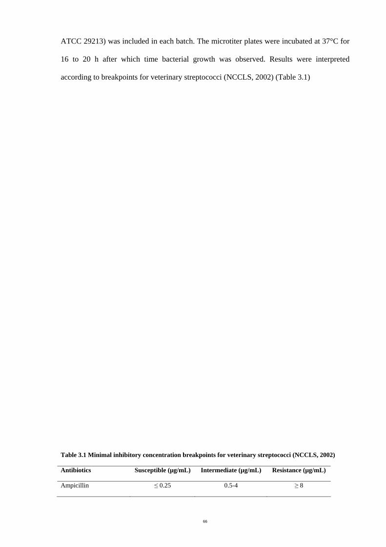
66
ATCC 29213) was included in each batch. The microtiter plates were incubated at 37°C for
16 to 20 h after which time bacterial growth was observed. Results were interpreted
according to breakpoints for veterinary streptococci (NCCLS, 2002) (Table 3.1)
Table 3.1 Minimal inhibitory concentration breakpoints for veterinary streptococci (NCCLS, 2002)
Antibiotics Susceptible (µg/mL) Intermediate (µg/mL) Resistance (µg/mL)
Ampicillin ≤ 0.25 0.5-4 ≥ 8
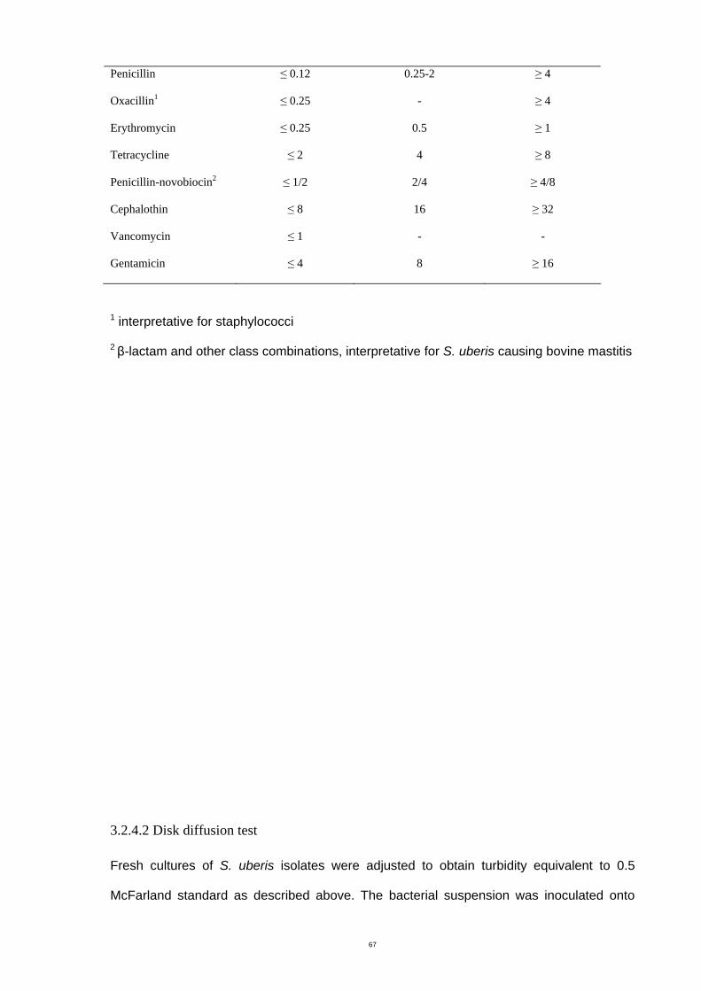
67
Penicillin ≤ 0.12 0.25-2 ≥ 4
Oxacillin1 ≤ 0.25 - ≥ 4
Erythromycin ≤ 0.25 0.5 ≥ 1
Tetracycline ≤ 2 4 ≥ 8
Penicillin-novobiocin2 ≤ 1/2 2/4 ≥ 4/8
Cephalothin ≤ 8 16 ≥ 32
Vancomycin ≤ 1 - -
Gentamicin ≤ 4 8 ≥ 16
1 interpretative for staphylococci
2 β-lactam and other class combinations, interpretative for S. uberis causing bovine mastitis
3.2.4.2 Disk diffusion test
Fresh cultures of S. uberis isolates were adjusted to obtain turbidity equivalent to 0.5
McFarland standard as described above. The bacterial suspension was inoculated onto
68
Mueller-Hinton agar (MHA, Appendix C) supplemented with 5% defibrinated sheep blood
(Appendix C) using a sterile cotton swab. The excess moisture was allowed to be absorbed
for five min before applying the antibacterial disks. The disks were placed on the agar
plates (no more than seven disks per plate). The plates were incubated at 37°C for 16 to 18
h and the diameter of inhibition zones was recorded.
3.2.4.3 Minimum bactericidal concentration (MBC)
The MBC is the lowest concentration of antibiotic that reduces the viable count by 99.9%.
After observing the results of MIC, 50 µl of the content of wells at and below the MIC value
were cultured on BHA, incubated at 37°C for approximately 16 h, and the number of
colonies were counted.
3.2.5 Erythromycin resistance gene PCR
Detection of the erythromycin-resistance gene (erm A) by PCR was as follows. The primers
were as described by Sutcliffe et al. (1996) with modification of PCR reaction conditions.
The product size was 645 bp. for Staphylococcus aureus 344/2-7 which was used as a
positive control. A 50 µl PCR reaction mix contained 40.19 µl of molecular grade water,
6.66 µl of 10X PCR buffer, 0.4 µl of dNTPs, 0.625 µl of 20 µM primer (ermA 5’ – CTA AAA
AGC ATG ATA AAA GAA - 3’ and 5’-CTT CGA TAG TTT ATT TAA TAT TAG - 3’), 0.5 µl of
5 U/µl Taq polymerase and 1 µl of DNA template. The PCR cycle consisted of 94°C for 1
min, 30 cycles of 94°C for 30 s, 46°C for 30 s and 72°C for 30 s followed by final extension
at 72°C for 5 min. The PCR reaction was performed using a PCR Thermal Cycler (Corbett
Research Model GP001).
3.2.6 Transmission electron microscopy
Transmission electron microscopy was performed as described in section 5.2.2.2.
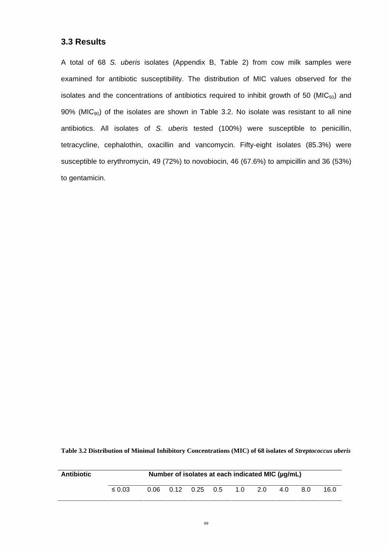
69
3.3 Results
A total of 68 S. uberis isolates (Appendix B, Table 2) from cow milk samples were
examined for antibiotic susceptibility. The distribution of MIC values observed for the
isolates and the concentrations of antibiotics required to inhibit growth of 50 (MIC50) and
90% (MIC90) of the isolates are shown in Table 3.2. No isolate was resistant to all nine
antibiotics. All isolates of S. uberis tested (100%) were susceptible to penicillin,
tetracycline, cephalothin, oxacillin and vancomycin. Fifty-eight isolates (85.3%) were
susceptible to erythromycin, 49 (72%) to novobiocin, 46 (67.6%) to ampicillin and 36 (53%)
to gentamicin.
Table 3.2 Distribution of Minimal Inhibitory Concentrations (MIC) of 68 isolates of Streptococcus uberis
Number of isolates at each indicated MIC (µg/mL) Antibiotic
≤ 0.03 0.06 0.12 0.25 0.5 1.0 2.0 4.0 8.0 16.0
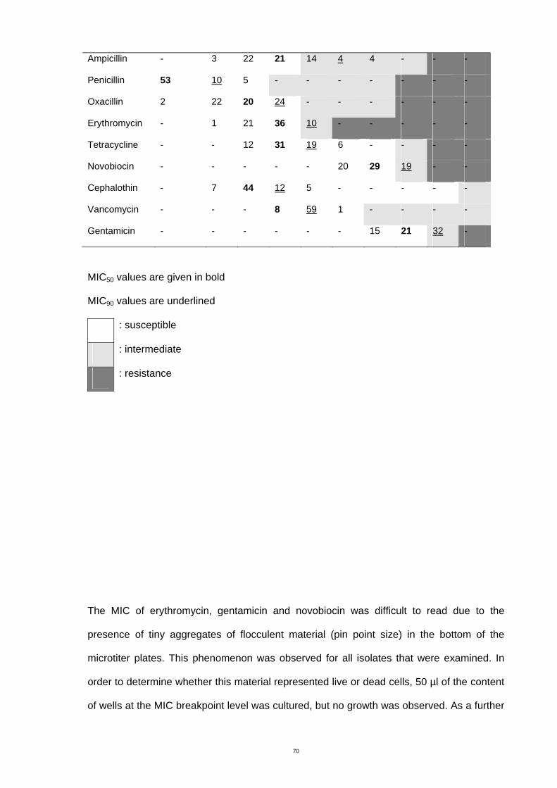
70
Ampicillin - 3 22 21 14 4 4 - - -
Penicillin 53 10 5 - - - - - - -
Oxacillin 2 22 20 24 - - - - - -
Erythromycin - 1 21 36 10 - - - - -
Tetracycline - - 12 31 19 6 - - - -
Novobiocin - - - - - 20 29 19 - -
Cephalothin - 7 44 12 5 - - - - -
Vancomycin - - - 8 59 1 - - - -
Gentamicin - - - - - - 15 21 32 -
MIC50 values are given in bold
MIC90 values are underlined
: susceptible
: intermediate
: resistance
The MIC of erythromycin, gentamicin and novobiocin was difficult to read due to the
presence of tiny aggregates of flocculent material (pin point size) in the bottom of the
microtiter plates. This phenomenon was observed for all isolates that were examined. In
order to determine whether this material represented live or dead cells, 50 µl of the content
of wells at the MIC breakpoint level was cultured, but no growth was observed. As a further
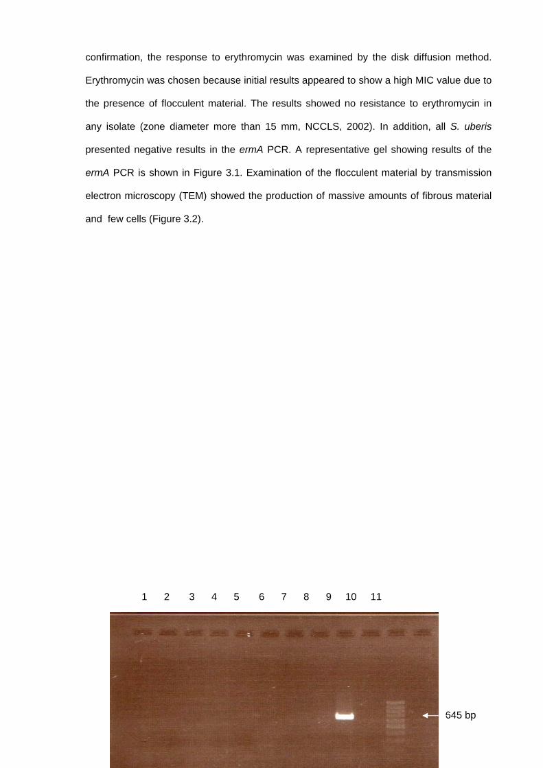
71
confirmation, the response to erythromycin was examined by the disk diffusion method.
Erythromycin was chosen because initial results appeared to show a high MIC value due to
the presence of flocculent material. The results showed no resistance to erythromycin in
any isolate (zone diameter more than 15 mm, NCCLS, 2002). In addition, all S. uberis
presented negative results in the ermA PCR. A representative gel showing results of the
ermA PCR is shown in Figure 3.1. Examination of the flocculent material by transmission
electron microscopy (TEM) showed the production of massive amounts of fibrous material
and few cells (Figure 3.2).
645 bp
1 2 3 4 5 6 7 8 9 10 11
72
Figure 3.1 PCR of ermA gene erythromycin-resistant isolate. Lane 1-8; Streptococcus uberis isolates 796-2, 2169-2, 2481-1, 2530-2, 2530-4, 2530-5, 2565-2 and 2730-5. Lane 9; Staphylococcus aureus 344/2-7 (positive control). Lane 10; Molecular grade water (negative control). Lane 11; DNA marker.
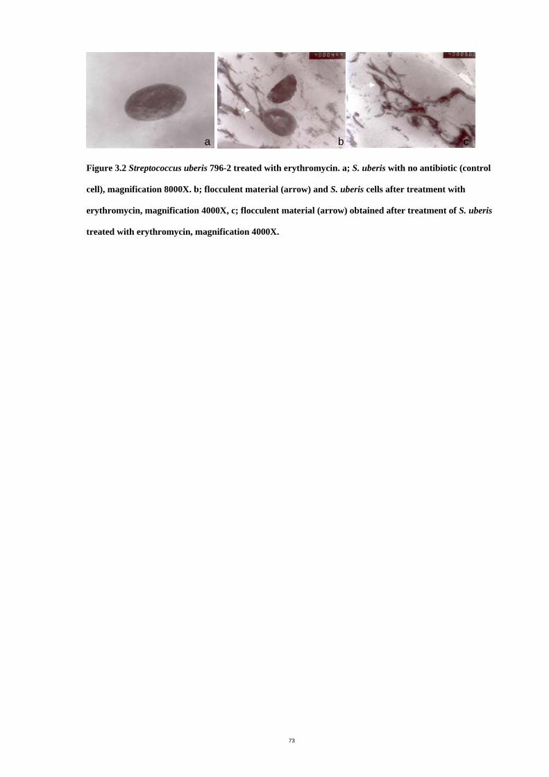
73
Figure 3.2 Streptococcus uberis 796-2 treated with erythromycin. a; S. uberis with no antibiotic (control
cell), magnification 8000X. b; flocculent material (arrow) and S. uberis cells after treatment with
erythromycin, magnification 4000X, c; flocculent material (arrow) obtained after treatment of S. uberis
treated with erythromycin, magnification 4000X.
a b c
74
3.4 Discussion
Several reports have published the results of antibiotic susceptibility testing of streptococci
isolated from bovine mastitis (Jousimies-Somer, et al., 1996, Salmon, et al., 1998, Erskine,
et al., 2002, Guerin-Faublee, et al., 2002, Rossitto, et al., 2002). In this study S. uberis
isolates were examined from Victoria for resistance to antibiotics recommended for routine
testing by veterinary microbiology laboratories (NCCLS, 2002).
Comparison of antibiotic susceptibility results reported in the literature must be made with
caution since different techniques have been used including disk diffusion (Owens, et al.,
1990, Denamiel, et al., 2005), agar dilution (Owens, et al., 1990, Guerin-Faublee, et al.,
2002) and broth dilution (Salmon, et al., 1998, Rossitto, et al., 2002). Agar or broth dilution
methods provide a quantitative result (MIC), while disk diffusion presents a qualitative
result (usually susceptibile, intermediate or resistant). Also the interpretative guidelines
used to categorized isolates as susceptible or resistant are not always the same, and some
studies used criteria for human isolates rather than the criterion for veterinary isolates
(Owens, et al., 1990, Guerin-Faublee, et al., 2002). The criteria for human isolates can be
used only as a comparative guide in the veterinary field. Additionally, some reports did not
offer data by individual bacterial species, but for example grouped all streptococci together
(Jousimies-Somer, et al., 1996).
Comparison of the levels of resistance to antibiotic agents (MIC50 and MIC90) for S. uberis
isolated from America, Denmark, New Zealand and Australia is shown in Table 3.3. Using
NCCLS criteria (2002) for determining antibiotic susceptibility of bacteria isolated from
animals, the results showed that more than 90% of S. uberis examined in the USA and in the
present study 100% were susceptible to cephalothin, but variable results were obtained for
75
most other antibiotics. Resistance to penicillin and oxacillin is not common in Australia and
New Zealand but strains with reduced susceptibility appear to be emerging in the USA and
Denmark. Streptococcus uberis appear to be less susceptible to ampicillin than to other β-
lactams in the USA and in this present study. This is of concern, since β-lactams are widely
used for dry cow treatment. Resistance to erythromycin and tetracycline was not found in
the present study or in a study from New Zealand, but was common in the USA and
Denmark. Susceptibility to novobiocin in Denmark appeared to be higher than in this study
and New Zealand. Vancomycin has not been approved for use in cattle (Jousimies-Somer, et
al., 1996), however, there was no resistance of S. uberis to vancomycin in this study, which
agrees with the results from the USA (Owens, et al., 1990). No resistant isolates to
gentamicin were found in this study but 47% resistance from the USA was reported (Owens,
et al., 1990). The different use of antibiotics in different geographical regions may cause
different patterns of resistance. In general, resistance to erythromycin and tetracycline
appeared to be a problem in the USA. Antibiotic resistance of S. uberis was not found to be
a problem in this study, and MIC90 values were relatively low when compared with those of
other continents. This may be because of strict regulation of antibiotic use in food-
producing animals in Australia (Joint Expert Advisory Committee on Antibiotic Resistance,
1999). However, resistance to erythromycin has been demonstrated in Denmark which has
similar regulations (Aarestrup, 1999). It is important to monitor and to continue to regulate
the use of antibiotics for food producing animals, in order to decrease the risk of resistant
strains emerging and to detect those strains if resistance does emerge.
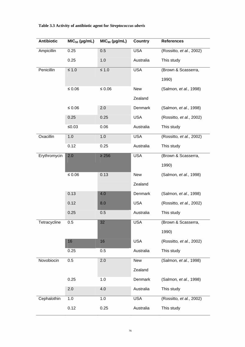
76
Table 3.3 Activity of antibiotic agent for Streptococcus uberis
Antibiotic MIC50 (µg/mL) MIC90 (µg/mL) Country References
Ampicillin 0.25 0.5 USA (Rossitto, et al., 2002)
0.25 1.0 Australia This study
Penicillin ≤ 1.0 ≤ 1.0 USA (Brown & Scasserra,
1990)
≤ 0.06 ≤ 0.06 New
Zealand
(Salmon, et al., 1998)
≤ 0.06 2.0 Denmark (Salmon, et al., 1998)
0.25 0.25 USA (Rossitto, et al., 2002)
≤0.03 0.06 Australia This study
Oxacillin 1.0 1.0 USA (Rossitto, et al., 2002)
0.12 0.25 Australia This study
Erythromycin 2.0 ≥ 256 USA (Brown & Scasserra,
1990)
≤ 0.06 0.13 New
Zealand
(Salmon, et al., 1998)
0.13 4.0 Denmark (Salmon, et al., 1998)
0.12 8.0 USA (Rossitto, et al., 2002)
0.25 0.5 Australia This study
Tetracycline 0.5 32 USA (Brown & Scasserra,
1990)
16 16 USA (Rossitto, et al., 2002)
0.25 0.5 Australia This study
Novobiocin 0.5 2.0 New
Zealand
(Salmon, et al., 1998)
0.25 1.0 Denmark (Salmon, et al., 1998)
2.0 4.0 Australia This study
Cephalothin 1.0 1.0 USA (Rossitto, et al., 2002)
0.12 0.25 Australia This study
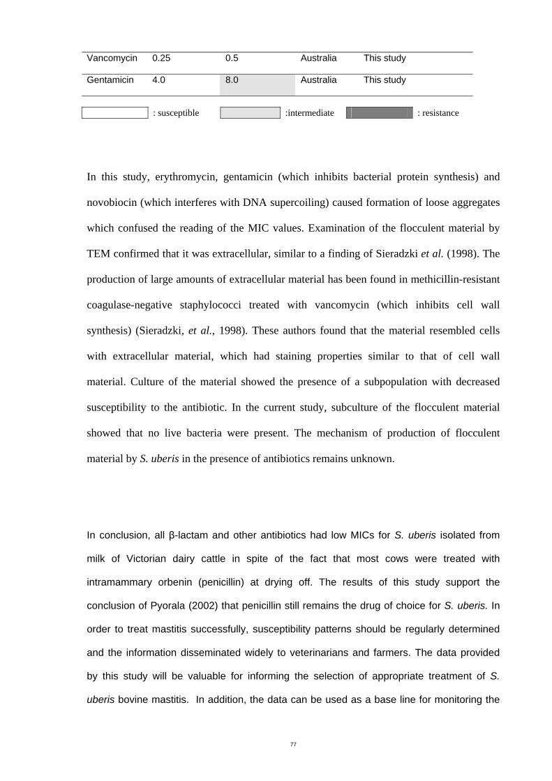
77
Vancomycin 0.25 0.5 Australia This study
Gentamicin 4.0 8.0 Australia This study
: susceptible :intermediate : resistance
In this study, erythromycin, gentamicin (which inhibits bacterial protein synthesis) and
novobiocin (which interferes with DNA supercoiling) caused formation of loose aggregates
which confused the reading of the MIC values. Examination of the flocculent material by
TEM confirmed that it was extracellular, similar to a finding of Sieradzki et al. (1998). The
production of large amounts of extracellular material has been found in methicillin-resistant
coagulase-negative staphylococci treated with vancomycin (which inhibits cell wall
synthesis) (Sieradzki, et al., 1998). These authors found that the material resembled cells
with extracellular material, which had staining properties similar to that of cell wall
material. Culture of the material showed the presence of a subpopulation with decreased
susceptibility to the antibiotic. In the current study, subculture of the flocculent material
showed that no live bacteria were present. The mechanism of production of flocculent
material by S. uberis in the presence of antibiotics remains unknown.
In conclusion, all β-lactam and other antibiotics had low MICs for S. uberis isolated from
milk of Victorian dairy cattle in spite of the fact that most cows were treated with
intramammary orbenin (penicillin) at drying off. The results of this study support the
conclusion of Pyorala (2002) that penicillin still remains the drug of choice for S. uberis. In
order to treat mastitis successfully, susceptibility patterns should be regularly determined
and the information disseminated widely to veterinarians and farmers. The data provided
by this study will be valuable for informing the selection of appropriate treatment of S.
uberis bovine mastitis. In addition, the data can be used as a base line for monitoring the

78
patterns of resistance in order to detect increasing resistance to antibiotic agents for
mastitis treatment.
79
Chapter 4 Bacteriocins isolation and production
4.1 Introduction
Mastitis pathogens, particularly environmental bacteria, are still a serious problem on dairy
farms (Leigh, 1999). Antibiotics are used to treat clinical mastitis and to control subclinical
mastitis during the dry period. After antibiotic treatment of clinical mastitis, it is important to
withhold milk for some days to ensure no antibiotic residues are in the milk sold to
customers. This causes problems economically due to loss of milk production. Moreover,
there has been increased concern regarding the widespread over prescrition of antibiotics
(Dairy Australia, 2007) with resultant increased development of antibiotic-resistant bacteria
(Erskine, et al., 2002). Thus, the investigation of an alternative strategy to control the
disease is essential.
Studies of bacterial isolates from mammary glands of dairy cows have suggested that
members of the normal teat flora of cows or minor mastitis pathogens influence the ability
of major pathogens to establish infection. For example, the presence of Corynebacterium
(Green, et al., 2005), CoNS and Staphylococcus chromogenes (Todhunter, et al., 1993,
Vliegher, et al., 2004) resulted in reduction of mastitis pathogens. Therefore, a possible use
of normal flora bacteria from cows or inhibitory substances isolated from these bacteria
was suggested for the mastitis biological control.
A few studies showed success in vitro and in vivo in the use of bacteriocins for mastitis
control (Sears, et al., 1992, Ryan, et al., 1999, Twomey, et al., 2000, Wu, et al., 2007).
These authors reported the inhibition of different bacteriocins against S. dysgalactiae, S.
aureus and E. coli. Bacteriocins can be used in combination with teat seals or as a teat
disinfectant. Since bacteriocins are antibiotic peptides, non-toxic and inactivated by
digestive enzymes in the gut of humans (Jack, et al., 1995, Chen & Hoover, 2003), it
should be possible to show that these agents present no hazard to milk and are able to be
80
used during lactation. Bacteriocins or inhibitory substances from CoNS may play an
important role in S. uberis control. The aims of this chapter were to examine a collection of
CoNS and related bacteria isolated from cow’s milk for bacteriocin production.
4.2 Materials and methods
4.2.1 Bacterial isolates and control strains
4.2.1.1 Coagulase negative staphylococci (CoNS) as test strains
Milk samples (described in Chapter 2) were processed and CoNS were isolated and
identified by the following method. An aliquot of 50 µl of milk was plated onto horse blood
agar (HBA, Appendix C) and the culture was incubated at 37°C for 48 hours. Coagulase
negative staphylococci were purified by repeated streaking. Strains were characterized as
described in 4.2.3. Seventy eight bovine CoNS isolates from a previous study (Abadin,
M.Sc. Thesis, RMIT, 1994) were also included in this study. The isolates were maintained
at -70°C in Protect Bacterial Preservers. They were cultured on brain heart infusion agar
(BHA, Appendix C) at 37°C for approximately 18 h before use. A bacterial reference strain,
Pedicoccus acidilactica FSAW 2702, kindly provided by Food Science Australia from the
culture collection maintained was used as a positive control strain for bacteriocin
production. The strain was stored at -70°C in Protect Bacterial Preservers and retrieved from
storage by aseptically culturing from the bacterial beads onto De Man Rogosa Sharpe agar
medium (MRS, Appendix C). Listeria innocua (a strain from the Microbiology Laboratory,
RMIT University) was used as an indicator strain against Pedicoccus acidilactica FSAW
2702. The strain was also stored at -70°C in Protect Bacterial Preservers and retrieved from
storage by aseptically culturing from the bacterial beads onto BHA.
4.2.1.2 Streptococcus uberis as indicator strains
81
The same S. uberis isolates described in previous chapter (n = 68) (Appendix B, Table 2)
were used as indicator organisms for the bacteriocin assay. The isolates were maintained at -
70°C in Protect Bacterial Preservers. They were propagated at 37°C on either HBA or BHA
at 37°C overnight before use.
4.2.2 Identification procedures for coagulase negative staphylococci
Conventional methods for CoNS identification included Gram staining, catalase test,
coagulase test and deoxyribonuclease (DNase) test. After screening for bacteriocin activity,
one isolate that showed inhibitory activity against S. uberis was further identified using 16S
rRNA sequencing (2.2.3.1).
4.2.2.1 Coagulase test (Isenberg, 1992)
Staphylococcus aureus has the ability to clot plasma by production of the enzyme
coagulase. It is an important criterion to distinguish Staphylococcus aureus (coagulase
positive) from other Staphylococcus species (CoNS).
Lyophilized rabbit plasma (Bio Merieux) was diluted with sterile distilled water according
to the manufacturer’s instruction. Working stocks of rabbit plasma was prepared by adding
three volumes of sterile peptone water to the diluted rabbit plasma. An aliquot of 400 µl of
working rabbit plasma was pipetted into 13 x 100 mm sterile test tubes, then one loopful of
bacterial culture was added. After incubation at 37°C, cultures were examined for clots at 4
h and again at 24 h. S. aureus ATCC 29213 was used as a positive control.
4.2.2.2 Deoxyribonuclease (DNase) test
This test detects microbial DNase based on its ability to hydrolyze polymerized DNA in the
agar to a mixture of mono and oligonucleotides. The bacteria were spot inoculated on the
surface of DNase agar and incubated at 37°C for 24 h, after which the plates were flooded
with 1 M HCl. DNase producing colonies were surrounded by a clear area that contained
82
nucleotide fractions which were not precipitated by acid (Isenberg, 1992). The control
strain, S. chromogenes NTCC 10530, showed non-hemolytic, opaque, yellow pigmented
colonies and an intermediate zone in the DNase test (Devriese, et al., 2002).
4.2.2.3 16S rRNA sequencing
The sequencing method was described in 2.2.3.2
4.2.3 Detection and isolation of bacteriocin-producing strains
Three different methods for assaying bacteriocin were used in the present study. The agar
inversion method and the spot on lawn assay were used for testing inhibitory activity
directly from cultures, whereas the agar diffusion assay was used for testing inhibitory
activity from liquid containing bacteriocin.
4.2.3.1 Agar inversion method (Vliegher, et al., 2004)
The agar inversion method was used as a screening test for bacteriocin production. This
method is not only a practical and simple method of screening for bacteriocin-producing
bacteria but it is able to distinguish inhibition by bacteriocin from inhibition by bacteriophage
(Vliegher, et al., 2004). The plates were prepared by measuring 25 mL of HBA to ensure
the same thickness of the agar for all plates. Freshly grown CoNS isolates were inoculated
approximately five mm in width across the centre of an agar plate and incubated at 37°C
overnight. The agar was then turned upside down into the cover of the Petri dish and
cultures of indicator strains were inoculated perpendicular to the test organisms. Cross
streaks of CoNS against themselves were also inoculated as a self control to exclude
inhibition caused by nutrient depletion. After incubation at 37°C for a further 24 h, patterns
of S. uberis inhibition were observed as total, partial or no inhibition. Total inhibition
referred to no growth of S. uberis whereas partial inhibition indicated a reduction in size of
colonies of S. uberis. The inhibition zone sizes were measured.
4.2.3.2 Spot on lawn method (Oscariz, et al., 1999)
83
The spot on lawn method is a simple method to detect bacteriocin activity from test strain
cultures. For screening of bacteriocin producers, five to ten colonies were spot-inoculated
onto BHA using sterile toothpicks, such that the spots were equidistant from each other and
approximately 2 cm from the edge of the plate. The cultures were incubated at 37°C
overnight. S. uberis at a concentration of 106 CFU/mL (1:100 dilution of a suspension with
turbidity equivalent to 0.5 McFarland standard) in BHI soft agar was poured over the
sample of the inoculated plates and the plates were further incubated at 37°C overnight.
The diameter of inhibition zones produced by CoNS against S. uberis was then measured.
Strains that produced inhibition zones were further investigated (section 4.2.4) before being
designated as bacteriocin producers. Pedicoccus acidilactica FSAW 2702 was used as
positive control producer strain against the indicator strain, Listeria innocua.
4.2.3.3 Agar diffusion assay
The agar diffusion assay was used for detection of antibacterial activity from liquid solution.
Twenty five mL of molten BHA was cooled to 45°C and seeded with fresh indicator cells to
a final inoculum at 106 CFU/mL by adding 250 µl of a suspension with turbidity equivalent
to a 0.5 McFarland standard. The plate was allowed to solidify and put into a laminar flow
cabinet with the lid open for 30 min to ensure the plate was completely dry. Wells 6 mm in
diameter were cut in the seeded agar plate with a sterile Pasteur pipette. Crude bacteriocin
from culture supernates and bacteriocin ammonium sulphate precipitate, which had been
filter-sterilized by passing through a filter membrane (pore size of 0.45 µm) or
chromatographic fractions were used for assessing inhibitory activity. Aliquots of 100 µl of
samples were added into the wells. The plates were incubated at 37°C overnight then
diameters of inhibition zones were measured.
For assay of bacteriocins, 100 µl of two-fold dilutions of the samples were transferred to the
wells. The plates were incubated at 37°C overnight then the diameters of the inhibition
zones were measured. Activity was defined as the reciprocal of the last serial dilution giving
84
a zone of inhibition and expressed as arbitrary units (AU) per milliliter (Crupper & Iandolo,
1997).
4.2.4 Confirmation that observed activity is due to a bacteriocin
Bacteriocins are proteins excreted by bacteria that inhibit the growth of other closely related
bacteria (Tagg, et al., 1976). In order to prove that the observed antibiotic activity is due to
a bacteriocin, it was necessary to confirm the protein nature of the antibiotic activity and to
exclude inhibition caused by bacteriophage (refer to 4.2.3.1) or metabolic by-products such
as organic acids and hydrogen peroxide.
4.2.4.1 Effect of proteases (proteinase K, trypsin and chymotrypsin), lipase and catalase on
bacteriocin (Villani, et al., 2001)
The bacteriocin producer strain was spotted onto BHA using a sterile toothpick and
incubated at 37°C overnight. A series of holes, two mm in diameter, were aseptically cut
three mm from the edge of the culture. A 10 µl aliquot of 10 mg/mL of each enzyme
(proteinase K, trypsin, chymotrypsin, lipase and catalase) was pipetted into each hole, after
which the plates were allowed to stand at room temperature for 30 min to allow the
enzymes to diffuse into the agar. BHI soft agar containing 106 CFU/mL of S. uberis 796-2
as an indicator strain was poured onto the surface of the plates. The control consisted of
untreated cell-free supernate of potential bacteriocin producer strains, and enzyme solution
alone incubated under the same conditions. After incubation at 37°C for approximately 16
h, the shape of inhibition zones was observed.
An even annular radius indicated the substance/s produced by the test strain was resistant
to the test enzyme. An irregular shaped annular radius (reduction of the diameter close to
the enzyme spot) indicated that the substance/s produced by the test strains was sensitive
to the enzyme.
4.2.4.2 Neutralization of acid production
85
Inhibitory activity due to acid production was excluded by adding CaCO3 to the media
(Heng, et al., 2006). The bacteriocin producer strain was assayed for inhibitory activity by
spot on lawn method on BHA containing 0.1% (w/v) CaCO3.
4.2.5 Studies of bacteriocin production
Most bacteriocins are secreted by the producer cells and can be detected in culture
supernates, but some bacteriocins cannot be detected in liquid media. Optimization of
culture media or culture conditions was studied in an attempt to find the optimum conditions
for bacteriocin production. Each of the following conditions was examined for bacteriocin
production and repeated at least twice.
4.2.5.1 Media selection
Ten S. uberis isolates (684-1, 686-5, 796-2, 853-4, 883-7, 2042-5, 2655-5, 2690-2, 3147-1
and H2581-3) were selected at random to be examined for susceptibility to the bacteriocin
produced by isolate 2042-4. The isolate 2042-4 was spot inoculated onto BHA, milk agar
(MA), nutrient agar (NA), and trypticase soy agar (TSA) (Appendix C) and the degree of
inhibition was determined by the spot on lawn method. The size of inhibition zones
obtained on each of the media was compared.
4.2.5.2 Extracellular bacteriocin
Effect of culture conditions and broth compositions on production of the bacteriocin
A fresh culture of the bacteriocin producer (108 CFU/mL, 200 µl) was transferred to 20 mL
of the following media and incubated under the following conditions. After incubation, the
inhibitory activity was determined by the agar diffusion method against S. uberis.
• The bacteriocin producer was cultured in brain heart infusion broth (BHB, Appendix
C) at room temperature, 30°C and 37°C with and without agitation. The initial pH of
the media ranged from 2 to 11. The culture supernate was collected after incubation
for 0, 3, 6, 9, 12, 18, 24 and 48 h and was tested for inhibitory activity.
86
• To obtain anaerobic conditions, BHB was placed in boiling water for 10 min to
eliminate oxygen from the broth. The culture tube was cooled immediately in tap
water. The producer strain was inoculated into the broth and sterile liquid paraffin
was gently overlaid onto the surface of the broth to maintain anaerobic conditions.
The culture was incubated at 37°C overnight and examined for inhibitory activity.
• The effect of sub-lethal concentrations of nine antibiotics (ampicillin, cephalothin,
erythromycin, gentamicin, oxacillin, penicillin, tetracycline, vancomycin and
novobiocin) on production of inhibitory substance by the bacteriocin producer strain
was examined. The minimal inhibitory concentration (MIC) of the bacteriocin
producer strain was determined (refer to method in chapter 3). BHB was
supplemented with each antibiotic at final concentration of 1/2, 1/4 and 1/8 of the
MIC value. The isolate was grown at 37°C overnight then the culture supernate was
tested for inhibitory activity. Media with antibiotics alone were used as negative
controls.
• BHB was supplemented with increasing concentrations of glucose, lactose, skim
milk, full cream milk, yeast extract and casein to a final concentration of 0, 0.1, 0.2,
0.3, 0.4, 0.5, 0.6, 0.7, 0.8, 0.9, 1.0, 1.5, 2.0, 2.5 and 3.0 % (w/v). After inoculation,
the culture was incubated at 37°C overnight then the culture supernate was tested for
inhibitory activity. Non inoculated media was use as a negative control.
• Inhibitory activity was assayed in BHB supplemented with different concentrations
of Tween 20 and Tween 80 at a final concentration of 0, 0.1, 0.2, 0.3, 0.4, 0.5, 0.6,
0.7, 0.8, 0.9, 1.0, 2.0, 3.0, 4.0 and 5.0 % (v/v). After inoculation, the culture was
incubated at 37°C overnight then culture supernate was tested for inhibitory activity.
Non inoculated media was use as a negative control.
Bacteriocin production from semisolid culture
87
Since inhibitory activity was not reliably detected in broth culture, extraction of inhibitory
substance from semi-solid agar was attempted (Mayr-Harting, et al., 1972, Balakrishnan, et
al., 2000). An inoculum of approximately 108 cfu/mL (a suspension with the turbidity
equivalent to 0.5 McFarland standard) of the bacteriocin producer strain was prepared in
BHB. The strain was inoculated with a sterile swab onto BHI soft agar plates and incubated
at 37°C overnight. The plate was frozen at -20°C overnight, then thawed at 37°C for 1 h.
This procedure was used to extract any bacteriocin that was embedded in the agar. The
slurry was transferred to a centrifuge tube. After centrifugation at 10,000 × g at 4°C for 30
min, the supernatant fluid was collected and sterilized by passage through a 0.45 µm
membrane filter. This liquid was referred to as crude bacteriocin. The inhibitory activity of
the crude bacteriocin was then examined by the agar diffusion assay.
4.2.5.3 Attempt to extract intracellular bacteriocin
Physical extraction
Sonication is physical cell disruption procedure using high frequency sound waves to shear
cells. The bacteriocin producer cells from cultures in 500 mL BHB, after incubation at 37°C
for 24 h, were harvested by centrifugation at 10,000 × g for 15 min then washed three times
with PBS (pH 7.0). The cells were suspended in 2 mL distilled water and sonicated for 3
min (6 times, 30 s each) in an ice bath or until the cloudy cell suspension became
translucent. The sonicator probe was set below the surface of the solution at all times. The
suspension was centrifuged at 10,000 × g, 4°C for 15 min to eliminate bacterial cells and
cell debris. The supernate was collected and tested for the bacteriocin activity by the agar
diffusion assay.
Chemical extraction
Attempts to extract bacteriocin from the producer cells were made using a mixture of HCl
and ethanol (Mota-Meira, et al., 1997, Wescombe, et al., 2006). Cultures in 1,000 mL BHB
were incubated at 37°C overnight. The cells were then harvested by centrifugation at
10,000 × g for 15 min. A mixture consisting of 20 mL HCl (20 mM) and 80 mL ethanol
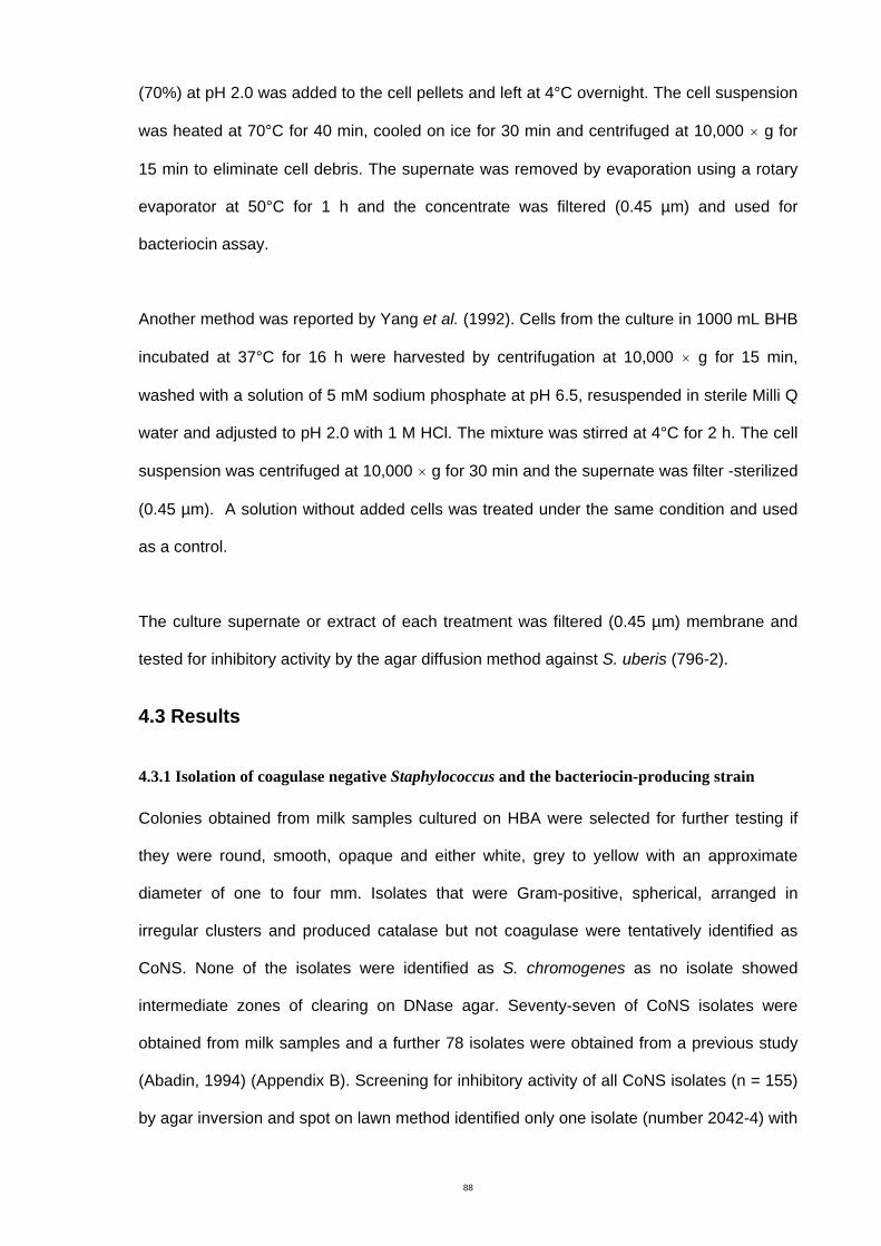
88
(70%) at pH 2.0 was added to the cell pellets and left at 4°C overnight. The cell suspension
was heated at 70°C for 40 min, cooled on ice for 30 min and centrifuged at 10,000 × g for
15 min to eliminate cell debris. The supernate was removed by evaporation using a rotary
evaporator at 50°C for 1 h and the concentrate was filtered (0.45 µm) and used for
bacteriocin assay.
Another method was reported by Yang et al. (1992). Cells from the culture in 1000 mL BHB
incubated at 37°C for 16 h were harvested by centrifugation at 10,000 × g for 15 min,
washed with a solution of 5 mM sodium phosphate at pH 6.5, resuspended in sterile Milli Q
water and adjusted to pH 2.0 with 1 M HCl. The mixture was stirred at 4°C for 2 h. The cell
suspension was centrifuged at 10,000 × g for 30 min and the supernate was filter -sterilized
(0.45 µm). A solution without added cells was treated under the same condition and used
as a control.
The culture supernate or extract of each treatment was filtered (0.45 µm) membrane and
tested for inhibitory activity by the agar diffusion method against S. uberis (796-2).
4.3 Results
4.3.1 Isolation of coagulase negative Staphylococcus and the bacteriocin-producing strain
Colonies obtained from milk samples cultured on HBA were selected for further testing if
they were round, smooth, opaque and either white, grey to yellow with an approximate
diameter of one to four mm. Isolates that were Gram-positive, spherical, arranged in
irregular clusters and produced catalase but not coagulase were tentatively identified as
CoNS. None of the isolates were identified as S. chromogenes as no isolate showed
intermediate zones of clearing on DNase agar. Seventy-seven of CoNS isolates were
obtained from milk samples and a further 78 isolates were obtained from a previous study
(Abadin, 1994) (Appendix B). Screening for inhibitory activity of all CoNS isolates (n = 155)
by agar inversion and spot on lawn method identified only one isolate (number 2042-4) with
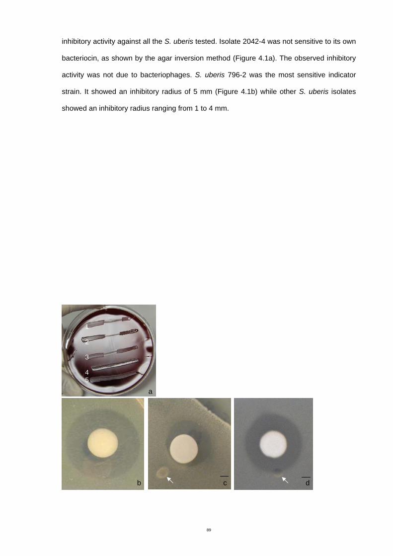
89
inhibitory activity against all the S. uberis tested. Isolate 2042-4 was not sensitive to its own
bacteriocin, as shown by the agar inversion method (Figure 4.1a). The observed inhibitory
activity was not due to bacteriophages. S. uberis 796-2 was the most sensitive indicator
strain. It showed an inhibitory radius of 5 mm (Figure 4.1b) while other S. uberis isolates
showed an inhibitory radius ranging from 1 to 4 mm.
a
b c d
1 2 3 4 5
90
Figure 4.1 Inhibitory effect of the strain 2042-4. a: agar inversion method against S. uberis 796-2, 686-5,
684-1 (1, 2, 3), S. aureus ATCC 29213 (4) and self control the strain 2042-4 (5), b: inhibitory activity
against S. uberis 796-2 by spot on lawn method, c: inhibitory activity against S. uberis 796-2 in the
presence of proteinase K (arrow), d: inhibitory activity against S. uberis 796-2 in presence of catalase
(arrow). Scale bar = 2 mm
Isolate number 2042-4 was a Gram-positive clustering coccus producing catalase but not
coagulase. Figure 4.2 shows the 16S rRNA sequence of the strain 2042-4 from nucleotide
number 1 to 366. A blast sequence search in the NCBI database revealed a close
relationship of the strain 2042-4 with Macrococcus caseolyticus. Figure 4.2 shows the
multiple alignments of 16S rRNA gene partial sequences of the isolate 2042-4 and four
strains of Macrococcus caseolyticus (accession numbers AY126154, DQ518592,
EF204307 and EU048336). isolate 2042-4 showed 97 - 98% similarity to M. caseolyticus
and was therefore designated as M. caseolyticus 2042-4.
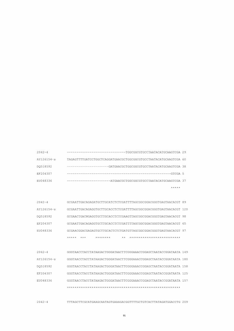
91
2042-4 -------------------------------TGGCGGCGTGCCTAATACATGCAAGTCGA 29
AY126154-s TAGAGTTTTGATCCTGGCTCAGGATGAACGCTGGCGGCGTGCCTAATACATGCAAGTCGA 60
DQ518592 ----------------------GATGAACGCTGGCGGCGTGCCTAATACATGCAAGTCGA 38
EF204307 -------------------------------------------------------GTCGA 5
EU048336 -----------------------ATGAACGCTGGCGGCGTGCCTAATACATGCAAGTCGA 37
*****
2042-4 GCGAATTGACAGAGATGCTTGCATCTCTCGATTTTAGCGGCGGACGGGTGAGTAACACGT 89
AY126154-s GCGAATTGACAGAGGTGCTTGCACCTCTCGATTTTAGCGGCGGACGGGTGAGTAACACGT 120
DQ518592 GCGAACTGACNGAGGTGCTTGCACCTCTCGAAGTTAGCGGCGGACGGGTGAGTAACACGT 98
EF204307 GCGAATTGACAGAGGTGCTTGCACCTCTCGATTTTAGCGGCGGACGGGTGAGTAACACGT 65
EU048336 GCGAACGGACGAGAGTGCTTGCACTCTCTGATGTTAGCGGCGGACGGGTGAGTAACACGT 97
***** *** ******** ** ***************************
2042-4 GGGTAACCTACCTATAAGACTGGGATAACTTCGGGAAACCGGAGCTAATACCGGATAATA 149
AY126154-s GGGTAACCTACCTATAAGACTGGGATAACTTCGGGAAACCGGAGCTAATACCGGATAATA 180
DQ518592 GGGTAACCTACCTATAAGACTGGGATAACTTCGGGAAACCGGAGCTAATACCGGATAATA 158
EF204307 GGGTAACCTACCTATAAGACTGGGATAACTTCGGGAAACCGGAGCTAATACCGGATAATA 125
EU048336 GGGTAACCTACCTATAAGACTGGGATAACTTCGGGAAACCGGAGCTAATACCGGATAATA 157
************************************************************
2042-4 TTTAGCTTCGCATGAAGCAATAGTGAAAGACGGTTTTGCTGTCACTTATAGATGGACCTG 209
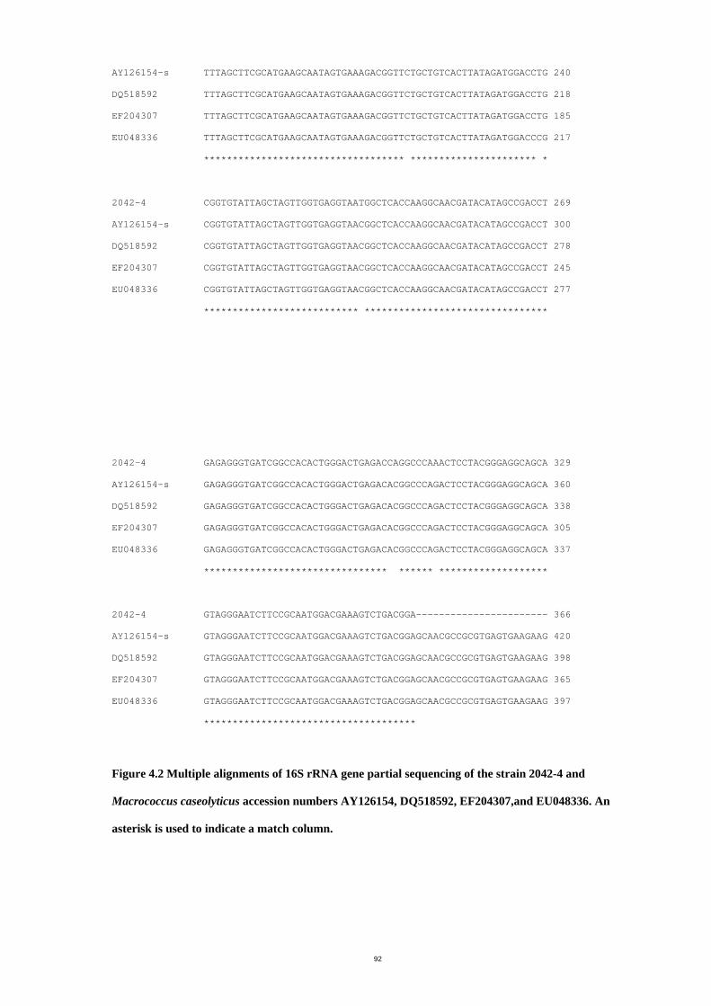
92
AY126154-s TTTAGCTTCGCATGAAGCAATAGTGAAAGACGGTTCTGCTGTCACTTATAGATGGACCTG 240
DQ518592 TTTAGCTTCGCATGAAGCAATAGTGAAAGACGGTTCTGCTGTCACTTATAGATGGACCTG 218
EF204307 TTTAGCTTCGCATGAAGCAATAGTGAAAGACGGTTCTGCTGTCACTTATAGATGGACCTG 185
EU048336 TTTAGCTTCGCATGAAGCAATAGTGAAAGACGGTTCTGCTGTCACTTATAGATGGACCCG 217
*********************************** ********************** *
2042-4 CGGTGTATTAGCTAGTTGGTGAGGTAATGGCTCACCAAGGCAACGATACATAGCCGACCT 269
AY126154-s CGGTGTATTAGCTAGTTGGTGAGGTAACGGCTCACCAAGGCAACGATACATAGCCGACCT 300
DQ518592 CGGTGTATTAGCTAGTTGGTGAGGTAACGGCTCACCAAGGCAACGATACATAGCCGACCT 278
EF204307 CGGTGTATTAGCTAGTTGGTGAGGTAACGGCTCACCAAGGCAACGATACATAGCCGACCT 245
EU048336 CGGTGTATTAGCTAGTTGGTGAGGTAACGGCTCACCAAGGCAACGATACATAGCCGACCT 277
*************************** ********************************
2042-4 GAGAGGGTGATCGGCCACACTGGGACTGAGACCAGGCCCAAACTCCTACGGGAGGCAGCA 329
AY126154-s GAGAGGGTGATCGGCCACACTGGGACTGAGACACGGCCCAGACTCCTACGGGAGGCAGCA 360
DQ518592 GAGAGGGTGATCGGCCACACTGGGACTGAGACACGGCCCAGACTCCTACGGGAGGCAGCA 338
EF204307 GAGAGGGTGATCGGCCACACTGGGACTGAGACACGGCCCAGACTCCTACGGGAGGCAGCA 305
EU048336 GAGAGGGTGATCGGCCACACTGGGACTGAGACACGGCCCAGACTCCTACGGGAGGCAGCA 337
******************************** ****** *******************
2042-4 GTAGGGAATCTTCCGCAATGGACGAAAGTCTGACGGA----------------------- 366
AY126154-s GTAGGGAATCTTCCGCAATGGACGAAAGTCTGACGGAGCAACGCCGCGTGAGTGAAGAAG 420
DQ518592 GTAGGGAATCTTCCGCAATGGACGAAAGTCTGACGGAGCAACGCCGCGTGAGTGAAGAAG 398
EF204307 GTAGGGAATCTTCCGCAATGGACGAAAGTCTGACGGAGCAACGCCGCGTGAGTGAAGAAG 365
EU048336 GTAGGGAATCTTCCGCAATGGACGAAAGTCTGACGGAGCAACGCCGCGTGAGTGAAGAAG 397
*************************************
Figure 4.2 Multiple alignments of 16S rRNA gene partial sequencing of the strain 2042-4 and
Macrococcus caseolyticus accession numbers AY126154, DQ518592, EF204307,and EU048336. An
asterisk is used to indicate a match column.
93
4.3.2 Characterization of antibiotic substance
The inhibitory activity of M. caseolyticus 2042-4 against S. uberis 796-2 was lost after
treatment with three proteolytic enzymes (Table 4.1): proteinase K, trypsin and
chymotrypsin. An example of reduction in inhibitory activity by proteinase K is shown in
Figure 4.1 c. Treatment with catalase and lipase (result not shown) did not result in loss of
activity (Table 4.1, Figure 4.1 d). The inhibitory activity on BHA containing CaCO3 (Table
4.1) showed the same activity as in BHA alone (Figure 4.1b). Together these results
suggested that the antibacterial activity demonstrated by M. caseolyticus 2042-4 was due
to production of a bacteriocin.
Table 4.1 Factors affecting antibiotic activity of Macrococcus caseolyticus 2042-4
Treatment Activity
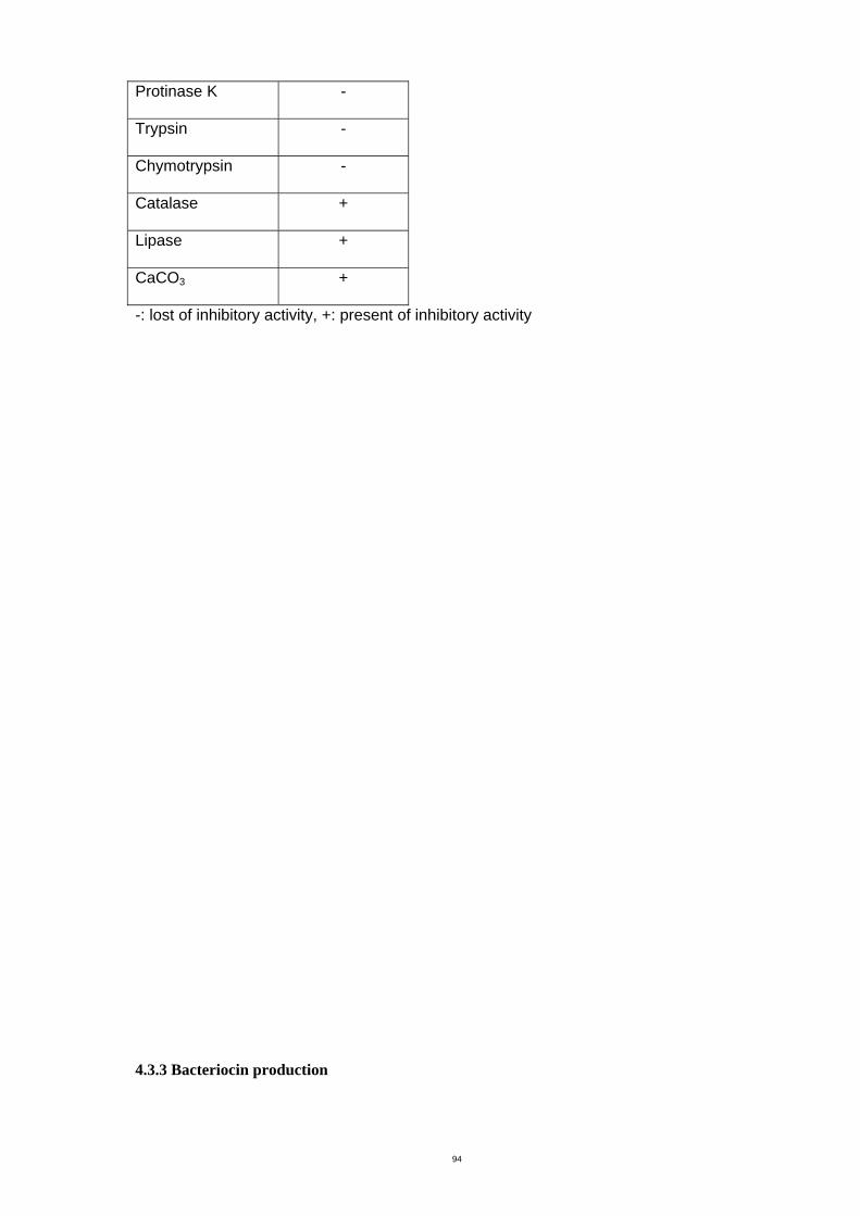
94
Protinase K -
Trypsin -
Chymotrypsin -
Catalase +
Lipase +
CaCO3 +
-: lost of inhibitory activity, +: present of inhibitory activity
4.3.3 Bacteriocin production
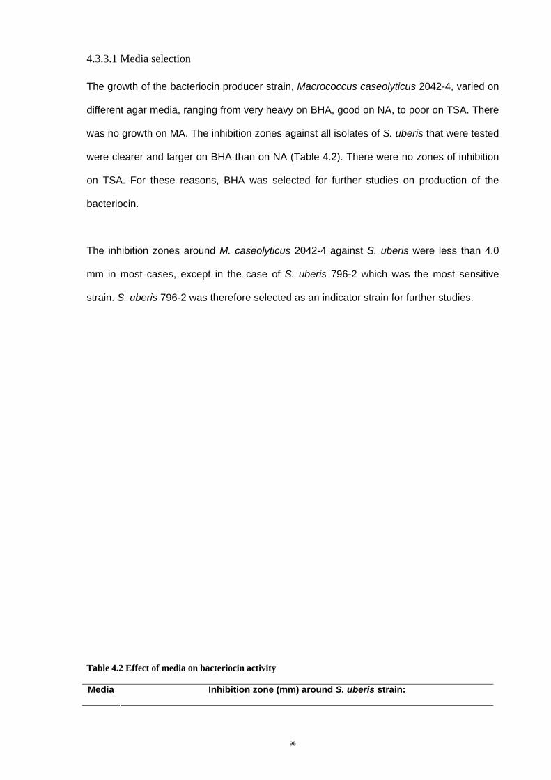
95
4.3.3.1 Media selection
The growth of the bacteriocin producer strain, Macrococcus caseolyticus 2042-4, varied on
different agar media, ranging from very heavy on BHA, good on NA, to poor on TSA. There
was no growth on MA. The inhibition zones against all isolates of S. uberis that were tested
were clearer and larger on BHA than on NA (Table 4.2). There were no zones of inhibition
on TSA. For these reasons, BHA was selected for further studies on production of the
bacteriocin.
The inhibition zones around M. caseolyticus 2042-4 against S. uberis were less than 4.0
mm in most cases, except in the case of S. uberis 796-2 which was the most sensitive
strain. S. uberis 796-2 was therefore selected as an indicator strain for further studies.
Table 4.2 Effect of media on bacteriocin activity
Media Inhibition zone (mm) around S. uberis strain:
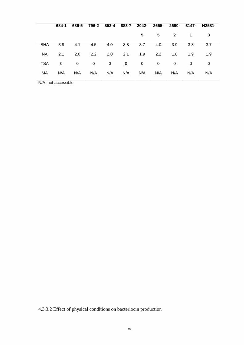
96
684-1 686-5 796-2 853-4 883-7 2042-
5
2655-
5
2690-
2
3147-
1
H2581-
3
BHA 3.9 4.1 4.5 4.0 3.8 3.7 4.0 3.9 3.8 3.7
NA 2.1 2.0 2.2 2.0 2.1 1.9 2.2 1.8 1.9 1.9
TSA 0 0 0 0 0 0 0 0 0 0
MA N/A N/A N/A N/A N/A N/A N/A N/A N/A N/A
N/A: not accessible
4.3.3.2 Effect of physical conditions on bacteriocin production
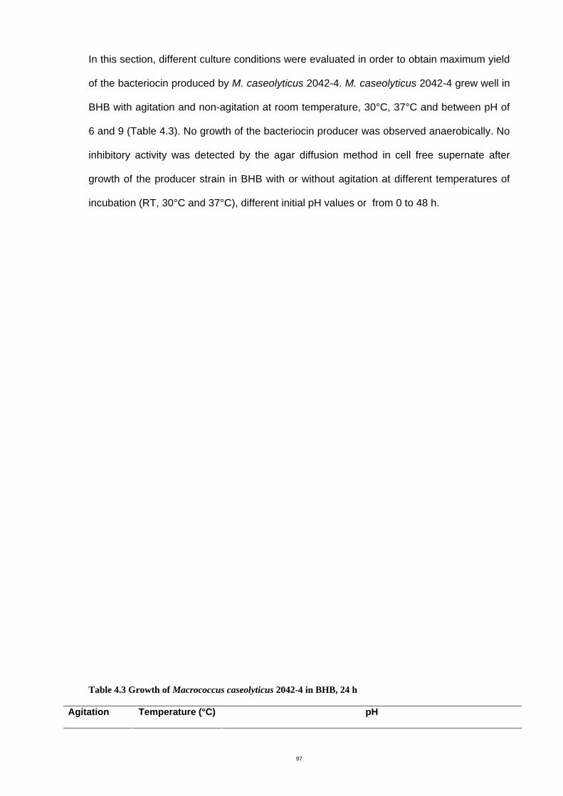
97
In this section, different culture conditions were evaluated in order to obtain maximum yield
of the bacteriocin produced by M. caseolyticus 2042-4. M. caseolyticus 2042-4 grew well in
BHB with agitation and non-agitation at room temperature, 30°C, 37°C and between pH of
6 and 9 (Table 4.3). No growth of the bacteriocin producer was observed anaerobically. No
inhibitory activity was detected by the agar diffusion method in cell free supernate after
growth of the producer strain in BHB with or without agitation at different temperatures of
incubation (RT, 30°C and 37°C), different initial pH values or from 0 to 48 h.
Table 4.3 Growth of Macrococcus caseolyticus 2042-4 in BHB, 24 h
Agitation Temperature (°C) pH
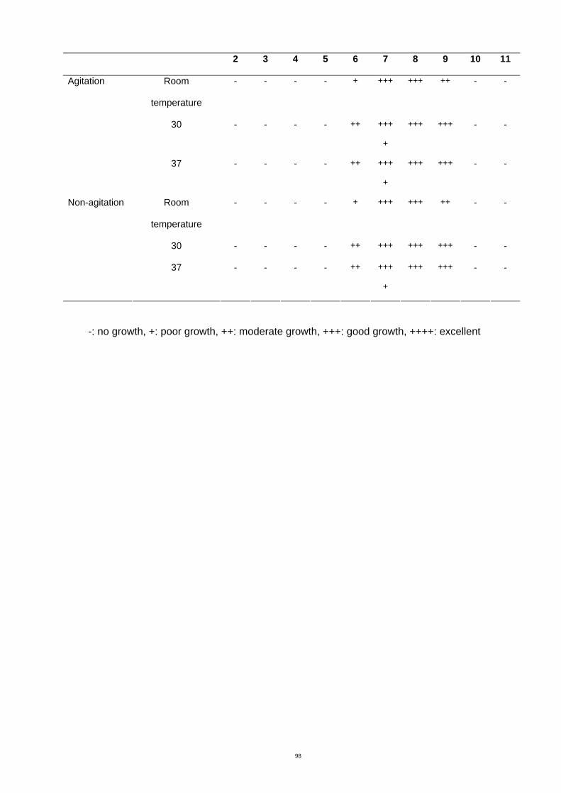
98
2 3 4 5 6 7 8 9 10 11
Room
temperature
- - - - + +++ +++ ++ - -
30 - - - - ++ +++
+
+++ +++ - -
Agitation
37 - - - - ++ +++
+
+++ +++ - -
Room
temperature
- - - - + +++ +++ ++ - -
30 - - - - ++ +++ +++ +++ - -
Non-agitation
37 - - - - ++ +++
+
+++ +++ - -
-: no growth, +: poor growth, ++: moderate growth, +++: good growth, ++++: excellent
99
The MICs of ampicillin, cephalothin, erythromycin, gentamicin, novobiocin, oxacillin,
penicillin, tetracycline and vancomycin for the producer strain (M. caseolyticus 2042-4) are
shown in Table 4.4. After culturing of the bacteriocin producer strain, in broth containing
sub-lethal concentrations (1/2, 1/4 and 1/8 of MIC) of these nine antibiotic agents, no
inhibitory activity was observed by the agar diffusion assay after growth in antibiotics at any
concentrations. The negative control of the broth containing sub-lethal concentration of
antibiotics alone against the indicator strains did not show activity, even though the sub-
lethal concentrations in the broth were higher than the MICs of the indicator strain
(Appendix B, Table 2). These results could be explained by the fact that the concentration
of indicator strain used in the agar diffusion method (106 CFU/mL) was higher than in the
MIC procedure (104 CFU/mL) (NCCLS, 2002) therefore no inhibition was observed.
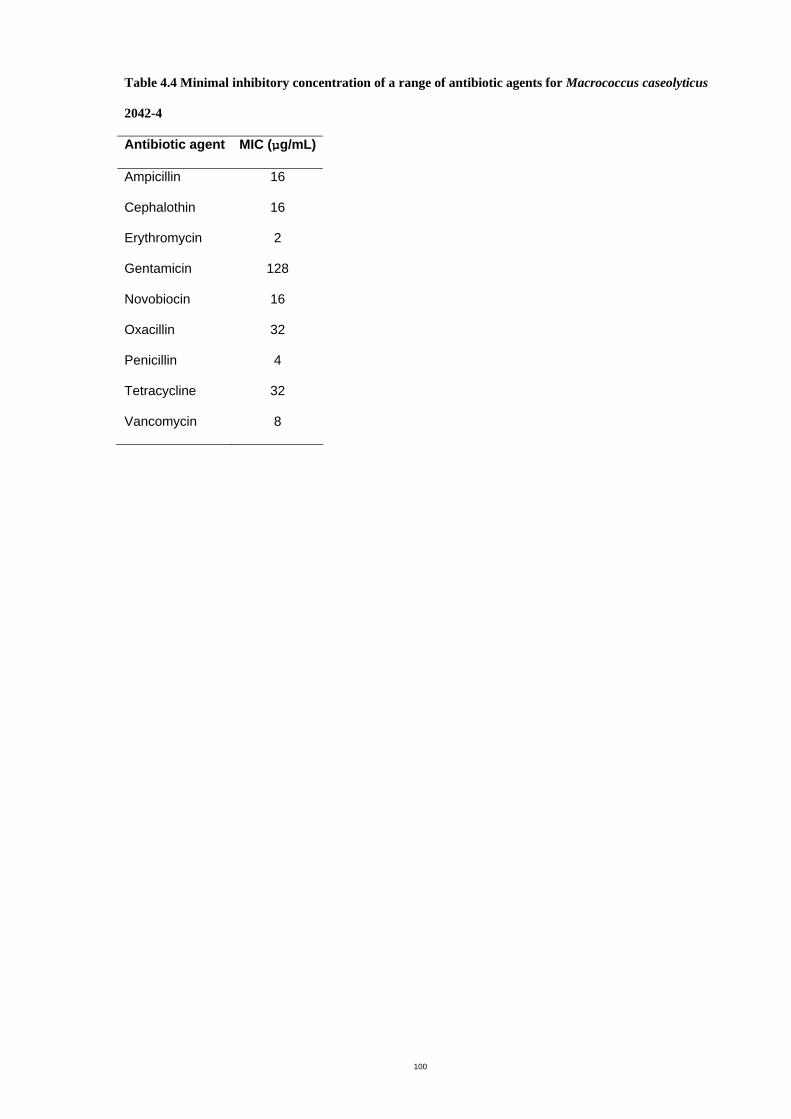
100
Table 4.4 Minimal inhibitory concentration of a range of antibiotic agents for Macrococcus caseolyticus
2042-4
Antibiotic agent MIC (µg/mL)
Ampicillin 16
Cephalothin 16
Erythromycin 2
Gentamicin 128
Novobiocin 16
Oxacillin 32
Penicillin 4
Tetracycline 32
Vancomycin 8
101
No inhibitory activity was detected in the culture supernate of M. caseolyticus 2042-4
grown in BHB supplemented with glucose, lactose, skim milk, full cream milk, casein or
yeast extract in the range of 0 to 3% at 37 °C or in BHB containing Tween 20 or Tween 80
at concentrations of 0 to 5%.
Extraction of intracellular bacteriocin was also performed by sonication and chemical
extractions but no inhibitory activity was discovered. Inhibitory activity of 10 AU/mL was
finally observed by culturing the producer strain on semi-solid agar (0.7% of agar in BHI)
followed by freezing and thawing as shown in Figure 4.3.
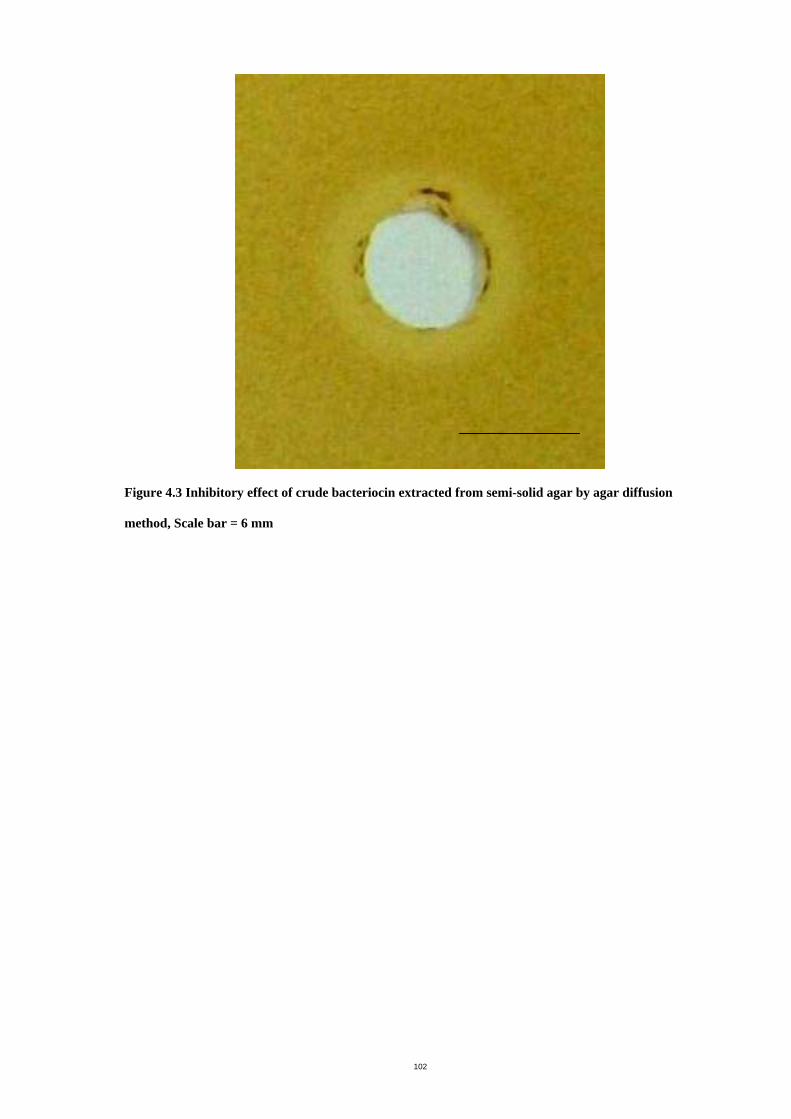
102
Figure 4.3 Inhibitory effect of crude bacteriocin extracted from semi-solid agar by agar diffusion
method, Scale bar = 6 mm
103
4.4 Discussion
Of the 155 isolates from milk, which were initially identified as CoNS, one had antagonistic
activity against all S. uberis that were examined in this study. The antagonistic activity was
shown to be due to the production of a bacteriocin rather than other types of inhibitory
substances such as bacteriophages, hydrogen peroxide or acid production (Jack, et al.,
1995). Bacteriophages were ruled out because activity was detected by the agar inversion
method, which requires the inhibitory agent to pass through the agar, which is not possible
for a bacteriophage. Hydrogen peroxide was also excluded as a cause of the antibacterial
activity since the inhibition was demonstrated in the presence of catalase. Activity was also
retained on BHA containing CaCO3, indicating that the inhibition was not caused by acid
production, as CaCO3 neutralizes acidity of organic acids (Nicolas, et al., 2004). Nutrient
depletion was also eliminated as a cause of the observed inhibition, since no activity was
observed in the agar inversion method if the producer strain was used as both producer
and indicator. Depletion of inhibitory activity was observed on treatment with proteolytic
enzymes but not with lipase. These results suggested that the inhibitory activity of the
isolate 2042-4 is due to the production of bacteriocin and that no lipid moieties are
contained in the molecule.
The bacteriocin producer was initially thought to be a CoNS, but was identified by 16S
rRNA sequencing as Macrococcus caseolyticus 2042-4 with 98% similarity to M.
caseolyticus in the NCBI database. In 1998, Macrococcus was separated from most
species of staphylococci and designated as a novel genus (Kloos, et al., 1998). The genus
Macrococcus contains four species: M. bovicus, M. caseolyticus, M. carouselicus and M.
equipercicu. Members of this genus are coagulase negative and catalase positive and can
be distinguished from CoNS by molecular identification techniques including 16S rRNA
sequencing. To date, there have been no reports of Macrococcus as pathogens, in contrast
to CoNS which are classified as minor mastitis pathogens (Radotits, et al., 1994). M.
caseolyticus can be isolated from the skin of cattle, goats, horses, whales and dolphins
104
(Kloos, et al., 1998), dairy products (Randazzo, et al., 2002), fermented sausages (Blaiotta,
et al., 2003) and meat products (Olsson, et al., 2003). Over the last few decades, a variety
of bacteriocins from CoNS have been characterized but no bacteriocins from M.
caseolyticus or Staphylococcus caseolyticus with activity against S. uberis have been
described so far in the literature. Thus, this antibacterial substance is of interest for further
characterization and identification.
In contrast to most published data, which describes the harvest of bacteriocins released
from the producer cells into liquid culture, no bacteriocin activity was detected in liquid
cultures of M. caseolyticus even though the bacteriocin activity was produced on solid
media and apparently diffused from the site of inoculation of the indicator strain to create a
zone of inhibition. Optimal growth conditions usually result in optimal bacteriocin production
(Parente & Ricciardi, 1999). Attempts were made to recover bacteriocin from liquid cultures
by optimizing culture conditions such as temperature, pH, incubation time and agitation but
these were unsuccessful. Also, the producer cells did not grow under anaerobic conditions
and thus inhibitory activity was not detected. Moreover, in contrast to other studies
(Ogunbanwo, et al., 2003, Avonts, et al., 2004, Nicolas, et al., 2004), optimization of the
media composition with addition of carbon sources, milk or yeast extract did not assist in
enhancing bacteriocin production from M. caseolyticus 2042-4. Additionally, sub-lethal
concentrations of nine antibiotics failed to induce bacteriocin synthesis from M.
caseolyticus 2042-4 in broth cultures. These results are in contrast to those of Jerman et al.
(2005), which showed that sub-lethal concentrations of ciprofloxacin induced bacteriocin
synthesis in E. coli. They suggested that ciprofloxacin interferes with DNA replication and
cell wall synthesis, which may induce production of bacteriocins by inducing the SOS
response. In this study, Tween 20 and Tween 80 did not stimulate bacteriocin production
which is in contrast to the study of Garver and Muriana (1994) that showed an increase in
curvaticin FS47 (a bacteriocin from Lactobacillus curvatus FS47) production by adding
Tween 20 or Tween 80 to the medium at concentrations from 0.75 to 1%. Other studies
showed that Tween did not enhance bacteriocin production but could possibly bind to
105
producer cells. For example, adding Tween 80 to the culture media reduced adsorption of
lacticin RM to producer cells, Lactobacillus lactis (Keren, et al., 2004). However Li, et al.
(2002), reported that addition of Tween 20 caused increased sensitivity of L.
monocytogenes to nisin by increasing the binding of nisin to the indicator cells.
In this study, no bacteriocin activity was detected after physical and chemical extraction of
producer cell pellets, suggesting that the bacteriocins did not adhere to the surface of the
producer cells. This is contrast to some investigators who successfully extracted
bacteriocins from cell pellets for example, mutacin B-Ny266 from S. mutans (Mota-Meira, et
al., 1997) and bacteriocins produced by different genera of lactic acid bacteria (Yang, et al.,
1992).
Limited success in detection of these substances may be due to inactivation of the
bacteriocin by proteolytic enzymes present in the culture supernate. Therefore the final
attempt to extract bacteriocin used culture media supplemented with agar and this method
proved successful. Similar problems had been described by Kelstrup and Gibbons (1969).
They explained that the recovery of the bacteriocin produced by non-group A streptococci
was dependent on the viscosity of the media which could be manipulated by the amount of
agar, starch, glycerol or dextran in the culture medium. In culture on solid media, the
bacteriocins may diffuse ahead of the active proteinase and would be protected from
inactivation.
Results from this study showed that a non-pathogenic bacterium found in cow’s milk,
Macrococcus caseolyticus 2042-4, had inhibitory activity against S. uberis. Since
Macrococcus caseolyticus was isolated from the same niche as S. uberis, this raises the
possibility that the inhibitory substance produced by this organism could be used to
regulate pathogenic species in the same environment. This newly found antibiotic,
therefore has the potential for beneficial use as a method of natural control of bovine
mastitis caused by S. uberis.

106
The next chapter describes further characterization of this bacteriocin as well as
investigation of an extraction method suitable for scale up of production in sufficient
amounts for purification and identification of this molecule.
107
Chapter 5 Bacteriocin characterization and purification
5.1 Introduction
The previous chapter presented results showing that a crude bacteriocin produced by
Macrococcus caseolyticus has antibacterial activity against all isolates of S. uberis that
were tested. Characteristics of bacteriocins, which include a narrow inhibitory spectrum,
thermostability and pH stability, could make them attractive as chemotherapeutic agents. In
particular, antibacterial activity against mastitis pathogens makes them attractive
candidates for the treatment of clinical mastitis or protection against from mastitis. Each
bacteriocin is unique; therefore characterization of this bacteriocin is necessary. The aim of
this chapter was to report on the characterization of purified bacteriocin from culture
supernate.
5.2 Material and methods
5.2.1 Bacteriocin characterization
Characterization of the bacteriocin involved determining the spectrum of activity against a
variety of Gram-positive and Gram-negative bacteria, thermostability, pH stability and rate
of killing. These tests used concentrated crude bacteriocin that was prepared by extraction
from semi-solid agar followed by ammonium sulphate precipitation as described in 5.2.3.2
and 5.2.3.3.
5.2.1.1 Spectrum of inhibitory activity
Indicator strains from stock cultures held by the Microbiology Laboratory, RMIT University
were used in this experiment. The strains included varieties of Gram-positive, Gram-
negative bacteria and spiral bacteria (Appendix B, Table 4). The strains were stored at -
80°C in Protect Bacterial Preservers. They were retrieved from storage at -80°C by
aseptically transferring the bacterial preserver beads onto horse blood agar (HBA, Appendix
108
C) or brain heart infusion agar (BHA, Appendix C), streaking and incubating for 24 hours
for further testing. Determination of the spectrum of activity of the bacteriocin culture was
performed by spot on lawn method (4.2.4.2) using a culture of the producer strain. The
bacteriocin precipitate (80 AU/mL) obtained by ammonium sulphate precipitation (5.2.3.3)
was examined for its spectrum of activity by the agar diffusion assay (4.2.4.3).
5.2.1.2 Thermostability
Thermostability of the ammonium sulphate precipitate (5.2.3.3) was determined by
incubating the precipitate (160 AU/mL) in PBS at room temperature, 37°C, 60°C, 80°C and
100°C. Samples were removed at intervals of 15, 30, 60, 120, 180 min and 24 h (only in
the case of the sample held at 37°C). The ammonium sulphate precipitate was also
incubated at 121°C for 15 min. After exposure to different temperatures, samples were
cooled to 4°C and assayed for the remaining inhibitory activity by the agar diffusion
method.
5.2.1.3 pH stability
The ammonium sulphate precipitate (5.2.3.3) (320 AU/mL) in PBS was adjusted to pH 2, 3,
4, 5, 6, 7, 8, 9, 10, 11 and 12 with 1M HCl or 1 M NaOH. Samples were incubated in a
waterbath at 37°C for 1 h. The samples were then adjusted to pH 7.0 with 1 M HCl or 1 M
NaOH before testing for the remaining inhibitory activity by the agar diffusion assay.
5.2.1.4 Kill curves
The effect of the ammonium sulphate precipitate (5.2.3.3) on indicator cells using S. uberis
796-2 was determined. After the indicator strain was grown on BHA at 37°C overnight, it
was inoculated into 10 mL of brain heart infusion broth (BHB, Appendix C) with agitation at
37°C until the OD reached 0.15 – 0.20 at wavelength 600 nm. Five mL of culture broth was
transferred into each of two tubes. Five mL of BHB was added to the first tube as a control.
Five mL of the ammonium sulphate precipitate (80 AU/mL in PBS) was added to the
second tube to treat the indicator cells. The control and treated cells were incubated at
109
37°C with agitation. Viable cell counts were performed at hourly intervals, by plating
aliquots of appropriate dilutions of broth culture onto BHA and incubating at 37°C overnight.
Colonies were counted and the CFU/mL was calculated.
5.2.2 Electron microscopy
The cell morphology of S. uberis treated with ammonium sulphate precipitate (160 AU/mL)
was examined by scanning and transmission electron microscopy and compared with the
morphology of control cells incubated under the same conditions but without bacteriocin.
5.2.2.1 Scanning electron microscopy
Scanning electron microscopy was employed to observe bacterial surfaces. Fresh cultures
of S. uberis and S. uberis treated with the ammonium sulphate precipitate (160 AU/mL) for
6 h were prepared as described under 5.2.1.4 above. The bacterial sample was prepared
by pipetting the cells onto a 0.45 µm membrane and the cells with the membrane were air-
dried overnight. After the sample was coated with gold (Magnetron sputter coater,
Dynavac, SC100M), the cells were viewed on a scanning electron microscope (XL30,
Philips).
5.2.2.2 Transmission electron microscopy
The method of specimen preparation for transmission electron microscopy was modified
from Almeida and Oliver (1993) and Bellemann et. al (1994). Fresh cultures of S. uberis
and S. uberis treated with the ammonium sulphate precipitate (160 AU/mL, refer to 5.2.3.3)
for 6 h were prepared as described in section 5.2.1.4. The cells were harvested by
centrifugation at 10,000 × g for 3 min to attain approximately 50 µl of bacterial pellet, then
washed once in PBS. The washed cells were fixed in 200 µl of 3% (v/v) glutaraldehyde in
0.1 M cacodylate buffer (pH 7.0) for 2 h at room temperature. Fixed bacteria were
suspended in 200 µl of 1 mg/mL cationized ferritin in 0.1 M cacodylate buffer, pH 7.0 for 30
min. The reaction was stopped by adding 2 mL of 0.1 M cacodylate buffer, pH 7.0. The
bacteria were washed three times in 0.1 M cacodylate buffer, pH 7.0 then immobilized in
4% (w/v) agar. Agar cubes containing the bacterial specimen were washed five times in 0.1
110
M cacodylate buffer, pH 7.0 and post-fixed in 200 µl of 1% (v/v) osmium tetroxide for 2 h,
then washed again in cacodylate buffer. The cubes were dehydrated by exposure to 1 mL
of the following increasing concentrations of acetone in Milli Q water: 25% (v/v), 50% (v/v),
70% (v/v), 90% (v/v) and 100% (v/v). Each dehydration step was performed for 30 min and
the final step (100% acetone) was performed three times for 30 min, 30 min and 1h
respectively. Dehydration was continued in propylene oxide three times, each for 30 min.
The sample was gradually infiltrated with embedding medium using a rotary mixer at room
temperature by the following three steps. First, the sample was infiltrated by placing it in 1
mL of a 1:1 solution of propylene oxide and embedding medium for 1 h. Embedding
medium contained 20 mL of Procure 812, 16 mL of DDSA, 8 mL of NMA and 1.1 mL of
BDMA (Appendix A). Second, the sample was infiltrated in 1 mL of a 2:1 solution of
propylene oxide and embedding medium overnight. Finally, the sample was placed in 1 mL
of 100% embedding medium for 1 h.
After infiltration, each sample was transferred to an embedding capsule and the capsule
was filled with embedding medium. The medium was cured in an oven at 50°C for 24 h.
Blocks of sample were returned to room temperature then trimmed with a blade and
sectioned with an ultratome (Reichert-Jung, Austria). The sections were placed on copper
grids.
The final step of specimen preparation consisted of uranyl acetate followed by lead citrate
staining. First, uracetate-saturated solution was prepared by adding aliquots of uranyl
acetate to a mixture of methanol and distilled water (1:1) until saturated, then filtering
through a 0.45 µm filter membrane before use. A drop of saturated solution of uranyl
acetate was placed on dental wax in a Petri dish. A copper grid containing the specimen
was placed on the drop of uranyl acetate and left for 20 min at room temperature. The grid
was rinsed with distilled water three times and allowed to air dry. The second staining was
performed with lead citrate in a CO2-free chamber. The chamber was prepared by dipping
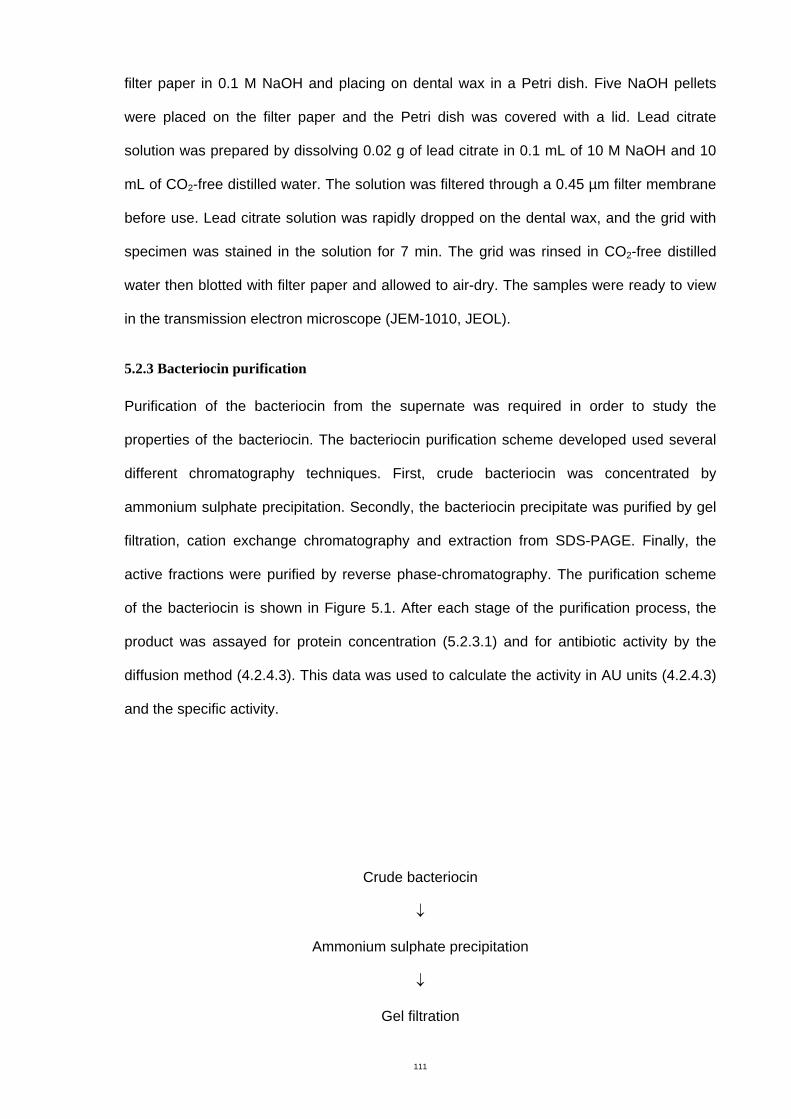
111
filter paper in 0.1 M NaOH and placing on dental wax in a Petri dish. Five NaOH pellets
were placed on the filter paper and the Petri dish was covered with a lid. Lead citrate
solution was prepared by dissolving 0.02 g of lead citrate in 0.1 mL of 10 M NaOH and 10
mL of CO2-free distilled water. The solution was filtered through a 0.45 µm filter membrane
before use. Lead citrate solution was rapidly dropped on the dental wax, and the grid with
specimen was stained in the solution for 7 min. The grid was rinsed in CO2-free distilled
water then blotted with filter paper and allowed to air-dry. The samples were ready to view
in the transmission electron microscope (JEM-1010, JEOL).
5.2.3 Bacteriocin purification
Purification of the bacteriocin from the supernate was required in order to study the
properties of the bacteriocin. The bacteriocin purification scheme developed used several
different chromatography techniques. First, crude bacteriocin was concentrated by
ammonium sulphate precipitation. Secondly, the bacteriocin precipitate was purified by gel
filtration, cation exchange chromatography and extraction from SDS-PAGE. Finally, the
active fractions were purified by reverse phase-chromatography. The purification scheme
of the bacteriocin is shown in Figure 5.1. After each stage of the purification process, the
product was assayed for protein concentration (5.2.3.1) and for antibiotic activity by the
diffusion method (4.2.4.3). This data was used to calculate the activity in AU units (4.2.4.3)
and the specific activity.
Crude bacteriocin
↓
Ammonium sulphate precipitation
↓
Gel filtration
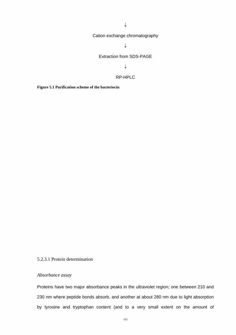
112
↓
Cation exchange chromatography
↓
Extraction from SDS-PAGE
↓
RP-HPLC
Figure 5.1 Purification scheme of the bacteriocin
5.2.3.1 Protein determination
Absorbance assay
Proteins have two major absorbance peaks in the ultraviolet region; one between 210 and
230 nm where peptide bonds absorb, and another at about 280 nm due to light absorption
by tyrosine and tryptophan content (and to a very small extent on the amount of
113
phenylalanine and disulfide bonds). Monitoring the absorbance at 214 nm is fast and
convenient for peptide determination and is commonly used for determining the protein
concentration of fractions from chromatography columns (Aitken and Learmounth, 2002,
The biotechnology project, 2007)
The spectrophotometer was switched on for about 15 min before adjusting the wavelength
at 214 nm. The buffer solution was placed in a quartz cuvette for zero calibration. After
cleaning the cuvette with distilled water, the first sample was placed in the cuvette and the
absorbance value was read. The cuvette was cleaned with distilled water before each new
sample was assayed (Bollag & Edelstein, 1991).
Bicinchoninic (BCA) assay (Bollag & Edelstein, 1991)
The BCA assay is a variation of the Lowry method. The reaction is simpler to perform and
has fewer interfering substances and is more sensitive than the Lowry method (Hansen,
2007). The BCA protein assay kit (Pierce, USA) consisted of BCA reagent A and B.
Working BCA reagent was prepared by mixing 50 parts of BCA reagent A and 1 part of
BCA reagent B. The mixture was stable in a closed container at room temperature for at
least one day.
A fresh set of protein standards, using bovine serum albumin (BSA), was prepared by
diluting 1mg/mL of BSA as shown in Table 5.1. Twenty-five microlitres of each BSA
standard or sample was transferred into the wells of a microtiter plate, then 200 µl of
prepared BCA reagent was added into each well and mixed appropriately. The plate was
incubated at 37°C for 30 min then at room temperature for 15 min. The absorbance of wells
containing standards and test samples were determined at 600 nm with a microplate
reader (Dynatech MR7000, Germany).
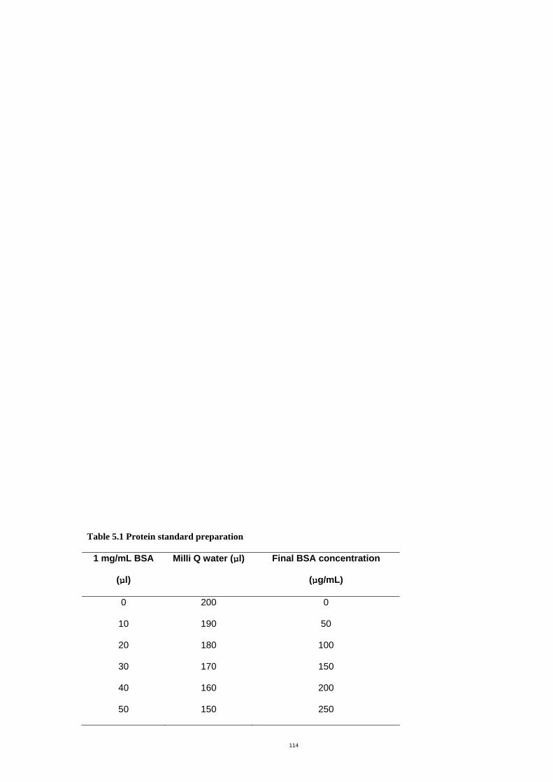
114
Table 5.1 Protein standard preparation
1 mg/mL BSA
(µl)
Milli Q water (µl) Final BSA concentration
(µg/mL)
0 200 0
10 190 50
20 180 100
30 170 150
40 160 200
50 150 250
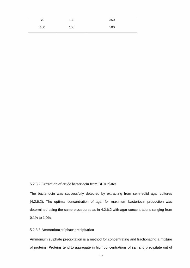
115
70 130 350
100 100 500
5.2.3.2 Extraction of crude bacteriocin from BHA plates
The bacteriocin was successfully detected by extracting from semi-solid agar cultures
(4.2.6.2). The optimal concentration of agar for maximum bacteriocin production was
determined using the same procedures as in 4.2.6.2 with agar concentrations ranging from
0.1% to 1.0%.
5.2.3.3 Ammonium sulphate precipitation
Ammonium sulphate precipitation is a method for concentrating and fractionating a mixture
of proteins. Proteins tend to aggregate in high concentrations of salt and precipitate out of
116
solution. Ammonium sulphate is the salt of choice because it is effective, highly soluble,
dissolves at low temperatures and at a range of pH values and is cheap (Bollag &
Edelstein, 1991). The crude bacteriocin was initially concentrated by ammonium sulphate
precipitation. The following procedures were performed at 4°C. Solid ammonium sulphate
was slowly added to the crude bacteriocin solution. A range of concentrations of
ammonium sulphate was added to the bacteriocin solution to optimize precipitation.
Ammonium sulphate reaches 40% (w/v), 50% (w/v), 60% (w/v), 70% (w/v), 80% (w/v) and
90% (w/v) saturation when 242, 314, 390, 472, 561 and 657 g of this salt is added to 1 L of
salt-free crude bacteriocin solution respectively (Bollag & Edelstein, 1991). The system was
held with constant stirring overnight. The precipitate was recovered by centrifugation at
10,000 × g for 30 min. The precipitate was then solubilized in 30 mL of 20 mM sodium
phosphate buffer (pH 5.5) and dialyzed in the same buffer using dialysis tubing with a
3,500 Dalton cut-off to eliminate ammonium sulphate residue. After dialyzing, the solution
was filtered through a 0.45 µm membrane. The product of this stage of purification process
was designated ammonium sulphate precipitate. The protein concentration and inhibitory
activity against S. uberis were determined as described in 4.2.4.3. Sodium phosphate
buffer was used as negative control. The precipitate was stored at -20°C until use.
5.2.3.4 Gel filtration chromatography
Gel filtration separates a mixture of proteins based on their differences in molecular size
(Wu, 2004). Gel beads have pores in them which allow smaller molecules to enter but
exclude molecules larger than the pore diameter. As a result, the smaller molecules are
retained in the column longer and pass through slower than the larger molecules.
Sephadex G-100 was mixed slowly and degassed to remove air bubbles prior to packing
into a 10 × 1 cm column (Pharmacia). The resin was washed with at least 100 mL of 20
mM sodium phosphate buffer, pH 5.5, at a flow rate of 0.5 mL/min. Three milliliters of the
bacteriocin precipitate was loaded onto the column followed by the same buffer and
fractions were collected (1 mL/tube). The column was washed again before each new run.
117
The peptide concentration of each fraction was assayed manually by determining the
absorbance at 214 nm (5.2.3.1), then the chromatogram was developed. Every fraction
was also analyzed for inhibitory activity against S. uberis by the agar diffusion assay
(4.2.4.3). The fractions with inhibitory activity were then further purified by cation
chromatography. Sodium phosphate buffer was used as a negative control.
5.2.3.5 Cation exchange chromatography
The stationary phase in ion exchange chromatography is made of porous polymer to which
anionic or cationic exchange groups have been attached. The retention and separation of
solutes are performed according to the degree of ionization of the solute and its affinity to
the ionic sites on stationary phase. The eluent is usually an aqueous buffer and the
retention controlled by changes in ionic strength (Hansen, et al., 1984).
The SP Sepharose Fast Flow (strong cation exchange) media was mixed and packed in a
10 × 1 cm column (Pharmacia). There were five steps for ion exchange chromatography;
equilibration, sample loading, washing unbound material, gradient elution and regeneration
of the column.
One hundred milliliters of 20 mM sodium phosphate buffer at the desired pH was used to
equilibrate the column at a flow rate of 6 mL/min. The sample and a starting buffer were
maintained at the same pH. An aliquot of 3 mL of sample or buffer was applied to the
column at a flow rate of 0.5 mL/min. The column was washed with 20 mL of buffer to
remove unbound material. Gradient mixtures consisting of equal volumes of buffer and 1
M NaCl in a gradient maker (Bio-Rad) were used for elution. At the end of elution, 50 mL of
1 M NaCl was applied to the column to elute any remaining bound material. One milliliter of
each fraction was collected and the protein content was determined manually by
absorbance at 214 nm (5.2.3.1) then the chromatogram was observed. Every fraction was
also analyzed for inhibitory activity against S. uberis 796-2 by the agar diffusion assay
118
(4.2.4.3). A new cycle of cation exchange chromatography was started by repeating the
equilibration step. Buffer alone and buffer with NaCl were used as negative controls.
5.2.3.6 Protein gel electrophoresis, bioassay and purification
Gel electrophoresis was not only a tool to determine the size and assess the purity of the
protein, but it was also used for purification of the protein of interest from the gel.
Polyacrylamide gel electrophoresis under denaturing conditions (Laemmli, 1970)
Sodium dodecyl sulphate polyacrylamide gel electrophoresis (SDS-PAGE) separates
proteins based on their differences in molecular weight. SDS binds along the polypeptide
chain, and the length of the reduced unfolded SDS-protein complex is proportional to its
molecular weight. The composition of the stacking and resolving gels are given in Table
5.2. The gel sandwich of the Mini-PROTEAN® II System (Bio-Rad) with gel plates was
assembled (Figure 5.2). The resolving gel solution was pipetted to the gel sandwich until it
reached 1.5 cm from the top of the front plate. Distilled water was gently layered on top of
the resolving gel solution to keep the gel surface flat. After polymerization of the gel for 1 h,
the water was decanted and the stacking gel solution was added until the solution again
reached the top of the front plate. Then, the comb was carefully inserted into the gel
sandwich. After the gel was allowed to polymerize for 30 min, the comb was removed.
Analysis by SDS-PAGE was performed using a discontinuous buffer system.
Electrophoresis was conducted at 150 V for 1 h under denaturing or reducing conditions.
The bacteriocin sample was prepared for loading by mixing four volumes of protein sample
and one volume of 5 X sample buffer in a microcentrifuge tube and heating at 100 °C for 2
min. The sample buffer (5 X) consisted of 0.6 mL of 1 M Tris-HCl, pH 6.8, 5 mL of 50%
glycerol, 2 mL of 10% SDS, 0.5 mL of 2-mercaptoethanol, 1 mL of 1% bromophenol blue
and 0.9 mL of distilled water. Samples were loaded into wells with a syringe, ensuring all
lanes were loaded, to keep samples running in straight lines. The electrophoresis buffer
consisted of 0.3% (w/v) Tris, 1.44% (w/v) glycine and 0.1% (w/v) SDS.
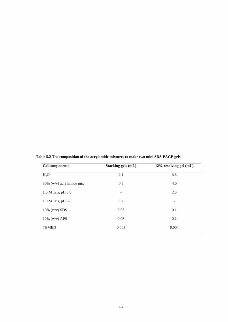
119
Table 5.2 The composition of the acrylamide mixtures to make two mini SDS-PAGE gels
Gel components Stacking gels (mL) 12% resolving gel (mL)
H2O 2.1 3.3
30% (w/v) acrylamide mix 0.5 4.0
1.5 M Tris, pH 8.8 - 2.5
1.0 M Tris, pH 6.8 0.38 -
10% (w/v) SDS 0.03 0.1
10% (w/v) APS 0.03 0.1
TEMED 0.003 0.004
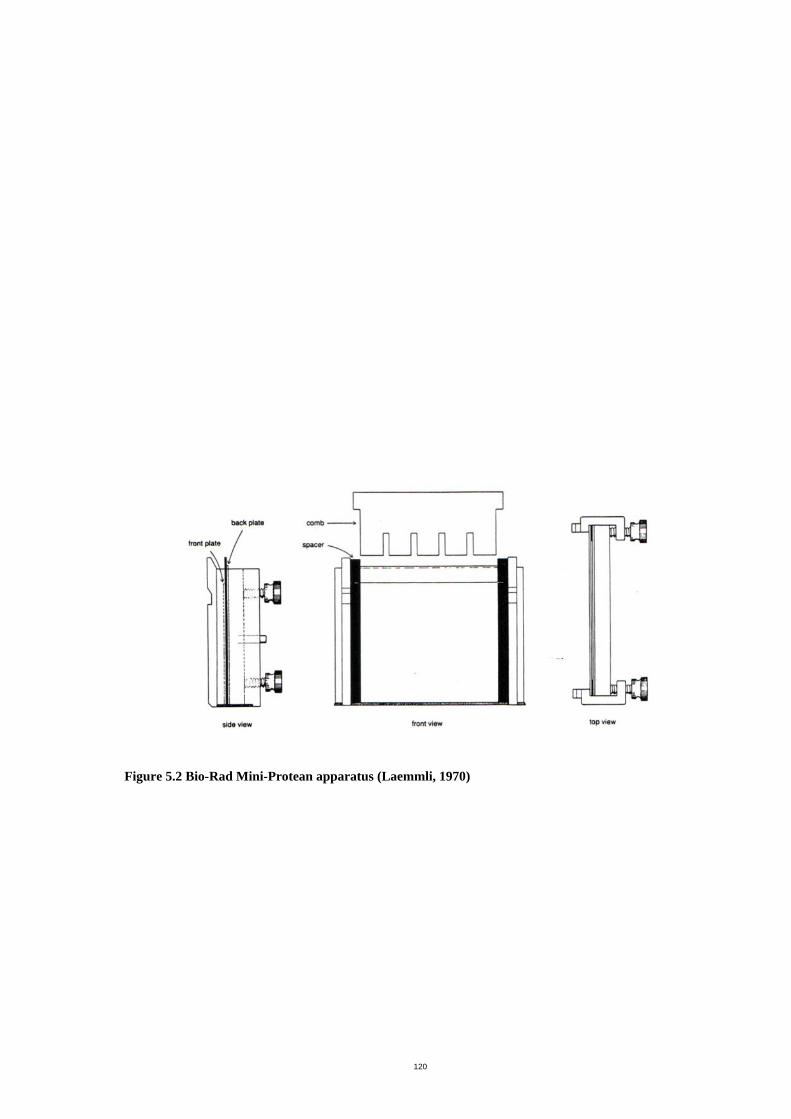
120
Figure 5.2 Bio-Rad Mini-Protean apparatus (Laemmli, 1970)
121
Coomassie blue R-250 staining (Laemmli, 1970)
The gel was stained on a slow shaker for 5-10 min in a solution of 0.1% Coomassie blue R-
250, 45% (w/v) methanol and 10% (w/v) glacial acetic acid. The stain was decanted and the
gel was rinsed with two changes of water. The Coomassie blue destaining solution,
consisting of 10% (w/v) methanol and 10% (w/v) glacial acetic acid, was added to the gel
which was destained for 1 h. To destain completely, the solution was changed and the
destaining was continued overnight.
Assay of inhibitory activity from electrophoresis gel (Crupper, et al., 1997)
The protein marker (Mark 12, Invitrogen) and duplicate samples were loaded into wells of a
native gel. After running the gel, the second sample lane was excised using a clean razor
blade and the remainder was stained. The unstained section of the gel was assayed for
inhibitory activity. It was first soaked in a solution consisting of 20% (v/v) isopropanol and
10% (v/v) acetic acid for 2 h to eliminate SDS, then the gel was rinsed in distilled water for
4 h. The washed gel was placed on a BHA plate which was overlaid with BHI soft agar
(0.7% agar) containing an indicator strain of S. uberis 796-2 at a concentration of 106
CFU/mL. The plate was incubated at 37°C overnight, examined for zones of inhibition and
aligned with the stained gel to determine the approximate molecular weight of the inhibitory
activity.
Protein purification from polyacrylamide gel (Retamal, et al., 1999)
Protein purification by SDS-PAGE gel is used to resolve individual components of complex
mixtures. The protein of interest was extracted from the gel as follows: after the bands of
interest on the unstained gel were localized by alignment with the stained gel, they were
cut with a clean razor blade and transferred to a sterile tube. The gel slices were then
washed three times for 5 min with a solution consisting of 250 mM EDTA in 250 mM Tris
(pH 7.4) then followed by three rinses of 5 min with distilled water. After removing the
water, the elution buffer, which consisted of 50 mM Tris-HCl, 150 mM NaCl, 0.1 mM EDTA
122
and 0.1% (w/v) SDS (pH 7.5), was added. The sample was sonicated (6 passes of 30 s) in
an ice bath then incubated in a rotary shaker at 30°C overnight. After centrifugation at
10,000 g for 10 min, the supernatant fluid containing the bacteriocin was carefully removed
and stored at -20°C until use. This bacteriocin preparation was dialyzed with PBS and
further purified by RP-HPLC.
5.2.3.7 Reversed phase-high performance liquid chromatography (RP-HPLC) (Lough &
Wainer, 1995)
Reversed phase chromatography separates proteins and peptides with differing
hydrophobicity based on their reversible interaction with the hydrophobic surface of a
chromatographic medium. The stationary phase is non-polar (hydrophobic) and the mobile
phase is polar. After loading, non-polar components are more attracted to the non-polar
stationary phase, while the polar compounds tend to remain in the polar mobile phase and
move faster. The length of carbon chains in the stationary phase of the column are
expressed as the number of carbon atom such as C4, C8 and C18
The samples with inhibitory activity were further purified by RP-HPLC on a C4 column
(Brownlee Labs, USA., 7 µM, 30 x 4.6 mm., Butyl C4 300) using a gradient of 0 to 60%
(v/v) acetronitrile. Buffer A was composed of 0.1% (v/v) TFA and buffer B was composed of
60% (v/v) acetronitrile in 0.1% (v/v) TFA. These mobile phase solutions were filtered
through a 0.45 µm membrane and degassed before use. Each run consisted of 3 min of
buffer A, a 30 min of linear gradient of 0-100% buffer B and 3 min of 100% buffer B. The
detector was set at 214 nm. One ml of each fraction was collected, lyophilised (Dynavac
FD12, Australia) and resuspended in 20 µl of PBS. The total activity of each fraction was
measured by the agar diffusion assay (4.2.4.3) and protein content was determined by the
BCA assay (5.2.3.1). Buffer A and buffer B were used as negative controls.
RP-HPLC fractions were cleaned with a C18 Zip tip (Millipore) to minimize salt
contamination before processing by mass spectrometry. The Zip tip (C18) was pre-wet with
100% methanol and equilibrated by 10 aspirations in wash solution, which contained 5%
123
(v/v) methanol in 0.1% (v/v) formic acid. The peptides were absorbed to the Zip tip matrix
by aspirating 10 µl aliquots through the Zip tip and transferring to another eppendorf tube.
The aspiration step was repeated so that each aliquot was aspirated through the Zip tip
twice. The Zip tip was washed with two aliquots of wash solution to remove unbound
peptides. The peptides were eluted by aspirating through the Zip tip 10 times with 10 µl of
elution buffer (80% (v/v) methanol in 0.1% (v/v) formic acid). The eluted peptides were
transferred to an Eppendorf tube.
5.2.3.8 Mass Spectrometry
An analysis of purified bacteriocins by matrix assisted laser desorption ionization-time-of-
flight mass spectrometry (MALDI-TOF-MS) was performed by Frank Antolasic at Applied
Chemistry, RMIT university. The results were analyzed by Bruker Data Analysis Software
for TOF 1.6 g.
124
5.3 Results
5.3.1 Bacteriocin characterization
5.3.1.1 Spectrum of inhibitory activity
Both the producer culture (Macrococcus caseolyticus 2042-4) and the ammonium sulphate
precipitate (80 AU/mL) showed the same inhibitory profiles (Appendix B, Table 4). An
inhibitory effect was detected against all species of streptococci tested (n = 12) including all
isolates of S. uberis, and there was partial inhibition against Enterococcus faecium. No
inhibitory activity was detected against other gram-positive bacterial strains. None of the
gram-negative and spiral bacteria examined were inhibited by producer cells or the
ammonium sulphate precipitate. The producer strain was resistant to its own bacteriocin.
5.3.1.2 Bacteriocin thermostability
The ammonium sulphate precipitate (160 AU/mL) showed 100% residual activity after
incubation for 60 min at 37°C and 60°C and 15 min at 80°C (Figure 5.3). The activity
gradually decreased when longer incubation periods were used. Some inhibitory activity
still remained after one day of exposure at 37°C. The bacteriocin activity decreased by 50%
when incubated at 100°C for 15 min. The bacteriocin was inactive after autoclaving at
120oC for 20 min.
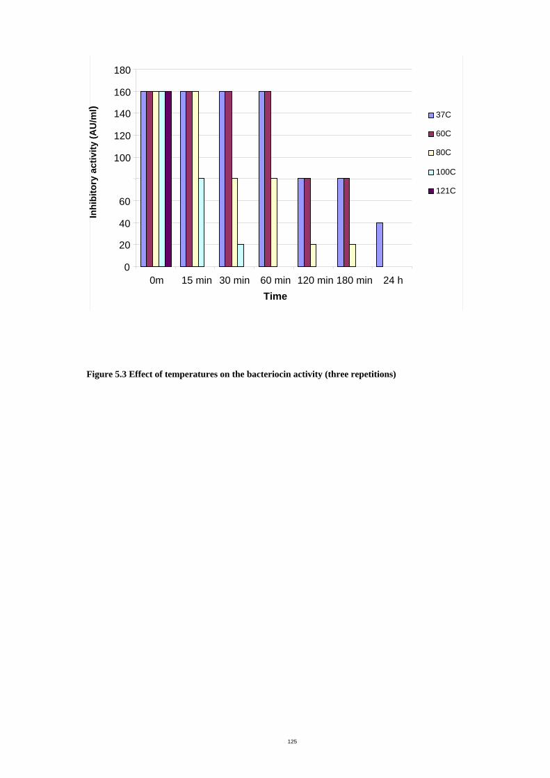
125
Figure 5.3 Effect of temperatures on the bacteriocin activity (three repetitions)
0
20
40
60
100
120
140
160
180
0m 15 min 30 min 60 min 120 min 180 min 24 h Time
37C
60C
80C
100C
121C
Inhi
bito
ry a
ctiv
ity (A
U/m
l)
126
5.3.1.3 Bacteriocin pH stability
The ammonium sulphate precipitate (320 AU/mL) showed great stability over a wide pH
range when incubated at 37°C for 1h (Figure 5.4). The bacteriocin had optimum activity at
relatively acidic and neutral pH and the activity decreased after incubation with strong acid
(pH 2) and in basic conditions (pH 8 and 9). No activity was detected at very strong basic
pH values of 10, 11 and 12.
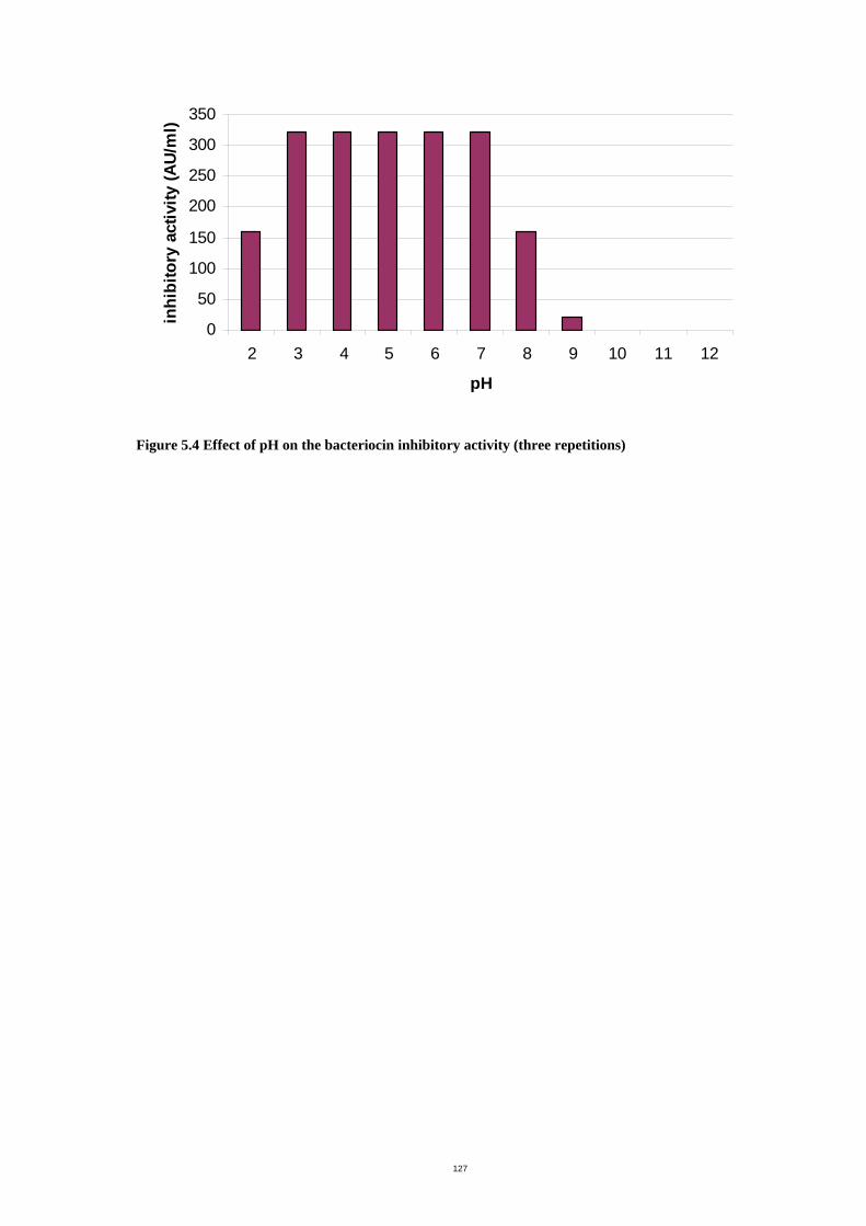
127
0
50
100
150
200
250
300
350
2 3 4 5 6 7 8 9 10 11 12
pH
inhi
bito
ry a
ctiv
ity (A
U/m
l)
Figure 5.4 Effect of pH on the bacteriocin inhibitory activity (three repetitions)
128
5.3.1.4 Kill curves
Addition of 80 AU/mL ammonium sulphate precipitate to S. uberis 796-2 resulted in a
dramatic decrease in the number of viable cells (Figure 5.5). The viability was reduced 4
log in 1 h and approximately 6 log when incubated further for 6 to 7 h. After 7 and 24 h of
treatment, only 1,000 and 100 CFU/mL of cells survived. Prolonged incubation beyond 24 h
did not result in any further killing (data not shown). S. uberis 796-2 without bacteriocin
showed a normal growth pattern.
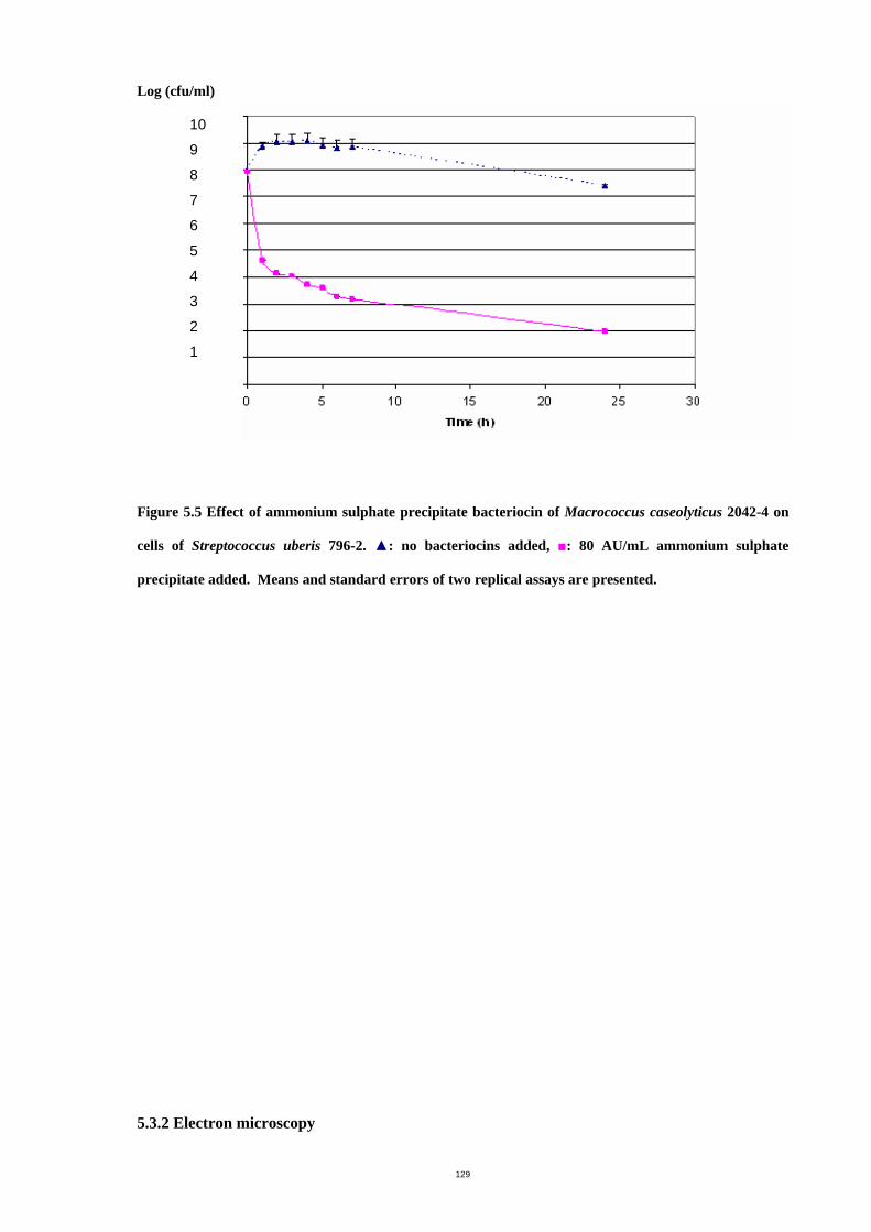
129
Log (cfu/ml)
10
9
8
7
6
5
4
3
2
1
Figure 5.5 Effect of ammonium sulphate precipitate bacteriocin of Macrococcus caseolyticus 2042-4 on
cells of Streptococcus uberis 796-2. ▲: no bacteriocins added, ■: 80 AU/mL ammonium sulphate
precipitate added. Means and standard errors of two replical assays are presented.
5.3.2 Electron microscopy
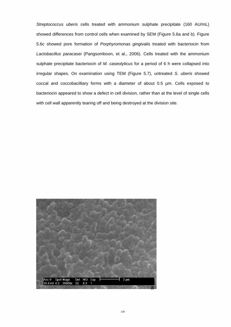
130
Streptococcus uberis cells treated with ammonium sulphate precipitate (160 AU/mL)
showed differences from control cells when examined by SEM (Figure 5.6a and b). Figure
5.6c showed pore formation of Porphyromonas gingivalis treated with bacteriocin from
Lactobacillus paracasei (Pangsomboon, et al., 2006). Cells treated with the ammonium
sulphate precipitate bacteriocin of M. caseolyticus for a period of 6 h were collapsed into
irregular shapes. On examination using TEM (Figure 5.7), untreated S. uberis showed
coccal and coccobacilliary forms with a diameter of about 0.5 µm. Cells exposed to
bacteriocin appeared to show a defect in cell division, rather than at the level of single cells
with cell wall apparently tearing off and being destroyed at the division site.
a
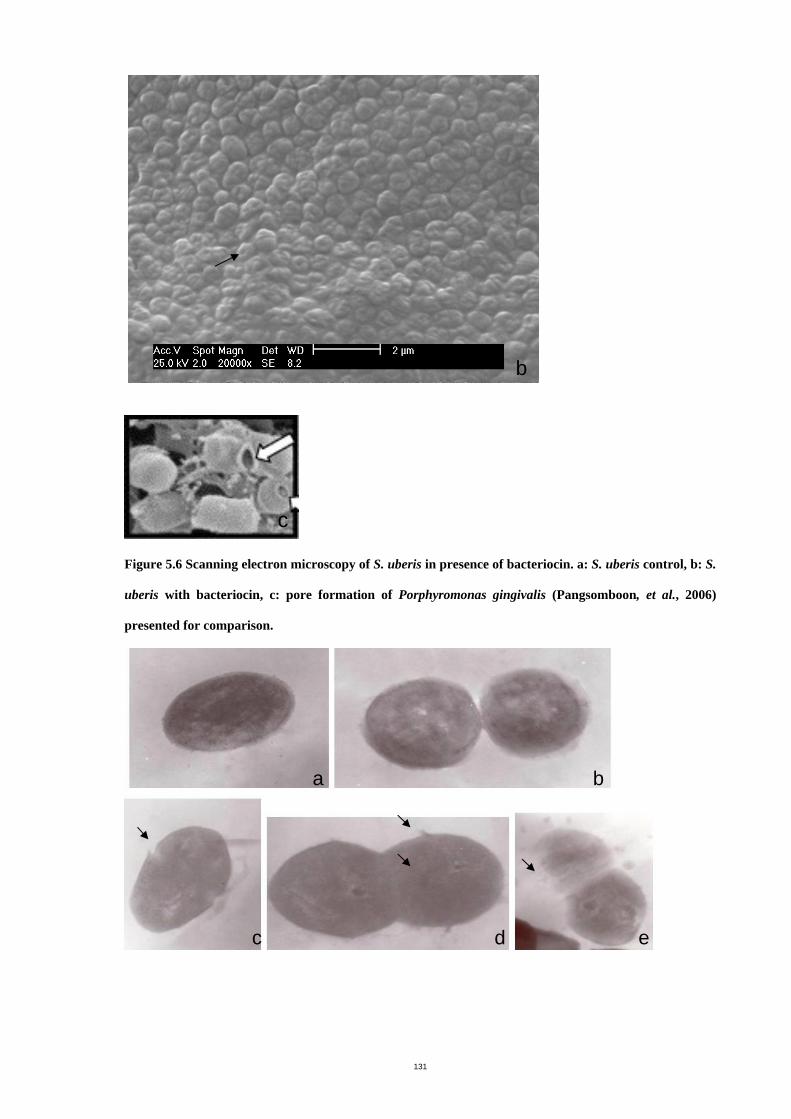
131
Figure 5.6 Scanning electron microscopy of S. uberis in presence of bacteriocin. a: S. uberis control, b: S.
uberis with bacteriocin, c: pore formation of Porphyromonas gingivalis (Pangsomboon, et al., 2006)
presented for comparison.
b
c
a b
c d e
132
Figure 5.7 Transmission electron microsopy of S. uberis treated with bacteriocin (8000X). a, b:
untreated cell, c, d and e: bacteriocin treated cell. Arrows showed cells defect.
5.3.3 Bacteriocin purification
Some antibiotic activity was detected in BHI containing 0.5% of agar (Table 5.3). BHI
containing 0.7% agar resulted in the maximum levels of bacteriocin production and the
activity level remained stable with further increases in agar concentration. Increasing the
incubation time from overnight to 24 or 48 h or increasing the number of thawing cycles up
to 5 times did not increase or reduce bacteriocin recovery. One liter of 0.7% agar in BHI
provided 600 mL of extracted liquid containing bacteriocin (Table 5.5), which was
designated as crude bacteriocin. The activity of the crude bacteriocin was 10 AU/mL and
yield was arbitrarily defined as 100%.
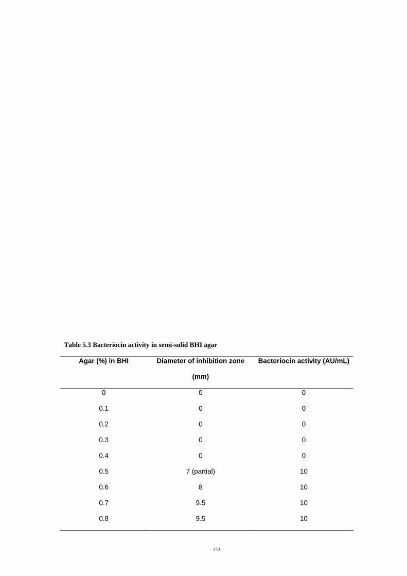
133
Table 5.3 Bacteriocin activity in semi-solid BHI agar
Agar (%) in BHI Diameter of inhibition zone
(mm)
Bacteriocin activity (AU/mL)
0 0 0
0.1 0 0
0.2 0 0
0.3 0 0
0.4 0 0
0.5 7 (partial) 10
0.6 8 10
0.7 9.5 10
0.8 9.5 10
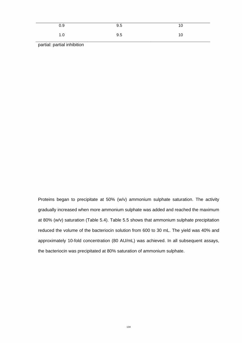
134
0.9 9.5 10
1.0 9.5 10
partial: partial inhibition
Proteins began to precipitate at 50% (w/v) ammonium sulphate saturation. The activity
gradually increased when more ammonium sulphate was added and reached the maximum
at 80% (w/v) saturation (Table 5.4). Table 5.5 shows that ammonium sulphate precipitation
reduced the volume of the bacteriocin solution from 600 to 30 mL. The yield was 40% and
approximately 10-fold concentration (80 AU/mL) was achieved. In all subsequent assays,
the bacteriocin was precipitated at 80% saturation of ammonium sulphate.
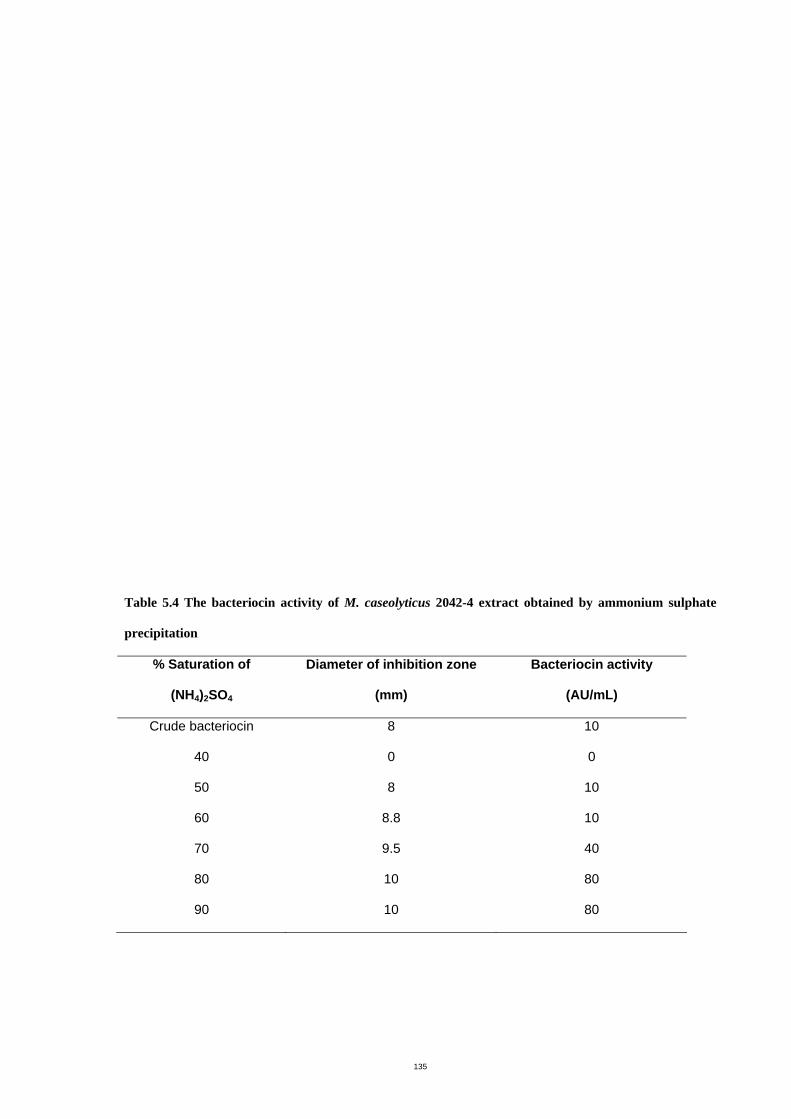
135
Table 5.4 The bacteriocin activity of M. caseolyticus 2042-4 extract obtained by ammonium sulphate
precipitation
% Saturation of
(NH4)2SO4
Diameter of inhibition zone
(mm)
Bacteriocin activity
(AU/mL)
Crude bacteriocin 8 10
40 0 0
50 8 10
60 8.8 10
70 9.5 40
80 10 80
90 10 80
136
Following ammonium sulphate precipitation and gel filtration chromatography with
Sephadex G100, the elution profile showed several fractions (Figure 5.8). Examination of
each fraction for biological activity by the diffusion assay demonstrated three fractions with
activity. Comparison of stained and unstained gels overlayed with S. uberis 796-2 showed
that the molecular mass of the active fraction was less than 6,000 Da (Figure 5.9). Both
denaturing and non-denaturing conditions of the PAGE gel demonstrated inhibition activity.
Examination of PAGE gel prepared from samples at different stages of purification showed
that significant amounts of contaminating proteins of similar molecular weight to the
bacteriocin were removed by the gel filtration step (Figure 5.9). The yield of bacteriocin
following gel filtration was 33% and there was a 38-fold increase in concentration of the
bacteriocin (Table 5.5).
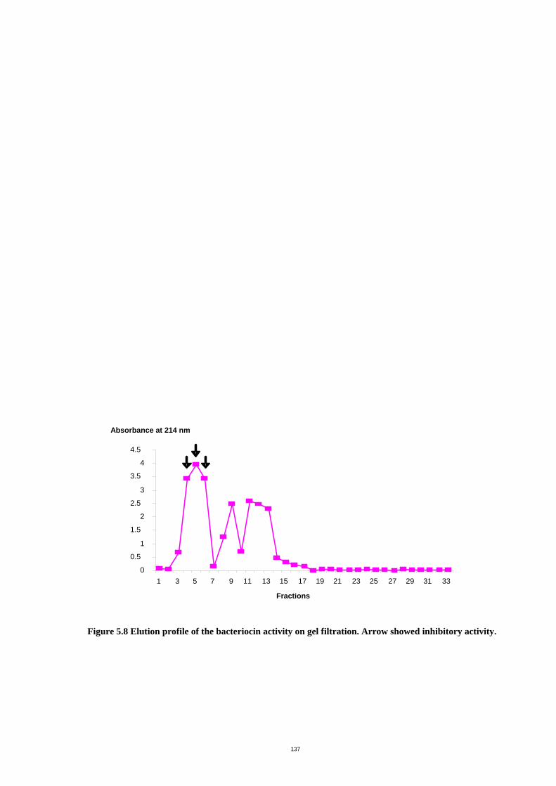
137
Figure 5.8 Elution profile of the bacteriocin activity on gel filtration. Arrow showed inhibitory activity.
0 0.5
1 1.5
2 2.5
3 3.5
4 4.5
1 3 5 7 9 11 13 15 17 19 21 23 25 27 29 31 33 Fractions
Absorbance at 214 nm
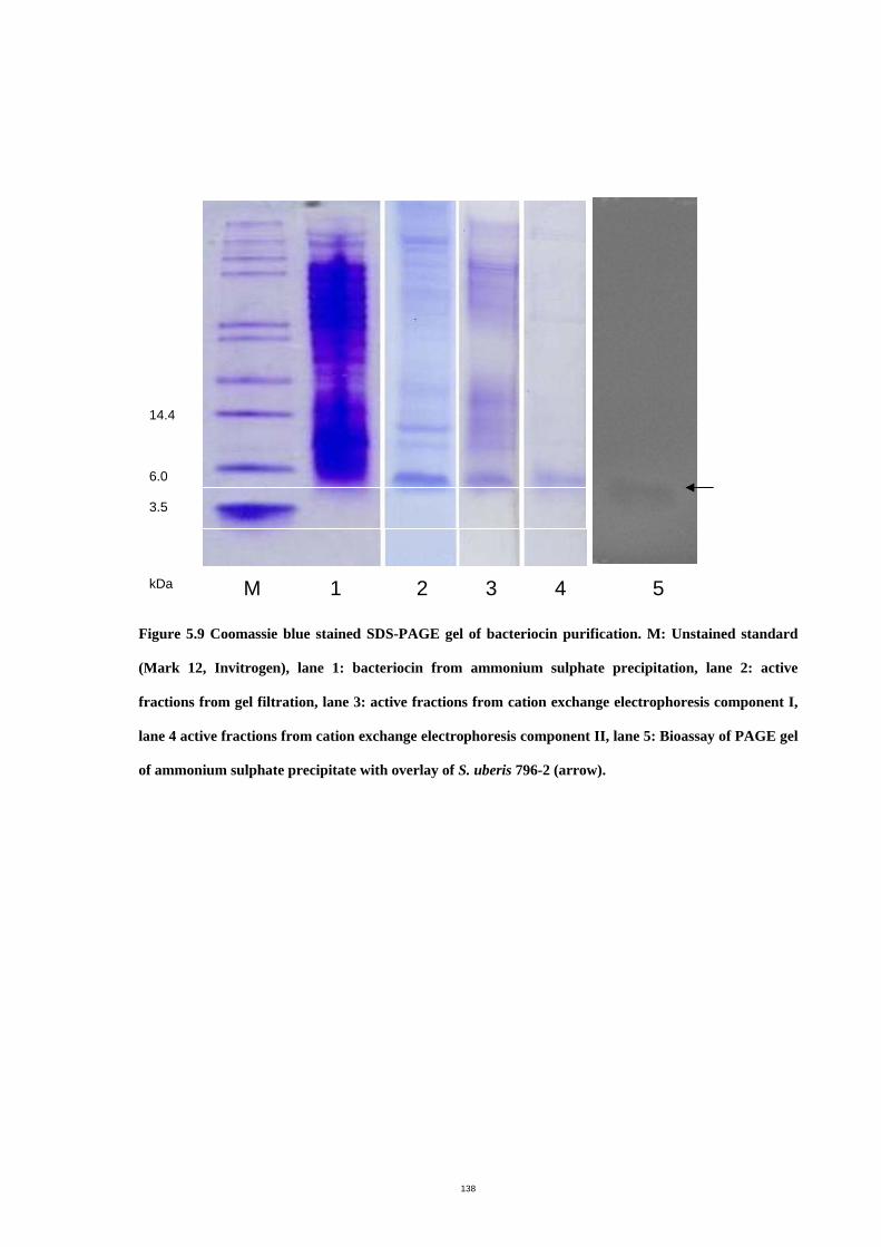
138
Figure 5.9 Coomassie blue stained SDS-PAGE gel of bacteriocin purification. M: Unstained standard
(Mark 12, Invitrogen), lane 1: bacteriocin from ammonium sulphate precipitation, lane 2: active
fractions from gel filtration, lane 3: active fractions from cation exchange electrophoresis component I,
lane 4 active fractions from cation exchange electrophoresis component II, lane 5: Bioassay of PAGE gel
of ammonium sulphate precipitate with overlay of S. uberis 796-2 (arrow).
14.4 6.0 3.5 kDa M 1 2 3 4 5
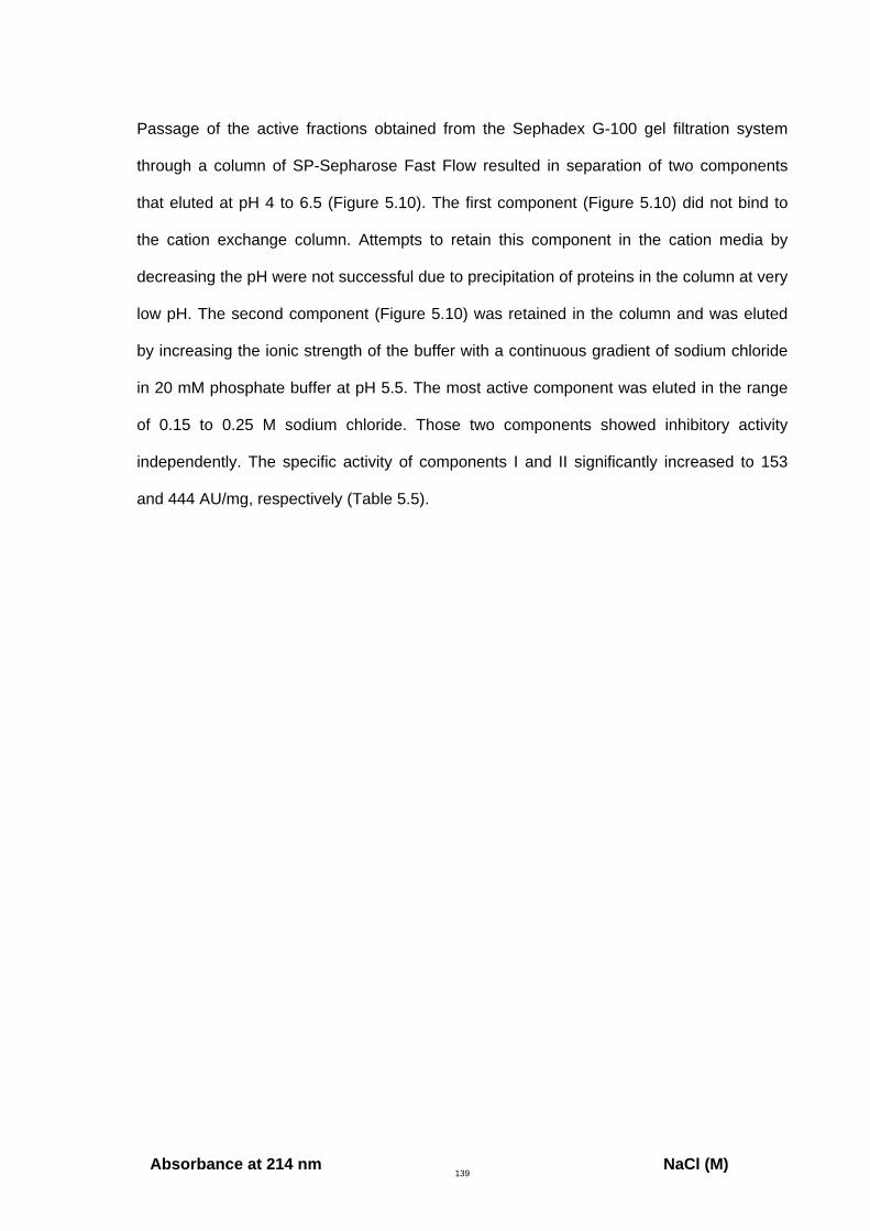
139
Passage of the active fractions obtained from the Sephadex G-100 gel filtration system
through a column of SP-Sepharose Fast Flow resulted in separation of two components
that eluted at pH 4 to 6.5 (Figure 5.10). The first component (Figure 5.10) did not bind to
the cation exchange column. Attempts to retain this component in the cation media by
decreasing the pH were not successful due to precipitation of proteins in the column at very
low pH. The second component (Figure 5.10) was retained in the column and was eluted
by increasing the ionic strength of the buffer with a continuous gradient of sodium chloride
in 20 mM phosphate buffer at pH 5.5. The most active component was eluted in the range
of 0.15 to 0.25 M sodium chloride. Those two components showed inhibitory activity
independently. The specific activity of components I and II significantly increased to 153
and 444 AU/mg, respectively (Table 5.5).
Absorbance at 214 nm NaCl (M)
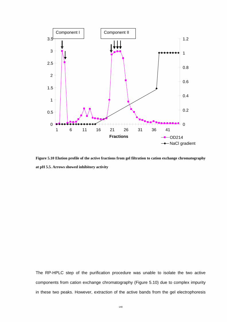
140
Figure 5.10 Elution profile of the active fractions from gel filtration to cation exchange chromatography
at pH 5.5. Arrows showed inhibitory activity
The RP-HPLC step of the purification procedure was unable to isolate the two active
components from cation exchange chromatography (Figure 5.10) due to complex impurity
in these two peaks. However, extraction of the active bands from the gel electrophoresis
0
0.5
1
1.5
2
2.5
3
3.5
1 6 11 16 21 26 31 36 41 Fractions
0
0.2
0.4
0.6
0.8
1
1.2
OD214 NaCl gradient
Component I Component II
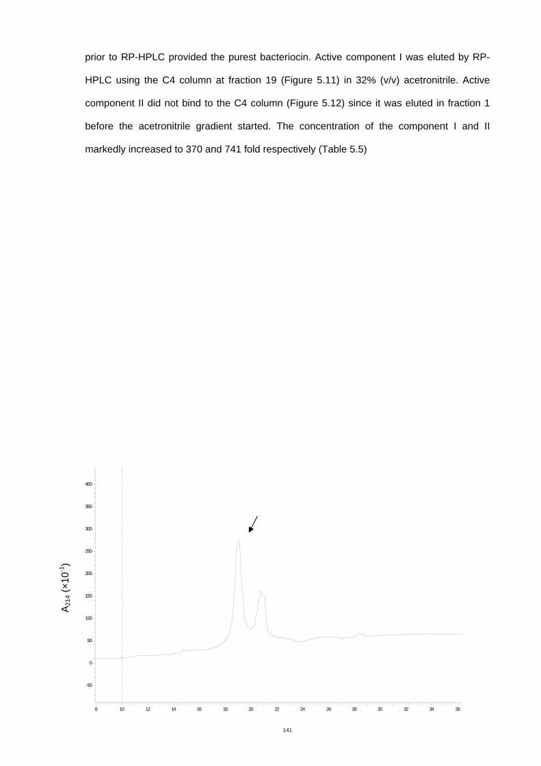
141
prior to RP-HPLC provided the purest bacteriocin. Active component I was eluted by RP-
HPLC using the C4 column at fraction 19 (Figure 5.11) in 32% (v/v) acetronitrile. Active
component II did not bind to the C4 column (Figure 5.12) since it was eluted in fraction 1
before the acetronitrile gradient started. The concentration of the component I and II
markedly increased to 370 and 741 fold respectively (Table 5.5)
-50
0
50
100
150
200
250
300
350
400
8 10 12 14 16 18 20 22 24 26 28 30 32 34 36
A21
4 (×1
0-1)
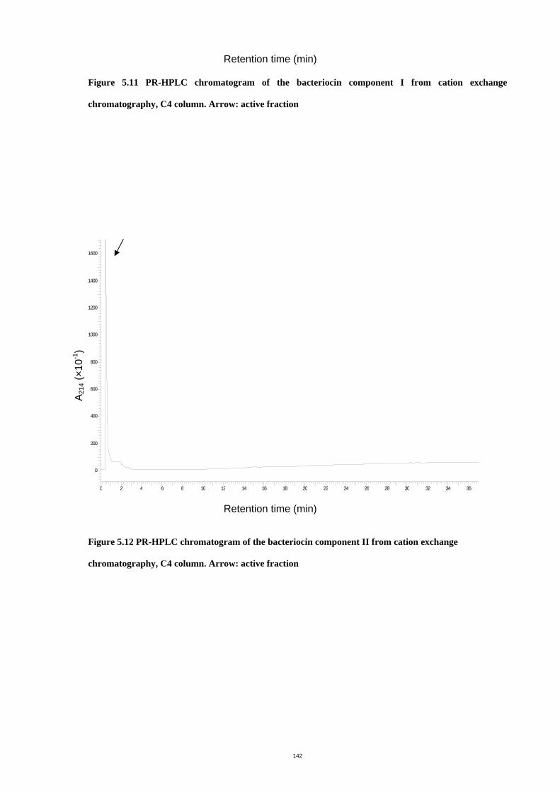
142
Retention time (min) Figure 5.11 PR-HPLC chromatogram of the bacteriocin component I from cation exchange
chromatography, C4 column. Arrow: active fraction
0
200
400
600
800
1000
1200
1400
1600
0 2 4 6 8 10 12 14 16 18 20 22 24 26 28 30 32 34 36
Retention time (min)
Figure 5.12 PR-HPLC chromatogram of the bacteriocin component II from cation exchange
chromatography, C4 column. Arrow: active fraction
A21
4 (×1
0-1)
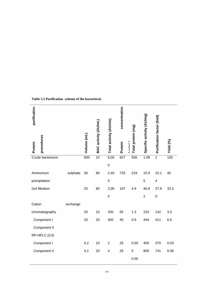
143
Table 5.5 Purification scheme of the bacteriocin
Prot
ein
purif
icat
ion
proc
edur
es
Volu
me
(mL)
BA
C a
ctiv
ity (A
U/m
L)
Tota
l act
ivity
(AU
nits
)
Prot
ein
conc
entr
atio
n
(µg/
mL)
Tota
l pro
tein
(mg)
Spec
ific
activ
ity (A
U/m
g)
Purif
icat
ion
fact
or (f
old)
Yiel
d (%
)
Crude bacteriocin 600 10 6,00
0
927 556 1.08 1 100
Ammonium sulphate
precipitation
30 80 2,40
0
733 219 10.9
5
10.1
4
40
Gel filtration 25 80 2,00
0
197 4.9 40.8
2
37.8
0
33.3
Cation exchange
chromatography
Component I
Component II
20
20
10
20
200
400
65
45
1.3
0.9
153
444
142
411
3.3
6.6
RP-HPLC (C4)
Component I
Component II
0.2
0.2
10
20
2
4
25
25
0.00
5
0.00
400
800
370
741
0.03
0.06
144
5
5.3.4 Mass spectrometry
The purified bacteriocin component I was analyzed by MALDI-TOF-MS (Figure5.13).
MALDI could not identify an individual peptide and there was no regular pattern of mass.
The spectrum showed mixtures of molecules ranging between 1640 to 3169 m/z. Analysis
of the purified bacteriocin component II showed three sets of peptide that had regular
patterns (Figure 5.14). The first set consisted of three peaks (1852, 1875 and 1897). The
second set consisted of four peaks (1940, 1962, 1983 and 2006). The third set consisted of
four peaks (2039, 2060, 2082 and 2106). After desalting purified bacteriocin component II
with the Zip tip (C18), no peptide peak was observed by MALDI-TOF-MS.
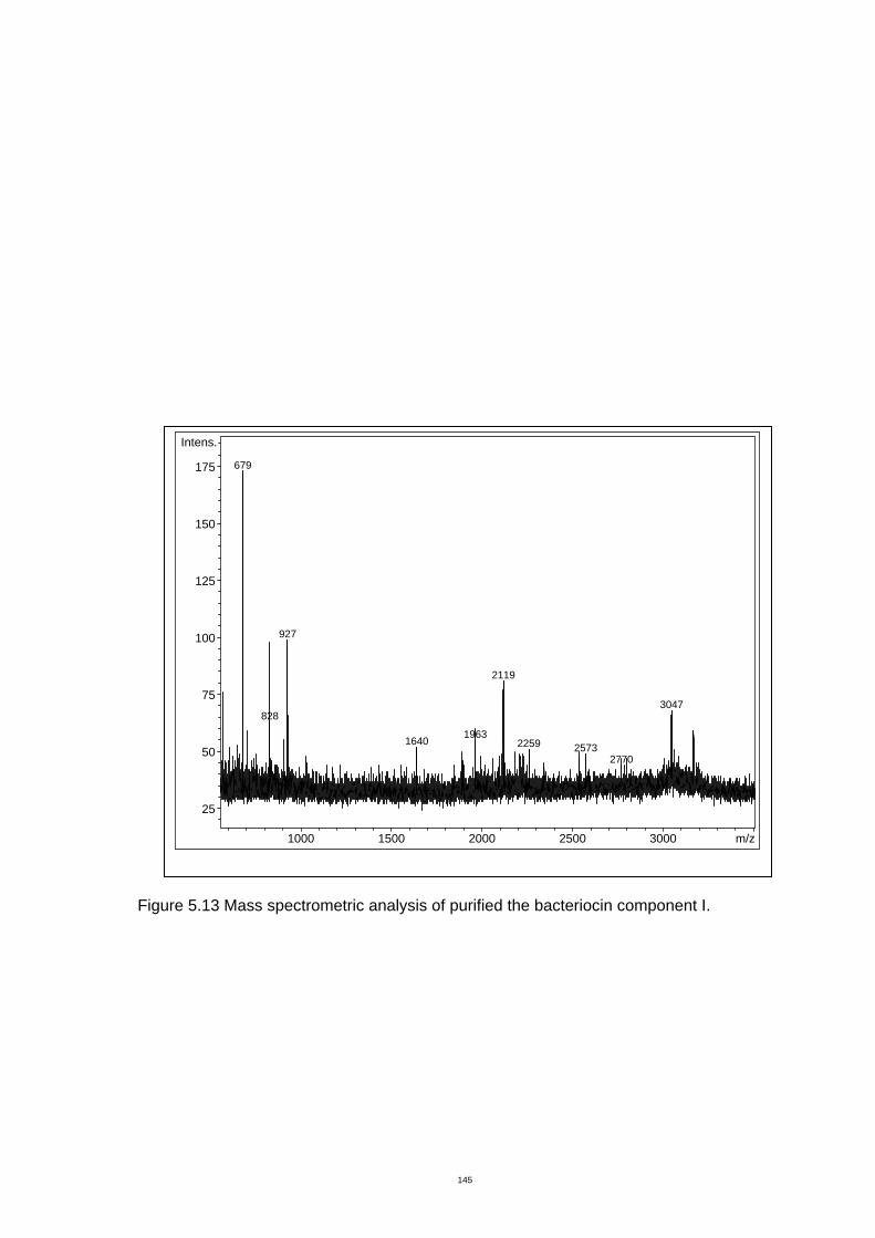
145
Figure 5.13 Mass spectrometric analysis of purified the bacteriocin component I.
1000 1500 2000 2500 3000 m/z
25
50
75
100
125
150
175
Intens.
679
927
828
2119
19631640
3047
2259 25732770
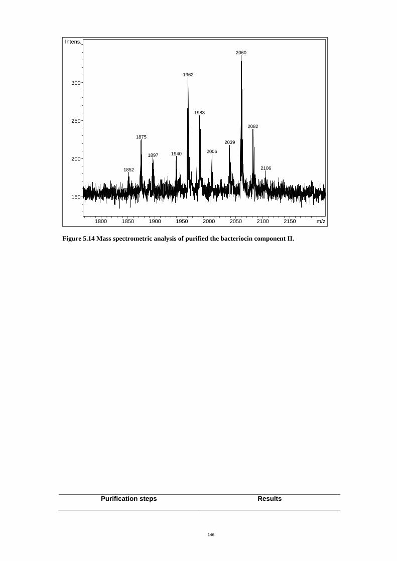
146
1800 1850 1900 1950 2000 2050 2100 2150 m/z
150
200
250
300
Intens.
2060
1962
1875
1852
19401897
1983
2039
2006
2082
2106
Figure 5.14 Mass spectrometric analysis of purified the bacteriocin component II.
Purification steps Results
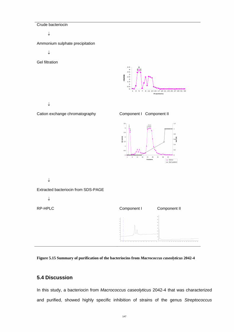
147
Crude bacteriocin
↓
Ammonium sulphate precipitation
↓
Gel filtration
0
0.5
1
1.5
2
2.5
3
3.5
4
4.5
1 3 5 7 9 11 13 15 17 19 21 23 25 27 29 31 33
Fractions
OD(A
214)
↓
Cation exchange chromatography
Component I Component II
0
0.5
1
1.5
2
2.5
3
3.5
1 6 11 16 21 26 31 36 41
Fractions
OD
(A21
4)
0
0.2
0.4
0.6
0.8
1
1.2
NaC
l (M
)
OD214NaCl gradient
↓
Extracted bacteriocin from SDS-PAGE
↓
RP-HPLC
Component I Component II
-50
0
50
100
150
200
250
300
350
400
8 10 12 14 16 18 20 22 24 26 28 30 32 34 36
0
200
400
600
800
1000
1200
1400
1600
0 2 4 6 8 10 12 14 16 18 20 22 24 26 28 30 32 34 36
Figure 5.15 Summary of purification of the bacteriocins from Macrococcus caseolyticus 2042-4
5.4 Discussion
In this study, a bacteriocin from Macrococcus caseolyticus 2042-4 that was characterized
and purified, showed highly specific inhibition of strains of the genus Streptococcus
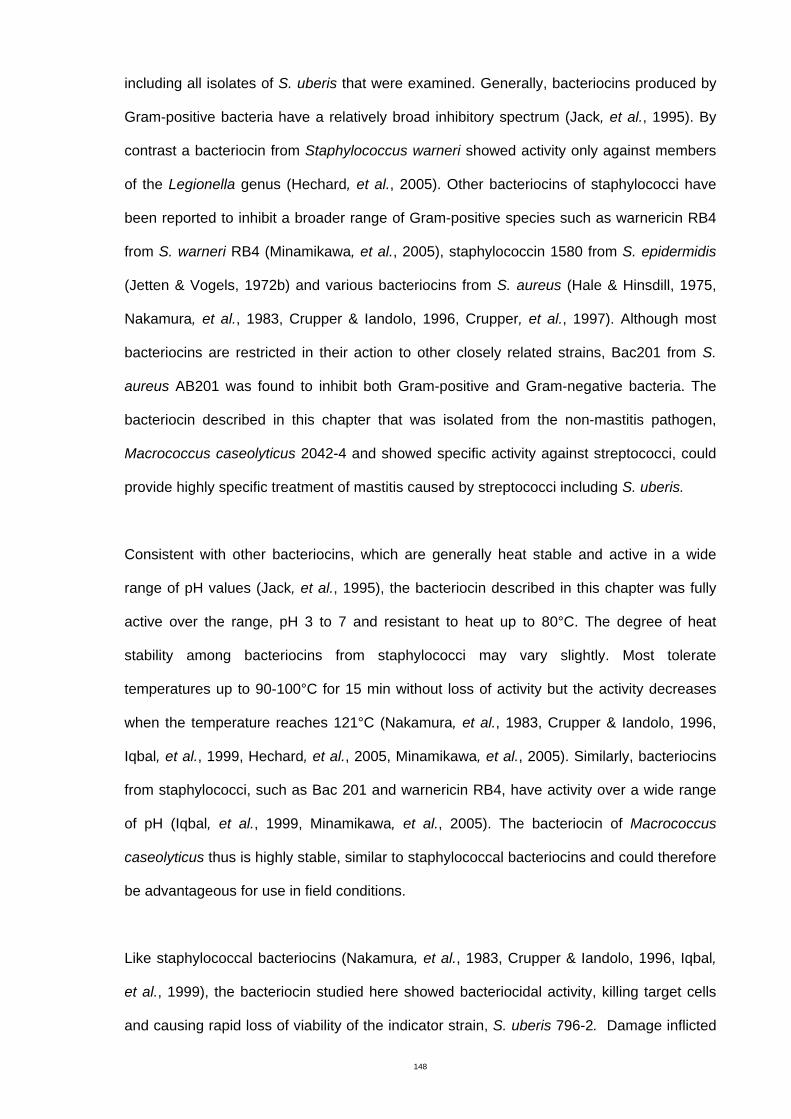
148
including all isolates of S. uberis that were examined. Generally, bacteriocins produced by
Gram-positive bacteria have a relatively broad inhibitory spectrum (Jack, et al., 1995). By
contrast a bacteriocin from Staphylococcus warneri showed activity only against members
of the Legionella genus (Hechard, et al., 2005). Other bacteriocins of staphylococci have
been reported to inhibit a broader range of Gram-positive species such as warnericin RB4
from S. warneri RB4 (Minamikawa, et al., 2005), staphylococcin 1580 from S. epidermidis
(Jetten & Vogels, 1972b) and various bacteriocins from S. aureus (Hale & Hinsdill, 1975,
Nakamura, et al., 1983, Crupper & Iandolo, 1996, Crupper, et al., 1997). Although most
bacteriocins are restricted in their action to other closely related strains, Bac201 from S.
aureus AB201 was found to inhibit both Gram-positive and Gram-negative bacteria. The
bacteriocin described in this chapter that was isolated from the non-mastitis pathogen,
Macrococcus caseolyticus 2042-4 and showed specific activity against streptococci, could
provide highly specific treatment of mastitis caused by streptococci including S. uberis.
Consistent with other bacteriocins, which are generally heat stable and active in a wide
range of pH values (Jack, et al., 1995), the bacteriocin described in this chapter was fully
active over the range, pH 3 to 7 and resistant to heat up to 80°C. The degree of heat
stability among bacteriocins from staphylococci may vary slightly. Most tolerate
temperatures up to 90-100°C for 15 min without loss of activity but the activity decreases
when the temperature reaches 121°C (Nakamura, et al., 1983, Crupper & Iandolo, 1996,
Iqbal, et al., 1999, Hechard, et al., 2005, Minamikawa, et al., 2005). Similarly, bacteriocins
from staphylococci, such as Bac 201 and warnericin RB4, have activity over a wide range
of pH (Iqbal, et al., 1999, Minamikawa, et al., 2005). The bacteriocin of Macrococcus
caseolyticus thus is highly stable, similar to staphylococcal bacteriocins and could therefore
be advantageous for use in field conditions.
Like staphylococcal bacteriocins (Nakamura, et al., 1983, Crupper & Iandolo, 1996, Iqbal,
et al., 1999), the bacteriocin studied here showed bacteriocidal activity, killing target cells
and causing rapid loss of viability of the indicator strain, S. uberis 796-2. Damage inflicted
149
on the cell envelope showed tearing of the cell on examination by TEM. The observation of
collapsed cells on examination by SEM was in agreement with other studies that showed
similar morphological changes to the foodborne pathogens, Listeria monocytogenes,
Salmonella typhimurium and Escherichai coli, exposed to nisin and pediocin
(Kalchayanand, et al., 2004). The collapsed cells observed in this study were different in
appearance from the pore formation observed when Porphyromonas gingivalis was
exposed to a bacteriocin produced by Lactobacillus paracasei (Figure 5.6c). Examination of
S. uberis following exposure to the bacteriocin suggested that the bacteriocin attacked cells
during their division. These findings are supported by other studies that reported
bacteriocins showed activity on log-phase cells (Nakamura, et al., 1983, Crupper & Iandolo,
1996, Iqbal, et al., 1999).
The laboratory purification protocols for bacteriocins tend to be cumbersome procedures,
usually including precipitation followed by various combinations of chromatographic steps
that normally vary from two to three stages (Nakamura, et al., 1983, Crupper & Iandolo,
1996, Crupper, et al., 1997, Crupper & Iandolo, 1997, Minamikawa, et al., 2005). For each
bacteriocin, an optimized purification scheme has to be developed. During purification of
the bacteriocin described in this chapter, several different strategies were applied. Liquid
containing bacteriocin was obtained by extraction from semi-solid agar. The purest active
preparations were obtained by ammonium sulphate precipitation, gel filtration, cation
exchange chromatography, extraction from SDS-PAGE and RP-HPLC.
The prolonged stability of the bacteriocin in culture was similar to that of other bacteriocins
(Nakamura, et al., 1983, Crupper & Iandolo, 1996, Crupper, et al., 1997). Stability of the
bacteriocin during freezing and thawing extraction was beneficial and it allowed the culture
to be harvested and stored for later use without loss of activity. The bacteriocin was
precipitated from the culture supernate by adding solid ammonium sulphate to reach 80%
(w/v) saturation. No increase in activity was observed at higher salt concentration and this
precipitation provided a convenient way to concentrate large volumes of the sample.
150
Using the gel filtration chromatography performed on the Sephadex G100 column, almost
all the activity was eluted early (fraction 4-6), indicating that the bacteriocin was possibly in
the form of aggregates or was associated with higher molecular weight substances. The
bacteriocin bands appeared with larger proteins but smaller proteins were eluted from the
column in later fractions which showed no activity (data not shown). In ultrafiltration studies,
all the activity was retained by membranes, with molecular mass exclusion sizes of 10 kDa
and 30 kDa (data not shown). These findings support the hypothesis that the bacteriocins
produced by M. caseolyticus are associated with higher molecular weight proteins.
Two antibiotic peptides with similar molecular weight (less than 6.5 kDa) were observed
after cation exchange chromatography. These two peptides showed different profiles on
RP-HPLC. These findings suggested that these two bacteriocins had independent activity,
different pI values and hydrophobicity by RP-HPLC retention.
Attempts to separate and extract active bacteriocin fractions following ion exchange
chromatography by SDS-PAGE, prior to applying to HPLC, resulted in low recovery of
product, probably due to loss during extraction from SDS-PAGE. The extraction of target
peptides from SDS-PAGE by electroelution may have improved the yield, but suitable
equipment was not available.
In contrast to the bacteriocins from Macrococcus caseolyticus 2042-4, many bacteriocins
are very hydrophobic in nature (Jack, et al., 1995). The chromatogram obtained by RP-
HPLC (Figure 5.11), indicated that the bacteriocin component II was not retained in the C4
column as it was detected in the first fraction. Component I was purified by the same
column and was eluted at 32% (v/v) acetronitrile. Both bacteriocins were therefore
hydrophilic.
151
Mass spectrometry was used to determine the exact molecular mass of each peptide.
Identification of the individual mass of component I was not feasible as no regular spectrual
pattern could be obtained. This could be due to the molecule degrading during
lyophilization or transportation. The three sets of peptides of the purified bacteriocin
component II shared a common base mass. The difference in mass between the major
peak of the first set (1875) and the second set (1962) was 87. The different of mass
between the major peak of the second set (1962) and the third set 2060 was 98. These
could imply that the second set and the third set of peptides consisted of extra serine
(mass = 87) and valine (mass = 99) respectively. Each peak of the same set of peptides
showed approximately a 23 mass interval. This could reveal that the purified bacteriocin
component II was contaminated with sodium. No peptide was detected by MALDI-TOF-MS
after attempts to remove salt contamination using the Zip tip (C18). This could be explained
if the purified bacteriocin component II was not retained in the Zip Tip. This result is also
supported by the observation that the peptide did not retain in the RP-HPLC column (C4
column), which was thought to be because of the hydrophilic character of the peptide. The
complete identification of the bacteriocin components (I and II) was beyond the scope of
this study.
Five steps as described above, were involved in the purification of the two bacteriocin
compounds. Consequently, a massive reduction in yield was obtained. Media used for
bacteriocin production may affect the purification procedures required due to the presence
of contaminants arising from the media. Barefoot and Klaenhammer (1984) used a semi-
defined medium for bacteriocin production from Lactobacillus and suggested that this
media eliminated excessive levels of medium contaminants compared with MRS. The
authors suggested the evaluation of the most suitable media for bacteriocin production
should include studies aimed to reduce contaminating proteins in order to reduce the steps
required for purification. Further development of culture media formulations to optimise
production of the bacteriocin described in this thesis may improve and assist the
purification scheme.
152
The two bacteriocin components I and II that were identified in this study had independent
inhibitory activity but inhibitory spectra was not preformed due to the limit of those
components available. Taking these biochemical characteristics into consideration, the
bacteriocin compounds from M. caseolyticus 2042-4 are likely to be monomers or multimer
pairs or they could have been formed as a result of post-translational modification that has
changed the charge and hydrophobicity of portion of the primary translation product.
The use of bacteriocins as mastitis biological control agents is expected to increase in the
future as a result of consumer awareness of the potential risks derived from pathogens as
well as from the extensive use of antibiotics to control mastitis. Because bacteriocin
treatment is effective and non-toxic to animals and humans, it has been already proposed
as an alternative strategy for disease control (Sears, et al., 1992, Ryan, et al., 1998, Ryan,
et al., 1999, Twomey, et al., 2000). The specific activity of the bacteriocins from
Macrococcus caseolyticus 2042-4 against S. uberis could be very beneficial to the dairy
industry if prepared as an antibiotic substance for the prevention and treatment of bovine
mastitis. It is anticipated that bacteriocins will prove to be valuable alternative therapies
against this emerging pathogen.
In summary, Macrococcus caseolyticus is a normal inhabitant of the teat flora and milk of
dairy cows. Strain 2042-4 produces antibacterial substances against an important bovine
mastitis pathogen, S. uberis. No bacteriocin from Macrococcus has been described so far
in the literature. Activity was restricted to the genus Streptococcus. The possibility of
controlling S. uberis mastitis with these bacteriocins should be investigated. Studies should
also be commenced to increase the yield of the bacteriocins, possibly by improving culture
conditions and purification methods, in order to obtain sufficient quantities of the two
components for pharmacology tests, toxicology tests, and to further evaluate their stability
and efficacy. Studies are also required to determine the mechanisms of action and genetic
determinants of the two bacteriocins produced by Macrococcus caseolyticus 2042-4. For
153
example, a gene of enterocin P from Enterococcus faecium P13 a was cloned into the
yeast, Pichia pastoris, and it has proven valuable for protein production at high level
(Gutierrez, et al., 2005). A construct of E. coli produced recombinant dysgalacticin that
provided a good yield of purified product after purification by affinity chromatography
(Heng, et al., 2006). The gene coding for bacteriocin production by M. caseolyticus 2042-4
could be identified and this information used to construct strains that express larger
quantities of the peptides.
154
Final discussion
In this study, identification, antibiotic susceptibility and biological control of bovine mastitis
caused by S. uberis were studied.
Infection with contagious mastitis pathogens has been markedly reduced over the past
three decades due to the implementation of mastitis controls but this has had little effect on
the incidence of environmental mastitis pathogens. As a result S. uberis has become a
major streptococci mastitis problem worldwide.
The inadequacy of the current identification methods of bovine mastitis caused by S. uberis
in diagnostic veterinary laboratories has lead to the search for additional measures to
better identify this organism. Four biochemical tests (hippurate hydrolysis, inulin and
mannitol fermentation and growth tolerance in 6.5% NaCl) were established for esculin
degrading streptococci. Accuracy in S. uberis identification was 91.4%. The set of four
tests provided a simple and cost effective method for use in veterinary diagnostic
laboratories. The set of four tests could not distinguish S. uberis and S. parauberis which
are similar in phenotype. These two species can only be distinguished by genotypic
methods. Pulsed field gel electrophoresis analysis showed that S. parauberis is a diverse
species that is most likely acquired from the environment, not by cow to cow transmission.
Samples from a greater number and variety of sites (including farm environments) are
required to confirm the mode of transmission for this bacterium.
Antibiotic treatment of cows infected with S. uberis is unavoidable. To have prudent use of
antibiotics, regular examination of antibiotic susceptibility patterns is required. A very low
level of antibiotic resistance was found in this study compared with studies of S. uberis
isolates from other countries. This is likely to be due to the strict regulations for antibiotic
use in animals in Australia. Regulation monitoring of susceptibility patterns is
recommended as S. uberis is a major problem in the Australian dairy industry. It is
155
important to report susceptibility patterns of individual streptococcal species rather than
reporting all streptococci as a group.
Biological control of bovine mastitis caused by S. uberis is an attractive possible addition to
antibiotic treatment. Antibacterial protein (bacteriocin) from a strain of a non-pathogenic
bacterium, M. caseolyticus, isolated from bovine milk produced active substances that were
active against the genus Streptococcus including all S. uberis isolated in this study (n = 68).
This species has not been previously reported to produce bacteriocins. Although, further
work is required to improve the yield and purity to define the structure of the bacteriocins
from Macrococcus, the exceptional characteristics of bacteriocins may make it possible to
use the product for mastitis control. These bacteriocins appear highly specific to
streptococci, and they have the potential to be used in field conditions because they are
heat stable and active over a wide range of pH values. Moreover, bacteriocins are
generally regarded as safe to use as a natural food preservative because of their low
toxicity and because they are digested by many proteolytic enzymes. However, if used
commercially, toxological investigations of any new product would need to be undertaken
for regulatory approval. To determine whether the bacteriocins described in this study,
which inhibit mastitis pathogens in vitro, would also be inhibitory in vivo, it would be
necessary to undertake a clinical trial of the bacteriocins as a teat dip. If in vivo inhibition
occurs, they may be a useful addition for mastitis control.
The yield of purified bacteriocins was low (component I 0.03% and component II 0.06%)
and improved production and purification methods will be necessary to facilitate further
characterization. Optimization of the broth media formulations may result in improved
bacteriocin production and would be more efficient than extraction of the bacteriocins from
semi-solid media. To improve purity, suitable equipment (HPLC columns for hydrophilic
proteins) for purification is necessary for the hydrophilic bacteriocins decribed in this study.
Also, reduction in the number of the purification steps could be examined in order to
improve the yield. Determination of the structure would lead to a better understanding of
156
the bacteriocins. Furthermore, improvement of production and purification methods of the
bacteriocin based on genetic engineering could lead to improved yields of more pure
product.
162
Appendix A List of equipment, reagents and their
manufacturers
Equipment Manufacturers
96-well microtiter plate, round bottom Techno-Plas, St. Mary, South Australia,
Australia
Butyl C4 300 Brownlee, USA
Copper grid ProScitech, Queensland, Australia
Dialysis tube, molecular weight cut off 3,500
dalton
Cellu-Sep, Membrane Filtration Products,
Inc., USA
Disposable syringe filter unit, pore size 0.45
µm
Advantec MFS, Inc., USA
Embedding capsule ProScitech, Queensland, Australia
Freeze-drier Dynavac FD12, Australia
Gel Documentation Imaging system BioRad, CA, USA
High Performance Liquid Chromatography
(HPLC), series 200 LC pump
Perkin Elmer, USA
Knifemaker LKB 7800 B, Sweden
Magnetron sputter coater, Dynavac SC 100, Australia
Microplate reader Dynatech MR7000, Germany
Mini-PROTEAN® II System Bio-Rad, CA, USA
PCR Thermal cycler Corbett Research GP001, Sydney, Australia
Protect bacterial preservers Technical service consultants Ltd.,
Lancashire, UK
Rotary evaporator Rotavapor-R Büchi, Switzerland
Rotary mixer, R2 Pelco Ted Pella Inc, CA, USA
Scanning electron microscope XL30, Philips
Sephadex G-100 GE-Healthcare Bio-Sciences AB, Sweden
163
Sonicator Branson, USA
SP Sepharose Fast Flow GE-Healthcare Bio-Sciences AB, Sweden
Spectrophotometer Varian, Cary 50 Bio, Australia
Transmission electron microscope JEM-1010, JEOL
Ultratome Reichert-Jung, Austria
Vortex mixer Vortex Genie-2, Scientific Industries, USA
Zip tip Millipore, USA
Reagents Manufacturer
164
2-mercaptoethanol Bio-Rad, USA
40% Acrylamide/Bis solution Bio-Rad, CA, USA
Acetronitrile Mallinckrodt, USA
Aesculin hydrate Sigma, UK
Agar bacteriological (Agar No. 1) Oxoid, Hampshire, England
Ampicillin Sigma, UK
API 20 Strep Bio Merieux, France
BCA Protein Assay Kit Pierce, Illinois, USA
Benzyldimethylamine (BDMA) ProSciTech, Queensland, Australia
Brain heart infusion Oxoid, Hampshire, UK
Catalase Sigma, St. Louise MO, USA
Cationized ferritin (11mg/mL) Sigma, Steinheim, Germany
Cephalothin Sigma, USA
CHEF DNA size standards lambda ladder Bio-Rad, CA, USA
Chymotrypsin SIGMA, St. Louise MO, USA
Columbia agar base Oxoid, Hampshire, UK
Defibrinated horse blood Oxoid, Australia
Deoxyribonuclease (DNase) test agar Oxoid, Hampshire, UK
DNA grade agarose Progen Industries Limited, Australia
Dodecenyl succinic anhydride (DDSA) ProSciTech, Queensland, Australia
Edwards medium (modified) Oxoid, Hampshire, UK
Erythromycin Sigma, USA
erythromycin disc15 µg Becton Dickinson, MD, USA
Gentamicin Sigma, USA
Glutaraldehyde 25% solution Electronmicroscopy Science, PA, USA
Lipase SIGMA, St. Louise MO, USA
Broad range resolution
of fragments from 1-20 kb
Progen industries limited, Australia
Low-melting temperature agarose Progen industries limited, Australia
Lyophilized rabbit plasma bioMérieux®, Marcy-I’Etoile, France
165
Mark 12 TM Unstained standard Invitrogen, CA, USA
MassRulerTM DNA ladder, Low range Fermentas, USA
Milk agar Oxoid, Hampshire, England
MRS Oxoid, Hampshire, England
Mueller-Hinton broth Oxoid, Hampshire, England
N,N,N′N′-Tetra-methylethylenediamine Bio-Rad, CA, USA
Nadic methyl anhydride (NMA) ProSciTech, Queensland, Australia
Ninhydrin Sigma, USA
Novobiocin Sigma, USA
Nutrient agar broth No.2 Oxoid, Hampshire, UK
Oxacillin Sigma, USA
Penicillin Sigma, USA
Phosphate buffer saline (PBS) Oxoid, Hampshire, UK
Procure 812 ProSciTech, Queensland, Australia
Propylene oxide ProScitTech, Queensland, Australia
Protease inhibitor cocktail tablets RocheDiagnostics GmbH, Mannheim,
Germany
Protect Bacterial Preservers Technical Service Consultants Ltd, UK
Proteinase K SIGMA, St. Louise MO, USA
Rabbit plasma Bio Merieux, France
SmaI Fermentas, USA
Sodium cacodylate trihydrate ProSciTech, Queensland, Australia
Sodium hippurate hydrate Sigma, USA
166
Sodium phosphate dibasic, anhydrous
(Na2HPO4)
AnalaR® , BDH, Victoria, Australia
Sodium phosphate monobasic, monohydrate
(NaH2PO4.H2O)
AnalaR® , BDH, Victoria, Australia
SP Sepharose TM Fast Flow GE Healthcare, Uppsala, Sweden
Tango buffer Fermentas, USA
Taq polymerase Roche, USA
Tetracycline Sigma, USA
Todd Hewitt broth Oxoid, Hampshire, UK
Trifluoroacetic acid (TFA) Auspep, Australia
Trypsin Sigma, St. Louise MO, USA
Trypticase soy agar Oxoid, Hampshire, UK
Vancomycin Sigma, USA

167
Appendix B Strains data
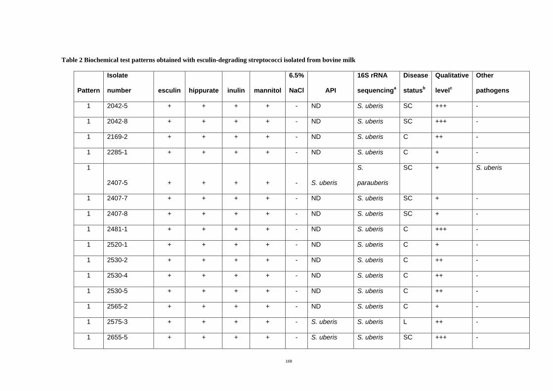
168
Table 2 Biochemical test patterns obtained with esculin-degrading streptococci isolated from bovine milk
Pattern
Isolate
number esculin hippurate inulin mannitol
6.5%
NaCl API
16S rRNA
sequencinga
Disease
statusb
Qualitative
levelc
Other
pathogens
1 2042-5 + + + + - ND S. uberis SC +++ -
1 2042-8 + + + + - ND S. uberis SC +++ -
1 2169-2 + + + + - ND S. uberis C ++ -
1 2285-1 + + + + - ND S. uberis C + -
1
2407-5 + + + + - S. uberis
S.
parauberis
SC + S. uberis
1 2407-7 + + + + - ND S. uberis SC + -
1 2407-8 + + + + - ND S. uberis SC + -
1 2481-1 + + + + - ND S. uberis C +++ -
1 2520-1 + + + + - ND S. uberis C + -
1 2530-2 + + + + - ND S. uberis C ++ -
1 2530-4 + + + + - ND S. uberis C ++ -
1 2530-5 + + + + - ND S. uberis C ++ -
1 2565-2 + + + + - ND S. uberis C + -
1 2575-3 + + + + - S. uberis S. uberis L ++ -
1 2655-5 + + + + - S. uberis S. uberis SC +++ -
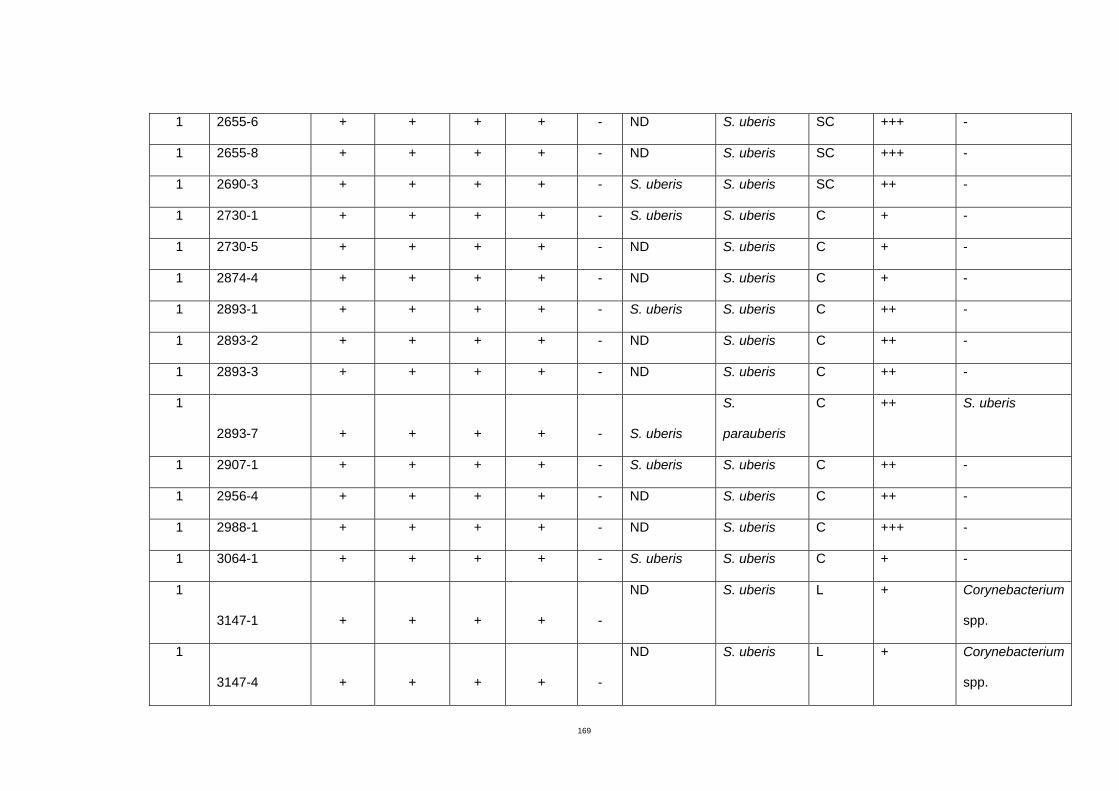
169
1 2655-6 + + + + - ND S. uberis SC +++ -
1 2655-8 + + + + - ND S. uberis SC +++ -
1 2690-3 + + + + - S. uberis S. uberis SC ++ -
1 2730-1 + + + + - S. uberis S. uberis C + -
1 2730-5 + + + + - ND S. uberis C + -
1 2874-4 + + + + - ND S. uberis C + -
1 2893-1 + + + + - S. uberis S. uberis C ++ -
1 2893-2 + + + + - ND S. uberis C ++ -
1 2893-3 + + + + - ND S. uberis C ++ -
1
2893-7 + + + + - S. uberis
S.
parauberis
C ++ S. uberis
1 2907-1 + + + + - S. uberis S. uberis C ++ -
1 2956-4 + + + + - ND S. uberis C ++ -
1 2988-1 + + + + - ND S. uberis C +++ -
1 3064-1 + + + + - S. uberis S. uberis C + -
1
3147-1 + + + + -
ND S. uberis L + Corynebacterium
spp.
1
3147-4 + + + + -
ND S. uberis L + Corynebacterium
spp.
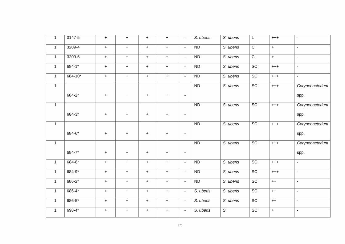
170
1 3147-5 + + + + - S. uberis S. uberis L +++ -
1 3209-4 + + + + - ND S. uberis C + -
1 3209-5 + + + + - ND S. uberis C + -
1 684-1* + + + + - ND S. uberis SC +++ -
1 684-10* + + + + - ND S. uberis SC +++ -
1
684-2* + + + + -
ND S. uberis SC +++ Corynebacterium
spp.
1
684-3* + + + + -
ND S. uberis SC +++ Corynebacterium
spp.
1
684-6* + + + + -
ND S. uberis SC +++ Corynebacterium
spp.
1
684-7* + + + + -
ND S. uberis SC +++ Corynebacterium
spp.
1 684-8* + + + + - ND S. uberis SC +++ -
1 684-9* + + + + - ND S. uberis SC +++ -
1 686-2* + + + + - ND S. uberis SC ++ -
1 686-4* + + + + - S. uberis S. uberis SC ++ -
1 686-5* + + + + - S. uberis S. uberis SC ++ -
1 698-4* + + + + - S. uberis S. SC + -
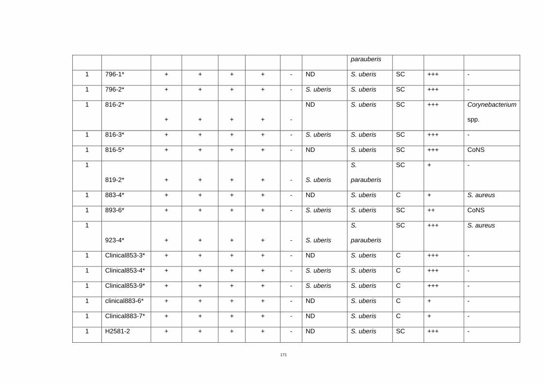
171
parauberis
1 796-1* + + + + - ND S. uberis SC +++ -
1 796-2* + + + + - S. uberis S. uberis SC +++ -
1 816-2*
+ + + + -
ND S. uberis SC +++ Corynebacterium
spp.
1 816-3* + + + + - S. uberis S. uberis SC +++ -
1 816-5* + + + + - ND S. uberis SC +++ CoNS
1
819-2* + + + + - S. uberis
S.
parauberis
SC + -
1 883-4* + + + + - ND S. uberis C + S. aureus
1 893-6* + + + + - S. uberis S. uberis SC ++ CoNS
1
923-4* + + + + - S. uberis
S.
parauberis
SC +++ S. aureus
1 Clinical853-3* + + + + - ND S. uberis C +++ -
1 Clinical853-4* + + + + - S. uberis S. uberis C +++ -
1 Clinical853-9* + + + + - S. uberis S. uberis C +++ -
1 clinical883-6* + + + + - ND S. uberis C + -
1 Clinical883-7* + + + + - ND S. uberis C + -
1 H2581-2 + + + + - ND S. uberis SC +++ -
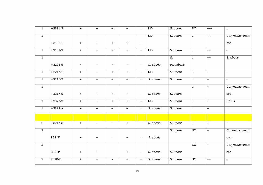
172
1 H2581-3 + + + + - ND S. uberis SC +++ -
1
H3133-1 + + + + -
ND S. uberis L ++ Corynebacterium
spp.
1 H3133-3 + + + + - ND S. uberis L ++ -
1
H3133-5 + + + + - S. uberis
S.
parauberis
L ++ S. uberis
1 H3217-1 + + + + - ND S. uberis L + -
1 H3217-2 + + + + - S. uberis S. uberis L + -
1
H3217-5 + + + + - S. uberis S. uberis
L + Corynebacterium
spp.
1 H3327-3 + + + + - ND S. uberis L + CoNS
1 H3333 a + + + + - S. uberis S. uberis L + -
2 H3217-3 + + - + - S. uberis S. uberis L + -
2
868-3* + + - + - S. uberis
S. uberis SC + Corynebacterium
spp.
2
868-4* + + - + - S. uberis S. uberis
SC + Corynebacterium
spp.
2 2690-2 + + - + - S. uberis S. uberis SC ++ -
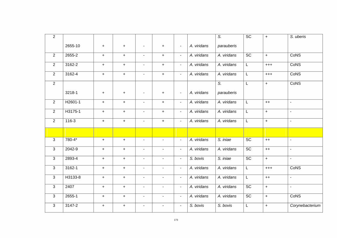
173
2
2655-10 + + - + - A. viridans
S.
parauberis
SC + S. uberis
2 2655-2 + + - + - A. viridans A. viridans SC + CoNS
2 3162-2 + + - + - A. viridans A. viridans L +++ CoNS
2 3162-4 + + - + - A. viridans A. viridans L +++ CoNS
2
3218-1 + + - + - A. viridans
S.
parauberis
L + CoNS
2 H2601-1 + + - + - A. viridans A. viridans L ++ -
2 H3175-1 + + - + - A. viridans A. viridans L + -
2 116-3 + + - + - A. viridans A. viridans L + -
3 780-4* + + - - - A. viridans S. iniae SC ++ -
3 2042-9 + + - - - A. viridans A. viridans SC ++ -
3 2893-4 + + - - - S. bovis S. iniae SC + -
3 3162-1 + + - - - A. viridans A. viridans L +++ CoNS
3 H3133-8 + + - - - A. viridans A. viridans L ++ -
3 2407 + + - - - A. viridans A. viridans SC + -
3 2655-1 + + - - - A. viridans A. viridans SC + CoNS
3 3147-2 + + - - - S. bovis S. bovis L + Corynebacterium
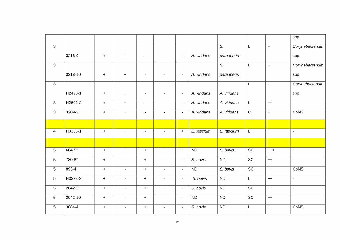
174
spp.
3
3218-9 + + - - - A. viridans
S.
parauberis
L + Corynebacterium
spp.
3
3218-10 + + - - - A. viridans
S.
parauberis
L + Corynebacterium
spp.
3
H2490-1 + + - - - A. viridans A. viridans
L + Corynebacterium
spp.
3 H2601-2 + + - - - A. viridans A. viridans L ++ -
3 3209-3 + + - - - A. viridans A. viridans C + CoNS
4 H3333-1 + + - - + E. faecium E. faecium L + -
5 684-5* + - + - - ND S. bovis SC +++ -
5 780-8* + - + - - S. bovis ND SC ++ -
5 893-4* + - + - - ND S. bovis SC ++ CoNS
5 H3333-3 + - + - - S. bovis ND L ++ -
5 2042-2 + - + - - S. bovis ND SC ++ -
5 2042-10 + - + - - ND ND SC ++ -
5 3084-4 + - + - - S. bovis ND L + CoNS
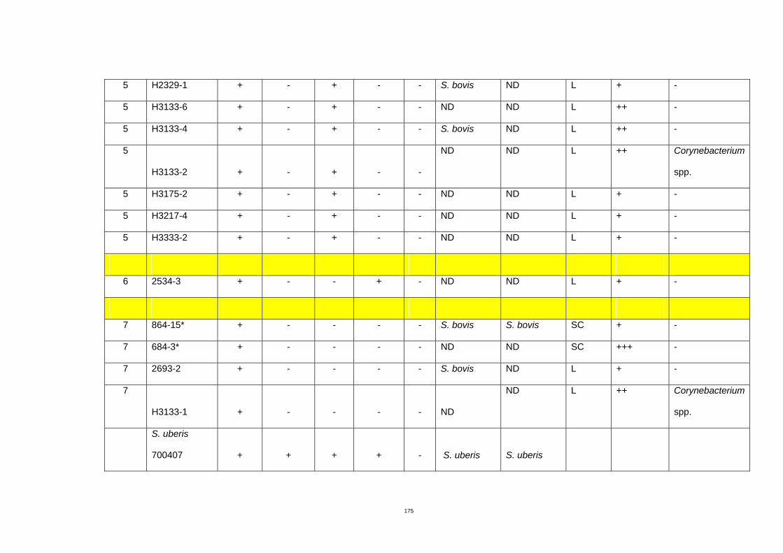
175
5 H2329-1 + - + - - S. bovis ND L + -
5 H3133-6 + - + - - ND ND L ++ -
5 H3133-4 + - + - - S. bovis ND L ++ -
5
H3133-2 + - + - -
ND ND L ++ Corynebacterium
spp.
5 H3175-2 + - + - - ND ND L + -
5 H3217-4 + - + - - ND ND L + -
5 H3333-2 + - + - - ND ND L + -
6 2534-3 + - - + - ND ND L + -
7 864-15* + - - - - S. bovis S. bovis SC + -
7 684-3* + - - - - ND ND SC +++ -
7 2693-2 + - - - - S. bovis ND L + -
7
H3133-1 + - - - - ND
ND L ++ Corynebacterium
spp.
S. uberis
700407 + + + + - S. uberis
S. uberis
176
-: negative
+: positive
ND: not determined
*: isolates were from Timboon farm and the others from Newry farm
CoNS: Coagulase negative staphylococci
a Results of S. uberis sequencing were from Takehiro Tomita (PhD. Student). Isolates that were not S. uberis were characterized in this study.
b Disease status is indicated as follow: C, clinical mastitis; SC, subclinical mastitis; L, low somatic cell count
c Quantitative growth of S. uberis is defined as follow: +, 1-10 colonies; ++, 11-50 colonies; +++, more than 50 colonies
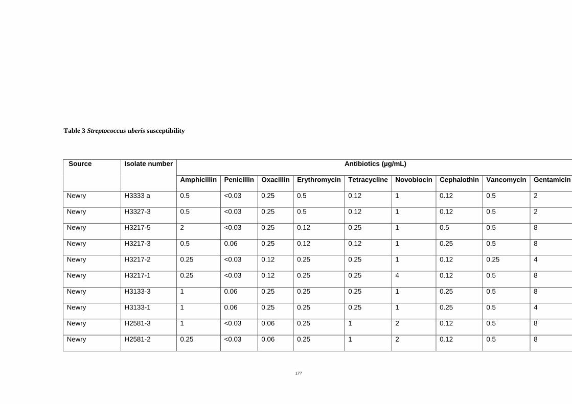
177
Table 3 Streptococcus uberis susceptibility
Antibiotics (µg/mL) Source
Isolate number
Amphicillin Penicillin Oxacillin Erythromycin Tetracycline Novobiocin Cephalothin Vancomycin Gentamicin
Newry H3333 a 0.5 <0.03 0.25 0.5 0.12 1 0.12 0.5 2
Newry H3327-3 0.5 <0.03 0.25 0.5 0.12 1 0.12 0.5 2
Newry H3217-5 2 <0.03 0.25 0.12 0.25 1 0.5 0.5 8
Newry H3217-3 0.5 0.06 0.25 0.12 0.12 1 0.25 0.5 8
Newry H3217-2 0.25 <0.03 0.12 0.25 0.25 1 0.12 0.25 4
Newry H3217-1 0.25 <0.03 0.12 0.25 0.25 4 0.12 0.5 8
Newry H3133-3 1 0.06 0.25 0.25 0.25 1 0.25 0.5 8
Newry H3133-1 1 0.06 0.25 0.25 0.25 1 0.25 0.5 4
Newry H2581-3 1 <0.03 0.06 0.25 1 2 0.12 0.5 8
Newry H2581-2 0.25 <0.03 0.06 0.25 1 2 0.12 0.5 8
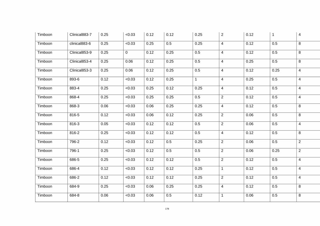
178
Timboon Clinical883-7 0.25 <0.03 0.12 0.12 0.25 2 0.12 1 4
Timboon clinical883-6 0.25 <0.03 0.25 0.5 0.25 4 0.12 0.5 8
Timboon Clinical853-9 0.25 0 0.12 0.25 0.5 4 0.12 0.5 8
Timboon Clinical853-4 0.25 0.06 0.12 0.25 0.5 4 0.25 0.5 8
Timboon Clinical853-3 0.25 0.06 0.12 0.25 0.5 4 0.12 0.25 4
Timboon 893-6 0.12 <0.03 0.12 0.25 1 4 0.25 0.5 4
Timboon 883-4 0.25 <0.03 0.25 0.12 0.25 4 0.12 0.5 4
Timboon 868-4 0.25 <0.03 0.25 0.25 0.5 2 0.12 0.5 4
Timboon 868-3 0.06 <0.03 0.06 0.25 0.25 4 0.12 0.5 8
Timboon 816-5 0.12 <0.03 0.06 0.12 0.25 2 0.06 0.5 8
Timboon 816-3 0.05 <0.03 0.12 0.12 0.5 2 0.06 0.5 4
Timboon 816-2 0.25 <0.03 0.12 0.12 0.5 4 0.12 0.5 8
Timboon 796-2 0.12 <0.03 0.12 0.5 0.25 2 0.06 0.5 2
Timboon 796-1 0.25 <0.03 0.12 0.5 0.5 2 0.06 0.25 2
Timboon 686-5 0.25 <0.03 0.12 0.12 0.5 2 0.12 0.5 4
Timboon 686-4 0.12 <0.03 0.12 0.12 0.25 1 0.12 0.5 4
Timboon 686-2 0.12 <0.03 0.12 0.12 0.25 2 0.12 0.5 4
Timboon 684-9 0.25 <0.03 0.06 0.25 0.25 4 0.12 0.5 8
Timboon 684-8 0.06 <0.03 0.06 0.5 0.12 1 0.06 0.5 8
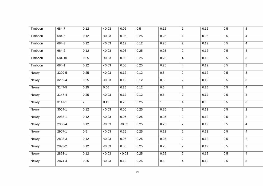
179
Timboon 684-7 0.12 <0.03 0.06 0.5 0.12 1 0.12 0.5 8
Timboon 684-6 0.12 <0.03 0.06 0.25 0.25 1 0.06 0.5 4
Timboon 684-3 0.12 <0.03 0.12 0.12 0.25 2 0.12 0.5 4
Timboon 684-2 0.12 <0.03 0.06 0.25 0.25 2 0.12 0.5 8
Timboon 684-10 0.25 <0.03 0.06 0.25 0.25 4 0.12 0.5 8
Timboon 684-1 0.12 <0.03 0.06 0.25 0.25 4 0.12 0.5 8
Newry 3209-5 0.25 <0.03 0.12 0.12 0.5 2 0.12 0.5 8
Newry 3209-4 0.25 <0.03 0.12 0.12 0.5 2 0.12 0.5 8
Newry 3147-5 0.25 0.06 0.25 0.12 0.5 2 0.25 0.5 4
Newry 3147-4 0.25 <0.03 0.12 0.12 0.5 2 0.12 0.5 8
Newry 3147-1 2 0.12 0.25 0.25 1 4 0.5 0.5 8
Newry 3064-1 0.12 <0.03 0.06 0.25 0.25 2 0.12 0.5 2
Newry 2988-1 0.12 <0.03 0.06 0.25 0.25 2 0.12 0.5 2
Newry 2956-4 0.12 <0.03 <0.03 0.25 0.25 2 0.12 0.5 4
Newry 2907-1 0.5 <0.03 0.25 0.25 0.12 2 0.12 0.5 4
Newry 2893-3 0.12 <0.03 0.06 0.25 0.25 2 0.12 0.5 2
Newry 2893-2 0.12 <0.03 0.06 0.25 0.25 2 0.12 0.5 2
Newry 2893-1 0.12 <0.03 <0.03 0.25 0.25 2 0.12 0.5 4
Newry 2874-4 0.25 <0.03 0.12 0.25 0.5 4 0.12 0.5 8
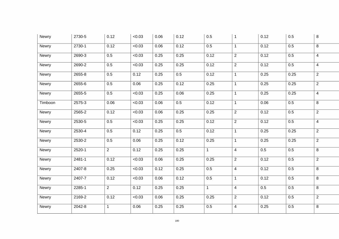
180
Newry 2730-5 0.12 <0.03 0.06 0.12 0.5 1 0.12 0.5 8
Newry 2730-1 0.12 <0.03 0.06 0.12 0.5 1 0.12 0.5 8
Newry 2690-3 0.5 <0.03 0.25 0.25 0.12 2 0.12 0.5 4
Newry 2690-2 0.5 <0.03 0.25 0.25 0.12 2 0.12 0.5 4
Newry 2655-8 0.5 0.12 0.25 0.5 0.12 1 0.25 0.25 2
Newry 2655-6 0.5 0.06 0.25 0.12 0.25 1 0.25 0.25 2
Newry 2655-5 0.5 <0.03 0.25 0.06 0.25 1 0.25 0.25 4
Timboon 2575-3 0.06 <0.03 0.06 0.5 0.12 1 0.06 0.5 8
Newry 2565-2 0.12 <0.03 0.06 0.25 0.25 2 0.12 0.5 2
Newry 2530-5 0.5 <0.03 0.25 0.25 0.12 2 0.12 0.5 4
Newry 2530-4 0.5 0.12 0.25 0.5 0.12 1 0.25 0.25 2
Newry 2530-2 0.5 0.06 0.25 0.12 0.25 1 0.25 0.25 2
Newry 2520-1 2 0.12 0.25 0.25 1 4 0.5 0.5 8
Newry 2481-1 0.12 <0.03 0.06 0.25 0.25 2 0.12 0.5 2
Newry 2407-8 0.25 <0.03 0.12 0.25 0.5 4 0.12 0.5 8
Newry 2407-7 0.12 <0.03 0.06 0.12 0.5 1 0.12 0.5 8
Newry 2285-1 2 0.12 0.25 0.25 1 4 0.5 0.5 8
Newry 2169-2 0.12 <0.03 0.06 0.25 0.25 2 0.12 0.5 2
Newry 2042-8 1 0.06 0.25 0.25 0.5 4 0.25 0.5 8

181
Newry 2042-5 0.5 0.06 0.25 0.12 0.5 4 0.5 0.5 8
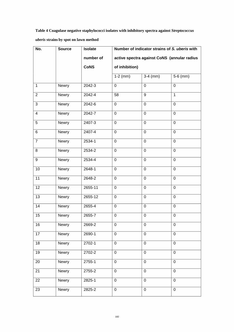
182
Table 4 Coagulase negative staphylococci isolates with inhibitory spectra against Streptococcus
uberis strains by spot on lawn method
Number of indicator strains of S. uberis with
active spectra against CoNS (annular radius
of inhibition)
No. Source Isolate
number of
CoNS
1-2 (mm) 3-4 (mm) 5-6 (mm)
1 Newry 2042-3 0 0 0
2 Newry 2042-4 58 9 1
3 Newry 2042-6 0 0 0
4 Newry 2042-7 0 0 0
5 Newry 2407-3 0 0 0
6 Newry 2407-4 0 0 0
7 Newry 2534-1 0 0 0
8 Newry 2534-2 0 0 0
9 Newry 2534-4 0 0 0
10 Newry 2648-1 0 0 0
11 Newry 2648-2 0 0 0
12 Newry 2655-11 0 0 0
13 Newry 2655-12 0 0 0
14 Newry 2655-4 0 0 0
15 Newry 2655-7 0 0 0
16 Newry 2669-2 0 0 0
17 Newry 2690-1 0 0 0
18 Newry 2702-1 0 0 0
19 Newry 2702-2 0 0 0
20 Newry 2755-1 0 0 0
21 Newry 2755-2 0 0 0
22 Newry 2825-1 0 0 0
23 Newry 2825-2 0 0 0
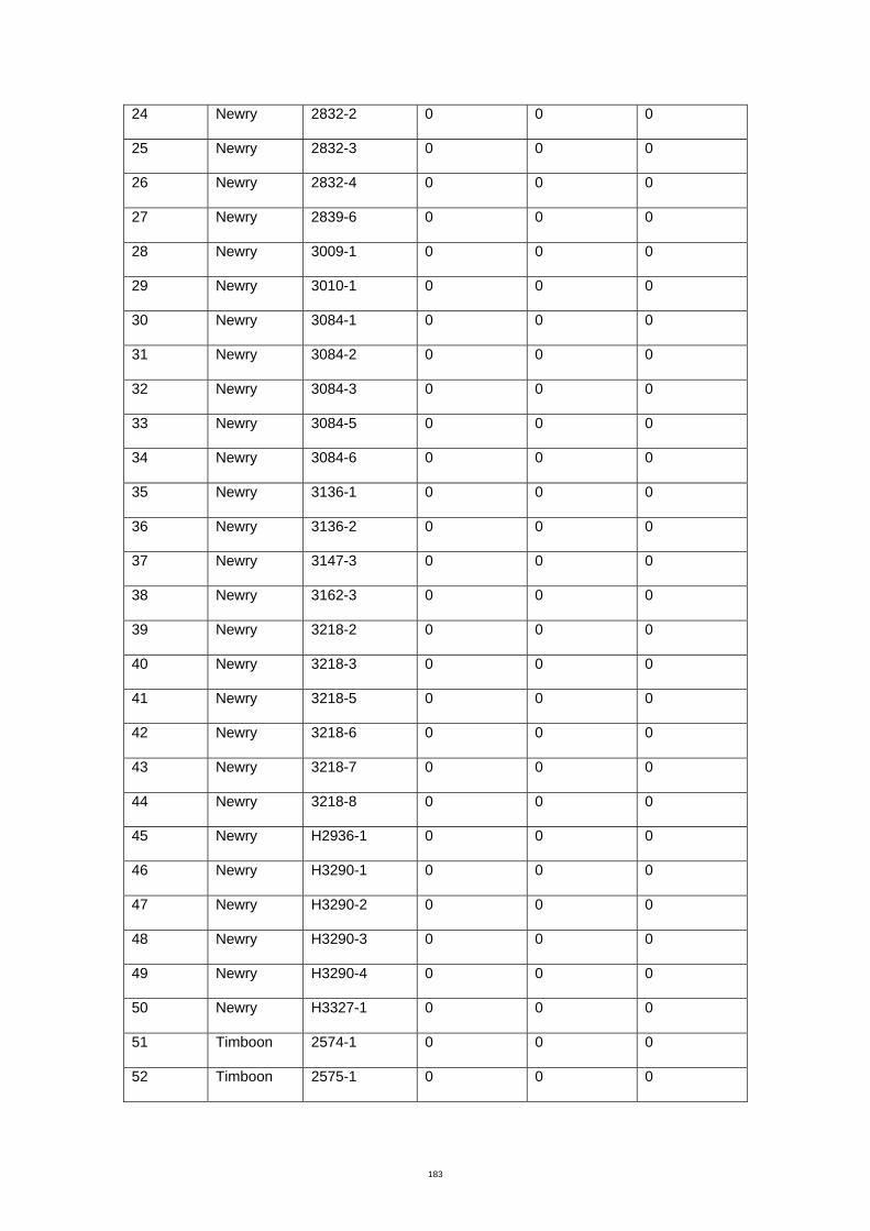
183
24 Newry 2832-2 0 0 0
25 Newry 2832-3 0 0 0
26 Newry 2832-4 0 0 0
27 Newry 2839-6 0 0 0
28 Newry 3009-1 0 0 0
29 Newry 3010-1 0 0 0
30 Newry 3084-1 0 0 0
31 Newry 3084-2 0 0 0
32 Newry 3084-3 0 0 0
33 Newry 3084-5 0 0 0
34 Newry 3084-6 0 0 0
35 Newry 3136-1 0 0 0
36 Newry 3136-2 0 0 0
37 Newry 3147-3 0 0 0
38 Newry 3162-3 0 0 0
39 Newry 3218-2 0 0 0
40 Newry 3218-3 0 0 0
41 Newry 3218-5 0 0 0
42 Newry 3218-6 0 0 0
43 Newry 3218-7 0 0 0
44 Newry 3218-8 0 0 0
45 Newry H2936-1 0 0 0
46 Newry H3290-1 0 0 0
47 Newry H3290-2 0 0 0
48 Newry H3290-3 0 0 0
49 Newry H3290-4 0 0 0
50 Newry H3327-1 0 0 0
51 Timboon 2574-1 0 0 0
52 Timboon 2575-1 0 0 0
184
53 Timboon 2575-2 0 0 0
54 Timboon 2618-1 0 0 0
55 Timboon 2627-1 0 0 0
56 Timboon 2627-2 0 0 0
57 Timboon 2659-1 0 0 0
58 Timboon 2659-2 0 0 0
59 Timboon 2725-1 0 0 0
60 Timboon 2741-1 0 0 0
61 Timboon 2741-2 0 0 0
62 Newry 2169-1 0 0 0
63 Newry 2530-1 0 0 0
64 Newry 2565-1 0 0 0
65 Newry 2730-1 0 0 0
66 Newry 2730-2 0 0 0
67 Newry 2730-3 0 0 0
68 Newry 2730-4 0 0 0
69 Newry 2874-1 0 0 0
70 Newry 2874-2 0 0 0
71 Newry 2874-3 0 0 0
72 Newry 2956-1 0 0 0
73 Newry 2956-2 0 0 0
74 Newry 2956-3 0 0 0
75 Newry 3209-1 0 0 0
76 Newry 3209-2 0 0 0
77 Newry 2907-2 0 0 0
78 RMIT 10 0 0 0
79 RMIT 18 0 0 0
80 RMIT 19 0 0 0
81 RMIT 31W 0 0 0
185
82 RMIT 31Y 0 0 0
83 RMIT 33W 0 0 0
84 RMIT 33Y 0 0 0
85 RMIT 35 0 0 0
86 RMIT 37 0 0 0
87 RMIT 30W 0 0 0
88 RMIT 30Y 0 0 0
89 RMIT 41 0 0 0
90 RMIT 42W 0 0 0
91 RMIT 42Y 0 0 0
92 RMIT 43 0 0 0
93 RMIT 45W 0 0 0
94 RMIT 45Y 0 0 0
95 RMIT 46DY 0 0 0
96 RMIT 50LW 0 0 0
97 RMIT 50SW 0 0 0
98 RMIT 51 0 0 0
99 RMIT 68 0 0 0
100 RMIT 70 0 0 0
101 RMIT 75 0 0 0
102 RMIT 76 0 0 0
103 RMIT 77 0 0 0
104 RMIT 83 0 0 0
105 RMIT 84W 0 0 0
106 RMIT 84Y 0 0 0
107 RMIT 86 0 0 0
108 RMIT 82 0 0 0
109 RMIT 95 0 0 0
110 RMIT 103 0 0 0
186
111 RMIT 104 0 0 0
112 RMIT 106 0 0 0
113 RMIT 108W 0 0 0
114 RMIT 108Y 0 0 0
115 RMIT 109W 0 0 0
116 RMIT 109Y 0 0 0
117 RMIT 110W 0 0 0
118 RMIT 110Y 0 0 0
119 RMIT 111 0 0 0
120 RMIT 112 0 0 0
121 RMIT 114 0 0 0
122 RMIT 115 0 0 0
123 RMIT 1 0 0 0
124 RMIT 3 0 0 0
125 RMIT 4 0 0 0
126 RMIT 5 0 0 0
127 RMIT 6 0 0 0
128 RMIT 7 0 0 0
129 RMIT 9 0 0 0
130 RMIT 11 0 0 0
131 RMIT 12 0 0 0
132 RMIT 13 0 0 0
133 RMIT 14W 0 0 0
134 RMIT 15 0 0 0
135 RMIT 16W 0 0 0
136 RMIT 16Y 0 0 0
137 RMIT 17 0 0 0
138 RMIT 21 0 0 0
139 RMIT 22 0 0 0
187
140 RMIT 23 0 0 0
141 RMIT 24W 0 0 0
142 RMIT 24Y 0 0 0
143 RMIT 25 0 0 0
144 RMIT 27 0 0 0
145 RMIT 28W 0 0 0
146 RMIT 36 0 0 0
147 RMIT 37Y 0 0 0
148 RMIT 39 0 0 0
149 RMIT 40 0 0 0
150 RMIT 46LY 0 0 0
151 RMIT 47Y 0 0 0
152 RMIT 48G 0 0 0
153 RMIT 48 0 0 0
154 RMIT 49 0 0 0
155 RMIT 50Y 0 0 0
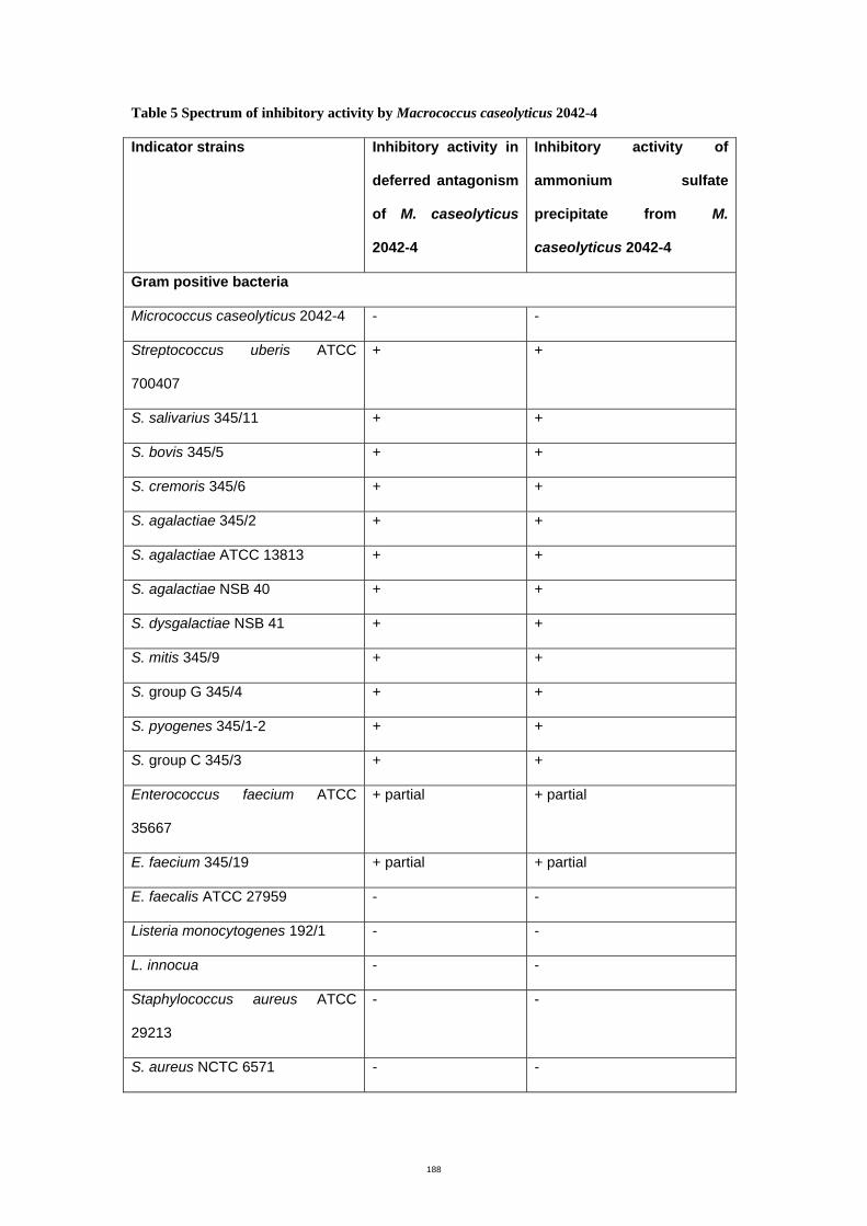
188
Table 5 Spectrum of inhibitory activity by Macrococcus caseolyticus 2042-4
Indicator strains Inhibitory activity in
deferred antagonism
of M. caseolyticus
2042-4
Inhibitory activity of
ammonium sulfate
precipitate from M.
caseolyticus 2042-4
Gram positive bacteria
Micrococcus caseolyticus 2042-4 - -
Streptococcus uberis ATCC
700407
+ +
S. salivarius 345/11 + +
S. bovis 345/5 + +
S. cremoris 345/6 + +
S. agalactiae 345/2 + +
S. agalactiae ATCC 13813 + +
S. agalactiae NSB 40 + +
S. dysgalactiae NSB 41 + +
S. mitis 345/9 + +
S. group G 345/4 + +
S. pyogenes 345/1-2 + +
S. group C 345/3 + +
Enterococcus faecium ATCC
35667
+ partial + partial
E. faecium 345/19 + partial + partial
E. faecalis ATCC 27959 - -
Listeria monocytogenes 192/1 - -
L. innocua - -
Staphylococcus aureus ATCC
29213
- -
S. aureus NCTC 6571 - -
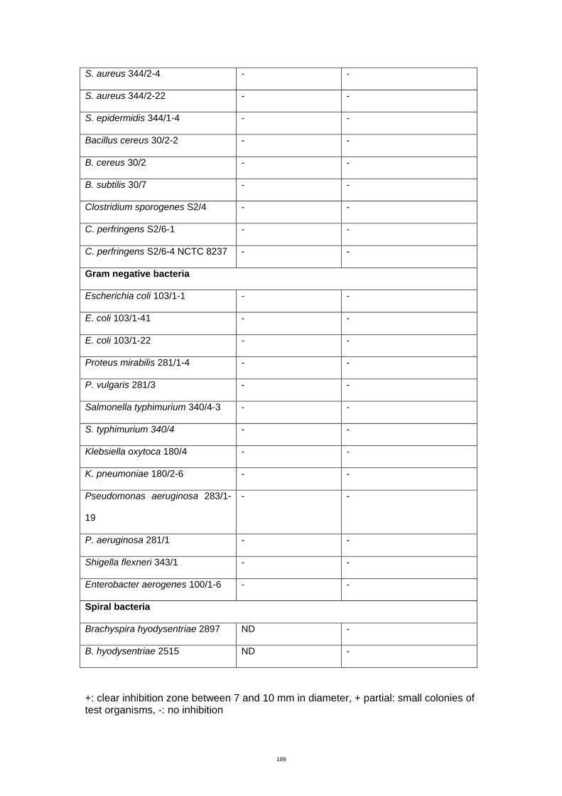
189
S. aureus 344/2-4 - -
S. aureus 344/2-22 - -
S. epidermidis 344/1-4 - -
Bacillus cereus 30/2-2 - -
B. cereus 30/2 - -
B. subtilis 30/7 - -
Clostridium sporogenes S2/4 - -
C. perfringens S2/6-1 - -
C. perfringens S2/6-4 NCTC 8237 - -
Gram negative bacteria
Escherichia coli 103/1-1 - -
E. coli 103/1-41 - -
E. coli 103/1-22 - -
Proteus mirabilis 281/1-4 - -
P. vulgaris 281/3 - -
Salmonella typhimurium 340/4-3 - -
S. typhimurium 340/4 - -
Klebsiella oxytoca 180/4 - -
K. pneumoniae 180/2-6 - -
Pseudomonas aeruginosa 283/1-
19
- -
P. aeruginosa 281/1 - -
Shigella flexneri 343/1 - -
Enterobacter aerogenes 100/1-6 - -
Spiral bacteria
Brachyspira hyodysentriae 2897 ND -
B. hyodysentriae 2515 ND -
+: clear inhibition zone between 7 and 10 mm in diameter, + partial: small colonies of test organisms, -: no inhibition
188
Appendix C Bacteriological media
Brain heart infusion broth/agar/soft agar (BHB/BHA/BHI soft
agar)
This medium was prepared according to the manufacturer’s instructions, and
sterilized by autoclaving at 121°C for 15 min. Bacteriological agar was added 1.5%
(w/v) for BHA and 0.7% (w/v) for BHI soft agar prior to autoclaving and poured in
Petri dishes.
De Man Rogosa Sharpe agar medium (MRS), DNase agar,
Edwards Milk agar (MA), Muller Hintion (MH), Nutrient agar
(NA) and Trypticase soy agar (TSA)
This medium was prepared according to the manufacturer’s instructions, and
sterilized by autoclaving at 121°C for 15 min and poured in Petri dishes.
Horse blood agar (HBA)
Horse blood was prepared by incorporating 5% horse blood in Columbia agar base.
The medium was prepared according to the manufacturer’s instruction, and sterilized
by autoclaving at 121°C for 15 min and cooled down to 50°C. Finally, 5% of
defibrinated horse blood was added aseptically and mixed gently before pouring into
Petri dishes.
The medium was prepared according to the manufacturer’s instruction, and sterilized
by autoclaving at 121°C for 15 min and poured in Petri dishes.
Todd Hewitt broth (THB)
189
The medium was prepared according to the manufacturer’s instruction, and
transferred to 16 × 150 mm tubes and sterilized by autoclaving at 121°C for 15 min.
189
References
Aarestrup FM (1999) The European Perspective on Antimicrobial Related Regulations and Trade. Retrieved December 19, 2007 from http://www.omafra.gov.on.ca/english/livestock/animalcare/amr/facts/moller. htm Abadin E (1994) Characterization of Staphylococcal isolates from Bovine Mastitis, Honours Thesis, RMIT, Bundoora, Vic. Abriouel H, Valdivia E, Martinez-Bueno M, Maqueda M & Galvez A (2003) A Simple Method for Semi-Preparative-Scale Production and Recovery of Enterocin AS-48 Derived from Enterococcus faecalis subsp. liquefaciens. Journal of Microbiological Methods 55: 599-605. Aitken A & Learmounth MP (2002). Protein Determination by UV Absorption. In Walker JM (Ed.), The Protein Protocols Handbook (pp. 3-6). Humana Press Inc. Alber J, El-Sayed A, Lammler C, Hassan AA & Zschock M (2004) Polymerase Chain Reaction Mediated Identification of Streptococcus uberis and Streptococcus parauberis Using Species-Specific Sequences of the Genes Encoding Superoxide Dimutase A and Chaperonin 60*. Journal of Veterinary Medicine B 51: 180-184. Almeida RA & Oliver SP (1993) Growth Curve, Capsule Expression and Characterization of the Capsular Material of Selected Strains of Streptococcus uberis. Journal of Veterinary Medicine B 40: 697-706. Almeida RA, Batcha T & Oliver SP (2005) Persistence of Streptococcus uberis in Bovine Mammary Epithelial Cells. In Hogeveen H (Ed), Mastitis in Dairy Production (pp. 137-142) Wageningen Academic Publishers.
Anonymous (1990) Procedures for the Identification of Specific Groups or Species of Microorganisms that Cause Mastitis. Microbiological Procedures for the Diagnosis of Bovine Udder Infection (pp. 10) National Mastitis Council Inc.
Aslim B, Yuksekdag ZN, Sarikaya E & Beyatli Y (2005) Determination of the Bacteriocin-Like Substances Produced by Some Lactic Acid Bacteria Isolated from Turkish Dairy Products. LWT 38: 691-694. Aso Y, Okuda K, Nagao J, Kanemasa Y, Phuong NTB, Koga H, Shioya K, Sashihara T, Nakayama J & Sonomoto K (2005a) A Novel Type of Immunity Protein, NukH, for the Lantibiotic Nukacin ISK-1 Produced by Staphylococcus warneri ISK-1. Bioscience, Biotechnology and Biochemistry 69: 1403-1410. Aso Y, Koga H, Sashihara T, Jun-ichi Nagao, Kanemasa Y, Nakayama J & Sonomoto K (2005b) Description of Complete DNA Sequence of Two Plasmids from the Nukacin ISK-1 Producer, Staphylococcus warneri ISK-1. Plasmid 53: 164-178.
190
Atroshi F, Työppönen J, Sankari S, Kangasniemi R & Parantainen J (1987) Possible Roles of Vitamin E and Glutathione Metabolism in Bovine Mastitis. International Journal of Vitamin Nutritional Research 57: 37-43. Avonts L, Uytven EV & Vuyst LD (2004) Cell Growth and Bacteriocin Production of Probiotic Lactobacillus Strains in Different Media. International Dairy Journal 14: 947-955. Ayad EHE, Verheul A, Wouters JTM & Smit G (2002) Antimicrobial-Producing Wild Lactococci Isolated from Artisanal and Non-Dairy Origins. International Dairy Journal 12: 145-150. Aymerich MT, Garriga M, Monfort JM, Nes I & Hugas M (2000) Bacteriocin-Producing Lactobacilli in Spanish-Style Fermented Sausages: Characterization of Bacteriocins. Food Microbiology 17: 33-45. Baba T & Schneewind O (1996) Target Cell Specificity of a Bacteriocin Molecule: a C-terminal Signal Directs Lysostaphin to the Cell Wall of Staphylococcus aureus. The EMBO Journal 15: 4789-4797. Balakrishnan M, Simmonds RS, Carne A & Tagg JR (2000) Streptococcus mutans Strain N Produces a Novel Low Molecular Mass Non-Lantibiotic Bacteriocin. FEMS Microbiology Letters 183: 165-169. Bartlett PC, Miller GY, Lance SE & Heider LE (1992) Clinical Mastitis and Intramammary Infections on Ohio Dairy Farms. Preventine Veterinary Medicine 12: 59-71. Baseggio N, Mansell PD, Browning JW & Browning GF (1997) Strain Differentiation of Isolates of Streptococci from Bovine Mastitis by Pulsed-Field Gel Electrophoresis. Molecular and Cellular Probes 11: 349-354. Beck HS, Wise WS & Dodd FH (1992) Cost Benefit Analysis of Bovine Mastitis in the UK. Journal of Dairy Research 59: 449-460. Beever DE (2006) The Impact of Controlled Nutrition During the Dry Period on Dairy Cow Health, Fertility and Performance. Animal Reproduction Science 96: 212-226. Bellemann P, Bereswill S, Berger S & Geider K (1994) Visualization of Capsule Formation by Erwinia amylovora and Assays to Determine Amylovoran Synthesis. International Journal of Biology and Macromolecule 16: 290-296. Berry EA & Hillerton JE (2002a) The Effect of an Intramammary Teat Seal on New Intramammary Infections. Journal of Dairy Science 85: 2512-2520. Berry EA & Hillerton JE (2002b) The Effect of Selective Dry Cow Treatment on New Intramammary Infections. Journal of Dairy Science 85: 112-121. Berry EA & Hillerton JE (2007) Effect of An Intramammary Teat Seal and Dry Cow Antibiotic in Relation to Dry Period Length on Postpartum Mastitis. Journal of dairy science 90: 760-765. Bizani D & Brandelli A (2002) Characterization of a Bacteriocin Produced by a Newly Isolated Bacillus sp. Strain 8A. Journal of Applied Microbiology 93: 512-519.
191
Blaiotta G, Pennacchia C, Ercolini D, Moschetti G & Villani F (2003) Combining Denaturing Gradient Gel Electrophoresis of 16S rDNA V3 Region and 16S-23S rDNA Spacer Region Polymorphism Analyses for the Identification of Staphylococci from Italian Fermented Sausages. Systematic of Applied Microbiology 26: 423-433. Blom H, Katla T, Nissen H & Holo H (2001) Characterization, Production and Purification of Carnocin H, a Bacteriocin Produced by Carnobacterium 377. Current Microbiology 43: 227-231. Bollag DM & Edelstein SJ (1991) Protein methods. Wiley-Liss, Inc. Booth SJ, Johnson JL & Wilkins TD (1977) Bacteriocin Production by Strains of Bacteroides Isolated from Human Feces and the Role of these Strains in the Bacterial Ecology of the Colon. Antimicrobial Agents and Chemotheraphy 11: 718-724. Bradley AJ (2002) Bovine Mastitis: An Evolving Disease. The Veterinary Journal 164: 116-128. Bramley AJ, Hillerton JE, Higgs TM & Hogben EM (1984) The Carriage of Summer Mastitis Pathogens by Muscid Flies. British Veterinary Journal 141: 618-627. Brightling P, Mein GA, Malmo J & Ryan DP (1998) Countdown Downunder: Farm Guidlines for Mastitis Control. Dairy Research and Development Corporation. Brown MB & Scasserra AE (1990) Antimicrobial Resistance in Streptococcal Species Isolated from Bovine Mammary Glands. American Journal of Veterinary Research 51: 2015-2018. Burvenich C, Monfardini E, Mehrzad J, Capuco AV & Paape MJ (2004) Role of Neutrophil Polymorphonuclear Leukocytes During Bovine Coliform Mastitis: Physiology or Pathology? Verhandelingnen-Koninklijke Academic voor Geneeskdunde van Belgie 66: 97-150. Busato A, Trachsel P, Schallibaum M & Blum JW (2000) Udder Health and Risk Factors for Subclinical Mastitis in Organic Dairy Farms in Switzerland. Preventive Veterinary Medicine 44: 205-220. Carnio MC, Holtzel A, Rudolf M, Henle T, Jung G & Scherer S (2000) The Macrocyclic Peptide Antibiotic Micrococcin P1 Is Secreted by the Food-Borne Bacterium Staphylococcus equorum WS 2733 and Inhibits Listeria monocytogenes on Soft Cheese. Applied and Environmental Microbiology 66: 2378-2384. Catry B, Laevens H, Devriese LA, Opsomer G & Kruif A (2003) Antimicrobial Resistance in Livestock. Journal of Veterinary Pharmacology Therapy 26: 81-93. Chen H & Hoover DG (2003) Bacteriocins and their Food Applications. Comprehensive Reviews in Food Science and Food Safety 2: 82-100. Chumchalova J, Stiles J, Josephsen J & Plockova M (2004) Characterization and Purification of Acidocin CH5, a Bacteriocin Produced by Lactobacillus acidophilus CH5. Journal of Applied Microbiology 96: 1082-1089.
192
Cladera-Olivera F, Caron GR & Brandelli A (2004) Bacteriocin-Like Substance Production by Bacillus licheniformis Strain P40. Letters in Applied Microbiology 38: 251-256. Coventry MJ, Wan J, Gordon JB, Mawson RF & Hickey MW (1996) Production of Brevicin 286 by Lactobacillus brevis VB286 and Partial Characterization. Journal of Applied Bacteriology 80: 91-98. Coventry MJ, Gordon JB, Wilcock A, et al. (1997) Detection of Bacteriocins of Lactic Acid Bacteria Isolated from Foods and Comparison with Pediocin and Nisin. Journal of Applied Microbiology 83: 248-258.
Cowan ST, Steel KJ, Barrow GI & Feltham RKA (1993) Cowan and Steel's Manual for the Identification of Medical Bacteria. Cambridge University Press.
Crupper SS & Iandolo JJ (1996) Purification and Partial Characterization of a Novel Antibacterial Agent (Bac1829) Produced by Staphylococcus aureus KSI1829. Applied and Environmental Microbiology 62: 3171-3175. Crupper SS & Iandolo JJ (1997) Exploiting the Unique Biophysical Properties of Bacteriocins to Purify Bac 1829 from Staphylococcus aureus KSI 1829. Protein Expression and Purification 9: 228-232. Crupper SS, Gies AJ & Iandolo JJ (1997) Purification and Characterization of Staphylococcin BacR1, a Broad-Spectrum Bacteriocin. Applied and Environmental Microbiology 63: 4185-4190. Crupper SS, Gies AJ & Iandolo JJ (1997) Purification and Characterization of Staphylococcin BacR1, a Broad-Spectrum Bacteriocin. Applied and Environmental Microbiology 63: 4185-4190. Cui S, Li J, Hu C, Jin S & Ma Y (2007) Development of a Method for the Detection of Beta-Lactamases in Milk Samples. Journal of AOAC International 90: 1128-1132. Cullen GA (1966) The Ecology of Streptococcus uberis The British Veterinary Journal 122: 333-339. Dairy Australia (2007) Countdown Downunder. Retrieved June 2007 from http://www.countdown.org.au. Dais FM & Allaire FR (1982) Dry Period to Maximize Milk Production Over Two Consecutive Lactations. Journal of Dairy Science 65: 136-145. Daw MA & Falkiner FR (1996) Bacteriocins: Nature, Function and Structure. Micron 27: 467-479. DeGraves FJ & Fetrow J (1993) Economics of Mastitis and Mastitis Control. Veterinary Clinics of North America: Food Animal Practice 9: 421-434.
193
Delgado A, Brito D, Fevereiro P, Tenreiro R & Peres C (2005) Bioactivity of Crude Bacteriocin Solutions. Journal of Microbiological Methods 62: 121-124. Denamiel G, Llorente P, Carabella M, Rebuelto M & Gentilini E (2005) Anti-Microbial Susceptibility of Streptococcus spp. Isolated from Bovine Mastitis in Argentina. Journal of veterinary medicine B 52: 125-128. Deraz SF, Karlsson EN, Hedstrom M, Anderson MM & Mattiasson B (2005) Purification and Characterization of Acidocin D20079, a Bacteriocin Produced by Lactobacillus acidophilus DSM 20079. Journal of Biotechnology 117: 343-354. Detilleux JC (2002) Genetic Factors Affecting Susceptibility of Dairy Cows to Udder Pathogens. Veterinary Immunology and Immunopathology 88: 103-110.
Devriese LA, Hommez J, Laevens H, Pot B, Vandamme P & Haesebrouck F (1999) Identification of Aesculin-Hydrolyzing Streptococci, Lactococci, Aerococci and Enterococci from Subclinical Intramammary Infections in Dairy Cows. Veterinary Microbiology 70: 87-94.
Devriese LA, Baele M, Vaneechoutte M, Martel A & Haesebrouck F (2002) Identification and Antimicrobial Susceptibility of Staphylococcus chromogenes Isolates from Intramammary Infections of Dairy Cows. Veterinary Microbiology 87: 175-182. Dingwell RT, Duffield TF, Leslie KE, et al. (2002) The Efficacy of Intramammary Tilmicosin at Drying-Off, and Other Risk Factors for the Prevention of New Intramammary Infections During the Dry Period. Journal of Dairy Science 85: 3250-3259.
Dingwell RT, Leslie KE, Schukken YH, Sargeant JM & Timms LL (2003) Evaluation of the California Mastitis Test to Detect an Intramammary Infection with a Major Pathogen in Early Lactation Dairy Cows. Canadian Veterinary Journal 44: 413-416.
Eberhart RJ (1986) Management of Dry Cows to Reduce Mastitis. Journal of Dairy Science 69: 1721-1732.
Ekkelenkamp MB, Hanssen M, Hsu STD, Jong A, Milatovic D, Verhoef J & Nuland NAJ (2005) Isolation and Structural Characterization of Epilancin 15X, a Novel Lantibiotic from a Clinical Strain of Staphylococcus epidermidis. FEBS Letters 579: 1917-1922. Erskine RJ, Walker RD, Bolin CA, Bartlett PC & White DG (2002) Trends in Antimicrobial Susceptibility of Mastitis Pathogens During a Seven-Year Period. Journal of Dairy Science 85: 1111-1118.
194
Facklam R & Elliott JA (1995) Identification, Classification, and Clinical Relevance of Catalase-Negative, Gram-Positive Cocci, Excluding the Streptococci and Enterococci. Clinical Microbiology Reviews 8: 479-495.
Facklam R (2002) What Happened to the Streptococci: Overview of Taxonomic and Nomenclature Changes. Clinical Microbiology Reviews 15: 613-630.
Finch JM, Hill AW, Field TR & Leigh JA (1994) Local Vaccination with Killed Streptococcus uberis Protects the Bovine Mammary Gland Against Experimental Intramammary Challenge with the Homologous Strain. Infection and Immunity 62: 3599-3603. Finch JM, Winter A, Walton AW & Leigh JA (1997) Further Studies on the Efficacy of a Live Vaccine Against Mastitis Caused by Streptococcus uberis. Vaccine 15: 1138-1143.
Fortin M, Messier S, Pare J & Higgins R (2003) Identification of Catalase-Negative, Non-Beta Hemolytic, Gram-Postive Cocci Isolated from Milk Samples. Journal of Clinical Microbiology 41: 106-109.
Francis PG, Wilesmith JW & Wilson CD (1986) Observation on the Incidence of Clinical Bovine Mastitis in Non-Lactating Cows in England and Wales. Veterinary Records 118: 549-552. Galton DM, Peterson LG & Erb HN (1986) Milk Iodine Residues in Herds Practicing Iodophor Premilking Teat Disinfection. Journal of Dairy Science 69: 267-271.
Garvie EI & Bramley AJ (1979) Streptococcus uberis: an Approach to Its Classification. Journal of Applied Bacteriology 46: 295-304.
Grave K, Greko C, Nilsson L, Odensvik K, Mork T & Ronning M(1999) The Usage of Veterinary Antimicrobial Drugs for Mastitis in Cattle in Norway and Sweden During 1990-1997. Preventive Veterinary Medicine 42: 45-55. Ghrairi T, Frere J, Berjeaud JM & Manai M (2005) Lactococcin MMT24, a Novel Two-Peptide Bacteriocin Produced by Lactococcus lactis Isolated from Rigouta Cheese. International Journal of Food microbiology 105: 389-398. Green MJ, Green LE, Bradley AJ, Burton PR, Schukken YH & Medley GF (2005) Prevalence and Associations Between Bacterial Isolates from Dry Mammary Glands of Dairy Cows. The Veterinary Record 156: 71-77. Grenier D (1996) Antagonistic Effect of Oral Bacteria Towards Treponema denticola. Journal of Clinical Microbiology 34: 1249-1252.
195
Guerin-Faublee V, Tardy F, Bouveron C & Carret G (2002) Antimicrobial Susceptibility of Streptococcus Species Isolated from Clinical Mastitis in Dairy Cows. International Journal of Antimicrobial Agents 19: 219-226. Gustafson RH & Bowen RE (1997) A Review: Antibiotic Use in Animal Agriculture. Journal of Applied Microbiology 83: 531-541. Guterbock WM, Eenennaam ALV, Anderson RJ, Gardner IA, Cullor JS & Holmberg CA (1993) Efficacy of lntramammary Antibiotic Therapy for Treatment of Clinical Mastitis Caused by Environmental Pathogens. Journal of Dairy Science 76. Gutierrez J, Criado R, Martin M, Herranz C, Cintas LM & Hernandez PE (2005) Production of Enterocin P, an Antilisterial Pediocin-Like Bacteriocin from Enterococcus faecium P13, in Pichia pastoris. Antimicrobial Agents and Chemotheraphy 49: 3004-3008. Hale EM & Hinsdill RD (1973) Characterization of a Bacteriocin from Staphylococcus aureus Strain 462. Antimicrobial Agents and Chemotherapy 4: 634-640. Hale EM & Hinsdill RD (1975) Biological Activity of Staphylococcin 462: Bacteriocin from Staphylococcus aureus. Antimicrobial Agents and Chemotherapy 7: 74-81. Haltia L, Honkanen-Buzalski T, Spiridonova I, Olkonen A & Myllys V (2006) A study of Bovine Mastitis, Milking Procedures and Management Practices on 25 Estonian Dairy Herds. Acta Veterinaria Scandinavica 48. Hames BD (1998) Gel Electrophoresis of Proteins: Oxford University Press. Hansen SH, Helboe P & Lund U (1984) Separation methods, Elsevier Science Publishers B.V. Hansen PJ (2007) Lowry Protein Assay. Retrieved December 20, 2007 from http:// www.animal. ufl.edu/hansen/protocols/LOWRY.htm. Harris ELV & Angal S (1990) Protein Purification Applications: a Practical Approach. Oxford University Press. Harting M, Hedges A, Berkeley AJ & W. RC (1972) Methods for Studying Bacteriocins. Methods in Microbiology 7A: 315-422.
Hassan AA, Khan IU, Abdulmawjood A & Lammler C (2001) Evaluation of PCR Methods for Rapid Identification and Differentiation of Streptococcus uberis and Streptococcus parauberis. Journal of Clinical Microbiology 39: 1618-1621.
He L, Chen W & Liu Y (2006) Production and Partial Characterization of Bacteriocin-Like Peptides by Bacillus licheniformis. Microbiological Research 161: 321-326. Hechard Y, Ferraz S, Bruneteau E, Steinert M & Berjeaud J-M (2005) Isolation and Characterization of a Staphylococcus warneri Strain Producing an Anti-Legionella Peptide. FEMS Microbiology Letters 252: 19-23.
196
Heng NCK, Ragland NL, Swe PM, Baird HJ, Inglis MA, Tagg JR & Jack RW (2006) Dysgalacticin: a Novel, Plasmid-Encoded Antimicrobial Protein (bacteriocin) Produced by Streptococcus dysgalactiae subsp. equisimilis. Microbiology 152: 1991-2001. Hill AW, Shears AL & Hibbit KG (1978) The Elimination of Serum-Resistant Escherichia coli from Experimentally Infected Single Mammary Glands of Healthy Cows. Research of Veterinary Science 25: 89-93. Hill AW (1988) Protective Effect of Previous Intramammary Infection with Streptococcus uberis Subsequent Clinical Mastitis in Cow. Research of Veterinary Science 44: 386-387. Hill AW, Finch JM, Field TR & Leigh JA (1994) Immune Modification of the Pathogenesis of Streptococcus uberis Mastitis in the Dairy Cow. FEMS Immunology and Medical Microbiology 8: 109-117. Hillerton JE & Kliem KE (2002) Effective Treatment of Streptococcus uberis Clinical Mastitis to Minimize the Use of Antibiotics. Journal of Dairy Science 85: 1009-1014. Hillerton JE & Berry EA (2005) A Review: Treating Mastitis in the Cow – A Tradition or an Archaism. Journal of Applied Microbiology 98: 1250-1255. Hogan JS, Smith KL, Hoblet KH, et al. (1989a) Bacterial Counts in Bedding Materials Used on Nine Commercial Dairies. Journal of Dairy Science 72: 250-258. Hogan JS, Smith KL, Hoblet KH, et al. (1989b) Field Survey of Clinical Mastitis in Low Somatic Cell Count Herds. Journal of Dairy Science 72. 1547-1556. Hogan JS, Smith KL, Todhunter DA & Schoenberger PS (1990) Bacterial Counts Associated with Recycled Newspaper Bedding. Journal of Dairy Science 73. 1756-1761. Holt JG, Krieg NR, Sneath PHA, Staley JT & Williams ST (1994) Bergey's Manual of Determinative Bacteriology: Williams & Wilkins. Houlihan AJ & Russell JB (2006) Factors Affecting the Activity of Bovicin HC5, a Bacteriocin from Streptococcus bovis HC5: Release, Stability and Binding to Target Bacteria. Journal of Applied Microbiology 100: 168-174. Iqbal A, Ahmed S, Ali SA & Rasool SA (1999) Isolation and Partial Characterization of Bac201: A Plasmid-Associated Bacteriocin-Like Inhibitory Substance from Staphylococcus aureus AB201. Journal of Basic Microbiology 39: 325-336.
Isenberg HD (1992) Clinical Microbiology Procedures Handbook. American Society for Microbiology.
Jack RW, Tagg JR & Ray B (1995) Bacteriocins of Gram-Positive Bacteria. Microbiological Reviews 59: 171-200.
197
Jayaro BM, J.J.L. Dore J, Baumbach GA, Matthews KR & Oliver SP (1991) Differentiation of Streptococcus uberis from Streptococcus parauberis by Polymerase Chain Reaction and Restriction Fragment Length Polymorphism Analysis if 16S Ribosomal DNA. Journal of Clinical Microbiology 29: 2774-2778.
Jayarao BM, Gillespie BE, Lewis MJ, Dowlen HH & Oliver SP (1999) Epidemiology of Streptococcus uberis Intramammary Infections in Dairy Herd. Journal of Veterinary Medicine B46: 433-442. Jerman B, Butala M & Zgur-Bertok D (2005) Sublethal Concentrations of Ciprofloxacin Induce Bacteriocin Synthesis in Escherichia coli. Antimicrobial Agents and Chemotheraphy 49: 3087-3090. Jetten AM & Vogels GD (1972a) Mode of Action of a Staphylococcus epidermidis Bacteriocin. Antimicrobial Agents and Chemotherapy 2: 456-463. Jetten AM & Vogels GD (1972b) Nature and Properties of a Staphylococcus epidermidis Bacteriocin. Journal of Bacteriology 112: 243-250. Jimenez-Diaz R, Ruiz-Barba JL, Cathcart DP, Holo H, Nes IF, Sletten KH & Warner PJ (1995) Purification and Partial Amino Acid Sequence of Plantaricin S, a Bacteriocin Produced by Lactobacillus lantarum LPCO10, the Activity of Which Depends on the Complementary Action of Two Peptides. Applied and Environmental Microbiology 61: 4459-4463. Joint Expert Advisory Committee on Antibiotic Resistance (JETACAR) (1999) The Use of Antibiotics in Food-Producing Animals: Antibiotic-Resistant Bacteria in Animals and Humans. Commonwealth of Australia. Jones D (1978) Streptococci. Academic Press Inc., Ltd. Jousimies-Somer H, Pyorala S & Kanervo A (1996) Susceptibility of Bovine Summer Mastitis Bacteria to Antimicrobial agents. Antimicrobial Agents and Chemotherapy 40: 157-160. Kalchayanand N, Dunne P, Sikes A & Ray B (2004) Viability Loss and Morphology Change of Foodborne Pathogens Following Exposure to Hydrostatic Pressures in the Presence and Absence of Bacteriocins. International Journal of Food microbiology 91: 91-98. Kelstrup J & Gibbons RJ (1969) Bacteriocins from Human and Rodent Streptococci. Archives Oral Biology 14: 251-258. Keren T, Yarmus M, Halevy G & Shapira R (2004) Immunodetection of the Bacteriocin Lacitin RM; Analysis of the Influence of Temperature and Tween 80 and Its Expression and Activity. Applied and Environmental Microbiology 70: 2098-2104. Khan IU, Hassan AA, Abdulmawjood A, Lammler C, Wolter W & Zschock M (2003) Identification and Epidemiological Characterization of Streptococcus uberis Isolated from Bovine Mastitis Using Conventional and Molecular Methods. Journal of Veterinary Science 4: 213-223.
198
Kim MH, Kong YJ, Baek H & Hyun HH (2006) Optimization of Culture Conditions and Medium Composition for the Production of Micrococcin GO5 by Micrococcus sp. GO5. Journal of Biotechnology 121: 54-61. Klaenhammer TR (1993) Genetics of Bacteriocins Produced by Lactic Acid Bacteria. FEMS Microbiology Reviews 12: 39-86. Kloos WE, Ballard DN, George CG, et al. (1998) Delimiting the Genus Staphylococcus through Description of Macrococcus caseolyticus gen. nov., comb. nov. and Macrococcus equipercicus sp. nov., Macrococcus bovicus sp. nov. and Macrococcus carouselicus sp. nov. International Journal of Systematic Bacteriology 48: 859-877. Laemmli UK (1970). Cleavage of Structural Proteins During the Assembly of the Head of Bacteriophage T4". Nature 227: 680–685. Lammler C (1991) Biochemical and Serological Properties of Streptococcus uberis. Journal of Veterinary Medicine B38: 737-742. Lancefield RC (1933) A serological Differentiation of Human and other Groups of Hemolytic Streptococci. The Journal of Experimental Medicine 57: 571-595. Leigh JA (1999) Streptococcus uberis: A Permanent Barrier to the Control of Bovine Mastitis? The Veterinary Journal 157: 225-238. Leigh JA, Finch JM, Field TR, Real NC, Winter A, Walton AW & Hodgkinson SM (1999) Vaccination with the Plasminogen Activator from Streptococcus uberis Induces an Inhibitory Response and Protects Against Experimental Infection in the Dairy Cow. Vaccine 17: 851-857. Leven M (2005) Molecular Methods for the Detection of Antibacterial Resistance Genes. Antibiotics in Laboratory Medicine: Lippincott Williams & Wilkins. Li H & O’Sullivan DJ (2002) Heterologous Expression of the Lactococcus lactis Bacteriocin, Nisin, in a Dairy Enterococcus Strain. Applied and Environmental Microbiology 68: 3392-3400. Lough WJ & Wainer IW (1995) High Performance Liquid Chromatography Fundamental Principles and Practice: Blackie Academic & Professional. Mayr-Harting A, Hedges AJ & Berkeley RCW (1972) Methods for Studying Bacteriocins. Methods in Microbiology 7A: 315-422. McDonald WL (2003) Rapid Identification of Streptococcus spp. Causing Bovine Mastitis Using Species-Specific DNA Sequence, PhD Thesis, RMIT, Bundoora, Vic.
McDonald WL, Fry BN & Deighton MA (2005) Identification of Streptococcus spp. Causing Bovine Mastitis by PCR-RFLP of 16S-23S Ribosomal DNA. Veterinary Microbiology 111: 241-246.
199
McDougall S, Parkinson TJ, Leyland M, Anniss FM & Fenwick SG (2004) Duration of Infection and Strain Variation in Streptococcus uberis Isolated from Cows' Milk. Journal of Dairy Science 87: 2062-2072. McEwen SA, Black WD & Meek AH (1992) Antibiotic Residues (Bacterial Inhibitory Substances) in the Milk of Cows Treated Under Label and Extra-label Conditions. Canadian Veterinary Journal 33: 527-534. McGregor A (2003) Transposon and Transposome Mutagenesis of Plasmids, Cosmids, and BACs. E. coli Plasmid Vectors Methods and Applications: Humana Press Inc. McVey DS, Shi J, Leigh JA, Rosey EL, Ward PN, Field TR & Yancey RJ (2005) Identification of Multiple Linear Epitopes of the Plasminogen Activator A (PauA) of Streptococcus uberis with Murine Monoclonal Antibodies. Veterinary Immunology and Immunopathology 104: 115-162. Miller RH, Paape MJ & Acton JC (1986) Comparison of Milk Somatic Cell Counts by Coulter and Fossomatic Counters. Journal of Dairy Science 69: 1942--1946. Milner P, Page KL & Hillerton JE (1997) The Effects of Early Antibiotic Treatment Following Diagnosis of Mastitis Detected by a Change in the Electrical Conductivity of Milk. Journal of Dairy Sciences 80: 859-863. Minamikawa M, Kawai Y, Inoue N & Yamazaki K (2005) Purification and Characterization of warnericin RB4, anti-Alicyclobacillus Bacteriocin, Produced by Staphylococcus warneri RB4. Current Microbiology 51: 22-26. Moon G-S, Pyun Y-R & Kim WJ (2006) Expression and Purification of a Fusion-typed Pediocin PA-1 in Escherichia coli and Recovery of Biological Active Pediocin PA-1. International Journal of Food Microbiology 108: 136-140. Mota-Meira M, Lacroix C, LaPointe G & Lavoie MC (1997) Purification and Structure of Mutacin B-Ny266: a New Lantibiotic Produced by Streptococcus mutans. FEBS Letters 410: 275-279. Mundt JO (1982) The Ecology of the Streptococci. Microbial Ecology 8: 355-369. Muriana PM & Klaenhammer TR (1991) Purification and Partial Characterization of Lactacin F, a Bacteriocin Produced by Lactobacillus acidophilus 11088. Applied and Environmental Microbiology 57: 114-121. Nakamura T, Yamazaki N, Taniguchi H & Fujimura S (1983) Production, Purification and Properties of a Bacteriocin from Staphylococcus aureus Isolated from Saliva. Infection and Immunity 39: 609-614. Nascimento JS, Santos KRN, Gentilini E, Sordelli D & Bastos MCF (2002) Phenotypic and Genetic Characterisation of Bacteriocin-Producing Strains of Staphylococcus aureus Involved in Bovine Mastitis. Veterinary Microbiology 85: 133-144. Nascimento JS, Fagundes PC, Brito MAVP, Santos KRN & Bastos MCF (2005) Production of Bacteriocins by Coagulase-Negative Staphylococci Involved in Bovine Mastitis. Veterinary Microbiology 106: 61-71.
200
National Committee for Clinical Laboratory Standards (2002) Performance standards for antimicrobial disk and dilution susceptibility tests for bacteria isolated from animals; Approved standard. NCCLS document M31-A2. Wayne. Pa. National Mastitis Council (2004) Bovine mastitis pathogens and trends in resistance to antimicrobial drugs: (pp. 400-414), NMC, Charlotte.
Ndiweni N & Finch JM (1995) Effects of In Vitro Supplementation of Bovine Mammary Gland Macrophages and Peripheral Blood Lymphocytes with Alpha-tocopherol and Sodium Selenite: Implications for Udder Defenses. Veterinary Immunology and lmmunopathology 47: 111-121. Ndiweni N & Finch JM (1996) Effects of In Vitro Supplementation with Alpha-tocopherol and Selenium on Bovine Neutrophil Function: Implications for Resistance to Mastitis. Veterinary Immunology and Immunopathology 51: 67-78. Netz DJA, Sahl H-G, Marcolino R, Nascimento JS, Oliveira SS, Soares MB & Bastos MCF (2001) Molecular Characterization of Aureocin A70, a Muti-peptide Bacteriocin Isolated from Staphylococcus aureus. Journal of Molecular Biology 311: 939-949. Netz DJA, Pohl R, Beck-Sickinger AG, Selmer T, Pierik AJ, Bastos MCF & Sahl H-G (2002a) Biochemical Characterization and Genetic Analysis of Aureocin A53, a New, Atypical Bacteriocin from Staphylococcus aureus. Journal of Molecular Biology 319: 745-756. Netz DJA, Bastos MdCdF & Sahl H-G (2002b) Mode of Action of the Antimicrobial Peptide Aureocin A53 from Staphylococcus aureus. Applied and Environmental Microbiology 68: 5274-5280. Nicolas G, Auger I, Beaudoin M, Halle F, Morency H, LaPointe G & Lavoie MC (2004) Improved Methods for Mutacin Detection and Production. Journal of Microbiological Methods 59: 351-361. Nilsen T, Nes IF & Holo H (2003) Enterolysin A, a Cell Wall-Degrading Bacteriocin from Enterococcus faecalis LMG 2333. Applied and Environmental Microbiology 69: 2975-2984. Odierno L, Calvinho L, Traverssa P, Lasagno M, Bogni C & Reinoso E (2006) Conventional Identification of Streptococcus uberis Isolated from Bovine Mastitis in Argentinean Dairy Herds. Journal of Dairy Science 89: 3886-3890. Ogunbanwo ST, Sanni AI & Onilude AA (2003a) Characterization of Bacteriocin Produced by Lactobacillus plantarum F1 and Lactobacillus brevis OG1. African Journal of Biotechnology 2: 219-227. Ogunbanwo ST, Sanni AI & Onilude AA (2003b) Influence of Cultural Conditions on the Production of Bacteriocin by Lactobacillus brevis OG1. African Journal of Biotechnology 2: 179-184. Oliveira SSd, Abrantes J, Cardoso M, Soedelli D & Bastos MCF (1988) Staphylococcal Strains Involved in Bovine Mastitis are Inhibited by Staphylococcus aureus Antimicrobial Peptides. Letters in Applied Microbiology 27: 287-291.
201
Oliver SP, King SH, Torre PM, Shull EP, Dowlen HH, Lewis MJ & Sordillo LM (1989) Prevention of Bovine Mastitis by a Postmilking Teat Disinfectant Containing Chlorous Acid and Chlorine Dioxide in a Soluble Polymer Gel. Journal of Dairy Science 72: 3091-3097. Oliver SP, Lewis MJ, Ingle L, Gillespie BE & Matthews KR (1993) Prevention of Bovine Mastitis by a Premilking Teat Disinfectant Containing Chlorous Acid and Chlorine Dioxide. Journal of Dairy Science 76: 287-292. Oliver SP, Gillespie BE & Jayarao BM (1998) Detection of New and Persistent Streptococcus uberis and Streptococcus dysgalactiae Intramammary Infections by Polymerase Chain Reaction-Based DNA Fingerprinting. FEMS Microbiology Letters 160: 69-73. Olsson C, Ahrne S, Petterson B & Molin G (2003) The Bacterial Flora of Fresh and Chill-Stored Pork Analysis by Cloning and Sequencing of 16S rRNA Genes International Journal of Food Microbiology 83: 245-252. Onda T, Yanagida F, Tsuji M, Shinohara T & Yokotsuka K (2003) Production and Purification of a Bacteriocin Peptide Produced by Lactococcus sp. Strain GM005, Isolated from Miso-Paste. International Journal of Food Microbiology 87: 153-159. Oscariz JC, Lasa I & Pisabarro AG (1999) Detection and Characterization of Cerein 7, a New Bacteriocin Produced by Bacillus cereus with a Broad Spectrum of Activity. FEMS Microbiology Letters 178: 337-341. Østerås O (2000) The Cost of Mastitis - An Opportunity to Gain More Money, 67-77. British Mastitis Conference, Institute for Animal Health/Milk Development Council.
Owens WE, Watts JL, Greene BB & Ray CH (1990) Minimum Inhibitory Concentrations and Disk Diffusion Zone Diameter for Selected Antibiotics Against Streptococci Isolated from Bovine Intramammary Infections. Journal of Dairy Science 73: 1225-1231. Parente E, Brienza C, Moles M & Ricciardi A (1995) A Comparison of Methods for the Measurement of Bacteriocin Activity. Journal of Microbiological Methods 22: 95-108. Pangsomboon K, Kaewnopparat S, Pitakpornpreecha T & Srichana T (2006) Antibacterial Activity of a Bacteriocin from Lactobacillus paracasei HL32 Against Porphyromonas gingivalis. Archives of Oral Biology 51: 784-793. Parente E & Ricciardi A (1999) Production, Recovery and Purification of Bacteriocins from Lactic Acid Bacteria. Applied and Microbiological Biotechnology 52: 628-638. Pedersen LH, Aalbek B, Rontved CM, Ingvartsen KL, Sorensen NS, Heegaard PMH & Jensen HE (2003) Early Pathogenesis and Inflammatory Response in Experimental Bovine Mastitis due to Streptococcus uberis. Journal of Comparative Pathology. 128: 156-164.
202
Peeler EJ, Green MJ, Fitzpatrick JL, Margan KL & Green LE (2000) Risk Factors Associated with Clinical Mastitis in Low Somatic Cell Count British Dairy Herds. Journal of Dairy Science 83: 2464-2472. Peeler EJ, Green MJ, Fitzpatrick JL & Green LE (2003) The Association Between Quarter Somatic-Cell Counts and Clinical Mastitis in Three British Dairy Herds. Preventive Veterinary Medicine 59: 169-180. Petrovski KR, Trajcev M & Buneski G (2006) A Review of the Factors Affecting the Costs of Bovine Mastitis. Journal of the South African Veterinary Association 77: 52-60. Phuektes P, Mansell PD, Dyson RS, Hooper ND, Dick JS & Browning GF (2001) Molecular Epidemiology of Streptococcus uberis Isolates from Dairy Cows with Mastitis. Journal of Clinical Microbiology 39: 1460-1466. Pullinger GD, Lopez-Benavides M., Coffey TJ, Williamson JH, Cursons RT, Summers Lacy-Hulbert EJ, Maiden MC, and Leigh JA (2006) Application of Streptococcus uberis multilocus sequence typing: analysis of the population structure detected among environmental and bovine isolates from New Zealand and the United Kingdom. Appllied Environtal Microbiology 72: 1429-1436. Pyorala S (2002) Antimicrobial treatment of mastitis - choice of the route of administration and efficacy. The British Mastitis Conference pp. 20-29. Brockworth, UK: Institute for Animal Health/Milk Department Council. Radostits OM, Blood DC & Gay CC (1994) Veterinary Medicine Text Book of the Diseases of Cattle, Sheep, Pigs Goats and Horses. Mastitis, 563-614. Bailliere Tindall. Rainard P & Poutrel B (1988) Effect of Naturally Occurring Intramammary Infections by Minor Pathogens on New Infections by Major Pathogens in Cattle. American Journal of Veterinary Research 49: 327-329. Rambeaud M, Almeida RA, Pighetti GM & Oliver SP (2003) Dynamics of Leukocytes and Cytokines During Experimentally Induced Streptococcus uberis Mastitis. Veterinary Immunology and Immunopathology 96: 193-205. Randazzo CL, Torriani S, Akkermans AD, Vos WMd & Vaughan EE (2002) Diversity, Dynamics, and Activity of Bacterial Communities During Production of an Artisanal Sicilian Cheese as Evaluated by 16S rRNA Analysis. Applied and Environmental Microbiology 68: 1882-1892. Rendos JJ, Eberhart RJ & Kesler EM (1975) Microbial Population of Teats Ends of Dairy Cows, and Bedding Materials. Journal of Dairy Science 58: 1492-1500. Retamal CA, Thiebaut P & Alves EW (1999) Protein Purification from Polyacrylamide Gels by Sonication Extraction. Analytical Biochemistry 268: 15-20. Riley MA (1993) Molecular Mechanisms of Colicin Evolution. Molecular Biology and Evolution 10: 1380-1395. Rogers AH (1972) Effect of the Medium on Bacteriocin Production Among Strains of Streptococcus mutans. Applied Microbiology 24: 294-295.
203
Rossitto PV, Ruiz L, Kikuchi Y, Glenn K, Luiz K, Watts JL & Cullor JS (2002) Antibiotic Susceptibility Patterns for Environmental Streptococci Isolated from Bovine Mastitis in Central California Dairies. Journal of Dairy Sciences. 85: 132-138. Ryan MP, Meaney WJ, Ross RP & Hill C (1998) Evaluation of Lacticin 3147 and a Teat Seal Containing This Bacteriocin for Inhibition of Mastitis Pathogens. Applied and Environmental Microbiology 64: 2287-2290. Ryan MP, Flynn J, Hill C, Ross RP & Meaney WJ (1999) The Natural Food Grade Inhibitor, Lacticin 3147, Reduced the Incidence of Mastitis After Experimental Challenge with Streptococcus dysgalactiae in Nonlactating Dairy Cows. Journal of Dairy Science 82: 2625-2631. Saeed S, Ahmad S & Rasool SA (2004) Antimicrobial Spectrum, Production and Mode of Action of Staphylococcin 188 Produced by Staphylococcus aureus 188. Pakistan Journal of Pharmacological Science 17: 1-8. Sahl HG (1994) Staphylococcin 1580 is Identical to the Lantibiotic Epidermin: Implications for the Nature of Bacteriocins from Gram-Positive Bacteria. Applied and Environmental Microbiology 60: 752-755. Salmon SA, Watts JL, Aarestrup FM, Pankey JW & R.J. Yancey J (1998) Minimum Inhibitory Concentrations for Selected Antimicrobial Agents Against Organisms Isolated from the Mammary Glands of Dairy Heifers in New Zealand and Denmark. Journal of Dairy Science 81: 570-578. Sashihara T, Dan M, Kimura H, Matsusaki H, Sonomoto K & Ishizaki A (2001) The Effect of Osmotic Stress on the Production of Nukacin ISK-1 from Staphylococcus waneri ISK-1. Applied and Microbiological Biotechnology 56: 496-501. Schalm OW, Lasmanis J & Jain NC (1976) Conversion of Chronic Staphylococcal Mastitis to Acute Gangrenous Mastitis After Neutropenia in Blood and Bone Marrow Produced by an Equine Anti-bovine Leukocyte Serum. American Journal of Veterinary Research 37: 885-890.
Schaufuss P, Lammler C & Blobel H (1986) Rapid Differentiation of Streptococci Isolated from Cows with Mastitis. Journal of Clinical Microbiology 24: 1098-1099.
Schleifer KH (1986) Gram-Positive Cocci. Bergey's Manual of Systematic Bacteriology, The Williams & Wilkins Co. Schukken YH, Grommers FJ, Geer DVD, Erb HN & Brand A (1990) Risk Factors for Clinical Mastitis in Herds with a Low Bulk Milk Somatic Cell Count. 1. Data and Risk Factors for All Cases. Journal of Dairy Science 73: 3463-3471. Scott JC, Sahl H-G, Carne A & Tagg JR (1992) Lantibiotic-mediated Anti-lactobacillus Activity of a Vaginal Staphylococcus aureus Isolate. FEMS Microbiology Letters 93: 97-102.
204
Sears PM, Smith BS, Stewart WK & Gonzalez RN (1992) Evaluation of a Nisin-Based Germicidal Formulation on Teat Skin of Live Cows. Journal of Dairy Science 75: 3185-3190. Seegers H, Fourichon C & Beaudeau F (2003) Production Effects Related to Mastitis and Mastitis Economics in Dairy Cattle Herds. Veterinary Research 34: 475-491. Seguya AG & Mansell PD (2000) An Evaluation of a Hand-held Electrical Resistance Meter for the Diagnosis of Bovine Subclinical Mastitis in Late Lactation Under Australian Conditions. Australian Veterinary Journal 78: 608-611. Seppala H, Haanpera M, Al-Juhaish M, Jarvinen H, Jalava J & Huovinen P (2003) Antimicrobial Susceptibility Patterns and Macrolide Resistance Genes of Viridans Group Streptococci from Normal Flora. Journal of Antimicrobial Chemotherapy 52: 636-644. Shagger H & Jagow Gv (1987) Tricine-Sodium Dodesyl Sulfate-Polyacrylamide Gel Electrophoresis for the Separation of Proteins in the Range from 1 to 100 kDa. Analytical Biochemistry 166: 368-379. Sieradzki K, Villari P & Tomasz A (1998) Decreased Susceptibilities to Teicoplanin and Vancomycin among Coagulase-Negative Methicillin-Resistant Clinical Isolates of Staphylococci. Antimicrobial Agents and Chemotherapy 42: 100-107. Smith KL, Harrison JH, Hancock DD, Todhunter DA & Conrad HR (1984) Effect of Vitamin E and Selenium Supplementation on Incidence of Clinical Mastitis and Duration of Clinical Symptoms. Journal of Dairy Science 67: 1293-1300. Smith CL & Cantor CR (1987) Purification, Specific Fragmentation, and Separation of Large DNA Molecules. Methods in Enzymology 155: 449-467. Smith KL, Todhunter DA & Schoenberger PS (1985a) Environmental Pathogens and Intramammary Infection During the Dry Period. Journal of Dairy Science 68: 402-417. Smith KL, Todhunter DA & Schoenberger PS (1985b) Symposium: Environmental Effects on Cow Health and Performance. Journal of Dairy Science 68: 1531-1553. Stentz R, Jury K, Eaton T, Parker M, Narbad A, Gasson M & Shearman C (2004) Controlled Expression of CluA in Lactococcus lactis and Its Role in Conjugation. Microbiology 150: 2503-2512. Steven KA, Sheldon BW, Klapes NA & Klaenhammer TR (1991) Nisin Treatment for Inactivation of Salmonella Species and Other Gram-Negative Bacteria. Applied and Environmental Microbiology 57: 3613-3615. Stevens KA, Klapes NA, Sheldon BW & Klaenhammer TR (1992) Antimicrobial Action of Nisin Against Salmonella typhimurium Lipopolysaccharide Mutant. Applied and Environmental Microbiology 58: 1786-1788. Tagg JR, Read RSD & McGiven AR (1973) Bacteriocin of a Group A Streptococcus: Partial Purification and Properties. Antimicrobial agents and Chemotheraphy 4: 214-221. Tagg JR, Dajani AS & Wannamaker LW (1976) Bacteriocins of Gram-Positive Bacteria. Bacteriological Reviews 40: 722-756.
205
Tenover FC, Arbeit RD, Goering RV, Mickelsen PA, Murray BE, Persing DH & Swaminathan B (1995) Interpreting Chromosomal DNA Restriction Patterns Produced by Plused-Field Gel Electrophoresis: Criteria for Bacterial Strain Typing. Journal of Clinical Microbiology 33 2233-2239.
The biotechnology project (2007) The UV Absorbance Spectra of Proteins. Retrieved March 7, 2006 from http://matcmadison.edu/biotech. Thomas LH, Haider W, Hill AW & Cook RS (1994) Pathologic Findings of Experimentally Induced Streptococcus uberis Infection in the Mammary Gland of Cows. American Journal of Veterinary Research 55: 1723-1728. Todhunter DA, Cantwell LL, Smith KL, Hoblet KH & Hogan JS (1993) Characteristics of Coagulase-negative Staphylococci Isolated from Bovine Intramammary Infections. Veterinary Microbiology 34: 373-380. Todhunter DA, Smith KL & Hogan JS (1995) Environmental Streptococcal Intramammary Infections of the Bovine Mammary Glands. Journal of Dairy Science 78: 2366-2374. Tomita T, Meehan B, Wongkattiya N, Malmo J, Pullinger G, Leigh J & Deighton M (2008) Identification of Streptococcus uberis Multilocus Sequence Types Highly Associated with Mastitis. Applied and Environmental Microbiology 74: 114-124.
Turcotte C, Lacroix C, Kheadr E, Grignon L & Fliss I (2004) A Rapid Turbidometric Microplate Bioassay for Accurate Quantification of Lactic Acid Bacteria Bacteriocins. International Journal of Food Microbiology 90: 283-293. Twomey DP, Wheelock AI, Flynn J, Meaney WJ, Hill C & Ross RP (2000) Protection Against Staphylococcus aureus Mastitis in Dairy Cows Using a Bismuth-Based Teat Seal Containing the Bacteriocin, Lacticin 3147. Journal of Dairy Science 83: 1981-1988.
Upreti GC & Hinsdill RD (1975) Production and Mode of Action of Lactocin 27: Bacteriocin from a Homofermentative Lactobacillus. Antimicrobial Agents and Chemotheraphy 7: 139-145.
Vliegher SD, Laevens H, Devriese LA, Opsomer G, Leroy JLM, Barkema HW & Kruif A (2003) Prepartum Teat Apex Colonization with Staphylococcus chromogenes in Dairy Heifers is Associated with Low Somatic Cell Count in Early Lactation. Veterinary Microbiology 92: 245-252. Vliegher SD, Opsomer G, Vanrolleghem A, Devriese LA, Sampimon OC, Sol J, Barkema HW, Haesebrouck F & Kruif A (2004) In Vitro Growth Inhibition of Major
206
Mastitis Pathogens by Staphylococcus chromogenes Originating from Teat Apices of Dairy Heifers. Veterinary Microbiology 101: 215-221. Villani F, Aponte M, Blaiotta G, Mauriello G, Pepe O & Moschetti G (2001) Detection and Characterization of a Bacteriocin, Garviecin L1-5, Produced by Lactococcus garvieae Isolated from Raw Cow's Milk. Journal of Applied Microbiology 90. Wan J, Harmark K, Davidson BE, Hillier AJ, Gordon JB, Wilcock A, Hickey MW & Conventry MJ (1997) Inhibition of Listeria monocytogenes by Piscicolin 126 in Milk and Camembert Cheese Manufactured with a Thermophilic Starter. Journal of Applied Microbiology 82: 273-280. Wang SM, Deighton MA, Capstick JA & Gerraty N (1999) Epidemiological Typing of Bovine Streptococci by Pulsed-field Gel Electrophoresis. Epidemiological Infection 123: 317-324. Ward WR, Hughes JW, Faull WB, Cripps PJ, Sutherland JP & Sutherst JE (2002) Observational Study of Temperature, Moisture, pH and Bacteria in Straw Bedding, and Faecal Consistency, Cleanliness and Mastitis in Cows in Four Dairy Herds The Veterinary Record 151: 199-206. Ward PN & Leigh JA (2004) Genetic Analysis of Streptococcus uberis Plasminogen Activators. Indian Journal of Medical Reserach 119: 136-140.
Watts JL (1988) Characterization and Identification of Streptococci from Bovine Mammary Glands. Journal of Dairy Science 71: 1616-1624.
Wescombe PA, Upton M, Dierksen KP, Ragland NL, Sivabalan S, Wirawan RE, Inglis MA, Moore CJ, Walker GV, Chilcott CN, Jenkinson HF & Tagg JR (2006) Production of the Lantibiotic Salivaricin A and Its Variants by Oral Streptococci and Use of a Specific Induction Assay to Detect their Presence in Human Saliva. Applied and environmental microbiology 72: 1459-1466. White DG, Harmon RJ, Matos JES & Langlois BE (1989) Isolation and Identification of Coagulase-Negative Staphylococcus Species from Bovine Body Sites and Streak Canals of NuIliparous Heifers. Journal of Dairy Science 72: 1886-1892. Williams AM & Collins MD (1990) Molecular Taxonomic Studies on Streptococcus uberis Type I and II. Journal of Applied Bacteriology 68: 485-490. Wirawan RE, Klesse NA, Jack RW & Tagg JR (2006) Molecular and Genetic Characterization of a Novel Nisin Variant Produced by Streptococcus uberis. Applied and Environmental Microbiology 72: 1148-1156. Woodward WD, Besser TE, Ward AC & Corbeil LB (1987) In vitro Growth Inhibition of Mastitis Pathogens by Bovine Teat Skin Normal Flora. Canadian Journal of Veterinary Research 51: 27-31. Woodward WD, Ward AC, Fox LK & Corbeil LB (1988) Teat Skin Normal Flora and Colonization with Mastitis Pathogen Inhibitors. Veterinary Microbiology 17: 357-365.
207
Wu C (2004) Handbook of Size Exclusion Chromatography. Marcel Dekker. Wu J, Hu S & Cao L (2007) Therapeutic Effect of Nisin Z on Subclinical Mastitis in Lactating Cows Antimicrobial Agents and Chemotheraphy 51: 3131-3135. Yang R, Johnson MC & Ray B (1992) Novel Method to Extract Large Amounts of Bacteriocin from Lactic Acid Bacteria. Applied and Environmental Microbiology 58: 3353-3359. Zadoks RN, Gillespie BE, Barkema HW, Sampimon OC, Oliver SP & Schukken YH (2003) Clinical, Epidemiological and Molecular Characteristic of Streptococcus uberis Infections in Dairy Herds. Epidemiology Infection 130: 335-349. Zadoks RN, Tikofsky LL & Boor KJ (2005) Ribotyping of Streptococcus uberis from a Dairy's Environment, Bovine Feces and Milk. Veterinary Microbiology 109: 257-265. Zhao X & Lacasse P (2007) Mammary Tissue Damage During Bovine Mastitis: Causes and Control. Retrieved November 18, 2007 from http://jas.fass.org/cgi/content/abstract/jas.2007-0302v1. Zink R, Loessner MJ & Scherer S (1995) Characterization of Cryptic Prophages (Monocins) in Listeria and Sequence Analysis of a Holin/endolysin Gene. Microbiology 141: 2577-2584.