chapter 7 apical periodontitis - metzger-endo.co.il€¦ · 113 chapter 7 apical periodontitis zvi...
TRANSCRIPT

113
Chapter 7
Apical periodontitisZvi Metzger, Itzhak Abramovitz and Gunnar Bergenholtz
Introduction
Apical periodontitis is an inflammatory lesion in the periodontal tissues that is caused mostly by bacterial elements derived from the infected root canal system of teeth (Core concept 7.1). In non-treated teeth apical peri-odontitis represents a defensive response to a primary infection in a necrotic pulp. Apical periodontitis may also develop due to a secondary infection subsequent to endodontic treatment procedures. Post-treatment apical periodontitis is most commonly due to either unsuccess-ful control of primary root canal infection by endodontic treatment measures, or infection or reinfection of the root canal system due to inadequate obturation and/or inadequate coronal seal that allowed bacterial leakage to take place. Inadvertent extrusion of certain medicaments and root filling materials into the periapical tissue compartment may also cause tissue toxic effects as well as precipitate foreign body reactions (Chapter 12). In this chapter the focus will be on apical periodontitis associated with non-endodontically treated teeth affected by root canal infection. It will be described in terms of biological function, pathogenesis and clinical as well as histological presentation.
The nature of apical periodontitis
Apical periodontitis serves an important protective func-tion, aimed at confining bacteria discharged from the root canal space and preventing them from spreading into adjacent bone marrow spaces and other remote sites. The process is unique in the sense that it cannot eradicate the source of infection. The reason is that once a pulp has become necrotic, defense mechanisms cannot operate far into the root canal due to the lack of vascular support. Although these mechanisms can act at the apical margins of the necrotic tissue, they are unable to pene-trate it in a fully developed tooth. Consequently, without proper endodontic treatment apical periodontitis may prevail chronically. Bone resorption is a most conspicuous feature of apical periodontitis and is an unavoidable side-effect of the defensive process. Some may view it as a “price” paid by the host to provide the necessary, effective, immune response to the root canal infection. Bone loss that appears in radiographs serves as the main clinical indicator for
Core concept 7.1 Apical periodontitis
As indicated by the term “apical periodontitis”, the lesion typically develops near the tips of roots, where the root canal communicates with the periodontium via the apical foramen. Inflammatory peri-odontal lesions of endodontic origin may also emerge at other ana-tomical or iatrogenic openings, which may be present in the lateral aspects of roots or in furcations of multi-rooted teeth (Fig. 7.1). In these instances the causative agents are released along lateral or accessory canals. This led to the more comprehensive term periradicular lesion. Since most portals of exit are in the apical portion of roots, and for the sake of convenience, the term periapical or apical lesion is used in this text to designate all inflammatory lesions of endodontic origin in the periodontium.
1. Apical
2. Lateral
Fig. 7.1 Potential sites for emergence of endodontic lesions in the periodontium.

114 The Necrotic Pulp
the presence of apical periodontitis (Fig. 7.2c), as many of these lesions are silent and prevail without overt clinical symptoms. Such lesions will be referred to in this text as asymptomatic apical periodontitis. Asymptomatic apical periodontitis is mostly biofilm derived (39). Yet, acute forms do occur and may develop during the expanding phase of the initial lesion. Lesions of symptom-atic apical periodontitis may also emerge as a result of a disturbed equilibrium between the host defense and the bacterial infection in an already established lesion. Symptomatic apical periodontitis results mostly from the action of planktonic bacteria (11, 39). Hence, single lesions of apical periodontitis can be symptomatic or asymptomatic at different stages of their development and progress ion. By far, most cases of apical periodontitis are asymptomatic. Pain, tenderness to biting pressure, percussion or palpation as well as swellings are typical clinical expres-sions of symptomatic apical periodontitis (Fig. 7.2a,b). The symptoms may vary from mild to severe. More dra-matic clinical symptoms of apical periodontitis may appear and dominate when the local immune defense has failed to detain the infection. Although very rare, acute apical periodontitis may develop into a very serious and even life-threatening condition. Phlegmonous spread of the infection into the connective tissue around the upper respiratory tract, orbit, neck or even to the brain is alarming. Infectious elements released in con-junction with symptomatic apical periodontits may also be distributed viag the blood stream and cause heart valve and myocardial infections (see Chapter 8). Even though apical periodontitis may be associated with such severe clinical manifestations, it needs to be stressed that its basic function is to contain root canal bacteria and not allow their dissimenation to distant sites (Core concept 7.2). Identification of apical periodontitis is directly relat-ed to the resolution of our examination tools; many apical periodontitis lesions that are not seen on conven-
tional radiographs are identified on CBCT (cone beam computed tomography) and many more may be iden-tifed only upon histological examination. On a microscopic level, different structural frame-works of apical periodontitis can be identified. These forms include apical granuloma, apical abscess and apical cyst. These lesions will be described here in terms of their general histological features while their clinical presenta-tion will be detailed later in the chapter. Clinically and radiographically these histopathological entities cannot be distinguished from each other or recognized, with the exception of abscesses with a sinus tract. Apical granuloma is the most common form of apical periodontitis and consists of an inflammatory lesion dominated by lymphocytes, macrophages and plasma cells (see also Key literature 7.1 and Advanced concept 7.1) (Fig. 7.3). Numerous fibroblasts and connective tissue fibers are usually present with abundant capillaries. At its periphery an encapsulation attempt may often be found but great structural heterogeneity is the norm for apical periodontitis (31, 34). Epithelial cell proliferation is a common finding in long-standing apical granulomas and may occur in up to 50% of the lesions (31, 33, 34, 45). The epithelium is believed to originate from the epithelial cell rests of
Core concept 7.2 Nature of the periapical lesion
Apical periodontitis develops following pulp tissue breakdown and the emergence of root canal infection. It represents an important host defense response aiming to confine root canal bacteria and prevent them from spreading into adjacent bone marrow spaces and other remote sites. Single lesions of apical periodontitis may or may not present with clinical symptoms including pain, tenderness and swell-ing. Periapical bone resorption, although representing tissue destruc-tion, occurs as part of the defensive process. When bacteria are eliminated by root canal treatment the active inflammatory lesion gradually subsides and bone regeneration usually takes place.
Fig. 7.2 Different clinical presentations of apical periodontitis due to an infected necrotic pulp: (a) extra-oral swelling in the left cheek region; (b) intra-oral vesi-tibular swelling associated with the severely broken down first premolar; (c) apically positioned radiolucent area (upper left incisor).
(c)

Apical periodontitis 115
Mallasez. Under the influence of cytokines and growth factors released in the inflammatory process, the nor-mally resting cells divide and migrate. They may form more or less continuous strands that seem to take a ran-dom course (Fig. 7.4). They may also become attached to the root surface (Fig. 7.5).
Key literature 7.1 Antigen presentation in apical periodontitis
Major histocompatibility complex class II (MHC II) molecule-express-ing macrophages and dendritic cells were found by Kaneko et al. in experimentally induced periapical lesions in rats (19). The presence of HLA-DR+ cells, which are the human equivalent of the MHC II posi-tive cells, was studied in human granulomas, in a dispersed-cell cyto-metric flow immunochemistry study (23). Activated macrophages (HLA-DR+, CD14+) and mature dendritic cells (HLA-DR+, CD83+) were found in great numbers. Thus, antigen presentation is possible and likely to occur within the apical granuloma.
Advanced concept 7.1 Dendritic cell function
Mature dendritic cells regulate the specific immune responses occur-ring during the initial phases of apical periodontitis. The activation phase includes cloning in regional lymph nodes of antigen specific lymphocytes (T-cells), which soon appear at the lesion site and later become a dominant cell type in the lesion. During the early active phase helper T-cells (CD4+) predominate over cytotoxic T-cells (CD8+), whereas in the more established chronic phase the situation becomes reversed (42, 43). This feature suggests that helper T-cells are active during the expansion of the inflammatory process. Thus, they are likely to be involved with the bone-resorptive process by activating macrophages to produce bone-resorptive mediators.
Fig. 7.3 Cells of the periapical granulomas: (a) lymphocytes (Ly), macrophages (M) and connective tissue fibroblasts (F) are the three main cells in the periapical granuloma. (b) Other cells like PMNs are also found but they are newcomers, not residents of the granuloma: each of them was recently recruited and will die in few days.
(a) (b)
Fig. 7.4 Epithelial strands in a periapical granuloma. Epitelial cell masses, originating from the epithelial rests of Mallasez, proliferate in periapical granu-lomas and form strands. Some are cut longitudinally (A) and some perpendicu-lar to the long axis of the strand (B).
Fig. 7.5 Epithelial strands in a periapical granuloma that seem to attach to the root tip (arrows).

116 The Necrotic Pulp
Polymorphonuclear leukocytes (PMNs) are found in varying numbers. Often they appear close to the bacterial front. They may reach local dominance within a given area of the lesion and even form an abscess cavity. Thus, abscess formation can be a transient or persistent event within an existing apical granuloma. Apical abscess denotes the presence of pus within the lesion. Abscess formation may reflect a shift in cellular dynamics within a pre-existing apical granuloma or be a direct outcome of an acute primary infection. The influx of PMNs is now dramatically increased. Upon the intense phagocytic activity of these cells and upon their death, tissue-destructive elements (e.g. hydrolytic enzymes and reactive oxygen species) are released to the extent that macrophages are no longer able to keep up with clearing and repairing the cell and tissue damage induced. Connective tissue constituents such as collagen and hyaluronic acid are degraded and the tissue in the center of the lesion is liquefied. In the periphery, granuloma-tous tissue may persist. Consequently, a continuum exists between apical abscesses and apical granulomas. While in some cases apical granulomas may contain only small numbers of infiltrating PMNs, other cases present with a massive influx of PMNs, leading to tissue liquefaction and pus formation. An apical cyst is an epithelium-lined cavity that contains fluid or semi-solid material and is commonly surrounded by dense connective tissue variably infiltrat-ed by mononuclear leukocytes and PMNs (Figs. 7.6 and 7.7). The cyst cavity is most commonly lined with stratified squamous epithelium of varying thickness that originates from the epithelial rest cells of Malassez. Rarely, the lining may be ciliary epithelium originating from the adjacent maxillary sinus. The epithelial lining may be continuous, but may also be disrupted or even completely missing in certain areas of the cavity. It should be noted that apical cysts may also become abscessed. In contrast to periapical granulomas, some apical cysts appear never to arrive at a steady state. They may slowly expand over time and eventually, if left untreated, may consume a considerable portion of the surrounding bone (Fig. 7.8). Apical cysts are divided into pocket cysts (bay cyst) and true cysts (29, 33, 34) (Fig. 7.9). A pocket cyst is an apical inflammatory cyst that contains a sac-like, epithelium-lined cavity that is open to and continuous with the root canal space. True apical cysts, on the other hand, are located within the periapical granuloma with no appar-ent connection between their cavity and that of the root canal space. The mechanism by which cysts are formed, grow and expand is not well understood, but is most likely to involve inflammatory mediators that are present in the
tissue lesion (13) (for cyst formation theories see also Advanced concept 7.2). Given the presumed mechanisms of pathogenesis (Advanced concept 7.2), epithelial growth in some apical cysts ceases when the stimulating factors are eliminated, for example after proper endodontic treatment. Subsequently the epithelium lining may become thin and even disappear and thus provide conditions for healing. In some cases both the cyst capsule and the cyst cavity may contain cholesterol, which forms oblong needle-like crystals. In tissue sections the crystals are not seen but appear as typical tissue clefts resulting from the dissolu-tion of the cholesterol during histological tissue process-ing. The crystals are formed in the connective tissue of the cyst capsule and are gradually moved towards and into the cyst cavity. They attract multinuclear giant cells of the foreign body type and, thus, elicit a foreign body
Fig. 7.6 Lining of a periapical cyst. Stratified squamous epithelium forms the lining of a periapical cyst which formed within a granuloma. A = granuloma; B = epithelial lining; C = cyst cavity.
Fig. 7.7 Root tip with a soft-tissue lesion encompassing an apical cyst (a). Bacterial condensations are seen in the foraminal areas of the root tip (blue stains) as well as in the cyst cavity surrounded primarily by PMNs (b). From Ricucci and Bergenholtz (38). Reproduced with permission from Wiley-Blackwell.
(a) (b)

Apical periodontitis 117
Fig. 7.8 Different clinical presentations of apical cysts. Radiographs in (a) and (b) demonstrate two radiolucent areas; one small associated with tooth 33 and one large with extension in a distal–coronal direction on tooth 34. Although the size and shape of a radiolucent lesion are not definitive criteria for cyst formation, there are other features suggestive of apical cyst. On opening tooth 33 for endodontic treatment, clear exudates drew off from the root canal (c). It was not possible to stop the exudation and this prevented completion of endodonic therapy. At the buccal and distal aspects of tooth 34 there was a distinct prominence that was hard and not tender to palpation (d). On raising a flap for enucleation of both processes, the expansive process is more clearly visible (e). Thin bone tissue limited the fluid-filled process at the surface. Histological examination of a tissue specimen confirmed the diagnosis.
True cystPocket cyst
)b()a(
Fig. 7.9 Radicular cysts may appear in two configurations: a pocket cyst (a) where there is direct communication between the cyst cavity and the root canal space; and a true cyst (b) where no such pathway exists.
Advanced concept 7.2 Cyst formation mechanisms
The mechanism of cyst formation in periapical inflammatory lesions has been the subject of much debate (34, 45, 52). Two main theories were proposed. The “nutritional deficiency theory” assumes that epi-thelial proliferation results in an epithelial mass that is too large for nutrients to reach its core, resulting in necrosis and liquefaction of the cells in the center. PMNs are attracted by the necrotic material, which, together with tissue exudate, result in microcavities that eventually coalesce to form the cystic cavity (34). The “abscess theory” assumes that tissue liquefaction occurs first, at the central part of an abscess. Its outer aspect is later lined by proliferating epithelium, due to the inherent nature of epithelial cells to cover exposed connective tissue surfaces. The exact mechanism for the subsequent slow increase in the size of radicular cysts has not received its explanation. Some believe that increased osmotic pressure in the cyst’s cavity is a key element (41). Increased osmosis leading to passage of fluid from the surrounding tissue into the cyst lumen is likely to occur due to breakdown of epi-thelial and inflammatory cells. Furthermore, cyst expansion is also related to the release of bone-resorbing factors from mononuclear leukocytes present in the cyst wall, including interleukins, mast cell tryptase and prostaglandins (10, 13, 24, 53).

118 The Necrotic Pulp
response in the connective tissue (28). The crystals are thought to derive from disintegrating red blood cells in stagnant vessels of the lesion. Inflammatory cells and circulating plasma lipids are other proposed sources (2).
Interactions with the infecting microbiota
Microbial infection is an absolute prerequisite for the emergence of apical periodontitis in a non-treated tooth (6, 46). Necrotic tissue alone in the pulpal space is unable to sustain inflammatory lesions in the periapical tissue environment. For example, following an ischemic injury by trauma, total pulp necrosis may develop without bacterial involvement (see Chapter 15); apical periodon-titis will not be established unless the pulpal space is infected (6, 46). This may occur sooner or later as necrotic tissue, like anywhere in the body, serves as a growth medium ready for microbial colonization. The course and the severity of the tissue response to root canal infection depend on the microbial load
(number of organisms and their pathogenic potential, see Chapter 6), the state of the defense potential of the host and time. Acute and severe forms may develop at initial stages when micro-organisms have rapidly increased in numbers and overwhelmed the local immune defense. This scenario is related mostly to bacte-ria in their planktonic state, that allows fast growth of organisms. Certain pathogens seem prone to cause acute processes. Organisms belonging to the anaerobic seg-ment of the microbiota, including genera of Porphyromonas, Prevotella, Fusobacterium and Peptostreptococcus, are more often associated with symptomatic and painful lesions of apical periodontitis than are other types of organisms (12, 46, 49).
Bacterial elimination
Bacterial elimination in periapical lesions is carried out by PMNs. All other constituents and processes in apical periodontitis may be viewed as serving this ultimate goal (Fig. 7.10). Local recruitment of PMNs from the capillar-
Specific IgG
LPS
IL- 8
IL-1
Activatedmacrophage
Plasma cell
IL-6
-IL 5
IL- 4
IL-1β
TNFβ
B
TINF?
APC
OC
IL-1
Bacterial antigen
Fig. 7.10 Cells and functions in the periapical granuloma. Bacterial antigens derived from the infected root canal are taken by antigen presenting cells (APC), processed and presented to the T-lymphocytes (T). A dual signal of antigen presentation with IL-1 activates the T-lymphocytes. Cytokines produced by these activated cells include (a) IL-4, IL-5 and IL-6, which induce proliferation and maturation of a specific clone of B-lymphocytes (B) that were exposed to this specific antigen, to result in plasma cells producing IgG specific to this antigen. (b) INFγ which serves to activate macrophages which in turn will produce the IL-1 essential for local recruitment of circulating PMNs and IL-8 which activates these PMNs. Bacterial endotoxin (LPS), derived from Gram-negative bacteria, synergistically participates in the activation of macrophages. All the above is aimed to allow effective specific phagocytosis by the PMNs of any bacterium emerging from the apical foramen. Bone resorption is a side-effect of the above defensive process, mediated by TNFβ produced by the activated T-lymphocytes and IL-1β, produced by the activated macrophages. Both activate osteoclastic bone resorption (OC).

Apical periodontitis 119
ies, by ICAM-1–mediated margination, depends on inter-leukin-1 (IL-1) and tumor necrosis factor alpha (TNFα) derived from activated macrophages. Specific immuno-globulins are essential for effective phagocytosis. Their local production requires antigen presentation followed by activation of specific T-lymphocytes to produce a set of cytokines that will (i) allow proliferation of antigen-specific B-lymphocytes and their maturation into specific IgG-producing plasma cells and (ii) interferon gamma (INFγ) that activates macrophages to produce the IL-1 and TNFα required for the above tasks, as well as IL-8 which further activates the PMNs. All this elaborate network of cells and events serves one cause: bacterial elimination by PMNs (Fig. 7.10). See also Advanced concept 7.3.
The bacterial front line
Bacteria causing apical periodontitis will normally remain confined to the root canal space and rarely be able to survive and establish themselves in the apical lesion per se. In the process of pulpal breakdown, for example after carious exposure, bacteria will gradually gain terrain and move their front towards the apex, depending on how effective the host response is in limit-ing further bacterial colonization in the pulp tissue. In due course, however, the host tissue–bacterial front will be established in the vicinity of or at the exit of the apical foramen. In fact observations by Nair (28), following histological examination of teeth with severe caries, indicated that the exact position of the bacterial front is unpredictable. He found that the level could often be well inside the apical foramen, where PMNs were engaged in phagocytic activities and obviously prevented further dispersion of bacterial elements detached from the root canal biofilm. Yet, PMNs have a hard task deal-ing with those organisms that are already attached in a
biofilm structure. This means that there is a constant battle between the attempts of organisms to spread and the host defense to curb dissemination of detached bacteria. Although bacteria are most often only transient visi-tors which are readily killed, they may nevertheless find their way into the lesion tissue and establish themselves in the lesion more or less permanently (Fig. 7.11). The term extraradicular infection has been introduced in this context. Two types of such infections can be discerned:
(1) Bacteria in chronic abscesses, with a persistent sinus tract. Viable bacteria can usually be isolated from the exu-date in these cases. The source may be bacteria that emerged from the root canal space which the PMNs failed to kill. This may be attributed to various phagocytosis-evading mechanisms possessed by these bacteria. Such bacterial presence will cease in most cases after adequate endodontic treatment (see below).
(2) Bacterial cluster formation in the lesion tissue per se. Some strains of Actinomyces israeli and Propionibacterium propionicum have an inherent ability to grow in clumps and thereby prevail in the soft-tissue lesion of apical periodontitis (39). Such aggregations may become too large for phagocytosis. Because the bac-terial cells are out of reach for phagocytosis a chronic infection may ensue that cannot be managed by conventional endodontic treatment.
Evidence has also accumulated to indicate that other bacteria may aggregate and survive outside the root canal space (Advanced concept 7.4). Another form of bacterial aggregate may sometimes be found on the external root tip surface with a biofilm structure (36, 37, 54) (Fig. 7.12). The frequency at which such bacterial biofilms occur is not well established, but they have been described in several case reports.
Advanced concept 7.3 Complement functions in apical periododontitis
The complement system has a crucial role in recruitment of PMNs to the lesion site. Engagement of specific IgG or IgM may induce comple-ment activation, through bacterial antigens reacting with the anti-bodies (the classical pathway), or complement may be activated directly by bacterial cell wall constituents, such as LPS (the properi-dine pathway). It results in formation of C3a and C5a that cause mast cell degranulation thus releasing vaso-active amines that cause local vasodilatation and increased vascular permeability. C5a serves also as a chemotactic factor, which controls the migration of PMNs. The opsonin C3b is another essential complement activation product, which binds to the surface of bacteria to allow effective phagocytosis by PMNs via their C3b receptor. Thus, complement activation is essential for bacterial elimination by the PMNs. It also has a major role in causing edema, a key mechanism in endodontic flare-ups.
3. Withinthe lesion per se
1. Insidethe root canal
2. Atthe apicalforamen
Fig. 7.11 Potential positions of the bacterial front in a necrotic pulp: (1) inside the root canal at a small distance from the apical foramen; (2) at the apical foramen; (3) within the lesion per se.

120 The Necrotic Pulp
Bacterial clusters within the tissue as well as bacterial biofilms on the external root structure may persist after conventional endodontic treatment even though the root canal infection has been eliminated. Both types of infection, thus, represent a true extraradicular infection.
The formation of the lesion
Bone resorption is the hallmark of apical periodontitis. It has been, traditionally, presented as a deliberate removal of the bone from the source of infection to form a “buffer zone” but should rather be viewed as a negative, unavoidable, side-effect of the battle between the immune response and the infection in the root canal. The process of bone resorption is carried out by osteo-clasts. The recruitment and activation of these cells are mediated by a variety of potential mediator molecules. Among these the cytokines IL-1β and TNFβ are the two most important and are produced by activated macro-phages (IL-1β) and T-lymphocytes (TNFβ) (see Key liter-ature 7.2, 7.3 and Advanced concept 7.5). These cells are activated in the periapical tissue in order to enable an
effective protective immune response (see Core concept 7.2 and Advanced concept 7.6). Cyclo-oxygenase prod-ucts, such as PGE2, also contribute to the process of periapical bone resorption (43, 56).
Advanced concept 7.4 Evading phagocytosis by bacterial coaggregation
Certain bacteria, such as Actinomyces israeli and Propniobacterium proprionicum, may survive and grow in the periapical tissue as aggre-gates that can escape phagocytosis by PMNs. Other bacteria may gain a similar benefit from coaggregation between cells of different species, mediated by cell surface adhesins. Fusobacterium nucleatum and certain strains of Porphyromonas gingivalis coaggregate through surface adhesins on the former (F. nucleatum) that recognize galac-tose residues on the latter (P. gingivalis) (26, 57). The same adhesin also allows adhesion of F. nucleatum to host cells that express galac-tose residues (57). This may in turn mediate attachment of coaggre-gates to host cells in the periapical lesion, thus facilitating colonization and extraradicular persistance of bacteria.
Bacterialfilm onthe externalroot surface
Nests of Actinomyces
Fig. 7.12 Bacteria may occur in the lesion as either a film on the external root surface or as nests, as in this example.
Key literature 7.2 Bone resorption in apical periodontitis
Many molecules that may cause bone resorption in organ culture in vitro were also found in periapical lesions. It was postulated that they were involved in the process of bone resorption, allowing develop-ment and maintenance of the lesions. It was not until the critical study by Wang and Stashenko (56) that the agents actually responsible for bone resorption in human periapical lesions were identified from the large number of those that could potentially be involved in the process. Extracts from surgically removed human periapical lesions were processed by gel chromatography and fractions with bone resorbing activity were subjected to the neutralizing effect of anti-bodies specific to the various inflammatory cytokines. It was conclud-ed that the two agents responsible for most of the bone resorbing activity found in these extracts were IL-1β and TNFβ (56). The first is mainly produced by activated macrophages; the second by activated T-lymphocytes.
Key literature 7.3 Are T-cells essential for formation of periapical lesions?
For a long time activated T-lymphocytes were considered the key cells responsible for the formation of periapical inflammatory lesions. Studies by Tani-Ishii et al. (50) and Wallstrom et al. (55) were con-comitantly carried out with the aim of confirming this point. In both studies athymic animals, that lack T-lymphocytes, were used. Contrary to previous assumptions, both studies observed that experimental periapical lesions developed at the same rate in both group of ani-mals, i.e. in those lacking T-lymphocytes as well as in normal animals. This finding served as a turning point for the understanding of the central role of activated macrophages in the process (25, 27). In the athymic animals macrophages were probably activated directly by bacterial endotoxin (LPS) with no vital role for the T-lymphocyte-derived interferon-γ (INFγ); until then INFγ was considered to be essential to the formation of the lesion (42).
Advanced concept 7.5 Helper T-cell functions in apical periodontitis
T helper (CD4+) lymphocytes may be divided according to their cyto-kine expression pattern and classified as either T helper-1 (Th1) cells that produce and secrete IL-2 and interferon-γ (INFγ) or Th2 cells that produce and secrete IL-4, IL-5, IL-6, IL-10 and IL-13. Th1 type cyto-kines augment cytotoxic T-cell functions and stimulate proinflamma-tory cytokine production in other cells, such as macrophages, while Th2-type cytokines participate in B-cell stimulation, to mount a humoral immune response, and in downregulation of inflammatory reactions (14, 35).

Apical periodontitis 121
In the early phases of apical periodontitis, osteoclasts are abundant and outperform bone-forming osteoblasts. Consequently the net result is loss of bone tissue within a limited area near the exit of main apical foramina and/or accessory canals. For the lesion to be visible radio-graphically, a certain amount of bone must be removed. Therefore early developing lesions are not detectable in radiographs. Along with the process of bone resorption some apical parts of the root may be lost as well. However, root
resorption will be much less pronounced, and is often visible only in microscopic sections and seldom in radio-graphs. Yet, root tips may sometimes be shortened and the apical end of the root canal widened to the extent that the original configuration of the apical foramen anatomy is altered (Fig. 7.14) (38).
The equilibrium between bacteria and the host
Apical granuloma and apical abscesses represent two distinct types of equilibrium established between the bacterial attack and the host defense. A major difference between these two conditions is in the extent of PMN influx to the area. In the apical granuloma bacteria that occasionally emerge through the apical foramen are readily phagocy-tized and eliminated (Core concept 7.2). PMNs recruited to the lesion site play a central role here and form the first line of defense. Following leaving the vasculature they are relatively short-lived and, once engaged in phagocytosis and bacterial killing, PMNs will spontane-ously die within a short period of time. The disintegrated PMNs and the remaining dead bacteria will be taken up by an abundance of macrophages. Histological sections of such lesions will show mainly the long-lived cells such as macrophages and lymphocytes. PMNs will be seen only in small numbers within established periapical granulomas, either at the apical foramen or on their way from adjacent blood vessels to where bacteria are found. As already stated, an apical granuloma may occasion-ally develop either into an acute apical abscess or into a chronic abscess that maintains a suppurating sinus tract. This event is most likely induced by a shift in the bacteria–host equilibrium. The shift may be quantitative, qualitative, or both. If the amount of bacteria introduced into the lesion suddenly increases, the amount of
Advanced concept 7.6 Regulation of local bone resorption.
IL-1β, produced by activated macrophages, and TNFβ, produced by activated T-lymphocytes, are responsible for the local bone resorption in apical periodontitis. Nevertheless, osteoclasts do not have recep-tors for these cytokines and require other cells to trigger them. IL-1β and TNFβ engage receptors on osteoblasts and bone stromal cells, thus triggering these cells to express the surface ligand osteoprote-gerin ligand (OPGL, RANK ligand) (21, 44, 51) (Fig. 7.17). OPGL-receptors (receptor activator of nuclear factor κB, RANK) are found on pre-osteoclasts and on osteoclasts. Engagement of the receptors on nearby pre-osteoclasts activates these cells to become multinucleated osteoclasts. Similar engagement of the receptors on existing osteo-clasts activates them to express an active ruffled border and resorb bone. Both OPGL (RANK ligand) and its receptor on the pre-osteo-clasts and osteoclasts (RANK) are cell-bound molecules. Thus, prox-imity of the cells expressing OPGL and those carrying its receptor is essential for this process to take place (51). This activation process is downregulated by a soluble mediator, osteoprotegerin (OPG), which competitively inhibits osteoclast activation by binding to the OPGL, thus blocking it and preventing the engagement of the receptors on the osteoclasts and pre-osteoclasts (51). This process explains how osteoclasts, which do not have IL-1β- or TNFβ-receptors, are locally activated by these cytokines.
Stimulation of osteoblasts/stromal cells
IL -1
TNFβPTH
OPGL
Osteoclast activation
OPG
Osteoclastprecursor
OPGL
RANK
Fig. 7.13 Osteoclast activation by RANK. When exposed to bone-resorbing cytokines, osteoblasts and stromal cells express RANK on their surfaces. Engagement of OPGL (RANK ligand) on the surface of osteoclasts with RANK activates them to express ruffled border and resorb bone. Mononuclear pre-osteoclasts are simi-larly activated to develop into mature multinucleated osteoclasts. The process is downregulated by a soluble mediator OPG which competitively inhibits the above activation.

122 The Necrotic Pulp
complement-derived chemotactic factors will increase, followed by an increased number of PMNs reaching the area per given time period as well as increased vascular permeability and fluid extravasation. As this response can be associated with pain and swelling it is known as an endodontic flare-up (see further bellow). In cases when the increased amount of bacteria is transient, host defense will gradually clean the area and the lesion will return to one that contains mainly mononuclear leuko-cytes, i.e. macrophages and lymphocytes. The shift in the bacteria–host equilibrium may also be of a qualitative nature. Some bacterial organisms have developed mechanisms that allow them to circumvent killing by PMNs. Some strains of Porphyromonas gingivalis, isolated from suppurative apical periodontitis, for exam-ple, have developed an antiphagocytic capsule allowing each individual bacterial cell to evade phagocytosis (48). Other P. gingivalis strains contain a specific enzyme that cleaves IgG at the hinge region, thus removing the Fc portion of the immunoglobulin and leaving the bacte-rial cell coated with host-derived Fab fragments (18). Other strains have a survival strategy based on the capacity to cleave the complement C3 molecules in the area, thus off-setting essential components of the host response. This in turn may allow them, as well as other bacterial by-standers, to colonize the site (47). Another strategy is the ability to cleave cell-bound C3b molecules
faster than they attach to the bacterial cell walls (8). Consequently, all this will cause a frustrated host response, which will continue to pour PMNs into the area with no ability to eradicate the infectious elements. The PMN influx will be large and continuous, well beyond the cleaning capacity of the macrophages. Continuous formation of liquefied tissue (pus) will result in a chronic abscess with a suppurating sinus tract. Clump formation by Actinomyces israeli and Propionibacterium propionicum, as discussed above, repre-sents another strategy of evading phagocytosis. A similar strategy has been adopted by bacterial strain combina-tions that coaggregate to form mixed bacterial clumps (26, 57) or grow as a biofilm on the outer surface of the apical area of the root (36, 37, 54). Bacteria in all these structures are protected from the phagocytes, thus resulting in a chronic abscess.
The endodontic flare-up
A flare-up represents a shift from or disruption of an established balance between bacteria and the host and is induced when bacteria are suddenly dispersed into the lesion. Exacerbation is another term used, which implies worsening of a clinical condition from a silent, asymp-tomatic process to one presenting with overt clinical symptoms, i.e. pain, tenderness and swelling.
Fig. 7.14 Lower molar with extensive caries and apical periodontitis (a) presenting with a large area of root resorption inside the most apical portion of the mesial root (b). From Ricucci and Bergenholtz (38). Reproduced with permission from Wiley-Blackwell.
(a) (b)

Apical periodontitis 123
An endodontic flare-up may have a variety of causes. It may occur spontaneously without any obvious cause and is likely to be explained by an ecological change within the root canal microbiota favoring growth and dispersion of especially aggressive endodontic patho-gens. Exacerbation may also be iatrogenic in nature (iatrogenic = a condition caused by the practitioner). Such a condition is not uncommon in conjunction with endodontic treatment of infected root canals whereupon canal content may be inadvertently moved into the lesion site. The risk is particularly high following pierc-ing and passing through the apical foramen with an endodontic instrument or when a piston-like movement of a larger endodontics instrument pushes some of the root canal content into the lesion. While the mechanical trauma is inconsequential per se, the immediate immune reaction to the bacterial antigens may result in a flare-up. An exacerbation may also be explained by an increased flow of nutrients into the root canal space following mechanical opening and widening of the apical foramen. Release of blood or inflammatory exudates into the root canal may thus break the harsh nutritional conditions that normally existed in the infected root canal and favor enhanced growth of those proteolytic bacteria that may have escaped killing by the endodontic treatment procedure. The symptoms associated with an endodontic flare-up are a function of edema formation in the periapical area, resulting from local activation of the complement system. As bacterial antigens are introduced in the periapical tissue, specific immunoglobulins present against these bacteria will immediately engage the antigen and form immune complexes that activate the complement system. C3a and C5a formation will cause mast cell degranula-tion which will induce local vasodilatation and increased vascular permeability resulting in edema (see also Key literature 7.4). Since the tissue is encased in a solid struc-ture, the only direction a swelling can take is pushing the tooth occlusally, as far as the periodontal ligament will allow. The effect is a tooth which is extremely sensitive to occlusal load and percussion. Viable bacteria carried into the periapical tissue may also start multiplying in the lesion per se, to an extent that an acute apical abscess and/or facial cellulitis may be initiated (see below).
Clinical manifestations and diagnostic terminology
Diagnostic terminology discussed here includes normal periapical conditions, asymptomatic apical periodontitis (syn.; chronic apical/periradicular periodontitis), symptomatic apical periodontitis (syn.: acute apical/periradicular peri-odontitis), acute apical abscess (syn.: acute periradicular
abscess), chronic apical abscess (syn.: chronic periradicular abscess, suppurative apical/periradicular periodontitis), cellulitis, condensing osteitis (syn.: focal sclerosing osteo-myelitis, periradicular osteosclerosis, sclerosing osteitis, sclerotic bone). It needs be recognized that these entities reflect clinical conditions and may not directly define the histopathological character of the lesion. Indeed there is a rather limited potential, under clinical conditions, to tell a granuloma from a cyst or even from an abscess. This applies in particular to asymptomatic lesions including abscesses with no evident sinus tract (see also Chapter 14).
Normal periapical conditions
Recognition of “normality” is essential to estimate changes that may occur with disease, as well as their gradual disappearance with healing (1). It is therefore necessary to recognize what is a normal healthy condi-tion. A healthy tooth is comfortable to the patient, it is not tender to percussion or occlusal pressure neither is it sensitive to palpation of the mucosa overlying the peri-apical region. There should furthermore be no sinus tract, or swelling or complaint of painful symptoms. Pocket-probing should not be deep: while indicating periodontal disease, pocketing may also imply a sinus tract from a chronic periapical abscess draining along the periodontal ligament space. Normal periapical condi-tions are also recognized in periapical radiographs by unbroken lamina dura and a distinct periodontal liga-ment space of normal width, comparable to adjacent and contralateral teeth.
Asymptomatic apical periodontitis
Asymptomatic apical periodontitis is a long-standing periapical inflammatory process with radiographically visible periapical bone resorption but with no clinical signs and symptoms. The designation of this diagnostic term should only be done in association with a tooth
Key literature 7.4 Specificity of immunoglobulins to root canal bacteria
The presence of immunoglobulins in apical periodontitis may be ran-dom and does not necessarily mean that they are specific for the bac-teria present in the necrotic pulp. Baumgartner and Falkler (3) and Kettering et al. (20) have tested immunoglobulins from human apical granulomas for reactivity against a large number of oral bacteria, using a modified enzyme-linked immunosorbent assay (ELISA). They demonstrated that the immunoglobulins found in apical periodontitis are specific for the bacteria commonly residing in the apical part of necrotic root canals. Baumgartner and Falkler extended these find-ings further and demonstrated that local production of IgG takes place in human periapical tissues (4,5).

124 The Necrotic Pulp
with a non-vital pulp (untreated or treated). It needs be recognized that progressive pulpitis, e.g. in response to a carious exposure, can also manifest itself radiographically by loss of lamina dura, and a small periapical radiolucent area. Such a periapical tissue response may emerge before major breakdown of the pulp tissue. The development of asymptomatic apical periodontitis may go unnoticed by the patient and is often discovered only by routine radiographic examination. It may also be surmised from a carefully taken disease history in cases when patient has experienced a prior painful event. The radiological appearance may take a wide range of forms. This has tempted clinicians to search for a correlation between size and morphology of the lesion with its histological nature, but there are no diagnostic means currently available to distinguish a granuloma from a cyst.
Symptomatic apical periodontitis
Symptomatic apical periodontitis may develop as a direct consequence of the breakdown and infection of the pulp within a previously healthy periapical region. It then reflects a response to an initial exposure of the periapical periodontium to bacteria or their products emerging from the infected root canal. Symptomatic apical periodontitis may also appear in a tooth with previous asymptomatic apical periodontitis. This may reflect either a natural shift in the balance previously established between the bacteria and the host or may occur in response to endodontic treatment (end-odontic flare-up).
The typical symptoms include pain of an aching nature that may become severe or even unbearable to the patient and which brings the patient to the dentist. The tooth will usually be tender to percussion, with the mucosa and bone overlying the apical area sensitive to palpation. The tooth may occlude prematurely, due to occlusal dis-placement caused by the edema in the periapical area.
Acute apical abscess
An acute apical abscess is characterized by rapid onset, spontaneous pain, tenderness of the tooth to pressure, pus formation and eventual swelling of associated tissues. At the initial stages of its formation, the process may be extremely painful, as pressure builds up in the restricted periapical bony crypt or periodontal space. The overlying cortical plate may eventually perforate and pus will accumulate under the periosteum produc-ing a most severe painful condition. Only with the perfo-ration of the periosteum will the pus be able to drain and allow pain to subside. At this stage, a tender local swell-ing will appear. In some cases, natural drainage will be established within a few days by perforation of the covering tissue. In other cases, the swelling will remain for some time before it gradually subsides. Drainage of an apical abscess will take the “path of least resistance” which is usually defined by the thick-ness of overlying bone (Fig. 7.15). Following penetration of the bone and periosteum, drainage will often be visible in the oral cavity but it may also occur into peri-oral tissues or into the maxillary sinus.
Mylohyoid muscle
Sublingual space,in sublingual tissueabove mylohyoid muscle
Palatal abscess
Maxillary sinus
Vestibule
Buccal space betweenbuccinator muscle andoverlying skin
Submandibular spacebelow mylohyoid muscle
Buccinator muscle
Fig. 7.15 Common pathways of an apical abscess. The route depends on the location of the roots in relation to the surrounding ana-tomical structures: (1) sublingual space, in the sublingual tissue above the mylohyoid muscle; (2) submandibular space below the mylohyoid muscle; (3) palatal abscess; (4) buccal space between buccinator muscle and overlying skin; (5) maxillary sinus; (6) vestibule.
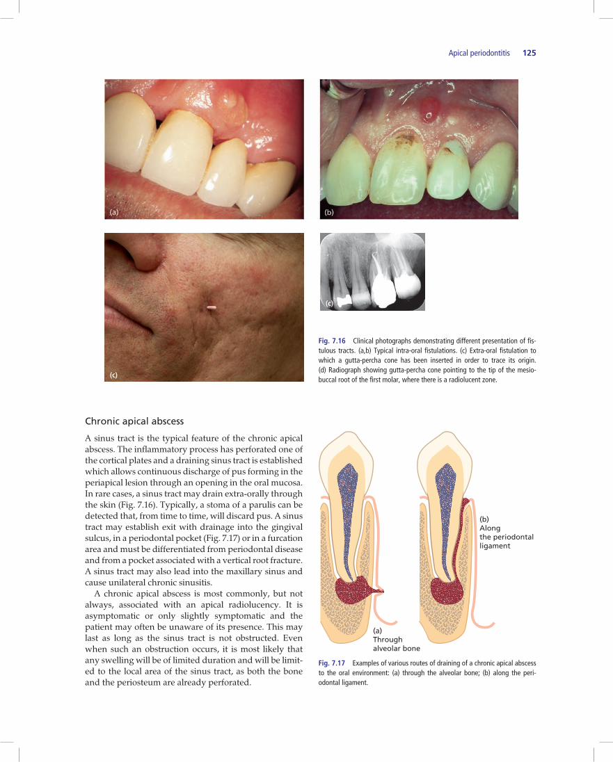
Apical periodontitis 125
Chronic apical abscess
A sinus tract is the typical feature of the chronic apical abscess. The inflammatory process has perforated one of the cortical plates and a draining sinus tract is established which allows continuous discharge of pus forming in the periapical lesion through an opening in the oral mucosa. In rare cases, a sinus tract may drain extra-orally through the skin (Fig. 7.16). Typically, a stoma of a parulis can be detected that, from time to time, will discard pus. A sinus tract may establish exit with drainage into the gingival sulcus, in a periodontal pocket (Fig. 7.17) or in a furcation area and must be differentiated from periodontal disease and from a pocket associated with a vertical root fracture. A sinus tract may also lead into the maxillary sinus and cause unilateral chronic sinusitis. A chronic apical abscess is most commonly, but not always, associated with an apical radiolucency. It is asymptomatic or only slightly symptomatic and the patient may often be unaware of its presence. This may last as long as the sinus tract is not obstructed. Even when such an obstruction occurs, it is most likely that any swelling will be of limited duration and will be limit-ed to the local area of the sinus tract, as both the bone and the periosteum are already perforated.
Fig. 7.16 Clinical photographs demonstrating different presentation of fis-tulous tracts. (a,b) Typical intra-oral fistulations. (c) Extra-oral fistulation to which a gutta-percha cone has been inserted in order to trace its origin. (d) Radiograph showing gutta-percha cone pointing to the tip of the mesio-buccal root of the first molar, where there is a radiolucent zone.
(c)
(c)
(a)Throughalveolar bone
(b)Alongthe periodontalligament
Fig. 7.17 Examples of various routes of draining of a chronic apical abscess to the oral environment: (a) through the alveolar bone; (b) along the peri-odontal ligament.

126 The Necrotic Pulp
Cellulitis
Cellulitis is a symptomatic edematous inflammation associated with diffuse spreading of invasive micro-organisms through connective tissue and fascial planes. Its main clinical feature is diffuse swelling of facial or cer-vical tissues. Cellulitis is usually a sequel of an apical abscess that penetrated the bone, allowing the spread of pus along paths of least resistance, between facial struc-tures. This usually implies the fascial planes between the muscles of the face or neck. Spreading of an infection may or may not be associated with systemic symptoms such as fever and malaise. Since cellulitis is usually a sequel of an uncontrolled apical abscess, other clinical features typical of an apical abscess are also expected. Spreading of an infection into adjacent and more remote connective tissue compartments may, rarely, result in serious or even life-threatening complications. Cases of Ludwig’s angina (17), orbital cellulitis (7), cavernous sinus thrombosis (9) and even death from a brain abscess (16, 22) originating from a spreading dental infection have been reported.
Condensing osteitis
Condensing osteitis is a diffuse radiopaque lesion believed to represent a localized bone reaction to a low-grade inflammatory stimulus, usually seen at an apex of a tooth (or its extraction site) in which there has been a long-standing pulp disease. It is characterized by over-production of bone in the periapical area, mostly around the apices of mandibular molars and premolars that had long-standing chronic pulpitis. The pulp of the involved tooth may be vital and chronically inflamed or may have become necrotic with time, leaving the radiopaque sequella. Normally the condition does not prompt treat-ment per se, unless the pulp undergoes further break-down leading to necrosis. The radiopacity may or may not disappear after endodontic treatment or tooth extrac-tion (15) (see also Chapter 14).
References
1. Abbott PV. Classification, diagnosis and clinical manifesta-tion of apical periodontitis. Endod. Topics 2004; 8: 36–54.
2. Arwill T, Heyden G. Histochemical studies on cholesterol formation in odontogenic cysts and granulomas. Scand. J. Dent. Res. 1973; 81: 406–410.
3. Baumgartner JC, Falkler WA Jr. Reactivity of IgG from explant cultures of periapical lesions with implicated micro-organisms. J. Endod. 1991; 17: 207–12.
4. Baumgartner JC, Falkler WA Jr. Biosynthesis of IgG in peri-apical lesion explant cultures. J. Endod. 1991; 17: 143–6.
5. Baumgartner JC, Falkler WA Jr. Detection of immunoglobu-lins from explant cultures of periapical lesions. J. Endod. 1991; 17: 105–110.
6. Bergenholtz G. Microorganisms from necrotic pulp in trauma tized teeth. Odontol. Revy. 1974; 25: 347–58.
7. Bullock JD, Fleishman JA. The spread of odontogenic infec-tions to the orbit: diagnosis and management. J. Oral Maxill. Surg. 1985; 43: 749–55.
8. Cutler CW, Arnold RR, Schenkein HA. Inhibition of C3 and IgG proteolysis enhances phagocytosis of Porphyromonas gingivalis. J. Immunol. 1993; 151: 7016–29.
9. Fielding AF, Cross S, Matise JL, Mohnac AM. Cavernous sinus thrombosis: report of case. J. Am. Dent. Ass. 1983; 106: 342–5.
10. Formigli L, Orlandini SZ, Tonelli P, Giannelli M, Martini M, Brandi ML, Bergamini M, Orlandini GE. Osteolytic process-es in human radicular cysts: morphological and biochemical results. J. Oral Pathol. Med. 1995; 24: 216–20.
11. Furukawa S, Kuchma SL, O’Toole GA. Keeping their options open: acute versus persistent infections. J. Bacteriol. 2006; 188: 1211–7.
12. Gomes BPFA, Drucker DB, Lilly JD. Association of specific bacteria with some signs and symptoms. Int. Endod. J. 1994; 27: 291–8.
13. Harris M, Jenkins MV, Bennett A, Wills MR. Prostaglandin production and bone resorption by dental cysts. Nature 1973; 245: 213–5.
14. Harris DP, Goodrich S, Mohrs K, Mohrs M, Lund FE. Cutting edge: the development of IL-4-producing B cells (B effector 2 cells) is controlled by IL-4, IL-4 receptor alpha, and Th2 cells. J. Immunol. 2005; 175: 7103–7.
15. Hedin M, Polhagen L . Follow-up study of periradicular bone condensation. Scand. J. Dent. Res. 1971; 79: 436–40.
16. Henig EF, Derschowitz T, Shalit M, Toledo E, Tikva P, Aviv T. Brain abcess following dental infection. Oral Surg. Oral Med. Oral Pathol. Oral Radiol. Endod. 1978; 45: 955–8.
17. Hought RT, Fitzgerald BE, Latta JE, Zallen RD. Ludwig’s angina: report of two cases and review of the literature from 1945 to January 1979. J. Oral Surg. 1980; 38: 849–55.
18. Jansen HJ, van-der Hoeven JS, van-den Kkiboom CWA, Goertz JH, Camp PJ, Bakkeren JA. Degradation of immuno-globulin G by periodontal bacteria. Oral Microbiol. Immunol. 1994; 9: 345–51.
19. Kaneko T, Okiji T, Kan L, Takagi M, Suda H. Ultrastructural analysis of MHC class II molecule-expressing cells in experi-mentally induced periapical lesions in the rat. J. Endod. 2001; 27: 337–42.
20. Kettering JD, Torabinejad M, Jones SL. Specificity of anti-bodies present in human periapical lesions. J. Endod. 1991; 17: 213–6.
21. Lacey DL, Timms E, Tan HL, Kelley MJ, Dunstan CR, Burgess T. Osteoprotegerin ligand is a cytokine that regu-lates osteoclast differentiation and activation. Cell 1998; 93: 165–76.
22. Li X, Tronstad L, Olsen I. Brain abscess caused by oral infec-tion. Endod. Dent. Traumatol. 1999; 15: 95 -101.
Review paper analyzing potential pathways by which root canal infection can initiate brain abscess.
23. Lukic A, Vasilijic S, Majstorovic I, Vucevic D, Mojsilovic S, Gazivoda D, Danilovic V, Petrovic R, Colic M. Characterization of antigen-presenting cells in human apical periodontitis lesions by flow cytometry and immunocytochemistry. Int. Endod. J. 2006; 39: 626–36.

Apical periodontitis 127
24. Meghji S, Harvey W, Harris M. Interleukin 1-like activity in cystic lesions of the jaw. Br. J. Oral Maxillofac. Surg. 1989; 27: 1–11.
25. Metzger Z. Macrophages in periapical lesions. Endod. Dent. Traumatol. 2000; 16: 1–8.
26. Metzger Z, Featherstone L, Ambrose W, Trope M, Arnold RR. Kinetics of coaggregation of Porphyromonas gingivalis HG405 with Fusobacterium nucleatum PK1594. A study with a novel V-max automated kinetic coaggregation assay. Oral Microbiol. Immunol. 2001; 16: 163–9.
27. Metzger Z, Abramovitz I. Periapical lesions of endodontic origin. In: Ingle’s Endodontics, 6th edn (Ingle JI, Bakland LK, Baumgartner JC, eds). Hamilton, Ontario: BC Decker, 2008; 494–519.
28. Nair PNR. Light and electron microscopic studies of root canal flora and periapical lesions. J. Endod. 1987; 13: 29–39.
Classic paper describing patterns of bacterial colonization in teeth with infected pulp necrosis and the microscopic features of the associated periapical inflammatory lesions.
29. Nair PNR, Sjögren U, Schumacher E, Sundqvist G. Radicular cyst affecting a root-filled human tooth: a long-term post-treatment follow-up. Int. Endod. J. 1993; 26: 225–33.
30. Nair PNR, Pajarola G, Schroeder HE. Types and incidence of human periapical lesions obtained with extracted teeth. Oral Surg. Oral Med. Oral Pathol. Oral Radiol. Endod. 1996; 81: 93–102.
31. Nair PNR. Apical periodontitis: a dynamic encounter between root canal infection and host response. Periodontol. 2000 1997; 13: 121–48.
32. Nair PNR, Henry S, Cano V, Vera J. Microbial status of apical root canal system of human mandibular first molars with primary apical periodontitis after ”one visit” endodon-tic treatment. Oral Surg. Oral Med. Oral Pathol. Oral Radiol. Endod. 2005; 99: 231–52.
33. Nair PNR. On the causes of persistent apical periodontitis: a review. Int. Endod. J. 2006; 39: 249–81.
34. Nair PNR. Pathobiology of primary apical periodontitis. In: Pathways of the Pulp, 9th edn (Cohen S, Hargreaves KM, eds). Philadelphia: Elseiver, 2006; 541–579.
35. Naldini A, Morena E, Filippi I, Pucci A, Bucci M, Cirino G, Carraro F . Thrombin inhibits IFN-gamma production in human peripheral blood mononuclear cells by promoting a Th2 profile. J. Interfer. Cytok. Res. 2006; 26: 793–9.
36. Noguchi N, Noiri Y, Narimatsu M, Ebisu S. Identification and localizationof extraradicular biofilm-forming bacteria associated with refractory endodontic pathogens. App. Envir. Microbiol. 2005; 71: 8738–43.
37. Noiri Y, Ehara A, Kawahara T, Takemura N, Ebisu S. Participation of bacterial biofilms in refractory and chronic apical periodontitis. J. Endod. 2002; 28: 679–83.
38. Ricucci D, Bergenholtz G. Histologic features of apical peri-odontitis in human biopsies. Endod. Topics 2004; 8: 68–87.
39. Siqueria JF. Periapical actinomicosis and infection with Propionobacterium propionicum. Endod. Topics 2003; 6: 78–95.
40. Siquerira JF, Rocas IN. Bacterial pathogenesis and media-tors in apical periodontitis. Braz. Dent. J. 2007; 18: 267–80.
41. Skaug N. Soluble proteins in fluid from non-keratinizing jaw cysts in man. Int. J. Oral Surg. 1977; 6: 107–21.
42. Stashenko P, Yu SM. T helper and T suppressor cell reversal during the development of induced rat periapical lesions. J. Dent. Res. 1989; 68: 830–4.
43. Stashenko P, Teles R, D’Souza R. Periapical inflammatory responses and their modulation. Crit. Rev. Oral Biol. Med. 1998; 9: 498–521.
The nature of the periapical inflammatory response to root canal infection and the immunopathological mechanisms involved by which it is controled are the focus of this review article.
44. Suda T, Takahashi N, Udagawa N, Jimi E, Gillespie MT, Martin TJ. Modulation of osteoclast differentiation and function by the new members of the tumor necrosis factor receptor and ligand families. Endocr. Rev. 1999; 20: 345–57.
45. Summers L. The incidence of epithelium in periapical gran-ulomas and the mechanism of cavitation in apical dental cysts in man. Arch. Oral Biol. 1974; 19: 1177–1180.
46. Sundqvist G. Bacteriological studies of necrotic dental pulps. Thesis. Umeå University, Umeå, Sweden, 1976.
47. Sundqvist G, Carlsson J, Herrmann B, Tarnvik A. Degradation of human immunoglobulins G and M and complement factors C3 and C5 by black-pigmented Bacteroides. J. Med. Microbiol. 1985; 19: 85–94.
48. Sundqvist G, Figdor D, Hanström L, Sorlin S, Sandström G. Phagocytosis and virulence of different strains of Porphyromonas gingivalis. Scand. J. Dent. Res. 1991; 99: 117–29.
49. Sundqvist G. Associations between microbial species in dental root canal infections. Oral Microbiol. Immunol. 1992; 7: 257–62.
50. Tani-Ishii N, Kuchiba K, Osada T, Watanabe Y, Umemoto T. Effect of T-cell deficiency on the formation of periapical lesions in mice: histological comparison between periapical lesion formation in BALB/c and BALB/c nu/nu mice. J. Endod. 1995; 21: 195–9.
51. Teitelbaum SL. Bone resorption by osteoclasts. Science 2000; 289: 1504–8.
52. Ten Cate AR. The epithelial cell rests of Malassez and the genesis of the dental cyst. Oral Surg. Oral Med. Oral Pathol. Oral Radiol. Endod. 1972; 34: 956–64.
53. Teronen O, Hietanen J, Lindqvist C, Salo T, Sorsa T, Eklund KK, Sommerhoff CP, Ylipaavalniemi P, Kontinen Y. Mast cell-derived tryptase in odontogenic cysts. J. Oral Pathol. Med. 1996; 25: 376–81.
54. Tronstad L, Barnett F, Gervone F. Periapical bacterial plaque in teeth refractory to endodontic treatment. Endod. Dental Traumatol. 1990; 6: 73–7.
55. Wallstrom JB, Torabinejad M, Kettering J, McMillan P. Role of T cells in the pathogenesis of periapical lesions. A prelimi-nary report. Oral Surg. Oral Med. Oral Pathol. Oral Radiol. Endod. 1993; 76: 213–8.
56. Wang CY, Stashenko P. Characterization of bone-resorbing activity in human periapical lesions. J. Endod. 1993; 19: 107–11.
57. Weiss EI, Shaniztki B, Dotan M, Ganeshkumar N, Kolenbrander PE, Metzger Z. Attachment of Fusobacterium nucleatum PK1594 to mammalian cells and its coaggregation with periopathogenic bacteria are mediated by the same galactose-binding adhesin. Oral Microbiol. Immunol. 2000; 15: 371–7.