chapter 5 marangoni convection and protein crystallisation · marangoni convection and protein...
TRANSCRIPT

175
Chapter 5
Marangoni convection and protein crystallisation
5.1 Introduction
Protein crystallisation is thought to yield better results when performed in a microgravityenvironment. It is assumed that the absence of gravity-driven buoyancy is one of the mainfactors responsible for an improvement of the quality of the grown crystals. However, whilebuoyancy is reduced significantly in microgravity, Marangoni convection might still be present.In co-operation with crystal growers and developers of protein crystallisation facilities a short-duration microgravity experiment was performed in order to investigate possible Marangonieffects in a hanging drop configuration. This experiment also served to test newly developedprotein crystallisation units.
Firstly, an introduction to protein crystallisation (which is still considered more an artthan a science [1]) and its relation to microgravity is provided. Then the Marangoni effect isdiscussed, as well as its possible occurrence in protein crystallisation systems. Convection isthought to be detrimental to protein crystal growth, and the arguments generally provided tosupport this hypothesis are presented subsequently. Finally, a description is given of the proteincrystallisation hardware and the results of the microgravity experiment.
Some of the information in this chapter has already been provided in earlier chapters(specifically chapter 1). Since this chapter constitutes a separate part of this thesis, it wasdecided not to refer to these earlier chapters to improve readability.
5.2 Protein crystallisation
The knowledge of the three-dimensional structure of a protein is essential for theunderstanding and explanation of the biological activity of such a protein. For larger molecules(> 20000 Dalton), the only way to determine this structure is X-ray diffraction, which requiressingle crystals of the protein (see e.g. [2, 3, 4]). Failure to produce any crystals at all or crystalsof high enough quality is currently the most important obstacle in protein X-raycrystallography.
Protein crystallisation is generally performed by dissolving the protein in a solutioncontaining a precipitating agent, a pH-buffer and in some cases additional additives, such asanti-fungal agents. The physical properties of this solution are then altered in such a way thatthe solution becomes supersaturated. This can be done by changing the precipitantconcentration, the temperature or the pH-value. Currently, most protein crystallisationmethods are based on supersaturating the solution by changing the precipitant concentrationand there are four such methods:

176 Chapter 5
1. Vapour diffusion.2. Liquid-liquid diffusion.3. Dialysis.4. Batch crystallisation.Details concerning these methods can be found in various publications (for example [5, 6,
7, 8]). The vapour diffusion method, especially the hanging drop method, is the most widelyused method for protein crystallisation. With this method, a hanging drop containing theprotein solution is separated by an air space from a reservoir containing precipitant solutionwith a higher precipitant concentration than the protein solution. Usually, a buffered proteinsolution is mixed 1:1 with a buffered precipitant solution, forming the drop solution, while thepure precipitant solution is placed in the precipitant reservoir.
Three kind of precipitants are used. These are salts, such as. sodium chloride andammonium sulphate, organic solvents, such as ethanol and acetone, and polyethylene glycol(PEG) of various molecular weights. In salt or PEG systems water is transferred from theprotein to the precipitant solution, while in organic solvent systems the organic solvent istransferred to the protein solution from the precipitant solution. The use of organic solvents islimited, because organic solvents tend to induce protein denaturation [9].
The supersaturation is a function of pH, ionic strength, dielectric constant of the liquid,temperature and protein concentration. The influence of the various factors is described, forexample, by Arakawa and Thimasheff [9]. The level of supersaturation should be higher fornucleation than for protein crystal growth. This explains the need for accurate control ofsupersaturation.
One phenomenon often seen in protein crystal growth is the “cessation of growth”phenomenon [10]. It is found frequently that protein crystals grow to a fixed terminal size,after which no further growth takes place. When these crystals are cleaved subsequently, bothpieces start growing on their newly exposed faces till both have reached their terminal size.The phenomenon is probably due to surface contamination, which can be influenced byconvection.
5.3 Microgravity
Under normal terrestrial conditions, gravity is generally assumed to disturb the crystalgrowth from solutions in two ways:1. Due to mass transfer during the crystallisation process, concentration gradients develop
around the growing crystal. The associated density gradients result in buoyancy forcesand thereby initiate Rayleigh convection. The influence of convection on protein crystalquality has been the subject of many investigations. It is believed to be detrimental tocrystal quality. There is no general agreement on the subject, however.
2. Crystals growing free in solution are subjected to sedimentation. This causesasymmetrical growth and intergrowth of crystals.

Marangoni convection and protein crystallisation. 177
For these two reasons it is believed that the use of a microgravity environment canimprove the quality of the growing crystal. In addition, under microgravity conditions it mightbe possible to grow crystals of proteins, which do not crystallise at all under terrestrialcircumstances. Additional advantages of a microgravity environment might be found incontainerless processing, as foreign surfaces affect nucleation and growth of protein crystals[11, 12].
Another aspect of the influence of gravity on protein crystals is outlined by Noever [13].In his paper it is stated that gravitational strain can lead to a maximum stable size for proteinaggregates and protein crystals, above which the aggregate becomes unstable or the crystalbecomes susceptible to strain-induced flaws or cracks. The higher the gravity level is, thesmaller the critical size becomes.
Protein crystallisation in microgravity has been the subject of many experiments.Experiments have been performed on board of longer duration flights such as Russian satellites(Photon and COSIMA-2) [14, 15, 16], the space station MIR [17], Chinese spacecrafts(COSIMA-1 [18], FSW-2 [19]), and on board of the US Space Shuttle [11, 12, 20, 21 ,22, 23,24, 25, 26], but also on shorter duration sounding rocket flights (TEXUS [20, 21] andMASER [27]).
Some experiments thus far indicated that crystals grown in microgravity are on averagelarger and of better quality (with respect to X-ray diffraction properties). Usually, theimprovement found is only with respect to experiments conducted in parallel on earth. Inrecent investigations, however, improvement in crystal quality (enhancement of X-raydiffraction resolution) with respect to the best crystals ever grown on earth has been found forvarious proteins [23, 24, 26]. Among the best results are those obtained by Day andMcPherson [12] for the crystallisation of satellite tobacco mosaic virus by liquid-liquiddiffusion. They found the X-ray resolution of microgravity crystals to be as good as 1.8 Åcompared to 2.3 Å for the best crystals ever grown on earth.
Not only did the microgravity experiments sometimes produce better [12, 15, 17, 18, 19,22, 23, 24, 25, 26, 27] and larger [11, 12, 14, 17, 18, 19, 20, 21, 22, 24, 25, 27] crystals (thanin the reference experiments), in some cases a different morphology (space group and unit celldimensions) [11, 12] and a faster growth was observed. Occasionally, a different (better)crystal habit was found [11, 12, 14, 15, 16, 17, 19, 22], sometimes as a result of the absence ofsedimentation. Uniform quality is the major difference between space- and lab-grown crystalsaccording to Day and McPherson [12, 23]. They also observed an absence of secondarynucleation and crystal aggregation. In some other experiments no crystals were found on earthin contrast to the microgravity experiments [14, 21], from which it was concluded thatmicrogravity may favour the nucleation step. Other authors, however, have concluded from thefact that multiple crystals were found on earth in contrast to only a small number of crystals inspace, that nucleation may be suppressed in microgravity [11, 15, 24]. McPherson and Dayargue that, in the case of vapour diffusion, nucleation is increased in microgravity, whilenucleation is reduced in microgravity when liquid-liquid diffusion is used [12]. They also founda faster equilibration of the protein droplet and the precipitant reservoir (vapour diffusion) in

178 Chapter 5
microgravity, contrary to the (preliminary) findings of Sibille and Baird [28]. Strong et al.found that in microgravity a higher lysozyme concentration was necessary to produce crystalsthan on earth [17]. During the STS-29 flight showers of small crystals were produced in eachcrystallisation chamber for each of 15 proteins. No explanation was given [22]. Littke and Johnused Schlieren photography to demonstrate that no convection plumes arise around growingcrystals in microgravity in a liquid-liquid diffusion set-up [21]. Hilgenfeld et al. [16] were theonly investigators finding a reproducibly worse crystal size and quality in microgravity. Theycrystallised lysozyme (from Streptomyces coelicolor) and thermolysin on the COSIMA-1 and 2missions.
All these findings are somehow contradictory and no sufficient explanation has yet beengiven for all results obtained in microgravity. A summary of observations regarding theinfluence of microgravity has been given by McPherson [29].
5.4 Marangoni convection in protein crystallisation systems
Although microgravity might prevent the density-driven Rayleigh convection fromdeveloping, it does not rule out another type of convection; the surface-tension-drivenMarangoni convection. In many protein crystallisation systems, solvent evaporates from thesolution in order to create and maintain the supersaturation conditions necessary for crystalgrowth. Due to differences in surface tension along these evaporating interfaces, theMarangoni effect may introduce unwanted flows, even under microgravity conditions. Itshould be noted that Marangoni effects are only possible in the vapour diffusion and the liquid-liquid diffusion crystallisation methods, as a phase boundary is only present when thesemethods are used.
Relatively little is know about the possibility of Marangoni convection occurring duringprotein crystallisation. In one paper, some qualitative remarks have been made about thesubject [30]. Another paper describes Marangoni convection as a result of temperaturegradient in solidifying lysozyme solution drops, but this paper is not pertinent to proteincrystallisation [31]. In two more recent papers [32, 33], Monti and Savino try to model vapourdiffusion systems from a fluid dynamic point of view. They included both Marangoni andRayleigh convection in their model. These papers are commented on at the end of this section.Firstly, the possibility of a Marangoni effect occurring in protein crystallisation is discussedqualitatively. Then, some aspects of interfacial behaviour of protein solutions are reviewed.Finally, some aspects relating to Marangoni convection in protein crystallisation systems areassessed quantitatively.
5.4.1 Qualitative description of the phenomenon

Marangoni convection and protein crystallisation. 179
Marangoni convection can be the result of concentration and temperature gradients alongan interface. Increasing, or sometimes decreasing, temperature as well as increasing theconcentration of a surface-tension-lowering solute may locally decrease the surface tension,and liquid is drawn from places with low surface tension to high surface tension areas. It hasbeen demonstrated in the past [e.g. 34, 35] that Marangoni convection can occur in twodifferent forms:1. Macro-convection, where convection originates from concentration or temperature
differences due to an asymmetry in the system.2. Micro-convection, where the convection is initiated by small (random) temperature or
concentration disturbances that grow in time.Micro-convection can only develop when the system under consideration is stationary
unstable with respect to the Sternling-Scriven criteria [36, 37], i.e. roll-cell instabilities canappear if mass transfer takes place out of the phase of higher kinematic viscosity and lowermolecular diffusivity if the gradient of static surface tension with concentration is negative, orinto this phase if the gradient of static surface tension with concentration is positive. Whenanalysing protein crystallisation systems, distinction should be made between the variousprecipitant systems. The analysis is complicated by the fact that a protein crystallisation systemis a multi-component system. When a salt is used as a precipitant, water is transferred from theprotein solution to the air. Both salt and protein concentration then increase. Salt increases thesurface tension while proteins decrease it (see section 5.4.2). Protein is likely to have a largerinfluence on surface tension. Therefore, according to Sternling and Scriven, the system isoscillatory unstable, rather than stationary unstable. When an organic solvent is used as aprecipitant, the organic solvent is transferred to the protein solution from the air, therebylowering the surface tension. Also in this case, the protein crystallisation system is oscillatoryunstable with respect to the Sternling-Scriven criteria. In either case, Marangoni convection istheoretically possible, but micro-convection is very unlikely.
Macro-convection in a hanging drop configuration could occur as a result of anasymmetry with respect to the concentration field. Asymmetry is present in a proteincrystallisation system, for example, due to the fact that the distance between precipitantreservoir and drop is not the same everywhere, and that generally the drop is shapedasymmetrically. Macro-convection is more intense in a stationary unstable system, and istherefore less intense in a protein/salt system.
Even when no mass transfer occurs between the liquid and the gas phase, concentrationgradients might develop along the liquid-gas interface in a crystallisation system. An example isgiven for a protein/salt system. According to various authors [10, 29, 38] a protein depletionzone might develop around a growing crystal. This depletion zone can have dimensions of theorder of 10-4-10-3 m [10, 39], about an order of magnitude smaller than crystallisation dropsize. It is disturbed by Rayleigh convection in normal gravity, which is believed to lead toinferior crystal growth (see section 5.5). However, Marangoni effects might also disturb thedepletion zone profile. Suppose the distance of a crystal from the interface is comparable to thethickness of the depletion zone. The protein concentration (cp) at the interface nearest to thecrystal is then lower than in the surrounding areas. Liquid with higher protein concentration is

180 Chapter 5
drawn to this spot, increasing the protein concentration and increasing the growth speed of thecrystal face facing the liquid-gas interface (figure 1). In this way the Marangoni effect mayserve to augment the crystallisation growth rate or cause the crystal habit to change.
figure Fout! Bladwijzer niet gedefinieerd. Crystal growing in a drop, surrounded bya protein depletion halo. When the crystal is growing close to the interface,the Marangoni effect increases the mass transfer rate of protein toward thecrystal face which is closest to the interface. The protein concentration at 1is larger than at 2.
A Marangoni effect caused by temperature gradients, also called thermocapillarity, canoccur in a protein crystallisation system whenever a macroscopic temperature gradient isapplied, or whenever releasing or absorbing heat of solution causes temperature gradients.When surface tension decreases with temperature, as is usually the case, then a system thatreleases heat during mass transfer is stable, e.g. an organic solvent crystallisation system, whilea system that needs heat during mass transfer, is always unstable, like a protein/salt system[40].
A macroscopic gradient can occur when the supersaturation is brought about by slowlychanging the temperature (however, in this case no gas-liquid interface is usually present) or byplacing heat sources such as lamps in the vicinity of the drop. This should be avoided in anycase.

Marangoni convection and protein crystallisation. 181
One additional comment should be made. It is the requirement of some proteincrystallographers to control the supersaturation levels in a protein solution [41, 42, 43]. Forexample, it could be advantageous to decrease supersaturation levels after nucleation has takenplace. In that case, mass and heat transfer to the drop are exactly the other way as describedabove. The mass transfer systems described above are then stationary unstable with respect tothe Sternling-Scriven criteria and convection is more likely.
The intensity of Marangoni convection due to concentration gradients can be expressedby the dimensionless Marangoni number (analogous to e.g. [44]).
Mac
H
c=−
∂γ∂µD
∆ (1)
In this equation, γ is the static surface tension, c the concentration of the componentinfluencing the surface tension, H a characteristic dimension of the system (film thickness, dropdiameter), µ the dynamic viscosity and D the diffusivity of the component causing the surfacetension gradients. When the Marangoni convection is a result of temperature gradients, theMarangoni number reads:
MaT
H
aT=
−
∂γ∂µ
∆ (2)
In this equation, a is the thermal diffusivity. Marangoni convection is enhanced by astrong dependence of surface tension on either concentration or temperature, low viscosity,small (thermal) diffusivity and large gradients in concentration or temperature. Largeconcentration and temperature gradients parallel to the interface are favoured by large drivingforces for mass and heat transfer, and by approximately equal mass (or heat) transferresistances in the two phases.
Given an unstable system with respect to the Sternling-Scriven criteria, micro-convectiononly develops when the Marangoni number is larger than a critical Marangoni number. Thiscritical number depends on the geometry and the boundary conditions of the system [45].Furthermore, these critical numbers depend on various factors concerning the condition of theliquid-gas interface, such as Gibbs absorption [46], the presence of insoluble surfactants, andthe surface viscosity of the liquid [47]. These factors are discussed in more detail in section5.4.3.
5.4.2 Surface tension of protein solutions.
The size of surface tension gradients is a function of, among other things, theconcentration dependence of the surface tension. For aqueous solutions of organic solvents,this surface tension dependence on concentration is often well known, as for example with

182 Chapter 5
acetone [48], or it can be calculated from surface tension values of the pure components [49].The surface tension of solutions of various salts in water is also known [48]. For proteinsolutions less is known about surface tension and its dependence on concentration.
Greenley reports on the surface tension of various proteins in aqueous solutions as afunction of concentration [50]. Surface tensions are measured in salt-free and 3 M NaClsolutions. All proteins show quite a large influence on surface tension. An increase of theprotein concentration always results in a decrease of the surface tension. This is also found forvarious plasma proteins by Katona et al. [51]. However, the magnitude of this dependence candiffer a factor 15 from one protein to another. Some proteins show surfactant-like behaviour.When the concentration is increased beyond a critical micelle concentration, no decrease ofstatic surface tension is found when the protein concentration is further increased [52]. Addinga salt to a protein solution sometimes alters the surface tension of the solution. In general, thesurface tension of enzymatic protein solutions is more sensitive to salt than that of non-enzymatic proteins. The salt usually increases the surface tension [50]. Changing thetemperature and pH can also affect the surface tension [50,51]. The higher the temperature is,the lower the surface tension. Some authors state that all these changes in surface tension (pH,temperature, effect of a salt) reflect conformational changes of the protein in solution [51, 53].
The surface tension of a protein solution also depends on the time after which a new gas-liquid interface is created [52, 54]. During times varying between somewhat more than 60minutes and 150 hours [51,52,54] the surface tension of a protein solution decreases (at firstexponentially) to a fixed value. The rate of decrease depends on the protein (and determinesthe foamability of the protein solution). Graham and Phillips studied the kinetics of theabsorption and subsequent denaturation of three different proteins at the air-water interface[55]. A more flexible protein such as β-casein is absorbed and changes its conformation at thesame time, in contrast with lysozyme, which has a more static structure.
Greenley also reported on the dependence of surface tension on concentration of PEG-4000. For very small concentrations the dependence is quite large and negative. However, inthe concentration ranges often encountered in protein crystallisation, the surface tensionbecomes independent of the PEG-concentration [50].
In our laboratory, a preliminary investigation was performed into the dependence ofsurface tension on concentration of lysozyme under typical protein crystallisation conditions.Results of these experiments indicate that the value of (dγ/dc) directly after forming the gas-liquid interface has the same order of magnitude as the value for the water/acetone systemaround 5 % w/w, which is -1.6 10-4 m3/ s2. It is important to note that the results are onlyapplicable for freshly formed interfaces. Details of these experiments are presented elsewhere[56].
All these observations show that the surface tension of a protein solution is a complicatedfunction of protein-concentration, salt or organic solvent concentration, temperature, pH,concentration of impurities and time.

Marangoni convection and protein crystallisation. 183
5.4.3 Quantitative analysis of Marangoni effects.
As a protein crystallisation system is a very difficult system to model from first principles,this section presents an assessment of the magnitude of the Marangoni effect based oncomparison.
In the first part of this section, an estimate is presented on the importance of thermalMarangoni convection relative to solutal Marangoni convection, both originating from masstransfer during crystallisation. Subsequently, with the help of a simple model, an attempt ismade to describe the mass transfer of water in a protein/salt crystallisation system and the masstransfer of organic solvent in a protein/organic solvent crystallisation system. From this model,the rates of mass transfer are found, as well as the magnitudes of the mass transfer resistance inliquid and gas phase. This leads to a comparison between the two different crystallisationsystems with respect to the magnitude of gradients in concentration of the surface tensiondetermining solute parallel to the interface. From this a ratio is estimated, indicating the size ofa possible solutal Marangoni effect in one protein crystallisation system compared to the other.Finally, some remarks are made on the interfacial properties of a protein solution.

184 Chapter 5
Solutal versus thermal Marangoni convection.
In order to compare the effect of temperature gradients caused by evaporation with theeffect of concentration gradients caused by the same evaporation, the equations describing theproblem need to be studied. When Newtonian behaviour of the liquid is assumed and interfacialproperties other than the surface tension are neglected (which is very crude as the last part ofthis section indicates), and the influence of salt concentration on surface tension is ignored, thefollowing dimensionless boundary condition to the Navier-Stokes equations remains (time (t),velocity (v) and length are non-dimensionalised using the kinematic viscosity (ν) and the dropdiameter (dd); protein concentration (c) and temperature are non-dimensionalised using theinitial concentration (c0) and temperature(T0)).
( )∇∇ ∇∇ ∇∇ΓΓ ΓΓ⊥ ⋅ = +v Γ
Ma
Scc
MaTc T
Pr (3)
Mac
d c
c
d
=
−∂γ∂
µ
0
D(4)
MaT
d T
aT
d
=
−∂γ∂
µ
0
(5)
In these equations, the Schmidt number (Sc = ν/D) and the Prandtl number (Pr = ν/a)have been introduced. The symbol ∇⊥ indicates the nabla operator normal to the gas-liquidinterface. The symbol ∇Γ indicates the nabla operator parallel to the interface. To get animpression of which term on the right hand side of equation (3) is more important, it is alsonecessary to involve the convection-diffusion equation and the heat balance. These equationsdetermine the size of the gradients in protein concentration and temperature parallel to theinterface present in equation (3).
( )∂∂t
c cSc
c= − ⋅ + ∇v ∇∇1 2 (6)
( )∂∂t
T T T= − ⋅ + ∇v ∇∇1 2
Pr(7)
The approximated boundary conditions to these two equations are (all heat is assumed tobe dissipated in the liquid phase):
∇∇ ⊥ = −cmd
wdp
&
Dc0
(8)
∇∇ ⊥ = =Thd
T
md
aT
H
cd d v
p
& &
λ ρ0 0
∆(9)

Marangoni convection and protein crystallisation. 185
In these equations, λ is the thermal conductivity, ∆Hv is the heat of vaporisation, ρ and cp
are the density and the heat capacity of the liquid, &h is the local heat flux and &m is the localmass flux, which depends amongst other things on local interface concentration, Biot numberand gas phase concentration. Furthermore, wp is the mass fraction of protein. The term -wp inequation (8) accounts for the fact that water evaporates while this equation relates to theprotein concentration.
To get an idea of the ratio of ∇Γc and ∇ΓT, a relationship needs to be obtained for theinfluence of diffusivity and boundary conditions on the interfacial concentration. Consider aone-dimensional problem in which a flux of mass or heat is dissipated by diffusion (a pre-convective state). The larger the coefficient of diffusion is, the larger the interfaceconcentration or temperature is. The problem is the problem of a semi-infinite space (y ≥ 0) inwhich at t = 0 and y = 0 a flux is applied to the boundary. The quantity of interest is called X:
∂∂
∂∂
2
2
X t y
yk
X t y
t
( , ) ( , )= (10)
boundary conditions t X t y= =0 1( , )
tX
yQ
y
>
=
=
00
κ∂∂
A solution for the interfacial quantity (at y = 0) can be derived [57].
X tQ t
k( , )0 1
2= −
κ π(11)
Suppose the flux differs from one place to another by a factor (1+f). The difference in thevalue of the interfacial quantity between these two places can be expressed as:
∆XQf t
k=
2
κ π(12)
When one wants to compare the ratio of differences in interfacial concentration andinterfacial temperature caused by evaporation, the following substitutions should be made (seeequations (6) - (9)) for the quantities X, Q, k and κ.
concentration: X = c kD
=ν
κ = D Qmd
cwd
p= −&
0
temperature: X = T k =νa
κ λ= Qmd
THd
v=&
0
∆
These substitutions lead to:
∆∆ ∆
Γ
Γ
X
X
c
Tw
T
c
c
H
ap
p
v
(
(
concentration)
temperature)=
∇∇
= − 0
0
ρ
D(13)
186 Chapter 5
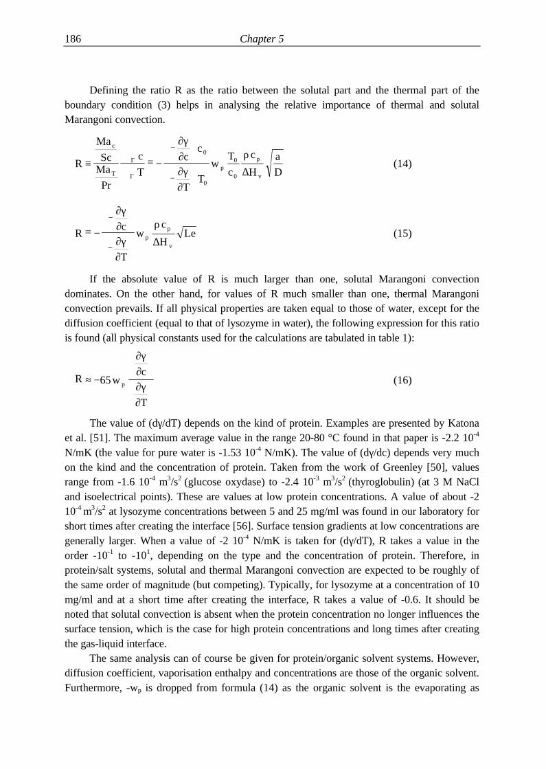
Defining the ratio R as the ratio between the solutal part and the thermal part of theboundary condition (3) helps in analysing the relative importance of thermal and solutalMarangoni convection.
R
Ma
ScMa
c
T
cc
TT
wT
c
c
H
ac
Tp
p
v
≡∇∇
= −
−
−Pr
Γ
Γ ∆
∂γ∂∂γ∂
ρ0
0
0
0 D(14)
Rc
T
wc
HLep
p
v
= −
−
−
∂γ∂∂γ∂
ρ
∆(15)
If the absolute value of R is much larger than one, solutal Marangoni convectiondominates. On the other hand, for values of R much smaller than one, thermal Marangoniconvection prevails. If all physical properties are taken equal to those of water, except for thediffusion coefficient (equal to that of lysozyme in water), the following expression for this ratiois found (all physical constants used for the calculations are tabulated in table 1):
R wc
T
p≈ −
65
∂γ∂∂γ∂
(16)
The value of (dγ/dT) depends on the kind of protein. Examples are presented by Katonaet al. [51]. The maximum average value in the range 20-80 °C found in that paper is -2.2 10-4
N/mK (the value for pure water is -1.53 10-4 N/mK). The value of (dγ/dc) depends very muchon the kind and the concentration of protein. Taken from the work of Greenley [50], valuesrange from -1.6 10-4 m3/s2 (glucose oxydase) to -2.4 10-3 m3/s2 (thyroglobulin) (at 3 M NaCland isoelectrical points). These are values at low protein concentrations. A value of about -210-4 m3/s2 at lysozyme concentrations between 5 and 25 mg/ml was found in our laboratory forshort times after creating the interface [56]. Surface tension gradients at low concentrations aregenerally larger. When a value of -2 10-4 N/mK is taken for (dγ/dT), R takes a value in theorder -10-1 to -101, depending on the type and the concentration of protein. Therefore, inprotein/salt systems, solutal and thermal Marangoni convection are expected to be roughly ofthe same order of magnitude (but competing). Typically, for lysozyme at a concentration of 10mg/ml and at a short time after creating the interface, R takes a value of -0.6. It should benoted that solutal convection is absent when the protein concentration no longer influences thesurface tension, which is the case for high protein concentrations and long times after creatingthe gas-liquid interface.
The same analysis can of course be given for protein/organic solvent systems. However,diffusion coefficient, vaporisation enthalpy and concentrations are those of the organic solvent.Furthermore, -wp is dropped from formula (14) as the organic solvent is the evaporating as

Marangoni convection and protein crystallisation. 187
well as the surface-tension-determining component. When acetone is chosen as an example, Rtakes the value of 84 ((dγ/dc) of this system is taken to be equal to that of 5 % w/w acetone-water without protein). R is generally in the order 101 to 102, indicating that in organic solvent-protein systems solutal Marangoni convection is dominant.
table 1 Values used for the calculations in this chapter
quantity value dimension reference
a (water) 1.43 10-7 m2 s-1 [58]
cp (water) 4.185 106 J kg-1 K-1 [58]
(dγ/dc)o (acetone in water, 25 °C) -1.6 10-4 m3 s-1 [59]
(dγ/dc)o (ethanol in water, 25 °C) -1.9 10-4 m3 s-1 [59]
(dγ/dc)p (lysozyme) -2 10-4 m3 s-1 [56]
(dγ/dT) (water, close to 20 °C) -1.53 10-4 kg s-2 K-1 [58]
(dγ/dT) (protein in water, maximal) -2.2 10-4 kg s-2 K-1 [52]
Doa (acetone in air, 21 °C) 1.04 10-5 m2 s-1 [60]
Doa (ethanol in air, 25 °C) 1.19 10-5 m2 s-1 [58]
Dwa (water in air, 25 °C) 2.56 10-5 m2 s-1 [58]
Dwo (acetone in water, 21 °C) 1.27 10-9 m2 s-1 [60]
Dwo (ethanol in water, 25 °C) 1.27 10-9 m2 s-1 [61]
Dwp (lysozyme in water, 20 °C) 1.04 10-10 m2 s-1 [62]
Dws (NaCl in water, 25 °C) 1.5 10-9 m2 s-1 [48]
m (acetone in air/water, 25 °C) 1.6 10-3 - [63]
m (ethanol in air/water, 25 °C) 2.3 10-4 - [64]
p0 (water, 20 °C) 2337 Pa [58]
W (NaCl) 2 - [65]
∆Hv (acetone) 5.5 105 J kg-1 [48]
∆Hv (water, 25 °C) 2.4 106 J kg-1 [48]
ρ (water, 20 °C) 998 kg m-3 [58]
Solutal Marangoni convection in organic solvent- and salt-protein systems.
In this subsection, the magnitude of the solutal Marangoni effect caused by mass transferin a protein/salt system is compared to the magnitude in a protein/organic solvent system. To

188 Chapter 5
do this, an estimate is required for the size of the concentration gradients parallel to theinterface existing in both systems as a result of geometrical effects. In the first part of thissection it was established that these concentration gradients parallel to the interface aredependent on the flux at the interface and the diffusion coefficient of the surface tensiondetermining component. Therefore, the mass transfer rate in protein crystallisation systems isfirst calculated with the help of a simple one-dimensional, stationary model.
In this model it is assumed that there is no convection yet and all mass transfer is bydiffusion (pre-convective state). To calculate the flux of water, the interfacial concentration ofprotein and the interfacial concentration of water in a protein/salt system, equations are neededfor transfer of water in the drop, transfer of protein in the drop, transfer of water in the vapourphase and an equilibrium relation describing the vapour pressure of water as a function of thesalt concentration. For the transport equations the simplified Maxwell-Stefan equations [66]are used. It is presumed that no mass transfer limitation is present between the vapour phaseand the precipitant reservoir and that there is no mass transfer limitation at the vapour-liquidinterface itself. The latter assumption can in some cases be an oversimplification [67], assurface active agents can form a mass transfer barrier. Further, the water and proteinconcentration in the centre of the drop is considered to be constant for this analysis. The molefraction of protein is neglected compared to that of water and salt. This leads to (see figure2.a):
x xx x
D CN
x x
D CNw i w b l
w i w b
ws totw l
p i p b
wp totw, ,
, , , ,− = −− −
−+
δ δ2
2 2(17)
C CN
Dw g L w g i gw
wa, , , ,− = −δ (18)
x xx x
D CNp i p b l
p i p b
wp totw, ,
, ,− =+
δ2
(19)
Cp
R Tew g i
Wxw i
xw i, ,
,
,=−
−
0
1
(20)

Marangoni convection and protein crystallisation. 189
figure 2 Sketch of the system used for the calculations in this subsection. The filmthicknesses (δl and δg) used are either calculated using the penetrationtheory or equal to drop radius and distance between drop and reservoirrespectively.a. A protein/salt system. The concentration in the graph is the mole fractionof water. Water is evaporating from the drop to the precipitant reservoir.b. A protein/organic solvent system. The concentration in this graph is themole fraction of organic solvent. Organic solvent is evaporating from theprecipitant reservoir to the drop.

190 Chapter 5
In these equations, C is a concentration in mole/m3, x is a mole fraction and N is a moleflux. The subscripts p, s, w, g, i, a, tot, and b refer to protein, salt, water, gas phase, air, total,and bulk of the drop, respectively. R and p0 are the universal gas constant and the vapourpressure of pure water. The D’s in the equations are Maxwell-Stefan diffusion coefficients, andthe two subscripts refer to the components. For example, Dwp is the diffusion coefficientresulting from the friction between protein and water. The last equation is taken from a paperby Fowlis et al. [68]. The δ's in the equations are film thicknesses (from film theory). Themagnitude of this film thickness is estimated from penetration theory for short times t afterstarting the evaporation process (using the diffusion coefficient of salt in water and of water inair respectively):
δ π= D t (21)
Whenever these film thicknesses (δl and δg) are calculated to be larger than drop radiusand distance between drop and precipitant reservoir, respectively, these last parameters areused as film thicknesses. The water concentration in the air adjacent to the precipitant reservoir(Cw,g,L) can be calculated from equation (20) by substituting xw,i with the water fraction in theprecipitant reservoir (xw,L, taken to be constant). These equations can now be solved for Nw,cw,g,i, xw,i and xp,i.
To calculate the flux of organic solvent and the interfacial concentration of organicsolvent in a protein/organic solvent system, equations for the transfer of organic solvent in thedrop and in the air and an equilibrium relation are needed. For this system, the contribution ofprotein to the transfer equations and to a possible Marangoni effect are neglected. Theequations are (see figure 2.b):
x xx x
D CNo i o b l
o i o b
ow toto, ,
, ,− = −− −
δ2
(22)
C CN
Do g L o g i go
oa, , , ,− = −δ (23)
C x C mo g i o i tot, , ,= (24)
In these equations, m is a distribution coefficient, defined as the ratio between theconcentration of organic solvent in the gas phase and the liquid phase at the interface (m = Cg,i
/ Cl,i ). Film thicknesses are calculated from penetration theory as well. The organic solventconcentration in the air can be calculated using equation (24) by substituting xo,i with theorganic solvent fraction in the precipitant reservoir, xo,L.
Some calculations have been performed with typical values listed in tables 1 and 2. Fromthese calculations an indication of the magnitude of the ratio of mass transfer resistances can beobtained by evaluating the Biot number:
Bix x
x xb i
i L
=−−
(25)

Marangoni convection and protein crystallisation. 191
If Bi is much larger than one, the resistance to mass transfer is located in the liquid phaseand if it is much smaller than one, the resistance is located in the gas phase. Calculations for thelysozyme-NaCl system indicate that Biot numbers are in the order 10-3-10-5 for a typicalcrystallisation system (parameters are indicated in table 2) depending on time. For very shorttimes, Biot numbers are smaller but insensitive to the dimensions of the system. For longertimes, Biot numbers and fluxes depend on the distance between drop and reservoir. Thesecalculations indicate that mass transfer resistance in protein/salt systems, even duringmicrogravity, is almost completely located in the gas phase. When convection is present, theBiot number is even smaller. This is contradictory to the hypothesis of Fowlis et al [68]. Theysuspect that during microgravity the evaporation rate of water might be determined by thetransfer of water in the drop. This result is in agreement with the experimental results of Sibilleet al. [69] for evaporation rates in Plaas-Link capillaries, and the more recent experimentalwork of DeTitta and Luft [70].
table 2 System constants used for calculating fluxes, Biot numbers and Rsol
geometric constants
distance between drop and reservoir (m) 0.01
drop radius (m) 2 10-3
lysozyme/NaCl
bulk protein concentration (kg/m3) 50
droplet salt concentration (w/w %) 5
reservoir salt concentration (w/w %) 10
organic solvent
bulk organic solvent concentration (v/v %) 2.5
reservoir organic solvent concentration (v/v %) 5
The model equation (17) shows that, although mass transfer for water is limited by thegas phase resistance, protein crystallisation gradients are nevertheless present. The gradients inprotein concentration are determined both by the magnitude of the water flux, which is limitedby the gas phase resistance, and the film thickness for the protein transport.
Biot numbers for organic solvent systems have been calculated for ethanol and acetone,and are in the order of 10-1-101 for large times and 10-2-10-1 for small times. The different Biotnumbers in the two systems are due to the difference in distribution coefficient, which is 10times as high for acetone as for ethanol, leading to ten times higher Biot numbers. As the Biotnumber in an organic solvent system is much closer to one than in a protein/salt system, aMarangoni effect is anticipated to be stronger for the former system.

192 Chapter 5
To obtain an indication of the magnitude of concentration gradients of the surface-tension-determining component parallel to the interface, a second interface concentration iscalculated by disturbing the flux by a factor. A new perturbed interface concentration iscalculated by either keeping the bulk liquid concentration of the transferring componentconstant when Bi < 1 or the precipitant concentration of the transferring component when Bi >1. The final step in the calculation is the computation of the ratio Rsol, indicating the relativemagnitude of a solutal Marangoni effect in a protein/salt and a protein/organic solvent system.
Rc
c
c
c
solps
po
p
o
=
∆
∆
∂γ∂
∂γ∂
(26)
In these calculations, a thermal Marangoni effect is neglected as well as differences inrheological and other interfacial properties (see next subsection) in the two systems. Values ofRsol have been calculated to compare lysozyme-NaCl to either acetone or ethanol systems. Forthe typical conditions listed in table 2, Rsol takes a value in the order of 10-2-10-1, the values foracetone being smaller than for ethanol, and the values for small times being smaller than forlarge times. The value of Rsol proves to be approximately independent of the factor used todisturb the flux. From these calculations it can be concluded that solutal Marangoni effects as aresult of mass transfer between drop and precipitant reservoir are of the order 101-102 times assmall in protein/salt as in protein/organic solvent systems. These numbers should be readkeeping the assumptions made in mind.
Interfacial properties of protein-solutions.
The gas-liquid interface in a protein crystallisation system rarely behaves as a cleanNewtonian interface with low surface shear and dilatational properties. Apart from proteins,crystallisation systems contain, more often than not, (traces of) other substances, which canaffect the behaviour of the gas-liquid interface seriously. Detergents, trace amount of fats, fattyacids and other proteins can all function in a surfactant-like manner, i.e. they can lower thesurface tension of a solution when present in only small concentrations. Apart from loweringthe surface tension, gas-liquid interfaces can be influenced in other ways by these substancesand the various influences are discussed below.
- Proteins or impurities that form an insoluble monolayer on the interface inhibit theMarangoni effect by their surface elasticity stabilisation (Plateau-Marangoni-Gibbseffect). When an interface is disturbed in such a way that it is partly cleared and absorbedsurfactant is swept outward, the concentration of surfactant is lowered on the clearedpatch and increased in its immediate surroundings. The movement is thereforecounteracted by a surface tension gradient caused by the surfactant surface concentration

Marangoni convection and protein crystallisation. 193
gradient. This effect is able to make an interface completely stable towards micro-convection. The effect is quantified by the elasticity number Nel.
NH
Dels
= −
Γ0
0
∂γ∂Γ
(27)
In this equation, Γ0 is the surface concentration in kg/m2. Berg and Acrivos [47] showedthat even a layer of an insoluble contamination of relatively low surface-tension-loweringcapability can dramatically increase the critical Marangoni number for the onset ofthermocapillary (micro-)convection. As quite a number of proteins change theirconformation in the interface relatively slowly and irreversibly (denaturation) to aconformation which resembles an insoluble surfactant, this effect may render Marangoniconvection non-existent in many protein crystallisation systems after a certain amount oftime. When data by Graham and Phillips [71] is used, the elasticity number of lysozyme,bovine serum albumin (BSA) and β-casein is calculated to be in the order 2 105, in theregion where surface tension decreases linearly with surface concentration. The criticalMarangoni number for the onset of convection is thereby increased thousand-fold [47].However, this elasticity number is not big enough to inhibit convection completely. Forhigher bulk concentrations, which are more common in protein crystal growth,equilibrium surface tension is independent of surface and bulk concentration, making theelasticity number and the Plateau-Marangoni-Gibbs effect vanish.
- Brian and Ross [46] studied the theoretical instability limits of a system in which masstransfer induces solutal Marangoni convection. They demonstrated that the more surfaceactive the transferring component is, the larger the critical Marangoni number for theonset of convection is. They quantified the Gibbs absorption effect using the absorptionnumber Na and found that increasing the absorption number beyond 0.5 renders thesystem completely stable.
( )N
c cab i
=−
Γ0
δ(28)
However, Brian and Ross' analysis is not applicable to the protein/salt crystallisationsystem as the protein is the surface tension determining component, but not thetransferring component. A more quantitative analysis, using their results, is therefore notpossible.
- As mentioned in section 5.4.2, some protein solutions do not change their static surfacetension when the concentration is increased beyond a specific concentration. This doesnot mean that in such a case any kind of surface tension gradient is impossible, since thedynamic surface tension can still vary and also gradients caused by temperaturefluctuations are possible. Nevertheless, it limits the possibility of Marangoni effectsoccurring in protein/salt systems. Graham and Phillips investigated the equilibriumsurface tension of solutions of lysozyme, BSA and β-casein as a function of protein

194 Chapter 5
concentration and found it to be a constant in the concentration ranges used in proteincrystallisation. Moreover, they found that protein was irreversibly absorbed to theinterface and that decreasing the protein concentration beneath the surface layer did notchange the surface tension for any of the three proteins investigated [71]. This indicatesthat solutal Marangoni effects are impossible once the equilibrium surface tension isattained, i.e. once the protein has formed a skin of denatured protein on the gas-liquidinterface.
- For a planar (x-y plane), non-deformable Newtonian interface the stress boundarycondition in one direction becomes (cf. [72]):
( )∂γ∂
µ∂∂
µ∂∂
κ µ∂∂x
v
z
v
y
v
xx s x s s x= − − +
2
2
2
2(29)
In this equation, z is the co-ordinate normal to the interface. This equation was obtainedby ignoring the velocity components in y-direction. Apart from the bulk viscosity two(Newtonian) surface viscosity coefficients appear in this equation, the interfacial shear(µs) and the interfacial dilatational viscosity (κs). For most systems the extra transport ofmomentum constituted by these coefficients is neglected. Usually the surface viscosity istoo small. The relative importance of interfacial shear and dilatational viscosity isexpressed by the Boussinesq number Boµ or Boκ:
BoH
BoH
s s
µ κ
µµ
κµ
= = (30)
When either Bo is larger than 1, bulk flow is strongly influenced by the interfacial shearproperties [73]. Normally, the interfacial shear viscosity is in the order of 10-5 to 10-7 kg/s[74]. Therefore, the interfacial shear viscosity does not play a significant role unless thecharacteristic dimension H (e.g. drop diameter) is smaller than respectively 10-2 to 10-4 m.However, in protein crystallisation systems, gel-like films of protein can form at theliquid-gas interface. These films consist of partly denatured protein [71, 75] and can bethicker than 100 Å [71]. The surface shear viscosity of absorbed proteins has beenmeasured by Graham and Phillips for lysozyme, BSA and β-casein and has maximumvalues of 10 kg/s, 4 10-1 kg/s and smaller than 1 10-3 kg/s respectively [76, see also 77].In a hanging drop configuration (typical dimension is 1 mm), this leads to values of Boµ
of the order of 107, 105 and smaller than 103, respectively. These data indicate thatvelocities due to Marangoni convection are strongly reduced by the presence of proteinfilms.Apart from reducing flows, a large interfacial viscosity also increases the criticalMarangoni number for the onset of micro-convection, as discussed by Berg and Acrivos[47]. This effect is also quantified by the Boussinesq number Boµ. The influence of thesurface viscosity is expressed in the sum (α2 Boµ + Nel). In this sum α is thedimensionless wave number, which typically has a value of 2. Since in their examples theelasticity number was always much larger than the Boussinesq number, Berg and Acrivos
Marangoni convection and protein crystallisation. 195
concluded the effect of interfacial viscosity to be negligible. However, in proteincrystallisation systems the Boussinesq number can be of the same order of magnitude asthe elasticity number, or even larger. Therefore, the large interfacial shear viscosity thatcan occur in protein crystallisation systems has a stabilising influence. A flexible protein,such as β-casein, generally forms interfacial films of lower viscosity than a globularprotein, such as lysozyme [76].Generally, surface rheology of protein solutions is not Newtonian. The interfacial stresscan depend on the history of the interface, resulting in viscoelastic behaviour [73, 78].For some interfaces a viscoplastic behaviour (Bingham surface fluid) has been suggested.A Bingham surface only moves when the applied stress is larger than a specific yieldstress. Such a surface is completely rigid when the applied stress is smaller. However, nosuch models have yet been proposed for protein solutions [73].
In the papers by Monti and Savino [32, 33], Marangoni convection is modellednumerically in vapour diffusion systems. From their calculations they conclude that Marangonieffects may play a role in microgravity for non-symmetrical drops, and around a growingcrystal. However, in their calculations none of the interfacial properties of protein solutionsdiscussed in this section were taken into account. For their calculations, a value for thedependence of surface tension on concentration has been taken from our previous paper [56],which is only valid for short times after creating the interface. Ground experiments were usedto validate their numerical code, and this validation was based on the observation of nucleationzones and not on the observation of any convection. Not surprisingly, crystals nucleated closeto the interface in hanging drop configurations (sitting drops were not tested), from which itwas concluded that the numerical code was qualitatively all right. At best, this reflects thebuoyancy part of their model, but this part should be checked with sitting drop experiments aswell. With respect to the Marangoni part of their calculations, their findings give at best anupper bound to the effect of Marangoni convection on the distortion of the concentration field.
5.5 Protein crystallisation in relation to convection
In this section a summary is presented with theory and proof for the influence ofconvection in protein crystallisation.
Evidence for the existence of convection during protein crystallisation and its detrimentaleffect on protein crystal quality.
Various studies have demonstrated the existence of Rayleigh convection during proteincrystallisation. Using Schlieren optics, some authors found plumes of low density fluid risingfrom crystals growing in solution [79, 80]. Convection was observed during proteincrystallisation in two microscope studies, in which precipitated protein acted as tracer particles[81, 82].

196 Chapter 5
Direct proof for the detrimental effect of convection on protein crystal growth has beengiven in several papers [79, 83, 84]. Pusey et al. observed that the growth of one of the facesof a growing lysozyme crystal was progressively and irreversibly inhibited when it was exposedto an increasing solution flow [79, 83]. Nyce and Rosenberger found that tetragonal lysozymecrystals did not grow when suspended in a thermosyphon, but similar crystals started growingwhen placed in a stagnant solution. They speculated that this might be due to the shear forcesexerted by the solution flow on the crystals, but it could also be a result of the varyingtemperatures used in their experiment. The influence of the solution flow was not found fororthorhombic crystals [84]. Miller et al. compared gel-grown crystals of human serum albuminwith crystals obtained with a normal vapour diffusion method, both in microgravity and onearth. From this comparison it was concluded that crystal quality is probably deteriorated bysolutal convection as the best crystal quality was obtained with gel-grown crystals and crystalsgrown in microgravity [85]. Broom et al. demonstrated that orientation of a crystal withrespect to solutal flow can have a pronounced influence on crystal morphology [86].
Pusey et al. demonstrated by means of a simple model combined with experiments thatRayleigh convection plays an important role (relative to diffusion) for crystals larger than 10microns and that even in microgravity convection starts to be significant for crystalsapproaching sizes of 1 cm [87, 88].
Indirect proof for the detrimental effect of convection on crystallisation is found in theobservation that crystals grown in gels can be of better quality than crystals grown in ordinarysolutions [85]. Convection is suppressed by the gel network, which is believed to be one of theadvantages of gels. Nevertheless, other factors may also be of influence, such as suppression ofheterogeneous and secondary nucleation and the reduction of sedimentation rates [89, 90, 91].Besides positive effects, gels can also have negative effects on protein crystal growth, such asrupture of mechanically weak protein crystals [21] or increasing the number of primarycrystallisation nuclei [19].
Theories explaining the influence of convection on protein crystal growth.
Theories explaining the effect of convection on protein crystal growth can be divided intwo: they are either fluid-dynamic in nature or they are related to the influence of convectionon mass transfer to and from a growing crystal. Hypotheses with a fluid-dynamic nature arepartly summarised by McPherson [29]:1. As proteins are held in the crystal by relatively weak bonds, shear flow along the crystal
might break these crystal bonds and remove protein molecules from the crystal(suggested in various papers, for example [11, 22, 23]).
2. As protein molecules can be oriented in the flow, they are not always susceptible toincorporation in the crystal lattice.
3. Protein molecules are deformed by the flow, which might reduce their attachmentprobability, but which might also cause deformed molecules to get incorporated in thelattice, resulting in crystal defects.

Marangoni convection and protein crystallisation. 197
4. The attachment probability of protein molecules is extremely low and this might even befurther reduced by a convective flow which sweeps the molecules past the interface.Furthermore, the incorporation of impurities which have a higher attachment probabilitythan the protein might be enhanced by convection.
5. Whenever shear flow is present, fluctuations in liquid temperature, concentration, andvelocity can occur, which could result in local growth irregularities. This is a recent resultfrom low velocity turbulence theory.However, by means of order-of-magnitude analyses, Grant and Saville [92] established
that the effect of shear flow on protein deformation and orientation is negligible, and that theforces exerted by shear flow are not large enough to break the bonds in the protein crystal.
Convection around a growing crystal can influence the mass transfer of protein from bulksolution to the crystal interface. This influence is also thought to be important in explaining thedetrimental effect of convection on protein crystal growth. The crystal growth rate isdetermined by the rate of transport of protein from bulk solution to the crystal interface and therate of incorporation in the crystal of protein present at the solid-liquid interface. If theincorporation rate is much faster than the transport rate in the concentration boundary layersurrounding the crystal, the protein concentration at the crystal face is minimal, i.e. it is equalto the saturation concentration. If, to the contrary, the incorporation rate is much slower thanthe transport rate, then the concentration close to the crystal face is maximal, i.e. it is equal tothe bulk protein concentration. The rate of transport of protein in the concentration boundarylayer is determined by convection and diffusion, and since convection increases this transportrate, protein concentration close to the crystal interface generally increases due to the presenceof convection. It is the hypothesis of McPherson [29] that low protein concentration favourscontrolled growth kinetics. High protein concentrations in the boundary layer close to theinterface might lead to secondary nucleation (cf. [93]), which leads to crystal showers andintergrowing crystals. If, to the contrary, the concentration near the crystal interface is lowerthan in bulk solution, concentrations could be in the so-called metastable region of thesolubility diagram. In the metastable region, the so-called Ostwald region, crystal growth issustained, but crystal nucleation is impossible. Furthermore, low concentrations lead to lowergrowth rates, reducing the chance for lattice defects (cf. [10]) and can also lead to differentgrowth kinetics [94]. Another way in which lower concentrations at the interface can influencecrystal growth, is the way concentration determines protein cluster size close to the interface.Tay et al. [93] explain that this influence can work two ways. A higher supersaturation caneither increase cluster size or diminish it. As protein crystallisation might take place by additionof monomer clusters of a specific size (see also [95, 96, 97]), the way convection influencescluster size affects growth rate and crystal habit.
The hypothesis of McPherson is supported by the fact that protein diffusion is a veryslow process (diffusion coefficients are of the order of 10-10 m2/s or even smaller). It istherefore very likely that growth rates in microgravity are limited by diffusion, resulting in verylow concentrations close to the crystal interface. An observation supporting this hypothesis hasbeen made by Malkin and McPherson [96]. Using a light-scattering technique, they found that

198 Chapter 5
growth rates of satellite tobacco mosaic virus (STMV) crystals were limited by transport of thevirus from the bulk to the growth interface (diffusion-limited aggregation). Feher and Kam[10] found quite large concentration gradients around a growing lysozyme crystal of 100 to200 microns (up to 50 % of bulk concentration) using UV-absorption. They used a set-up inwhich convection was probably hindered seriously. Contrary to these findings, experimentaland modelling results of Pusey, Grant, and co-authors on the crystallisation of lysozymeindicate that attachment kinetics is the limiting step up to crystal dimensions of 100 microns[87, 88, 92]. For lysozyme the hypothesis stated above is therefore not sufficient to explain thedetrimental effect of convection. Nevertheless, it might still explain why crystals grown inmicrogravity can grow substantially larger. In a later paper [83], Pusey found experimentallythat growth rates temporarily increased when small (~20 µm) lysozyme crystals were subjectedto solution flows, indicating that mass transfer is more important, when compared toattachment kinetics, than his previous modelling results had indicated. Using interferometry,Miyashita and co-workers found only slight concentration gradients (2.5 % of a bulkconcentration of 2 w/v %) across the boundary layer around a lysozyme crystal ofapproximately 500 microns [38]. Summarising, no experimental and theoretical agreement onthe validity of the hypothesis stated by McPherson has been found. The validity of thehypothesis might very well vary from protein to protein.
Apart from influencing protein transport to the crystal, convection can also influencetransport of impurities to and precipitant from the crystal [30]. Grant and Saville [92]suggested that this might explain the effect of convection on crystal growth for particularcrystals and particular impurities (convection can change the ratio of protein and impurityincorporation, i.e. segregation). Pusey [83] rejected this hypothesis since it cannot explain whysolutal flow does not seem to hinder orthorhombic lysozyme crystal growth, while it doeshinder tetragonal lysozyme crystal growth. As these two crystal forms are very much alike itdoes not seem logical that the same impurity has so great an influence on crystal growth of oneform and so little on the other.
The influence of convection on the transport of rejected precipitant is a beneficial one,when the same line of reasoning is applied as is used to explain the detrimental effect ofconvection on the protein transport. Convection decreases the concentration gradient ofprecipitant across the boundary layer, which leads to a reduction of precipitant concentration atthe interface and subsequently a lower supersaturation. Also in other cases, convection can bebeneficial to crystal quality. If segregation of an impurity takes place at a crystal interface andthis impurity is preferentially rejected, then convection can increase segregation with respect tothe bulk concentrations of protein and impurity [30]. As mentioned before, crystal growthkinetics depend on supersaturation and growth may either be favoured by large or small valuesof supersaturation. Convection usually increases the value of supersaturation at the crystalinterface and this might for some proteins and for some crystal habits be a preferable situation.Furthermore, studies of protein crystals growing in a flow cell showed that forced convectioncan provide stable, uniform growth conditions [94].
Considering all the evidence in literature, our opinion is that the influence of convectionon protein crystallisation should mainly be sought in the influence convection has on mass

Marangoni convection and protein crystallisation. 199
transfer rates from and to the protein crystal. Mass transfer rates influence concentration andthereby supersaturation levels. The exact value of the supersaturation close to the crystalinterface determines attachment rates, local cluster size of the protein and incorporation rate ofimpurities. However, it is our view that the influence of convection on protein crystallisationcan not be generalised too much. That is, the situation changes from protein to protein,crystallisation condition to crystallisation condition, and it even changes from crystal habit tocrystal habit (cf. orthorhombic and tetragonal lysozyme).
figure 3 Sketch of a protein crystallisation unit. Figure 3a shows the launch positionof the crystallisation cell. Figure 3b shows the microgravity configuration.

200 Chapter 5
1. Driving head. 2. Lamp. 3. Turning knob. 4. Syringe (crystallisation cell).5. Glass plunger. 6. Teflon tube (containing protein solution). 7.Crystallisation chamber. 8. Protein solution. 9. Observation window. 10.Teflonised sealing plate. 11. Precipitant chamber. 12. Precipitant solution.13. Metal ring. 14. Teflon ring. 15. Teflon ultrafiltration membrane. 16.Precipitant container.
5.6 A microgravity experiment
A microgravity experiment was performed to investigate the possibility of Marangoniconvection occurring during protein crystallisation. The experiment consisted of observinghanging drops during microgravity. The experiment also served to evaluate crystallisationunits, which are to be used in a Dutch Protein Crystallisation Facility (DPCF) [98]. Theevaluation of the crystallisation units is described elsewhere [56].
The experimental hardware
The design of the crystallisation units used in the experiment is sketched in figure 3. Thecrystallisation unit consisted of a crystallisation cell (a kind of syringe), a precipitant containerand the body of the crystallisation unit. A space was drilled out of the stainless steel body, inwhich the cell could be screwed and the precipitant container could be clasped.

Marangoni convection and protein crystallisation. 201
figure 4 Photograph of the experiment ring with the experiment cells. The windowsvisible on the outer face of the ring were sealed with a metal insert beforethe experiment. The drops were observed through the windows on the innerface of the ring.

202 Chapter 5
figure Fout! Bladwijzer niet gedefinieerd. Photograph of the experiment ringintegrated in the mechanism, which rotates the mirror and opens and closesthe experiment cells. The windows visible on the outer face of the ring weresealed with a metal insert before the experiment. The cylinders protrudingfrom the equipment are the motors driving the mechanism.
The crystallisation cell (figure 4) contained a glass plunger, a Teflon cylinder and aturning knob to move the cylinder. The glass plunger was glued into the turning knob. Whenthe knob was turned, the glass plunger turned around, but stayed in the same axial position.The Teflon cylinder, however, moved in axial direction. In this way, a droplet could be

Marangoni convection and protein crystallisation. 203
extruded from the cylinder. The droplet keeps hanging from the tip of the plunger. Figure 3ashows the cylinder position during launch and re-entry, while figure 3b depicts the cylinder inmicrogravity position. In launch position, the cylinder was pressed on a Teflonised metal plate.A conical bulge was present on the metal plate. This bulge exactly fitted into the cylinder andserved to facilitate the detachment of the drop from the plate. The Teflon tubes used during theexperiment had inner diameters of 1.0, 1.5 and 1.8 mm, corresponding to drop volumes of 6,13 and 19 µl.
The precipitation solution was contained in a precipitant container, manufactured fromTeflon. This container was sealed with an ultrafiltration membrane (the diameter of the pores is0.2 µm). A Teflon membrane on a supporting polyester layer was used (Schleicher & Schuell,TE35). No liquid could pass this membrane. However, vapour transport was not restricted bythe membrane.
The drops could be observed via a window with which each crystallisation chamber isprovided. The droplet was illuminated by a LED, positioned at the other end of the glassplunger (by which the light is transported). Tracer particles were added to the droplets in orderto visualise possible flows. No measurable heat is transported from the LED to the drop. Thehardware for the microgravity experiment consisted of one ring with 16 crystallisation units.All 16 windows were facing towards the centre of the ring (figure 4). The system wasequipped with one CCD-camera with which 4 drops could be observed at the same time bymeans of a pyramid-shaped mirror in the centre of the ring.
Apart from the experiment ring with the cells, the experimental equipment consisted of amechanism, which could rotate the mirror and the knobs on the cells (figure 5), a CCD-camera,a video-8 recorder, an electronic control unit, a temperature logger and a battery package. Allthese elements were accommodated in a vacuum tight late access container.
Overview of the experiments
Solutions were prepared in the laboratory in Groningen and transported in a cooling boxto the launch site Esrange near Kiruna (North-Sweden). In the laboratory and at Esrange thesolutions were kept at 2-4 °C. Precipitant chambers and experiment cells were filled 2.5 daysbefore launch (the launch was postponed for two days due to weather conditions). Experimentcells 2 to 4 were refilled one day before launch (lysozyme started to crystallise in these cells).The late access container with the experiment cells was transferred from the refrigerator to therocket 4 hours before the start of each countdown, where the experiment units were given timeto equilibrate to the temperature in the launch tower (18-19 °C). After the first twocountdowns the container was replaced in the refrigerator.
Maser 5 was launched on April 9, 1992. Microgravity levels during flight were ofexceptional quality and continuously less than 10-6 g [99]. The late access container wasrecovered from the rocket within two hours after impact. The container was then transportedin thermal insulation with a helicopter back to the laboratory at the launch range. After

204 Chapter 5
demounting, the experiment ring was transported back to the University of Groningen where itwas opened for inspection 15 days after the experiment. The ring was kept at a temperature of2-4 °C.
Two months after the microgravity experiment a reference experiment was performed atthe University of Groningen using exactly the same equipment.
Description of the 16 experiments.
The contents of the various experiment cells and of the various precipitant containers arelisted in table 3.
The various lysozyme (Boehringer, crystallised from hen egg white) and NaCl- solutionswere prepared in a 0.2 M HAc/Ac- buffer (pH 4.70). Microcentrifuge cups were filled withlysozyme and filled up with buffer solution till solutions of 50 mg/ml were obtained. The cupswere centrifuged for 10 minutes at 10000 rpm and supernatant solution was transferred inclean cups. NaCl solutions were prepared by dissolving NaCl (analytical grade) in buffersolution (the pH of the resulting salt solutions was 4.43).
The human serum albumin (HSA) and PEG-400 solutions were prepared with 500 mMpotassium phosphate buffer (pH 6.72). Microcentrifuge cups were filled with HSA (Sigma,essentially fatty acid free, fraction V) and filled up with buffer solution and water till 100mg/ml HSA in 50 mM K-phosphate solutions were obtained. The cups were centrifuged for 10minutes at 10000 rpm and the supernatant solution was transferred in clean cups. PEG-400solution was prepared by dissolving PEG-400 (Merck) in buffer solution and water till a 25 %(v/v) PEG in 50 mM K-phosphate solution was obtained.
The phospholipase A2 and accompanying organic solvent solutions were prepared in a 10mM TRIS (2-amino-2(hydroxymethyl)-1,3-propanediol)-HCl /5 mM CaCl2 buffer. The TRIS(Boehringer, crystallised) buffer had pH 7.01. One microcentrifuge cup was filled with 10mg/ml porcine phospholipase A2, centrifuged for 10 minutes at 10000 rpm and clean cups werefilled with supernatant solution. The 10 % methanol, 20 % acetone and 20 % ethanol solutionswere prepared by mixing the respective organic solvents (Merck, analytical grade) with theappropriate amount of buffer solution.
The 5 % (w/w) acetone-solutions were prepared in water. De-ionised water was used forall solutions. To all buffer solutions NaN3 was added in 1 mM concentration.
Table 3. Description of the cell contents
cellnumber
cell contents contents precipitantcontainer
celldiameter
1 water 8 % (w/v) NaCl 1.8 mm

Marangoni convection and protein crystallisation. 205
2 lysozyme / 2 % (w/v) NaCl 8 % (w/v) NaCl 1.8 mm
3 lysozyme / 2 % (w/v) NaCl solid NaCl 1.8 mm
4 lysozyme / 4 % (w/v) NaCl 8 % (w/v) NaCl 1.8 mm
5 human serum albumin /
12.5 % (v/v) PEG 400
25 % PEG 400 1.8 mm
6 phospholipase A2 /
5 % (v/v) methanol
10 % v/v methanol 1.8 mm
7 phospholipase A2 /
10 % (v/v) acetone
20 % v/v acetone 1.8 mm
8 phospholipase A2 /
10 % (v/v) ethanol
20 % v/v ethanol 1.8 mm
9 lysozyme /
2 % (w/v) NaCl /
5 % (w/v) acetone
active charcoal 1.8 mm
10 5 % (w/w) acetone / water 8 % w/v NaCl 1.8 mm
11 lysozyme /
2 % (w/v) NaCl /
5 % (w/v) acetone
8 % w/v NaCl 1.8 mm
12 lysozyme /
4 % (w/v) NaCl /
5 % (w/v) acetone
solid NaCl 1.8 mm
13 water active charcoal 1.8 mm
14 5 % (w/w) acetone / water active charcoal 1.8 mm
15 5 % (w/w) acetone / water active charcoal 1.0 mm
16 5 % (w/w) acetone / water active charcoal 1.5 mm
Tracer particles consisted of ceramic microballoons (Grace), coated (by precipitationreaction) with silver to improve their visibility and to increase their density. The particles wereclassified according to size (diameter between 100 and 200 µm) and density (approximately thesame as water).
The final protein solutions, which were placed in the experiment cells, were prepared atthe launch base by mixing the protein solution with the precipitant solution (and in some cases

206 Chapter 5
buffer solution and/or acetone) in appropriate volumetric amounts. Tracer particles were firstinserted in the experiment cells. Special equipment, devised to select the right amount oftracers, was used for this task. In some cases problems arose, because particles moved due tostatic electricity. In these cases the procedure was repeated. The crystallisation cells weresubsequently filled with the appropriate solutions using syringes equipped with small needles.Slow filling speeds were required, due to the foamability of the protein solutions. Finally, thecells were screwed into the experiment ring.
The precipitant containers were filled with the appropriate precipitant solutions, withsolid NaCl or small cylinders of active carbon (Ceca, AC35, 1.8 mm).
All crystallisation cells, precipitant containers and syringes as well as the crystallisationring were ultrasonically cleaned with water, acetone (p.a.) and a mixture of those liquids. Theywere subsequently flushed with these liquids and dried with compressed nitrogen (absolutelyoil free).
Three experiments were chosen to investigate a protein/salt crystallisation system (2-4,table 3). Lysozyme was chosen, because this protein has been the subject of manycrystallisation studies. Different salt concentrations were used to vary driving forces forevaporation. Acetone was added in two cases (11-12) to investigate whether the protein mightinhibit Marangoni effects which are usually observed in a pure acetone-water system. Only oneexperiment was chosen to investigate a protein/PEG crystallisation system (5). Human serumalbumin was selected, because it is characteristically crystallised with PEG as a precipitant.Three experiments were chosen to examine a protein/organic solvent system (6-9).Phospholipase A2 was selected as a protein as it is easily crystallised with methanol, acetoneand ethanol using the conditions employed in this experiment [100]. Three experiments with anacetone-water system were performed (14-16) to check the occurrence of Marangoni effects inthis protein crystallisation set-up. With this stationary unstable system, violent Marangonieffects are usually observed. Three different experiments were done to investigate whetherdrop volume might have any influence. Finally, three reference experiments were included (1,10 and 13).
Timeline
As described before, four different cells can be observed at the same time. The cells canalso be opened four at a time. The first eight drops were observed twice and only theseexperiment cells were closed towards the end of the experiment. The other eight drops werelost during re-entry. A precise time schedule is given in table 4.
The experiment sequence was activated by the microgravity signal received from theService Module of the sounding rocket.

Marangoni convection and protein crystallisation. 207
Table 4 Timeline of microgravity experiment
time (seconds) action
0-36 cells 1-4 opened and observed
41-82 cells 5-8 opened and observed
87-207 cells 9-12 opened and observed
212-332 cells 13-16 opened and observed
337-378 cells 1-4 observed and closed
382-420 cells 5-8 observed and closed
Results
The contents of cells 1-8 were checked for crystals when they were opened at thelaboratory. This was done by transferring the solution to a vapour diffusion set-up as they arecommonly used in the lab. This set-up consists of plastic depression trays in which theprecipitant solution is brought prior to placing the protein drop on a siliconised glass coverslip. This cover slip is then placed upside down above the precipitant solution. The slip issealed with vacuum grease. The drops were checked under a normal light microscope.
All lysozyme drops contained many imperfect crystals. However, these crystals probablygrew during the entire time the liquid was contained in the cell, because the solutions werealready supersaturated. The HSA drop contained many, very small, needles, which were notbirefringent, however. Movement of these needles in the liquid was observed under themicroscope, indicating some kind of convection. When the phospholipase A2/methanol and/ethanol drops were unscrewed, tracers started circulating very violently, due to evaporation ofthe alcohol. After the drops were placed in the microscope set-up no further tracer movementswere observable with the naked eye. However, microscope observations showed thatconvection was occurring in the ethanol drop. Very small particles (probably precipitatedprotein) were circulating during quite a long time in the drops to and from the place where theliquid-gas interface met the glass wall of the objective plate. The convection was probablysurface tension gradient induced, as the particles seemed to accelerate at the interface. Theconvection was of considerable intensity (larger than in the HSA drop). The convection couldhave been influenced by temperature gradients caused by the lamp of the microscope, heatingthe drop. Nevertheless, it can be concluded from this that the tracers used in the experimentswere too large to show microscopic convection in the drops. It should be remarked that othermicroscope studies in literature also demonstrated the existence of various convection patternsduring protein crystallisation by observation of precipitated protein [81, 82].

208 Chapter 5
Hard copies were made of pictures of drops 1-4 and 5-8 at the beginning and at the endof the experiment and tracer positions were compared. Results of this and other video tapeobservations are summarised in table 5.
The observations of drops 1 and 7 give reason to believe that the contents of these cellshave been interchanged accidentally. It is not very likely that a pure water drop showsconvection as is also demonstrated by the reference experiment (which only showedsedimentation of tracer particles). In cell 7 no convection was observed, in contrast to cells 6and 8. Moreover, cells 1 and 7 appeared to be empty when unscrewed in the lab. Therefore,these experiments were not taken into account.
The water-acetone drops (10, 14, 15, 16) showed Marangoni convection in all cases. Itappeared that neither drop volume nor the kind of precipitant used was of considerableinfluence on convection intensity.
No Marangoni convection was observed for the lysozyme-salt systems. In drops 6 and 8(phospholipase A2 / organic solvent) Marangoni convection was observed for a short time,despite these systems not being stationary unstable with respect to mass transfer. Extruding thedrops might cause some unexpected effects as no movements were observed after ten seconds.However, comparing the drops at the end of the first observation period with the start of thesecond observation period showed that some tracer movements have occurred in between. Oneother reason for decreasing Marangoni activity might be that phospholipase inhibits interfacialmovements. Violent tracer movements which occurred when the drop was transferred to themicroscope equipment showed nevertheless that the protein does not completely stopinterfacial movements (especially not in a stationary unstable system) as could also beconcluded from the microscope observations.
Comparing 9, 11, 12 and 14 leads to the conclusion that lysozyme tends to make theinterface more rigid. No explanation can yet be given for differences between 9,11 and 12.
Table 5 Observations during microgravity experiment.
cell observation
1 movement of tracers first minute; air bubble present;no movement of tracers after 6 minutes
2 no movement of tracers
3 no movement of tracers
4 no movement of tracers
5 drop not observable

Marangoni convection and protein crystallisation. 209
6 tracers move during first ~10 seconds after which they come to astop; tracers seem to have moved slightly after 7 minutes
7 low tracer content; no movements observed after unscrewing;tracers are still at the same place after 7 minutes
8 tracers move during first ~10 seconds after which they come to astop; tracers seem to have moved slightly after 7 minutes
9 only part of small drop visible; no movement of tracers
10 tracers move during the whole observation time
11 tracers move during first ~35 seconds after unscrewing the drop;movements are at most minimal afterwards
12 tracers move during first ~10 seconds after unscrewing the drop;movements are at most minimal afterwards
13 only a small drop is observable; no tracer movements observed
14 tracer movements during the whole observation time
15 tracer movements during the whole observation time
16 tracer movements during the whole observation time;no discernible differences in tracer velocities are observedbetween drops 10, 14, 15 and 16; initial movements in drop 1, 6,11 and 12 are somewhat less than in 10, 14, 15 and 16
One reference experiment showed anomalous behaviour compared to the microgravityexperiment (11), as no convection was observed on earth and tracer movements were observedin microgravity. Some other reference experiment observations were affected by the presenceof air bubbles (5-8, 12). As the microgravity equipment and the tracers used for thisexperiment impose some extra limitations on the observation of convection, it was decided notto repeat this reference experiment. However, some other reference experiments wereperformed with hanging drop equipment as it is commonly used in the laboratory (as describedabove). The drops have been seeded with cross-linked (DVB) polystyrene spheres (chemicallyinert) of 1-85 µm diameter (Duke Scientific) and observed under the microscope, using aninfra-red mirror to avoid thermal Marangoni effects. Sedimentation was observed in everyexperiment. In some of the protein experiments a layer of protein was formed at the interface.Convection was observed for experiments 7, 10 (very minimal) and 14 (violently) andfurthermore for experiments similar to 9, in which b-casein was used instead of lysozyme (veryminimal as well). Even the smaller particles were too large to observe every movement in thedrop, as was demonstrated for experiment number 8, in which no movement was found of thepolystyrene spheres, whereas an experiment with precipitated phospholipase A2 showedconvection on a very small scale. The movement in the drop was definitely not entirely due to

210 Chapter 5
sedimentation, as precipitated protein moved upwards as well as downwards. The intensity ofconvection was less than found after the microgravity experiment, indicating that convectioninduced by temperature gradients had been reduced by the infra-red mirror.
5.7 Conclusions and discussion
The major drawbacks to the microgravity experiment were the small number of cells andthe magnification of the drop observation. Convection magnitudes due to buoyancy have beenmeasured for lysozyme and are of the order of 10-5 m/s for a crystal of 1 micron [79]. Thesevelocities are very small, but supposedly still sufficient to be detrimental to crystal growth. Inorder to establish whether Marangoni convection might have any influence on protein crystalgrowth it is necessary to check for the occurrence of very small flows, not seen with normalmagnifications. This requires larger magnifications and smaller tracer particles than were usedin the experiment described in this study.
Despite this, the experiments revealed that Marangoni effects can occur in proteincrystallisation systems, at least for a short period, but possibly on a smaller scale for longerperiods. The experiment leads to the conclusion that Marangoni effects are probably larger inorganic solvent systems than in salt or PEG systems. This is partly explained in terms ofconcentration gradients in section 5.4.3. Another explanation could be that protein inhibitsinterfacial movements in an organic solvent system less than it would do in a protein/saltsystem. Protein that is not denatured by the organic solvent is probably also less prone todenaturation in the gas-liquid interface.
Both a protein/salt system and a protein/organic solvent system are oscillatory unstablewith respect to solutal Marangoni convection. Marangoni convection probably has lessinfluence on crystal quality than buoyancy has, as it is primarily relevant in the drop regionsclose to the gas-liquid interface. Furthermore, Marangoni convection is more probable inprotein/organic solvent systems than in protein/salt systems, is more likely induced by thermaleffects in protein/salt systems and by solutal effects in protein/organic solvent systems and ismore likely to occur shortly after creating the interface and with proteins with a flexible orrandom conformation. The considerations in the last subsection of section 5.4.3 give reason tobelieve that Marangoni convection does not occur in protein/salt crystallisation systems, atleast not a long time after creation of the interface and when proteins are used that can form aninsoluble layer on the air-liquid interface. Nevertheless, as long as no more definite answers canbe given, it should be taken into account. Marangoni convection can be minimised by:- Avoiding a gas-liquid interface (when dialysis is used, or when temperature is used to
control supersaturation, no gas-liquid interface is present; temperature control mighthave additional advantages [101, 102])
- Minimising mass transfer (equilibration) rates- Adding surfactants, which do not alter crystal quality (detergents [103])- Containerless processing. No macroscopic gradients due to evaporation develop in a
spherical drop.

Marangoni convection and protein crystallisation. 211
- Increase of viscosity (as with gel crystallisation [89, 90, 91] or the use of high PEGconcentrations)None of these measures is in conflict with other protein crystallisation objectives, except,
in some cases, the first and the last [21].
With the simple multi-component model presented in section 5.4.3, it can be concludedthat in the hanging drop configuration for protein/salt systems mass transfer resistance islocated exclusively in the gas phase. The model does not need the assumptions of Fowlis et al.[68] to demonstrate this. Protein gradients in the liquid exist nevertheless. For protein/organicsolvent systems, mass transfer resistance is usually located in the gas phase as well, but undercertain conditions, the resistance to mass transfer may also be mainly located in the liquidphase. The model indicates that when the Biot number is close to one, Marangoni effects arelargest.
List of symbols
a thermal diffusivity [m2 s-1]Bi Biot number [-]Bo Boussinesq number [-]c concentration [kg m-3] or [-]C concentration [mole m-3]dd drop diameter [m]D diffusivity [m2 s-1]Ds surface diffusivity [m2 s-1]f factor used in section 5.4.3 [-]&h heat flux [J m2 s-1]H characteristic liquid dimension (equation (1)) [m]k constant in equation (10) [s m-2]Le Lewis number [-]m distribution coefficient (Cg,i/Cl,i) [-]&m mass flux [kg m-2 s-1]
Ma Marangoni number [-]N mole flux [mole m-2 s-1]Na absorption number [-]Nel elasticity number [-]p0 vapour pressure of pure water [Pa]Q flux at interface in equation (10) [m2/s] or [W m-1 K-1]Pr Prandtl number [-]R ratio as defined in equation (14) [-]R gas constant [J mole-1 K-1]Rsol ratio as defined in equation (26) [-]

212 Chapter 5
Sc Schmidt number [-]T temperature [K] or [-]v velocity [m s-1]w mass fraction [-]W coefficient in equation (20) [-]x mole fraction in liquid [-]X dimensionless quantity in equation (10) [-]y co-ordinate normal to the interface [-]γ surface tension [N m-1]Γ0 surface concentration [kg m-2]δ mass transfer film thickness [m]∆c concentration difference [kg m-3]∆Hv heat of vaporisation [J kg-1]∆T temperature gradient [K]κ constant in equation (10) [m2 s-1] or [W m-1 K-1]κs surface dilatational viscosity [kg s-1]λ thermal conductivity [W m-1 K-1]ρ density [kg m-3]µ dynamic viscosity [Pa s]µs surface shear viscosity [kg s-1]
subscripts
a airb bulk (of droplet)g gas phasei interfaciall liquid phaseL precipitant reservoiro organic solventp proteins saltw water0 initial
Literature
[1] C.E. Bugg, “The future of protein crystal growth”, J. Cryst. Growth, 76, 535-544, 1986[2] T.L. Blundell, L.N. Johnson, “Protein Crystallography”, Academic Press, London, 1976

Marangoni convection and protein crystallisation. 213
[3] J. Drenth, J.R. Heliwell, W. Littke, Chapter 13 in: “Fluid sciences and Materials Sciencein Space”, H.U. Walter (ed.), Springer-Verlag, ESA, Berlin, 1987
[4] J. Drenth, “Principles of Protein X-Ray Crystallography”, Springer Verlag, New York,1994
[5] A. McPherson, “Crystallization of macromolecules: general principles”, Meth.Enzymology, 114, 112-120, 1985
[6] A. McPherson, “Preparation and analysis of protein crystals”, Krieger, Malabar, 1989[7] A. McPherson, Chapter 1 in: “Crystallization of membrane proteins”, ed. by H. Michel,
CRC Press, Boca Raton, 1-51, 1991[8] A. Ducruix, R. Giegé, “Methods of crystallization”, Chapter 4 in “Crystallization of
nucleic acids and proteins. A practical approach.”, A. Ducruix, R. Giegé (eds.), OxfordUniversity Press, Oxford, 73-98, 1992
[9] T. Arakawa, S.N. Thimasheff, “Theory of protein solubility”, Meth. Enzymology, 114,49-77, 1985
[10] G. Feher, Z. Kam, “Nucleation and growth of protein crystals: general principles andassays”, Meth. Enzymology, 114, 77-112, 1985
[11] L.J. DeLucas, F.L. Suddath, R. Snyder, R. Naumann, M.B. Broom, M. Pusey, V. Yost,B. Herren, D. Carter, B. Nelson, E.J. Meehan, A. McPherson, C.E. Bugg, “Preliminaryinvestigations of protein crystal growth using the space shuttle”, J. Cryst. Growth, 76,681-693, 1986
[12] J. Day, A. McPherson, “Macromolecular crystal growth experiments on InternationalMicrogravity Laboratory-1”, Protein Science, 1, 1254-1268, 1992
[13] D.A. Noever, “Size effects in models for mechanically-stressed protein crystals andaggregates”, J. Cryst. Growth, 122, 120-135, 1992
[14] S.D. Trakhanov, A.I. Grebenko, V.A. Shirokov, A.V. Gudkov, A.V. Egorov, I.N.Barmin, B.K. Vainstein, A.S. Spirin, “Crystallization of protein and ribosomal particles inmicrogravity”, J. Cryst. Growth, 110, 317-321, 1991
[15] K. Asano, S. Fujita, T. Senda, Y. Mitsui, “Crystal growth of ribonuclease S undermicrogravity”, J. Cryst. Growth, 122, 323-329, 1992
[16] R. Hilgenfeld, A. Liesum, R. Storm, A. Plaas-Link, “Crystallization of two bacterialenzymes on an unmanned space mission”, J. Cryst. Growth, 122, 330-336, 1992
[17] R.K. Strong, B.L. Stoddard, A. Arrott, G.K. Farber, “Long duration growth of proteincrystals in microgravity aboard the MIR space station”, J. Cryst. Growth, 119, 200-214,1992
[18] V.A. Erdmann, C. Lippmann, C. Betzel, Z. Dauter, K. Wilson, R. Hilgenfeld, J. Hoven,A. Liesum, W. Saenger, A. Müller-Fahrnow, W. Hinrichs, M. Düvel, G.E. Schulz, C.W.Müller, H.G. Wittmann, A. Yonath, G. Weber, K. Stegen, A. Plaas-Link, “Crystallizationof proteins under microgravity”, FEBS Letters, 259(1), 194-198, 1989
[19] R.-C. Bi, L.-L. Gui, Q. Han, F.-L. Shen, K. Shi, Y.-P. Wang, S.-Z. Chen, Y.-L. Hu, X.-T. Niu, J. Dong, Y.-C. Zhou, N.-Q. Lin, “Protein crystallization in space”, MicrogravitySci. Technol. VII/2, 203-206, 1994

214 Chapter 5
[20] W. Littke, C. John, “Protein single crystal growth under microgravity”, Science, 225,203-204, 1984
[21] W. Littke, C. John, “Protein single crystal growth under microgravity”, J. Cryst. Growth,76, 663-672, 1986
[22] L.J. DeLucas, C.D. Smith, W. Smith, S. Vijay-Kumar, S.E. Senadhi, S.E. Ealick, D.C.Carter, R.S. Snyder, P.C. Weber, F. Raymond Salemme, D.H. OhAlendorf, H.M.Einspahr, L.L. Clancy, M.A. Navia, B.M. McKeever, T.L. Nagabhushan, G. Nelson, A.McPherson, S. Koszelak, G. Taylor, D. Stammers, K. Powell, G. Darby, C.E. Bugg,“Protein crystal growth results for shuttle flights STS-26 and STS-29”, J. Cryst. Growth,110, 302-311, 1991
[23] L.J. DeLucas, C.D. Smith, D.C. Carter, R.S. Snyder, A. McPherson, S. Koszelak, C.E.Bugg, “Microgravity protein crystal growth: results and hardware development”, J.Cryst. Growth, 109, 12-16, 1991
[24] L.J. DeLucas, M.M. Long, K.M. Moore, W.M. Rosenblum, T.L. Bray, C. Smith, M.Carson, S.V.L. Narayana, M.D. Harrington, D. Carter, A.D. Clark Jr., R.G. Nanni, J.Ding, A. Jacobo-Molina, G. Kamer, S.H. Hughes, E. Arnold, H.M. Einspahr, L.L.Clancy, G.S.J. Rao, P.F. Cook, B.G. Harris, S.H. Munson, B.C. Finzel A. McPherson,P.C. Weber, F.A. Lewandowski, T.L. Nagabhushan, P.P. Trotta, P. Reichert, M.A.Navia, K.P. Wilson, J.A. Thomson, R.N. Richards, K.D. Bowersox, C.J. Meade, E.S.Baker, S.P. Bishop, B.J. Dunbar, E. Trinh, J. Prahl, A. Sacco Jr., C.E. Bugg, “Recentresults and new hardware developments for protein crystal growth in microgravity”, J.Cryst. Growth, 135, 183-195, 1994
[25] M.M. Long, L.J. DeLucas, C. Smith, M. Carson, K. Moore, M.D. Harrington, D.J.Pillion, S.P. Bishop, W.M. Rosenblum, R.J. Naumann, A. Chait, J. Prahl, C.E. Bugg,“Protein crystal growth in microgravity - temperature induced large scale crystallizationof insulin”, Microgravity Sci. Technol. VII/2, 196-202, 1994
[26] C. Betzel, N. Günther, S. Poll, K. Moore, L.J. DeLucas, C.E. Bugg, W. Weber,“Crystallization of the EGF receptor ectodomain on US Space Mission STS-47”,Microgravity Sci. Technol. VII/3, 242-245, 1994
[27] L. Sjölin, A. Wlodawer, G. Bergqvist, P. Holm, K. Loth, H. Malmström, J. Zaar, L.Anders Svensson, G.L. Gilliland, “Protein crystal growth of ribonuclease A andpancreatic trypsin inhibitor aboard the MASER 3 rocket”, J. Cryst. Growth, 110, 322-332, 1991
[28] L. Sibille, J.K. Baird, “Analysis of solvent evaporation rates in the vapor diffusion proteincrystal growth experiments from the STS-61C Space Shuttle Mission”, J. Cryst. Growth,110, 72-79, 1991
[29] A. McPherson, “Effects of a microgravity environment on the crystallization of biologicalmacromolecules”, Microgravity Sci. Technol. VI/2, 101-109, 1993
[30] F. Rosenberger, “Inorganic and protein crystal growth - similarities and differences”, J.Cryst. Growth, 76, 618-636, 1986
[31] J. Liu, W-J. Yang, “A model simulating transport phenomena in solidifying sessile dropsof protein solutions”, in “Advanced Materials '93, III/A: Computations, glassy materials,

Marangoni convection and protein crystallisation. 215
microgravity and non-destructive testing”, eds. T. Masumoto et al., Elsevier Science,Amsterdam, 643-646, 1994
[32] R. Monti, R. Savino, “Fluid-dynamic modelling of protein crystallizers”, in “Materialsand fluids under low gravity”, eds. L. Ratke, H. Walter, B. Feuerbacher, Springer-Verlag, Berlin, 171-194, 1996
[33] R. Savino, R. Monti, “Buoyancy and surface-tension-driven convection in a hanging dropprotein crystallizer”, J. Cryst. Growth, 165, 308-318, 1996
[34] C.A.P. Bakker, P.M. van Buytenen, W.J. Beek, “Interfacial phenomena and masstransfer”, Chem. Eng. Sci., 21, 1039-1046, 1966
[35] H.C.J. Hoefsloot, L.P.B.M. Janssen, R.T. Sibbald, H.W. Hoogstraten, “Experimentalresults from Marangoni convection in V-shaped containers under microgravityconditions”, Microgravity Sci. Techn. IV/1, 55-59, 1991
[36] C.V. Sternling, L.E. Scriven, “Interfacial turbulence: hydrodynamic instability and theMarangoni effect”, A.I.Ch.E. J., 5 (4), 514-523, 1959
[37] H. Sawistowski, “Interfacial convection”, Ber. Bunsenges. Phys. Chem., 85, 905-909,1981
[38] S. Miyashita, H. Komatsu, Y. Suzuki, T. Nakada, “Observation of the concentrationdistribution around a growing lysozyme crystal”. J. Cryst. Growth, 141, 419-424, 1994
[39] J.K. Baird, E.J. Meehan Jr., A.L. Xidis, S.B. Howard, “Convective diffusion in proteincrystal growth”, J. Cryst. Growth, 76, 694-700, 1986
[40] H. Linde, E. Schwarz, K. Gröger, “Zum Auftreten des Oszillatorischen Regimes derMarangoni-instabilität beim Stoffübergang”, Chem. Eng. Sci., 22, 823-836, 1967
[41] W. Heinrichs, M. Heinrichs, H. Schönert, “Growth of protein single crystals in aperiodically changing solubility gradient: description of the method and first results”, J.Cryst. Growth, 122, 186-193, 1992
[42] L.J. Wilson, T.L. Bray, F.L. Suddath, “Crystallization of proteins by dynamic control ofevaporation”, J. Cryst. Growth, 110, 142-147, 1991
[43] L.J. Wilson, F.L. Suddath, “Control of solvent evaporation in hen egg white lysozymecrystallization”, J. Cryst. Growth, 116, 414-420, 1992
[44] H.W. Hoogstraten, H.C.J. Hoefsloot, L.P.B.M. Janssen, “Marangoni convection in V-shaped containers”, J. Eng. Math., 26, 21-37, 1992
[45] J.R.A. Pearson, “On convection cells induced by surface tension”, J. Fluid Mech., 4, 489-500, 1958
[46] P.L.T. Brian, J.R. Ross, “The effect of Gibbs adsorption on Marangoni instability inpenetration mass transfer”, A.I.Ch.E. J., 18 (3), 582-591, 1972
[47] J.C. Berg, A. Acrivos, “The effect of surface active agents on convection cells induced bysurface tension”, Chem. Eng. Sci., 20, 737-745, 1965
[48] CRC Handbook of Chemistry and Physics, R.C. Weast (ed.), 70th ed., CRC Press,Cleveland, 1989
[49] J.T. Suarez, C. Torres-Marchal, P. Rasmussen, “Prediction of surface tensions ofnonelectrolyte solutions”, Chem. Eng. Sci., 44(3), 782-786, 1989

216 Chapter 5
[50] R.Z. Greenley, “Surface tension of proteins in aqueous solutions”, J. Bioactive andCompatible Polymers, 2, 293-304, 1987
[51] E. Katona, A.W. Neumann, M.A. Moscarello, “The temperature dependence of thesurface tension of aqueous solutions of plasma proteins”, Biochimica et Biophysica Acta,534, 275-284, 1984
[52] K. Kalischewski, W. Bumbullis, K. Schügerl, “Foam behavior of biological media. I.Protein foams”, Eur. J. Appl. Microbiol. Biotechnol., 7, 21-31, 1979
[53] C.J. van Oss, D.R. Absolom, A.W. Neumann, “The “Hydrophobic effect”: Essentially avan der Waals interaction”, Colloid and Polymer Science, 258, 424-427, 1980
[54] N. Kitabatake, E. Doi, “Surface tension and foaming of protein solutions”, J. Food Sci.,47, 1218-1225, 1982
[55] D.E. Graham, M.C. Phillips, “Proteins at liquid interfaces. I. Kinetics of adsorption andsurface denaturation”, J. Colloid Interface Sci. 70(3), 403-414, 1979
[56] T. Molenkamp, L.P.B.M. Janssen, J. Drenth, “Protein crystallisation and Marangoniconvection”, Final Reports of Sounding Rocket Experiments in Fluid Science andMaterials Sciences, O. Minster, B. Kaldeich (Eds.) (ESA SP-1132), Noordwijk, 4, 22-43, 1994
[57] H.S. Carslaw, J.C. Jaeger, “Conduction of heat in solids”, Clarendon Press, Oxford, 50-91, 1973
[58] L.P.B.M. Janssen, M.M.C.G. Warmoeskerken, “Transport phenomena data companion”,Edward Arnold, London, 1987
[59] International Critical Tables of Numerical Data, Physics, Chemistry and Technology, IV,McGraw-Hill, New York, 1928
[60] H.W. van der Klooster, “The influence of gradients in surface tension on the masstransfer in gas liquid systems.”, Ph.D. thesis, University of Groningen, 1978
[61] H.R. Perry, D.W. Green, “Chemical Engineers' Handbook”, 6th ed., McGraw-Hill, NewYork, 1984
[62] C. Tanford, “Physical chemistry of macromolecules.”, Wiley, New York, 1961[63] J.H. Lichtenbelt, B.J. Schram, “Vapor-liquid equilibrium of water-acetone air at ambient
temperatures and pressures. An analysis of different VLE-fitting methods”, Ind. Eng.Chem. Process Des. Dev., 24, 391-397, 1985
[64] J. Gmehling, U. Onken, “Vapour-liquid equilibrium data collection Part 1. Aqueous-organic systems”, 1, DECHEMA, Frankfurt am Main, 1977
[65] E.A. Guggenheim, R.H. Stokes, “Equilibrium properties of aqueous solutions of singlestrong electrolytes”, Pergamon, Oxford, 1969
[66] J.A. Wesselingh, R. Krishna, Mass Transfer, Ellis Horwood, Chichester, 1990[67] V. Mikol, J-L. Rodeau, R. Giegé, “Experimental determination of water equilibration
rates in the hanging drop method of protein crystallization”, Anal. Biochem., 186, 332-339, 1990
[68] W.W. Fowlis, L.J. DeLucas, P.J. Twigg, S.B. Howard, E.J. Meehan Jr., J.K. Baird,“Experimental and theoretical analysis of the rate of solvent equilibration in the hangingdrop method of protein crystal growth”, J. Cryst. Growth, 90, 117-129, 1988
Marangoni convection and protein crystallisation. 217
[69] L. Sibille, J.C. Clunie, J.K. Baird, “Solvent evaporation rates in the closed capillary vapordiffusion method of protein crystal growth”, J. Crystal Growth, 110, 80-88, 1991
[70] G.T. DeTitta, J.R. Luft, “Rate of water equilibration in vapor-diffusion crystallization:dependence on the residual pressure of air in the vapor space”, Acta Cryst., D51, 786-791, 1995
[71] D.E. Graham, M.C. Phillips, “Proteins at liquid interfaces. II. Adsorption isotherms”, J.Colloid Interface Sci., 70 (3), 415-426, 1979
[72] L.E. Scriven, “Dynamics of a fluid interface; equation of motion for Newtonian surfacefluids”, Chem. Eng. Sci., 12, 98-108, 1960
[73] D.A. Edwards, H. Brenner, D.T. Wasan, “Interfacial transport processes and rheology”,Butterworth-Heinemann, Boston, 1991
[74] A.W. Adamson, “Physical chemistry of surfaces”, Wiley, 5th ed., New York, 25-29,1990
[75] D.E. Graham, M.C. Phillips, “Proteins at liquid interfaces. III. Molecular structures ofadsorbed films”, J. Colloid Interface Sci., 70 (3), 427-439, 1979
[76] D.E. Graham, M.C. Phillips, “Proteins at liquid interfaces. V. Shear properties”. J.Colloid Interface Sci. 76(1), 240-250, 1980
[77] J.V. Boyd, J.R. Mitchell, L. Irons, P.R. Musselwhite, P. Sherman, “The mechanicalproperties of milk protein films spread at the air-water interface”, J. Colloid InterfaceSci., 45 (3), 1973
[78] D.E. Graham, M.C. Phillips, “Proteins at liquid interfaces. IV. Dilatational Properties”, J.Colloid Interface Sci., 76 (1), 227-239, 1980
[79] M. Pusey, W. Witherow, R. Naumann, “Preliminary investigations into solutal flow aboutgrowing tetragonal lysozyme crystals”, J. Cryst. Growth, 90, 105-111, 1988
[80] C.C. Young, R.C. De Mattei, R.S. Feigelson, W.A. Tiller, “Some implications of colloidstability theory for protein crystallization”, J. Cryst. Growth, 90, 79-85, 1988
[81] S. Koszelak, A. McPherson, “Time lapse microphotography of protein crystal growthusing a color VCR”, J. Cryst. Growth, 90, 340-343, 1988
[82] S. Koszelak, D. Martin, J. Ng, A. McPherson, “Protein crystal growth rates determinedby time lapse microphotography”, J. Cryst. Growth, 110, 177-181, 1991
[83] M.L. Pusey, “Continuing adventures in lysozyme crystal growth”, J. Cryst. Growth, 122,1-7, 1992
[84] T.A. Nyce, F. Rosenberger, “Growth of protein crystals suspended in a closed loopthermosyphon”, J. Cryst. Growth, 110, 52-59, 1991
[85] T.Y. Miller, X. He, D.C. Carter, “A comparison between protein crystals grown withvapor diffusion methods in microgravity and protein crystals using a gel liquid-liquiddiffusion ground-based method”, J. Cryst. Growth, 122, 306-309, 1992
[86] M.B.H. Broom, W.K. Witherow, R.S. Snyder, D.C. Carter, “Preliminary observations ofthe effect of solutal convection on crystal morphology”, J. Cryst. Growth, 90, 130-135,1988
[87] M. Pusey, R. Naumann, “Growth kinetics of tetragonal lysozyme crystals”, J. Cryst.Growth, 76, 593-599, 1986

218 Chapter 5
[88] M.L. Pusey, R.S. Snyder, R. Naumann, “Protein crystal growth: growth kinetics fortetragonal lysozyme crystals”, J. Biol. Chem., 261 (14), 6524-6529, 1986
[89] M.C. Robert, F. Lefaucheux, “Crystal growth in gels: principle and applications”, J.Cryst. Growth, 358-367, 1988
[90] K. Provost, M.C. Robert, “Application of gel growth to hanging drop technique”, J.Cryst. Growth, 110, 258-264, 1991
[91] M.C. Robert, K. Provost, F. Lefaucheux, “Crystallization in gels and related methods”,Chapter 6 in “Crystallization of nucleic acids and proteins. A practical approach.”, A.Ducruix, R. Giegé (eds.), Oxford University Press, Oxford, 127-143, 1992
[92] M.L. Grant, D.A. Saville, “The role of transport phenomena in protein crystal growth”, J.Cryst. Growth, 108, 8-18, 1991
[93] C.Y. Tai, J.-F. Wu, R.W. Rousseau, “Interfacial supersaturation, secundary nucleation,and crystal growth”, J. Cryst. Growth, 116, 294-306, 1992
[94] S.D. Durbin, G. Feher, “Crystal growth studies of lysozyme as a model for proteincrystallization”, J. Crystal Growth, 76, 583-592, 1986
[95] A.J. Malkin, A. McPherson, “Crystallization of satellite tobacco mosaic virus. II.Postnucleation events”, J. Cryst. Growth, 126, 555-564, 1993
[96] A.J. Malkin, J. Cheung, “Crystallization of satellite tobacco mosaic virus. I. Nucleationphenomena”, A. McPherson, J. Cryst. Growth, 126, 544-554, 1993
[97] M.L. Pusey, “Estimation of the initial equilibrium constants in the formation of tetragonallysozyme nuclei”, J. Cryst. Growth, 110, 60-65, 1991
[98] L.H.H.M. Meijlink, M. Jaspers, R.H. Huijser, R.J. Postema, L.C. van den Bergh, D. vanden Assem, A.J. Kramer, “Phase II, Dutch Protein Crystallization Facility. Definitionstudy. Final Report.”, report by Comprimo, Fokker Space & Systems, CCM and NLR,61085-31-03, February 1993
[99] K. Anflo, L. Dreier, E. Tillberg, “Maser 5 final report”, report by Swedish SpaceCorporation, SSC-RLL-5/1-11, 1992
[100]B.W. Dijkstra, R. Renetseder, K.H. Kalk, W.G.J. Hol, J. Drenth, J. Mol. Biol., 168, 163-179, 1983
[101]F. Rosenberger, E.J. Meehan, “Control of nucleation and growth in protein crystalgrowth”, J. Cryst. Growth, 90, 74-78, 1988
[102]F. Rosenberger, S.B. Howard, J.W. Sowers, T.A. Nyce, “Temperature dependence ofprotein solubility - determination and application to crystallization in X-ray capillaries”, J.Cryst. Growth, 129, 1-12, 1993
[103]A. McPherson, S. Koszelak, H. Axelrod, J. Day, L. Robinson, M. McGrath, R. Williams,D. Cascio, “The effects of neutral detergents on the crystallization of soluble proteins”, J.Cryst. Growth, 76, 547-553, 1986