chapter 2: human anatomy - albemarle county, · pdf filechapter 2: human anatomy ... axial...
TRANSCRIPT
Chapter 2:Human Anatomy
Chapter 2:Chapter 2:Human AnatomyHuman Anatomy
ACE Personal Trainer ManualThird Edition
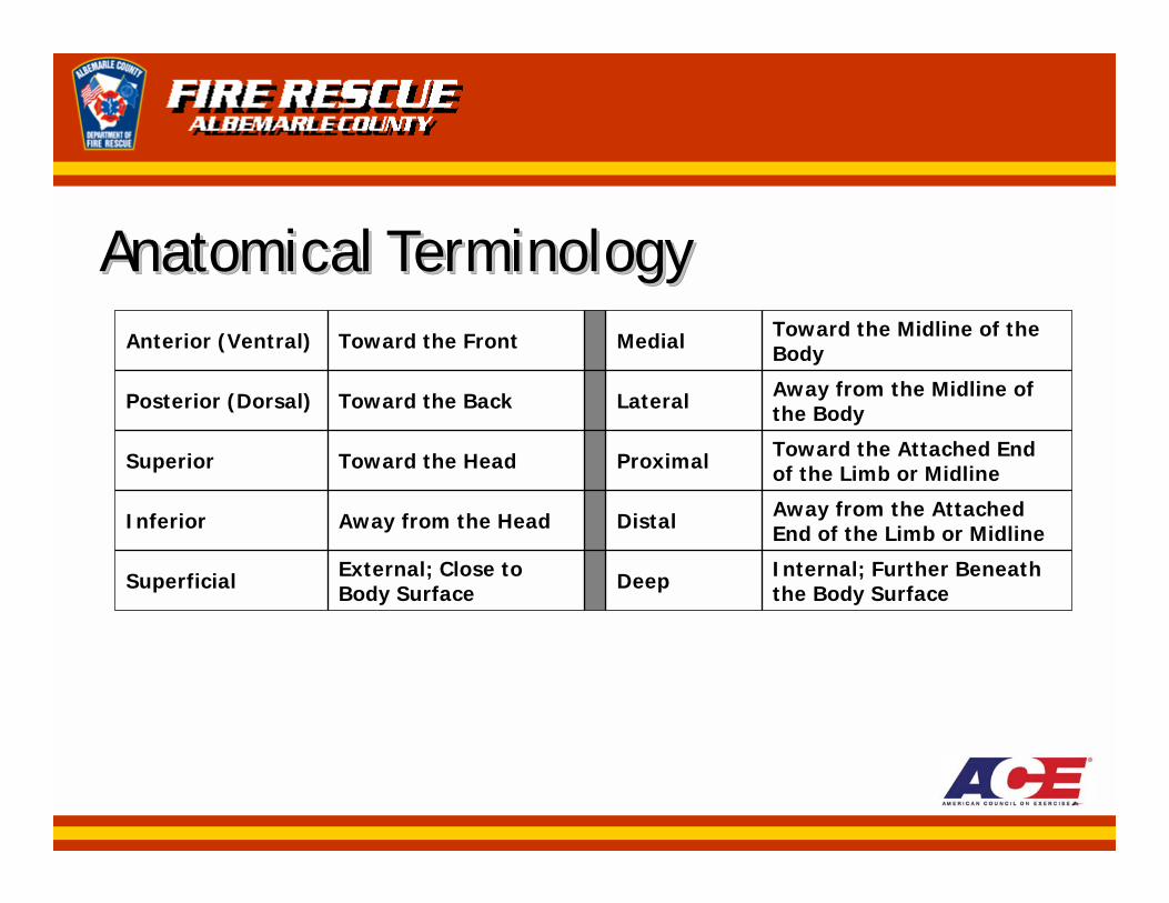
Anatomical TerminologyAnatomical TerminologyAnatomical Terminology
Internal; Further Beneath the Body SurfaceDeepExternal; Close to
Body SurfaceSuperficial
Away from the Attached End of the Limb or MidlineDistalAway from the HeadInferior
Toward the Attached End of the Limb or MidlineProximalToward the HeadSuperior
Away from the Midline of the BodyLateralToward the BackPosterior (Dorsal)
Toward the Midline of the BodyMedialToward the FrontAnterior (Ventral)
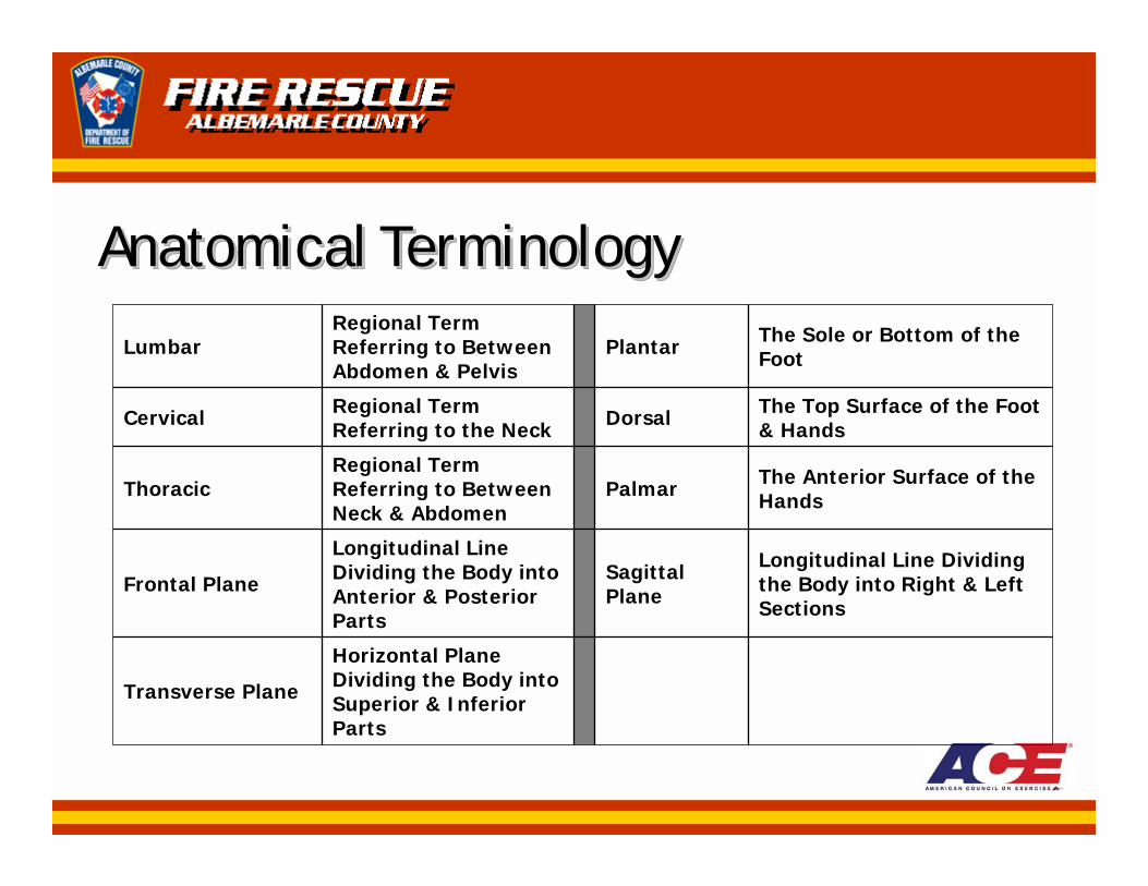
Anatomical TerminologyAnatomical TerminologyAnatomical Terminology
Horizontal Plane Dividing the Body into Superior & Inferior Parts
Transverse Plane
Longitudinal Line Dividing the Body into Right & Left Sections
SagittalPlane
Longitudinal Line Dividing the Body into Anterior & Posterior Parts
Frontal Plane
The Anterior Surface of the HandsPalmar
Regional Term Referring to Between Neck & Abdomen
Thoracic
The Top Surface of the Foot & HandsDorsalRegional Term
Referring to the NeckCervical
The Sole or Bottom of the FootPlantar
Regional Term Referring to Between Abdomen & Pelvis
Lumbar
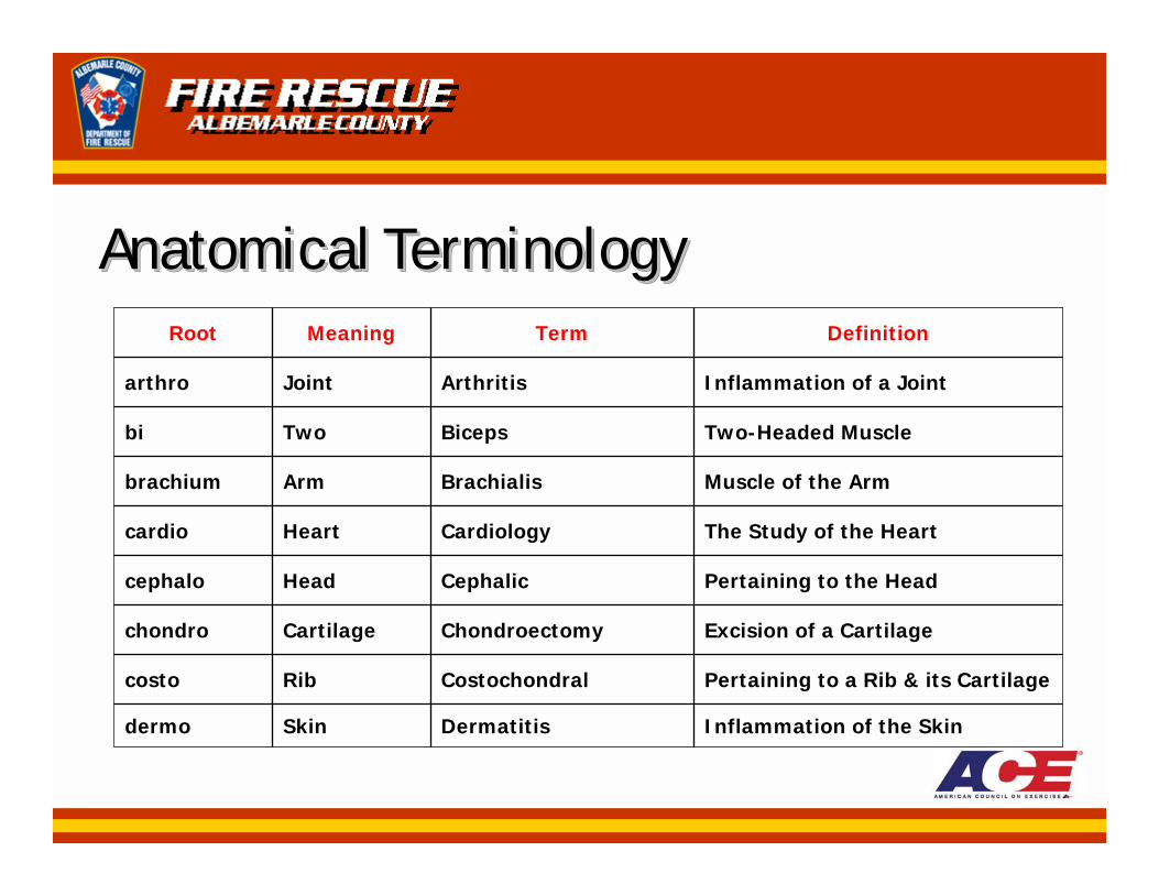
Anatomical TerminologyAnatomical TerminologyAnatomical TerminologyDefinitionTermMeaningRoot
Inflammation of the SkinDermatitisSkindermo
Pertaining to a Rib & its CartilageCostochondralRibcosto
Excision of a CartilageChondroectomyCartilagechondro
Pertaining to the HeadCephalicHeadcephalo
The Study of the HeartCardiologyHeartcardio
Muscle of the ArmBrachialisArmbrachium
Two-Headed MuscleBicepsTwobi
Inflammation of a JointArthritisJointarthro
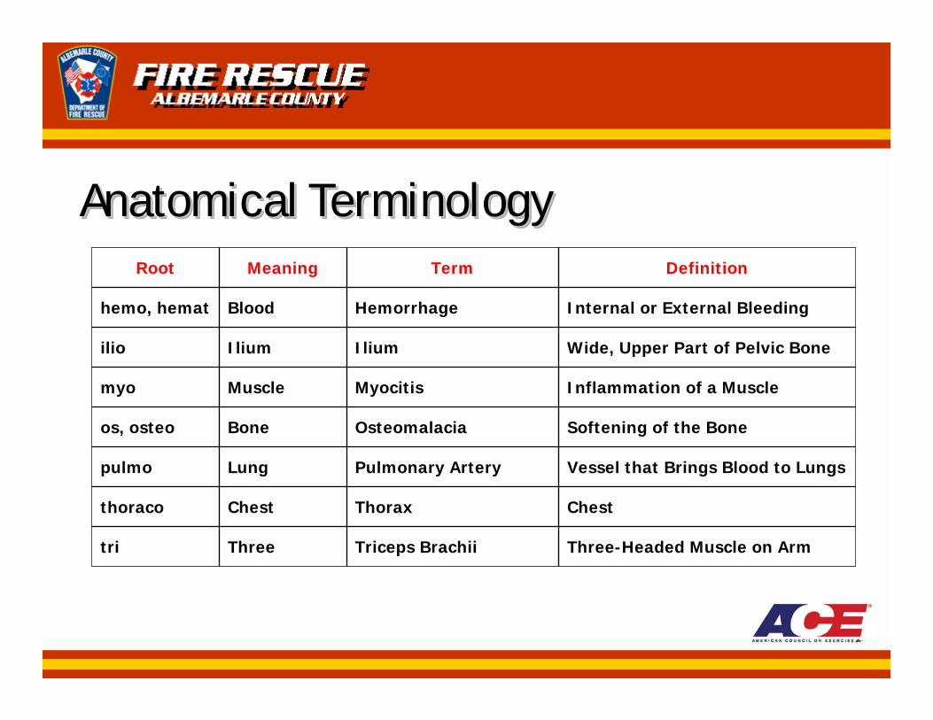
Anatomical TerminologyAnatomical TerminologyAnatomical TerminologyDefinitionTermMeaningRoot
Three-Headed Muscle on ArmTriceps BrachiiThreetri
ChestThoraxChestthoraco
Vessel that Brings Blood to LungsPulmonary ArteryLungpulmo
Softening of the BoneOsteomalaciaBoneos, osteo
Inflammation of a MuscleMyocitisMusclemyo
Wide, Upper Part of Pelvic BoneIliumIliumilio
Internal or External BleedingHemorrhageBloodhemo, hemat
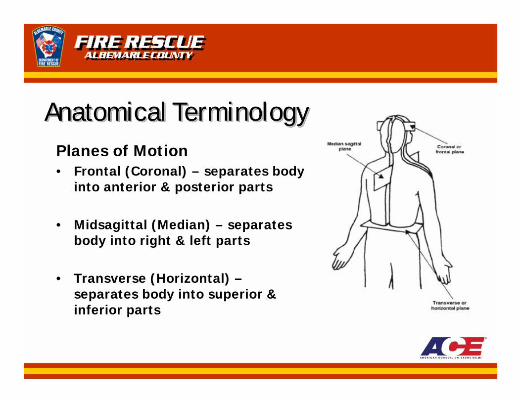
Anatomical TerminologyAnatomical TerminologyAnatomical TerminologyPlanes of Motion• Frontal (Coronal) – separates body
into anterior & posterior parts
• Midsagittal (Median) – separates body into right & left parts
• Transverse (Horizontal) –separates body into superior & inferior parts
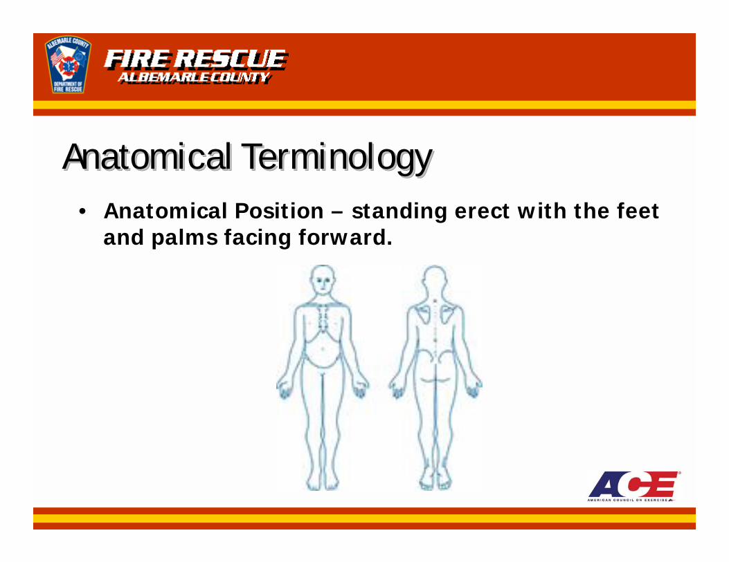
Anatomical TerminologyAnatomical TerminologyAnatomical Terminology• Anatomical Position – standing erect with the feet
and palms facing forward.

Anatomical TerminologyAnatomical TerminologyAnatomical Terminology• Five (5) Major Human Body Systems (pertinent
to physical activity):1. Cardiovascular2. Respiratory3. Nervous4. Skeletal5. Muscular
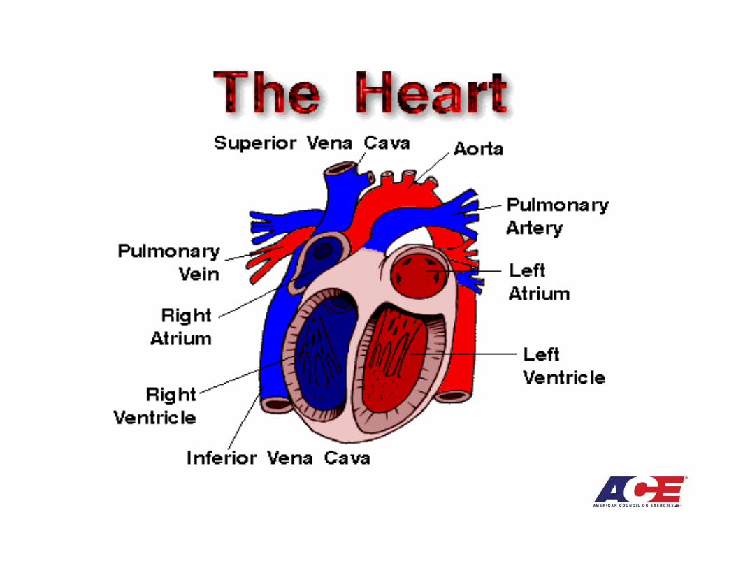
Cardiovascular SystemCardiovascular SystemCardiovascular System• Components:
– Blood– Blood Vessels– Heart
• Purposes: – Distribute O2 and nutrients to the cells– Carry CO2 and metabolic wastes from the cells– Protect against disease– Help regulate body temperature– Prevent serious blood loss after injury
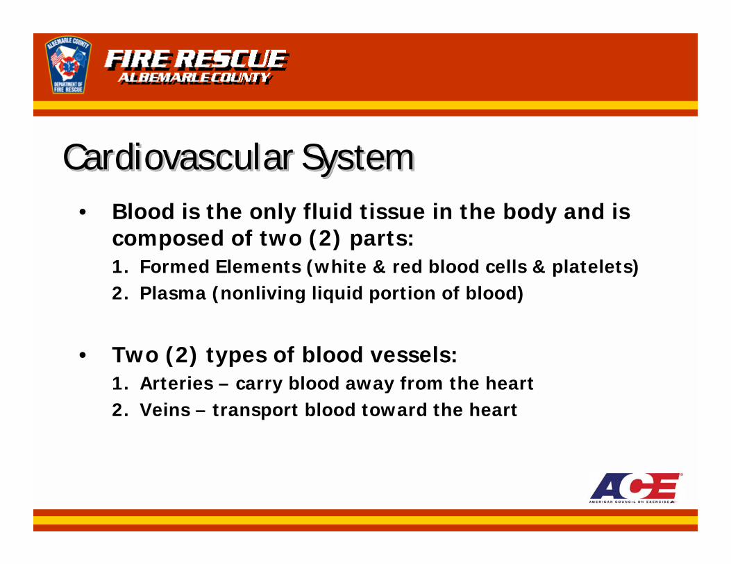
Cardiovascular SystemCardiovascular SystemCardiovascular System• Blood is the only fluid tissue in the body and is
composed of two (2) parts:1. Formed Elements (white & red blood cells & platelets)2. Plasma (nonliving liquid portion of blood)
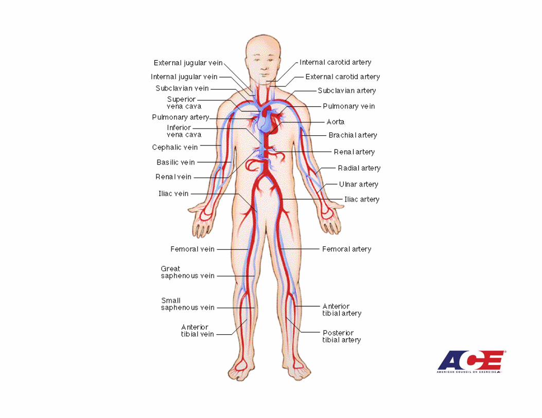
• Two (2) types of blood vessels:1. Arteries – carry blood away from the heart2. Veins – transport blood toward the heart
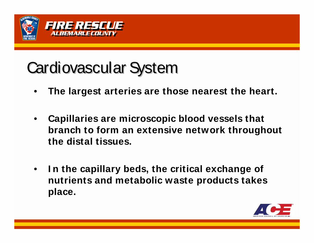
Cardiovascular SystemCardiovascular SystemCardiovascular System• The largest arteries are those nearest the heart.
• Capillaries are microscopic blood vessels that branch to form an extensive network throughout the distal tissues.
• In the capillary beds, the critical exchange of nutrients and metabolic waste products takes place.
Respiratory SystemRespiratory SystemRespiratory System• Respiration is the overall exchange of gases (O2,
CO2, and N) between the atmosphere, the blood, and the cells.
• There are three (3) general phases of respiration:1. External: exchange of O2 and CO2 between the
atmosphere and the blood within the large capillaries in the lungs.
2. Internal: exchange of those gases between the blood and the cells of the body.
3. Cellular: utilization of the O2 and the production of CO2by the metabolic activity within cells.
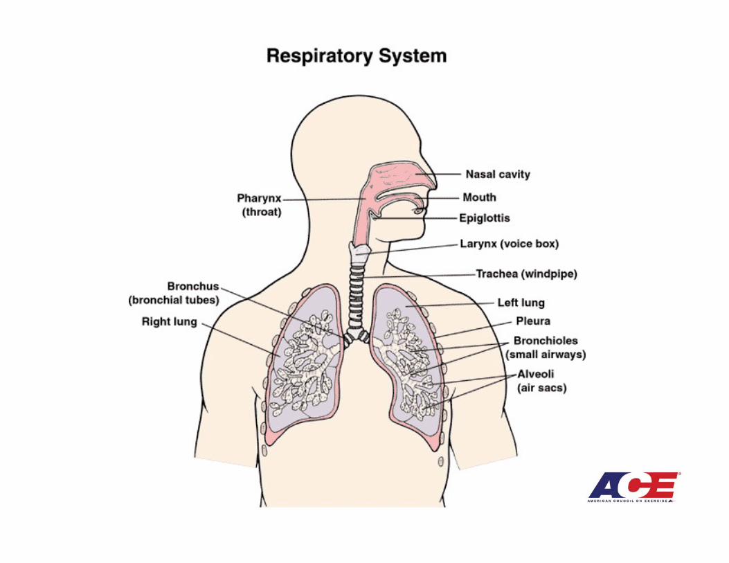
Respiratory SystemRespiratory SystemRespiratory System• The actual exchange of respiratory gases, such as
O2 and CO2, between the lungs and the blood occurs at the bronchioles.
• The lungs are the final component of the respiratory system.
• The lungs are paired, cone-shaped organs lying in the thoracic cavity with three (3) lobes on the right and two (2) lobes on the left.
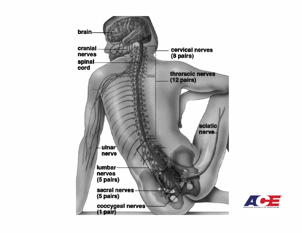
Nervous SystemNervous SystemNervous System• The nervous system is divided into two (2) parts
– according to location:1. Central Nervous System (CNS): brain and spinal cord2. Peripheral Nervous System (PNS): nerves that connect
the extremities of the body with their receptors in the CNS.
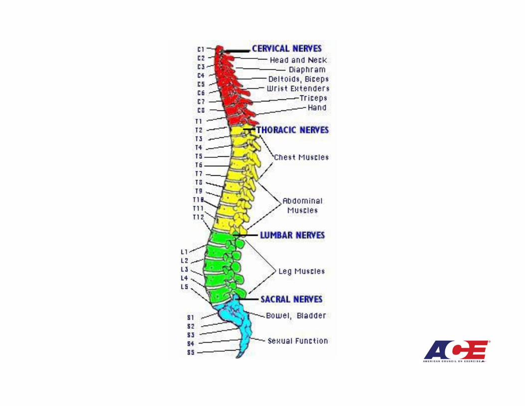
• The PNS is comprised of:– 12 pairs of cranial nerves– 31 pairs of spinal nerves
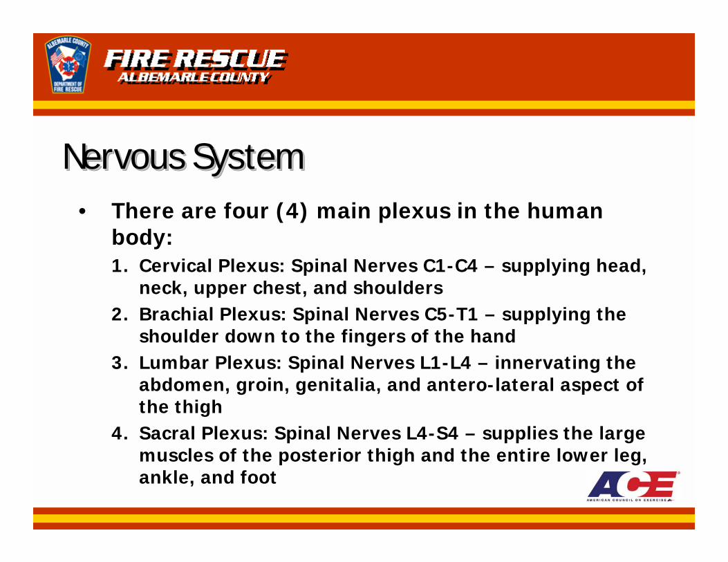
Nervous SystemNervous SystemNervous System• There are four (4) main plexus in the human
body:1. Cervical Plexus: Spinal Nerves C1-C4 – supplying head,
neck, upper chest, and shoulders2. Brachial Plexus: Spinal Nerves C5-T1 – supplying the
shoulder down to the fingers of the hand3. Lumbar Plexus: Spinal Nerves L1-L4 – innervating the
abdomen, groin, genitalia, and antero-lateral aspect of the thigh
4. Sacral Plexus: Spinal Nerves L4-S4 – supplies the large muscles of the posterior thigh and the entire lower leg, ankle, and foot
Nervous SystemNervous SystemNervous System• Receptors are specialized nerve cells located
throughout the body that carry messages (nerve impulses).
• Sensory nerve cells carry impulses from the peripheral receptors to the spinal cord and brain.
• Motor nerve cells carry impulses away from the CNS to respond to the perceived changes in the body’s internal or external environment.
Skeletal SystemSkeletal SystemSkeletal System• The human skeletal system consists of 206
bones.
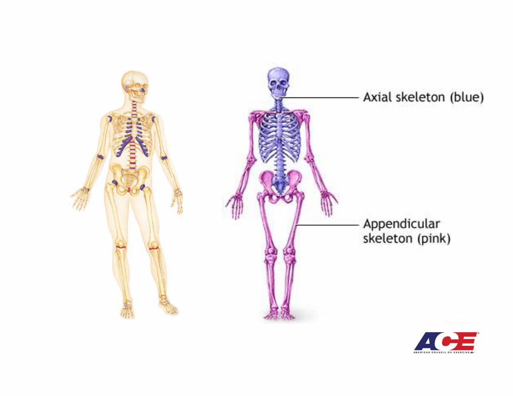
• The skeletal system can be divided into two (2) sections:1. Axial Skeleton – 80 bones that make up the head, neck,
and trunk2. Appendicular Skeleton – 126 bones that form the
extremities
Skeletal SystemSkeletal SystemSkeletal System• Bones have five (5) basic functions:
1. Protection – serves to protect the vital organs (heart, brain, and spinal cord).
2. Support – serves to support the soft tissue so that erect posture and the form of the body can be maintained.
3. Leverage – provides a framework of levers to which muscles are attached.
4. Blood Cell Production – produces certain blood cells in the red marrow of bone (primarily red blood cells, some white blood cells and platelets).
5. Storage – serves as storage for calcium, phosphorous, potassium, sodium, and other minerals.
Skeletal SystemSkeletal SystemSkeletal System• Bones are classified according to their shape:
– Long– Short– Flat– Irregular
• Bones are comprised of an organic component made of collagen (complex protein) and an inorganic component composed of mineral salts (calcium and potassium).
Skeletal SystemSkeletal SystemSkeletal System• The bones of persons with sedentary lifestyles
will become less dense over time as mineral salts are withdrawn.
• “Form follows function” – the form that bone will take (strong or weak) is in direct response to the recent function of that bone.
Skeletal SystemSkeletal SystemSkeletal System• Amenorrhea – two (2) or less menstrual periods
per year
• Menopause – loss of menstrual periods
• Low levels of estrogen (hormone) accompany amenorrhea and menopause; thus, women may have substantial bone mineral loss and need specific attention when developing strength-building programs.
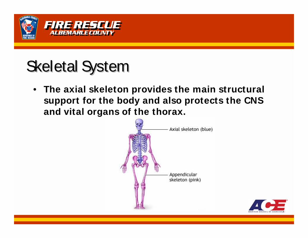
Skeletal SystemSkeletal SystemSkeletal System• The axial skeleton provides the main structural
support for the body and also protects the CNS and vital organs of the thorax.
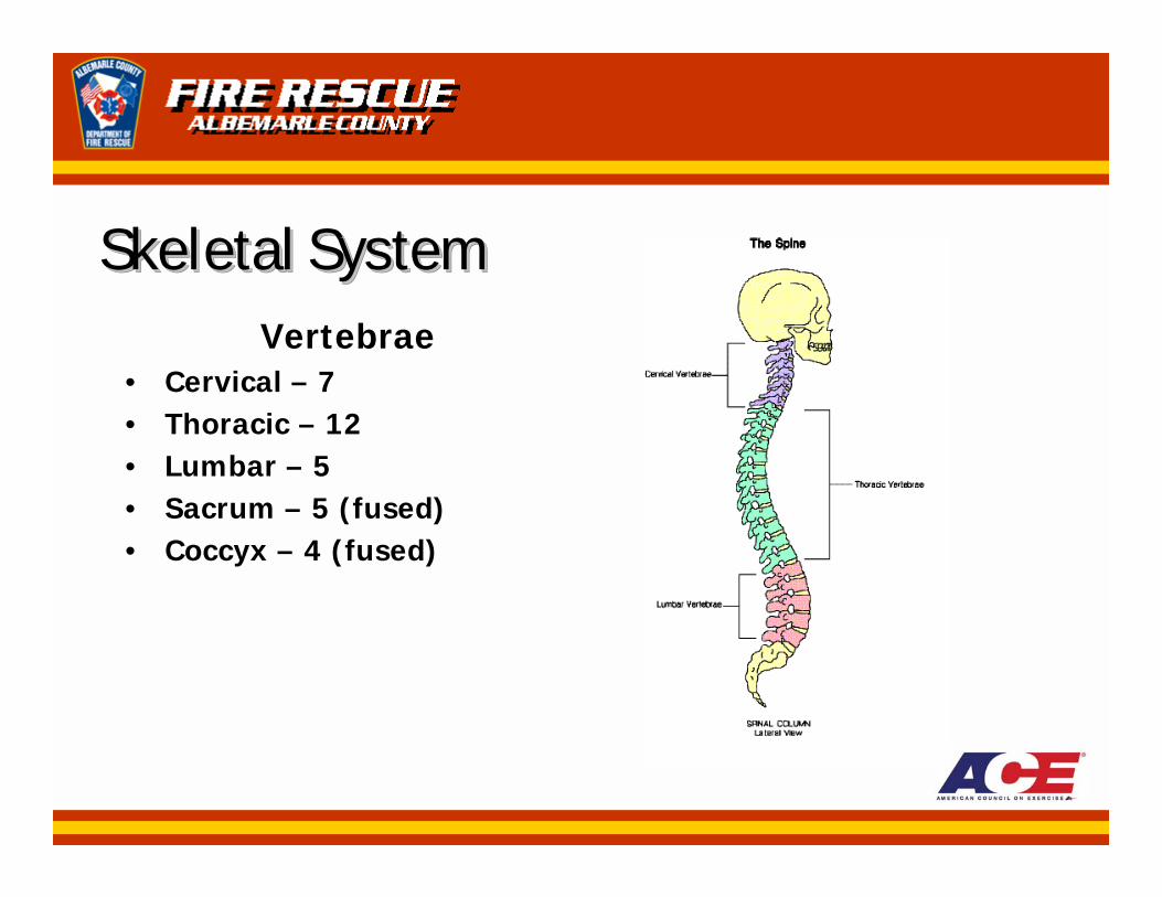
Skeletal SystemSkeletal SystemSkeletal SystemVertebrae
• Cervical – 7• Thoracic – 12• Lumbar – 5• Sacrum – 5 (fused)• Coccyx – 4 (fused)
Skeletal SystemSkeletal SystemSkeletal System• Articulation (Joint) – point of contact or
connection between bones, or between bones and cartilage.
• Ligament – dense, fibrous strands of connective tissue that link together the bony segments.
Skeletal SystemSkeletal SystemSkeletal System• Joints can be classified into two (2) general
categories according to:1. Structure of the joints2. Type of movement allowed by the joints
• Two (2) main characteristics differentiate the types of joints:1. Type of connective tissue that holds the bones of the
joint together2. Presence or absence of a joint cavity
Skeletal SystemSkeletal SystemSkeletal System• There are three (3) major structural categories of
joints:1. Fibrous – Ex., joints between the bones of the skull and
between the distal tibia and fibula2. Cartilaginous – Ex., joints formed by the hyaline
cartilages that connect the ribs to the sternum3. Synovial – Ex., knee, shoulder, most joints
• The most predominant type of joint in the body are synovial joints.
Skeletal SystemSkeletal SystemSkeletal System• The primary function of the synovial membrane
is the secretion of synovial fluid – a lubricant for the joint and nutrition for the articular cartilage.
• Acute injury to, or overuse of, a synovial joint can stimulate the synovial membrane to secrete excessive fluid, typically producing swelling and decreasing the pain-free range of motion.
Skeletal SystemSkeletal SystemSkeletal System• Menisci (meniscus) – articular disks made of
fibrous cartilage designed to help absorb shock, increase joint stability, aid in joint nutrition, and increase the joint contact area.
Skeletal SystemSkeletal SystemSkeletal System• The functional classification of joints is based on
the degree and type of movement they allow:– Fibrous (immovable)
• Between distal tibia & fibula– Cartilaginous (slightly moveable)
• Ribs & sternum– Diarthrodial (synovial free-moving joints)
• Foot, ankle, knee, hip, hand, thumb, wrist, elbow, shoulder
Skeletal SystemSkeletal SystemSkeletal System• For a joint to move in a given plane, there must
be an axis of rotation.
• An axis of rotation is an imaginary line perpendicular to (at a right angle to) the plane of movement about which a joint rotates. Many joints have several axes of rotation, enabling bones to move in various planes.
Skeletal SystemSkeletal SystemSkeletal System• Uniplanar joint – joint with one (1) axis of
rotation; can only move in one (1) plane; also known as “hinge” joints
Ex., ankle and elbow
• Biplanar joint – joint with two (2) axes of rotation; can move in two (2) planes that are right angles to one another; “modified hinge”and condyloid joints
Ex., knee (modified hinge); joints of hand & fingers, feet & toes (condyloid)
Skeletal SystemSkeletal SystemSkeletal System• Multiplanar joint - joint with three (3) axes of
rotation; can move in three (3) planes; ball-and-socket and saddle joints
Ex., Hip & shoulder (ball-and-socket); thumb (saddle)
• There are two (2) basic types of movement in the synovial joints: – Angular – flexion, extension, abduction, adduction– Circular - rotation
Skeletal SystemSkeletal SystemSkeletal System• Flexion and extension occur in the sagittal plane.
• Adduction and abduction occur in the frontal plane.
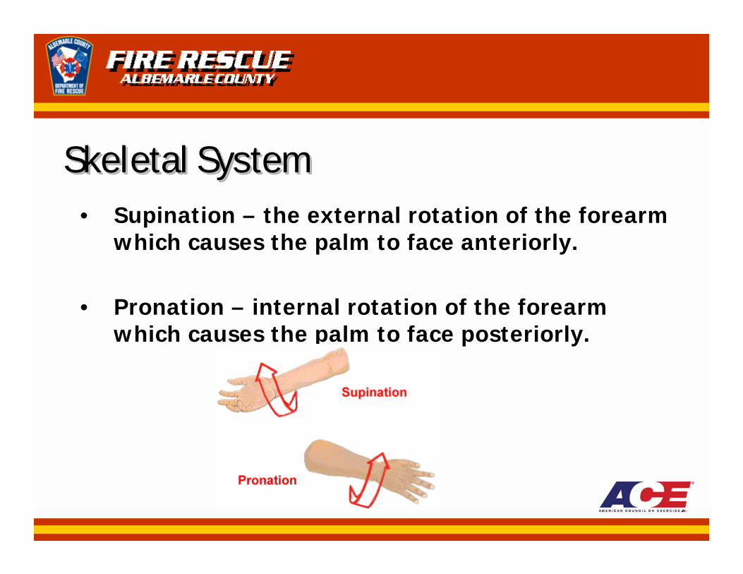
Skeletal SystemSkeletal SystemSkeletal System• Supination – the external rotation of the forearm
which causes the palm to face anteriorly.
• Pronation – internal rotation of the forearm which causes the palm to face posteriorly.
Muscular SystemMuscular SystemMuscular System• The anatomical system most directly affected by
exercise is the muscular system.
• There are three (3) types of muscle tissue:– Skeletal – voluntary muscle attached to bones by
tendons– Cardiac – involuntary muscle that forms the walls of the
heart– Visceral (Smooth) – involuntary muscle found in the
walls of internal organs such as the stomach and intestines

Muscular SystemMuscular SystemMuscular System• Aponeurosis – a broad, flat type of tendon that
sometimes attaches skeletal muscle to bone.Ex., wide flat insertion of the rectus abdominus
• There are more than 600 muscles in the human body.
Muscular SystemMuscular SystemMuscular SystemMuscles are named according to:
1. Location (posterior tibialis, rectus abdominis)2. Shape (deltoid, trapezius, serratus, anterior, rhomboid)3. Action (various muscle names include the terms
extensor, flexor, abductor, adductor)4. Number of Divisions (biceps brachii, triceps brachii,
quadriceps femoris)5. Bony Attachments (coracobrachialis, iliocostalis)6. Size Relationships (pectoralis major, pectoralis minor)
Some muscles also include the terms “longus” (long) and “brevis” (short).
Muscular SystemMuscular SystemMuscular System• Agonist – a muscle that is directly engaged in
contraction; oppose the action of an antagonist.
• Antagonist – a muscle that acts in opposition to the action produced by an agonist muscle.
• When one muscle (agonist) is contracting to achieve a desired movement, its opposite muscle (antagonist) is being stretched.
Muscular SystemMuscular SystemMuscular System• At most joints, several muscles combine to help
perform the same anatomical function; these muscles are known as synergists.
Muscular SystemMuscular SystemMuscular SystemThe lower extremity (leg) has four (4) compartments:
1. Anterior Tibial Compartment – extend toes & dorsiflex (flex) the ankle
2. Lateral Tibial Compartment – eversion (abduction) of the foot & plantraflexion of the ankle
3. Superficial Posterior Tibial Compartment – largest muscles of calf (soleus and gastrocnemius) responsible for plantarflexion(extension) of the ankle, flexion of the toes, & inversion (adduction of the foot)
4. Deep Posterior Tibial Compartment – popliteus, posterior tibialis, flexor hallucis longus, & flexor digitorum longus also responsible for plantarflexion (extension) of the ankle, flexion of the toes, and inversion (adduction of the foot)
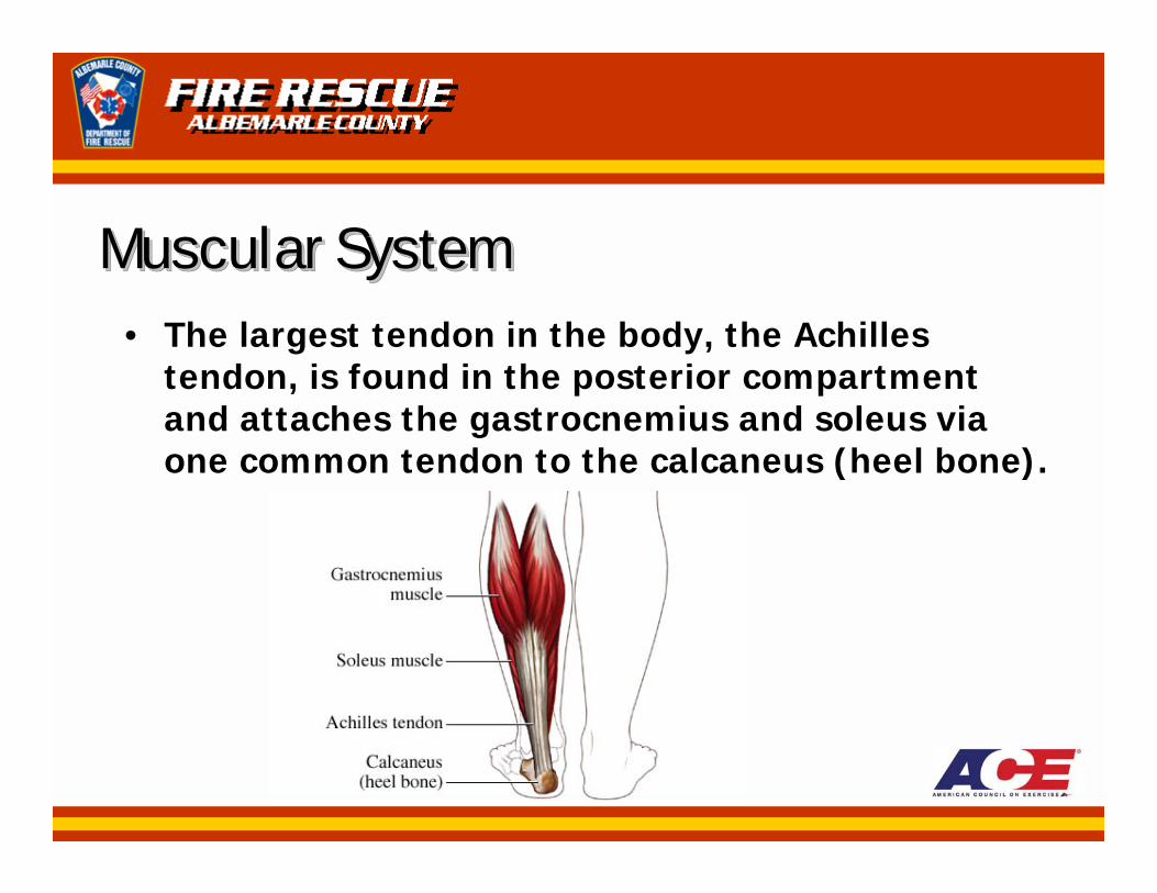
Muscular SystemMuscular SystemMuscular System• The largest tendon in the body, the Achilles
tendon, is found in the posterior compartment and attaches the gastrocnemius and soleus via one common tendon to the calcaneus (heel bone).
Muscular SystemMuscular SystemMuscular SystemThe muscles that cross the knee joint to act on the
leg (tibia & fibula) can be divided into three (3) separate groups:
1. Quadriceps femoris (rectus femoris, vastus medialis, vastus intermedius, & vastus lateralis)
2. Hamstrings (biceps femoris, semitendinosus, & semimembranosus)
3. Pes anserine group (semitendinosus, sartorius, & gracilis)
The sartorius is the longest muscle in the body.
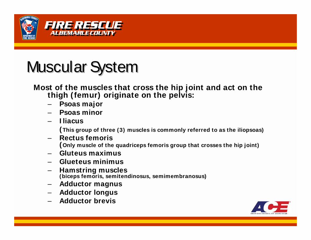
Muscular SystemMuscular SystemMuscular SystemMost of the muscles that cross the hip joint and act on the
thigh (femur) originate on the pelvis:– Psoas major– Psoas minor– Iliacus
(This group of three (3) muscles is commonly referred to as the iliopsoas)– Rectus femoris
(Only muscle of the quadriceps femoris group that crosses the hip joint)– Gluteus maximus– Glueteus minimus– Hamstring muscles
(biceps femoris, semitendinosus, semimembranosus)– Adductor magnus– Adductor longus– Adductor brevis
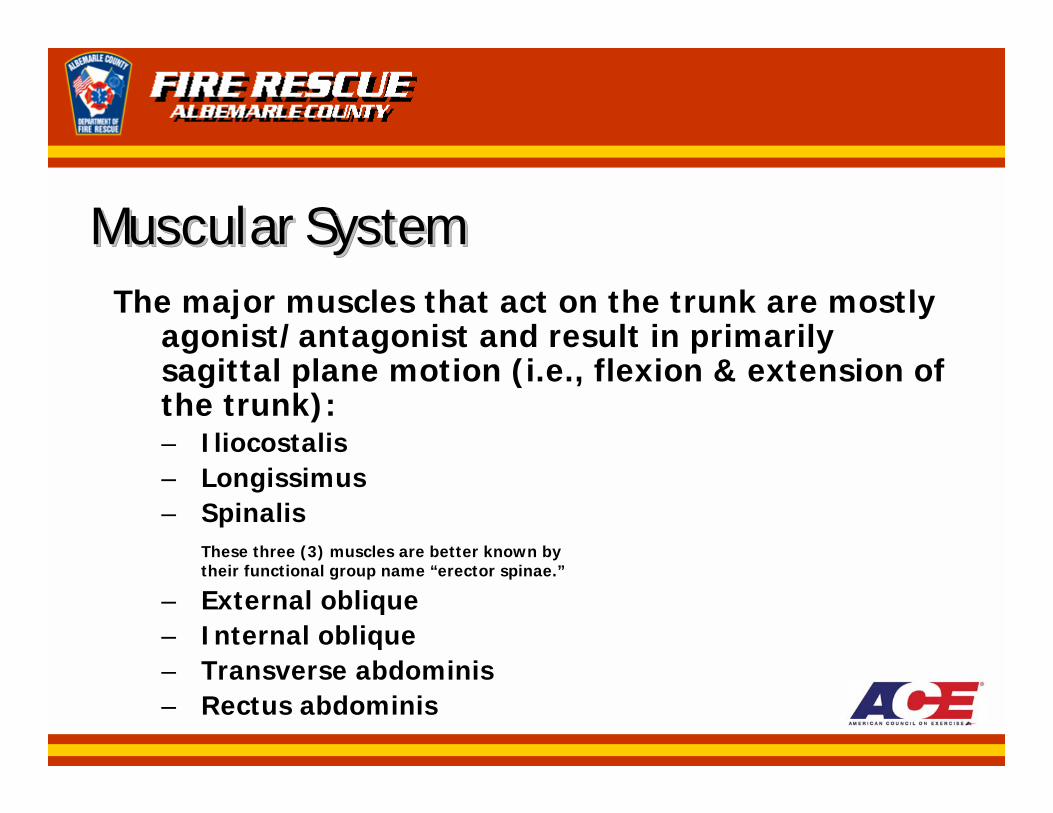
Muscular SystemMuscular SystemMuscular SystemThe major muscles that act on the trunk are mostly
agonist/antagonist and result in primarily sagittal plane motion (i.e., flexion & extension of the trunk):– Iliocostalis– Longissimus– Spinalis
These three (3) muscles are better known by their functional group name “erector spinae.”
– External oblique– Internal oblique– Transverse abdominis– Rectus abdominis
Muscular SystemMuscular SystemMuscular SystemThe three (3) major muscles responsible for
movement of the vertebral column are:1. Iliocostalis2. Longissimus3. Spinalis
These three (3) muscles are better known by their functional group name “erector spinae.”
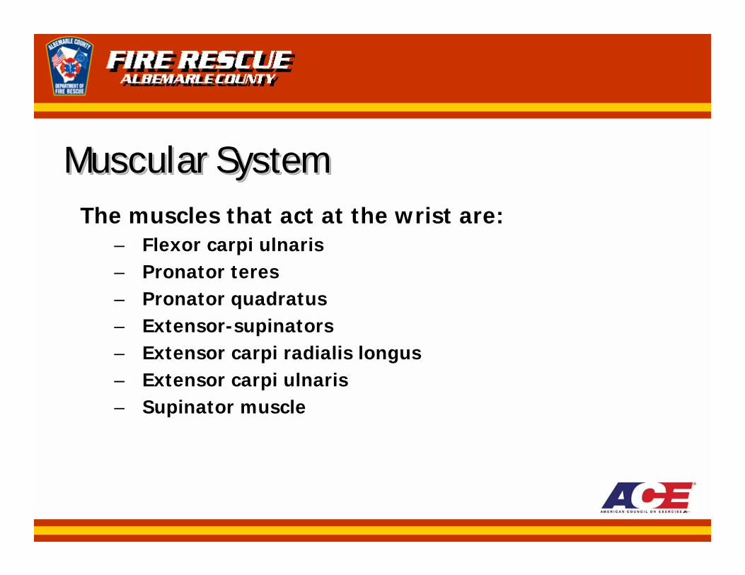
Muscular SystemMuscular SystemMuscular SystemThe muscles that act at the wrist are:
– Flexor carpi ulnaris– Pronator teres– Pronator quadratus– Extensor-supinators– Extensor carpi radialis longus– Extensor carpi ulnaris– Supinator muscle
Muscular SystemMuscular SystemMuscular System• The muscles that act at the elbow joint are:
– Biceps brachii– Brachialis– Brachioradialis– Triceps brachii
Muscular SystemMuscular SystemMuscular System• The muscles that act at the shoulder joint are:
– Pectoralis major– Latissimus dorsi– Deltoid muscles– Rotator cuff muscles (“SITS” muscles: supraspinatus,
infraspinatus, terres minor, & subscapularis)
Muscular SystemMuscular SystemMuscular System• The muscles that act at the scapulothoracic
articulation are:– Trapezius– Rhomboid major– Rhomboid minor– Levator scapulae