chapter 1 apical periodontitis: microbial infection and …€¦ · · 2008-03-15chapter 1 apical...
TRANSCRIPT
1.1 Introduction
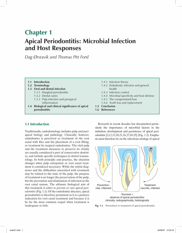
Traditionally, endodontology includes pulp and peri-apical biology and pathology. Clinically, however, endodontics is perceived as treatment of the root canal with fi les and the placement of a root fi lling, or treatment by surgical endodontics. The vital pulp and the treatment measures to preserve its vitality are usually considered a part of conservative dentist-ry, and include specifi c techniques in dental trauma-tology. In both principle and practice, the situation changes when pulp extirpation or root canal treat-ment is considered necessary. While the initial diag-noses and the diffi culties associated with treatment may be related to the state of the pulp, the purpose of treatment is no longer the preservation of the pulp, but the prevention and elimination of infection in the root canal system. The ultimate biological aim of this treatment is either to prevent or cure apical peri-odontitis (Fig. 1.1). Of the endodontic diseases, apical periodontitis is therefore prominent as it is a primary indication for root canal treatment and because it is by far the most common sequel when treatment is inadequate or fails.
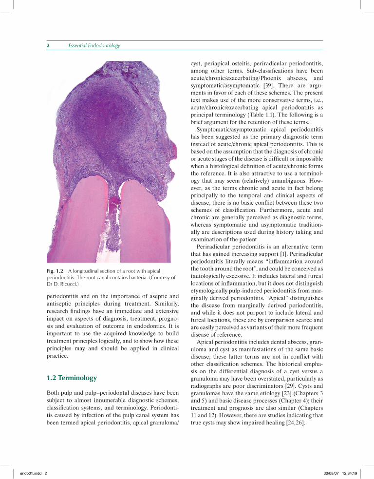
Research in recent decades has documented partic-ularly the importance of microbial factors in the initia tion, development and persistence of apical peri-odontitis [3,12,13,20,21,26,27,30,35] (Fig. 1.2). Empha-sis must therefore be on the infectious etiology of apical
1.1 Introduction1.2 Terminology1.3 Oral and dental infection 1.3.1 Marginal periodontitis 1.3.2 Dental caries 1.3.3 Pulp infection and periapical
infl ammation1.4 Biological and clinical signifi cance of apical
periodontitis
1.4.1 Infection theory 1.4.2 Endodontic infection and general
health 1.4.3 Infection control 1.4.4 Microbial specifi city and host defense 1.4.5 The compromised host 1.4.6 Tooth loss and replacement1.5 Conclusion 1.6 References
Chapter 1Apical Periodontitis: Microbial Infection and Host ResponsesDag Ørstavik and Thomas Pitt Ford
Prevention:vital, inflamed
Treatment:necrotic, infected
Success =absence of apical periodontitis:
clinically, radiographically, histologically
Fig. 1.1 Prevention or treatment of apical periodontitis.
endo01.indd 1endo01.indd 1 30/08/07 12:34:1830/08/07 12:34:18
2 Essential Endodontology
periodontitis and on the importance of aseptic and antiseptic principles during treatment. Similarly, research fi ndings have an immediate and extensive impact on aspects of diagnosis, treatment, progno-sis and evaluation of outcome in endodontics. It is important to use the acquired knowledge to build treatment principles logically, and to show how these principles may and should be applied in clinical practice.
1.2 Terminology
Both pulp and pulp–periodontal diseases have been subject to almost innumerable diagnostic schemes, classifi cation systems, and terminology. Periodonti-tis caused by infection of the pulp canal system has been termed apical periodontitis, apical granuloma/
cyst, periapical osteitis, periradicular periodontitis, among other terms. Sub-classifi cations have been acute/chronic/exacerbating/Phoenix abscess, and symptomatic/asymptomatic [39]. There are argu-ments in favor of each of these schemes. The present text makes use of the more conservative terms, i.e., acute/chronic/exacerbating apical periodontitis as principal terminology (Table 1.1). The following is a brief argument for the retention of these terms. Symptomatic/asymptomatic apical periodontitis has been suggested as the primary diagnostic term instead of acute/chronic apical periodontitis. This is based on the assumption that the diagnosis of chronic or acute stages of the disease is diffi cult or impossible when a histological defi nition of acute/chronic forms the reference. It is also attractive to use a terminol-ogy that may seem (relatively) unambiguous. How-ever, as the terms chronic and acute in fact belong principally to the temporal and clinical aspects of disease, there is no basic confl ict between these two schemes of classifi cation. Furthermore, acute and chronic are generally perceived as diagnostic terms, whereas symptomatic and asymptomatic tradition-ally are descriptions used during history taking and examination of the patient. Periradicular periodontitis is an alternative term that has gained increasing support [1]. Periradicular periodontitis literally means “infl ammation around the tooth around the root”, and could be conceived as tautologically excessive. It includes lateral and furcal locations of infl ammation, but it does not distinguish etymologically pulp-induced periodontitis from mar-ginally derived periodontitis. “Apical” distinguishes the disease from marginally derived periodontitis, and while it does not purport to include lateral and furcal locations, these are by comparison scarce and are easily perceived as variants of their more frequent disease of reference. Apical periodontitis includes dental abscess, gran-uloma and cyst as manifestations of the same basic disease; these latter terms are not in confl ict with other classifi cation schemes. The historical empha-sis on the differential diagnosis of a cyst versus a granuloma may have been overstated, particularly as radiographs are poor discriminators [29]. Cysts and granulomas have the same etiology [23] (Chapters 3 and 5) and basic disease processes (Chapter 4); their treatment and prognosis are also similar (Chapters 11 and 12). However, there are studies indicating that true cysts may show impaired healing [24,26].
Fig. 1.2 A longitudinal section of a root with apical periodontitis. The root canal contains bacteria. (Courtesy of Dr D. Ricucci.)
endo01.indd 2endo01.indd 2 30/08/07 12:34:1930/08/07 12:34:19
Apical Periodontitis: Microbial Infection and Host Responses 3
The use of the term “periapical osteitis” has been limited and the connotation that bone is infl amed seems incorrect. Rather, the infl ammation of the api-cal periodontium causes resorption of the bone and prevents it from becoming infected. In summary, “apical periodontitis” takes pre ference by etymology and usage; “acute” and “chronic” are preferred and used as clinical supplementary terms of disease. On the other hand, terminology should not be used as a straightjacket for authors. There-fore, variants of the terms and references to other diagnostic schemes, in this book and other texts, are inevitable, even desirable.
1.3 Oral and dental infection
The oral cavity is an extension of the skin/mucosal barrier to the external environment. In the digestive tract, it may be viewed as the fi rst battleground for the body’s efforts to maintain homeostasis and keep infection away from the vulnerable interior parts of
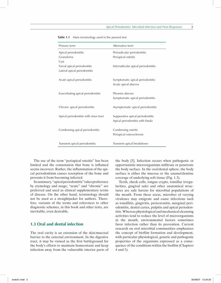
the body [5]. Infection occurs when pathogenic or opportunistic microorganisms infi ltrate or penetrate the body surface. In the oral/dental sphere, the body surface is either the mucosa or the enamel/dentine coverage of underlying soft tissue (Fig. 1.3). Teeth, cheek cells, tongue crypts, tonsillar irregu-larities, gingival sulci and other anatomical struc-tures are safe havens for microbial populations of the mouth. From these areas, microbes of varying virulence may emigrate and cause infections such as tonsillitis, gingivitis, pericoronitis, marginal peri-odontitis, dental caries, pulpitis and apical periodon-titis. Whereas physiological and mechanical cleansing activities tend to reduce the level of microorganisms in the mouth, environmental factors sometimes favor infection rather than its prevention. Current research on oral microbial communities emphasizes the concept of biofi lm formation and development, with particular physiological, genetic and pathogenic properties of the organisms expressed as a conse-quence of the conditions within the biofi lm (Chapters 4 and 5).
Table 1.1 Main terminology used in the present text
Primary term Alternative term
Apical periodontitis Periradicular periodontitisGranuloma Periapical osteitisCystFurcal apical periodontitis Interradicular apical periodontitisLateral apical periodontitis
Acute apical periodontitis Symptomatic apical periodontitisAcute apical abscess
Exacerbating apical periodontitis Phoenix abscessSymptomatic apical periodontitis
Chronic apical periodontitis Asymptomatic apical periodontitis
Apical periodontitis with sinus tract Suppurative apical periodontitisApical periodontitis with fi stula
Condensing apical periodontitis Condensing osteitisPeriapical osteosclerosis
Transient apical periodontitis Transient apical breakdown
endo01.indd 3endo01.indd 3 30/08/07 12:34:2030/08/07 12:34:20
4 Essential Endodontology
1.3.1 Marginal periodontitis
The gingival sulcus is potentially a weak link in the sur-face cover, combining a thin epithelial coverage with a topographic structure that favors the accumulation of bacteria on the adjoining tooth. With time, the micro-bial challenge in this area frequently overwhelms the body defenses and causes gingivitis and subsequent marginal periodontitis. Untreated, the ensuing infl am-mation is often followed by loosening of the tooth, its loss and, fi nally, reestablishment of the mucosal bar-rier. Biologically, the infl ammation has a surface local-ization with easy drainage of pus, and deeper infection of tissues is not often seen. Moreover, tooth loss from marginal periodontitis usually happens late in the life-cycle of the individual, with limited consequences for the survivability of the individual, group or species.
1.3.2 Dental caries
Clinical dentistry and dental science have emerged in response largely to the other major, visible infection of the oral cavity: dental caries. The disease has primar-ily, but not exclusively, affected children and young adults; and the pain, impaired appearance, and tooth loss at an early age associated with dental caries have made it a major challenge to societies where it has become widespread. However, the epidemic of dental caries may have had a relatively short history in the lifespan of man. Although the history of dental caries goes back to the Iron Age and earlier, archaeological material and historical sources do not indicate that dental caries was widespread until a few centuries ago. Greater use of preventive measures in developed countries has caused a downturn in disease, although
it still constitutes a signifi cant health concern for some population groups and individuals.
1.3.3 Pulp infection and periapical infl ammation
Infection and infl ammation of the pulp and peri apical tissues have long been regarded as an extension of the dental caries process. This has been a reasonable interpretation in view of the dominance of caries as a source of infection of dentin during the last few cen-
(a) (b) (c)
A
C P
Fig. 1.3 Breaks in the mucocutaneous barrier associated with teeth. (a) Attrition (A), abrasion or trauma exposes the pulp. (b) Dental caries (C) reaches the pulp with subsequent infection of the pulp and periapical tissues. (c) Dental plaque (P) penetrates the gingival cuff and bacteria invade the gingival and periodontal tissues.
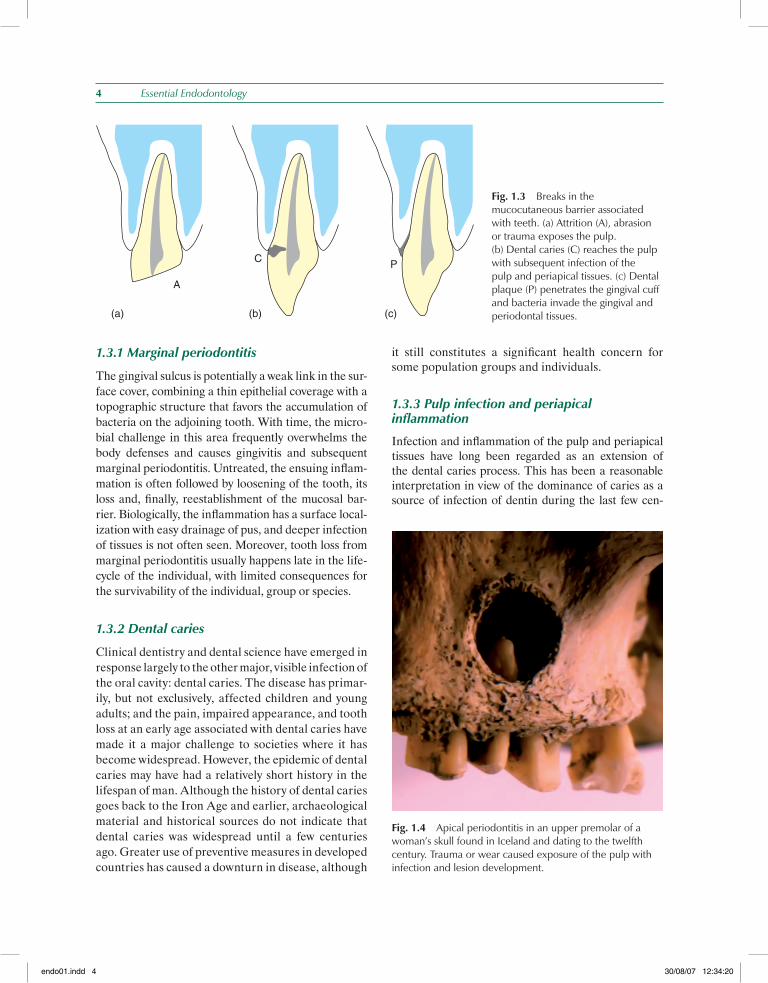
Fig. 1.4 Apical periodontitis in an upper premolar of a woman’s skull found in Iceland and dating to the twelfth century. Trauma or wear caused exposure of the pulp with infection and lesion development.
endo01.indd 4endo01.indd 4 30/08/07 12:34:2030/08/07 12:34:20
Apical Periodontitis: Microbial Infection and Host Responses 5
turies. However, infection of the pulp and periapical tissues and the tissue responses are probably an older and more general biological occurrence than dental caries (Fig. 1.4). Despite the common origin of infect-ing organisms, the microbial fl oras of dental caries and endodontic infections differ in many respects. The evolutionary biological development of the permanent dentition, which has nonreplaceable teeth, must also have included the development of effective responses to trauma and subsequent infection of the deeper tissues. Apical periodontitis may thus be viewed as a tissue response to pulp infection from trauma such as blows to or fracture of the teeth, attrition from mastication, and abrasion from the use of teeth as tools for surviv-al. Essentially, apical periodontitis may be seen as the body’s means of coping with the threat of infection following breaches in the mucosal/tooth barrier from trauma to, or attrition of, the teeth. An exposed pulp potentially leaves the body open to infection; the processes of apical periodontitis then usually work to create a second barrier within the body to prevent further spread of potentially threatening microbes [14,23] (Figs 1.5 and 1.6). Treatment of apical periodontitis must be seen in the context of preventing microbial access to the jaw-bone and the body beyond, and needs to be achieved by effective disinfection and obturation. A complete seal from the coronal to the apical end of the treated root reestablishes the mucocutaneo-odonto barrier, whereas voids or leaks may present an opportunity for bacteria to establish a foothold close to the body’s
interior. The recent emphasis on coronal leakage of bacteria and bacterial products as opposed to api-cal leakage is a refl ection of this more scientifi cally based line of reasoning [18,40].
1.4 Biological and clinical signifi cance of apical periodontitis
1.4.1 Infection theory
In the pre-antibiotic era, infections of the pulp and periapical tissues were considered potentially seri-ous and in need of close monitoring. In the early days of antibiotics, it was found that most of these infections were readily susceptible to penicillin, and therefore the spread of infection to regional spaces was believed to be easily controllable by antibiot-ics. Today, it is recognized that pulp infection may be caused by organisms of different virulence [36] (Chapter 5), and that control of the infection is not always easily obtained, particularly when endodontic treatment is ineffective. The fl ora of the mouth is fortunately composed of few species of pathogenic organisms, which usually have low virulence (Chapter 5). Most may be con-sidered opportunistic, causing disease only in mixed infections or in hosts compromised by other diseases. Teleologically, it is usually not to the advantage of microbial species living on other organisms to cause disease; the basis for their presence is rather the pres-ervation of the host.
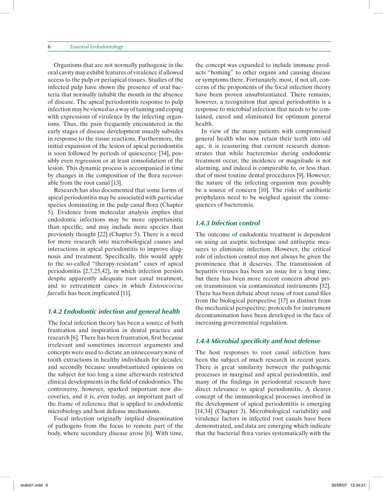
(a) (b)
Fig. 1.5 Infection of the pulp (a) is contained by development of an apical granuloma/cyst (b).
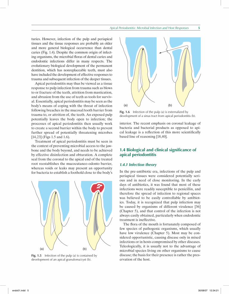
(a) (b)
Fig. 1.6 Infection of the pulp (a) is externalized by development of a sinus tract from apical periodontitis (b).
endo01.indd 5endo01.indd 5 30/08/07 12:34:2130/08/07 12:34:21
6 Essential Endodontology
Organisms that are not normally pathogenic in the oral cavity may exhibit features of virulence if allowed access to the pulp or periapical tissues. Studies of the infected pulp have shown the presence of oral bac-teria that normally inhabit the mouth in the absence of disease. The apical periodontitis response to pulp infection may be viewed as a way of taming and coping with expressions of virulence by the infecting organ-isms. Thus, the pain frequently encountered in the early stages of disease development usually subsides in response to the tissue reactions. Furthermore, the initial expansion of the lesion of apical periodontitis is soon followed by periods of quiescence [34], pos-sibly even regression or at least consolidation of the lesion. This dynamic process is accompanied in time by changes in the composition of the fl ora recover-able from the root canal [13]. Research has also documented that some forms of apical periodontitis may be associated with parti cular species dominating in the pulp canal fl ora (Chapter 5). Evidence from molecular analysis implies that endodontic infections may be more opportunistic than specifi c, and may include more species than previously thought [22] (Chapter 5). There is a need for more research into microbiological causes and interactions in apical periodontitis to improve diag-nosis and treatment. Specifi cally, this would apply to the so-called “therapy-resistant” cases of apical periodontitis [2,7,25,42], in which infection persists despite apparently adequate root canal treatment, and to retreatment cases in which Enterococcus faecalis has been implicated [11].
1.4.2 Endodontic infection and general health
The focal infection theory has been a source of both frustration and inspiration in dental practice and research [6]. There has been frustration, fi rst because irrelevant and sometimes incorrect arguments and concepts were used to dictate an unnecessary wave of tooth extractions in healthy individuals for decades; and secondly because unsubstantiated opinions on the subject for too long a time afterwards restricted clinical developments in the fi eld of endodontics. The controversy, however, sparked important new dis-coveries, and it is, even today, an important part of the frame of reference that is applied to endodontic microbiology and host defense mechanisms. Focal infection originally implied dissemination of pathogens from the focus to remote part of the body, where secondary disease arose [6]. With time,
the concept was expanded to include immune prod-ucts “homing” to other organs and causing disease or symptoms there. Fortunately, most, if not all, con-cerns of the proponents of the focal infection theory have been proven unsubstantiated. There remains, however, a recognition that apical periodontitis is a response to microbial infection that needs to be con-tained, cured and eliminated for optimum general health. In view of the many patients with compromised general health who now retain their teeth into old age, it is reassuring that current research demon-strates that while bacteremias during endodontic treatment occur, the incidence or magnitude is not alarming, and indeed is comparable to, or less than, that of most routine dental procedures [9]. However, the nature of the infecting organism may possibly be a source of concern [10]. The risks of antibiotic prophylaxis need to be weighed against the conse-quences of bacteremia.
1.4.3 Infection control
The outcome of endodontic treatment is dependent on using an aseptic technique and antiseptic mea-sures to eliminate infection. However, the critical role of infection control may not always be given the prominence that it deserves. The transmission of hepatitis viruses has been an issue for a long time, but there has been more recent concern about pri-on transmission via contaminated instruments [32]. There has been debate about reuse of root canal fi les from the biological perspective [17] as distinct from the mechanical perspective; protocols for instrument decontamination have been developed in the face of increasing governmental regulation.
1.4.4 Microbial specifi city and host defense
The host responses to root canal infection have been the subject of much research in recent years. There is great similarity between the pathogenic processes in marginal and apical periodontitis, and many of the fi ndings in periodontal research have direct relevance to apical periodontitis. A clearer concept of the immunological processes involved in the development of apical periodontitis is emerging [14,34] (Chapter 3). Microbiological variability and virulence factors in infected root canals have been demonstrated, and data are emerging which indicate that the bacterial fl ora varies systematically with the
endo01.indd 6endo01.indd 6 30/08/07 12:34:2130/08/07 12:34:21
Apical Periodontitis: Microbial Infection and Host Responses 7
clinical condition of the tooth involved (persistent infection, therapy-resistant infection) (Chapters 5 and 11). Thus, different strategies of antimicrobial measures may have to be applied depending on the micro biological diagnosis in a given case.
1.4.5 The compromised host
A characteristic feature of dentistry as it is practiced today is the shift of care from young to elderly peo-ple. The success of preventive dentistry in controlling and containing dental caries has resulted in a larger part of the population retaining their natural teeth longer and aspiring to keep them into old age. The improvement in dental health as regards dental car-ies has therefore not resulted in a decrease in demand for endodontic treatment; rather, more people in the older age groups are seeking to preserve their teeth by endodontic intervention. The net result is a sta-ble, or increasing, demand for endodontic treatment (Chapter 8) rather than a decline. With the increased demand for endodontic care among the elderly, there is also concern for the consequences of apical peri-odontitis and its treatment sequels in relation to other medical conditions. Fortunately, many of the concerns which previously were held as restrictions to endodontic treatment of the elderly have not been substantiated: both the prevention and cure of apical periodontitis by endodontics seem to be as successful in the old as in the young. Moreover, initial concerns about treatment outcome in HIV-positive patients seem unfounded [28]. There are a number of medical conditions that may infl uence the indications for, and choice of, end-odontic treatment and that occur more frequently among elderly patients. Generally, any disease for which bacteremia poses an additional hazard is of concern when endodontic treatment is being consid-ered. Particularly, a history of infective endocardi-tis, congenital heart disease, rheumatic heart fever or the presence of an artifi cial heart valve or other susceptible implants may necessitate the implemen-tation of an antibiotic regimen in conjunction with the endodontic procedures. The progression, pos-sibly also prognosis and healing pattern in diabetic patients may differ from that in people not affected by the disease [16,31]. Special consideration must be given to patients who are being treated with immu-nosuppressants, or otherwise have compromised immune systems [37,44]. A number of the blood dyscrasias, notably leukemias, are associated with
potentially serious sequels to apical periodontitis: infection spreads easily and may require extensive antimicrobial therapy [41]. A special case is presented by the irradiated patient: the high incidence of osteo-radionecrosis after oral surgical procedures places high demands on effective, conservative treatment of endodontic conditions. Smoking has been shown to have an adverse effect on marginal periodontitis and wound healing; the effect of smoking on apical periodontitis has largely been overlooked, and the limited evidence is confl icting [4,19].
1.4.6 Tooth loss and replacement
Untreated apical periodontitis represents a chronic infection of the oral tissues at locations closer to more vital organs than many other oral infections. While these infections may remain quiescent for decades, they may also develop and spread with serious consequences for the individual [8,33]. In the face of the risks of such chronic infection from involved teeth, their extraction and replacement by implants has been put forward and discussed as a viable alternative to endodontic treatment [43]. The variable success rates (by strict criteria) of treatment procedures for the cure of apical periodontitis [15] (Chapter 14) are sometimes put forth as arguments for the implant “treatment” concept. However, what little evidence there is does not indicate a lower sur-vival rate of endodontically treated teeth [38], and the superiority of tooth preservation compared to its replacement should be evident as a biological prin-ciple of preference. On the other hand, the challenge from other treatment concepts to endodontics as a discipline should act as a driving force to produce more and scientifi cally solid evidence for the modali-ties of cure and prevention applied to our disease of interest, apical periodontitis.
1.5 Conclusion
Pulp and periapical infl ammation, the associated pain and the consequences of root canal infection remain signifi cant aspects of dentistry today. New knowledge and insights provide better treatment opportunities and stimulate further research activities. The pre-vention and control of apical periodontitis has a solid scientifi c base, but the many variations in the clinical manifestations of the disease still leave technical and biological problems that need to be solved. Despite
endo01.indd 7endo01.indd 7 30/08/07 12:34:2230/08/07 12:34:22
8 Essential Endodontology
recent technological advances in treatment, evidence of improved outcome is still lacking. Alternative treatment involving implants is promoted as being better, but the criteria of evaluation of the outcome of the two forms of treatment are dissimilar; there is no true evidence-based comparison.
1.6 References
1. American Association of Endodontists (2003) Glos sary of endodontic terms, 7th edn. Chicago, IL: American Association of Endodontists.
2. Barnard D, Davies J, Figdor D (1996) Susceptibility of Actinomyces israelii to antibiotics, sodium hypo ch lorite and calcium hydroxide. International Endodontic Journal 29, 320–6.
3. Bergenholtz G (1974) Micro-organisms from necro-tic pulp of traumatized teeth. Odontologisk Revy 25, 347–58.
4. Bergström J, Babcan J, Eliasson S (2004) Tobacco smoking and dental periapical condition. European Journal of Oral Science 112, 115–20.
5. Bernard C (1927) An introduction to the study of experi-mental medicine, 1865. English translation by Henry Copley Greene, Macmillan & Co., 1927.
6. Billings F (1913) Chronic focal infection as a causative factor in chronic arthritis. Journal of the American Med-ical Association 61, 819–23.
7. Byström A, Claeson R, Sundqvist G (1985) The anti-bacterial effect of camphorated paramonochloro-phenol, camphorated phenol and calcium hydroxide in the treatment of infected root canals. Endodontics and Dental Traumatology 1, 170–5.
8. Caruso PA, Watkins LM, Suwansaard P, Yamamoto M, Durand ML, Romo LV, Rincon SP, Curtin HD (2006) Odontogenic orbital infl ammation: clinical and CT fi ndings – initial observations. Radiology 239, 187–94.
9. Debelian GJ, Olsen I, Tronstad L (1994) Systemic dis-eases caused by oral microorganisms. Endodontics and Dental Traumatology 10, 57–65.
10. Debelian GJ, Olsen I, Tronstad L (1995) Bacteremia in conjunction with endodontic therapy. Endodontics and Dental Traumatology 11, 142–9.
11. Evans M, Davies JK, Sundqvist G, Figdor D (2002) Mechanisms involved in the resistance of Enterococcus faecalis to calcium hydroxide. International Endodontic Journal 35, 221–8.
12. Fabricius L, Dahlén G, Holm SE, Möller ÅJR (1982) Infl uence of combinations of oral bacteria on peri-apical tissues of monkeys. Scandinavian Journal of Dental Research 90, 200–6.
13. Fabricius L, Dahlén G, Öhman AE, Möller ÅJR (1982) Predominant indigenous oral bacteria isolated from infected root canals after varied time of closure. Scandinavian Journal of Dental Research 90, 134–44.
14. Kawashima N, Okiji T, Kosaka T, Suda H (1996) Kinet-ics of macrophages and lymphoid cells during the devel-opment of experimentally induced periapical lesions in
rat molars: a quantitative immunohistochemical study. Journal of Endodontics 22, 311–16.
15. Kirkevang LL, Vaeth M, Hörsted-Bindslev P, Wenzel A (2006) Longitudinal study of periapical and end-odontic status in a Danish population. International Endodontic Journal 39, 100–7.
16. Kohsaka T, Kumazawa M, Yamasaki M, Nakamura H (1996) Periapical lesions in rats with streptozotocin-induced diabetes. Journal of Endodontics 22, 418–21.
17. Linsuwanont P, Parashos P, Messer HH (2004) Clean-ing of rotary nickel-titanium endodontic instruments. International Endodontic Journal 37, 19–28.
18. Madison S, Wilcox LR (1988) An evaluation of coronal microleakage in endodontically treated teeth. III. In vivo study. Journal of Endodontics 14, 455–8.
19. Marending M, Peters OA, Zehnder M (2005). Factors affecting the outcome of orthograde root canal therapy in a general dentistry hospital practice. Oral Surgery, Oral Medicine, Oral Pathology, Oral Radiology, and Endo dontics 99, 119–24.
20. Möller ÅJR, Fabricius L, Dahlén G, Öhman AE, Heyden G (1981) Infl uence on periapical tissues of indig-enous oral bacteria and necrotic pulp tissue in monkeys. Scandinavian Journal of Dental Research 89, 475–84.
21. Molven O, Olsen I, Kerekes K (1991) Scanning electron microscopy of bacteria in the apical part of root canals in permanent teeth with periapical lesions. Endodon-tics and Dental Traumatology 7, 226–9.
22. Munson MA, Pitt Ford T, Chong B, Weightman A, Wade WG (2002) Molecular and cultural analysis of the microfl ora associated with endodontic infections. Journal of Dental Research 81, 761–6.
23. Nair PN (2004) Pathogenesis of apical periodontitis and the causes of endodontic failures. Critical Reviews in Oral Biology and Medicine 15, 348–81.
24. Nair PNR, Pajarola G, Schroeder HE (1996) Types and incidence of human periapical lesions obtained with extracted teeth. Oral Surgery, Oral Medicine, Oral Pathol ology 81, 93–102.
25. Nair PNR, Sjögren U, Kahnberg KE, Sundqvist G (1990) Intraradicular bacteria and fungi in root-fi lled asymp-tomatic human teeth with therapy-resistant periapical lesions: a long-term light and electron microscopic follow-up study. Journal of Endodontics 16, 580–88.
26. Nair PNR, Sjögren U, Schumacher E, Sundqvist G (1993) Radicular cyst affecting a root-fi lled human tooth: a long-term post-treatment follow-up. Inter-national Endodontic Journal 26, 225–33.
27. Nair R (1987) Light and electron microscopic studies of root canal fl ora and periapical lesions. Journal of Endo dontics 13, 29–39.
28. Quesnell BT, Alves M, Hawkinson RW Jr, Johnson BR, Wenckus CS, BeGole EA (2005) The effect of human immunodefi ciency virus on endodontic treatment out-come. Journal of Endodontics 31, 633–6.
29. Ricucci D, Mannocci F, Pitt Ford TR (2006) A study of periapical lesions correlating the presence of a radio-paque lamina with histological fi ndings. Oral Surgery, Oral Medicine, Oral Pathology, Oral Radiology, and Endo dontics 101, 389–94.
30. Ricucci D, Pascon EA, Pitt Ford TR, Langeland K (2006) Epithelium and bacteria in periapical lesions.
endo01.indd 8endo01.indd 8 30/08/07 12:34:2230/08/07 12:34:22
Apical Periodontitis: Microbial Infection and Host Responses 9
Oral Surgery, Oral Medicine, Oral Pathology, Oral Radi-ology, and Endodontics 101, 239–49.
31. Segura-Egea JJ, Jimenez-Pinzon A, Rios-Santos JV, Velasco-Ortega E, Cisneros-Cabello R, Poyato-Ferrera M (2005) High prevalence of apical periodontitis amongst type 2 diabetic patients. International Endodontic Journal 38, 564–9.
32. Smith AJ, Bagg J, Ironside JW, Will RG, Scully C (2003) Prions and the oral cavity. Journal of Dental Research 82, 769–75.
33. Stalfors J, Adielsson A, Ebenfelt A, Nethander G, Westin T (2004) Deep neck space infections remain a surgical challenge. A study of 72 patients. Acta Otolaryngol 124, 1191–6.
34. Stashenko P, Wang CY, Tani-Ishii N, Yu SM (1994) Pathogenesis of induced rat periapical lesions. Oral Surgery, Oral Medicine, Oral Pathology 78, 494–502.
35. Sundqvist G (1976) Bacteriological studies of necrotic dental pulps. Umeå, Sweden: Umeå University Odonto-logical Dissertations.
36. Sundqvist G (1994) Taxonomy, ecology, and patho-genicity of the root canal fl ora. Oral Surgery, Oral Med icine, Oral Pathology 78, 522–30.
37. Teixeira FB, Gomes BP, Ferraz CC, Souza-Filho FJ, Zaia AA (2000) Radiographic analysis of the develop-ment of periapical lesions in normal rats, sialoadenecto-
mized rats and sialoadenectomized-immunosuppressed rats. Endodontics and Dental Traumatology 16, 154–7.
38. Torabinejad M, Goodacre CJ (2006) Endodontic or dental implant therapy: The factors affecting treatment planning. Journal of the American Dental Association 137, 973–7.
39. Tronstad L (2003) Clinical endodontics, 2nd edn. Stuttgart: Thieme.
40. Trope M, Chow E, Nissan R (1995) In vitro endo toxin penetration of coronally unsealed endodontically treated teeth. Endodontics and Dental Traumatology 11, 90–4.
41. Walsh LJ (1997) Serious complications of endodontic infections: Some cautionary tales. Australian Dental Journal 42, 156–9.
42. Weiger R, Manncke B, Werner H, Löst C (1995) Micro-bial fl ora of sinus tracts and root canals of non-vital teeth. Endodontics and Dental Traumatology 11, 15–19.
43. Wolcott J, Meyers J (2006) Endodontic re-treatment or implants: a contemporary conundrum. The Compen-dium of Continuing Education in Dentistry 27, 104–10.
44. Zyrianov GV, Apollonova LA, Bogomazov MIa, Fro-lova TM (1991) The characteristics of the development of experimental apical periodontitis in irradiation-induced immunodefi ciency. [In Russian.] Stomatologiia (Mosk) Nov–Dec;(6), 13–15.
endo01.indd 9endo01.indd 9 30/08/07 12:34:2330/08/07 12:34:23