changes of liver enzymes and bilirubin during ischemic stroke: mechanisms and possible significance
TRANSCRIPT

RESEARCH ARTICLE Open Access
Changes of liver enzymes and bilirubin duringischemic stroke: mechanisms and possiblesignificanceAntonio Muscari1,2*, Andrea Collini2, Elisa Fabbri2, Marco Giovagnoli2, Chiara Napoli2, Valentina Rossi2, Luca Vizioli2,Andrea Bonfiglioli1, Donatella Magalotti1, Giovanni M Puddu1 and Marco Zoli1,2
Abstract
Background: Small changes of bilirubin and liver enzymes are often detected during the acute phase of stroke, buttheir origin and significance are still poorly understood.
Methods: On days 0, 3, 7, and 14 after admission, 180 patients with ischemic stroke underwent serial determinationsof bilirubin, GOT, GPT, γGT, alkaline phosphatase, C-reactive protein (CRP) and complete blood count. On days 0 and 7common bile duct diameter was measured by ultrasound, and on day 3 cerebral infarct volume (IV) was calculatedfrom CT scan slices.
Results: During the first week GOT, GPT, γGT (P < 0.001) and CRP (P = 0.03) increased with subsequent plateau,while significant decrements (P < 0.001) concerned unconjugated bilirubin, erythrocytes and haemoglobin.Alkaline phosphatase, direct bilirubin and common bile duct diameter remained stable. IV correlated with CRP,leukocytes, GOT, γGT (r > 0.3, P < 0.001 for all) and direct bilirubin (r = 0.23, P = 0.008). In multivariate analysis onlyCRP and GOT remained independently associated with IV (P < =0.001). The correlation of IV with GOT increasedprogressively from admission to day 14. GOT independently correlated with GPT which, in turn, correlated withγGT. γGT was also highly correlated with leukocytes. Unconjugated bilirubin correlated with haemoglobin, whichwas inversely correlated with CRP.
Conclusions: The changes of bilirubin and liver enzymes during ischemic stroke reflect two phenomena, whichare both related to IV: 1) inflammation, with consequent increment of CRP, leukocytes and γGT, and decrease ofhaemoglobin and unconjugated bilirubin and 2) an unknown signal, independent from inflammation, leading toincreasing GOT and GPT levels.
Keywords: Bilirubin, C-reactive protein, γGT, GOT, GPT, Ischemic stroke, Liver
BackgroundSome previous studies have reported an increment ofbilirubin levels during ischemic or haemorrhagic stroke,and their association with the severity of symptoms[1,2]. These results (concerning in particular directbilirubin [1]), could be related to the anti-oxidant actionof this molecule, which might counteract the oxidativeprocesses contributing to cerebral damage during theacute phase [3,4].
Changes during the acute phase of stroke have also beenreported for gamma-glutamyl-transferase (γ-GT) [5], anenzyme that is typically associated with cholestasis andalcohol consumption. In particular, γ-GT might increasedue to a contraction of the sphincter of Oddi caused byalterations of the autonomic nervous system, or by afast-related reduction of cholecystokinin secretion [5].But even a direct release of γ-GT and alkaline phosphatase(ALP) from cerebral lesions cannot be ruled out, asthese enzymes are present in the endothelium of cerebralcapillaries and therefore they might behave as markers ofblood–brain barrier lesion [6]. In addition, γ-GT wouldhave a long term predictive significance for new cases of
* Correspondence: [email protected] Unit – Department of Internal Medicine, Aging and NephrologicalDiseases, S. Orsola-Malpighi Hospital, Via Albertoni, 15, Bologna 40138, Italy2Department of Medical and Surgical Sciences, University of Bologna,Bologna, Italy
© 2014 Muscari et al.; licensee BioMed Central Ltd. This is an Open Access article distributed under the terms of the CreativeCommons Attribution License (http://creativecommons.org/licenses/by/2.0), which permits unrestricted use, distribution, andreproduction in any medium, provided the original work is properly credited. The Creative Commons Public DomainDedication waiver (http://creativecommons.org/publicdomain/zero/1.0/) applies to the data made available in this article,unless otherwise stated.
Muscari et al. BMC Neurology 2014, 14:122http://www.biomedcentral.com/1471-2377/14/122

stroke [7,8], which could be due to its relation to alcohol,which in turn is a recognized risk factor for stroke [9,10].In the Atorvastatin During Ischemic Stroke (ADIS)
study, a double-blind controlled randomized investiga-tion, we assessed the short-medium term effects of theadministration of high-dose atorvastatin during thefirst week of ischemic stroke [11]. In the two groupsof patients very significant (although not very wide)increments of glutamate-oxaloacetate-transaminase (GOT),glutamate-piruvate-transaminase (GPT) and γ-GT serumlevels were detected during the week of study. Theincrement of transaminases, in particular, which hadpreviously been reported only sporadically and mainlyin the cerebrospinal fluid [12,13], occurred in mostpatients of both groups.Overall, these data suggest that, beyond the explana-
tions so far proposed to justify the increases in bilirubinand γ-GT, there may be a true involvement of the liverin stroke. The present prospective study was performedto describe more precisely the changes of bilirubin andliver enzymes in the acute phase of ischemic stroke,and to search for their possible explanations, consider-ing in particular the relationships with cerebral lesionvolume, markers of inflammation and common bileduct diameter.
MethodsPatientsThe study started on April 1, 2011, and the last patientwas enrolled on June 27, 2012. During that period thepatients with suspected stroke (sudden and persistentfocal neurological deficit) were admitted to the EmergencyDepartment of our Hospital, where they underwentthe first laboratory assessment and CT scan, and werepossibly treated with rT-PA. After a variable period oftime (depending on the type of treatment and the timeof admission, see Table 1) 313 patients with suspectedischemic stroke were transferred to our Stroke Unit.Subsequently, 133 of them were excluded due to thefollowing criteria: thrombolytic treatment (possible inter-ference with serum turnover of enzymes and bilirubin,29% of exclusions), diagnosis of TIA or other diseasedifferent from ischemic stroke (25%), delay from onsetof symptoms > 72 hours (17%), refusal to provide theinformed consent (11%), HBsAg or anti-HCV positivity(10%), alcohol abuse (>3 alcoholic units/day, 6%), orother exclusion criteria (jaundice of any type, presentor previous inflammatory or neoplastic pathology ofthe pancreas or biliary tree, 2%). Eventually the studyparticipants were 180 (mean age 73.2 ± 13.2 years, 53%women, see Table 1). In the ADIS Study [11], in 31patients with ischemic stroke a mean increment of 5 ± 8 Uof both GOT and γ-GT was obtained after 7 days fromadmission (P = 0.001). Hypothesizing, for the present
study, a mean increment of only 3 ± 8 U with a P valueof 0.01, 110 patients would be needed to get a power of90% (a size well below the number of our patients, evenconsidering missing data).The severity of neurological deficit was assessed by the
National Institutes of Health Stroke Scale (NIHSS) onadmission, at discharge, and every time it was deemednecessary according to clinical picture variations.
Table 1 General characteristics of the study population(N = 180)
Characteristic Value
Age (years) 73.2 ± 13.2
Male sex 85 (47.2)
Cerebral lesion volume (ml) 4.7 [0.4-35.5]
Admission to emergency dept. delay (hrs)* 1.7 [0.8-6.3]
Admission to stroke unit delay (hrs)* 16.8 [6–28.7]
OCSP classification
LACS 32 (17.8)
PACS 65 (36.1)
TACS 55 (30.6)
POCS 28 (15.6)
TOAST classification
Large artery 28 (15.6)
Cardioembolism 57 (31.5)
Small artery 45 (25.0)
Other cause 6 (3.3)
Undetermined 44 (24.4)
Initial NIHSS score 6 [3-15]
<10 112 (62.2)
> = 10 and < 18 36 (20.0)
> = 18 32 (17.8)
Diabetes 41 (22.8)
Hypertension 152 (84.4)
Current smoker 35 (19.4)
Ex-smoker 31 (17.2)
Hypercholesterolemia 112 (62.2)
Malignant neoplasm 12 (6.7)
Previous myocardial infarction 32 (17.8)
Previous stroke 29 (16.1)
Previous TIA 9 (5.0)
Statin (pre-admission) 46 (25.6)
Clopidogrel (pre-admission) 14 (7.8)
Oestro-progestinic (pre-admission) 2 (1.1)
Values are number (percentage), or mean ± SD, or median [25th-75th percentile].*Delays are referred to stroke onset.LACS= lacunar anterior circulation syndrome, NIHSS = National Institutes of HealthStroke Scale, OCSP =Oxfordshire Community Stroke Project, PACS= partial anteriorcirculation syndrome, POCS = posterior circulation syndrome, TACS = total anteriorcirculation symdrome, TOAST = Trial of ORG 10172 in Acute Stroke Treatment.
Muscari et al. BMC Neurology 2014, 14:122 Page 2 of 8http://www.biomedcentral.com/1471-2377/14/122
All patients (or a relative in case of severe motor deficitor inability of understanding) signed the informed consentform. The study was approved by the joint University-Hospital Ethics Committee, and was conducted accordingto the criteria of the Helsinki Declaration.
Study variablesOn admission to Stroke Unit (median delay from strokeonset 16.8 hours, see Table 1), and after 7 days, the patientsunderwent a venous blood drawing for a complete bloodcount and the determination of the following serumparameters: GOT, GPT, ALP, γ-GT, total biliribin, directbilirubin (unconjugated bilirubin = total – direct blirubin)and C-reactive protein (CRP). In addition, at the sametimes 2 abdominal ultrasound assessments were per-formed, for the measurement of common bile duct andinferior vena cava diameter, and the search for possibleintrahepatic bile duct dilatation, liver steatosis and gall-bladder stones.Moreover, on days 3 and 14 the measurements of liver
markers (GOT, GPT, ALP, γ-GT and fractionated biliru-bin) were repeated. Finally, the transaminases (GOT andGPT) were also measured on admission to the EmergencyDepartment (median delay from stroke onset 1.7 hours,see Table 1).All laboratory determinations were performed in the
Central Laboratory of our Hospital, using automatedprocedures and commercially available kits: AST and ALTCobas (UV test) for the measurement of transaminases,ALP2 Cobas (colorimetric test) for the measurement ofALP, GGT-2 Cobas (enzymatic colorimetric test) for themeasurement of γ-GT, TBILI and BILD2 Cobas (methodwith diazoreagent) for the measurement of fractionatedbilirubin, and Tina-quant CRP-Latex for the measurementof high sensitivity CRP. All kits were produced by RocheDiagnostics, Mannhein, Germany. Finally, the completeblood counts were performed by an automated counter(Bayer ADVIA 120).Generally on admission to Stroke Unit, or on the fol-
lowing day, a supra-aortic ultrasound assessment wasperformed, while on day 3 a second brain CT scan (inaddition to the one performed in the Emergency Depart-ment) was performed to precisely define the site andvolume of cerebral infarct.For each patient the infarct volume, expressed in ml,
was obtained by one of the authors (A.C.) from the 3rdday scan measuring the lesion area in each CT slice, thenadding up all the lesion areas, and multiplying the resultby the mean slice thickness.
Statistical analysisThe study variables are described by mean ± SD in case ofnormal distribution, or median and interquartile intervalin case of non-gaussian distribution. The differences
between means were tested by Student’s t, while the differ-ences between medians were assessed by Mann-Whitney’sU test. The comparisons between 2 different times ofthe same variable were assessed with Student’s t forpaired data or with Wilcoxon’s test, as appropriate. Thedifferences between percentages were tested by χ2. Allsimple correlations were assessed by Pearson’s r coeffi-cients after logarithmic transformation of the variableswith non-gaussian distribution. Multivariate analysis wasperformed by multiple linear regressions and standardizedβ coefficients, with backward elimination of the non-significant associations. Also in this case the log-normalvariables were previously log-transformed. P values < 0.05were considered significant and two-tail tests were usedthroughout. The analyses were performed using SYSTAT10 (SPSS Inc, Chicago, IL, USA).
ResultsThe general characteristics of the sample population areillustrated in Table 1. Overall there was a prevalenceof hypertensive, hypercholesterolemic and with mildsymptoms patients. The median value of cerebral infarctvolume was 4.7 ml (range 0–462.3).
Variables changing during the acute phase of strokeTable 2 reports the mean or median values of the studyvariables, both on admission and on the 7th day. Missingvalues increased with time, and concerned differentvariables from one patient to another. Thus, the variableshad different sample sizes, considering also that for eachvariable both values (on admission and on the 7th day)had to be available.Both on admission and on the 7th day the upper limit
of the interquartile interval of direct bilirubin, leukocytesand CRP exceeded their maximum normal value: thus,more than a quarter of the patients had high values ofthese variables already on admission. GOT, GPT, γGTand platelets displayed small (generally within thenormal range) but highly significant increments, whileCRP had a less significant increment. On the other hand,unconjugated bilirubin, erythrocytes, haemoglobin andleukocytes decreased significantly.The mean or median values on the 3rd and 14th day
are not reported since none of them differed significantlyfrom the mean or median values obtained, respectively,on admission and on the 7th day. Thus, during the secondweek all variables displayed a “plateau” course. Moreover,due to the random presence of missing values, theaddition of these data to the Table would have caused afurther reduction in sample size.Since ALP and direct bilirubin did not change, they will
not be further considered in the following analysis. Table 3shows the simple correlations between the logarithm ofthe infarct volume and the values on admission, the values
Muscari et al. BMC Neurology 2014, 14:122 Page 3 of 8http://www.biomedcentral.com/1471-2377/14/122
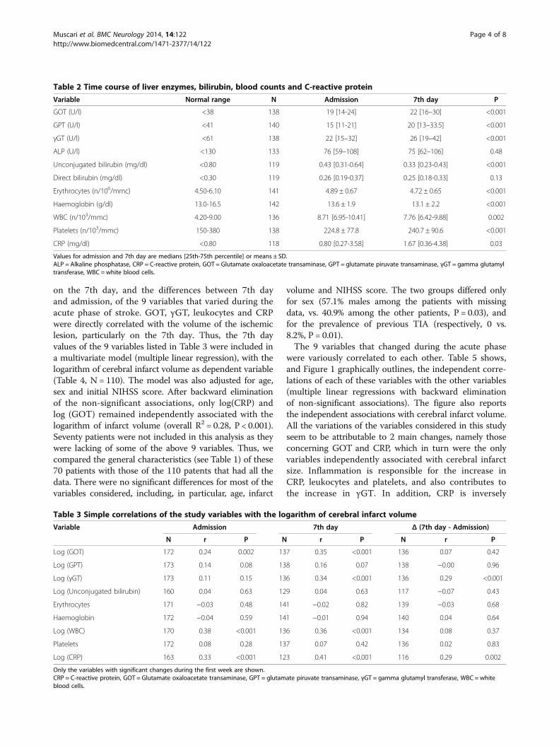
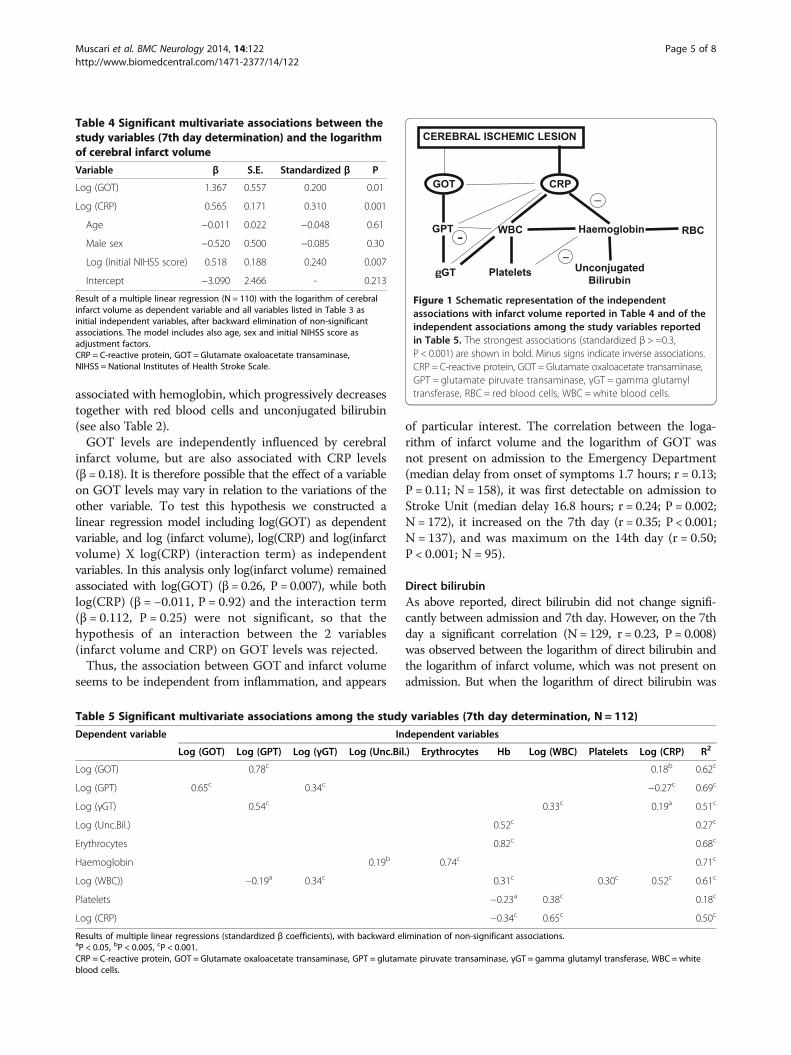
on the 7th day, and the differences between 7th dayand admission, of the 9 variables that varied during theacute phase of stroke. GOT, γGT, leukocytes and CRPwere directly correlated with the volume of the ischemiclesion, particularly on the 7th day. Thus, the 7th dayvalues of the 9 variables listed in Table 3 were included ina multivariate model (multiple linear regression), with thelogarithm of cerebral infarct volume as dependent variable(Table 4, N = 110). The model was also adjusted for age,sex and initial NIHSS score. After backward eliminationof the non-significant associations, only log(CRP) andlog (GOT) remained independently associated with thelogarithm of infarct volume (overall R2 = 0.28, P < 0.001).Seventy patients were not included in this analysis as theywere lacking of some of the above 9 variables. Thus, wecompared the general characteristics (see Table 1) of these70 patients with those of the 110 patents that had all thedata. There were no significant differences for most of thevariables considered, including, in particular, age, infarct
volume and NIHSS score. The two groups differed onlyfor sex (57.1% males among the patients with missingdata, vs. 40.9% among the other patients, P = 0.03), andfor the prevalence of previous TIA (respectively, 0 vs.8.2%, P = 0.01).The 9 variables that changed during the acute phase
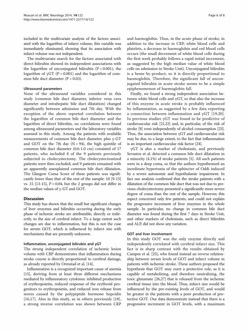
were variously correlated to each other. Table 5 shows,and Figure 1 graphically outlines, the independent corre-lations of each of these variables with the other variables(multiple linear regressions with backward eliminationof non-significant associations). The figure also reportsthe independent associations with cerebral infarct volume.All the variations of the variables considered in this studyseem to be attributable to 2 main changes, namely thoseconcerning GOT and CRP, which in turn were the onlyvariables independently associated with cerebral infarctsize. Inflammation is responsible for the increase inCRP, leukocytes and platelets, and also contributes tothe increase in γGT. In addition, CRP is inversely
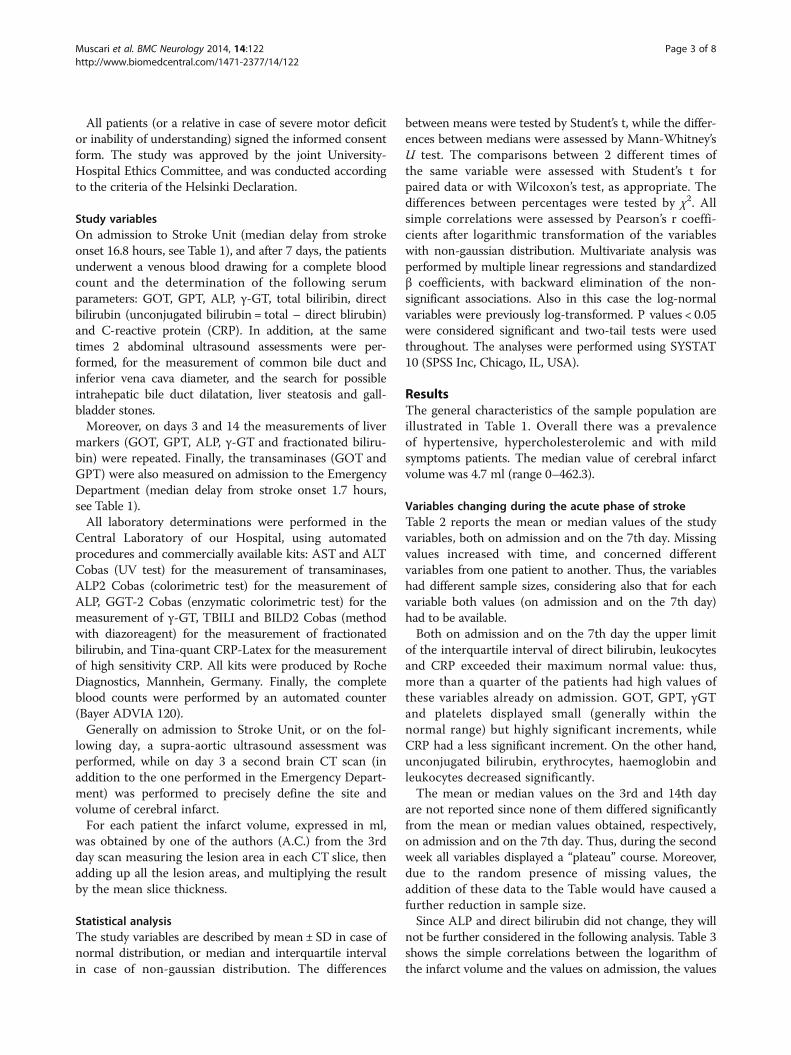
Table 2 Time course of liver enzymes, bilirubin, blood counts and C-reactive protein
Variable Normal range N Admission 7th day P
GOT (U/l) <38 138 19 [14-24] 22 [16–30] <0.001
GPT (U/l) <41 140 15 [11-21] 20 [13–33.5] <0.001
γGT (U/l) <61 138 22 [15–32] 26 [19–42] <0.001
ALP (U/l) <130 133 76 [59–108] 75 [62–106] 0.48
Unconjugated bilirubin (mg/dl) <0.80 119 0.43 [0.31-0.64] 0.33 [0.23-0.43] <0.001
Direct bilirubin (mg/dl) <0.30 119 0.26 [0.19-0.37] 0.25 [0.18-0.33] 0.13
Erythrocytes (n/106/mmc) 4.50-6.10 141 4.89 ± 0.67 4.72 ± 0.65 <0.001
Haemoglobin (g/dl) 13.0-16.5 142 13.6 ± 1.9 13.1 ± 2.2 <0.001
WBC (n/103/mmc) 4.20-9.00 136 8.71 [6.95-10.41] 7.76 [6.42-9.88] 0.002
Platelets (n/103/mmc) 150-380 138 224.8 ± 77.8 240.7 ± 90.6 <0.001
CRP (mg/dl) <0.80 118 0.80 [0.27-3.58] 1.67 [0.36-4.38] 0.03
Values for admission and 7th day are medians [25th-75th percentile] or means ± SD.ALP = Alkaline phosphatase, CRP = C-reactive protein, GOT = Glutamate oxaloacetate transaminase, GPT = glutamate piruvate transaminase, γGT = gamma glutamyltransferase, WBC =white blood cells.
Table 3 Simple correlations of the study variables with the logarithm of cerebral infarct volume
Variable Admission 7th day Δ (7th day - Admission)
N r P N r P N r P
Log (GOT) 172 0.24 0.002 137 0.35 <0.001 136 0.07 0.42
Log (GPT) 173 0.14 0.08 138 0.16 0.07 138 −0.00 0.96
Log (γGT) 173 0.11 0.15 136 0.34 <0.001 136 0.29 <0.001
Log (Unconjugated bilirubin) 160 0.04 0.63 129 0.04 0.63 117 −0.07 0.43
Erythrocytes 171 −0.03 0.48 141 −0.02 0.82 139 −0.03 0.68
Haemoglobin 172 −0.04 0.59 141 −0.01 0.94 140 0.04 0.64
Log (WBC) 170 0.38 <0.001 136 0.36 <0.001 134 0.08 0.37
Platelets 172 0.08 0.28 137 0.07 0.42 136 0.02 0.83
Log (CRP) 163 0.33 <0.001 123 0.41 <0.001 116 0.29 0.002
Only the variables with significant changes during the first week are shown.CRP = C-reactive protein, GOT = Glutamate oxaloacetate transaminase, GPT = glutamate piruvate transaminase, γGT = gamma glutamyl transferase, WBC = whiteblood cells.
Muscari et al. BMC Neurology 2014, 14:122 Page 4 of 8http://www.biomedcentral.com/1471-2377/14/122
associated with hemoglobin, which progressively decreasestogether with red blood cells and unconjugated bilirubin(see also Table 2).GOT levels are independently influenced by cerebral
infarct volume, but are also associated with CRP levels(β = 0.18). It is therefore possible that the effect of a variableon GOT levels may vary in relation to the variations of theother variable. To test this hypothesis we constructed alinear regression model including log(GOT) as dependentvariable, and log (infarct volume), log(CRP) and log(infarctvolume) X log(CRP) (interaction term) as independentvariables. In this analysis only log(infarct volume) remainedassociated with log(GOT) (β = 0.26, P = 0.007), while bothlog(CRP) (β = −0.011, P = 0.92) and the interaction term(β = 0.112, P = 0.25) were not significant, so that thehypothesis of an interaction between the 2 variables(infarct volume and CRP) on GOT levels was rejected.Thus, the association between GOT and infarct volume
seems to be independent from inflammation, and appears
of particular interest. The correlation between the loga-rithm of infarct volume and the logarithm of GOT wasnot present on admission to the Emergency Department(median delay from onset of symptoms 1.7 hours; r = 0.13;P = 0.11; N = 158), it was first detectable on admission toStroke Unit (median delay 16.8 hours; r = 0.24; P = 0.002;N = 172), it increased on the 7th day (r = 0.35; P < 0.001;N = 137), and was maximum on the 14th day (r = 0.50;P < 0.001; N = 95).
Direct bilirubinAs above reported, direct bilirubin did not change signifi-cantly between admission and 7th day. However, on the 7thday a significant correlation (N = 129, r = 0.23, P = 0.008)was observed between the logarithm of direct bilirubin andthe logarithm of infarct volume, which was not present onadmission. But when the logarithm of direct bilirubin was
Table 5 Significant multivariate associations among the study variables (7th day determination, N = 112)
Dependent variable Independent variables
Log (GOT) Log (GPT) Log (γGT) Log (Unc.Bil.) Erythrocytes Hb Log (WBC) Platelets Log (CRP) R2
Log (GOT) 0.78c 0.18b 0.62c
Log (GPT) 0.65c 0.34c −0.27c 0.69c
Log (γGT) 0.54c 0.33c 0.19a 0.51c
Log (Unc.Bil.) 0.52c 0.27c
Erythrocytes 0.82c 0.68c
Haemoglobin 0.19b 0.74c 0.71c
Log (WBC)) −0.19a 0.34c 0.31c 0.30c 0.52c 0.61c
Platelets −0.23a 0.38c 0.18c
Log (CRP) −0.34c 0.65c 0.50c
Results of multiple linear regressions (standardized β coefficients), with backward elimination of non-significant associations.aP < 0.05, bP < 0.005, cP < 0.001.CRP = C-reactive protein, GOT = Glutamate oxaloacetate transaminase, GPT = glutamate piruvate transaminase, γGT = gamma glutamyl transferase, WBC = whiteblood cells.
CEREBRAL ISCHEMIC LESION
GOT CRP
GPT WBC
GT Platelets
Haemoglobin RBC
Unconjugated Bilirubin
_
_-
Figure 1 Schematic representation of the independentassociations with infarct volume reported in Table 4 and of theindependent associations among the study variables reportedin Table 5. The strongest associations (standardized β > =0.3,P < 0.001) are shown in bold. Minus signs indicate inverse associations.CRP = C-reactive protein, GOT = Glutamate oxaloacetate transaminase,GPT = glutamate piruvate transaminase, γGT = gamma glutamyltransferase, RBC = red blood cells, WBC = white blood cells.
Table 4 Significant multivariate associations between thestudy variables (7th day determination) and the logarithmof cerebral infarct volume
Variable β S.E. Standardized β P
Log (GOT) 1.367 0.557 0.200 0.01
Log (CRP) 0.565 0.171 0.310 0.001
Age −0.011 0.022 −0.048 0.61
Male sex −0.520 0.500 −0.085 0.30
Log (Initial NIHSS score) 0.518 0.188 0.240 0.007
Intercept −3.090 2.466 - 0.213
Result of a multiple linear regression (N = 110) with the logarithm of cerebralinfarct volume as dependent variable and all variables listed in Table 3 asinitial independent variables, after backward elimination of non-significantassociations. The model includes also age, sex and initial NIHSS score asadjustment factors.CRP = C-reactive protein, GOT = Glutamate oxaloacetate transaminase,NIHSS = National Institutes of Health Stroke Scale.
Muscari et al. BMC Neurology 2014, 14:122 Page 5 of 8http://www.biomedcentral.com/1471-2377/14/122
included in the multivariate analysis of the factors associ-ated with the logarithm of infarct volume, this variable wasimmediately eliminated, showing that its association withinfarct volume was not independent.The multivariate search for the factors associated with
direct bilirubin showed its independent associations withthe logarithm of unconjugated bilirubin (P < 0.001), thelogarithm of γGT (P = 0.001) and the logarithm of com-mon bile duct diameter (P = 0.03).
Ultrasound parametersNone of the ultrasound variables considered in thisstudy (common bile duct diameter, inferior vena cavadiameter and intrahepatic bile duct dilatation) changedsignificantly between admission and 7th day. With theexception of the above reported correlation betweenthe logarithm of common bile duct diameter and thelogarithm of direct bilirubin, no correlations were foundamong ultrasound parameters and the laboratory variablesassessed in this study. Among the patients with availablemeasurements of common bile duct diameter, plus γ-GTand GOT on the 7th day (N = 94), the high quintile ofcommon bile duct diameter (0.6-1.0 cm) consisted of 17patients, who included 8 of the 9 patients previouslysubjected to cholecystectomy. The cholecystectomizedpatients were then excluded, and 9 patients remained withan apparently unexplained common bile duct dilatation.The Glasgow Coma Score of these patients was signifi-cantly lower than that of the rest of the sample: 10 [9-15]vs. 15 [13-15], P = 0.04, but the 2 groups did not differ inthe median values of γ-GT and GOT.
DiscussionThis study has shown that the small but significant changesof liver enzymes and bilirubin occurring during the earlyphase of ischemic stroke are attributable, directly or indir-ectly, to the size of cerebral infarct. To a large extent suchchanges are due to inflammation, but this is not the casefor serum GOT, which is influenced by infarct size withmechanisms that are presently unknown.
Inflammation, unconjugated bilirubin and γGTThe strong independent correlation of ischemic lesionvolume with CRP demonstrates that inflammation duringstroke course is directly proportional to cerebral damage,as already reported by Ormstad et al. [14].Inflammation is a recognized important cause of anemia
[15], deriving from at least three different mechanismsmediated by inflammatory cytokines: inhibited productionof erythropoietin, reduced response of the erythroid pro-genitors to erythropoietin, and reduced iron release fromstores caused by the polypeptide hormone hepcidin[16,17]. Also in this study, as in others previously [18],a strong inverse correlation was shown between CRP
and haemoglobin. Thus, in the acute phase of stroke, inaddition to the increase in CRP, white blood cells andplatelets, a decrease in haemoglobin and red blood cellsoccurs (the small decrement of white blood cells duringthe first week probably follows a rapid initial increment,as suggested by the high median value of white bloodcells on admission to Stroke Unit). Unconjugated bilirubinis a heme by-product, so it is directly proportional tohaemoglobin. Therefore, the significant fall of uncon-jugated bilirubin in acute stroke seems to be a simpleepiphenomenon of haemoglobin fall.Finally, we found a strong independent association be-
tween white blood cells and γGT, so that also the increaseof this enzyme in acute stroke is probably influencedby inflammation, as suggested by a few data reportinga connection between inflammation and γGT [19,20].In previous studies γGT was found to be predictive ofcardiovascular risk [21,22] and, in particular, of the risk ofstroke [8] even independently of alcohol consumption [23].Thus, the association between γGT and cardiovascular riskmay be due, to a large extent, to the fact that inflammationis an important cardiovascular risk factor [24].γGT is also a marker of cholestasis, and previously
Sevastos et al. detected a common bile duct dilatation ina minority (4.1%) of stroke patients [5]. All such patientswere in a deep coma, so that the authors hypothesized aninordinate hypertonia of the sphincter of Oddi inducedby a severe autonomic and hypothalamic impairment. Infact our analysis confirmed that the stroke patients with adilatation of the common bile duct that was not due to pre-vious cholecystectomy presented a significantly more severedegree of coma than the rest of the sample. However, thisaspect concerned only few patients, and could not explainthe progressive increment of liver enzymes in the wholesample. In particular, no change in common bile ductdiameter was found during the first 7 days in Stroke Unit,and other markers of cholestasis, such as direct bilirubinand ALP, did not show any variation.
GOT and liver involvementIn this study GOT was the only enzyme directly andindependently correlated with cerebral infarct size. Thisfact is in sharp contrast with the results obtained byCampos et al. [25], who found instead an inverse relation-ship between serum levels of GOT and infarct volume inpatients with ischemic stroke. These authors proposed thehypothesis that GOT may exert a protective role, as it iscapable of metabolizing, and therefore neutralizing, thetoxic glutamate [26,27] that is released from the ischemiccerebral tissue into the blood. Thus, infarct size would beinfluenced by the pre-existing levels of GOT, and wouldbe greater in the patients with a poor production of pro-tective GOT. Our data demonstrate instead that there is aprogressive increment in GOT levels, with a maximum
Muscari et al. BMC Neurology 2014, 14:122 Page 6 of 8http://www.biomedcentral.com/1471-2377/14/122
about on the 7th day after admission and subsequent plat-eau, and that the correlation with infarct volume is notonly direct, but also increasingly stronger with the passingof time after the acute event. Thus, our results show thatGOT production is influenced by cerebral infarct volume,and not vice versa. It seems probable that some substancereleased from cerebral infarct and different from inflam-matory cytokines (perhaps glutamate itself ), is capableof stimulating the production of GOT (whose possibleprotective effect is however not excluded).The differences with the study by Campos et al. [25]
may have several explanations. Firstly, these authorsmeasured GOT levels only on admission, i.e. on averagein their study nearly 3 hours after stroke onset, while inthe present study the strongest relationships betweenGOT and infarct volume were found on the 7th and14th day. However, in our patients even when GOT wasmeasured on admission to the Stroke Unit a significantdirect relationship with infarct volume was found, whilewhen GOT was measured in the Emergency Department(median delay from onset of symptoms 1.6 hours) nocorrelation with infarct volume was found, and in anycase the relationship was not of inverse type. Furthermore,the sample by Campos et al. was rather different fromours, as 35% of their patients had been thrombolysed(while thrombolysed patients had been excluded from oursample), cardioembolic strokes were over-represented(50%), and lacunar strokes were under-represented (4%).Finally, in our study the infarct volume was measuredin a more precise way, and log-transformed values wereused in the calculation of linear regressions betweennon-gaussian variables.Although GOT increments during stroke have been
reported also in cerebrospinal fluid [12,13], the chainedcorrelations among this enzyme, GPT and γGT suggestthat its synthesis probably occurs in the liver. Also a directrelease of GOT from necrotic cerebral tissue does notseem probable, as the appearance of the enzyme in blooddoes not follow a log-normal course (with early peak andsubsequent progressive decrease), as is the case, for ex-ample, after a myocardial infarction. Instead the enzymegradually accumulates in blood, with a progressive increase,and a plateau persisting at least until the 14th day.Further studies are needed to clarify the mechanisms
underlying the apparently independent association betweencerebral lesion size and serum GOT levels. At present, thehypothesis that the release of toxic glutamate by the ische-mic cerebral tissue [26,27] may induce the hepatic synthesisof the main enzymes involved in the metabolism of thisneurotransmitter remains the most probable one.
Direct bilirubinDirect bilirubin displayed no variations during the acutephase of stroke. However, in some cases its levels
exceeded the maximum normal value on both admissionand 7th day. Furthermore, the values measured on the7th day correlated with infarct volume, and previouslydirect bilirubin was found to be associated with strokeseverity as assessed by the NIH stroke scale [1,28]. Inour study multivariate analysis showed that the maindeterminants of direct bilirubin were unconjugatedbilirubin and γGT: the former is evidently the substratefrom which direct bilirubin originates, while the latter,during the course of stroke, is probably synthesized bythe liver in response to factors including inflammation(see above), which in part might also trigger the glucur-onoconjugation of bilirubin. Since during the first weekafter stroke onset unconjugated bilirubin decreasesand γGT increases, the net result on the trend of directbilirubin could be a plateau. Furthermore, the associationof direct bilirubin with γGT may explain the asso-ciation with infarct volume, and consequently withstroke severity.
LimitationsThe patients were admitted to our Stroke Unit afterhaving spent a variable period of time in the EmergencyDepartment. It is possible that during this early phasesome markers (especially CRP, leukocytes and direct bili-rubin) may have had a sharp rise that was not detected.The measurement of serum glutamate might have
allowed a better understanding of the pathophysiologicmechanisms involved.
ConclusionsThis study suggests that the liver participates to a variableextent in the acute phase of ischemic stroke, producingenzymes in response to signals coming from cerebralinfarct and proportional to its size. An important signalis likely represented by inflammatory cytokines, whichinduce the production of acute phase reactants such asCRP and probably γGT. Another signal, presently unknownand perhaps coinciding with toxic glutamate, triggersthe synthesis of GOT and indirectly of GPT. Instead,unconjugated bilirubin progressively decreases, togetherwith haemoglobin, in response to inflammation, whiledirect bilirubin does not show any significant variation.Also, the hypothesis that the changes of liver enzymesand bilirubin are related to extrahepatic cholestasis wasnot confirmed: this phenomenon probably concernsonly a small minority of comatose patients.In conclusion, GOT is the only liver enzyme directly
associated with the ischemic cerebral lesion independentlyfrom inflammation. Possibly this enzyme, neutralizingthe toxic glutamate, might play a protective role, as somereports on its favourable prognostic significance suggest[25,29].
Muscari et al. BMC Neurology 2014, 14:122 Page 7 of 8http://www.biomedcentral.com/1471-2377/14/122
Competing interestsThe authors declare that they have no competing interest.
Authors’ contributionsAM designed the study, analysed the data and wrote the manuscript. ACperformed infarct volume measurements and participated in data analysis.EF, MG, CN, VR and LV recruited patients and significantly contributed todata collection. AB and DM performed the ultrasound investigations andcritically revised the manuscript. GMP was involved in patients’ care and dataanalysis. MZ critically revised the manuscript. All authors read and approvedthe final version.
AcknowledgementsWe gratefully acknowledge the assistance of Dr. Carolina Tiani in preparing thestudy protocol to be submitted to the Ethics Committee. We are also indebtedwith Dr. Andrea Cenni for his help in recruiting patients and data entry. Thestudy was self-financed: no sources of funding have to be acknowledged.
Received: 28 September 2013 Accepted: 28 May 2014Published: 6 June 2014
References1. Pineda S, Bang OY, Saver JL, Starkman S, Yun SW, Liebeskind DS, Kim D, Ali
LK, Shah SH, Ovbiagele B: Association of serum bilirubin with ischemicstroke outcomes. J Stroke Cerebrovasc Dis 2008, 17:147–152.
2. Dohi K, Mochizuki Y, Satoh K, Jimbo H, Hayashi M, Toyoda I, Ikeda Y, Abe T,Aruga T: Transient elevation of serum bilirubin (a heme oxygenase-1metabolite) level in hemorrhagic stroke: bilirubin is a marker of oxidantstress. Acta Neurochir Suppl 2003, 86:247–249.
3. Stocker R, Yamamoto Y, McDonagh AF, Glazer AN, Ames BN: Bilirubin is anantioxidant of possible physiological importance. Science 1987,235(4792):1043–1046.
4. Doré S, Takahashi M, Ferris CD, Zakhary R, Hester LD, Guastella D, Snyder SH:Bilirubin, formed by activation of heme oxygenase-2, protects neuronsagainst oxidative stress injury. Proc Natl Acad Sci U S A 1999, 96:2445–2450.
5. Sevastos N, Savvas SP, Rafailidis PI, Manesis EK: Cholestasis in acute stroke:an investigation on its prevalence and etiology. Scand J Gastroenterol2005, 40:862–866.
6. Meyer J, Mischeck U, Veyhl M, Henzel K, Galla HJ: Blood–brain barriercharacteristic enzymatic properties in cultured brain capillary endothelialcells. Brain Res 1990, 514:305–309.
7. Korantzopoulos P, Tzimas P, Kalantzi K, Kostapanos M, Vemmos K,Goudevenos J, Elisaf M, Milionis H: Association between serum gamma-glutamyltransferase and acute ischemic nonembolic stroke in elderlysubjects. Arch Med Res 2009, 40:582–589.
8. Bots ML, Salonen JT, Elwood PC, Nikitin Y, Freire de Concalves A, Inzitari D,Sivenius J, Trichopoulou A, Tuomilehto J, Koudstaal PJ, Grobbee DE:Gamma-glutamyltransferase and risk of stroke: the EUROSTROKE project.J Epidemiol Community Health 2002, 56(Suppl 1):i25–i29.
9. Peck K, Shinton R, Beevers G: Is serum gamma-glutamyl transferase agood marker of alcohol intake in stroke patients? Postgrad Med J 1990,66:710–713.
10. Jousilahti P, Rastenyte D, Tuomilehto J: Serum gamma-glutamyl transferase,self-reported alcohol drinking, and the risk of stroke. Stroke 2000,31:1851–1855.
11. Muscari A, Puddu GM, Santoro N, Serafini C, Cenni A, Rossi V, Zoli M: TheAtorvastatin During Ischemic Stroke study: a pilot randomized controlledtrial. Clin Neuropharmacol 2011, 34:141–147.
12. Donnan GA, Zapf P, Doyle AE, Bladin PF: CSF enzymes in lacunar andcortical stroke. Stroke 1983, 14:266–269.
13. Nand N, Gupta S, Sharma M, Khosla SN, Saini AS, Lal H: Evaluation ofenzymes in serum and cerebrospinal fluid in cases of cerebrovascularaccidents. Angiology 1987, 38:750–755.
14. Ormstad H, Aass HC, Lund-Sørensen N, Amthor KF, Sandvik L: Serum levels ofcytokines and C-reactive protein in acute ischemic stroke patients, andtheir relationship to stroke lateralization, type, and infarct volume. J Neurol2011, 258:677–685.
15. Roy CN: Anemia of inflammation. Hematol Am Soc Hematol Educ Program2010, 2010:276–280.
16. Adamson JW: The anemia of inflammation/malignancy: mechanisms andmanagement. Hematol Am Soc Hematol Educ Program 2008, 2008:159–165.
17. Nicolas G, Viatte L, Bennoun M, Beaumont C, Kahn A, Vaulont S: Hepcidin, anew iron regulatory peptide. Blood Cells Mol Dis 2002, 29:327–35.
18. Hackeng CM, Beerenhout CM, Hermans M, Van der Kuy PH, Van der Dussen H,Van Dieijen-Visser MP, Hamulyák K, Van der Sande FM, Leunissen KM, KoomanJP: The relationship between reticulocyte hemoglobin content withC-reactive protein and conventional iron parameters in dialysis patients.J Nephrol 2004, 17:107–111.
19. Singh J, Chander J, Singh S, Singh G, Atal CK: Gamma-Glutamyltranspeptidase: a novel biochemical marker in inflammation.Biochem Pharmacol 1986, 35:3753–3760.
20. Bo S, Gambino R, Durazzo M, Guidi S, Tiozzo E, Ghione F, Gentile L, Cassader M,Pagano GF: Associations between gamma-glutamyl transferase, metabolicabnormalities and inflammation in healthy subjects from a population-based cohort: a possible implication for oxidative stress. World J Gastroenterol2005, 11:7109–7117.
21. Mason JE, Starke RD, Van Kirk JE: Gamma-glutamyl transferase: a novelcardiovascular risk biomarker. Prev Cardiol 2010, 13:36–41.
22. Ulus T, Yildirir A, Sade LE, Temiz A, Polat E, Bozbaş H, Aydinalp A, Eroğlu S,Ozin B, Müderrisoğlu H: Serum gamma-glutamyl transferase activity: newhigh-risk criteria in acute coronary syndrome patients? Coron Artery Dis2008, 19:489–495.
23. Fraser A, Harris R, Sattar N, Ebrahim S, Smith GD, Lawlor DA:Gamma-glutamyltransferase is associated with incident vascular eventsindependently of alcohol intake: analysis of the British Women’s Heartand Health Study and Meta-Analysis. Arterioscler Thromb Vasc Biol 2007,27:2729–2735.
24. Willerson JT, Ridker PM: Inflammation as a cardiovascular risk factor.Circulation 2004, 109(Suppl 1):II2–10.
25. Campos F, Rodríguez-Yáñez M, Castellanos M, Arias S, Pérez-Mato M,Sobrino T, Blanco M, Serena J, Castillo J: Blood levels of glutamateoxaloacetate transaminase are more strongly associated with goodoutcome in acute ischaemic stroke than glutamate pyruvatetransaminase levels. Clin Sci (Lond) 2011, 121:11–17.
26. Castillo J, Dávalos A, Naveiro J, Noya M: Neuroexcitatory amino acids andtheir relation to infarct size and neurological deficit in ischemic stroke.Stroke 1996, 27:1060–1065.
27. Castillo J, Dávalos A, Noya M: Progression of ischaemic stroke andexcitotoxic aminoacids. Lancet 1997, 349:79–83.
28. Luo Y, Li JW, Lu ZJ, Wang C, Guan DN, Xu Y: Serum bilirubin after acuteischemic stroke is associated with stroke severity. Curr Neurovasc Res2012, 9:128–132.
29. Campos F, Sobrino T, Ramos-Cabrer P, Castellanos M, Blanco M, Rodríguez-Yáñez M, Serena J, Leira R, Castillo J: High blood glutamate oxaloacetatetransaminase levels are associated with good functional outcome inacute ischemic stroke. J Cereb Blood Flow Metab 2011, 31:1387–1393.
doi:10.1186/1471-2377-14-122Cite this article as: Muscari et al.: Changes of liver enzymes and bilirubinduring ischemic stroke: mechanisms and possible significance. BMCNeurology 2014 14:122.
Submit your next manuscript to BioMed Centraland take full advantage of:
• Convenient online submission
• Thorough peer review
• No space constraints or color figure charges
• Immediate publication on acceptance
• Inclusion in PubMed, CAS, Scopus and Google Scholar
• Research which is freely available for redistribution
Submit your manuscript at www.biomedcentral.com/submit
Muscari et al. BMC Neurology 2014, 14:122 Page 8 of 8http://www.biomedcentral.com/1471-2377/14/122