ch. 6: cells & environment - francis marion...
TRANSCRIPT
11/15/2017
1
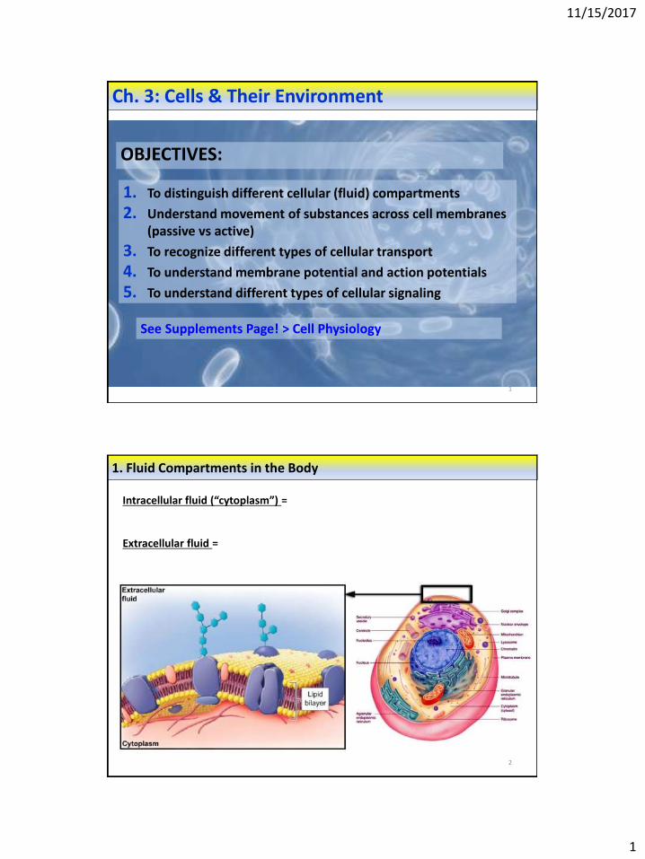
Ch. 3: Cells & Their Environment
1
OBJECTIVES:
1. To distinguish different cellular (fluid) compartments
2. Understand movement of substances across cell membranes (passive vs active)
3. To recognize different types of cellular transport
4. To understand membrane potential and action potentials
5. To understand different types of cellular signaling
See Supplements Page! > Cell Physiology
2
1. Fluid Compartments in the Body
Intracellular fluid (“cytoplasm”) =
Extracellular fluid =

11/15/2017
2
3
2. Movement of SubstancesAcross the Plasma Membrane
A) Categorize Substances by how permeable membrane is to them: easily they cross membranes.
B) Categorize by how permeable Membranes are:
• Permeable Membrane =
• Selectively Permeable Membrane =
11/15/2017
3
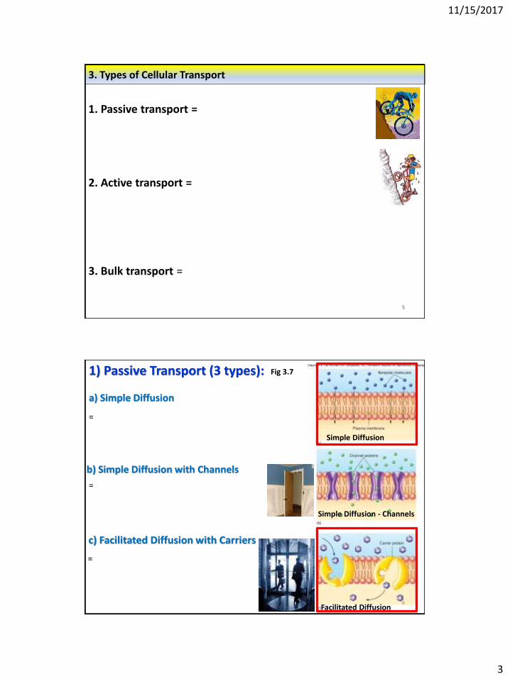
3. Types of Cellular Transport
5
1. Passive transport =
2. Active transport =
3. Bulk transport =
c) Facilitated Diffusion with Carriers
6
Simple Diffusion
Simple Diffusion - Channels
Facilitated Diffusion
a) Simple Diffusion
1) Passive Transport (3 types): Fig 3.7
=
=
b) Simple Diffusion with Channels
=

11/15/2017
4
c) Osmosis =
7
Fig 3.10
8
Osmosis depends on “Tonicity”
Fig 3.13
Isotonic solution =
(ex. Normal or physiologic saline)
Hypotonic solution =
Hypertonic solution =
Normal (isotonic) saline
11/15/2017
5
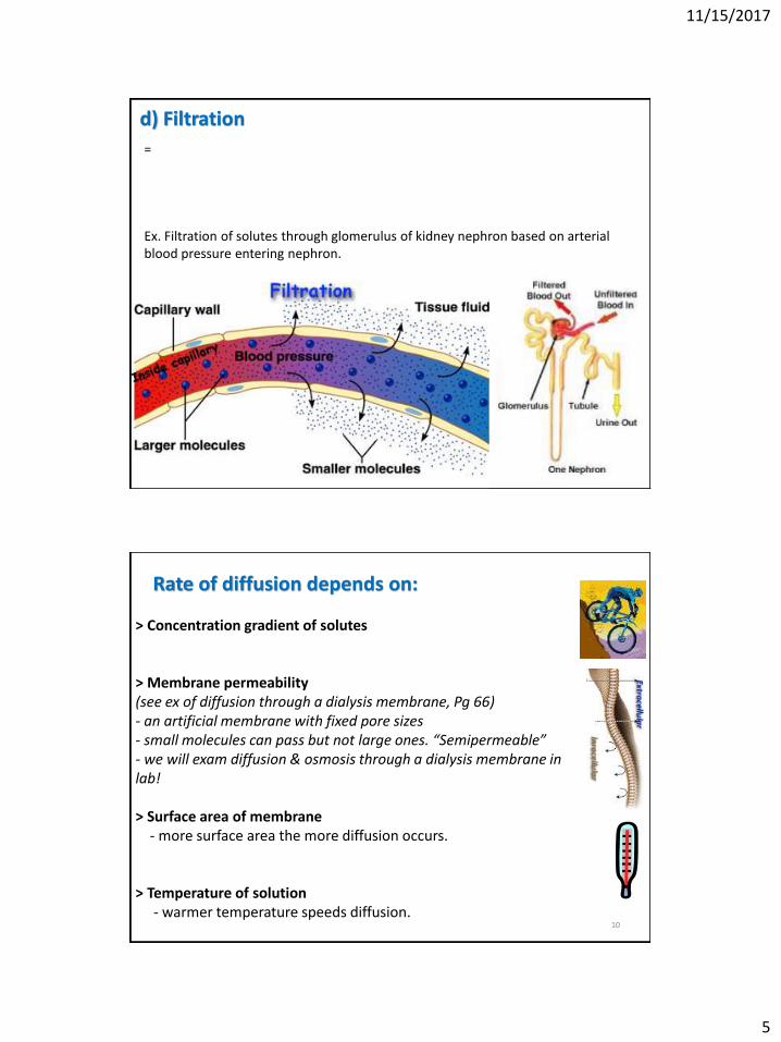
d) Filtration
9
=
Ex. Filtration of solutes through glomerulus of kidney nephron based on arterial blood pressure entering nephron.
Rate of diffusion depends on:
10
> Concentration gradient of solutes
> Membrane permeability (see ex of diffusion through a dialysis membrane, Pg 66)- an artificial membrane with fixed pore sizes - small molecules can pass but not large ones. “Semipermeable”- we will exam diffusion & osmosis through a dialysis membrane in lab!
> Surface area of membrane- more surface area the more diffusion occurs.
> Temperature of solution- warmer temperature speeds diffusion.

11/15/2017
6
2. Active Transport
11
a) Primary Active Transport = movement of ions with a pump fueled by ATP.
i) Calcium (Ca+2) Pumpkeeps Ca+2 concentrations low inside cells.
ii) Hydrogen (H+) Pumpused to increase acidity.Ex. Parietal cells of stomach have H+ pumps.Nexium® targets these cells for those with GERD.
iii) Sodium – Potassium (Na+/K+) Pump3 Na+ exit for every 2 K+ that enter cell.
Helps maintain cell membrane resting potential.
Fig 6.19

11/15/2017
7
b) Secondary Active Transport: Coupled transport
13
i) Co-transport (“symport”) = Energy gained from passive transport of one ion fuels the active transport of another ion in the same direction.
Ex. Passive transport of Na+ with its concentration gradient helps fuel the active transport of glucose against its concentration gradient in kidney tubules.
ii) Counter-transport (“antiport”) = Energy gained from passive transport of one ion fuels the active transport of another ion in the opposite direction.
Read “Physiology in Health & Disease” – Pg 80The importance of co-transport of Na+ and glucose in Oral Rehydration Therapy (ORT):
Chronic diarrhea (from acute gastroenteritis, cholera, etc…) limits ability of intestines to reabsorb salt & water, leading to risk of dehydration (life-threatening in children).
BUT diarrhea doesn’t interfere with co-transport of Na+ & glucose in intestines. Water follows Na+ by osmosis into cells, and into bloodstream. Patient gets hydrated.
So, ORT with salt AND glucose is vital!

11/15/2017
8
3. BulkTransport = A) Endocytosis = bulk movement of molecules into a cell.
i) phagocytosis =
ii) Pinocytosis =
iii) Receptor-mediated endocytosis =
B) Exocytosis = bulk movement of molecules out of a cell.
Cell Transport - Review
16
“Permeability” (of substances & membranes)
Passive transport = no energy, with concentration gradient (“downhill”)- Simple diffusion- Facilitated diffusion- Osmosis- Filtration
Active transport = ATP required, against concentration gradient (“uphill”)- Primary active transport (calcium, hydrogen, & Na+/K+ pumps)- Coupled transport (co-transport & counter-transport)
Bulk tranport- Endocytosis- Exocytosis

11/15/2017
9
17
4. Membrane Potential
Figs 3.21
Resting cell membrane potential (MP)= -70 mV inside of cell has “fixed number of anions” (neg charged particles)
> number of K+ ions entering /leaving cell changes intracellular negativity- The more K+ exits, the more neg inside becomes- The more K+ enters, the less neg inside becomes
MP maintained by Na+/K+ pump
18
Clinical Application– Hyperkalemia & Lethal Injections
Lethal injection is potassium chloride.
Hyperkalemia =
11/15/2017
10
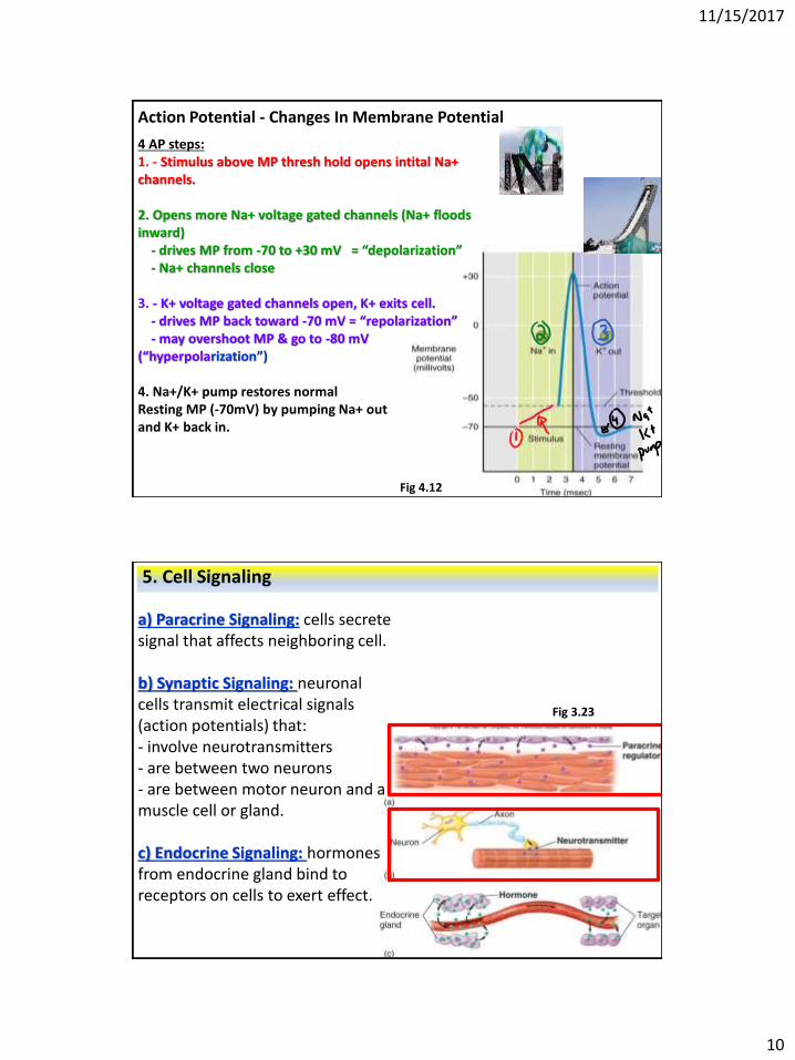
Action Potential - Changes In Membrane Potential
4 AP steps:1. - Stimulus above MP thresh hold opens intital Na+ channels.
2. Opens more Na+ voltage gated channels (Na+ floods inward)
- drives MP from -70 to +30 mV = “depolarization”- Na+ channels close
3. - K+ voltage gated channels open, K+ exits cell.- drives MP back toward -70 mV = “repolarization”- may overshoot MP & go to -80 mV
(“hyperpolarization”)
4. Na+/K+ pump restores normalResting MP (-70mV) by pumping Na+ out and K+ back in.
Fig 4.12
5. Cell Signaling
a) Paracrine Signaling: cells secrete signal that affects neighboring cell.
b) Synaptic Signaling: neuronal cells transmit electrical signals (action potentials) that:- involve neurotransmitters- are between two neurons- are between motor neuron and a muscle cell or gland.
c) Endocrine Signaling: hormones from endocrine gland bind to receptors on cells to exert effect.
Fig 3.23
11/15/2017
11
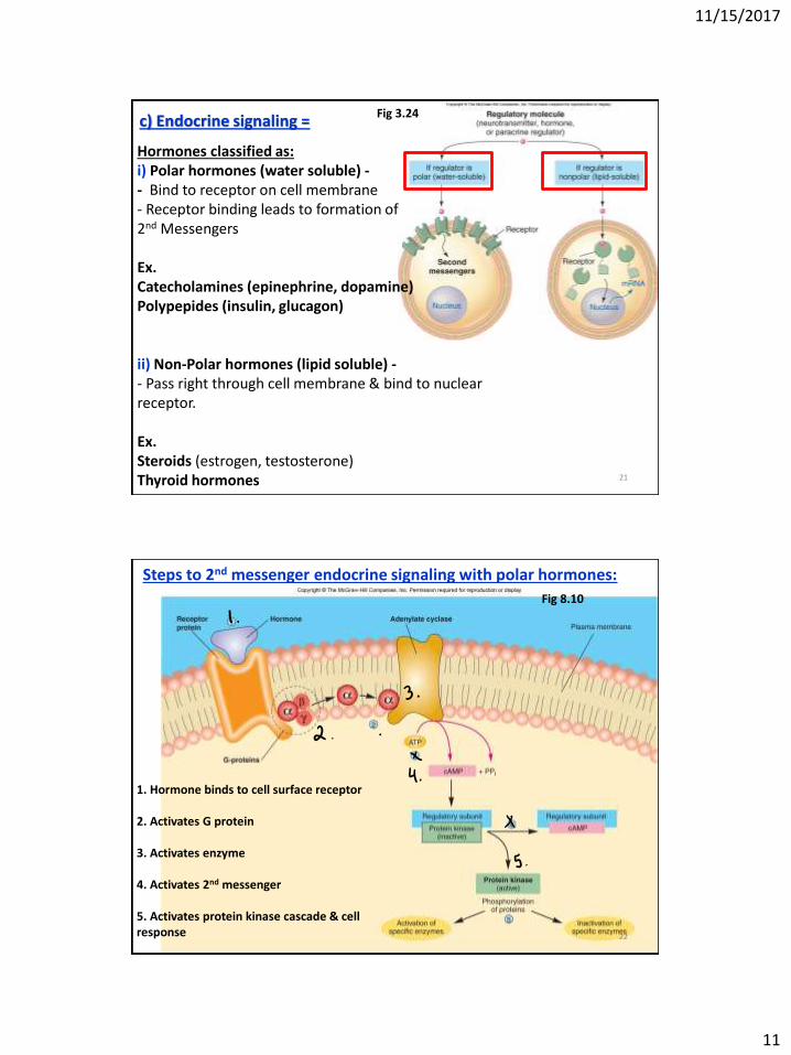
21
c) Endocrine signaling =
Hormones classified as:i) Polar hormones (water soluble) -- Bind to receptor on cell membrane- Receptor binding leads to formation of 2nd Messengers
Ex.Catecholamines (epinephrine, dopamine)Polypepides (insulin, glucagon)
ii) Non-Polar hormones (lipid soluble) -- Pass right through cell membrane & bind to nuclear receptor.
Ex.Steroids (estrogen, testosterone)Thyroid hormones
Fig 3.24
22
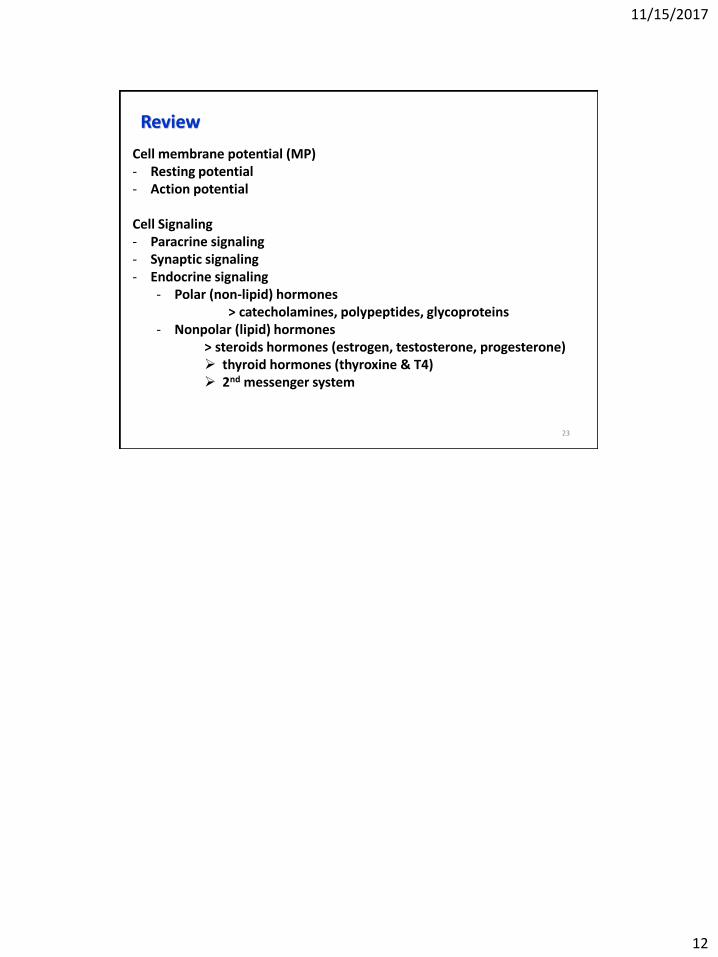
Fig 8.10
Steps to 2nd messenger endocrine signaling with polar hormones:
1. Hormone binds to cell surface receptor
2. Activates G protein
3. Activates enzyme
4. Activates 2nd messenger
5. Activates protein kinase cascade & cell response
11/15/2017
12
Review
23
Cell membrane potential (MP)- Resting potential- Action potential
Cell Signaling- Paracrine signaling- Synaptic signaling- Endocrine signaling
- Polar (non-lipid) hormones> catecholamines, polypeptides, glycoproteins
- Nonpolar (lipid) hormones> steroids hormones (estrogen, testosterone, progesterone) thyroid hormones (thyroxine & T4) 2nd messenger system