cervicofacial infection in a nigerian tertiary health institution: a ... · cervicofacial infection...
TRANSCRIPT

293
public health concern with regard to dental diseases and oro-
facial trauma, and they are common in poor patients lacking
proper health resources. Patients affected by such infections
typically present to emergency rooms2. These infections fre-
quently result in cellulitis or abscess formation involving at
least one facial space3.
Infection of the anatomic spaces of the head and neck are
graded with regard to severity based on the level to which
they threaten the airway or vital structures, such as the heart
and mediastinum or cranial contents. The severity can either
be low, moderate, or high4. A lower severity is noted when
the infection does not threaten the airway. Infections of the
spaces that can hinder access to the airway due to swelling or
trismus are considered to be of moderate severity, while those
that obstruct or deviate the airway or threaten vital structures
are graded as severe4.
Anatomical and microbial factors as well as impairment in
host resistance, compounded by a delay in receiving adequate
I. Introduction
Infection involving the orbit, zygomatic space, lateral
pharyngeal space, or multifacial (unilateral) and oral floor
phlegmon are known as cervicofacial infections (CFIs).
When diagnosis and/or adequate treatment are delayed, such
infections can be life-threatening1. Orofacial infections are a
ORIGINAL ARTICLE
Benjamin FometeDepartment of Maxillofacial Surgery, Ahmadu Bello University Teaching Hospital, P.O. Box 3772, Zaria, Kaduna, Nigeria TEL: +234-8034515494E-mail: [email protected]: http://orcid.org/0000-0003-4690-0496
*Current affiliation: Department of Dental and Maxillofacial Surgery, University of Jos Teaching Hospital, Jos, Nigeria.
This is an open-access article distributed under the terms of the Creative Commons Attribution Non-Commercial License (http://creativecommons.org/licenses/by-nc/4.0/), which permits unrestricted non-commercial use, distribution, and reproduction in any medium, provided the original work is properly cited.
CC
Cervicofacial infection in a Nigerian tertiary health institution: a retrospective analysis of 77 cases
Benjamin Fomete1, Rowland Agbara1,*, Daniel Otasowie Osunde2, Charles N Ononiwu1
1Department of Maxillofacial Surgery, Ahmadu Bello University Teaching Hospital, Zaria, 2Department of Dental and Maxillofacial Surgery, University of Calabar Teaching Hospital, Calabar, Nigeria
Abstract (J Korean Assoc Oral Maxillofac Surg 2015;41:293-298)
Objectives: Infection involving the orbit, zygomatic space, lateral pharyngeal space, or hemifacial and oral floor phlegmon is referred to as cervicofa-cial infection (CFI). When diagnosis and/or adequate treatment are delayed, these infections can be life-threatening. Most cases are the result of odon-togenic infections. We highlight our experiences in the management of this life-threatening condition.Materials and Methods: This was a retrospective study of patients who presented with CFI from December 2005 to June 2012 at the Oral and Max-illofacial Surgery Clinic or the Accident and Emergency Unit of Ahmadu Bello University Teaching Hospital (Zaria, Nigeria). The medical records of all patients who presented with either localized or diffuse infection of the maxillofacial soft tissue spaces were retrospectively collected. Data collected was analyzed using SPSS version 13.0 and are expressed as descriptive and inferential statistics.Results: Of the 77 patients, 49 patients (63.6%) were males, a male to female ratio of 1:7.5. The ages ranged from two years to 75 years with a mean of 35.0±19.3 years, although most patients were older than 40 years. The duration of symptoms prior to presentation ranged from 6 to 60 days, with a mean of 11.0±9.4 days. More than 90% of the patients presented to the clinic within the first 10 days. The most commonly involved anatomical space was the submandibular space (n=29, 37.7%), followed by hemifacial space (n=22, 28.6%) and buccal space (n=7, 9.1%). Ludwig angina accounted for about 7.8% of the cases.Conclusion: CFI most commonly involves the submandibular space, typically affects individuals with a low level of education, and is influenced by traditional medical practices. Despite improved health care delivery, CFI remains a significant problem in developing countries.
Key words: Cervicofacial, Infection, Management, Odontogengic[paper submitted 2015. 4. 3 / revised 2015. 7. 4 / accepted 2015. 8. 4]
Copyright Ⓒ 2015 The Korean Association of Oral and Maxillofacial Surgeons. All rights reserved.
http://dx.doi.org/10.5125/jkaoms.2015.41.6.293pISSN 2234-7550·eISSN 2234-5930

J Korean Assoc Oral Maxillofac Surg 2015;41:293-298
294
The data were analyzed using SPSS version 13.0 (SPSS
Inc., Chicago, IL, USA). Qualitative variables were expressed
as frequency and percentage, while quantitative variables
were reported as mean and standard deviation. Inferential sta-
tistics were performed using chi-square test or independent
sample t-test as appropriate. A P-value<0.05 was considered
statistically significant.
III. Results
Seventy-seven patients comprised of 49 male (63.6%) and
28 female (36.4%) were analyzed, resulting in a male to fe-
male ratio of 1:7.5. The ages ranged from 2 to 75 years, with
a mean of 35.0±19.3 years, although most were older than 40
years.(Fig. 1) The duration of symptoms prior to presenta-
tion ranged from 6 to 60 days, with a mean of 11.0±9.4 days.
More than 90% of the patients presented to the clinic within
the first 10 days.(Fig. 2) The majority of patients had only
primary school education (88.6%), and this was distantly
followed by those with secondary school education (5.7%),
while those who attained university and quaranic education
were equally distributed at 2.9%. All patients presented with
facial swelling, which was either diffuse (Fig. 3) or local-
ized. Other presentations included pain (90.9%), toothache
(58.4%), dysphagia (38.9%), and pus discharge (29.8%).
About 65.2% of patients presented with trismus, and 64.3%
reported having used traditional herbal medications prior to
presentation at the hospital. The most commonly involved an-
atomical space was the submandibular space (n=29, 37.7%),
followed by the multifacial space (unilateral) (n=22, 28.6%)
and the buccal space (n=7, 9.1%). Ludwig angina accounted
treatment in the early stages, can result in progression of a
localized infection into a CFI5. Predisposing factors such as
alcoholism, immunosuppression, uncontrolled diabetes mel-
litus, and multiple underlying medical conditions have been
reported to increase the risk of odontogenic infection2. The
long hospital stay often associated with these infections can
become an economic factor for both the patient and society.
These CFI generally respond to antimicrobial chemotherapy
and/or surgical intervention.
The aim of this paper is to present our experience in the
management of CFI and the challenges in a resource-limited
environment.
II. Materials and Methods
This is a retrospective study of patients who presented
with CFI to the Oral and Maxillofacial Surgery Clinic or the
Accident and Emergency Unit of Ahmadu Bello University
Teaching Hospital (Zaria, Nigeria), between December 2005
and June 2012. The medical records of all patients who pre-
sented with either localized or diffuse infection of the head
and neck soft tissue spaces were collected. Patients who
presented with dentoalveolar abscesses or osteomyelitis were
excluded. Information obtained were demographics, pre-
senting complaint, time of presentation, nature of swelling,
source of infection, presence of any underlying comorbidity,
complications, degree of mouth opening, as well as use of
self-medication and herbal medications. In addition, the tem-
perature at presentation; types of treatment; results of micros-
copy, culture, and sensitivity (MCS); and length of stay (LOS)
in the hospital were noted.
Patients
(%)
<10
40
30
20
10
0
Age (yr)
11-20 21-30 31-40 >41
10.7
16.0
20.0
16.0
37.3
Fig. 1. Age distribution of the patients.Benjamin Fomete et al: Cervicofacial infection in a Nigerian tertiary health institution: a retrospective analysis of 77 cases. J Korean Assoc Oral Maxillofac Surg 2015
Patients
(%)
<10
100
80
60
40
20
0
Duration (day)
11-20 21-30 >31
91.8
3.3 1.6 3.3
Fig. 2. Duration of presenting complaint.Benjamin Fomete et al: Cervicofacial infection in a Nigerian tertiary health institution: a retrospective analysis of 77 cases. J Korean Assoc Oral Maxillofac Surg 2015

Cervicofacial infection in a Nigerian tertiary health institution
295
treated on an out-patient basis with oral drugs such as linco-
mycin, metronidazole, and analgesic. Following I&D, corru-
gated rubber drains (Fig. 3) were inserted and secured using
sutures. The temperature range at admission was 37.5oC to
39.9oC.
About 15.6% of patients had one complication, and facial
nerve palsy was the most common, observed in 25% of the
cases. Other complications are shown in Table 2. Of the 12
patients with complications, four patients (33.3%) died, re-
sulting in an overall mortality among all patients of 5.2%.
The use of traditional herbal medications had no significant
influence on the complications (χ2=1.831, degree of freedom
[df]=1, P=0.176).
Anemia was the most common comorbidity (n=40, 75.5%),
followed by diabetes mellitus either in isolation or occurring
concurrently with other comorbidities, and this accounted for
about 17% of the cases.(Table 3) There was a strong associa-
tion between time of presentation and presence of comorbid-
ity (χ2= 65.648, df=14, P<0.001). Thus, patients who present-
ed late were more likely to develop comorbidity compared
with early presenters.
The modality of treatment was not stated for one of the
patients. Of the remaining 76 patients, about 98.7% received
for about 7.8% of the cases.(Table 1)
The admitted, non-diabetic patients received an infusion of
0.9% saline (Dana Pharmaceutical, Lagos, Nigeria) and 5%
dextrose (Juhel Nigeria Ltd., Enugu, Nigeria). All patients
underwent incision and drainage (I&D) under local anes-
thesia; pus or swab was collected for MCS, and 63.4% were
admitted to the hospital and received parenteral medications,
typically metronidazole (Juhel Nigeria Ltd.), crystalline peni-
cillin (Shanxi Federal Pharmaceutical Co., Ltd., Jinzhong,
China), gentamicin (Lek Pharmaceutical and Chemical Com-
pany, Ljubljana, Slovania), and rocephine (F. Hoffmann-La
Roche Ltd., Basel, Switzerland); the remaining 36.6% were
Table 1. Anatomical spaces involved in the patients (n=77)
Space Frequency
Anterior triangle of neckBuccal spaceMultifacial (unilateral) spaces1
SMD spaceLateral pharyngeal, anterior neckBilateral SMD and SLBilateral SMD, SL+pharyngealMasticator and perimandibularOrbitalParapharyngealRight multifacial and left SMDSL, SMDSMTTemporal, buccal, SMT, SMD
1 (1.3)7 (9.1)
22 (28.6)29 (37.7)1 (1.3)6 (7.8)1 (1.3)1 (1.3)1 (1.3)1 (1.3)1 (1.3)1 (1.3)4 (5.2)1 (1.3)
(SMD: submandibular, SL: sublingual, SMT: submasseteric)1All the spaces on one side. Values are presented as number (%).The sum of the percentages does not equal 100% because of rounding.Benjamin Fomete et al: Cervicofacial infection in a Nigerian tertiary health institution: a retrospective analysis of 77 cases. J Korean Assoc Oral Maxillofac Surg 2015
Table 2. Types of complications recorded in the patients (n=12)
Type of complications Frequency
Cavernous sinus thrombosisEmpyema thoracisFacial nerve palsyMediastinitisMetastatic brain abscessRestricted neck movement resulting from contractureDeath
1 (8.3)1 (8.3)3 (25.0)1 (8.3)1 (8.3)1 (8.3)4 (33.3)
Values are presented as number (%).Benjamin Fomete et al: Cervicofacial infection in a Nigerian tertiary health institution: a retrospective analysis of 77 cases. J Korean Assoc Oral Maxillofac Surg 2015
Table 3. Comorbidities in the patients (n=53)
Comorbidity Frequency
AnemiaAnemia+HIVAnemia in pregnancyDiabetes mellitusDiabetes mellitus+anemiaHIVHTNUncontrolled diabetes mellitus+HTN
40 (75.5)1 (1.9)1 (1.9)4 (7.5)4 (7.5)1 (1.9)1 (1.9)1 (1.9)
(HIV: human immunodeficiency virus, HTN: hypertension)Values are presented as number (%).The sum of the percentages does not equal 100% because of rounding.Benjamin Fomete et al: Cervicofacial infection in a Nigerian tertiary health institution: a retrospective analysis of 77 cases. J Korean Assoc Oral Maxillofac Surg 2015
Fig. 3. It is diffused cellulitis.Benjamin Fomete et al: Cervicofacial infection in a Nigerian tertiary health institution: a retrospective analysis of 77 cases. J Korean Assoc Oral Maxillofac Surg 2015
J Korean Assoc Oral Maxillofac Surg 2015;41:293-298
296
space, followed by the submandibular space13. The frequency
of submandibular space involvement recorded in this study
is lower than that in other reports3,5,12 but higher than findings
from other studies8,10. Of the previously reported involved
spaces, the hemifacial space was the most common, and in-
volvement of multiple spaces has been attributed to delay in
presentation.
Although different imaging techniques have been used for
diagnosing odontogenic infections, a computed tomography
scan is considered the gold standard owing to its superiority
in diagnosing deep space infections11,14. Ultrasound has been
suggested as one possible modality for imaging of infections;
however, thus far, it has not been successful in detecting deep
fascial space infections11.
Ludwig’s angina constituted 7.8% (6 cases) of all CFIs, and
this is lower than findings from a similar study in northern
Nigeria12. Ludwig’s angina is a fatal progressive gangrenous
cellulitis and edema of the soft tissues of the neck and floor
of the mouth. Airway compromise has been a leading cause
of death due to this infection. Two of the four mortalities re-
corded in this study were observed in patients with Ludwig’s
angina. Their causes of death were not known as no autopsy
was carried out. The probable cause is airway obstruction or
endotoxic shock resulting from septicemia. Although trache-
ostomy has been used to secure the airway in patients with
CFI5, none of our patients underwent tracheostomy. How-
ever, endotracheal intubation was attempted on one patient to
secure the airway after I&D. This was unsuccessful, and the
patient eventually died.
Infection of spaces that can hinder access to the airway as
a result of swelling or trismus carry a moderate threat to the
airway13. Even though 65.3% of the patients in this study had
I&D and medication. Other treatments administered are
shown in Table 4. About 13% of patients had repeat I&D,
and patients with trismus were encouraged to perform mouth
exercise using a wooden spatula or chewing sugarless gum.
The wounds were dressed twice daily with gauze soaked in
diluted hydrogen peroxide and eusol.
The results of microscopy showed that only about 23.3%
of the specimens were culture-positive. One possible reason
for this low finding is that patients were taking medication
(self-prescribed or prescribed at referring centers). The iso-
lated organisms were Streptococcus pyogenes, Pseudomonas
species, and Staphylococcus aureus. The length of stay in the
hospital ranged from 4 to 42 days, with a mean of 15.0±7.6
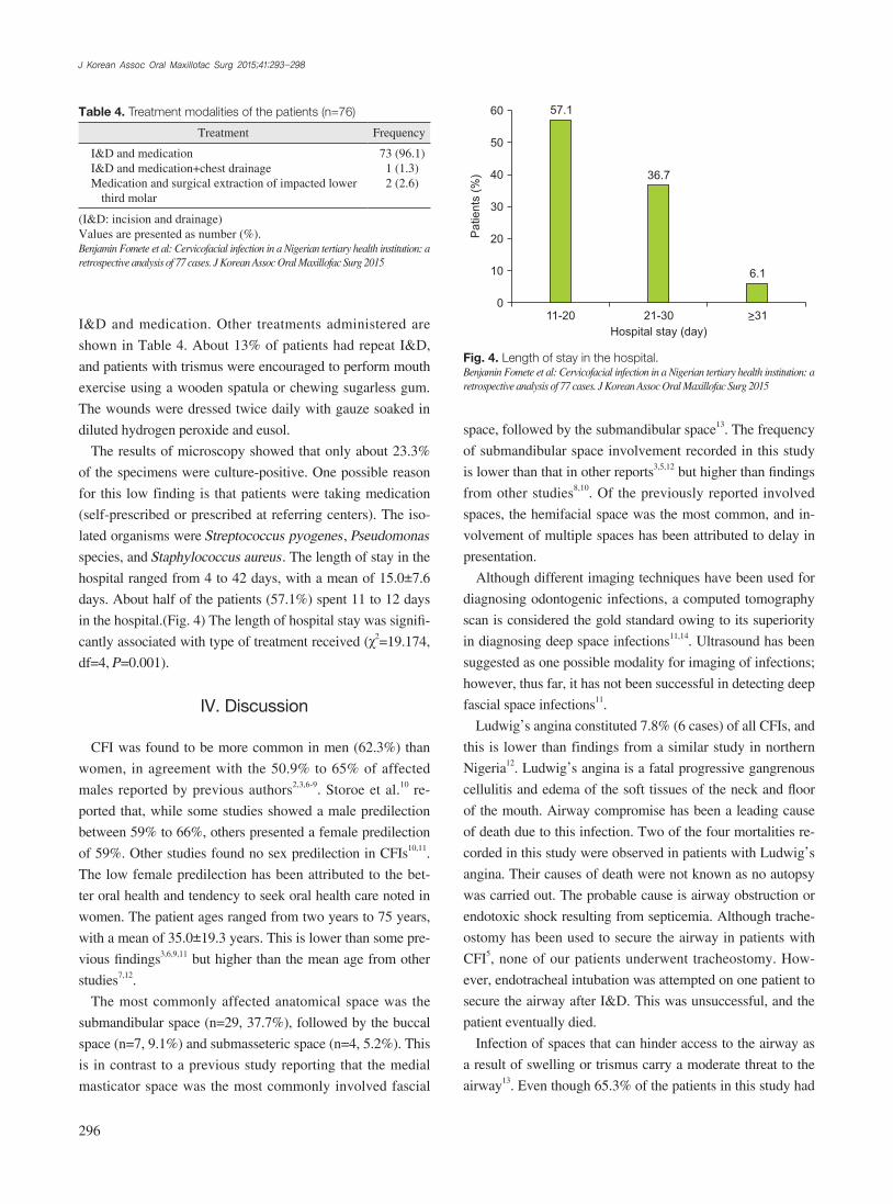
days. About half of the patients (57.1%) spent 11 to 12 days
in the hospital.(Fig. 4) The length of hospital stay was signifi-
cantly associated with type of treatment received (χ2=19.174,
df=4, P=0.001).
IV. Discussion
CFI was found to be more common in men (62.3%) than
women, in agreement with the 50.9% to 65% of affected
males reported by previous authors2,3,6-9. Storoe et al.10 re-
ported that, while some studies showed a male predilection
between 59% to 66%, others presented a female predilection
of 59%. Other studies found no sex predilection in CFIs10,11.
The low female predilection has been attributed to the bet-
ter oral health and tendency to seek oral health care noted in
women. The patient ages ranged from two years to 75 years,
with a mean of 35.0±19.3 years. This is lower than some pre-
vious findings3,6,9,11 but higher than the mean age from other
studies7,12.
The most commonly affected anatomical space was the
submandibular space (n=29, 37.7%), followed by the buccal
space (n=7, 9.1%) and submasseteric space (n=4, 5.2%). This
is in contrast to a previous study reporting that the medial
masticator space was the most commonly involved fascial
Table 4. Treatment modalities of the patients (n=76)
Treatment Frequency
I&D and medicationI&D and medication+chest drainageMedication and surgical extraction of impacted lower
third molar
73 (96.1)1 (1.3)2 (2.6)
(I&D: incision and drainage)Values are presented as number (%).Benjamin Fomete et al: Cervicofacial infection in a Nigerian tertiary health institution: a retrospective analysis of 77 cases. J Korean Assoc Oral Maxillofac Surg 2015
Patients
(%)
60
50
40
30
20
10
0
Hospital stay (day)
11-20 21-30 >31
57.1
36.7
6.1
Fig. 4. Length of stay in the hospital.Benjamin Fomete et al: Cervicofacial infection in a Nigerian tertiary health institution: a retrospective analysis of 77 cases. J Korean Assoc Oral Maxillofac Surg 2015

Cervicofacial infection in a Nigerian tertiary health institution
297
local anesthesia avoids the complications of general anesthe-
sia. The presence of comorbidity in some of our patients also
contraindicated any planned use of general anesthesia in the
present series.
Orofacial odontogenic infections are known to be poly-mi-
crobial, and the bacteriology, though complex, often reflects
the commensal oral flora15,18-20, consisting of both anaerobes
and aerobes. More than 65% of the isolated species are obli-
gate anaerobes, and these are isolated from virtually all odon-
togenic infections, although aerobes are also isolated from
about one-third of infections15. The inability to carry out an-
aerobic culture in our center has a negative influence on our
results, and this challenge has been previously highlighted12.
S. pyogenese and S. aureus have been previously isolated in
our environment17,21 and were found in microbial cultures in
the present study. In odontogenic infection, a whole variety
of microorganisms can be identified, many of which are not
culturable using standard methods22. The ability to deter-
mine rapid identification of all the infecting organisms will
definitely improve the treatment of odontogenic infections.
Mathew et al.5 had no growth on 83.6% of specimens sent
to the laboratory which was a little higher Of the microbio-
logical specimens analyzed in this study, 76.7% yielded no
growth. This is similar to findings from another study5. Other
studies did not consider routine microbiological culture nec-
essary but only indicated such a need when the patient failed
to respond effectively to antibiotic therapy6.
The mean LOS in our study was 15.0±7.6 days, and this
is higher than the 3.1 to 5.2 days observed in other stud-
ies3,4,6,11,12. The LOS has been reported to be positively
correlated with age, presence of co-morbidity, tempera-
ture on admission, number of involved anatomical spaces,
use of intensive care unit (ICU), and involvement of deep
spaces3,5,11,23. None of our patients required ICU admission.
Moreover, ICUs in developing countries tend to be poorly
equipped and possess virtually no functioning instruments.
The majority of the patients were of low socioeconomic
status, a population in which the length of hospital stay is
usually high. The high LOS noted in the present study could
be attributed to the presence of comorbidity and involvement
of multiple fascial spaces in the patients reviewed. The role
of comorbidity in the etiology and prognosis of CFI is well
documented12,17. Anemia (nutritional) was the single most
common comorbidity recorded in this study, followed by dia-
betes mellitus. All our patients with nutritional anemia were
placed on a special diet by the hospital nutrition unit. Other
authors have reported diabetes mellitus as the most common
trismus, only about 10% of them showed clinical features of
threat to the airway.
The mean duration of presenting complaint was 11.0±
9.4 days, which was higher than the result from a previous
study in Nigeria12. Although most patients reported recurring
symptoms such as pain prior to the onset of CFI, they only
presented when there as associated swelling5.
Fascial space infections of the head and neck region
are mainly caused by bacterial infection arising from pre-
existing dental caries-related sequelae such as pulpitis and
apical periodontitis, pericoronitis, or periodontal disease12,15.
Other documented causes include tonsillitis, gunshot injury,
peritonsillar or parapharyngeal abscess, mandibular fracture,
oral laceration/piercing, or submandibular sialadenitis12. We
found that 58.4% of the patients had odontogenic infection,
although the etiologies in children were unknown. It has been
suggested that ductal openings of salivary glands are a prob-
able portal of entry in infants12,16.
Management of CFI involves a combination of antibiotic,
surgical decompression of the localized or diffuse cellulitis,
rehydration, and removal of the source of infection. In the
present study, the offending tooth/teeth were extracted. In
addition, crystalline penicillin was administered empirically
together with metronidazole and gentamycin and then altered
based on the results of MCS. Crystalline penicillin is not
abused in our environment; hence it was used as a first-line
drug17. The effectiveness of penicillin in the treatment of CFI
has been documented in a similar study8. Our patients were
converted to oral drugs once they were able to swallow.
Although traditional herbal medications were used by most
of the patients, this did not significantly affect the morbidity
rate. This is surprising and contrary to a previous Nigerian
study12 and can be explained by the early presentation to the
hospital by a majority of our patients. More than 90% of the
patients in the present study presented within the first 10 days
of symptom onset, and this was higher than the 58.9% previ-
ously reported12.
According to our study, life-threatening CFI, including
Ludwig’s angina, can be managed under local anesthesia.
This is in agreement with a previous report in Nigeria12 but
disagrees with earlier published reports in which the majority
of Ludwig’s angina cases were treated under general anesthe-
sia5,11. The management of CFI under local anesthesia has the
advantages of safety, economy, and less technique-sensitive
compared to treatment under general anesthesia, especially in
developing countries where resources might be limited and
competent anesthetists are few or not available. In addition,
J Korean Assoc Oral Maxillofac Surg 2015;41:293-298
298
infections: correlation with DMFT and oral health impact profile 14 indexes. Oral Surg Oral Med Oral Pathol Oral Radiol 2012;113: 207-13.
7. Cunningham LL Jr, Madsen MJ, Van Sickels JE. Using prealbumin as an inflammatory marker for patients with deep space infections of odontogenic origin. J Oral Maxillofac Surg 2006;64:375-8.
8. Poeschl PW, Spusta L, Russmueller G, Seemann R, Hirschl A, Poeschl E, et al. Antibiotic susceptibility and resistance of the odontogenic microbiological spectrum and its clinical impact on severe deep space head and neck infections. Oral Surg Oral Med Oral Pathol Oral Radiol Endod 2010;110:151-6.
9. Farmahan S, Tuopar D, Ameerally PJ. The clinical relevance of mi-crobiology specimens in head and neck space infections of odonto-genic origin. Br J Oral Maxillofac Surg 2014;52:629-31.
10. Storoe W, Haug RH, Lillich TT. The changing face of odontogenic infections. J Oral Maxillofac Surg 2001;59:739-48.
11. Jundt JS, Gutta R. Characteristics and cost impact of severe odon-togenic infections. Oral Surg Oral Med Oral Pathol Oral Radiol 2012;114:558-66.
12. Osunde OD, Akhiwu BI, Efunkoya AA, Adebola AR, Iyogun CA, Arotiba JT. Management of fascial space infections in a Nigerian teaching hospital: a 4-year review. Niger Med J 2012;53:12-5.
13. Flynn TR, Shanti RM, Hayes C. Severe odontogenic infections, part 2: prospective outcomes study. J Oral Maxillofac Surg 2006; 64:1104-13.
14. Ariji Y, Gotoh M, Kimura Y, Naitoh M, Kurita K, Natsume N, et al. Odontogenic infection pathway to the submandibular space: imag-ing assessment. Int J Oral Maxillofac Surg 2002;31:165-9.
15. Gill Y, Scully C. Orofacial odontogenic infections: review of mi-crobiology and current treatment. Oral Surg Oral Med Oral Pathol 1990;70:155-8.
16. Adekeye EO, Brown AE, Adekeye JO. Cervicofacial abcesses of unknown origin: a survey of eighty-one cases. Oral Surg Oral Med Oral Pathol 1978;45:831-40.
17. Fomete B, Ononiwu CN, Agbara R, Idehen OK, Okeke UA. Cer-vicofacial necrotizing fasciitis: case series and review of literature. Case Study Case Rep 2013;3:26-33.
18. Gilmore WC, Jacobus NV, Gorbach SL, Doku HC, Tally FP. A pro-spective double-blind evaluation of penicillin versus clindamycin in the treatment of odontogenic infections. J Oral Maxillofac Surg 1988;46:1065-70.
19. Warnke PH, Becker ST, Springer IN, Haerle F, Ullmann U, Russo PA, et al. Penicillin compared with other advanced broad spectrum antibiotics regarding antibacterial activity against oral pathogens isolated from odontogenic abscesses. J Craniomaxillofac Surg 2008;36:462-7.
20. Al-Qamachi LH, Aga H, McMahon J, Leanord A, Hammersley N. Microbiology of odontogenic infections in deep neck spaces: a ret-rospective study. Br J Oral Maxillofac Surg 2010;48:37-9.
21. Fomete B, Saheeb BD, Obiadazie AC. A prospective clinical evalu-ation of the effects of chlorhexidine, warm saline mouth washes and microbial growth on intraoral sutures. J Maxillofac Oral Surg 2015;14:448-53.
22. Flynn TR, Paster BJ, Stokes LN, Susarla SM, Shanti RM. Molecu-lar methods for diagnosis of odontogenic infections. J Oral Maxil-lofac Surg 2012;70:1854-9.
23. Christensen B, Han M, Dillon JK. The cause of cost in the manage-ment of odontogenic infections 2: multivariate outcome analyses. J Oral Maxillofac Surg 2013;71:2068-76.
comorbidity5,11. The prevalence of complications in this study
was 15.6%, with an overall mortality of 5.2%.
V. Conclusion
CFI is a potentially lethal condition that necessitates ag-
gressive medical and surgical management. The major chal-
lenge in management of CFI in our environment is lack of
disease awareness, which can result in late presentation of
patients and the attendant morbidity and mortality. In addi-
tion, lack of appropriate resuscitative facilities, especially in a
resource-limited environment like ours, can further contribute
to poor treatment outcome.
Conflict of Interest
No potential conflict of interest relevant to this article was
reported.
ORCID
Benjamin Fomete, http://orcid.org/0000-0003-4690-0496Rowland Agbara, http://orcid.org/0000-0003-3415-6479Daniel Otasowie Osunde, http://orcid.org/0000-0001-9757-
8156
References
1. Adekeye EO, Adekeye JO. The pathogenesis and microbiology of idiopathic cervicofacial abscesses. J Oral Maxillofac Surg 1982;40: 100-6.
2. Wang J, Ahani A, Pogrel MA. A five-year retrospective study of odontogenic maxillofacial infections in a large urban public hospi-tal. Int J Oral Maxillofac Surg 2005;34:646-9.
3. Bouloux GF, Wallace J, Xue W. Irrigating drains for severe odon-togenic infections do not improve outcome. J Oral Maxillofac Surg 2013;71:42-6.
4. Flynn Thomas R. Principles of management of odontogenic infec-tions. In: Miloro M, Ghali GE, Larsen P, Waite P, eds. Peterson’s principle of oral and maxillofacial surgery. 2nd ed. London, Hamil-ton: BC Decker Inc.; 2004:277-93.
5. Mathew GC, Ranganathan LK, Gandhi S, Jacob ME, Singh I, Solanki M, et al. Odontogenic maxillofacial space infections at a tertiary care center in North India: a five-year retrospective study. Int J Infect Dis 2012;16:e296-302.
6. Boffano P, Roccia F, Pittoni D, Di Dio D, Forni P, Gallesio C. Man-agement of 112 hospitalized patients with spreading odontogenic