central corneal thickness in retinal detachment
TRANSCRIPT

A C T A O P H T H A L M O L O G I C A V O L . 4 9 1 9 7 1
Department of Ophthalmology, the State Hearing Centre and the Institute of Pathology,
Arhus Kommunehospital, University of Arhus, 8000 Arhus C , Denmark.
CENTRAL CORNEAL THICKNESS IN RETINAL DETACHMENT
BY
F. KRUSE HANSEN, N. EHLERS, 0. BENTZEN and H. S0GAARD
I n a previous study of central corneal thickness and intraocular pressure in patients with unilateral retinal detachment, the difference in pressure between the two eyes was found to be correlated to the difference in thickness between the two corneae (Ehlers & Riise 1967). I t was, however, not until the methods of measurement and the normal range of central corneal thickness had been further investigated (Ehlers & Kruse Hansen 1971, Kruse Hansen 1971) that the observed values were realized to be significantly lower than normal.
The purpose of the present investigation, therefore, has been to measure the central corneal thickness in patients with idiopathic retinal detachment. During the study it soon became evident that the corneal thickness was lower than normal, and consequently a histological study of the skin has been included in an attempt to demonstrate a more generalized abnormality in these patients.
Material and Methods
40 consecutively operated cases of retinal detachment (1 8 women and 22 men) were studied. From a comparison with a material previously reported from this clinic by Ehlers 8c Msterby (1970), the present series may be considered repre- sentative for idiopathic retinal detachment. Traumatic cases have been excluded from the present material.
An ophthalmological examination was made, including measurement of the intraocular pressure by applanation tonometry and of the central corneal thickness with the Haag-Streit pachometer. The procedure followed in the latter measurement has previously been described (Ehlers & Kruse Hansen 1971). Values of corneal thickness will be given as mean t standard error of mean.
I n 17 of the cases a histological examination was made of full skin biopsies
467 30'

taken from the anterior side of the femur 20 cm above the patella. Similar biopsies from 10 patients without retinal detachment were studied for com- parison. The biopsies were fixed in neutral buffered 4 ~ O / O formaldehyde, dehy- drated in ethanol, and embedded in paraffin. Sections were stained with haema- toxylin-eosin, van Gicson-Hansen, Mallory, sirius red, toluidin blue, Astra- blau, PAS, orcein for elastic fibers and for reticulin a.m. Foot.
Results
The mean value of the intraocular pressure in the 40 eyes with retinal detach- ment was 12.4 L- 0.7 mm Hg, and for the 40 contralateral eyes 13.3 ? 0.7 mm Hg. The difference, 0.9 mm Hg, is not statistically significant.
1. Cenlral corneal thickneu. As central vision is necessary to maintain fixa- tion in the measurement of central corneal thickness, both eyes were measured in 23 patients only. When the values for corneal thickness in the detachment eye and the contralateral eye were pairwise compared, no statistically signifi- cant difference was found (t = 1.48, 0.1 < p < 0.2). I n the remaining cases only one eye was measured.
In the total material (detachment eyes and contralateral eyes) a value of 0.491 k 0.004 mrn was found in 31 right eyes. In 33 left eyes a value of 0.508 III 0.005 mm was found. This difference between right and left cornea is explained by the systematic error of measurement, caused by the angle kappa (Ehlers & Kruse Hansen 1971). The values for right and left eyes are both significantly lower than our reference values. Table I shows the material divided according to scs and laterality. Reference values are included. The differences between the mean values for the groups of detachment eyes and the corresponding reference values are statistically significant. P values are seen from the table.
2. Histological examination of the skin. When the biopsies from patients with retinal detachment were compared with the controls, the rete pegs were found to be flattened, the collagenous fibers in the superficial part of the dermis were hypertrophic, and the amount of elastic and reticular fibers was reduced. In the middle and deeper sheets of the dermis, hypertrophic and fragmented collagenous and elastic fibers were found. No changes in the mucopolysaccarides of the ground substance were observed. The subcutaneous tissue, the hairs, glands, vessels and nerves showed no abnormalities.
The mean value for epidermal thickness was 44 p in the cases of retinal detachment, and 58 p in the control group, corresponding to a difference of 10-15 p or to 1-2 layers of cells (Figs. 1 & 2).
The thickness of the dermis was difficult to measure, due to great variation within the same section. In cases of retinal detachment, the mean value of the
468

Table I . Corneal thickness in retinal detachment.
Left eyes Retinal Reference
Right eyes I Retinal Reference
detachment values" detachment values"
Males
Females
0.494 ? 0.006 (N = 20)
0.519 ? 0.003 (N = 40)
P < 0.001
0.486 ? 0.007 (N = 11)
0.520 ? 0.003 (N = 36)
P < 0.001
0.504 ? 0.006 (N = 17)
0.525 ? 0.003 (N = 48)
P < 0.005
0.51 1 k 0.008 (N = 16)
0.525 k 0.004 (N = 26)
P < 0.05
" Reference values from Kruse Hansen (1971).
Fig . 1. Histological section of skin from patient with retinal detachment. Haematoxylin-Eosin.
dermal thickness was 1200 p; the corresponding value of the control g roup was 1600 p (Figs. 3 & 4).
469
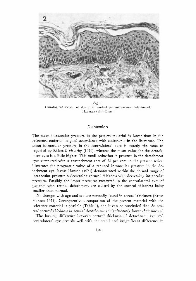
Fig . 2. Histological section of skin from control patient without detachment.
Haematoxylin-Eosin.
Discussion
The mean intraocular pressure in the present material is lower than in the reference material in good accordance with statements in the literature. The mean intraocular pressure in the contralateral eyes is exactly the same as reported by Ehlers & 0sterby (1970), whereas the mean value for the detach- ment eyes is a little higher. This small reduction in pressure in the detachment eyes compared with a reattachment rate of 85 per cent in the present series. illustrates the prognostic value of a reduced intraocular pressure in the de- tachment eye. Kruse Hansen (1971) demonstrated within the normal range of intraocular pressure a decreasing corneal thickness with decreasing intraocular pressure. Possibly the lower pressures measured in the contralateral eyes of patients with retinal detachment are caused by the corneal thickness being smaller than normal.
No changes with age and sex are normally found in corneal thickness (Kruse Hansen 197 1). Consequently a comparison of the present material with the reference material is possible (Table I), and it can be concluded that the cen- tral corneal thickness in retinal detachment is signif icantly lower than normal.
The lacking difference between corneal thickness of detachment eye and contralateral eye accords well with the small and insignificant difference in
470

Figs. 3 and 4. Histological sec- tions of skin from patient with retinal detachment ( 3 ) , and con- trol patient without detachment
(4). Haematoxylin-Eosin.
intraocular pressure (0.9 mm Hg), and indirectly supports the conclusion of Ehlers & Riise (1967) that the difference in thickness in their material was caused by the difference in intraocular pressure.
The different pathogenetical theories of retinal detachment (choroidal effu- sion, vitreous degeneration and retinal degeneration with hole) all originate from the last century (von Graefe 1854, Miiller 1858, Wecker 1870, Gonin 1904 - cited by Rosengren 1958), and are all still debated (Hervouet 1970). In addition to the demonstration of a reduced thickness of the cornea, the present study has also been an attempt to consider the pathogenesis of retinal detach- ment in relation to generalized abnormalities. Degenerative changes in the skin have been observed histologically, consisting in thinning of the epidermis and dermis and hypertrophic and fragmented collagenous and elastic fibers. Similar histological changes have been demonstrated in otosclerosis by Bentzen (1961) and Stadil (1961). These degenerative changes in the skin, and also reduced corneal thickness in the contralateral eyes, suggest a universal abnormality of constitution, possibly as a pre-disposition to development of retinal detachment. The rather high incidence of bilateral cases also suggests a universal abnor- mality. Generalized abnormalities have been described previously in familial
471

a n d bilateral retinal detachment (Edmund 1961, Pemberton e t al. 1966, d e Rotth 1939) and have also been described in some interesting case reports (Wol- ter & McVicar 1966, Epinay et al. 1969, Roaf et al. 1967, van den Berg 1965 a n d Delaney et al. 1965).
Summary
Forty consecutively operated cases of retinal detachment were studied. Central corneal thickness was found to be significantly lower than normal in detach- ment eyes as well as in contralateral eyes. Histological examination of skin biopsies f rom 1 7 of the patients revealed, f rom comparison with controls, a smaller thickness of epidermis and dermis and degenerative changes in col- lagenous a n d elastic fibers. These results suggest a universal abnormality of constitution, possibly as a predisposition to development of retinal detachment.
References
Bentzen, 0. (1961): 7eme Congr. Int. d’0to-rhino-laryng. Paris. In: Excerpta (Amst.)
van den Berg, E. 0. (1965): Hereditary disposition to retinal detachment in two fami-
Dclaney, W . V . (1965): Heredity and retinal detachment. Geriatrics 20: 584-588. Edmund, J . (1961): Familial retinal detachment. Acta ophthal. (Kbh.) 39: 644-6.54. Ehlers, N . & Kruse Hansen, F . (1971): On the optical measurement of corneal thickness.
Acta ophthal. (Kbh.) 49: 65-81. Ehlerc, N . & Xiise, D. (1967): On corneal thickness and intraocular pressure. Acta
ophthal. (Kbh.) 45: 809-813. Ehlers, N . & Osterby, E . (1970): On the prognostic value of intraocular pressure in
treatment of retinal detachment. Acta ophthal. (Kbh.) 48: 181-185. d’Epinay, S. L., Giedion, A . & Witmcr , R. (1969): Amotio retinae bei der spondy-
loepiphysaren Dysplasie. Klin. Mbl. Augenheilk. 155: 810-817. Hervoztt, F . (1970): Anatomie pathologique et pathoghie du dkcollement rktinien.
Ophthalmologica IG0: 25-53. Kruse Hansen, F. (1971): A clinical study of the normal human central corneal thick-
ness. Acta ophthal. (Kbh.) 49: 82-89. Pemberton, J . W., Mackenzie Freemann, H . & Schepens, C. L. (1966): Familial retinal
detachment and the Ehlers-Danlos syndrome. Arch. Ophthal. 76: 81 7-824. Roaf, R., Longmore, /. B. & Forrester, R. M . (1967): A childhood syndrome o f bone
dysplasia, retinal detachment and deafness. Develop. Med. Child Neurol. 9: 464- 473.
Rosengren, B. (1958): Nathinneavlossningens patogenes och operativa behandling. Acta Universitatis Gothoburgensis, 64: 1.
de Rotth, A. (1939): Bilateral detachment o f the retina. Arch. Ophthal. 22: 809-831. Stadil, P. (1961): Danish Med. Bull. 8: 131. Wolter , 1. R. & Mac Vicar, J . E. (1967): Blue sclerae, brittle bones and retinal detach-
Int. Congr. Ser. 35, 40.
lies. Ophthalmologica 149: 236-240.
ment. J. pediat. Ophthal. 4: 13-16.
472