central corneal thickness in nepalese glaucoma … · journal of clinical research and...
TRANSCRIPT
Journal of Clinical Research and Ophthalmology
Citation: Adhikari P, Chettry P, Thapa M (2015) Central Corneal Thickness in Nepalese Glaucoma Patients and Glaucoma Suspects. J Clin Res Ophthalmol 2(1): 003-006. DOI: 10.17352/2455-1414.000007
003
Abstract
Purpose: To compare central corneal thickness (CCT) among glaucoma patients, glaucoma suspects, and normal subjects and to determine its association with glaucoma severity in Nepalese population.
Methods: This study included 400 eyes (149 glaucoma, 157 glaucoma suspects, 94 controls) of 400 participants examined in a glaucoma clinic and eye OPD in Nepal. CCT was measured by ultrasonic pachymetry.
Results: CCT was significantly different among the study groups (P = 0.05), with the thinnest CCT in normal tension glaucoma (NTG) and thickest in ocular hypertension (OHT). CCT (in µm) was thinner in NTG (519.6 ± 31.6; P = 0.06) and primary open angle glaucoma (POAG) (524.5 ± 35.8; P = 0.026) than controls (536.6 ± 28.9); and it was thinner in POAG compared to primary angle closure glaucoma (PACG) (541.3 ± 50.5; P = 0.028) and OHT (559.8 ± 28.1; P = 0.017). In NTG, CCT was thinner compared to Glaucoma suspects (GS) (531.6 ± 35.0; P = 0.038), PACG (P = 0.008), and OHT (P = 0.008).There was no correlation between CCT and visual field defect and CCT was not statistically different between early, moderate and severe POAG groups.
Conclusions: We report that CCT in glaucoma suspects is similar to normal subjects and POAG, but thicker than NTG. These data will be important in clinically monitoring glaucoma suspects that are at increased risk of glaucoma. Our results may be population specific and further longitudinal studies are warranted to determine influence of CCT on glaucoma progression in this population.
developing glaucoma and the current study fills this research gap. It has also been shown that lower CCT is associated with visual field defect in glaucoma [11,12,14-18]. However it is controversial if CCT can predict glaucoma progression. Some studies have identified CCT as a risk factor for progression of glaucoma [14,19], and some have determined that CCT is not related to the severity of visual field defect [12,16,17]. Here, we try to address this controversy.
Different races and nationalities might have dissimilarities in CCT, which have been identified in normal population and glaucoma patients as well [13,15,20,21]. Thus, this study aims to compare CCT among glaucomatous, glaucoma suspects and normal individuals, to correlate CCT with severity of visual field loss, and to determine the association of the CCT with age and gender in Nepalese population. We are particularly interested to know the CCT characteristics of Nepalese population because this population has a lower overall prevalence of glaucoma (1.8%) [22] compared to the other south Asian regions (2.6 to 3.3%) [23-25].
Patients and MethodsGlaucoma patients and controls were recruited from glaucoma
clinic and eye outpatient department of Tribhuvan University Teaching Hospital, Nepal. The research was approved by Research
IntroductionIntra Ocular Pressure (IOP) is an important parameter in the
detection and monitoring of glaucoma. The Goldmann applanation tonometer (GAT) is the international “gold standard” for IOP measurement [1]. Central corneal thickness (CCT) has been shown to influence the pressure estimate [2], with thin corneas underestimating and thick corneas overestimating the readings [3].
Patients with normal tension glaucoma (NTG) may have thinner corneas than normal individuals resulting in underestimation of their IOP and under diagnosis; and patients with thicker cornea can be misdiagnosed to have glaucoma [4]. Copt et al. have described that many cases of glaucoma were reclassified after evaluating effect of CCT on measured IOP [5]. Thus, CCT should be considered to estimate actual IOP, to decide who requires closer observation or the initiation of treatment before definite damage occurs, and to establish a target IOP.CCT in patients with ocular hypertension (OHT) is greater and in patients with NTG lower compared to controls, with CCT in primary open angle glaucoma (POAG) falling in between OHT and NTG [3,5-13]. CCT in different types of glaucoma has been evaluated, but Glaucoma suspect (GS) excluding OHT has been ignored in this regard. We believe that CCT in glaucoma suspects should also be equally monitored as this group is always at a risk of
Research Article
Central Corneal Thickness in Nepalese Glaucoma Patients and Glaucoma Suspects
Prakash Adhikari1*, Pratik Chettry2 and Madhu Thapa3
1Visual Science and Medical Retina Laboratories, School of Optometry and Vision Science and Institute of Health and Biomedical Innovation, Queensland University of Technology, Australia2Male Optical Co. Ltd., Maldives3Eye Department, Institute of Medicine, Tribhuvan University, Nepal
Dates: Received: 28 October, 2014; Accepted: 22 November, 2014; Published: 25 November, 2014
*Corresponding author: Prakash Adhikari, PhD Candidate, Visual Science and Medical Retina Laboratories, Institute of Health and Biomedical Innovation, Queensland University of Technology, 60 Musk Avenue, Kelvin Grove, Brisbane 4059, Queensland, Australia, Work: +6173138 6450; Tel: 61431176244; Fax: +617 3138 6030; E-mail:
www.peertechz.com
Keywords: Central corneal thickness; Glaucoma; Glaucoma suspect; Intraocular pressure; Visual field defect
ISSN: 2455-1414
Citation: Adhikari P, Chettry P, Thapa M (2015) Central Corneal Thickness in Nepalese Glaucoma Patients and Glaucoma Suspects. J Clin Res Ophthalmol 2(1): 003-006. DOI: 10.17352/2455-1414.000007
Adhikari et al. (2015)
004
Ethics Committee of Tribhuvan University, Nepal. The tenets of the Declaration of Helsinki were followed and informed consent was obtained from the participants after explanation of nature of the study.
Different types of glaucoma were defined according to Preferred Practice Pattern Guidelines of American Academy of Ophthalmology [26]. Primary open angle glaucoma (POAG)was defined by typical glaucomatous disc, visual field defect and/or significant loss of retinal nerve fiber layer (RNFL) in the optic nerve head region in Heidelberg Retinal Tomography (HRT) or Optical Coherence Tomography (OCT), IOP > 21 mmHg, and an open anterior chamber angle on gonioscopy. POAG was further divided into early, moderate, and severe on the basis of mean deviation (MD) of Humphrey standard automated perimetry according to Hodapp, Parrish, and Anderson’s classification. [27] NTG was defined bytypical glaucomatous disc, visual field defect and/or significant loss of RNFL in the optic nerve head region in HRT or OCT, IOP ≤ 21 mmHg, and an open anterior chamber angle. Primary angle closure glaucoma (PACG) was defined by gonioscopic finding of more than 180° ofirido-trabecular contact, IOP > 21 mmHg, and optic nerve and visual field damage. OHT was defined by IOP > 21 mmHg, but normal disc, field, and angle. Glaucoma suspect (GS) was defined by family history of glaucoma and/or appearance of the optic disc or RNFL that is suspicious for glaucomatous damage including enlarged cup-disc ratio, asymmetric cup-disc ratio, narrowing of the neuroretinal rim, disc hemorrhage, nerve fiber layer defect, but with no visual field defect. For some analysis, OHT was also included in GS group; otherwise they are presented separately to reflect our new findings in GS group (Previous findings in OHT are discussed in introduction and discussion). All eyes with ocular disorders altering CCT, any active ocular disease other than glaucoma, any ocular surgery, corneal astigmatism > 4 D, and history of contact lens wear were excluded from the study. Age and gender matched individuals with healthy eyes were taken as controls.
For diagnosis and classification of glaucoma, detailed history taking, slit lamp examination, IOP measurement, gonioscopy, funduscopy, AVF examination, OCT, and HRT were performed in all cases. Central measurement system of USG Pachymetry (Axis II PR) was used to measure CCT in upright position by same examiner. Five consecutive readings with standard deviation (SD) <5 microns were taken and averaged.
Statistical AnalysisData were described as mean ± SD and 95% confidence interval;
and p < 0.05 was considered statistically significant. One-way ANOVA was applied to compute the differences in the CCT among the study groups. The association of CCT with age, IOP, and visual field defect was evaluated with Pearson Correlation and linear regression.
ResultsA total of 400 eyes of 400 subjects, comprising 149eyes with
glaucoma (72 eyes with POAG, 29 eyes with PACG, and 48 eyes with NTG), 157eyes with GS (6 with OHT) and 94 eyes of control subjects were examined. Among the subjects enrolled in the study, 180(45%) patients were male and 220(55%) patients were female. The mean ±
SD age of glaucoma patients, glaucoma suspect, and controls was 45.0 ± 21.1, 45.6 ± 21.5, 45.2 ± 20.8 years respectively and There was no significant difference in mean age among the study groups (P=0.140) (Tables 1-3).
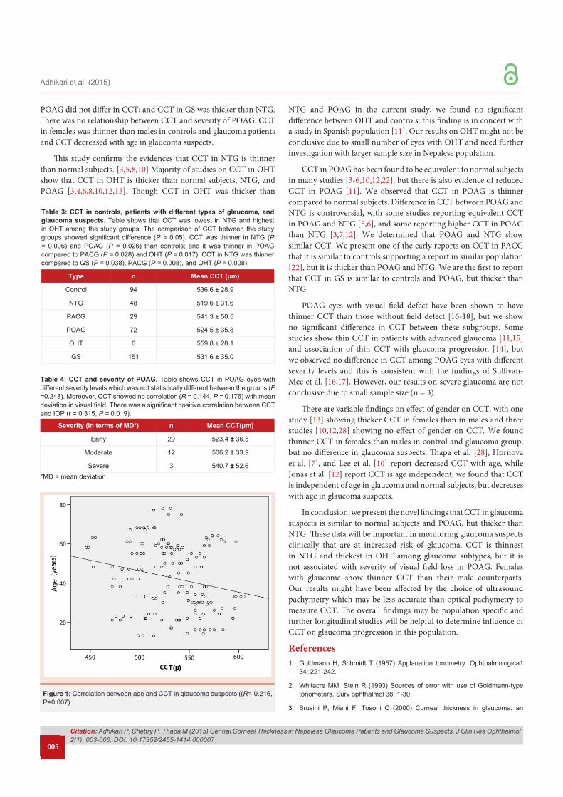
Out of 72 eyes with POAG, 44 eyes had visual field defects; rest of the cases were diagnosed on the basis of disc findings, HRT, and OCT. CCT in POAG eyes with field defect (519.8 ± 37.1) and without field defect (530.1 ± 32.3) was not statistically different (P = 0.241). Table 4 shows CCT in POAG eyes with different severity levels which was not statistically different between the groups (P =0.248). Moreover, CCT showed no correlation (R = 0.144, P = 0.176) with mean deviation in visual field. There was a significant positive correlation between CCT and IOP (r = 0.315, P = 0.019) (Figure 1).
DiscussionIOP is an important risk factor and has a significant influence
on diagnosis and management of glaucoma. GAT is used worldwide for IOP measurement because of its accuracy but its results may be affected by CCT. We measured CCT in different types of glaucoma and glaucoma suspects and evaluated the relationship between CCT and severity of glaucoma. CCT was significantly different between the study groups, with thinnest CCT in NTG and thickest CCT in OHT. CCT in PACG was thicker than in NTG and POAG, NTG and
Gender n Mean CCT(µm) p-value
ControlM 48 542.6 ± 31.4
0.040F 46 530.4 ± 25.0
GlaucomaM 66 533.5 ± 34.0
0.037F 83 520.3 ± 40.8
Glaucoma suspect
M 66 535.7 ± 35.40.368
F 91 530.6 ± 35.0
Table 1: Gender and CCT in different study groups. Table shows that CCT in females was significantly thinner than in males in controls and glaucoma patients, but not in glaucoma suspects.
Age Range Control Glaucoma Glaucoma suspect
n Mean CCT (µm) n Mean CCT
(µm) n Mean CCT (µm)
11-20 6 534.2 ± 14.5 8 504.4 ± 35.9 14 544.0 ± 28.4
21-30 6 554.2 ± 32.0 13 524.6 ± 39.0 37 541.6 ± 35.6
31-40 16 548.9 ± 37.4 23 532.6 ± 24.3 25 545.6 ± 35.7
41-50 30 528.4 ± 23.8 33 539.5 ± 43.9 21 512.0 ± 30.5
51-60 16 545.8 ± 28.9 28 521.0 ± 44.0 32 526.1 ± 29.3
61-70 12 537.6 ± 15.8 32 511.8 ± 34.1 22 526.7 ± 43.7
71-80 8 511.8 ± 30.0 12 544.0 ± 23.6 6 525.5 ± 10.8
Table 2: Age and CCT in different study groups. Table shows that CCT in different age groups among the study groups. There was no significant correlation between age and CCT in controls (R=-0.194, P =0.061) and glaucoma patients (R=0.008, P=0.927). However, in glaucoma suspects, CCT was negatively correlated with age (R=-0.216, P=0.007) (Figure 1). Multiple linear regression was done to evaluate how age and gender interact to affect CCT. The interaction was significant [F(2,91) = 4.9, P = 0.010] in controls and glaucoma suspects [F(2,154) = 3.8, P = 0.024], but not in glaucoma patients [F(2,146) = 2.5, P = 0.085].
Citation: Adhikari P, Chettry P, Thapa M (2015) Central Corneal Thickness in Nepalese Glaucoma Patients and Glaucoma Suspects. J Clin Res Ophthalmol 2(1): 003-006. DOI: 10.17352/2455-1414.000007
Adhikari et al. (2015)
005
POAG did not differ in CCT; and CCT in GS was thicker than NTG. There was no relationship between CCT and severity of POAG. CCT in females was thinner than males in controls and glaucoma patients and CCT decreased with age in glaucoma suspects.
This study confirms the evidences that CCT in NTG is thinner than normal subjects. [3,5,8,10] Majority of studies on CCT in OHT show that CCT in OHT is thicker than normal subjects, NTG, and POAG [3,4,6,8,10,12,13]. Though CCT in OHT was thicker than
NTG and POAG in the current study, we found no significant difference between OHT and controls; this finding is in concert with a study in Spanish population [11]. Our results on OHT might not be conclusive due to small number of eyes with OHT and need further investigation with larger sample size in Nepalese population.
CCT in POAG has been found to be equivalent to normal subjects in many studies [3-6,10,12,22], but there is also evidence of reduced CCT in POAG [11]. We observed that CCT in POAG is thinner compared to normal subjects. Difference in CCT between POAG and NTG is controversial, with some studies reporting equivalent CCT in POAG and NTG [5,6], and some reporting higher CCT in POAG than NTG [3,7,12]. We determined that POAG and NTG show similar CCT. We present one of the early reports on CCT in PACG that it is similar to controls supporting a report in similar population [22], but it is thicker than POAG and NTG. We are the first to report that CCT in GS is similar to controls and POAG, but thicker than NTG.
POAG eyes with visual field defect have been shown to have thinner CCT than those without field defect [16-18], but we show no significant difference in CCT between these subgroups. Some studies show thin CCT in patients with advanced glaucoma [11,15] and association of thin CCT with glaucoma progression [14], but we observed no difference in CCT among POAG eyes with different severity levels and this is consistent with the findings of Sullivan-Mee et al. [16,17]. However, our results on severe glaucoma are not conclusive due to small sample size (n = 3).
There are variable findings on effect of gender on CCT, with one study [13] showing thicker CCT in females than in males and three studies [10,12,28] showing no effect of gender on CCT. We found thinner CCT in females than males in control and glaucoma group, but no difference in glaucoma suspects. Thapa et al. [28], Hornova et al. [7], and Lee et al. [10] report decreased CCT with age, while Jonas et al. [12] report CCT is age independent; we found that CCT is independent of age in glaucoma and normal subjects, but decreases with age in glaucoma suspects.
In conclusion, we present the novel findings that CCT in glaucoma suspects is similar to normal subjects and POAG, but thicker than NTG. These data will be important in monitoring glaucoma suspects clinically that are at increased risk of glaucoma. CCT is thinnest in NTG and thickest in OHT among glaucoma subtypes, but it is not associated with severity of visual field loss in POAG. Females with glaucoma show thinner CCT than their male counterparts. Our results might have been affected by the choice of ultrasound pachymetry which may be less accurate than optical pachymetry to measure CCT. The overall findings may be population specific and further longitudinal studies will be helpful to determine influence of CCT on glaucoma progression in this population.
References1. Goldmann H, Schmidt T (1957) Applanation tonometry. Ophthalmologica1
34: 221-242.
2. Whitacre MM, Stein R (1993) Sources of error with use of Goldmann-type tonometers. Surv ophthalmol 38: 1-30.
3. Brusini P, Miani F, Tosoni C (2000) Corneal thickness in glaucoma: an
Type n Mean CCT (µm)
Control 94 536.6 ± 28.9
NTG 48 519.6 ± 31.6
PACG 29 541.3 ± 50.5
POAG 72 524.5 ± 35.8
OHT 6 559.8 ± 28.1
GS 151 531.6 ± 35.0
Table 3: CCT in controls, patients with different types of glaucoma, and glaucoma suspects. Table shows that CCT was lowest in NTG and highest in OHT among the study groups. The comparison of CCT between the study groups showed significant difference (P = 0.05). CCT was thinner in NTG (P = 0.006) and POAG (P = 0.026) than controls; and it was thinner in POAG compared to PACG (P = 0.028) and OHT (P = 0.017). CCT in NTG was thinner compared to GS (P = 0.038), PACG (P = 0.008), and OHT (P = 0.008).
Severity (in terms of MD*) n Mean CCT(µm)
Early 29 523.4 ± 36.5
Moderate 12 506.2 ± 33.9
Severe 3 540.7 ± 52.6
Table 4: CCT and severity of POAG. Table shows CCT in POAG eyes with different severity levels which was not statistically different between the groups (P =0.248). Moreover, CCT showed no correlation (R = 0.144, P = 0.176) with mean deviation in visual field. There was a significant positive correlation between CCT and IOP (r = 0.315, P = 0.019).
*MD = mean deviation
80
20
60
40
450 500 550 600
Age
(yea
rs)
CC
Figure 1: Correlation between age and CCT in glaucoma suspects ((R=-0.216, P=0.007).
Citation: Adhikari P, Chettry P, Thapa M (2015) Central Corneal Thickness in Nepalese Glaucoma Patients and Glaucoma Suspects. J Clin Res Ophthalmol 2(1): 003-006. DOI: 10.17352/2455-1414.000007
Adhikari et al. (2015)
006
Copyright: © 2015 Adhikari P, et al. This is an open-access article distributed under the terms of the Creative Commons Attribution License, which permits unrestricted use, distribution, and reproduction in any medium, provided the original author and source are credited.
important parameter? Acta Ophthalmol Scand Suppl 78: 41-42.
4. Bron AM, Creuzot-Garcher C, Goudeau-Boutillon S, d’Athis P (1999) Falsely elevated intraocular pressure due to increased central corneal thickness. Graefes Arch Clin Exp Ophthalmol 237: 220-224.
5. Copt R-P, Thomas R, Mermoud A (1999) Corneal thickness in ocular hypertension, primary open-angle glaucoma, and normal tension glaucoma. Arch Ophthalmol 117: 14-16.
6. Ventura AS, Böhnke M, Mojon D (2001) Central corneal thickness measurements in patients with normal tension glaucoma, primary open angle glaucoma, pseudoexfoliation glaucoma, or ocular hypertension. Br j Ophthalmol 85: 792-795.
7. Hornova J, Sedlak P (1999) Pachymetry in patients with glaucoma. Cesk Slov Oftalmol 55: 212-215.
8. Dave H, Kutschan A, Pauer A, Wiegand W (2004) Measurement of corneal thickness in glaucoma patients. Ophthalmologe 101: 919-924.
9. Jordan JF, Joergens S, Dinslage S, Dietlein TS, Krieglstein GK (2006) Central and paracentral corneal pachymetry in patients with normal tension glaucoma and ocular hypertension. Graefe’s Archive for Clinical and Experimental Ophthalmology 244: 177-182.
10. Lee ES, Kim CY, Ha SJ, Seong GJ, Hong YJ (2007) Central corneal thickness of Korean patients with glaucoma. Ophthalmology 114: 927-930.
11. Jiménez-Rodríguez E, López-de-Cobos M, Luque-Aranda R, López-Egea-Bueno MA, Vázquez-Salvi AI et al. (2009) Relationship between central corneal thickness, intraocular pressure and severity of glaucomatous visual field loss. Arch Soc Esp Oftalmol 84: 139-143.
12. Jonas JB, Stroux A, Velten I, Juenemann A, Martus P, et al. (2005) Central corneal thickness correlated with glaucoma damage and rate of progression. Invest Ophthalmol Vis Sci 46: 1269-1274.
13. Brandt JD, Beiser JA, Kass MA, Gordon MO (2001) Central corneal thickness in the ocular hypertension treatment study (OHTS). Ophthalmology 108: 1779-1788.
14. Kim JW, Chen PP (2004) Central corneal pachymetry and visual field progression in patients with open-angle glaucoma. Ophthalmology 111: 2126-2132.
15. Herndon LW, Weizer JS, Stinnett SS (2004) Central corneal thickness as a risk factor for advanced glaucoma damage. Arch Ophthalmol 122: 17-21.
16. Sullivan-Mee M, Halverson KD, Saxon MC, Saxon GB, Qualls C (2006) Central corneal thickness and normal tension glaucoma: a cross-sectional study. Optometry-Journal of the American Optometric Association 77: 134-
140.
17. Sullivan-Mee M, Halverson KD, Saxon GB, Saxon MC, Qualls C (2006) Relationship between central corneal thickness and severity of glaucomatous visual field loss in a primary care population. Optometry 77: 40-46.
18. Lin W, Aoyama Y, Kawase K, Yamamoto T (2009) Relationship between central corneal thickness and visual field defect in open-angle glaucoma. Jpn J Ophthalmol 53: 477-481.
19. Dueker DK, Singh K, Lin SC, Fechtner RD, Minckler DS, et al. (2007) Corneal thickness measurement in the management of primary open-angle glaucoma: a report by the American Academy of Ophthalmology. Ophthalmology 114: 1779-1787.
20. La Rosa FA, Gross RL, Orengo-Nania S (2001) Central corneal thickness of Caucasians and African Americans in glaucomatous and nonglaucomatous populations. Arch Ophthalmol 119: 23-27.
21. Aghaian E, Choe JE, Lin S, Stamper RL (2004) Central corneal thickness of Caucasians, Chinese, Hispanics, Filipinos, African Americans, and Japanese in a glaucoma clinic. Ophthalmology 111: 2211-2219.
22. Thapa SS, Paudyal I, Khanal S, Twyana SN, Paudyal G, et al. (2012) A population-based survey of the prevalence and types of glaucoma in Nepal: the Bhaktapur Glaucoma Study. Ophthalmology 119: 759-764.
23. Ramakrishnan R, Nirmalan PK, Krishnadas R, Thulasiraj R, Tielsch JM, et al. (2003) Glaucoma in a rural population of southern India: the Aravind comprehensive eye survey. Ophthalmology 110: 1484-1490.
24. Rahman M, Rahman N, Foster P, Haque Z, Zaman A, Dineen B, et al. (2004) The prevalence of glaucoma in Bangladesh: a population based survey in Dhaka division. Br J Ophthalmol 88: 1493-1497.
25. Raychaudhuri A, Lahiri S, Bandyopadhyay M, Foster P, Reeves B, et al. (2005) A population based survey of the prevalence and types of glaucoma in rural West Bengal: the West Bengal Glaucoma Study. Br J Ophthalmol 89: 1559-1564.
26. Committee AAOPPP (2010). Preferred Practice Pattern Guidelines. Comprehensive Adult Medical Eye Evaluation. San Francisco, CA: American Academy of Ophthalmology.
27. Hodapp E, Parrish R, II AD (1993) Clinical Decisions in Glaucoma. Mosby-Year Book St. In: Louis.
28. Thapa SS, Paudyal I, Khanal S, Paudel N, Mansberger SL, et al. (2012) Central Corneal Thickness and Intraocular Pressure in a Nepalese Population: The Bhaktapur Glaucoma Study. J Glauco 21: 481-485.