cellular cardiac electrophysiological effects of hcn...
TRANSCRIPT

1
Cellular cardiac electrophysiological effects of
HCN channel blockers
Ph.D. Thesis
István Koncz M.D.
Supervisors: András Varró M.D., Ph.D., DSc. and László Virág
Ph.D.
Szeged
2014
2
Cellular cardiac electrophysiological effects of HCN
channel blockers
Ph.D. Thesis
István Koncz M.D.
Supervisors: András Varró M.D., Ph.D., DSc. and László Virág Ph.D.
Department of Pharmacology and Pharmacotherapy University of Szeged
Szeged, Hungary
2014
3
TABLE OF CONTENTS
1. INTRODUCTION……………………………………………………………………7 1.1 HCN channels. General overview …………………………………………………....7
1.2 HCN channels in the heart………………...………………………………………….8
1.3 Characteristics of the If current and its role in the adrenergic and cholinergic modulation of heart rate…………………………….9 1.4 If current inhibitor heart rate-reducing agents………………………………………. 11 1.5 Heart rate reduction with a highly selective If inhibitor: ivabradine (S16257)………14
1.6 Therapeutical advantages of ivabradine in coronary artery disease and heart failure patients……………………………………………...15 1.7 Future possibilities: ivabradine as a reasonable alternative to beta receptor blockers in myocardial infarction, results with ivabradine in experimental models of myocardial infarction…………...16
1.8 HCN channels and arrhythmogenesis ……………………………………………….17 1.9 Recently synthesized HCN channel blockers ……………………………………….17 1.10 Aims of the study……………………………………………………………………18 2. MATERIALS AND METHODS…………………………………………………...19 3. RESULTS……………………………………………………………………………22
3.1 Effects of ivabradine on ionic currents ……………………………………………...22 3.2 Effects of ivabradine on transmembrane action potentials ………………………....23
3.3 Effects of compounds EC18 and MEL57A on transmembrane action potentials …..31 4. DISCUSSION..............................................................................................................34 4.1 Possible ionic mechanisms…………………………………………………………....34 4.2 Possible clinical implications ………………………………………………………...35 4.3 Electrophysiological effects of compounds EC18 and MEL57A …………………....36
5. CONCLUSIONS.........................................................................................................37 6. REFERENCES………………………………………………………………………38 7. ACKNOWLEDGEMENTS…………………………………………………………47

4
LIST OF FULL PAPERS RELATED TO THE SUBJECT OF THE
DISSERTATION
1. Koncz I, Szél T, Bitay M, Cerbai E, Jaeger K, Fülöp F, Jost N, Virág L, Orvos P, Tálosi L, Kristóf
A, Baczkó I, Papp JG, Varró A. Electrophysiological effects of ivabradine in dog and human cardiac preparations: potential antiarrhythmic actions. European Journal of Pharmacology 2011 Oct 15;668(3):419-26. Epub 2011 Aug 2.
2. Koncz I, Szél T, Jaeger K, Baczkó I, Cerbai E, Romanelli MN, Gy Papp J, Varró A. Selective
pharmacological inhibition of the pacemaker channel isoforms (HCN1-4) as new possible therapeutical targets. Current Medicinal Chemistry. 2011;18(24):3662-74.
3. Del Lungo M, Melchiorre M, Guandalini L, Sartiani L, Mugelli A, Koncz I, Szel T, Varro A,
Romanelli MN, Cerbai E. Novel blockers of hyperpolarization-activated current with isoform selectivity in recombinant cells and native tissue. British Journal of Pharmacology 2012 May;166(2):602-16. doi: 10.1111/j.1476-5381.2011.01782.x.
OTHER STUDIES (13)
Bönöczk P, Gulyás B, Adam-Vizi V, Nemes A, Kárpáti E, Kiss B, Kapás M, Szántay C, Koncz I, Zelles T, Vas A. Role of sodium channel inhibition in neuroprotection: effect of vinpocetine. Brain Res Bull. 2000 Oct;53(3):245-54. Zelles T, Franklin L, Koncz I, Lendvai B, Zsilla G. The nootropic drug vinpocetine inhibits veratridine-induced [Ca2+]i increase in rat hippocampal CA1 pyramidal cells. Neurochem Res. 2001 Sep;26(8-9):1095-100.
Pecze L, Szabó K, Széll M, Jósvay K, Kaszás K, Kúsz E, Letoha T, Prorok J, Koncz I, Tóth A, Kemény L, Vizler C, Oláh Z. Human keratinocytes are vanilloid resistant. PLoS One. 2008;3(10):e3419. Epub 2008 Oct 14.
Koini EN, Papazafiri P, Vassilopoulos A, Koufaki M, Horváth Z, Koncz I, Virág L, Papp GJ, Varró A, Calogeropoulou T. 5,7,8-Trimethyl-benzopyran and 5,7,8-trimethyl-1,4-benzoxazine aminoamide derivatives as novel antiarrhythmics against ischemia-reperfusion injury. J Med Chem. 2009 Apr 23;52(8):2328-40.
Bárándi L, Virág L, Jost N, Horváth Z, Koncz I, Papp R, Harmati G, Horváth B, Szentandrássy N, Bányász T, Magyar J, Zaza A, Varró A, Nánási PP. Reverse rate-dependent changes are determined by baseline action potential duration in mammalian and human ventricular preparations. Basic Res Cardiol. 2010 May;105(3):315-23. Epub 2010 Feb 3. Virág L, Jost N, Papp R, Koncz I, Kristóf A, Kohajda Z, Harmati G, Carbonell-Pascual B, Ferrero JM Jr, Papp JG, Nánási PP, Varró A. Analysis of the contribution of I(to) to repolarization in canine ventricular myocardium. Br J Pharmacol. 2011 Sep;164(1):93-105.
5
Szél T, Koncz I, Jost N, Baczkó I, Husti Z, Virág L, Bussek A, Wettwer E, Ravens U, Papp JG, Varró A. Class I/B antiarrhythmic property of ranolazine, a novel antianginal agent, in dog and human cardiac preparations. Eur J Pharmacol. 2011 Jul 15;662(1-3):31-9. doi: 10.1016/j.ejphar.2011.04.042. Kristóf A, Husti Z, Koncz I, Kohajda Z, Szél T, Juhász V, Biliczki P, Jost N, Baczkó I, Papp JG, Varró A, Virág L. Diclofenac prolongs repolarization in ventricular muscle with impaired repolarization reserve. PLoS One. 2012;7(12):e53255. doi: 10.1371/journal.pone.0053255. Jost N, Virág L, Comtois P, Ördög B, Szuts V, Seprényi G, Bitay M, Kohajda Z, Koncz I, Nagy N, Szél T, Magyar J, Kovács M, Puskás LG, Lengyel C, Wettwer E, Ravens U, Nánási PP, Papp JG, Varro A, Nattel S. Ionic mechanisms limiting cardiac repolarization-reserve in humans compared to dogs. J Physiol. 2013 Sep 1;591(Pt 17):4189-206. doi: 10.1113/jphysiol.2013.261198. Epub 2013 Jul 22. Szél T, Koncz I, Antzelevitch C. Cellular mechanisms underlying the effects of milrinone and cilostazol to suppress arrhythmogenesis associated with Brugada syndrome. Heart Rhythm. 2013 Nov;10(11):1720-7. doi: 10.1016/j.hrthm.2013.07.047.
Koncz I, Gurabi Z, Patocskai B, Panama BK, Szél T, Hu D, Barajas-Martínez H, Antzelevitch C.Mechanisms Underlying the Development of the Electrocardiographic and Arrhythmic Manifestations of Early Repolarization Syndrome. J Mol Cell Cardiol. 2014 Mar;68:20-8. doi: 10.1016/j.yjmcc.2013.12.012. Epub 2013 Dec 28. Gurabi Z, Koncz I, Patocskai B, Nesterenko VV, Antzelevitch C. Cellular Mechanism Underlying Hypothermia-Induced VT/VF in the Setting of Early Repolarization and the Protective Effect of Quinidine, Cilostazol and Milrinone. Circ Arrhythm Electrophysiol.2014 Feb;7(1):134-42. doi: 10.1161/CIRCEP.113.000919. Epub 2014 Jan 15. Koncz I, Szász BK, Szabó SI, Kiss JP, Mike A, Lendvai B, Sylvester Vizi E, Zelles T. The tricyclic antidepressant desipramine inhibited the neurotoxic, kainate-induced [Ca2+]i increases in CA1 pyramidal cells in acute hippocampal slices. 2014. Brain Res Bull. 2014 Apr 15;104C:42-51. doi: 10.1016/j.brainresbull.2014.04.003. [Epub ahead of print]

6
ACRONYMS AND ABBREVIATIONS
APA action potential amplitude APD50 action potential duration at 50% of repolarization APD90 action potential duration at 90% of repolarization ASSOCIATE evaluation of the Antianginal efficacy and Safety of the aSsociation Of the If Current Inhibitor ivAbradine with a beTa-blockEr BEAUTIFUL morBidity-mortality EvAlUaTion of the If inhibitor ivabradine in patients with coronary disease and left ventricULar dysfunction CHF congestive heart failure ERG electroretinogram HCN hyperpolarization-activated and cyclic nucleotide-gated INITIATIVE INternatIonal TrIal of the Antianginal effecT of IVabradinE compared with atenolol MDP maximum diastolic potential; MiRP1 MinK-related protein SAN sino-atrial node SBAs specific bradycardic agents SHIFT Systolic Heart failure treatment with the IF inhibitor ivabradine Trial SNI sinus node inhibitors STEMI ST-segment elevation myocardial infarction Vmax maximal rate of depolarization.
7
1. INTRODUCTION
1.1 HCN channels. General overview.
The hyperpolarization-activated cyclic nucleotide-gated (HCN) channel family implies four
members (HCN1-4) which are expressed in the heart and the nervous system. The current
flowing through HCN channels was termed as If or Ih or Iq. It was demonstrated that the
heterogeneity of native Ih currents is attributable to, at least partly, the tissue-specific
expression of HCN channel genes [1]. The hyperpolarization–activated current was named
"funny" current (If) in the heart because of its unusual electrophysiological characteristics [2],
while this current in the nervous system was designated Ih (for "hyperpolarization") or Iq (for
"queer" current). If/Ih current plays a substantial role in the initiation and control of the heart
rate [3]. If/Ih generates rhythmic activities particularly in thalamocortical circuits, has a
function in regulating synaptic integration (e.g. in prefrontal pyramidal neurons) [4], in
neuronal cells takes part in the determination of resting membrane potential [5] (Ih is inward,
and the others (Kir2 and Kleak) are outward, there is dynamic equilibrium between them),
probably plays an important role in olfactory information processing [6], generates and
controls oscillations of the membrane potential. The increased expression level of Ih and HCN
isoforms in sensory nerves indicates that they might play a prominent role in sensory function
[7] (e.g. by modulating pain thresholds) and could be targets in analgesic therapy in special
conditions e.g. nerve injury based neuropathic pain. Interestingly, it turned out that some
volatile anaethetic agents are able to influence the operation of HCN channels, opening an
other significant field in drug research. HCN isoforms have also been found in pancreatic
beta-cells and kidney, where they regulate important physiological functions [8-10]. HCN
channels are expressed in the retina [11]. Ivabradine is a novel heart rate-lowering antianginal
agent which inhibits the pacemaker current in the heart with high selectivity. According to the
BEAUTIFUL study, ivabradine can be used to reduce the incidence of coronary artery disease
outcomes in a subgroup of patients who have heart rates of 70 bpm or greater [12]. A large
proportion of patients suffering from heart failure die of sudden cardiac death caused by
arrhythmia. It emerged from experiments analyzing the expression pattern of HCN channel
isoforms in failing heart that suppressing the activity of the upregulated HCN channel
subtypes can probably preclude the development of that type of arrhythmias which are
triggered by abnormal pacemaker activity in the working myocardium, as it was found that
both mRNA and protein levels of HCN2 and HCN4 isoforms were significantly augmented in

8
failing ventricles [13]. Therefore, selective HCN blockers might serve as effective agents of
this profile. HCN4 is the predominant isoform in undiseased human heart, and its expression
is increased by disease (e.g. heart failure). The classic antiarrhythmic compounds might exert
little or insignificant effect on this isoform as recently it turned out that the blocking effects of
them /quinidine, disopyramide, cibenzoline, lidocaine, mexiletine, aprindine, propafenone,
flecainide, propranolol, and verapamil/ on the HCN4 channel current were weak, D,L-sotalol
hardly influenced the HCN4 channel current in clinical concentrations in HEK293 cells [14].
1.2 HCN channels in the heart
Heart rate is regulated by spontaneous electrical pacemaker activity in the sino-atrial node
mainly controlled by the If current. The If current determines the slope of diastolic
depolarization and cardiac frequency, and its inhibition causes heart rate reduction. The
´funny´ (If) current is so termed because of its unusual characteristics, including that of being
an inward current that is activated on hyperpolarization and not on depolarization like other
known currents [15]. The role of currents other than If and cellular mechanisms (ICaL, ICaT, INa,
IKr, Ist, INCX, intracellular Ca2+ cycling = "calcium clock") in pacemaker activity is also
important and it is still argued [16]. Nevertheless, it can be stated that If plays a principal role
in regulating sinoatrial node pacemaker activity [3]. It is generally agreed that it has
modulatory effect on pacemaker rate. HCN channels (hyperpolarization-activated cyclic
nucleotid-gated) are responsible for generating the If pacemaker current in the heart. The
channel relating to the cardiac If current is known to be a member of a family of channels
denominated the hyperpolarization-activated cyclic nucleotid-gated (HCN) channels. The If
current is controlled by opposite processes, i.e. the beta adrenergic stimulatory and muscarinic
cholinergic inhibitory actions occur via adenylate cyclase and cAMP [17]. The If is a mixed
Na+ and K+ current that is activated on hyperpolarization to voltages within the pacemaker
range. Recently, Ca2+ influx through HCN channels has indirectly been postulated and then
directly the Ca2+ permeability of native rat and human If has been shown [18].
Heart rate has a prominent role in the development and pathophysiology of myocardial
ischaemia. In patients with coronary artery disease, ischaemic episodes could be generated by
an elevation in heart rate that elicits imbalance between myocardial oxygen supply and
demand. Therefore, lowering the heart rate is regarded as an important therapeutic target in
preventing ischaemia by reducing myocardial oxygen consumption. Therefore, reducing heart
rate is one of the most important therapeutic possibilities in the treatment of stable angina
9
pectoris [19, 20]. Additionally, resting heart rate is associated with the severity and rate of
progression of coronary atherosclerosis and is an independent predictor of plaque rupture in
coronary arteries, therefore, heart rate lowering e.g. by ivabradine the specific HCN blocker
should prevent atherosclerosis and cardiovascular events [21].
The current pharmacological treatment of stable angina pectoris is based on beta adrenoceptor
blockers, calcium channel antagonists, nitrates and trimetazidine with beneficial metabolic
effect on the heart. The antiischemic effect of beta blockers is attributable, at least in part, to
heart rate reduction. Beta blockers apart from the numerous, proven, valuable effects in the
treatment of cardiovascular diseases (angina pectoris, myocardial infarction, heart failure,
arrhythmias) have several side effects including fatigue, depression, change in lipid status,
sexual dysfunction, bronchospasm, atrioventricular block, possible increase in blood sugar
level, rebound phenomena. They can cause paradoxical vasoconstriction of large epicardial
arteries [22] during exercise or in patients with resting angina associated to coronary
vasospams. Beta blockers (except carvedilol and nebivolol) may be contraindicated in patients
with peripheral vascular disease. Concerning the nitrates headache, regarding the calcium
channel blockers pedal edema are potential side effects. The hemodynamic effects might be
also limiting factors at application e.g. the negative inotropic effect of the beta blockers, the
considerable blood pressure lowering property of the calcium channel blockers. In ST-
segment elevation myocardial infarction (STEMI) early i.v. use of beta blockers is clearly
contraindicated in patients with clinical signs of hypotension or congestive heart failure [23].
Therefore, highly selective HCN channel blockers, producing solely heart rate lowering, e.g.
ivabradine, lacking the negative inotropic effect, may offer a new therapeutic perspective in
the treatment of ischaemic heart diseases e.g. in stable angina pectoris, particularly in patients
with left ventricular dysfunction. Based on the recent clinical trials and experiments, widening
the indication of ivabradine for the treatment of heart failure or myocardial infarction is
expected. Thus, to seek for specific blockers of the If current is in the focus of cardiovascular
pharmacological research.
1.3 Characteristics of the If current and its role in the adrenergic and cholinergic
modulation of heart rate
The sino-atrial node (SAN) is a small, specialized structure located in the right atrium. The
nodal cells (specialized myocytes) generate pacemaker activity, i.e. their diastolic membrane
potential is unstable, a gradual depolarization to a threshold occurs. If current is deactivated

10
during the upstroke of an action potential. Upon repolarization when membrane potential
reaches the If activation voltage (below -40 mV), If becomes activated, and its slow activation
counteracts the repolarizing IK and slowly depolarizes the membrane voltage [24]. Reaching
the threshold a new action potential is elicited. The potential ionic currents and mechanism
responsible for the pacemaker activity are as follows: If, ICaL, ICaT, INa, IKr, Ist, INCX and probably
the pacemaker function is dependent on complex interactions between localized,
subsarcolemmal Ca2+ releases and action potentials [25]. The role of If in the control of the
firing rate of the sino-atrial node and therefore determining the heart rate is heavily argued
[3,16,26]. The clinical studies might give proof of the considerable influence of If on the
pacemaker function, as the highly specific If current blocker ivabradine used in the trials
reduced heart rate by 10 to 15 beats/min [27].
The heart, regarding its control by the autonomic nervous system, receives both sympathetic
(responsible for increasing the heart rate, via stimulation of β1 adrenoceptors) and
parasympathetic (liable for decreasing the heart frequency, through activation of M2
cholinergic receptors) innervation. The predominant tone is the parasympathetic, mediated by
acetylcholine. At least in rabbits, functional inhomogeneity of the sinus node is possibly
responsible for the predominance of the effect of acetylcholine over that of adrenaline [28].
Cyclic AMP is a second messenger in the acetylcholine inhibitory action on the If pacemaker
current, the parasympathetic cholinergic-stimulated decrease of intracellular cyclic AMP
concentration occurs via inhibition of a high basal adenylate cyclase activity [29].
Sympathetic stimulation by noradrenaline (via increasing the activity of adenylate-cyclase
resulting in a higher intracellular cAMP level) increases the If current during
hyperpolarizations [30] and accelerates its activation rate. The second messenger cAMP
promotes HCN (If) channel opening by direct binding to the C-terminal channel domain.
Therefore, compounds that increase If current (e.g. beta1 adrenoceptor agonists) accelerate
heart rate and conversely, substances that decrease If current (e.g. acetylcholine) slow heart
rate.
In 1979, the If pacemaker current was first described in the sino-atrial node cells by Brown,
DiFrancesco and Noble in Oxford [2]. According to its special property, i.e. being activated
on hyperpolarization, this inward current was nominated as the "funny" current. Later, If was
identified in atrial and ventricular myocytes [31,32]. A similar current, denominated Ih, was
identified in several different types of neurons (responsible for controlling excitability,
integrating synaptic activity) and in retina cells [33]. The If pacemaker current is an inward
current activated by both voltage hyperpolarization and increase in intracellular cAMP. Cyclic
11
AMP activates If current by a mechanism other than phosphorylation, implying a direct
interaction with the channels at their cytoplasmic side [34]. Cyclic AMP stimulates If by
shifting its activation curve to more positive voltages [34]. According to an allosteric model
cAMP has a higher binding affinity to open than to closed channels [35]. If is enhanced by
beta1 adrenoceptor activation, and is decreased by acetylcholine. If current possesses slow
kinetics of activation and deactivation (i.e. most If/Ih channels activate quite slowly with time
constants ranging between hundreds of milliseconds and seconds [11]). If is carried by a
mixture of Na+ (the major contributor) and K+ ions [36]. Recently, Ca2+ entry through HCN
channels has indirectly been postulated and then directly the Ca2+ permeability of native rat
and, more importantly human If in the presence of physiological extracellular Ca2+
concentrations at the physiological resting membrane potential has been shown [18].
Representative values of mid-activation voltages of If current [37] measured in the sino-atrial
node cells are in the range of -60 to -70 mV. In human sino-atrial cells activation threshold
has been reported to be between -50 and -60 mV [38]. If kinetics and activation range are
modulated by cAMP (see above); auxiliary subunits e.g. MinK-related protein (MiRP1) co-
assembling with HCN2/ acting as a β subunit for HCN2 pacemaker channel subunits resulting
in larger If current with more rapid activation and deactivation kinetics [39]. Other processes
contributing to adjustment of the current activation range and kinetics are phosphorylation-
dependent mechanisms (possible candidates for the phosphorylation events are protein kinase
A, C, tyrosine kinases) as phosphorylation increases If conductance, potentiates the action of
isoproterenol on If [40]; interaction with structural proteins e.g. binding to filamin A slows
HCN1 channel kinetics and reduces the density of channel expression and whole-cell
conductance [41]. Recently, evidence for a major role of other regulatory elements of HCN
current has been indicated. Among these are phosphatidylinositol 4,5 bisphosphate
(PI(4,5)P2), and caveolin 3. PI(4,5)P2, can induce positive shifts in voltage dependence [42].
Disorganization of caveolin-rich microdomains in the membrane is able to shift the activation
curve of HCN channels toward more positive voltages [43]. HCN channels are also regulated
by intracellular pH, intracellular increase in protons shifts the voltage dependence of
activation to more negative potentials leading to inhibition of channel operation.
1.4 If current inhibitor heart rate-reducing agents
In accordance with the prominent role of If current [3,44] in the generation and autonomic
control of spontaneous activity in the sino-atrial node, selective inhibition of the pacemaker
12
current is expected to be able to modify the slope of diastolic depolarization, therefore to
modulate cardiac chronotropy without influencing other cardiovascular parameters e.g.
inotropy, impulse conduction or repolarization.
At the end of 1980s, relatively selective inhibitors of the If current, e.g. the first described
alinidine and falipamil, were termed as "specific bradycardic agents, SBAs" [45]. Later,
compounds acting by the same mechanism have been denominated as "heart rate-lowering"
agents. They perform cardiac slowing without the negative inotropic side effect and ideally
without slowing down atrioventricular (AV) conduction, characteristic for the classic,
currently used drugs (beta receptor blockers and Ca2+ antagonists) also aiming to reduce heart
rate.
Specific bradycardic agents can be grouped into three different chemical classes:
phenylalkylamines such as zatebradine, imidazolines such as alinidine, and ZD7288, which is
the only diaminopyrimidine whose activity on HCN channel has been reported.
Alinidine was the first bradycardic agent with known sino-atrial pacemaker current (If)
inhibitory property which is an imidazoline compound derived from clonidine. Further
bradycardic substances (also termed as newer "sinus node inhibitors" SNIs) have been derived
from the calcium channel inhibitor verapamil, e.g.: falipamil, cilobradine, zatebradine and
ivabradine.
Alinidine (ST567), (N-allyl-clonidine), reduced the spontaneous frequency and the slope of
diastolic depolarization dose-dependently and prolonged repolarization in rabbit [46] and
guinea pig [47] sino-atrial node cells. Due to the voltage clamp experiments, the underlying
ionic currents responsible for these changes in configuration of the action potentials turned
out to be –concerning the repolarization lengthening- the reduced outward current (Ik) and –
regarding the frequency and slope- the decreased hyperpolarization-activated inward current
(If) [46]. Regarding If current block no use or frequency dependence were reported indicating
that alinidine binds equally to open and closed channel states. Lack of specificity e.g. the
ability of alinidine to block of K+ channels in similar concentrations exerting bradycardic
effect and the consequent action potential prolongation and negative inotropism [47] and
visual side-effects [48] resulted that its development was discontinued.
The verapamil analogue falipamil (AQ-A39), (2-(3-[2-(3,4-dimethoxyphenyl)ethyl]methyl-
aminopropyl)-5,6-dimethoxy-2,3-dihydroisoindol-1-one hydrochloride) apart from reducing
sino-atrial frequency by blocking If [49], possesses several unwanted receptorial actions e.g. it
exhibits direct vagolytic effect, via a competitive action of the drug with acetylcholine at the
muscarinic receptor [49, 50], reduces (the time-dependent) potassium current (IK), blocks

13
calcium channels [49]. Similarly to zatebradine (below), the block of If conductance presents
use-dependence with falipamil. The proarrhythmic effects attributable to excessive potassium
and calcium channel blockade resulted in discontinuing the development of this drug.
The benzazepinone derivative zatebradine (UL-FS49; /1,3,4,5-tetrahydro-7,8-dimethoxy-3-[3-
[[2-(3,4-dimethoxy- phenyl)ethyl ]methylimino]propyl]-2H -3-benzazepin-2-one
hydrochloride/), is a bradycardic compound that blocks the hyperpolarization-activated
current (If) in the rabbit sinoatrial node. Zatebradine behaves as an open channel blocker of
(If) channels, block occurs within the pore, at a distance of about 39% of the membrane
thickness from its internal side [51]. Zatebradine-induced block is use dependent [52].
Zatebradine is able to block hKv1.5 [52], prolongs AP duration [53] in concentrations
exerting bradycardic effect. Zatebradine exerted positive inotropic actions in guinea pig
papillary muscles [47]. A phase III clinical trial ascertained that zatebradine seems to provide
no additional antianginal benefit to patients already receiving nifedipine [54]. Moreover,
patients taking zatebradine experienced visual disturbances. Patch-clamp recordings from
newt rod photoreceptor cells showed that zatebradine caused use- and concentration-
dependent (0.1-100 µM) inhibition of Ih , which may explain its effects on the
electroretinogram (ERG) in vivo and its adverse effects on vision in clinical studies [55].
Cilobradine (DK-AH269), a newer congener of zatebradine induced a more effective (2-fold
higher) and faster block of If in sino-atrial node myocytes than zatebradine [56]. Similarly to
falipamil and zatebradine, cilobradine produced a use-dependent block of If. Cilobradine
requires open channels to access the binding site and acts intracellularly. Block by cilobradine
is voltage-dependent and is relieved by strong hyperpolarization. Cilobradine performed
considerable (~22%) IK reduction in mouse sino-atrial node cells at 5 µM, only preclinical
studies were accomplished [57].
Zeneca ZD 7288 (also known as ICI-D7288), which is a lipophilic quaternary cation, is a
distant analogue of alinidine. The block of ZD7288 (4-(N-ethyl-N-phenylamino)-1,2-
dimethyl-6-(methylamino) pyrimidinium chloride) on If current is not use-dependent, whereas
zatebradine (UL-FS 49) displays use-dependent If current inhibition. The lack of use-
dependence in the effect of ZD7288 on If current might reflect an affinity of the drug for the
closed or resting state of the channel, resulting in the development of inhibition even in the
absence of activation. Distinctly, zatebradine (UL-FS 49) performs small or no inhibition of
the current in the absence of activation. ZD7288 had no significant effect on the delayed
rectifier potassium current (IK) in guinea-pig dissociated sinoatrial node cells, but both
alinidine and zatebradine (UL-FS 49) significantly inhibited IK at the same concentrations

14
which decreased If [58]. Although ZD7288 is usually represented as pyridinium ion, it is
likely that at physiological pH the non-ionic structure is present in sufficient amount to allow
the penetration of the compound through the cell membrane [58]. In different regions of the
central nervous system (rat hippocampal CA1 neurons, guinea pig substantia nigra neurons,
thalamocortical neurons of the cat ventrobasal thalamus) ZD7288 considerably blocked the
hyperpolarization-activated current (Ih) [59-61].
The compound YM758 (Fig.2), [(-)-N-2-[(R)-3-(6,7-dimethoxy-1,2,3,4-
tetrahydroisoquinoline-2-carbonyl)piperidin-1-yl]ethyl-4-fluorobenzamide monophosphate],
a novel If current blocker, was being developed as a treatment for stable angina and atrial
fibrillation by Astellas Pharma and has recently been discontinued [62].
1.5 Heart rate reduction with a highly selective If inhibitor: ivabradine (S16257)
The resolution of the benzocyclobutane derivative S 15544 (a racemate compound) into its
two isomers S 16257 (S configuration) and S 16260 (R configuration) provided the compound
S 16257, ivabradine, an optically pure molecule [63]. Both isomers reduced the spontaneous
firing of rabbit sinus node preparations. S 16260 lengthened ventricular repolarization [64],
which is a potential proarrhythmic effect. The electrophysiological selectivity of S 16257
made it possible its further preclinical evaluation. The chemical formula of ivabradine is [3-
(3-[((7S)-3,4-dimethoxybicyclo-[4,2,0]-octa-1,3,5-trien-7-yl)methyl]-methylamino-propyl)-
1,3,4,5-tetrahydro-7,8-dimethoxy-2H-3-benzazepin-2-one hydrochloride].
Ivabradine is a novel antianginal agent, a potent blocker of the pacemaker current, which has
significantly weaker ability to block other ion currents than its predecessors. Ivabradine
(Procoralan®, Coralan®, Corlantor®) is an anti-ischaemic agent, which inhibits the If current
with high selectivity [65, 66]. Ivabradine induces a marked exponential use-dependent
blockade of the hyperpolarization activated If current. The IC50 for the block of If is 2.8 µM
[65]. It has been shown that a high concentration of ivabradine (10 µM) has no detectable
effect on T-type calcium current and slightly decreases L-type calcium current without
significant use-dependent block. Also it moderately decreases the delayed outward potassium
current in rabbit sino-atrial node cells [65]. The effect of ivabradine on the action potential
repolarization was studied in guinea pig papillary muscle and rabbit sinus node pacemaker
cells [53, 64]. In these preparations ivabradine did not show considerable effect on action
potential duration at therapeutically relevant concentrations. Ivabradine blocks ion flow
(through HCN channels) from the cytoplasmic side of the membrane through competing with

15
permeating ions in their binding to specific ion binding sites in the single file permeation
pathway of the HCN channels [67]. Ivabradine blocks HCN channels from the cytoplasmic
side requiring drug molecules to enter the cell before acting ("open channel" block). Use
dependence appears as a slowly progressing block accumulation during repetitive channel
activation/deactivation cycles. The If inhibition by ivabradine is highly voltage-dependent,
being stronger at depolarized voltages [67, 44], and current dependent, since the direction of
current influences the stability of its binding in the pore of the channel. The study [68]
investigated the blocking action of ivabradine on mouse (m) HCN1 and human (h) HCN4
channels heterologously expressed in HEK293 cells revealed IC50s of 0.94 µM for mHCN1
and 2.0 µM for hHCN4.
1.6 Therapeutical advantages of ivabradine in coronary artery disease and heart failure
patients
Ivabradine is the only clinically available selective heart rate-reducing drug, and it exerts anti-
ischaemic effects in patients with chronic stable angina [69]. Ivabradine has been associated
with a good safety profile during its clinical development [70]. In a trial, mild, tolerable visual
symptoms (e.g. abrupt changes in light intensity) were reported as side effects [27]. Contrary
to several heart rate-reducing drugs, ivabradine lowers heart rate both at rest and during
exercise without producing any negative inotropic or vasoconstrictor effect. The bradycardic
effect of ivabradine is proportional to the resting heart rate, the effect tends to plateau.
Therefore, severe sinus bradycardia is not common. In a study [71] with healthy volunteers it
has been demonstrated that for a similar decrease in heart rate at rest and during sympathetic
stimulation, acute administration of ivabradine decreased myocardial oxygen demand to the
same extent as a reference beta-blocker, propranolol, but withot proof of depressant action on
cardiac function. The advantages of heart rate-lowering by beta-blockers are in part
counterbalanced by unmasked alpha-adrenergic coronary vasoconstriction, therefore selective
heart-reducing compounds might be able to decrease heart rate without unmasking alpha-
adrenergic coronary vasoconstriction [69].
The INITIATIVE study compared the anti-anginal and anti-ischaemic effects of ivabradine
and the beta receptor blocker atenolol and revealed that ivabradine is as effective as atenolol
in patients with stable angina [72]. The ASSOCIATE study evaluated the anti-anginal and
anti-ischaemic efficacy of ivabradine in patients with chronic stable angina pectoris receiving
beta-blocker therapy and found that the combination of ivabradine 7.5 mg b.i.d. and atenolol

16
at the commonly used dosage in clinical practice in patients with chronic stable angina
pectoris produced additional efficacy with no untoward effect on safety or tolerability [73].
According to the BEAUTIFUL trial, in patients with coronary artery disease and left-
ventricular systolic dysfunction ivabradine can be used to reduce the incidence of coronary
artery disease outcomes (e.g. hospitalization for myocardial infarction and coronary
revascularization) in a subgroup of patients who have heart rates of 70 bpm or greater [8]. The
placebo arm of this trial which was a large cohort of patients with stable coronary artery
disease and left-ventricular dysfunction showed that elevated heart rate was associated with
an increased risk of adverse fatal and non-fatal cardiac events. The results of the recent
SHIFT study (also) support the significance of heart-rate reduction with ivabradine for
improvement of clinical outcomes in heart failure and confirm the important role of heart rate
in the pathophysiology of this disorder [74].
Moreover, an increased heart rate has been demonstrated to be associated with more
pronounced atherosclerosis progression and a higher risk of coronary plaque rupture. Heart
rate lowering has slowed atherosclerosis progression and improved endothelial function in
experimental models [21].
1.7 Future possibilities: ivabradine as a reasonable alternative to beta receptor blockers in
myocardial infarction, results with ivabradine in experimental models of myocardial
infarction
In acute myocardial infarction, the negative inotropic and hypotensive effects of beta receptor
blockers contraindicate their use in patients with borderline blood pressure, pulmonary
congestion, manifest pulmonary edema, or cardiogenic shock. After myocardial infarction, for
chronic therapy beta receptor blockers are considered as a first-line therapy but side effects
e.g. fatigue, change in lipid status, sexual dysfunction, bronchospasm, atrioventricular block
may limit their use [75]. Ivabradine is devoid of negative inotropic effect and does not
influence coronary vasomotion, and it has no impact on blood pressure and AV node
conduction. These favourable properties might put ivabradine in a status to lower heart rate in
unstable patients. Ivabradine improves myocardial stunning following myocardial ischaemia
better than beta receptor blockade. In a recent study performed in a rabbit model of
myocardial infarction, ivabradine improved global and regional systolic function of the
reperfused heart [76]. Previously, improvement of regional myocardial blood flow and
function and reduction of infarct size by ivabradine have also been described [77]. Also,

17
chronic treatment with ivabradine positively affected global cardiac remodelling in post-
myocardial infarcted rats [78].
1.8 HCN channels and arrhythmogenesis
It has been shown that cardiac HCN4 isoforms are fundamental for physiological heart
impulse generation and conduction in adult mice supporting that dysfunctional HCN4
channels might be a direct cause of arrhythmias [79]. HCN4 is the main isoform (based on
mRNA analysis) expressed in human sino-atrial node (hSAN) and ivabradine blocks this
channel in a use-dependent way [80]. In rabbit atrioventricular node (AVN) and human
Purkinje fibres HCN4 is the main isoform expressed [81, 82]. In human, the main HCN
isoforms both in atria and ventricles are HCN4 and HCN2 [13, 57, 83]. HCN upregulation
probably contributes to increased If and may play a role in ventricular and atrial
arrhythmogenesis in heart failure. If overexpression has been reported in failing human hearts
[84, 85]. In diseased human hearts, current density is higher in ischemic than in dilated
cardiomyopathy, indicating that functional If overexpression is related to the etiology of the
disease [86]. At the mRNA and protein level, it has been documented that HCN2 and HCN4
isoforms are upregulated in failing human ventricles [13]. Single-channel, cell-attached
recordings carried out in human atrial myocytes [87], showed that activation potentials were
more positive than previously suggested in whole-cell measurements, supporting the idea that
If might contribute to arrhythmogenesis in disease states described by If overexpression e.g.
heart failure, cardiac hypertrophy (also HCN2 and HCN4 are upregulated) [44]. Altered
expression of HCN channels as a result of electrophysiological remodeling may represent an
arrhythmogenic mechanism in heart failure, therefore HCN4 or HCN2 specific or HCN4/2
preferring If blockers might be considered as potential therapeutic agents which may modulate
abnormal automaticity [13, 84, 85].
1.9 Recently synthesized HCN channel blockers
In order to design isoform-selective phenylalkylamines, Melchiorre et al. tested compound
EC4, and its cis isomer EC32, on HEK cells stably expressing mHCN1, mHCN2 and hHCN4
channel isoforms [88]. It was found that these molecules show different ability to block Ih
current: in particular, while EC4 shows a preference for the HCN1 channel over HCN2 and
HCN4, EC32 blocks the HCN2 channel more potently than HCN1 and HCN4. These findings

18
lead to the discovery of analogs with improved selectivity, allowing to derive some
preliminary structure-activity relationships. In particular: a R absolute configuration is
important for a strong interaction with the HCN2 channel (MEL55A), and therefore to gain
selectivity for one isoform over the others; the increase of steric bulkiness on the basic
nitrogen seems to drive the activity only on the HCN1 subtype, as suggested by the high
selectivity of compound MEL57A, R isomer. Moreover, the incorporation of spacer B into a
cyclohexane ring gave compound EC18, which interacts selectively with the HCN4 isoform
with respect to HCN1 and HCN2, [89] suggesting that cyclic constraints seem necessary to
achieve selectivity for the HCN4 isoform.
1.10 Aims of the study
The main goal of the present study was to describe in detail the cellular cardiac
electrophysiological effects of compounds ivabradine and EC18 and MEL57A in dog and
human heart preparations using standard microelectrode technique. The effect of ivabradine
on the action potential repolarization was only studied in guinea pig papillary muscle and
rabbit sinus node pacemaker cells [53, 64], i.e. in species having relatively small hearts with
high frequencies. In these preparations ivabradine did not show considerable effect on action
potential duration at therapeutically relevant concentrations, and at high concentration (50
µM) it produced frequency-dependent Vmax block in guinea-pig papillary muscle [90]. The
effects of ivabradine on Vmax, repolarization and spontaneous depolarization have not yet been
reported in human isolated cardiac preparations and in large animals close to human in heart
size and spontaneous frequency. We investigated the important question whether these
substances might possess subsidiary anti –and/or proarrhythmic actions.

19
2. MATERIALS AND METHODS
2.1 Whole cell configuration of the patch-clamp technique
Ventricular myocytes were enzymatically dissociated from hearts of mongrel dogs or rabbits
of either sex weighing 10 – 15 kg and 1-2 kg, respectively, as described earlier in detail
[91,92]. One drop of cell suspension was placed within a transparent recording chamber
mounted on the stage of an inverted microscope, and individual myocytes were allowed to
settle and adhere to the chamber bottom for at least 5 minutes before superfusion was
initiated. Only rod shaped cells with clear cross striations were used. HEPES buffered
Tyrode's solution served as the normal superfusate. This solution contained (in mM): NaCl
144, NaH2PO4 0.33, KCl 4.0, CaCl2 1.8, MgCl2 0.53, Glucose 5.5, and HEPES 5.0 at pH of
7.4. Patch-clamp micropipettes were fabricated from borosilicate glass capillaries using a P-
97 Flaming/Brown micropipette puller (Sutter Co, Novato, CA, USA). These electrodes had
resistances between 1.5 and 2.5 MΩ when filled with pipette solution containing (in mM):
KOH 100, KCl 40, K2ATP 5, MgCl2 5, EGTA 4, CaCl2 1.5 and HEPES 10. The pH of this
solution was adjusted to 7.2 by aspartic acid. Measuring K+ currents, nisoldipine (1 µM) (gift
from Bayer AG, Leverkusen, Germany) was added to the external solution to eliminate
inward L-type Ca2+ current (ICa). The rapid delayed rectifier potassium current (IKr) was
separated from slow IKs component by using the selective IKs blocker HMR-1556 (0.5 µM).
Membrane currents were recorded with Axopatch-200B patch-clamp amplifiers (Molecular
Devices –Axon Instruments, Union City, CA, USA) using the whole-cell configuration of the
patch-clamp technique. After establishing a high (1-10 Gohm) resistance seal by gentle
suction, the cell membrane beneath the tip of the electrode was disrupted by suction or by
application of 1.5 V electrical pulses for 1 - 5 ms. The series resistance was typically 4 - 8
MΩ before compensation (50 - 80%, depending on the voltage protocols). Experiments where
the series resistance was high, or substantially increased during measurement, were discarded.
Membrane currents were digitized using analog-to-digital converters (Digidata 1200 and
Digidata 1322, Molecular Devices –Axon Instruments, Union City, CA, USA) after low-pass
filtering at 1 kHz under software control (pClamp 8.0 Molecular Devices –Axon Instruments,
Union City, CA, USA). The same software was used for off-line analysis. All patch-clamp
data were collected at 37 °C.
2.2 Conventional microelectrode technique

20
2.2.1 Dog tissue
All experiments were carried out in compliance with the Guide for the Care and Use of
Laboratory Animals (USA NIH publication No 85-23, revised 1985). The protocols were
approved by the Review Board of the Committee on Animal Research of the University of
Szeged (54/1999 OEj). Ventricular muscles were obtained from the right ventricle, and free-
running (false tendons of) Purkinje fibres were isolated from both ventricles of hearts
removed through a right lateral thoracotomy from anesthetized (thiopental 30 mg/kg i.v.)
mongrel dogs of either sex weighing 10 - 15 kg. The preparations were placed in a tissue bath
and allowed to equilibrate for at least 2 h while superfused (flow rate 4-5 ml/min) with
Locke’s solution containing (in mM): NaCl 120, KCl 4, CaCl2 2, MgCl2 1, NaHCO3 22, and
glucose 11. The pH of this solution was 7.40 to 7.45 when gassed with 95% O2 and 5% CO2
at 37°C. During the equilibration period, the ventricular muscle tissues were stimulated at a
basic cycle length of 1000 ms, Purkinje fibres were stimulated at a basic cycle length of 500
ms. Electrical pulses of 2 ms in duration and twice diastolic threshold in intensity (S1) were
delivered to the preparations through bipolar platinum electrodes. Transmembrane potentials
were recorded with the use of glass capillary microelectrodes filled with 3 M KCl (tip
resistance: 5 to 15 MΩ). The microelectrodes were coupled through an Ag-AgCl junction to
the input of a high-impedance, capacitance-neutralizing amplifier (Experimetria 2004).
Intracellular recordings were displayed on a storage oscilloscope (Hitachi V-555) and led to a
computer system (APES) designed for on-line determination of the following parameters:
resting membrane potential, action potettial amplitude, action potential duration at 50% and
90% repolarization and the maximum rate of rise of the action potential upstroke (Vmax). The
following types of stimulation were applied in the course of the experiments: stimulation with
a constant cycle length of 1000 ms (ventricular muscles); stimulation with a constant cycle
length of 500 ms (Purkinje fibres); stimulation with different constant cycle lengths ranging
from 300 to 5000 ms (or to 2000 ms in the case of Purkinje fibres to prevent spontaneous
diastolic depolarizations at cycle lengths longer than 2000 ms). To determine the recovery
kinetics of Vmax, extra test action potentials were elicited by using single test pulses (S2) in a
preparation driven at a basic cycle length of 400 ms. The S1-S2 coupling interval was
increased progressively from the end of the refractory period. The effective refractory period
was defined as the longest S1-S2 interval at which S2 failed to elicit a propagated response. The
diastolic intervals preceding the test action potential were measured from the point
corresponding to 90% of repolarization of the preceding basic beat to the upstroke of the test

21
action potential and were increased progressively. To determine the onset kinetics the
preparations were continuously stimulated at cycle length of 1000 ms, the stimulation was
interrupted for 1 min, and then a train of 40-beat stimuli was applied with a cycle length of
400 ms. Control recordings were obtained after equilibrium period. The effects of ivabradine
were determined at the given concentrations, with recordings started 25 minutes after the
addition of each concentration of the drug in a cumulative manner. For all experiments
ivabradine (Sequoia Research Products Ltd, Pangbourne, United Kingdom) was dissolved in
distilled water at stock solution concentration of 1 or 10 mM. Compounds EC18 and
MEL57A were kindly provided in the frame of collaboration with Prof. Elisabetta Cerbai’s
research group, University of Florence; compounds EC18 and MEL57A were dissolved in
DMSO.
2.2.2 Human tissue
Non-diseased human hearts that were technically unusable for transplantation (based on
logistical, not patient-related, considerations) were obtained from organ donors. Before
cardiac explantation, organ donor patients did not receive medication except dobutamine,
furosemide and plasma expanders. The investigations conform to the principles outlined in the
Declaration of Helsinki of the World Medical Association. All experimental protocols were
approved by the Scientific and Research Ethical Committee of the Medical Scientific Board at
the Hungarian Ministry of Health (ETT-TUKEB), under ethical approval No 4991-0/2010-
1018EKU (339/PI/010). Human cardiac tissue was stored in cardioplegic solution at 4°C for
4–8 hour. Papillary muscles were obtained from the right ventricle. Preparations were initially
stimulated at a basic cycle length of 1000 ms and allowed at least 1 hour to equilibrate while
they were continuously superfused with Locke’s solution. Temperature of the superfusate was
kept constant at 37°C.
2.3 Statistical analysis
Results were analyzed by using Student’s t-test for paired and unpaired data. Differences were
considered significant when p < 0.05. Data are expressed as mean ± S.E.M. (standard error of
the mean).

22
3. RESULTS
3.1 Effects of ivabradine on ionic currents
The effects of the drug on rapid delayed rectifier (IKr), the transient outward (Ito) and the
inward rectifier (IK1) potassium currents were measured in rabbit and dog ventricular
myocytes. 10 µM ivabradine did not influence IK1 (control: 365±73 pA, drug: 361±83 pA, at -
60 mV test potential, n=5) in rabbit ventricular myocytes (Fig. 1A). The drug did not affect Ito
in rabbit (control: 496±101 pA, drug: 479±76 pA, at 50 mV test potential, Fig. 1B, n=5), and
in dog ventricular myocytes (control: 2592±577 pA, drug: 2573±516 pA, at 50 mV test
potential, Fig. 1C, n=4).
Fig. 1. Effect of ivabradine on IK1 in rabbit ventricular myocyte (A, n=5); effect of ivabradine
on Ito in rabbit ventricular myocyte (B, n=5); effect of ivabradine on Ito in dog ventricular
myocyte (C, n=4).
In rabbit ventricular myocytes the amplitude of IKr was decreased concentration-dependently
by ivabradine with an estimated IC50 value of 3.5 µM (Fig. 2A, 2B).

23
Fig. 2. Effect of ivabradine on IKr in rabbit ventricular myocyte (A, n=5); concentration-
dependent effect of ivabradine on IKr in rabbit ventricular myocyte (B, n=3-5).
3.2 Effects of ivabradine on transmembrane action potentials
In dog Purkinje strands, stimulation (2 Hz) was terminated to allow development of
spontaneous activity. The developed spontaneous frequency in the control preparations was
0.471 ± 0.06 Hz. Ivabradine concentration-dependently decreased the amplitude of
spontaneous diastolic depolarization (Fig. 3B, n=6) and slowed spontaneous rate of firing
(Fig. 3A, n=5) of the Purkinje fibres. Spontaneous activity was completely abolished by 10
µM ivabradine in all preparations.

24
0 1000 2000 3000 4000 5000
-100
-80
-60
-40
-20
0
20
40
60 A Potential (mV)
Time (ms)
Control
0.1 µM Ivabradine
1 µM Ivabradine
10 µM Ivabradine
Control Ivabradine 0.1 µM
Ivabradine 1 µM
Ivabradine 10 µM
-90
-80
-70
-60
-50
-40
-30
-20
-10
0
0 2000 4000 6000 8000
time (ms)
Po
ten
tia
l (m
V)
-90
-80
-70
-60
-50
-40
-30
-20
-10
0
0 2000 4000 6000 8000
time (ms)
Po
ten
tia
l (m
V)
-90
-80
-70
-60
-50
-40
-30
-20
-10
0
0 2000 4000 6000 8000
time (ms)
Po
ten
tia
l (m
V)
-90
-80
-70
-60
-50
-40
-30
-20
-10
0
0 2000 4000 6000 8000
time (ms)
Po
ten
tia
l (m
V)
25
Fig. 3. Effect of ivabradine on the spontaneous action potential (AP) firing (A, representative
figures) and the amplitude of spontaneous diastolic depolarization (B, results are mean ±
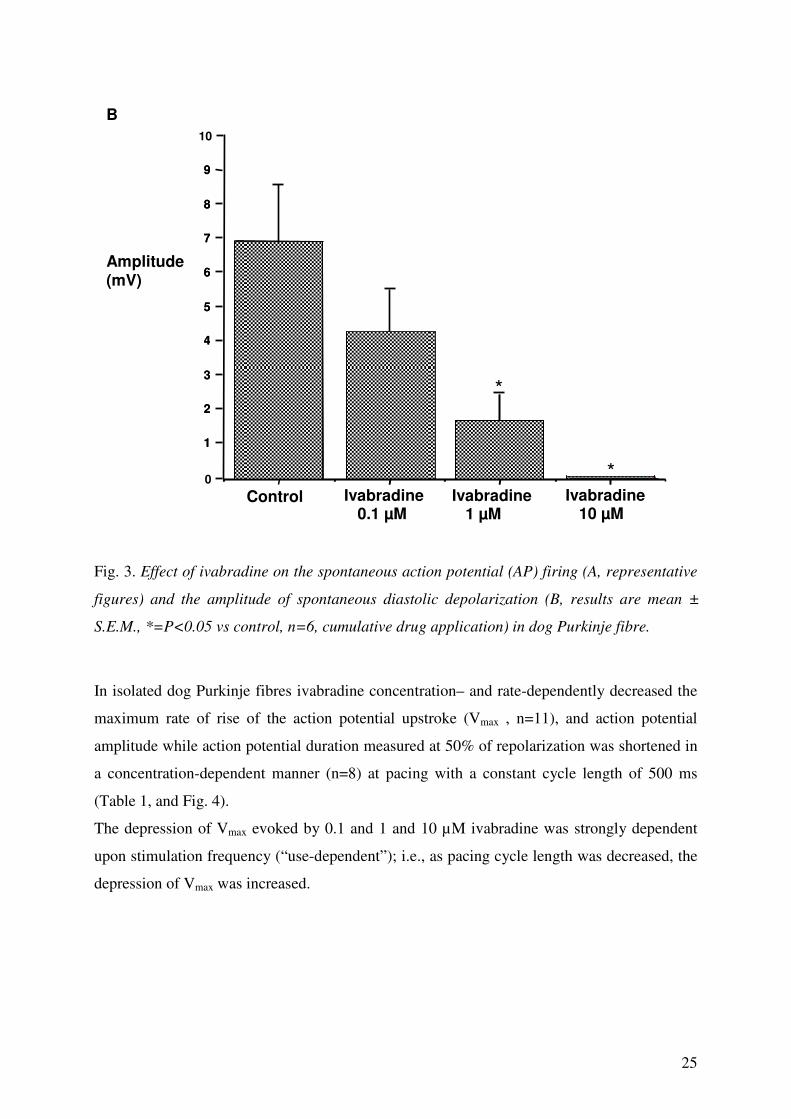
S.E.M., *=P<0.05 vs control, n=6, cumulative drug application) in dog Purkinje fibre.
In isolated dog Purkinje fibres ivabradine concentration– and rate-dependently decreased the
maximum rate of rise of the action potential upstroke (Vmax , n=11), and action potential
amplitude while action potential duration measured at 50% of repolarization was shortened in
a concentration-dependent manner (n=8) at pacing with a constant cycle length of 500 ms
(Table 1, and Fig. 4).
The depression of Vmax evoked by 0.1 and 1 and 10 µM ivabradine was strongly dependent
upon stimulation frequency (“use-dependent”); i.e., as pacing cycle length was decreased, the
depression of Vmax was increased.
Ivabradine 10 µM
Ivabradine 0.1 µM
Ivabradine 1 µM
B Amplitude (mV)
Control 0
1
2
3
4
5
6
7
8
9
10
1
2
3
4
5
6
7
8
9
*
*

26
Fig. 4. Rate- and dose-dependent effect of ivabradine on the maximum rate of depolarization
(Vmax) (A, results are mean ± S.E.M., *=P<0.05, **=P<0.01 vs control, n=11, cumulative
drug application) and concentration-dependent effect of the drug on the AP waveform/plateau
(B, representative figure ) in dog Purkinje fibre (recordings originate from the same
preparation).
Table 1: The electrophysiological effects of ivabradine in dog Purkinje fibers at basic cycle
length of 500 ms.
Parameters MDP (mV) APA (mV) APD50 (ms) APD90 (ms) Vmax (Vs-1
)
Control -88.3 ±1.0 129.4 ± 1.9 170.0 ± 9.8 245.7 ± 7.5 640.7 ± 38.4
Ivabraine
0.1 µM -88.0 ± 0.9 127.5 ± 1.9 168.8 ± 7.2 248.6 ± 6.2 591.7 ± 36.4
Ivabraine
1 µM -88.5 ± 1.5 126.5 ± 2.8 165.8 ± 8.8 252.0 ± 6.8 556.8 ± 42.8
Ivabraine
10 µM -88.1 ± 1.5 122.6 ± 2.4
a 137.7 ± 10.3
a 242.8 ± 7.7 471.8 ± 38.1
a
MDP, maximum diastolic potential; APA, action potential amplitude; APD50 and APD90,
action potential durations at 50% and 90% of repolarization; Vmax, maximal rate of
depolarization. Results are mean ± S.E.M. a
p < 0.05. n=8.

27
A
B

28
Fig. 5. Onset of frequency-dependent depression of Vmax induced by 10 µM ivabradine in dog
ventricular muscle at stimulation cycle length of 400 ms. Mean values are given from 7
experiments (A); effect of 10 µM ivabradine on recovery of Vmax in dog ventricular muscle at
stimulation cycle length of 400 ms. Mean values are given from 8 experiments (B).
In dog ventricular muscles (n=7) stimulated at the cycle length of 400 ms the onset kinetics of
Vmax block induced by 10 µM ivabradine was fitted to a single exponential, resulting in the
onset rate kinetic constant of 13.9 ± 3.2 beat -1 (Fig. 5A). In dog ventricular muscles (n=8) at
the basic cycle length of 400 ms, the recovery of Vmax during control was best fit to a single
exponential relation (Fig. 5B). The time constant for recovery of Vmax during control was fast
(τfast = 46.2 ± 4.3 ms) and before final repolarization of the basic action potential, it was
almost complete. In the presence of 10 µM ivabradine the recovery kinetics of Vmax was best
fit with a two exponential relation. In addition to a fast component (τfast = 41.2 ± 8.2 ms)
which reflects recovery of the drug-free sodium channels [93], a slow component (τslow = 8.76
± 1.34 s) of recovery of Vmax was revealed following exposure to ivabradine. This second
slow component for recovery of Vmax may reflect effects on drug-affected sodium channels
[93] (Fig. 3B).
In dog ventricular muscle (n=8) at a stimulation cycle length of 1000 ms the drug moderately
and in a concentration dependent manner prolonged the action potential duration (Fig. 6,
Table 2), while after attenuation of the repolarization reserve by inhibition of the inward
rectifier potassium current (n=5) by adding 30 µM BaCl2 it further lengthened the ventricular
repolarization at concentrations of 1 and 10 µM. (Fig. 6, Table 3).

29
Fig. 6. Effect of ivabradine on action potential waveform of dog ventricular muscle with
normal (on the left, top and bottom figures, showing the same representative experiment) and
attenuated repolarization reserve (on the right, top and bottom, illustrating 2 different
representative experiments) at stimulation cycle length of 1000 ms (action potential waveform
recordings originate from the same preparation, per experiment).
Table 2. The electrophysiological effects of ivabradine in dog ventricular muscle preparations
at basic cycle length of 1000 ms.
Parameters MDP (mV) APA (mV) APD50 (ms) APD90 (ms) Vmax (Vs-1
)
Control -86.3 ±1.2 106.5 ± 1.5 190.1 ± 4.1 223.9 ± 5.9 201.1 ± 17.2
Ivabraine
0.1 µM -86.3 ± 1.0 106.7 ± 1.8 195.0 ± 4.7
b 229.0 ± 5.7
a 205.7 ± 21.6
Ivabraine
1 µM -85.4 ± 0.8 107.2 ± 2.1 199.0 ± 5.3
b 233.5 ± 6.0
a 212.1 ± 20.2
Ivabraine
10 µM -87.6 ± 1.1 106.1 ± 2.4
205.0 ± 5.0
a 243.8 ± 6.5
a 181.8 ± 20.3
30
MDP, maximum diastolic potential; APA, action potential amplitude; APD50 and APD90,
action potential durations at 50% and 90% of repolarization; Vmax, maximal rate of
depolarization. Results are mean ± S.E.M. a
p < 0.01; bp < 0.05. n=8.
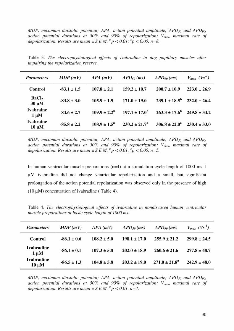
Table 3. The electrophysiological effects of ivabradine in dog papillary muscles after
impairing the repolarization reserve.
Parameters MDP (mV) APA (mV) APD50 (ms) APD90 (ms) Vmax (Vs-1
)
Control -83.1 ± 1.5 107.8 ± 2.1 159.2 ± 10.7 200.7 ± 10.9 223.0 ± 26.9
BaCl2
30 µM -83.8 ± 3.0 105.9 ± 1.9 171.0 ± 19.0 239.1 ± 18.5
b 232.0 ± 26.4
Ivabraine
1 µM -84.6 ± 2.7 109.9 ± 2.2
b 197.1 ± 17.0
b 263.3 ± 17.6
b 249.8 ± 34.2
Ivabraine 10 µM
-85.8 ± 2.2 108.9 ± 1.5a
230.2 ± 21.7a
306.8 ± 22.0a
230.4 ± 33.0
MDP, maximum diastolic potential; APA, action potential amplitude; APD50 and APD90,
action potential durations at 50% and 90% of repolarization; Vmax, maximal rate of
depolarization. Results are mean ± S.E.M. a
p < 0.01; bp < 0.05. n=5.
In human ventricular muscle preparations (n=4) at a stimulation cycle length of 1000 ms 1
µM ivabradine did not change ventricular repolarization and a small, but significant
prolongation of the action potential repolarization was observed only in the presence of high
(10 µM) concentration of ivabradine ( Table 4).
Table 4. The electrophysiological effects of ivabradine in nondiseased human ventricular
muscle preparations at basic cycle length of 1000 ms.
Parameters MDP (mV) APA (mV) APD50 (ms) APD90 (ms) Vmax (Vs-1
)
Control -86.1 ± 0.6 108.2 ± 5.0 198.1 ± 17.0 255.9 ± 21.2 299.8 ± 24.5
Ivabradine
1 µM -86.1 ± 0.1 107.3 ± 5.8 202.0 ± 18.9 260.6 ± 21.6 277.8 ± 48.7
Ivabradine
10 µM -86.5 ± 1.3 104.8 ± 5.8 203.2 ± 19.0 271.0 ± 21.8
a 242.9 ± 48.0
MDP, maximum diastolic potential; APA, action potential amplitude; APD50 and APD90,
action potential durations at 50% and 90% of repolarization; Vmax, maximal rate of
depolarization. Results are mean ± S.E.M. a
p < 0.01. n=4.

31
3.3 Effects of compounds EC18 and MEL57A on transmembrane action potentials
We investigated the effects of compounds EC18 and Mel 57A on the action potential
parameters of dog papillary muscle preparations and Purkinje fibers.
EC18 has similar action on the spontaneous diastolic depolarization (Fig. 7.) to ivabradine
(i.e. dose-dependently reduced the amplitude of diastolic depolarization phase) and at 10 µM,
decreased the AP plateau in dog Purkinje fibre (Fig. 8).
Fig. 7. Effect of EC18 on the amplitude of spontaneous (left, representative experiment)
diastolic depolarization (histograms represent mean ± S.E.M., *=p<0.05 vs control, n=5,
cumulative drug application) in dog Purkinje fibre.
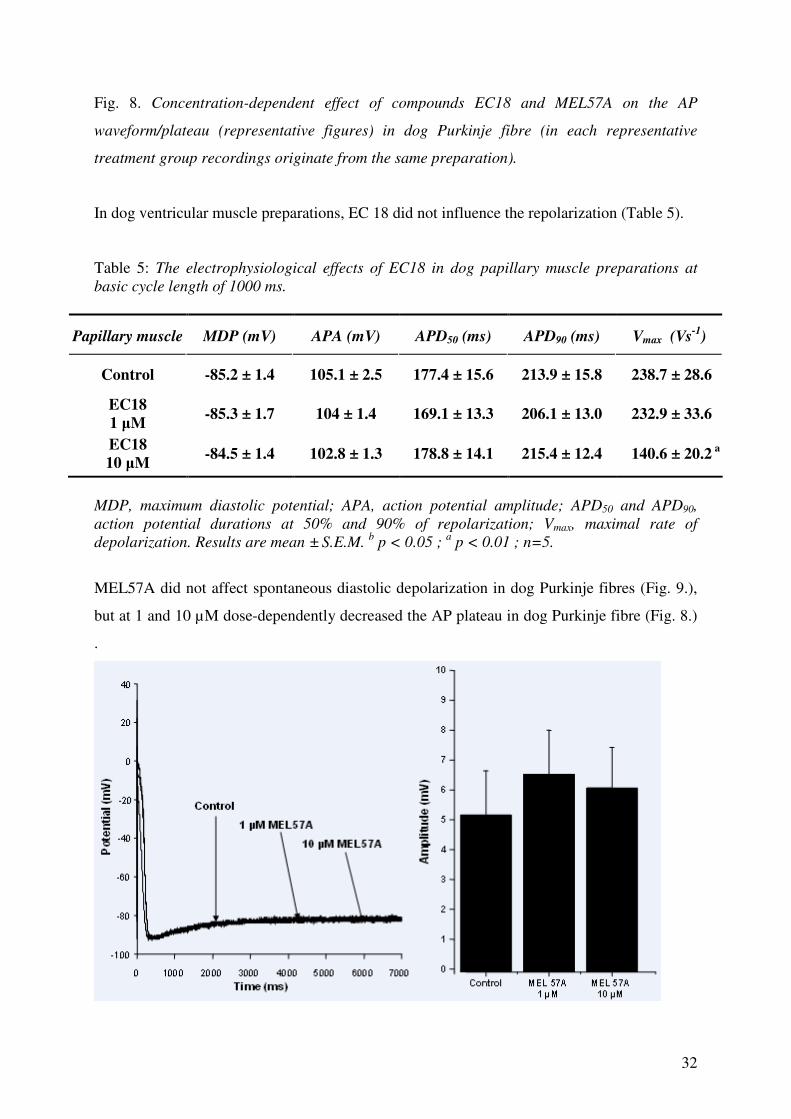
32
Fig. 8. Concentration-dependent effect of compounds EC18 and MEL57A on the AP
waveform/plateau (representative figures) in dog Purkinje fibre (in each representative
treatment group recordings originate from the same preparation).
In dog ventricular muscle preparations, EC 18 did not influence the repolarization (Table 5).
Table 5: The electrophysiological effects of EC18 in dog papillary muscle preparations at
basic cycle length of 1000 ms.
Papillary muscle MDP (mV) APA (mV) APD50 (ms) APD90 (ms) Vmax (Vs-1
)
Control -85.2 ± 1.4 105.1 ± 2.5 177.4 ± 15.6 213.9 ± 15.8 238.7 ± 28.6
EC18
1 µM -85.3 ± 1.7 104 ± 1.4 169.1 ± 13.3 206.1 ± 13.0 232.9 ± 33.6
EC18
10 µM -84.5 ± 1.4 102.8 ± 1.3 178.8 ± 14.1 215.4 ± 12.4
140.6 ± 20.2
a
MDP, maximum diastolic potential; APA, action potential amplitude; APD50 and APD90,
action potential durations at 50% and 90% of repolarization; Vmax, maximal rate of
depolarization. Results are mean ± S.E.M. b p < 0.05 ;
a p < 0.01 ; n=5.
MEL57A did not affect spontaneous diastolic depolarization in dog Purkinje fibres (Fig. 9.),
but at 1 and 10 µM dose-dependently decreased the AP plateau in dog Purkinje fibre (Fig. 8.)
.
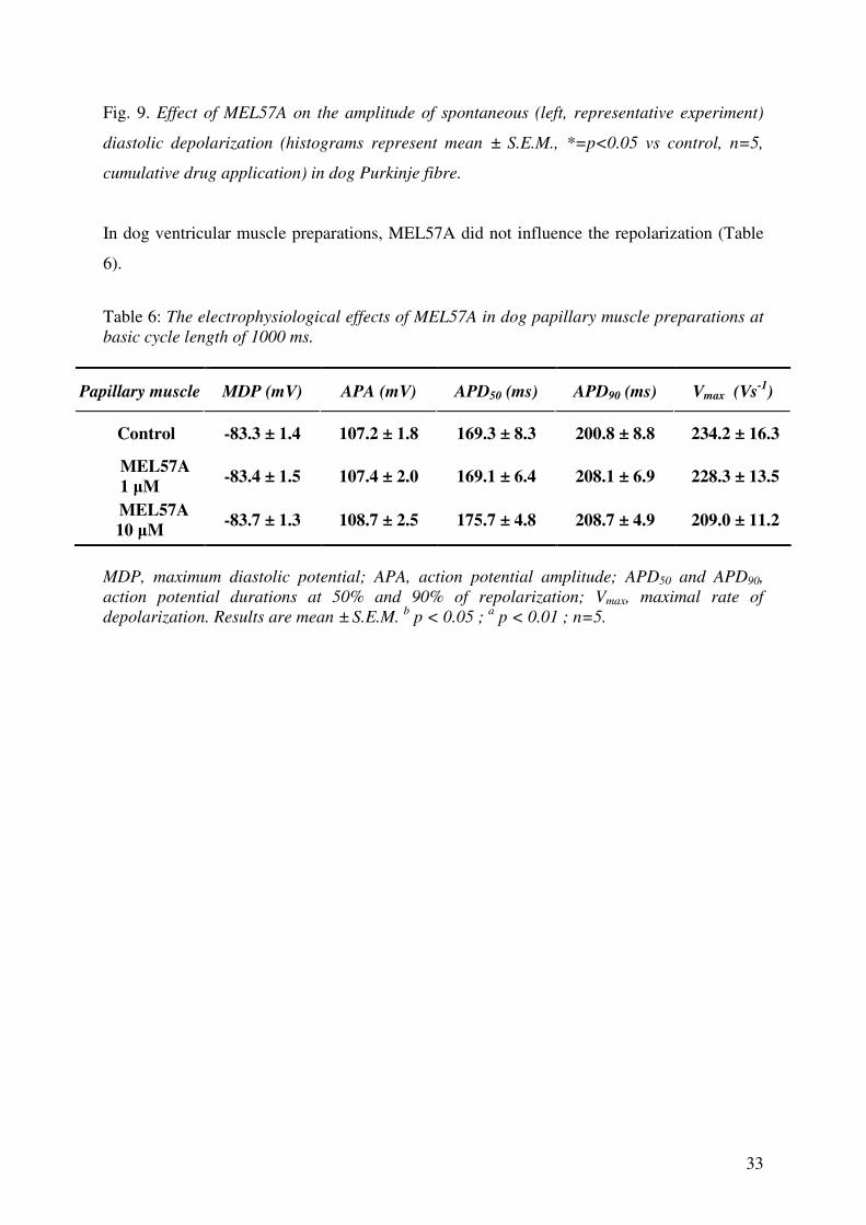
33
Fig. 9. Effect of MEL57A on the amplitude of spontaneous (left, representative experiment)
diastolic depolarization (histograms represent mean ± S.E.M., *=p<0.05 vs control, n=5,
cumulative drug application) in dog Purkinje fibre.
In dog ventricular muscle preparations, MEL57A did not influence the repolarization (Table
6).
Table 6: The electrophysiological effects of MEL57A in dog papillary muscle preparations at
basic cycle length of 1000 ms.
Papillary muscle MDP (mV) APA (mV) APD50 (ms) APD90 (ms) Vmax (Vs-1
)
Control -83.3 ± 1.4 107.2 ± 1.8 169.3 ± 8.3 200.8 ± 8.8 234.2 ± 16.3
MEL57A
1 µM -83.4 ± 1.5 107.4 ± 2.0 169.1 ± 6.4 208.1 ± 6.9 228.3 ± 13.5
MEL57A
10 µM -83.7 ± 1.3 108.7 ± 2.5 175.7 ± 4.8 208.7 ± 4.9
209.0 ± 11.2
MDP, maximum diastolic potential; APA, action potential amplitude; APD50 and APD90,
action potential durations at 50% and 90% of repolarization; Vmax, maximal rate of
depolarization. Results are mean ± S.E.M. b p < 0.05 ;
a p < 0.01 ; n=5.

34
4. DISCUSSION
Some of the results is confirmation of previous findings in small animals at high
concentration i.e. ivabradine (S16257) at 50 µM induces frequency-dependent Vmax block and
at 3 and 10 µM prolongs repolarization in guinea-pig papillary muscles [90, 53]. The main
new finding of the present study is that ivabradine in large animal (dog) and human cardiac
preparations, in addition to decreasing heart rate, inhibits Vmax , IKr , and at a concentration
higher than 1 µM moderately prolongs APD i.e. exerts combined Class I, Class III and Class
V antiarrhythmic actions.
4.1 Possible ionic mechanisms
We have examined the specific ionic mechanisms by which ivabradine affects action potential
configuration. Repolarization lengthening could be related to the IKr block found in rabbit
isolated ventricular myocytes. Dose- and frequency dependent Vmax block in dog Purkinje-
fibres could be attributed to the inhibitory effect of ivabradine on the fast Na+ current. Vmax
measurements are indicative for INa function, but can not be used for quantitative estimation of
sodium channel availability, which could be underestimated by such measurements [93], since
Vmax can be regarded as a nonlinear indicator of the fast inward sodium current [94]. Block of
sodium current has been demonstrated to be more pronounced when the resting membrane
potential is partly depolarized [95] e.g. in ischaemic tissues. The effects of ivabradine on
recovery of Vmax observed in isolated ventricular muscle indicate that ivabradine resembles
kinetically slow antiarrhythmic agents. According to reported data [96] at 10 µM
concentration ivabradine does not exhibit a 100% blockade of the If current, therefore
additional factors such as inhibitory effect on sodium channels can not be excluded as
possible contributor. A slowly activating sodium current, INa3, has been described by patch-
clamp analysis in the late diastolic depolarization of dog single Purkinje cells. This current is
sensitive to lidocaine and tetrodotoxin, and contributes to attainment of the threshold for the
upstroke in the oscillary range of late diastolic depolarization [97]. Another possible
explanation might be the functional subunit interspecies and tissue specific difference of
hyperpolarization-activated cyclic nucleotide gated (HCN) channels [98].

Inhibition of IKr by ivabradine at higher concentrations might also influence the spontaneous
diastolic depolarization in Purkinje fibres owing to the „K+ current decay hypothesis” [99].
Effect on the plateau slope might be related to the inhibitory effect of ivabradine on the
persistent, ’window’ Na+ current. Block of this Na+ current might also have an additional
therapeutic value [100] and would also limit action potential prolongation at slow rate due to
the IKr inhibition of the drug at higher concentration. In this context it has to be mentioned
that similar relatively high (2.8 µM) IC50 of ivabradine was reported on If in rabbit sinus node
cells [96]. Therefore, the possible decrease of INa by the drug may contribute to the inhibition
of the pacemaker function.
4.2 Possible clinical implications
As the present study shows, ivabradine, in addition to blocking the pacemaker current, at
micromolar concentration ranges also inhibits the rapid component of the delayed rectifier
potassium current, and presumably, the sodium channels. Though therapeutic plasma
concentrations of ivabradine are about 0.04-0.07 µM, the drug has been tested at higher (0.17
µM) concentration in healthy volunteers [101], therefore the (0.1-1-10 µM) concentrations
applied in our experiments may be relevant, since the tissue concentration can be expected to
be higher than that of the plasma, in accordance with the high volume of distribution (close to
100 L) value of the drug. In this context, the relatively low therapeutic plasma concentration
might be the result of possibly meaningful tissue appearance of ivabradine, therefore, we can
not underestimate the probable INa and the proven IKr blocking ability of ivabradine at higher
concentrations. Mostly, because CHF (congestive heart failure) -induced impairments (e.g.
deterioration in Vmax) in dog Purkinje tissue conduction have been lately reported [102]. It has
to be emphasized that the drug so far has proved to be safe and free from any proarrhythmic
events in clinical trials [103,104]. However, considering the bradycardic action of the drug,
possible very rare proarrhythmic episodes can not be completely ruled out in certain, special
cases during application of ivabradine, especially in some pathological conditions, e.g. in case
of drug accumulation or intoxication, or in case of attenuated repolarization reserve like in
heart failure, diabetes or in LQT. On the other hand, the mild sodium channel blocking ability
on Purkinje fibres might be considered as an antiarrhythmic property and might suppress the
initiation of an extrasystole, limit any repolarization lengthening and most importantly, it
could decrease dispersion of repolarization. The frequency-dependent Vmax block might
diminish conduction of early extrasystoles or bursts with fast rates which potentially elicit
36
cardiac arrhythmias in the presence of an ischaemic cardiac substrate. Therefore, the most
conspicious finding was that this bradycardic drug might possess subsidiary antiarrhythmic
actions, which can be utilized in the therapy.
4.3 Electrophysiological effects of compounds EC18 and MEL57A
We also investigated compounds EC18 and MEL57A on dog papillary muscles where they
did not influence repolarization and on cardiac Purkinje fibers, a cardiac subsidiary
pacemaker exhibiting spontaneous diastolic depolarization, where isoforms HCN4 and HCN2
have been suggested to be the prevalent f-channel isoforms at variance with isoform HCN1
which is no – or poorly – expressed in mammalian heart [105], including humans [106]. EC18
strongly decreased the amplitude of Purkinje fibers diastolic depolarization phase. On the
contrary, MEL57A did not change the diastolic depolarization phase. This latter observation
suggests that HCN1 selective inhibitors may be devoid of – or with less pronounced - cardiac
adverse effects while HCN4 blockers may be active also at ventricular level. Given HCN4
overexpression in cardiomyopathies such as in case of heart failure [84, 86, 107], such a
property might be advantageous possessing potential antiarrhythmic effects. Effect of EC18
and MEL57A on Purkinje fibre AP plateau might indicate subsidiary sodium channel or L-
type calcium channel inhibition.

37
5. CONCLUSIONS
Based on the cellular cardiac electrophysiological properties of ivabradine it can be concluded
that the drug, in addition to its well established bradycardic effect, also shows Class I and III
antiarrhythmic properties which can be advantageous to treat patients with ischaemic heart
disease, heart failure [108] liable to disturbances of cardiac rhythm.
Concerning –in general- the bradycardic agents the HCN4 isoform selectivity is desirable
according to its prominent role in cardiac pacemaking [79, 80]. Reaching the lack of visual
side effects (attributable to the retinal Ih block) would be advantageous in future development.
As to the arrhythmogenic potency of HCN4 and HCN2 upregulation in the working
myocardium of the failing heart, simultaneous block of them seems to be a feasible
opportunity. As it was seen in the history of development of beta blocker generations,
valuable subsidiary effects (alfa-blocker, antioxidant, very high beta1 selectivity) were
discovered at some relatively new representatives of them. In case of If blockers, similar
phenomen might happen, adding extra beneficial property to future bradycardic compounds.
HCN1 isoform selective Ih inhibitors (MEL57A) might be appropriate for the treatment of
neuropathic pain associated with nerve injury [7]. The HCN1 isoform also play an important
role in the effect of general anaesthetic agents. Regarding the potential adverse effects, and
considering that HCN channels are also expressed in liver, testis and pancreas it can be stated
that synthetizing a well tolerable, safe agent is very important, and therefore the isoform
selective approach might be the basis of future developments.

38
6. REFERENCES (1-108)
[1] Moosmang, S.; Stieber, J.; Zong, X.; Biel, M.; Hofmann, F.; Ludwig, A. Cellular expression and functional characterization of four hyperpolarization-activated pacemaker channels in cardiac and neuronal tissues. Eur J. Biochem., 2001, 268, 1646-1652.
[2] Brown, HF.; DiFrancesco, D.; Noble, SJ. How does adrenaline accelerate the heart? Nature, 1979, 280(5719), 235-236. [3] DiFrancesco, D. The pacemaker current (If) plays an important role in regulating SA node pacemaker activity. Cardiovasc Res., 1995, 30(2), 307-308. [4] Day, M.; Carr, DB.; Ulrich, S.; Ilijic, E.; Tkatch, T.; Surmeier, DJ. Dendritic excitability of mouse frontal cortex pyramidal neurons is shaped by the interaction among HCN, Kir2, and Kleak channels. J. Neurosci., 2005, 25(38), 8776-8787. [5] Ludwig, A.; Budde, T.; Stieber, J.; Moosmang, S.; Wahl, C.; Holthoff, K.; Langebartels, A.; Wotjak, C.; Munsch, T.; Zong, X.; Feil, S.; Feil, R.; Lancel, M.; Chien, KR.; Konnerth, A.; Pape, HC.; Biel, M.; Hofmann, F. Absence epilepsy and sinus dysrhythmia in mice lacking the pacemaker channel HCN2. EMBO J., 2003, 22, 216-224. [6] Fried, HU.; Kaupp, UB.; Müller, F. Hyperpolarization-activated and cyclic nucleotide-gated channels are differentially expressed in juxtaglomerular cells in the olfactory bulb of mice. Cell Tissue Res., 2010, 339(3), 463-479. [7] Wickenden, AD.; Maher, MP.; Chaplan, SR. HCN pacemaker channels and pain: a drug discovery perspective. Curr. Pharm. Des., 2009, 15, 2149-2168. [8] Wahl-Schott, C.; Biel, M. HCN channels: structure, cellular regulation and physiological function. Cell Mol. Life Sci., 2009, 66(3), 470-494. [9] El-Kholy, W.; MacDonald, P. E.; Fox, J. M.; Bhattacharjee, A.; Xue, T.; Gao, X.; Zhang, Y.; Stieber, J.; Li, R. A.; Tsushima, R. G.; Wheeler, M. B. Hyperpolarization-activated cyclic nucleotide-gated channels in pancreatic beta-cells. Mol. Endocrinol., 2007, 21, 753-764. [10] Hurtado, R.; Bub, G.; Herzlinger, D. The pelvis–kidney junction contains HCN3, a hyperpolarization-activated cation channel that triggers ureter peristalsis. Kidney Int., 2010, 77, 500-508. [11] Biel, M.; Wahl-Schott, C.; Michalakis, S.; Zong, X. Hyperpolarization-activated cation channels: from genes to function. Physiol. Rev., 2009, 89(3), 847-885. [12] Fox, K.; Ford, I.; Steg, PG.; Tendera, M.; Ferrari, R. Ivabradine for patients with stable coronary artery disease and left-ventricular systolic dysfunction (BEAUTIFUL): a randomised, double-blind, placebo-controlled trial. Lancet, 2008, 372(9641), 807-816.
[13] Stillitano, F.; Lonardo, G.; Zicha, S.; Varro, A.; Cerbai, E.; Mugelli, A.; Nattel, S. Molecular basis of funny current (If) in normal and failing human heart. J. Mol. Cell. Cardiol., 2008, 45(2), 289-299.

39
[14] Tamura, A.; Ogura, T.; Uemura, H.; Reien, Y.; Kishimoto, T.; Nagai, T.; Komuro, I.; Miyazaki, M.; Nakaya, H. Effects of antiarrhythmic drugs on the hyperpolarization-activated cyclic nucleotide-gated channel current. J Pharmacol Sci., 2009, 110, 150-159.
[15] DiFrancesco, D. If inhibition: a novel mechanism of action. Eur. Heart J. Suppl., 2003, 5(suppl. G), 19-25. [16] Lakatta, EG.; DiFrancesco, D. What keeps us ticking: a funny current, a calcium clock, or both? J. Mol. Cell. Cardiol., 2009, 47, 157-170. [17] DiFrancesco, D.; Mangoni, M. Modulation of single hyperpolarization-activated channels (If) by cAMP in the rabbit sino-atrial node. J. Physiol., 1994, 474(3), 473-482. [18] Michels, G.; Brandt, MC.; Zagidullin, N.; Khan, IF.; Larbig, R.; van Aaken, S.; Wippermann, Jens.; Hoppe, UC. Direct evidence for calcium conductance of hyperpolarization-activated cyclic nucleotide-gated channels and human native If at physiological calcium concentrations. Cardiovasc. Res., 2008, 78, 466-475. [19] Borer, JS.; Fox, K.; Jaillon, P.; Lerebours, G.; Ivabradine Investigators. Antianginal and antiischemic effects of ivabradine, an If inhibitor, in stable angina. Circ. 2003, 107, 817-823.
[20] Borer, JS. Heart rate slowing by If inhibition: therapeutic utility from clinical trials. Eur.
Heart J., 2005, Suppl. S7 (suppl H), 22-28.
[21] Tardif, JC. Heart rate and atherosclerosis. Eur. Heart J. Suppl., 2009, 11(suppl. D), 8-12. [22] Berdeaux, A.; Drieu la Rochelle, C.; Richard, V.; Giudicelli JF. Opposed responses of large and small coronary arteries to propranolol during exercise in dogs. Am. J. Physiol., 1991, 261(2), 265-270. [23] Van de Werf, F.; Bax, J.; Betriu, A.; Blomstrom-Lundqvist, C.; Crea, F.; Falk, V.; Filippatos, G.; Fox, K.; Huber, K.; Kastrati, A.; Rosengren, A.; Steg, PG.; Tubaro, M.; Verheugt, F.; Weidinger, F.; Weis, M. Management of acute myocardial infarction in patients presenting with persistent ST-segment elevation. Eur. Heart J., 2008, 29, 2909-2945. [24] DiFrancesco, D. Funny channels in the control of cardiac rhythm and mode of action of selective blockers. Pharm. Res., 2006, 53, 399-406. [25] Vinogradova, TM.; Zhou, YY.; Maltsev, V.; Lyashkov, A.; Stern, M.; Lakatta, EG. Rhythmic ryanodine receptor Ca2+ releases during diastolic depolarization of sinoatrial pacemaker cells do not require membrane depolarization. Circ. Res., 2004, 94, 802-809. [26] Vassalle, M. The pacemaker current If does not play an important role in regulating SA node pacemaker activity. Cardiovasc Res., 1995, 30(2), 309-310. [27] Borer, JS.; Fox, K.; Jaillon, P.; Lerebours, G. Antianginal and anti-ischemic effects of ivabradine, an If inhibitor, in stable angina: a randomized, double-blind, multicentered, placebo-controlled trial. Circulation, 2003, 107, 817-823.
40
[28] Mackaay, AJ.; Op't Hof , T.; Bleeker, WK.; Jongsma, HJ.; Bouman, LN. Interaction of adrenaline and acetylcholine on cardiac pacemaker function. Functional inhomogeneity of the rabbit sinus node. J. Pharmacol. Exp. Ther., 1980, 214(2), 417-422. [29] DiFrancesco, D.; Tromba, C. Muscarinic control of the hyperpolarization-activated current (If) in rabbit sino-atrial node myocytes. J. Physiol., 1988, 405, 493-510. [30] DiFrancesco, D.; Ferroni, A.; Mazzanti, M.; Tromba, C. Properties of the hyperpolarizing-activated current (If) in cells isolated from the rabbit sino-atrial node. J.
Physiol., 1986, 377, 61-88. [31] Thuringer, D.; Lauribe, P.; Escande, D. A hyperpolarization-activated inward current in human myocardial cells. J. Mol. Cell. Card., 1992, 24(5), 451-455. [32] Cerbai, E.; Barbieri, M.; Mugelli, A. Characterization of the hyperpolarization-activated current, If, in ventricular myocytes isolated from hypertensive rats. J. Physiol., 1994, 481(3), 585-591. [33] Pape, HC. Queer current and pacemaker: the hyperpolarization-activated cation current in neurons. Annu. Rev. Physiol., 1996, 58, 299-327. [34] DiFrancesco, D.; Tortora, P. Direct activation of cardiac pacemaker channels by intracellular cyclic AMP. Nature, 1991, 351, 145-147. [35] DiFrancesco, D. Dual allosteric modulation of pacemaker (f) channels by cAMP and voltage in rabbit SA node. J. Physiol., 1999, 515(2), 367–376. [36] DiFrancesco, D. Pacemaker mechanisms in cardiac tissue. Annu. Rev. Physiol., 1993, 55, 455-472. [37] Baruscotti, M.; Bucchi, A.; DiFrancesco, D. Physiology and pharmacology of the cardiac pacemaker ("funny") current. Pharm. & Ther., 2005, 107, 59-79.
[38] Verkerk, AO.; Wilders, R.; van Borren, MM.; Peters, RJ.; Broekhuis, E.; Lam, K.; Coronel, R.; de Bakker, JM.; Tan, HL. Pacemaker current If in the human sinoatrial node. Eur.
Heart J., 2007, 20, 2472-2478.
[39] Qu, J.; Kryukova, Y.; Potapova, IA.; Doronin, SV.; Larsen, M.; Krishnamurthy, G.; Cohen, IS.; Robinson, RB. MiRP1 modulates HCN2 channel expression and gating in cardiac myocytes. J. Biol. Chem., 2004, 279, 43497-43502. [40] Accili, EA.; Redaelli, G.; DiFrancesco, D. Differential control of the hyperpolarization-activated current (If) by cAMP gating and phosphatase inhibition in rabbit sino-atrial node myocytes. J. Physiol., 1997, 500.3, 643-651. [41] Gravante, B.; Barbuti, A.; Milanesi, R.; Zappi, I.; Viscomi, C.; DiFrancesco, D. Interaction of the pacemaker channel HCN1 with filamin A. J. Biol. Chem., 2004, 279, 43847-43853.
41
[42] Suh, BC.; Hille, B. Regulation of ion channels by phosphatidylinositol 4,5-bisphosphate. Curr. Opin. Neurobiol., 2005, 15, 370-378. [43] Barbuti, A.; Gravante, B.; Riolfo, M.; Milanesi, R.; Terragni, B.; DiFrancesco, D. Localization of pacemaker channels in lipid rafts regulates channel kinetics. Circ. Res., 2004,
94, 1325-1331. [44] DiFrancesco, D. The role of the funny current in pacemaker activity. Circ. Res., 2010, 106, 434-446. [45] Kobinger, W.; Lillie, C. Specific bradycardic agents – a novel pharmacological class? Eur. Heart J., 1987, Suppl. L, 7-15. [46] Satoh, H.; Hashimoto, K. Electrophysiological study of alinidine in voltage clamped rabbit sino-atrial node cells. Eur. J. Pharmacol., 1986, 121(2), 211-219. [47] Briggs, I.; BoSmith, RE.; Heapy, CG. Effects of Zeneca ZD 7288 in comparison with alinidine and UL-FS 49 on guinea pig sinoatrial node and ventricular action potentials. J.
Cardiovasc. Pharm., 1994, 24, 380-387. [48] Kobinger, W. Editorial: History of ’specific bradycardic agents’. Eur. Heart J., 1987, Suppl. L, 5-6. [49] Osterrieder, W.; Pelzer, D.; Yang, QF.; Trautwein W. The electrophysiological basis of the bradycardic action of AQA 39 on the sinoatrial node. Naunyn Schmiedebergs Arch
Pharmacol., 1981, 317(3), 233-237. [50] Boucher, M.; Chassaing, C.; Chapuy, E. Cardiac electrophysiological effects of falipamil in the conscious dog: comparison with alinidine. Eur. J. Pharmacol., 1996, 306(1-3), 93-98.
[51] DiFrancesco, D. Some properties of the UL-FS 49 block of the hyperpolarization-activated current (I(f)) in sino-atrial node myocytes. Pflugers Arch, 1994, 427(1-2), 64-70.
[52] Valenzuela, C.; Delpon, E.; Franqueza, L.; BS; Gay, P.; Perez, O.; Tamargo, J.; Snyders D.J. Class III Antiarrhythmic Effects of Zatebradine. Circulation, 1996, 94, 562-570. [53] Thollon, C.; Cambarrat, C.; Vian, J.; Prost, J-F.; Peglion, J.; Vilaine, J. Electrophysiological effects of S 16257, a novel sino-atrial node modulator, on rabbit and guinea-pig cardiac preparations: comparison with UL-FS 49. Br. J. Pharmacol. 1994, 112, 37-42. [54] Frishman, WH.; Pepine, CJ.; Weiss, RJ.; Baiker, WM. Addition of zatebradine, a direct sinus node inhibitor, provides no greater exercise tolerance benefit in patients with angina taking extended-release nifedipine: results of a multicenter, randomized, double-blind, placebo-controlled, parallel-group study. The zatebradine study group. J. Am. Coll. Cardiol., 1995, 26(2), 305-312. [55] Satoh, TO.; Yamada, M. Multiple inhibitory effects of zatebradine (UL-FS49) on the electrophysiological properties of retinal rod photoreceptors. Pflugers Arch., 2002, 443(4), 532-540.

42
[56] Van Bogaert, PP.; Pittoors, F. Use-dependent blockade of cardiac pacemaker current (If) by cilobradine and zatebradine. Eur. J. Pharmacol., 2003, 478(2-3), 161-171. [57] Baruscotti, M.; Barbuti, A.; Bucchi, A. The cardiac pacemaker current. J. Mol. Cell.
Cardiol., 2010, 48, 55-64. [58] BoSmith, RE.; Briggs, I.; Sturgess, N. Inhibitory actions of Zeneca ZD7288 on whole-cell hyperpolarization activated inward current (If) in guinea-pig dissociated sinoatrial node cells. Br. J. Pharmacol., 1993, 110, 343-349. [59] Gasparini, S.; DiFrancesco, D. Actions of the hyperpolarization-activated current (Ih) blocker ZD 7288 in hippocampal CA1 neurons. Pflugers Arch., 1997, 435(1), 99-106. [60] Harris, NC.; Constanti, A. Mechanism of block by ZD 7288 of the hyperpolarization-activated inward rectifying current in guinea pig substantia nigra neurons in vitro. J.
Neurophysiol., 1995, 74(6), 2366-2378. [61] Williams, SR.; Turner, JP.; Hughes, SW.; Crunelli, V. On the nature of anomalous rectification in thalamocortical neurones of the cat ventrobasal thalamus in vitro. J. Physiol., 1997, 505(3), 727-747. [62] Thollon, C.; Vilaine, JP. I(f) inhibition in cardiovascular diseases. Adv. Pharmacol., 2010, 59, 53-92. [63] Vilaine, JP. The discovery of the selective If current inhibitor ivabradine. A new therapeutic approach to ischemic heart disease. Pharm. Res., 2006, 53, 424-434. [64] Thollon, C.; Bidouard, JP.; Cambarrat, C.; Lesage, L.; Reure, H.; Delescluse, I.; Vian, J.; Peglion, JL.; Vilaine, JP. Stereospecific in vitro and in vivo effects of the new sinus node inhibitor (+)-S16257. Eur. J. Pharmacol., 1997, 339, 43-51. [65] Bois, P.; Bescond, J.; Renaudon, B.; Lenfant, J. Mode of action of bradycardic agent, S 16257, on ionic currents of rabbit sinoatrial node cells. Br. J. Pharmacol., 1996, 118, 1051-1057. [66] Vilaine, JP.; Thollon, C.; Villeneuve, N.; Peglion, JL. Procoralan, a new selective If
current inhibitor. Eur. Heart J. Suppl., 2003, 5(suppl. G), 26-35. [67] Bucchi; A.; Baruscotti, M.; DiFrancesco, D. Current-dependent block of rabbit sino-atrial node If channels by ivabradine. J Gen Physiol., 2002, 120(1), 1–13. [68] Bucchi, A.; Tognati, A.; Milanesi, R.; Baruscotti, M.; DiFrancesco, D. Properties of ivabradine-induced block of HCN1 and HCN4 pacemaker channels. J. Physiol. 2006, 572.2, 335-346. [69] Heusch, G.; Schulz, R. The role of heart rate and the benefits of heart rate reduction in acute myocardial ischaemia. Eur. Heart J. Suppl., 2007, 9(F), 8-14.

43
[70] Savelieva, I.; Camm, AJ. Novel If current inhibitor ivabradine: safety considerations. Adv. Cardiol., 2006, 43, 79-96. [71] Joannides, R.; Moore, N.; Iacob, M.; Compagnon, P.; Lerebours, G.; Menard, JF.; Thuillez, C. Comparative effects of ivabradine, a selective heart rate-lowering agent, and propranolol on systemic and cardiac haemodynamics at rest and during exercise. Br. J. Clin.
Pharmacol., 2005, 61(2), 127-137. [72] Tardif, JC.; Ford, I.; Tendera, M.; Bourassa, MG.; Fox, K.; INITIATIVE Investigators. Efficacy of ivabradine, a new selective I(f) inhibitor, compared with atenolol in patients with chronic stable angina. Eur. Heart J., 2005, 26(23), 2529-2536. [73] Tardif, JC.; Ponikowski, P.; Kahan, T.; ASSOCIATE Investigators. Efficacy of the If current inhibitor ivabradine in patients with chronic stable angina receiving beta-blocker therapy: a 4-month, randomized, placebo-controlled trial. Eur. Heart J., 2009, 30(5), 540–548. [74] Swedberg, K.; Komajda, M.; Böhm, M.; Borer, JS.; Ford, I.; Dubost-Brama, A.; Lerebours, G.; Tavazzi, L.; SHIFT Investigators. Ivabradine and outcomes in chronic heart failure (SHIFT): a randomised placebo-controlled study. Lancet, 2010, 376(9744), 875-885. [75] Martin, G.; Tardif, JC. Can If inhibition help after myocardial infarction? Dialogues in
Cardiovascular Medicine, 2006, Vol.11 No.1., 36-41. [76] Couvreur, N.; Tissier, R.; Pons, S.; Chetboul, V.; Gouni, V.; Bruneval, P.; Mandet, C.; Pouchelon, J-L.; Berdeaux, A.; Ghaleh, B. Chronic heart rate reduction with ivabradine improves systolic function of the reperfused heart through a dual mechanism involving a direct mechanical effect and a long-term increase in FKBP12/12.6 expression. Eur. Heart J., 2010, 31, 1529-1537. [77] Heusch, G.; Skyschally, A.; Gres, P.; van Caster, P.; Schilawa, D.; Schulz, R. Improvement of regional myocardial blood flow and function and reduction of infarct size with ivabradine: protection beyond heart rate reduction. Eur. Heart J., 2008, 29, 2265-2275. [78] Ceconi, C.; Comini, L.; Suffredini, S.; Stillitano, F.; Bouly, M.; Cerbai, E.; Mugelli, A.; Ferrari, R. Heart rate reduction with ivabradine prevents the global phenotype of left ventricular remodeling. Am. J. Physiol. Heart Circ. Physiol., 2011, 300(1), H366-373.
[79] Baruscotti, M.; Bucchi, A.; Viscomi, C.; Mandelli, G.; Consalez, G.; Gnecchi-Rusconi, T.; Montano, N.; Casali, KR.; Micheloni, S.; Barbuti, A.; DiFrancesco D. Deep bradycardia and heart block caused by inducible cardiac-specific knockout of the pacemaker channel gene HCN4. Proc. Natl. Acad. Sci. USA, 2011, 108, 1705-1710.
[80] Thollon, C.; Bedut, S.; Villeneuve, N.; Cogé, F.; Piffard, L.; Guillaumin, J-P.; Brunel-Jacquemin, C.; Chomarat, P.; Boutin, J-A.; Peglion, J-L.; Vilaine, J-P. Use-dependent inhibition of hHCN4 by ivabradine and relationship with reduction in pacemaker activity. Br.
J. Pharmacol., 2007, 150, 37-46.

44
[81] Dobrzynski, H.; Nikolski, VP.; Sambelashvili, AT.; Greener, ID.; Yamamoto, M.; Boyett, MR.; Efimov, IR. Site of origin and molecular substrate of atrioventricular junctional rhythm in the rabbit heart. Circ. Res., 2003, 93, 1102-1110.
[82] Gaborit, N.; Le Bouter, S.; Szuts, V.; Varro, A.; Escande, D.; Nattel, S.; Demolombe, S. Regional and tissue specific transcript signatures of ion channel genes in the non-diseased human heart. J. Physiol., 2007, 582, 675-693.
[83] Ludwig, A.; Zong, X.; Stieber, J.; Hullin, R.; Hofmann, F.; Biel, M. Two pacemaker channels from human heart with profoundly different activation kinetics. EMBO J., 1999, 18, 2323-2329. [84] Cerbai, E.; Pino, R.; Porciatti, F.; Sani, G.; Toscano, M.; Maccherini, M.; Giunti, G.; Mugelli, A. Characterization of the hyperpolarization-activated current, (If), in ventricular myocytes from human failing heart. Circulation, 1997, 95, 568-571.
[85] Hoppe, UC.; Jansen, E.; Südkamp, M.; Beuckelmann, DJ. Hyperpolarization-activated inward current in ventricular myocytes from normal and failing human hearts. Circulation,
1998, 97, 55-65.
[86] Cerbai, E.; Sartiani, L.; DePaoli, P.; Pino, R.; Maccherini, M.; Bizzarri, F.; DiCiolla, F.; Davoli, G.; Sani, G.; Mugelli, A. The properties of the pacemaker current (If) in human ventricular myocytes are modulated by cardiac disease. J. Mol. Cell. Cardiol., 2001, 33, 441-448.
[87] Michels, G.; Er, F.; Khan, I.; Südkamp, M.; Herzig, S.; Hoppe, UC. Single-channel properties support a potential contribution of hyperpolarization-activated cyclic nucleotide-gated channels and If to cardiac arrhythmias. Circulation, 2005, 111, 399-404.
[88] Melchiorre, M.; Del Lungo, M.; Guandalini, L.; Martini, E.; Dei, S.; Manetti, D.; Scapecchi, S.; Teodori, E.; Sartiani, L.; Mugelli, A.; Cerbai, E.; Romanelli, M. N. Design, synthesis, and preliminary biological evaluation of new isoform-selective f-current blockers. J. Med. Chem., 2010, 53(18), 6773-6777. [89] Romanelli, M. N.; Mugelli, A.; Cerbai, E.; Sartiani, L.; Del Lungo, M.; Melchiorre, M. New isoform-selective HCN blockers. 2010, PCT/EP2010/059369. [90] Pérez O., Gay P., Fanqueza L., Carrón R., Valenzuela C., Delpón E., Tamargo J.. Effects of the two enantiomers, S-16257-2 and S-16260-2, of a new bradycardic agent on guinea-pig isolated cardiac preparations. Br. J. Pharmacol., 1995, 115, 787-794. [91] Varró A., Baláti B., Iost N., Takács J., Virág L., Lathrop D.A., Lengyel C., Tálosi L., Papp J.Gy. The role of the delayed rectifier component IKs in dog ventricular muscle and Purkinje fibre repolarization. J. Physiol., 2000, 523, 67-81. [92] Virág L., Varró A., Papp J.Gy. Effect of disopyramide on potassium currents in rabbit ventricular myocytes. Naunyn-Schmiedeberg’s Arch. Pharmacol., 1998, 357, 268-275. [93] Sheets M.F., Hanck D.A., Fozzard H.A. Nonlinear relation between Vmax and INa in canine cardiac purkinje cells. Circ. Res., 1998, 63, 386-398.
45
[94] Cohen C.J., Bean B.P., Tsien R.W. Maximal upstroke velocity as an index of available sodium conductance. Circ. Res., 1984, 54, 636-651. [95] Pu J., Balser J.R., Boyden P. Lidocaine action on Na+ currents in ventricular myocytes from the epicardial border zone of the infarcted heart. Circ. Res., 1998, 83, 431-440. [96] Bois P., Bescond J., Renaudon B., Lenfant J. Mode of action of bradycardic agent, S 16257, on ionic currents of rabbit sinoatrial node cells. Br. J. Pharmacol., 1996, 118, 1051-1057. [97] Rota M., Vassalle M. Patch-clamp analysis in canine cardiac Purkinje cells of a novel sodium component in the pacemaker range. J. Physiol., 2003, 548, 147–165. [98] Koncz I, Szél T, Jaeger K, Baczkó I, Cerbai E, Romanelli MN, Gy Papp J, Varró A. Selective pharmacological inhibition of the pacemaker channel isoforms (HCN1-4) as new possible therapeutical targets. Current Medicinal Chemistry., 2011, 18(24):3662-74. [99] Vassalle M., Yu H., Cohen I.S. The pacemaker current in cardiac Purkinje myocytes. J. Gen. Physiol., 1995, 106, 559-578. [100] Saint D.A. The cardiac persistent sodium current: an appealing therapeutic target? Br. J. Pharmacol., 2008, 153, 1133-1142. [101] Joannides R., Moore N., Iacob M., Compagnon P., Lerebours G., Menard J.F., Thuillez C. Comparative effects of ivabradine, a selective heart rate-lowering agent, and propranolol on systemic and cardiac haemodynamics at rest and during exercise. Br. J. Clin. Pharmacol., 2005, 61, 127-137. [102] Maguy A., Le Bouter S., Comtois P., Chartier D., Villeneuve L., Wakili R., Nishida K., Nattel S. Ion channel subunit expression changes in cardiac Purkinje fibers: a potential role in conduction abnormalities associated with congestive heart failure. Circ. Res., 2009, 104, 1113-1122
[103] Camm A.J., Savelieva I. If inhibition with ivabradine: electrophysiological effects and safety. Drug Saf., 2008, 31, 95-107.
[104] Tardif J.C., Borer J.S. Efficacy of ivabradine, a selective I(f) inhibitor, in patients with chronic stable angina pectoris and diabetes mellitus. Am. J. Cardiol., 2010, 105, 29-35. [105] Han W, Bao W, Wang Z, Nattel S. Comparison of ion-channel subunit expression in canine cardiac Purkinje fibers and ventricular muscle. Circ Res., 2002, 91: 790-7. [106] Gaborit N, Le Bouter S, Szuts V, Varro A, Escande D, Nattel S et al. Regional and tissue specific transcript signatures of ion channel genes in the non-diseased human heart. J Physiol., 2007, 582: 675-93. [107] Sartiani L, Stillitano F, Cerbai E, Mugelli A. Electrophysiologic changes in heart failure: focus on pacemaker channels. Can J Physiol Pharmacol, 2009, 87(2):84-90.

46
[108] Tavazzi L., Mugelli A. Can If inhibition help in congestive heart failure? Dialogues in Cardiovascular Medicine, 2006, 11, 30–35.

47
7. ACKNOWLEDGEMENTS
I am very grateful to my supervisors Prof. Dr. András Varró (Director of the Department
of Pharmacology and Pharmacotherapy, Faculty of Medicine, University of Szeged) and
Dr. László Virág (associate professor) for their professional mentorship, scientific guidance
for my research.
I would like to express my gratitude to Prof. Dr. Julius Gy. Papp (academician) for his
continuous scientific and moral support to me.
I would like to express my thanks to my Family.

48