cell signaling, wound repair, and atp receptors kevin quirke and alex knobloch
TRANSCRIPT
Cell Signaling, Wound Repair, and ATP Receptors
Kevin Quirke and Alex Knobloch
Cell Signaling Overview
• typical cell exposed to numbers of different signal molecules– selective response according to cell function– cellular response dictated by:
• unique sets of cell surface receptors • cell-specific intracellular targets
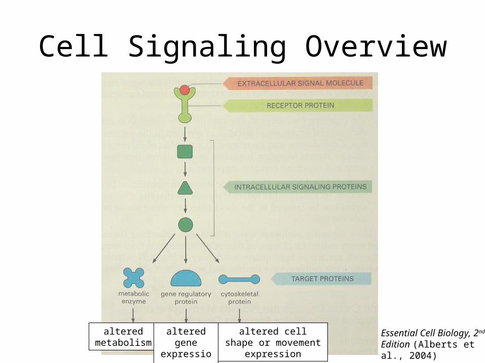
Cell Signaling Overview
Essential Cell Biology, 2nd Edition (Alberts et al., 2004)
altered metabolism
altered gene expression
altered cell shape or movement expression
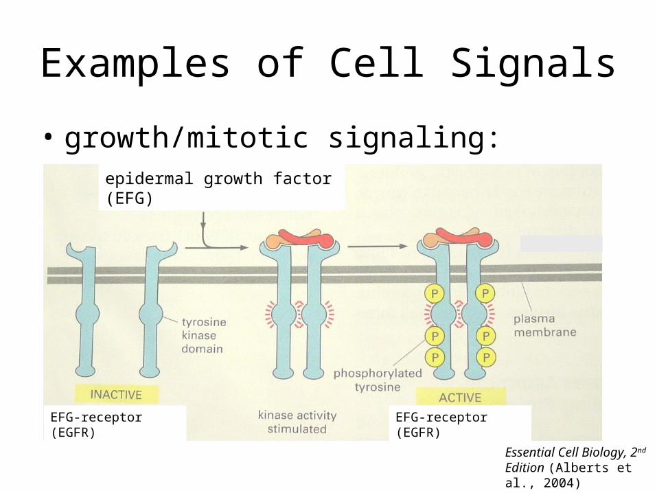
Examples of Cell Signals
• growth/mitotic signaling:epidermal growth factor (EFG)
EFG-receptor (EGFR) EFG-receptor (EGFR)
Essential Cell Biology, 2nd Edition (Alberts et al., 2004)
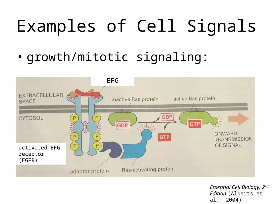
Examples of Cell Signals
• growth/mitotic signaling:
activated EFG-receptor (EGFR)
EFG
Essential Cell Biology, 2nd Edition (Alberts et al., 2004)
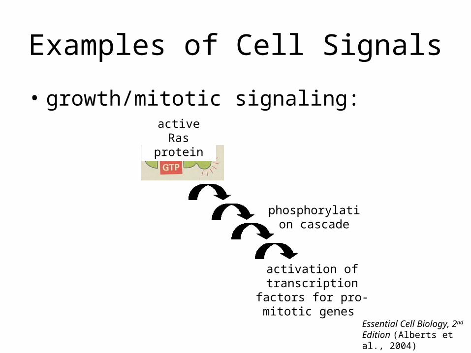
Examples of Cell Signals
• growth/mitotic signaling:active Ras
protein
activation of transcription factors for
pro-mitotic genes
phosphorylation cascade
Essential Cell Biology, 2nd Edition (Alberts et al., 2004)
Process Where Cell Signaling is Important
=Wound Repair
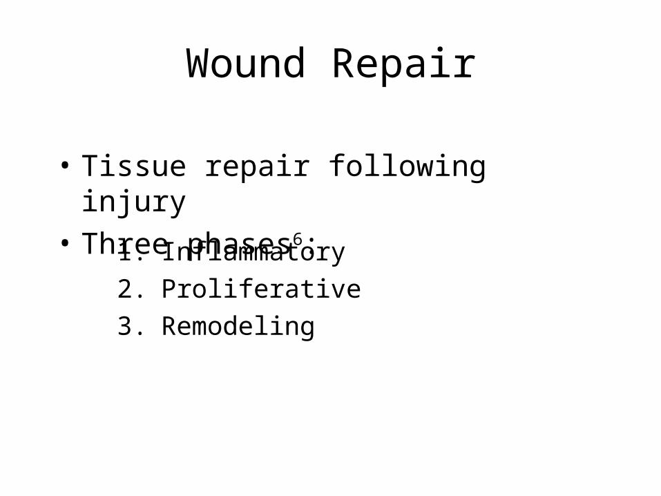
Wound Repair
1. Inflammatory
2. Proliferative
3. Remodeling
• Tissue repair following injury
• Three phases6:
Examples of Signals in Inflammatory Phase
• PAMPs
• DAMPs
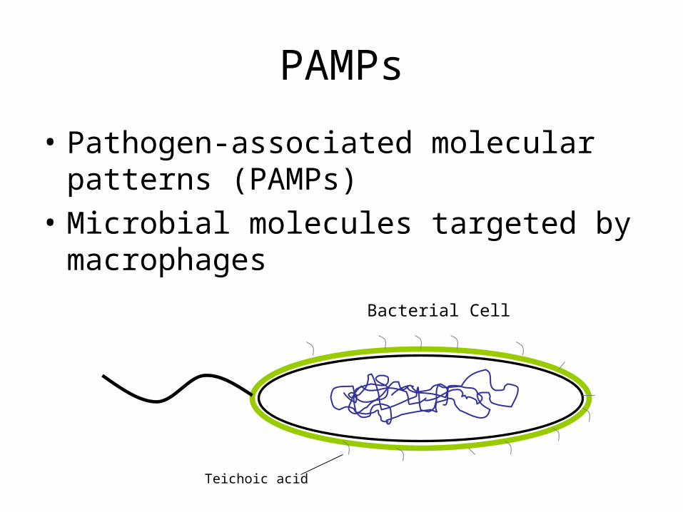
PAMPs
• Pathogen-associated molecular patterns (PAMPs)
• Microbial molecules targeted by macrophages
Teichoic acid
Bacterial Cell
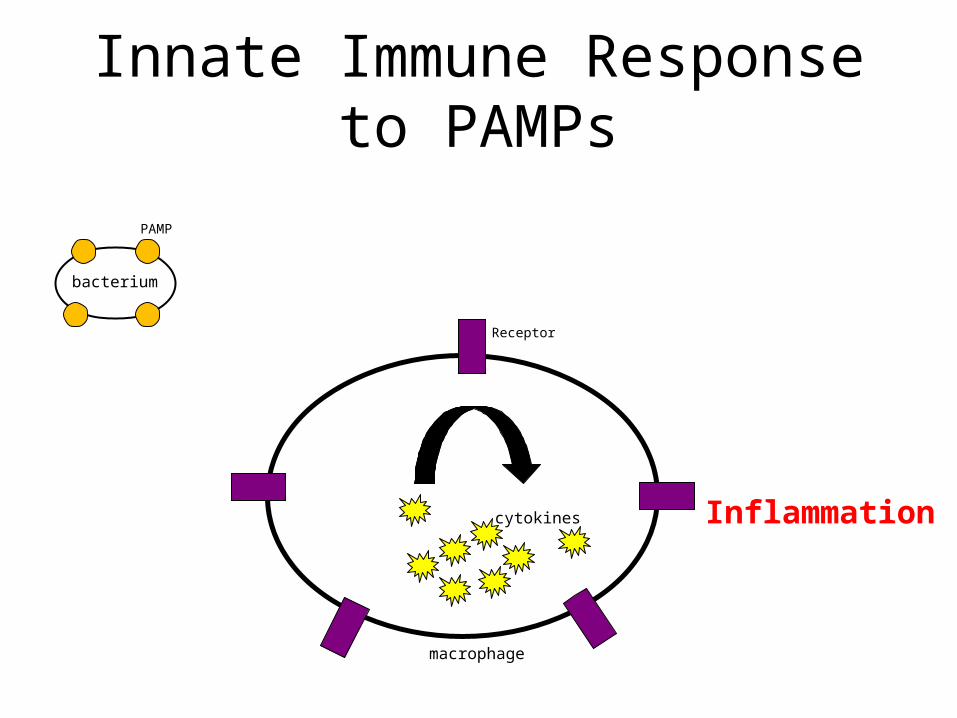
Innate Immune Response to PAMPs
macrophage
bacterium
PAMP
Receptor
cytokines Inflammation
DAMPs
• Damage-associated molecular-patterns (DAMPs)• Intracellular molecules• Released by cells undergoing stress or death• Initiate immune response
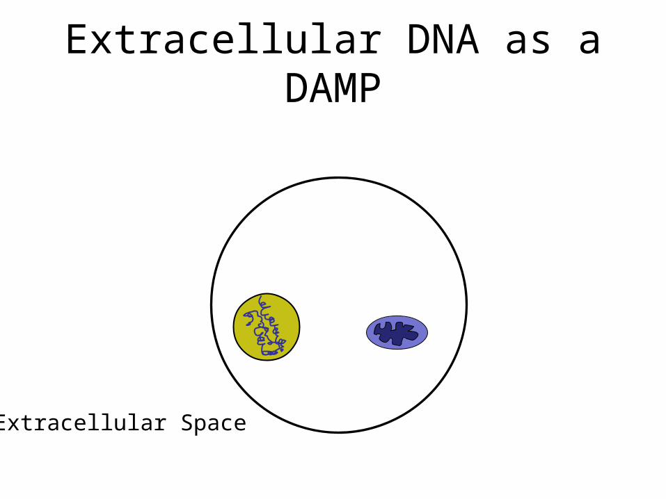
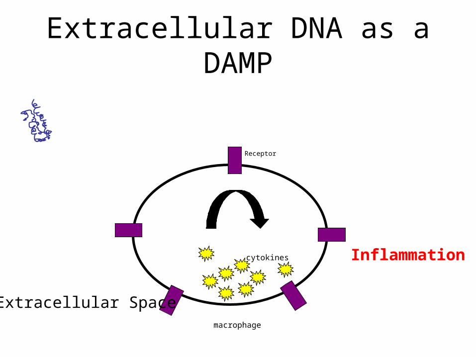
Extracellular DNA as a DAMP
Extracellular Space
Receptor
Extracellular DNA as a DAMP
Extracellular Spacemacrophage
cytokines Inflammation
Key Recipient and Producer of Inflammatory Phase Signals =
Monocytes
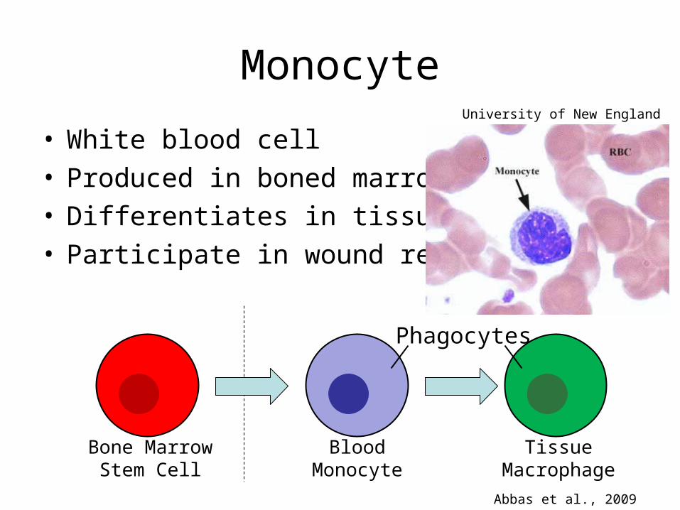
Monocyte
• White blood cell• Produced in boned marrow• Differentiates in tissues• Participate in wound repair
Bone Marrow Stem Cell
Blood Monocyte
Tissue Macrophage
Phagocytes
Abbas et al., 2009
University of New England
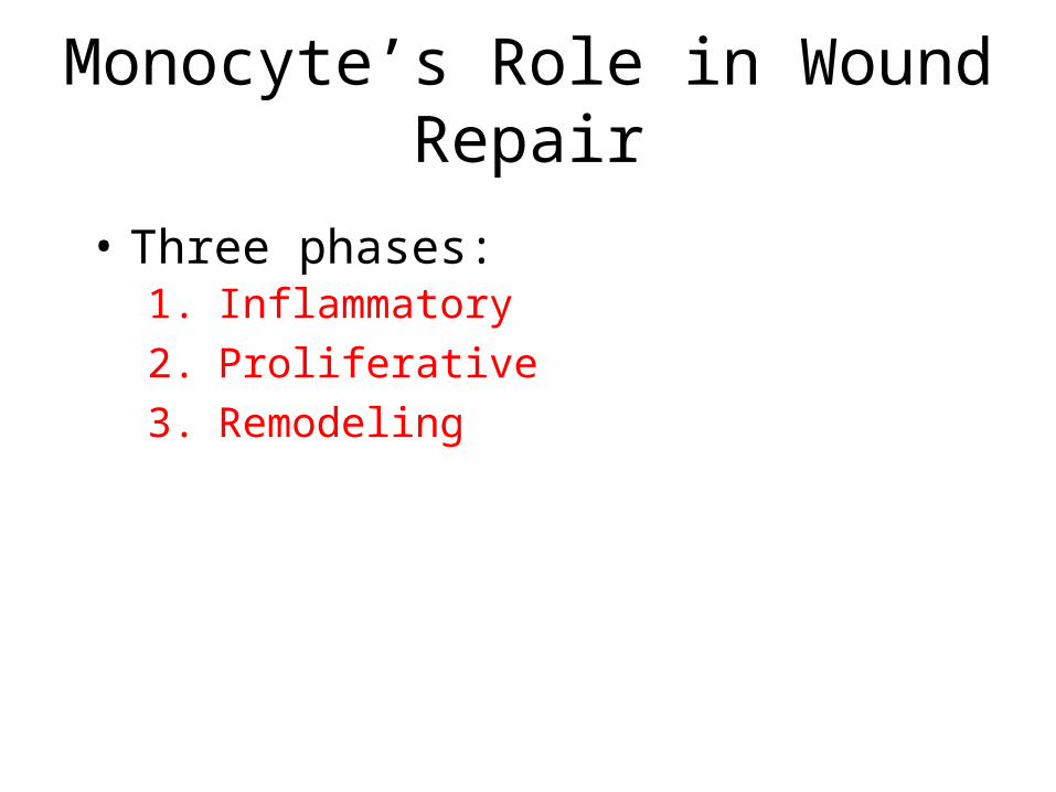
Monocyte’s Role in Wound Repair
1. Inflammatory
2. Proliferative
3. Remodeling
• Three phases:
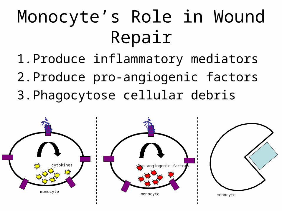
Monocyte’s Role in Wound Repair
1. Produce inflammatory mediators
2. Produce pro-angiogenic factors
3. Phagocytose cellular debris
monocyte
cytokines
monocyte
Pro-angiogenic factors
monocyte
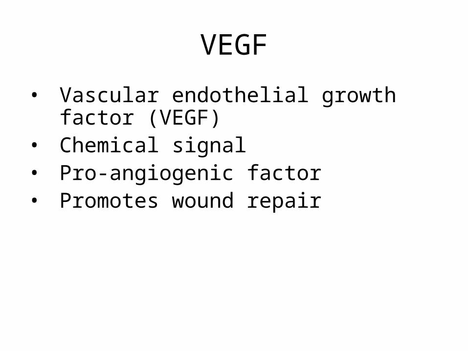
VEGF
• Vascular endothelial growth factor (VEGF)
• Chemical signal• Pro-angiogenic factor• Promotes wound repair
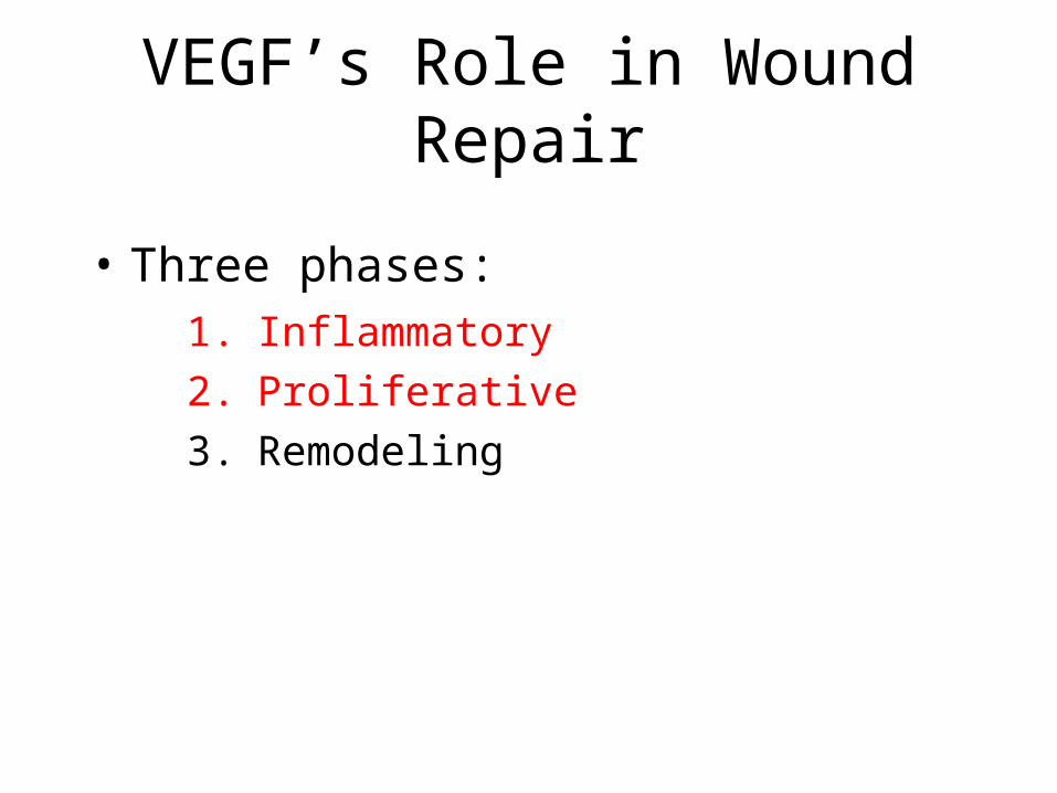
VEGF’s Role in Wound Repair
1. Inflammatory
2. Proliferative
3. Remodeling
• Three phases:
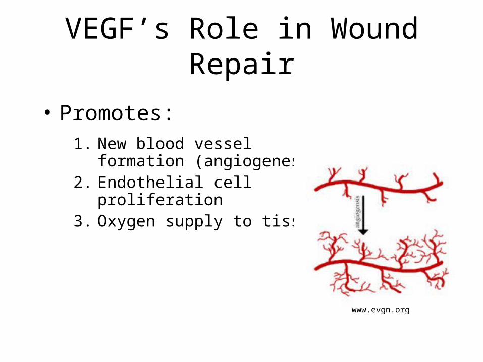
VEGF’s Role in Wound Repair
Endothelial cell
1. New blood vessel formation (angiogenesis)
2. Endothelial cell proliferation3. Oxygen supply to tissues
• Promotes:
www.evgn.org
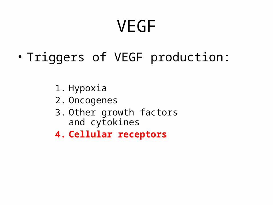
VEGF
• Triggers of VEGF production:
1. Hypoxia 2. Oncogenes3. Other growth factors and
cytokines4. Cellular receptors
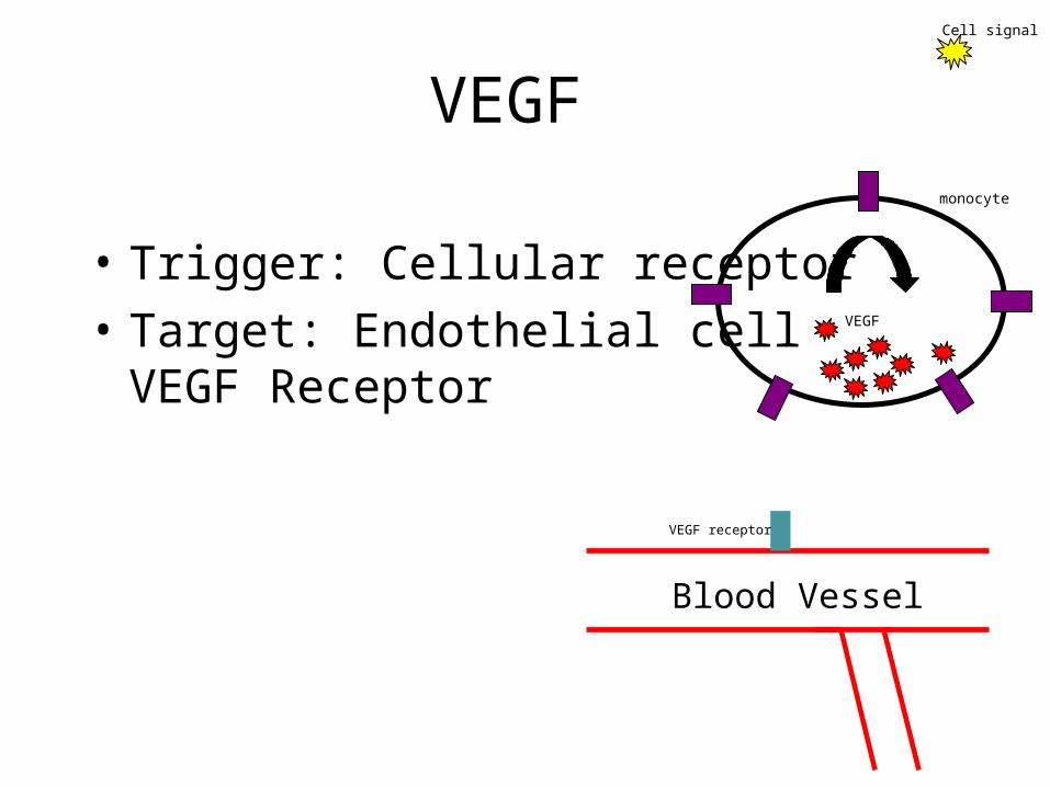
VEGF
VEGF receptor
VEGF
monocyte
Blood Vessel
Cell signal
• Trigger: Cellular receptor
• Target: Endothelial cellVEGF Receptor
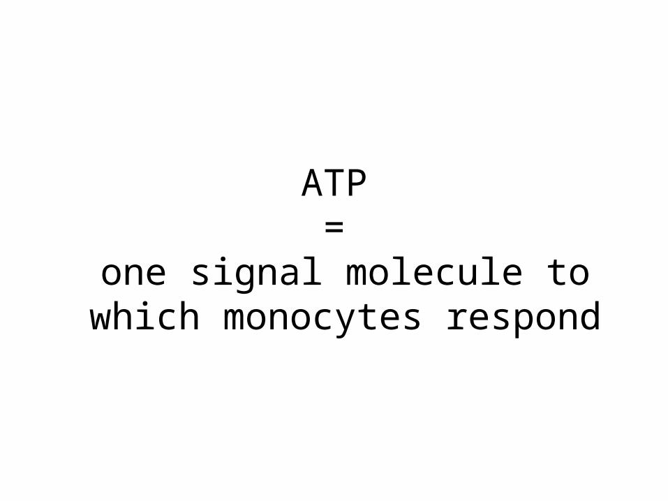
ATP =
one signal molecule to which monocytes respond
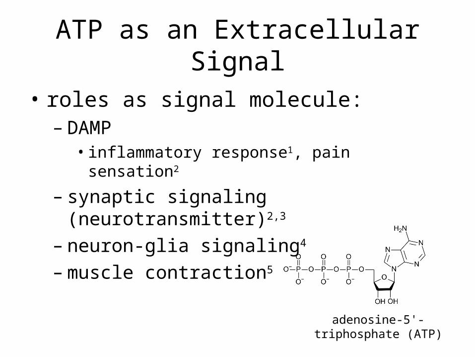
ATP as an Extracellular Signal
• roles as signal molecule:– DAMP
• inflammatory response1, pain sensation2
– synaptic signaling (neurotransmitter)2,3
– neuron-glia signaling4
– muscle contraction5
adenosine-5'-triphosphate (ATP)
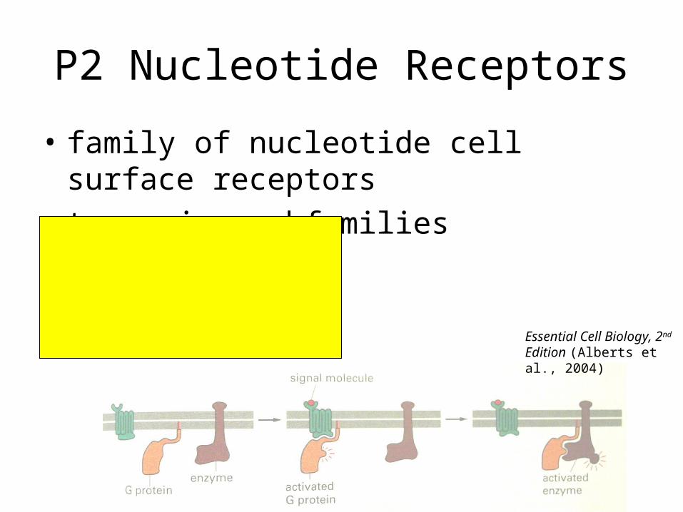
P2 Nucleotide Receptors
• family of nucleotide cell surface receptors
• two major subfamilies
P2Y Receptors
- G-protein coupled1
- bind ATP, ADP, UTP, and
UDP1Essential Cell Biology, 2nd Edition (Alberts et al., 2004)
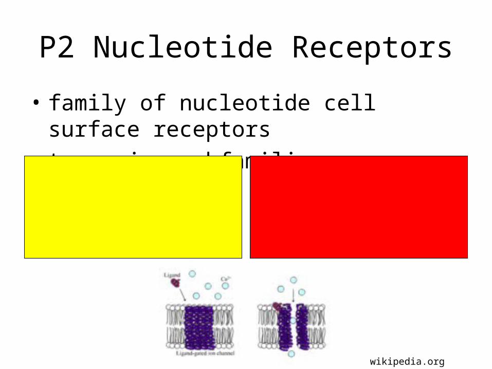
P2 Nucleotide Receptors
• family of nucleotide cell surface receptors
• two major subfamilies
P2Y Receptors
- G-protein coupled1
- bind ATP, ADP, UTP, and
UDP1
P2X Receptors
- ionotrophic (ligand-gated)1
- bind ATP1
wikipedia.org
P2X Subfamily
• ligand-gated ion channels
• cation selective2
– equal permeability to K+ and Na+
– significant permeability to Ca2+
• seven members (P2X1-7)
P2X7
• aka P2RX7
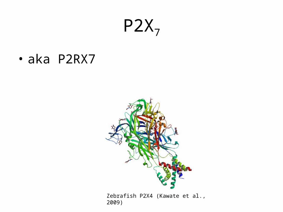
P2X7
• aka P2RX7
Zebrafish P2X4 (Kawate et al., 2009)
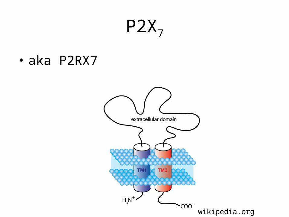
P2X7
• aka P2RX7
wikipedia.org
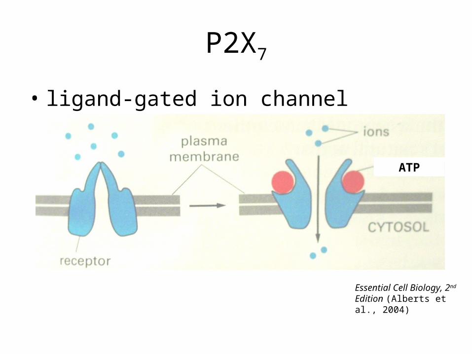
P2X7
• ligand-gated ion channel
Essential Cell Biology, 2nd Edition (Alberts et al., 2004)
ATP
P2X7
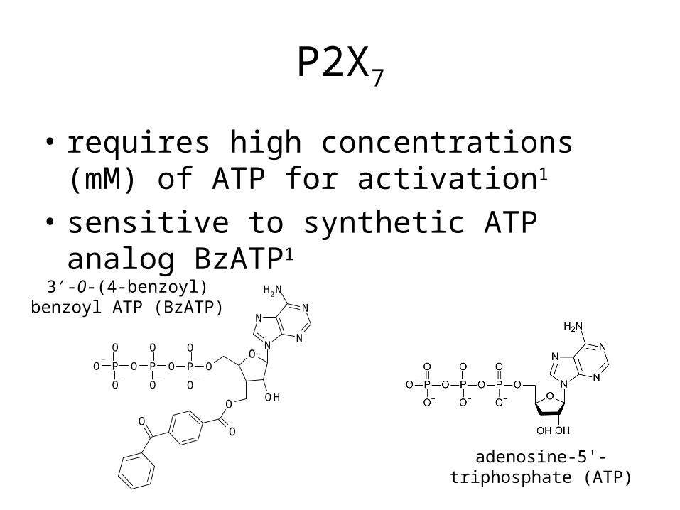
• requires high concentrations (mM) of ATP for activation1
• sensitive to synthetic ATP analog BzATP1
adenosine-5'-triphosphate (ATP)
O
N
NN
N
NH2
OPOPOPO
O O O
O O OOH
O
OO
3′-0-(4-benzoyl) benzoyl ATP (BzATP)

P2X7
• requires high concentrations (mM) of ATP for activation1
• sensitive to synthetic ATP analog BzATP1
adenosine-5'-triphosphate (ATP)
O
N
NN
N
NH2
OPOPOPO
O O O
O O OOH
O
OO
3′-0-(4-benzoyl) benzoyl ATP (BzATP)
P2X7
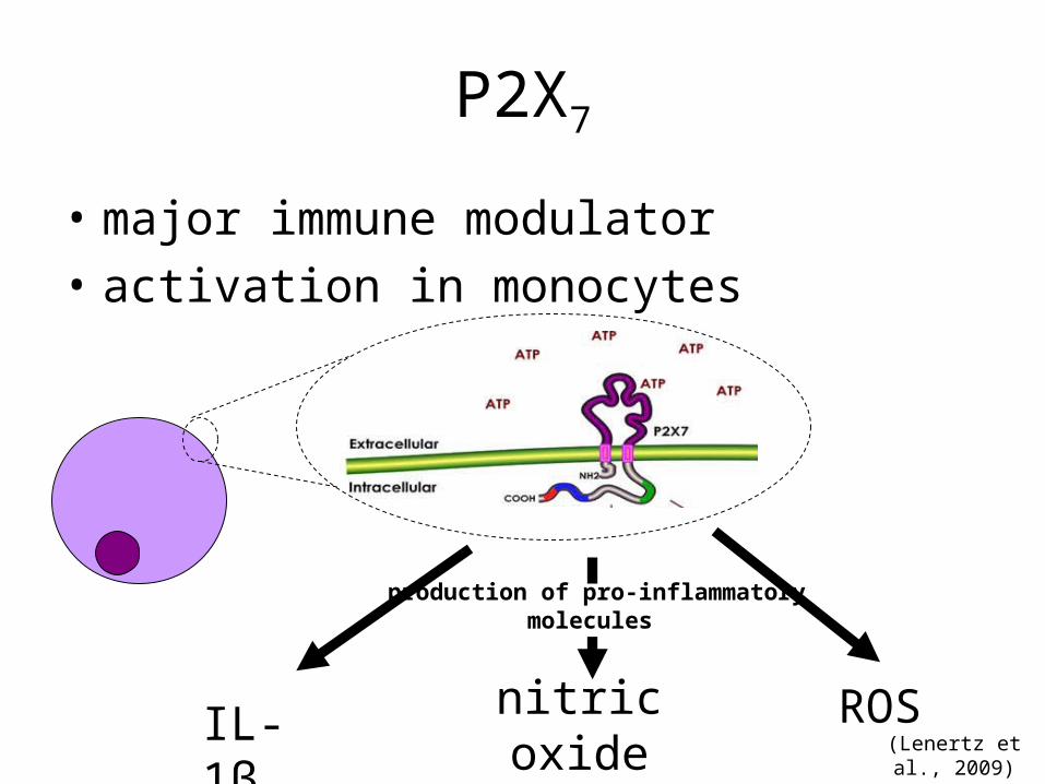
• major immune modulator
• activation in monocytes
IL-1βnitric oxide synthase
ROS
production of pro-inflammatorymolecules
(Lenertz et al., 2009)
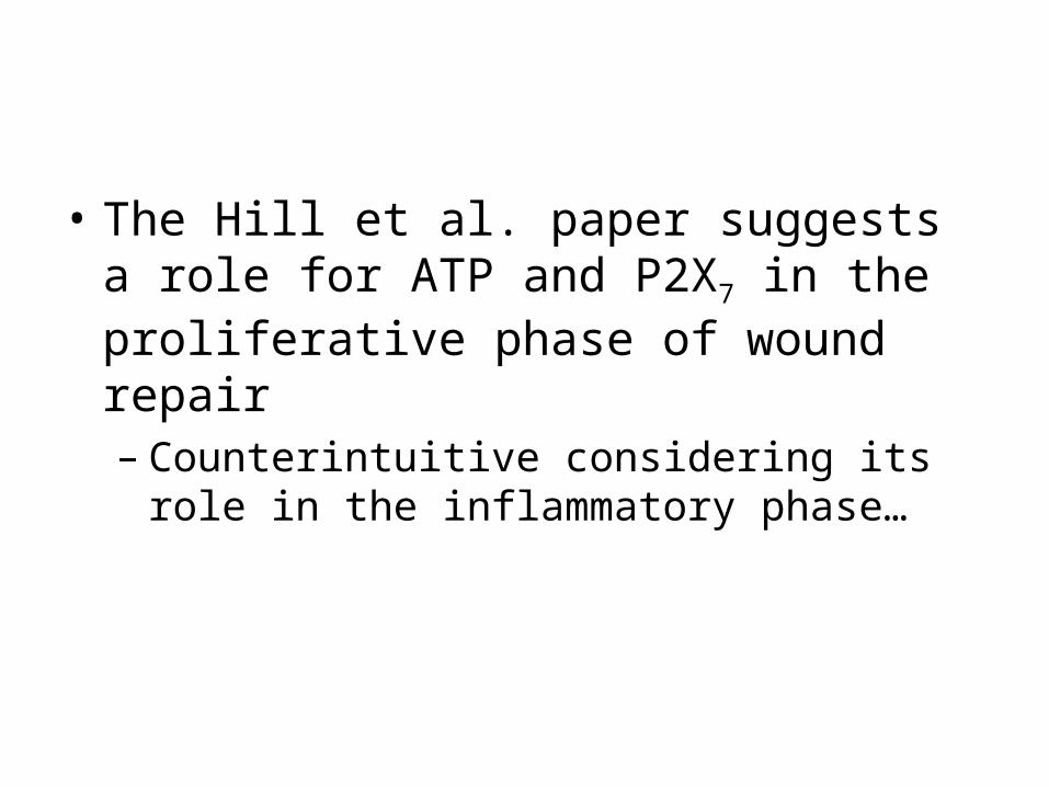
• The Hill et al. paper suggests a role for ATP and P2X7 in the proliferative phase of wound repair – Counterintuitive considering its role in the
inflammatory phase…
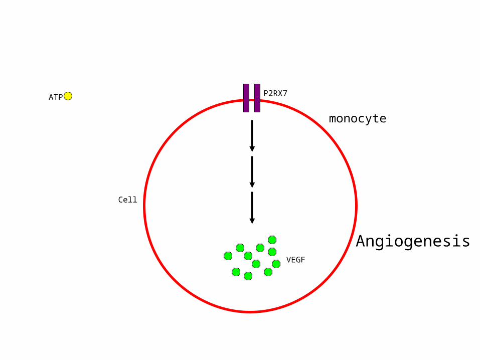
ATP
VEGF
Cell
P2RX7
Angiogenesis
monocyte
References1. Lenertz, L.Y., M.L. Gavala, L.M. Hill, and P.J. Bertics. 2009. Cell signaling
via the P2X7 nucleotide receptor: linkage to ROS production, gene transcription, and receptor trafficking. Purinergic Signal 5: 175-187.
2. Khakh, B.S. 2001. Molecular physiology of P2X receptors and ATP signalling at synapses. Nat. Rev. Neurosci. 2: 165-174.
3. Khakh, B.S., and R.A North. 2006. P2X receptors as cell-surface ATP sensors in health and disease. Nature 442: 527-532.
4. Fields, R.D., and G. Burnstock. 2006. Purinergic signalling in neuron-glia interactions. Nat. Rev. Neurosci. 7: 423-436.
5. Vassort, G. 2001. Adenosine 5′-triphosphate: a P2-purinergic agonist in the myocardium. Physiol. Rev. 81: 767-806.
6. Kirsner, R.S., and W.H. Eaglstein. 1993. The wound healing process. Dermatol. Clin. 11: 629-640.
7. Abbas, A.K., and A.H. Lichtman. 2009. Basic immunology: functions and disorders of the immune system. Saunders: Philadelphia, pp. 24-29.