cell biological effects of mechanical stimulations …...cell biological effects of mechanical...
TRANSCRIPT
Ava i l ab l e on l i ne a t www.sc i enced i r ec t . com
www.e l sev i e r . com/ loca te / sc r
Stem Cell Research (2013) 11, 951–964
Cell biological effects of mechanicalstimulations generated by focusedextracorporeal shock wave applications oncultured human bone marrow stromal cells
Frank Suhra,1,2, Yvonne Delhassea,1,3, Gerd Bungartz a,b,4,5,Annette Schmidt a,c,1,6, Kurt Pfannkuched,7,8, Wilhelm Blocha,⁎,9a Institute of Cardiovascular Research and Sport Medicine, Department of Molecular and Cellular Sport Medicine,German Sport University Cologne, Cologne, Germanyb Harvard Stem Cell Institute, Harvard University, Boston, MA, USAc Bundeswehr Institute of Pharmacology and Toxicology, Munich, Germanyd Institute of Neurophysiology, Medical School of University of Cologne, Cologne, Germany
Received 7 August 2012; received in revised form 20 March 2013; accepted 18 May 2013Available online 29 May 2013
Abstract Human bone marrow stromal cells (hBMSCs) bear tremendous clinical potential due to their immunomodulatoryproperties in transplantation settings and their contribution to tissue regeneration. In fact, they are among themost promising typesof stem-like cells for therapeutic applications and are the subject of intense research. However, the clinical use of hBMSCs has beenconfounded by limitations in their availability; they are scarce cells cumbersome to isolate and purify. Additionally, they are difficultto target to the site of injury in regeneration experiments. In order to combat these limitations, focused extracorporeal shock waves(fESW, 0.2/0.3 mJ ∗ mm−2) were applied to purified, cultured hBMSCs. fESW (0.2 mJ ∗ mm−2) stimulations were found to increasehBMSCs' growth rate (p b 0.05), proliferation (p b 0.05), migration, cell tracking andwound healing (p b 0.05, respectively), as wellas to reduce the rate of apoptosis activation (p b 0.05). The increase in hBMSC migration behavior was found to be mediated byactive remodeling of the actin cytoskeleton as indicated by increased directed stress fiber formations (p b 0.05). Furthermore,
⁎ Corresponding author at: Institute of Cardiovascular Research and Sport Medicine, Department of Molecular and Cellular Sport Medicine,German Sport University Cologne, Am Sportpark Müngersdorf 6, 50933 Cologne, Germany. Tel.: +49 221 4982 5440, +49 221 4982 5390;fax: +49 221 4982 8370.
E-mail addresses: [email protected] (F. Suhr), [email protected] (Y. Delhasse), [email protected] (G. Bungartz),[email protected] (A. Schmidt), [email protected] (K. Pfannkuche), [email protected] (W. Bloch).1 Am Sportpark Muengersdorf 6, 50933 Cologne, Germany. Tel.: +49 221 4982 5440; fax: +49 221 4982 8370.2 Conception and design of the study, assembly of data, data analysis and interpretation, manuscript writing.3 Conception and design, collection of data, data analysis.4 Present address: Am Sportpark Muengersdorf 6, 50933 Cologne, Germany. Tel.: +49 221 4982 5440; fax: +49 221 4982 8370.5 Data analysis, manuscript writing.6 Conception and design of the study.7 Robert-Koch-Straße 39, 50931 Cologne, Germany. Tel.: +49 221 478 6960; fax: +49 221 478 6965.8 Manuscript revision for critical intellectual impact.9 Financial support, manuscript revision for critical intellectual impact, final approval of the manuscript.
1873-5061/$ - see front matter © 2013 Elsevier B.V. All rights reserved.http://dx.doi.org/10.1016/j.scr.2013.05.010
952 F. Suhr et al.
hBMSCs maintain their differentiation potentials after fESW treatment, whereas 0.2 mJ ∗ mm−2 is themost effective application. Inconclusion, our results establish first-timely that hBMSCs' behavior can bemodified and optimized in response to definedmechanicalstimulation. These findings appear particularly promising as they suggest that mechanical stress preconditions hBMSCs for improvedtherapeutic performance without genetic manipulations and that mechanically preconditioned hBMSCs will be advantageous forhBMSC-based tissue regeneration. Therefore, this approach opens the door for exploiting the full potential of these cells inregenerative medicine.
© 2013 Elsevier B.V. All rights reserved.Introduction
Human bone marrow stromal cells (hBMSCs) are characterizedby their capability to differentiate into various mesenchymaltissues, including adipocytes, chondrocytes and osteocytes(Ciapetti et al., 2006; Kuznetsov et al., 1997a; Meinel et al.,2004; Pittenger et al., 1999; Tondreau et al., 2004). hBMSCsare a promising tool for the field of regenerative medicine forseveral reasons; they can be cultured ex vivo, have endoge-nous activation potential (Jiang et al., 2002; Pittenger et al.,1999), can be systemically delivered or undergo allogeneictransplantation (Krause et al., 2007; Wolf et al., 2009) andhave a high self-renewal potential (English et al., 2010; Kolf etal., 2007; Nombela-Arrieta et al., 2011; Song et al., 2006;Zaragosi et al., 2006). Their immunosuppressive propertiesmake them a promising tool to counter complications arisingfrom graft versus host rejections in transplantation settings ofcells other than hBMSCs (English et al., 2010; Prockop, 2009).It should be noted that the transplantations of BMSCs have notbeen highly successful, yet, in order to cure disease tissue, asBMSCs disappear within a short period of time after transplan-tation. However, a myriad of research is still geared towardsutilizing their differentiation potential for tissue engineer-ing approaches, particularly for cartilage and bone regen-eration (Nombela-Arrieta et al., 2011) indicating the unraveledpotential of BMSCs in tissue regeneration.
How hBMSCs exert their beneficial effects in tissue regener-ation is, however, less clear. It was first hypothesized thatBMSCs engraft directly into the degenerative tissue anddifferentiate into the respective cell type (Mahmood et al.,2003; Murphy et al., 2003). However, there is accumulatingevidence in favor of a second hypothesis postulating that BMSCspredominantly contribute to tissue regeneration by theircapability to secrete a variety of trophic factors contributingto paracrine effects in tissue repair (Gnecchi et al., 2005;Haynesworth et al., 1996; Ladage et al., 2007; Mirotsou et al.,2007) rather than tissue-specific differentiation (Prockop,2009). Regardless of how BMSCs unfold their remarkable healingproperties, it has been demonstrated in different studies thatonly a low percentage of transplanted hBMSCs reaches thetargeted disease area (Hofmann et al., 2005) and that after afew days, only a small amount (2–6%) persists in the affectedtissue (Hofmann et al., 2005; Kolf et al., 2007). In additionto the limitation of the presence of BMSCs at the site of injury,certain proportions of these cells need to display proliferativeand apoptotic, as well as migratory, behaviors in order tomediate tissue repair (Li and Jiang, 2011; Schmidt et al.,2006). Thus, the therapeutic success of using hBMSCs intissue regeneration has been limited and falls far behindtheir potential.
In order to increase the efficiency of targeting hBMSCs tothe disease area and to promote their beneficial cellular
response at those desired targets for therapeutic use, novelstrategies avoiding genetic manipulation are needed (Karpand Leng Teo, 2009; Peterson et al., 2011; Schumann et al.,2006; Yan et al., 2011) to precondition BMSCs (Ghanem etal., 2009). Extracorporeal shock waves (ESW) are transientpressure fluctuations that propagate 3-dimensionally andthat are widely applied in the context of therapeuticmechanotransduction with a high success of increased tissueregeneration (Nishida et al., 2004; van der Jagt et al., 2011,2013). Two major types of ESW are used in medicaltherapies, focused and radial ESW, whereas the formerESW type reflects high-peak pressure amplitudes and themajority of energy flux is concentrated on a small focus(Chang et al., 2012). These data show that focused ESW(fESW) types might be a suitable tool for preconditioninghBMSCs in order to improve their therapeutic potentials.
It is known that the expression of genes involved indifferentiation pathways of hBMSCs can be influenced bymechanical stimuli (Friedl et al., 2007). Different groupshave shown that ESW offer great beneficial potential in thetreatment of patients suffering from different conditions,including injured bone, cartilage and cardiac tissue (Nishidaet al., 2004; Wang et al., 2003). Unfortunately, themechanistic foundation for these improvements has notbeen addressed in these studies and the identity of cellularcontributors has not been confirmed. In this context, noknowledge exists concerning the potential effects of ESWapplication on cell biological properties of hBMSCs, such asproliferation and migration, that are crucial for successfulmedical BMSC-based therapies (Li and Jiang, 2011). Addi-tionally, it remains unclear whether hBMSCs maintain theirfull differentiation potentials after treatment with mechan-ical stimuli.
To address the unresolved, but highly significant questionof whether hBMSCs' behavior can be manipulated by ESWapplication and whether those manipulations depend onenergy and/or timing of ESW application as well as whetherhBMSCs maintain their full differentiation potentials afterESW treatment, the present study hypothesized (I) that ESWapplications induce cell biological effects on purified culturedhBMSCs that prove beneficial for tissue regeneration, (II) thatthe cell biological processes are dependent on the dose of ESWapplication and (III) that hBMSCs maintain their full differenti-ation potentials after ESW applications. The results clearlydemonstrate that ESW promote biological processes in hBMSCs,including increased proliferation, survival and migration,which are described to prove beneficial for tissue regenera-tion. Additionally, ESW do not disturb hBMSCs' differentiationpotentials. Therefore, the presented observations demonstratethat ESW stimulations of a defined nature are a dynamicapproach to manipulate hBMSC behavior in vitro in order toexploit their full regenerative capacity in vivo. Furthermore,
953Mechanical stimulations regulate hBMSC behavior
the benefits of ESW application are obtained without anymanipulation prohibitive to immediate therapeutic application.
Materials and methods
hBMSC isolation and culture
Adult hBMSCs were isolated from the bone marrow of humanfemoral heads from patients receiving hip joint replace-ments. The average age of the patients was 73 ± 7.4 years.Only material that tested negative for HIV and hepatitis wasaccepted. After abrading the marrow of the femoral headand filtering the marrow with PBS up to a volume of 50 mL, adensity gradient centrifugation was performed. The fractioncontaining hBMSCswas purified as described recently (Steingenet al., 2008). Cells were cultured in alpha modifications ofEagle's Medium (alpha-MEM, PAA with Glutamine, withoutNucleosides, Cölbe, Germany) supplemented with 20% FCS(Biowest, Nuaille, France). The medium was changed the firsttime after two days of culture. The cells were used up topassage 3 and plated at a density of 2000 cells ∗ cm−2 forculturing purposes (Schmidt et al., 2006). hBMSCs werepassaged by Accutase™ with an optimal confluence of approx-imately 80%. The studywas authorized by the ethics commissionof the Medical School of the University Cologne and was in linewith the declaration of Helsinki.
hBMSCs characterization
Briefly, hBMSCs were characterized using FACS analysis asdescribed by Steingen (2008). Freshly isolated hBMScs wereplated with a density of 1000 cells ∗cm−2 (adipogenic andosteogenic differentiation) or 2000 cells ∗ cm−2 (chondrogenicdifferentiation), respectively, and were characterized using acolony-forming unit-fibroblast (CFU-F) as described earlier(Friedenstein et al., 1966, 1976). Thereby, it should be notedthat populations of human marrow CFU-Fs are heterogeneouswith respect to their differentiation capacity (Kuznetsov et al.,1997b; Sacchetti et al., 2007). The identity of hBMSCs wasconfirmed by established methods in our lab culturingthe hBMSCs under conditions favorable for adipogenic [basicmedium: alpha-MEM; 2 mM L-glutamine (Sigma-Aldrich,Munich, Germany), 60 μM indomethacin (Sigma-Aldrich,Munich, Germany), 1 μM dexamethasone (Sigma-Aldrich,Munich, Germany), 0.5% antibiotics-antimycotics (Invitrogen,Karlsruhe, Germany), 5 μg ∗ mL−1 insulin (Sigma-Aldrich,Munich, Germany), 15% FCS (Biowest, Nuaille, France)], chon-drogenic [basic medium: D-MEM; 2 mM L-glutamine (Sigma-Aldrich, Munich, Germany), 50 μMascorbic acid (Sigma-Aldrich,Munich, Germany), 10 ng ∗ mL−1 transforming growth factorbeta (Sanver Tech, USA), 0.5 μg ∗ mL−1 insulin (Sigma-Aldrich,Munich, Germany), 0.5% antibiotics–antimycotics (Invitrogen,Karlsruhe, Germany), 1% FCS (Biowest, Nuaille, France)] orosteogenic [basic medium: alpha-MEM; 2 mM L-glutamine(Sigma-Aldrich, Munich, Germany), 60 μM ascorbic acid(Sigma-Aldrich, Munich, Germany), 10 mM β-glycerophosphate(Sigma-Aldrich, Munich, Germany), 0.1 μM dexamethasone(Sigma-Aldrich, Munich, Germany), 0.5% antibiotics–antimycotics (Invitrogen, Karlsruhe, Germany), 15% FCS(Biowest, Nuaille, France)] differentiation (Baumgartner etal., 2010; Steingen et al., 2008).
Adipogenic differentiation of hBMSCs was proven by OilRed staining. Osteogenic differentiation of hBMSCs wasproven by von Kossa staining. Chondrogenic differentiationwas either proven by morphological Alcian blue (Okamoto etal., 2002) or Toluidine blue staining or by RT-PCT (seeSection 2.3). Especially for the proof of maintenance of fulldifferentiation capacities of hBMSCs after fESW treatment(see Section 2.4) we used morphological methods ratherthan RT-PCR to evaluate both the differentiation indicatorsdescribed above and the morphology of hBMSCs. hBMSCmorphology cannot be obtained by RT-PCR; however, themorphology represents a highly relevant marker in order toassess the influence of mechanical stimuli on the integrity ofmechanically treated hBMSCs.
Proof of chondrogenic differentiation by RT-PCR
RNA of fESW-treated and -untreated hMSCs was extractedwith the Peq-Gold TriFast reagent (PeqLab, Erlangen,Germany) according to the manufacturer's protocol. RNAwas incubated with DNase (Invitrogen, Karlsruhe, Germany) for15 min at room temperature to avoid DNA contamination. 1 μgRNA was used for RT-PCR. RNA was reverse-transcribed intocDNA using OmniScript reverse transcriptase kit (Qiagen, CA,USA) according to themanufacturer's protocol. For PCR, 1 μg ofcDNA template was used, respectively. The following primerswere used to prove for chondrogenic (collagen type II, GenBanknumber: NM_001844.4; forward: GAA CAT CAC CTA CCA CTGCAA G, reverse: GCA GAG TCC TAG AGT GAC TGA G) (Steingenet al., 2008), β-actin served as internal control, (GenBanknumber: NM_001101.3; forward: ACC TTC AAC ACC CCA GCCATG TAC G, reverse: CTG ATC CAC ATC TGC TGG AAG GTG G).Resulting PCR productswere analyzed using gel electrophoresis.
Application of focused and radial extracorporealshock waves on hBMSCs
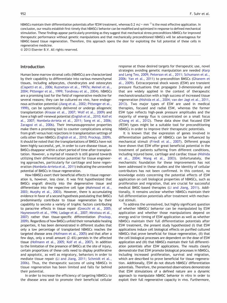
Cultured hBMSCs were subjected to focused (fESW) applica-tions using the extracorporeal shock wave systemDuolith®SD1(Storz Medical AG, Trägerwilen, Switzerland) in 100 mmdishes (ibidi, Martinsried, Germany). fESW modes wereapplied to hBMSCs on a heat-stable foil and on an absorbingsupport surface (freshly prepared pork skin). In order to mimicthe in vivo situation, culture dishes were filled completelywith media and covered with freshly prepared pork skin(Fig. 1). For optimal coupling with the Duolith®SD1 system, anultrasound gel (Parker Laboratories, Fairfield, NJ, USA) wasplaced on top of the pork skin. fESW applications wereconducted as follows: continuous pulse, 1000 impulses, 4 Hz.Energy densities were as follows: 0 mJ ∗ mm−2 (control),0.2 mJ ∗ mm−2, and 0.3 mJ ∗ mm−2. To evaluate the growthrate of hBMSCs before and after fESW applications, the cellswere splitted after either control or after 0.2 mJ ∗ mm−2
application with 2000 cells ∗ cm−2. After 24 h the cell numberwas again quantified. Every biological experiment wasperformed in quadruplicate and in a single session.
fESW treatments were also applied on cultured hBMSCs inorder to test the influence of fESW on hBMSC differentiationpotentials. Therefore, hBMSCs were treated with fESW asdescribed above in this section and subsequently differen-tiated as described in Section 2.1.
Adherent cells
Cell-specific medium
Pork skin
ESW applicator
Figure 1 Cartoon of the experimental setup.
954 F. Suhr et al.
Immunocytochemistry
Prior immunocytochemistry, hBMSCs were fixed with 4%paraformaldehyde (PFA) for 1 h at room temperature beforehistological analysis (Steingen et al., 2008). hBMSC prolifer-ation was detected by incubation with rabbit polyclonal antiKi67 (Scholzen and Gerdes, 2000) antibody (dilution: 1:200,Abcam, Cambridge, UK) overnight at 4 °C. hBMSC apoptosiswas detected by incubation with rabbit polyclonal anticaspase-3 (Patel et al., 1996) antibody (dilution: 1:500, BD,Heidelberg, Germany) overnight at 4 °C. Respective sec-ondary antibodies (Dako Deutschland GmbH, Hamburg,Germany) were used with a dilution of 1:400. hBMSCs wereexamined with a Zeiss Axiovert 200 M light microscopecoupled to a Axio Cam MR Video Camera (Carl Zeiss, Jena,Germany). Digitally captured images (200× magnification)were analyzed by assessment of optical densitometry withthe software ImageJ® (National Institutes of Health, USA).Negative control hBMSCs were processed as were experi-mental hMSCs; however, primary antibody incubations weresubstituted by incubation with 0.8% TBS.
Analysis of the cytoskeletal morphology
To visualize directed F-actin stress fibers and disorganizedactin fibers of the cytoskeleton, fESW-treated and -untreatedhBMSCs were washed in 0.05 M TBS followed by perme-abilization with 0.25% Triton X-100, washed again with TBSand subsequently dyedwith phalloidin/Alexa Fluor488 (dilution1:40 in TBS, Molecular Probes, Invitrogen, Leiden, Netherland)for 20 min (Pellegrin and Mellor, 2007; Spallarossa et al., 2010)and covered with 4,6-diamidino-2-phenyl-indol (DAPI) con-taining mounting medium (Vectashield, Vector Laborato-ries). For analysis of directed F-actin stress fibers anddisorganized actin fibers, the total number of DAPI-positivehBMSCs was quantified using a fluorescence microscope(Axiovert 200 M, Carl Zeiss, Jena, Germany). DirectedF-actin stress fibers as well as disorganized actin fiberswere related to the total number of DAPI-positive hMSCs.Additionally, the percentage of disorganized actin fibers ofhBMSCs was documented in fESW-treated -untreated
hBMSCs, indicating structural rearrangements of the cyto-skeleton (Jaalouk and Lammerding, 2009; Spallarossa et al.,2010) in response to fESW applications.
Boyden chamber assay
The migration assay was performed in a modified Boydenchamber assay as described previously (Schmidt et al.,2006). In brief, after hBMSCs were grown to a confluentmonolayer and the dishes were treated with singularapplications of fESW. Untreated dishes served as controls.After fESW treatments, hBMSCs were detached by Accutaseand counted. 5 ∗ 104 cells were seeded on top of the specificmigration filter (8 μm pores, Falcon® HTS Fluoro Blok™,Becton Dickinson Labware Europe, Le Pont De Claix, France)and were incubated for 8 h at 37 °C, 5% CO2, and 95%humidity in 20% FCS-containing alpha-MEM in a 24-well plate(Tissue Culture Plate, Becton Dickinson Labware Europe, LePont De Claix, France). Subsequently, hBMSCs were fixedusing 4% paraformaldehyde (PFA). The cells were coveredusing mounting medium containing DAPI (Vectashield,Vector Laboratories). The number of migrated hBMSCs wascounted and documented using a fluorescent microscope(Axiovert 200 M, Carl Zeiss Microscopy, Jena, Germany).Only completely migrated cells were taken into account forfurther analysis.
Wound healing and scratch assay of hMSCs
In vitro wound healing assays (scratch assays) were performedas described previously (Faber-Elman et al., 1996). After fESWapplication, the treated hBMSCs were incubated for 24 h inthe Axiovert 200 M light microscope (Carl Zeiss, Jena,Germany) under cell culture conditions (37 °C, 5% CO2, and95% humidity) in 20% FCS-containing alpha-MEM. Time lapsevideo analysis was used to document the wound healingprocess; every 10 min pictures were taken with a 5× objective(A-Plan 5×/0.12, Carl Zeiss, Jena, Germany). The wound areawas calculated with the Axiovision software (Carl Zeiss, Jena,Germany).
Cell tracking assay of hMSCs
To monitor cell movements, cell tracking analysis of hBMSCsin response to fESW applications was performed as described(Schmidt et al., 2006). In brief, hBMSCs were plated at adensity of 2000 cells ∗ cm−2 on cell culture dishes (Ibidi,Martinsried, Germany). ESW-treated and untreated controlhBMSCs were monitored by time lapse video for 24 h untilhBMSCs reached a confluence of about 40%. During thattime, the cells were incubated for 24 h in the Axiovert 200 Mlight microscope (Carl Zeiss, Jena, Germany) under cellculture conditions (37 °C, 5% CO2, and 95% humidity) in 20%FCS-containing alpha-MEM. Every 10 min, pictures weretaken with a 5× objective (A-Plan 5×/0.12, Carl Zeiss,Jena, Germany); data were exported to Metamorph micros-copy analysis software (Molecular Devices, Sunnyvale, CA,USA) for further analysis. The analyzed parameter includedthe velocity of hBMSCs, their distance [pixel] per 10 min andtheir total distance to the starting point after 24 h.
955Mechanical stimulations regulate hBMSC behavior
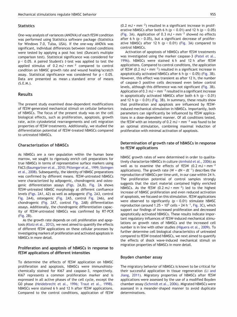
Statistics
One-way analysis of variances (ANOVA) of each fESW conditionwas performed using Statistica software package (Statisticafor Windows 7.0, Tulsa, USA). If the one-way ANOVA wassignificant, individual differences between tested conditionswere tested by applying a post hoc test (Duncan's multiplecomparison test). Statistical significance was considered forp b 0.05. A paired Student's t-test was applied to test theapplied stimulus of 0.2 mJ ∗ mm−2 compared to controlcondition on hBMSC growth rate and wound healing/scratchassay. Statistical significance was considered for p b 0.05.Data are presented as mean ± standard error of means(S.E.M.).
Results
The present study examined dose-dependent modificationsof fESW-generated mechanical stimuli on cellular behaviorsof hBMSCs. The focus of the present study was on the cellbiological effects, such as proliferation, apoptosis, growthrate, actin cytoskeletal rearrangements and cell migrationproperties of fESW treatments. Additionally, we studied thedifferentiation potential of fESW-treated hBMSCs comparedto untreated hBMSCs.
Characterization of hBMSCs
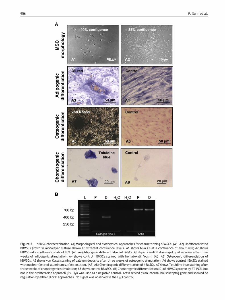
As hBMSCs are a rare population within the human bonemarrow, we sought to rigorously enrich cell preparations fortrue hBMSCs in terms of representative surface markers usingFACS (Baumgartner et al., 2010; Pittenger et al., 1999; Steingenet al., 2008). Subsequently, the identity of hBMSC preparationswas confirmed by different means. fESW-untreated hBMSCswere characterized by adipogenic, chondrogenic and osteo-genic differentiation assays (Figs. 2A,B). Fig. 2A showsfESW-untreated hBMSC morphology at different confluencelevels (Figs. 2A1, A2) as well as adipogenic (Fig. 2A3, controlFig. 2A4), osteogenic (Fig. 2A5, control Fig. 2A6), andchondrogenic (Fig. 2A7, control Fig. 2A8) differentiationassays. Additionally, the chondrogenic differentiation capac-ity of fESW-untreated hBMSCs was confirmed by RT-PCR(Fig. 2B).
As the growth rate depends on cell proliferation and apop-tosis (Klotz et al., 2012), we next aimed to quantify the effectsof different fESW applications on these cellular processes byinvestigatingmarkers of proliferation and activated apoptosis inhBMSCs in more detail.
Proliferation and apoptosis of hBMSCs in response tofESW applications of different intensities
To determine the effects of fESW application on hBMSCproliferation and apoptosis, hBMSCs were immunohisto-chemically stained for Ki67 and caspase-3, respectively.Ki67 represents a common proliferation marker and isexpressed in all active phases of the cell cycle, except theG0 phase (Heidebrecht et al., 1996; Traut et al., 1998).hBMSCs were stained 6 h and 12 h after fESW applications.Compared to the control conditions, application of fESW
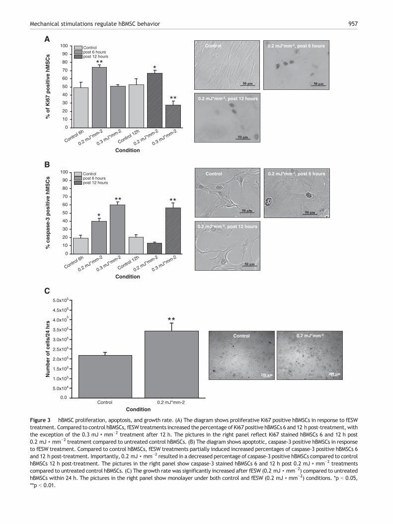
(0.2 mJ ∗ mm−2) resulted in a significant increase in prolif-erative hBMSCs after both 6 h (p b 0.01) and 12 h (p b 0.05)(Fig. 3A). Application of 0.3 mJ ∗ mm−2 showed no effectsafter 6 h (p N 0.05), but a significant decrease of prolifer-ative hBMSCs after 12 h (p b 0.01) (Fig. 3A) compared tocontrol hBMSCs.
Activation of apoptosis of hBMSCs after fESW treatmentswas investigated using the marker caspase-3 (Patel et al.,1996). hBMSCs were stained 6 h and 12 h after fESWapplications. Compared to control conditions, the applicationof fESW (0.2 mJ ∗ mm−2) resulted in a significant increase inapoptotically activated hBMSCs after 6 h (p b 0.05) (Fig. 3B).However, this effect was transient as after 12 h, the numberof caspase-3 positive cells decreased even below controllevels, although this difference was not significant (Fig. 3B).Application of 0.3 mJ ∗ mm−2 resulted in a significant increasein apoptotically activated hBMSCs after both 6 h (p b 0.01)and 12 h (p b 0.01) (Fig. 3B). In summary, these results showthat proliferation and apoptosis are influenced by fESW-induced mechanical stimulation in hBMSCs. Importantly, bothparameters can significantly be influenced by fESW applica-tions in a dose-dependent manner. Of all conditions tested,the fESW with an intensity of 0.2 mJ ∗ mm−2 was found to bean optimal stimulation, combining maximal induction ofproliferation with minimal activation of apoptosis.
Determination of growth rate of hBMSCs in responseto fESW applications
hBMSC growth rates of were determined in order to qualita-tively characterize hBMSCs in culture (Arnhold et al., 2006) aswell as to examine the effects of fESW (0.2 mJ ∗ mm−2
applications). The growth rate (W = dN ∗ dt−1) describes thereproduction of hBMSCs per time unit, in our case within 24 h.The proliferation potential of control samples stronglysuggests that the start material contained highly enrichedhBMSCs. As the fESW (0.2 mJ ∗ mm−2) led to the highestincrease of hBMSC proliferation and even reduced activationof apoptosis, we focused on this stimulation. fESW applicationswere observed to significantly (p b 0.01) stimulate hBMSCreproduction (around 1.25 ∗ 105 cells ∗ 24 h−1, Fig. 3C), whichsupport our findings of increased proliferation and decreasedapoptotically activated hBMSCs. These results indicate impor-tant regulatory influences of fESW-induced mechanical stimu-lations on growth rates of hBMSCs and the increased cellnumber is in line with other studies (Higuera et al., 2009). Tofurther determine cell biological characteristics of untreatedcompared to fESW-treated hBMSCs, we next aimed to quantifythe effects of shock wave-induced mechanical stimuli onmigration properties of hBMSCs in more detail.
Boyden chamber assay
The migratory behavior of hBMSCs is known to be critical fortheir successful application in tissue regeneration (Li andJiang, 2011). Migratory properties of hBMSCs after fESWapplications were assessed by the use of a modified Boydenchamber assay (Schmidt et al., 2006). Migrated hBMSCs wereassessed in a meander-shaped manner to avoid duplicatedeterminations.
A
A3 A450 µm 50 µm
A1 A2M
SC
m
orp
ho
log
yA
dip
og
enic
dif
fere
nti
atio
nO
steo
gen
icd
iffe
ren
tiat
ion
L P D H2O H2O P D
Collagen type II Actin
700 bp
400 bp
250 bp
≈40% confluence ≈ 85% confluence
Oil red Control
A6 50 µm
Control
50 µm
von Kossa
A5
B
20 µmA7
Toluidineblue
20 µmA8
Control
Ch
on
dro
gen
icd
iffe
ren
tiat
ion
Figure 2 hBMSC characterization. (A) Morphological and biochemical approaches for characterizing hBMSCs. (A1, A2) UndifferentiatedhBMSCs grown in monolayer culture shown at different confluence levels. A1 shows hBMSCs at a confluence of about 40%; A2 showshBMSCs at a confluence of about 85%. (A3, A4) Adipogenic differentiation of hMSCs. A3 depicts RedOil staining of lipid vacuoles after threeweeks of adipogenic stimulation; A4 shows control hBMSCs stained with hematoxylin/eosin. (A5, A6) Osteogenic differentiation ofhBMSCs. A5 shows von Kossa staining of calcium deposits after three weeks of osteogenic stimulation; A6 shows control hBMSCs stainedwith nuclear fast red-aluminum sulfate solution. (A7, A8) Chondrogenic differentiation of hBMSCs. A7 shows Toluidine blue staining afterthreeweeks of chondrogenic stimulation; A8 shows control hBMSCs. (B) Chondrogenic differentiation (D) of hBMSCs proven by RT-PCR, butnot in the proliferation approach (P). H2O was used as a negative control. Actin served as an internal housekeeping gene and showed noregulation by either D or P approaches. No signal was observed in the H2O control.
956 F. Suhr et al.
Control 6h
0.2 mJ*mm-2
0.3 mJ*mm-2
Control 12h
0.2 mJ*mm-2
0.3 mJ*mm-2
0
10
20
30
40
50
60
70
80
90
100 Control post 6 hours post 12 hours
% c
asp
ase-
3 p
osi
tive
hM
SC
s
Condition
Control 0.2 mJ*mm-20.0
5.0x10⁴
1.0x10⁵
1.5x10⁵
2.0x10⁵
2.5x10⁵
3.0x10⁵
3.5x10⁵
4.0x10⁵
4.5x10⁵
5.0x10⁵
Nu
mb
er o
f ce
lls/2
4 h
rs
Condition
A
B
Control 6h
0.2 mJ*mm-2
0.3 mJ*mm-2
Control 12h
0.2 mJ*mm-2
0.3 mJ*mm-2
0
10
20
30
40
50
60
70
80
90
100 Control post 6 hours post 12 hours
% o
f K
i67
po
siti
ve h
MS
Cs
Condition
**
**
*
*
****
Control 0.2 mJ*mm-2, post 6 hours
0.2 mJ*mm-2, post 12 hours
Control 0.2 mJ*mm-2, post 6 hours
0.2 mJ*mm-2, post 12 hours
C
**Control 0.2 mJ*mm-2
Figure 3 hBMSC proliferation, apoptosis, and growth rate. (A) The diagram shows proliferative Ki67 positive hBMSCs in response to fESWtreatment. Compared to control hBMSCs, fESW treatments increased the percentage of Ki67 positive hBMSCs 6 and 12 h post-treatment,withthe exception of the 0.3 mJ ∗ mm−2 treatment after 12 h. The pictures in the right panel reflect Ki67 stained hBMSCs 6 and 12 h post0.2 mJ ∗ mm−2 treatment compared to untreated control hBMSCs. (B) The diagram shows apoptotic, caspase-3 positive hBMSCs in responseto fESW treatment. Compared to control hBMSCs, fESW treatments partially induced increased percentages of caspase-3 positive hBMSCs 6and 12 h post-treatment. Importantly, 0.2 mJ ∗ mm−2 resulted in a decreased percentage of caspase-3 positive hBMSCs compared to controlhBMSCs 12 h post-treatment. The pictures in the right panel show caspase-3 stained hBMSCs 6 and 12 h post 0.2 mJ ∗ mm−2 treatmentscompared to untreated control hBMSCs. (C) The growth rate was significantly increased after fESW (0.2 mJ ∗ mm−2) compared to untreatedhBMSCs within 24 h. The pictures in the right panel show monolayer under both control and fESW (0.2 mJ ∗ mm−2) conditions. *p b 0.05,**p b 0.01.
957Mechanical stimulations regulate hBMSC behavior
958 F. Suhr et al.
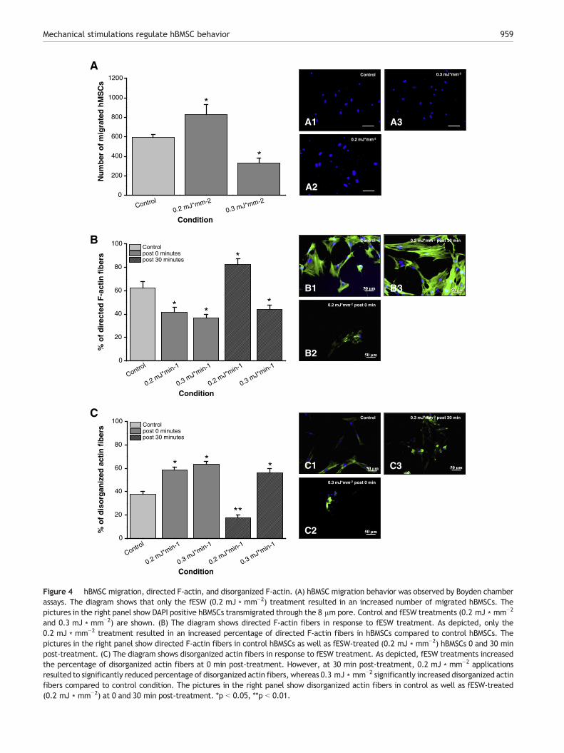
fESW applications (0.2 mJ ∗ mm−2) resulted in signifi-cantly increased numbers (p b 0.05, Fig. 4A) of completelymigrated hBMSCs, whereas application of 0.3 mJ ∗ mm−2
inhibited the migration of hBMSCs significantly (p b 0.05,Fig. 4A). These results demonstrate the existence of aphysiological threshold above which mechanical stimulationsseem to be counterproductive for the induction of hBMSCmigration. Therefore, fESW-induced mechanical stimula-tions induce dose-dependent biological effects on hBMSCs.
Actin cytoskeleton arrangement of hBMSCs
In hBMSCs F-actin is well-organized in linear stress fibers(Fig. 4B). Cytoskeletal architecture, such as directed F-actinstress fibers, is known to be crucial for cell migration(Pellegrin and Mellor, 2007). Therefore, it was testedwhether mechanical stimuli can modify the actin cytoskele-ton. Fluorescence-labeled phalloidin (Alexa Fluor488) wasused to visualize the F-actin cytoskeleton of hBMSCs (PellegrinandMellor, 2007; Spallarossa et al., 2010). hBMSC cytoskeletalorganization was determined 0 min and 30 min after fESWapplications.
fESW (0.2 mJ ∗ mm−2 and 0.3 mJ ∗ mm−2) treatmentsresulted in an initially significantly (p b 0.05) decreasedportions of directed F-actin fibers. However, 30 min after0.2 mJ ∗ mm−2 application the cells recovered and presentedwith a massive assembly of directed F-actin fibers (p b 0.05)(Fig. 4B), while the cells were only able to reassemble veryfew fibers within 30 min post 0.3 mJ ∗ mm−2 treatment(Fig. 4B).
These results suggest that the stimulation with fESW(0.2 mJ ∗ mm−2) might prove optimal to induce migration ofhBMSCs. We tested this assumption directly using differentassays.
Disorganized and partly destroyed actin fibers indicaterearrangements of the cellular cytoskeleton (Morita et al.,1994; Spallarossa et al., 2010). Therefore, the portion ofdisorganized actin fibers was also quantified in response to fESWapplications 0 min and 30 min after treatment. Compared tothe control condition, fESW treatment (0.2 mJ ∗ mm−2 and0.3 mJ ∗ mm−2) resulted in a significantly (p b 0.05 for each)increased portion of disorganized actin fibers in hBMSCs at0 min post-treatment (Fig. 4C). A similar picture was observedat 30 min (p b 0.05), but only after 0.3 mJ ∗ mm−2 fESWapplication (Fig. 4C). In contrast, but in line with the directedF-actin fibers (Fig. 4B), 0.2 mJ ∗ mm−2 significantly (p b 0.01)reduced disorganized actin fibers at 30 min post-treatment(Fig. 4C). These results indicate important divergent influencesof fESW application in a dose-dependent manner and indicate0.2 mJ ∗ mm−2 as most beneficial for directed F-actinassembly.
Cell tracking assay
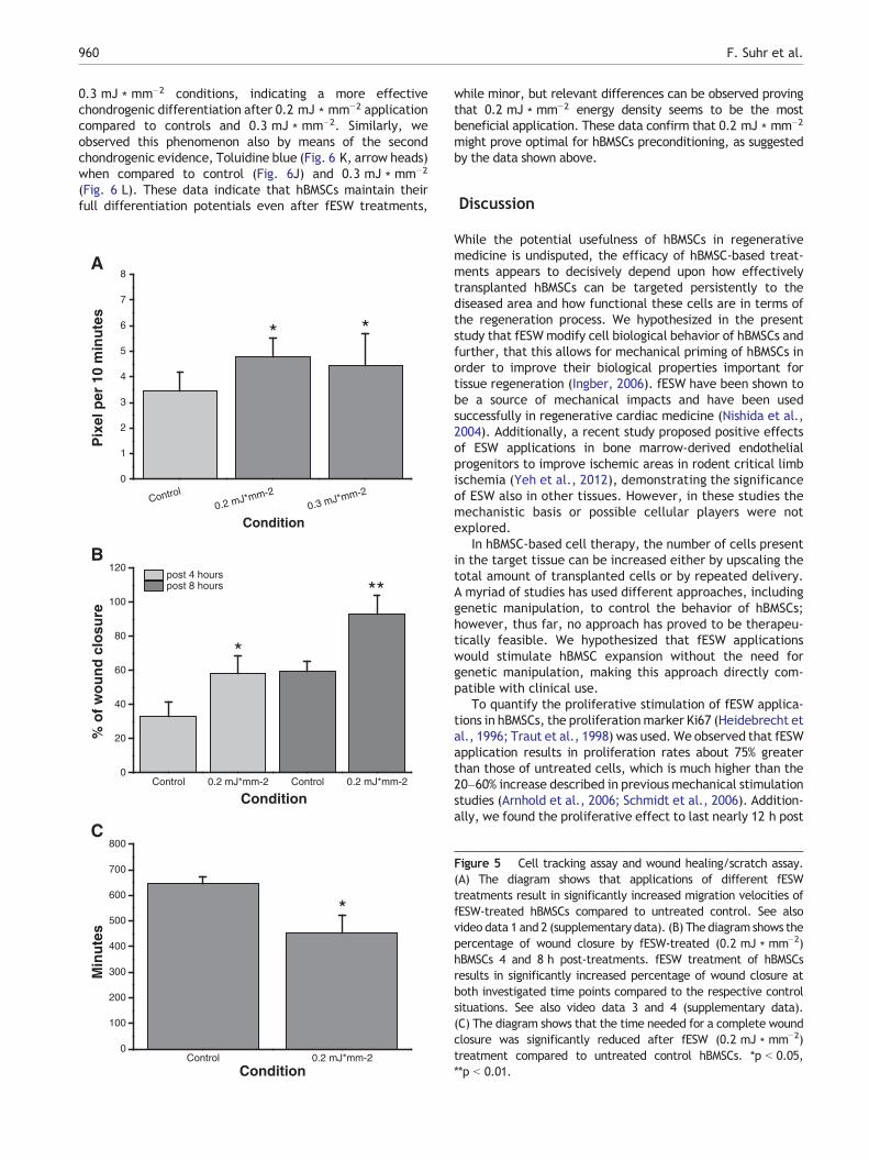
To assess hBMSC movements, cell-tracking assays wereperformed. fESW-treated and -untreated hBMSCs werephotographed every 10 min over a time course of 24 h todocument hBMSC movement in response to fESW applica-tions. The treatment of hBMSCs with any fESW conditionresulted in comparable significant increases in movementvelocities of hBMSCs compared to control hBMSCs (p b 0.05,
Fig. 5, see also video data 1 and 2 in the supplementarydata). These results lead to the assumption that mechanicalstimulations exerted by fESW of divergent intensities posi-tively influence migration and distance velocities of hBMSCs.This observation might be of great impact for medicaltherapies as hBMSCs often show low infiltrations into diseasedtissues (Hofmann et al., 2005).
Wound healing assay
Due to the results obtained from the Boyden chamber assays,which indicated a positive influence of fESW (0.2 mJ ∗ mm−2)applications on hBMSC migration, wound-healing assays wereconducted with hBMSCs using the same treatment. Theseassays test whether the increased cell movements aredirected or whether the mechanical stimulation causesrandom movements leaving the cells incapable of directedmigration. In this system only directed migration results in amore efficient wound closure. In comparison to the controlconditions, fESW application of 0.2 mJ ∗ mm−2 resulted in anincreased portion of wound closure at both 4 h (p b 0.05) and8 h (p b 0.01) post fESW treatment (Fig. 5B, see also videodata 3 and 4 in the supplementary data). Consequently, thetime required to complete wound closure was significantlydecreased (p b 0.05, Fig. 5C). These observations clearlyillustrate that ESW-induced mechanical stimulations have apositive influence on the movement and velocity of directedhBMSC migration, demonstrating the potential of mechanicalstimulations for clinical hMSC applications.
hBMSC differentiation potential into adipocytes,chondrocytes, and osteocytes after fESW treatment
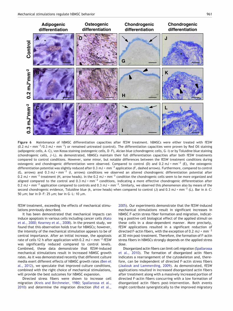
After investigating cell biological behaviors of hBMSCs inresponse to fESW treatment we sought to study whether thedifferentiation potential of mechanically treated hBMSCswere still viable and whether the differentiation potentialcorrelated in a certain manner to the cell biological resultswe obtained from our assays. Therefore, hBMSCs weresubjected to both 0.2 mJ ∗ mm−2 and 0.3 mJ ∗ mm−2 fESWtreatments and differentiated into adipogenic, chondrogenic,and osetogenic cells, whereas fESW-treated hBMSCs werecompared to mechanically untreated hBMSCs (Fig. 6). Asshown in Fig. 6, both fESW treatments directed hBMSCs intothe three lineages as also observed under control conditions.This is of paramount interest, because mechanically treatedhBMSCs do not lose their differentiation potentials, but remainable to differentiate into adipocytes, chondrocytes, andosteocytes. While the adipogenic differentiation was unaf-fected by fESW treatments (Figs. 6A–C), we observed someminor, but notable differences between the fESW treatmentconditions during osteogenic and chondrogenic differentia-tions. Compared to control (Fig. 6D) and 0.2 mJ ∗ mm−2
(Fig. 6E), the osteogenic differentiation potential was slightlyreduced after 0.3 mJ ∗ mm−2 application (Fig. 6F, dashedarrows). Furthermore, compared to control (Fig. 6 G, arrows)and 0.3 mJ ∗ mm−2 (Fig. 6I, arrows) conditions we observedan altered chondrogenic differentiation potential after0.2 mJ ∗ mm−2 treatment (Fig. 6 H, arrow heads). In the0.2 mJ ∗ mm−2 condition the chondrogenic cells seem to bemore organized and aligned compared to the control and
Control
0.2 mJ*mm-20.3 mJ*mm-2
0
200
400
600
800
1000
1200
Nu
mb
er o
f m
igra
ted
hM
SC
s
Condition
Control
0.2 mJ*min-1
0.3 mJ*min-1
0.2 mJ*min-1
0.3 mJ*min-1
0
20
40
60
80
100 Control post 0 minutes post 30 minutes
% o
f d
iso
rgan
ized
act
in f
iber
s
Condition
Control
0.2 mJ*min-1
0.3 mJ*min-1
0.2 mJ*min-1
0.3 mJ*min-1
0
20
40
60
80
100 Control post 0 minutes post 30 minutes
% o
f d
irec
ted
F-a
ctin
fib
ers
Condition
C Control 0.3 mJ*mm-2 post 30 min
0.3 mJ*mm-2 post 0 min
C1
C2
C3
A
*
*
A1
Control
A3
0.3 mJ*mm-2
A2
0.2 mJ*mm-2
B
* *
*
* 0.2 mJ*mm-2 post 0 min
B1
B2
B3
Control 0.2 mJ*mm-2 post 30 min
**
* **
Figure 4 hBMSC migration, directed F-actin, and disorganized F-actin. (A) hBMSC migration behavior was observed by Boyden chamberassays. The diagram shows that only the fESW (0.2 mJ ∗ mm−2) treatment resulted in an increased number of migrated hBMSCs. Thepictures in the right panel show DAPI positive hBMSCs transmigrated through the 8 μm pore. Control and fESW treatments (0.2 mJ ∗ mm−2
and 0.3 mJ ∗ mm−2) are shown. (B) The diagram shows directed F-actin fibers in response to fESW treatment. As depicted, only the0.2 mJ ∗ mm−2 treatment resulted in an increased percentage of directed F-actin fibers in hBMSCs compared to control hBMSCs. Thepictures in the right panel show directed F-actin fibers in control hBMSCs as well as fESW-treated (0.2 mJ ∗ mm−2) hBMSCs 0 and 30 minpost-treatment. (C) The diagram shows disorganized actin fibers in response to fESW treatment. As depicted, fESW treatments increasedthe percentage of disorganized actin fibers at 0 min post-treatment. However, at 30 min post-treatment, 0.2 mJ ∗ mm−2 applicationsresulted to significantly reduced percentage of disorganized actin fibers, whereas 0.3 mJ ∗ mm−2 significantly increased disorganized actinfibers compared to control condition. The pictures in the right panel show disorganized actin fibers in control as well as fESW-treated(0.2 mJ ∗ mm−2) at 0 and 30 min post-treatment. *p b 0.05, **p b 0.01.
959Mechanical stimulations regulate hBMSC behavior
960 F. Suhr et al.
0.3 mJ ∗ mm−2 conditions, indicating a more effectivechondrogenic differentiation after 0.2 mJ ∗ mm−2 applicationcompared to controls and 0.3 mJ ∗ mm−2. Similarly, weobserved this phenomenon also by means of the secondchondrogenic evidence, Toluidine blue (Fig. 6 K, arrow heads)when compared to control (Fig. 6J) and 0.3 mJ ∗ mm−2
(Fig. 6 L). These data indicate that hBMSCs maintain theirfull differentiation potentials even after fESW treatments,
Control
0.2 mJ*mm-20.3 mJ*mm-2
0
1
2
3
4
5
6
7
8
Pix
el p
er 1
0 m
inu
tes
Condition
* *
Control 0.2 mJ*mm-2 Control 0.2 mJ*mm-20
20
40
60
80
100
120 post 4 hours post 8 hours
% o
f w
ou
nd
clo
sure
Condition
B
*
**
Control 0.2 mJ*mm-20
100
200
300
400
500
600
700
800
Min
ute
s
Condition
C
*
A
while minor, but relevant differences can be observed provingthat 0.2 mJ ∗ mm−2 energy density seems to be the mostbeneficial application. These data confirm that 0.2 mJ ∗ mm−2
might prove optimal for hBMSCs preconditioning, as suggestedby the data shown above.
Discussion
While the potential usefulness of hBMSCs in regenerativemedicine is undisputed, the efficacy of hBMSC-based treat-ments appears to decisively depend upon how effectivelytransplanted hBMSCs can be targeted persistently to thediseased area and how functional these cells are in terms ofthe regeneration process. We hypothesized in the presentstudy that fESWmodify cell biological behavior of hBMSCs andfurther, that this allows for mechanical priming of hBMSCs inorder to improve their biological properties important fortissue regeneration (Ingber, 2006). fESW have been shown tobe a source of mechanical impacts and have been usedsuccessfully in regenerative cardiac medicine (Nishida et al.,2004). Additionally, a recent study proposed positive effectsof ESW applications in bone marrow-derived endothelialprogenitors to improve ischemic areas in rodent critical limbischemia (Yeh et al., 2012), demonstrating the significanceof ESW also in other tissues. However, in these studies themechanistic basis or possible cellular players were notexplored.
In hBMSC-based cell therapy, the number of cells presentin the target tissue can be increased either by upscaling thetotal amount of transplanted cells or by repeated delivery.A myriad of studies has used different approaches, includinggenetic manipulation, to control the behavior of hBMSCs;however, thus far, no approach has proved to be therapeu-tically feasible. We hypothesized that fESW applicationswould stimulate hBMSC expansion without the need forgenetic manipulation, making this approach directly com-patible with clinical use.
To quantify the proliferative stimulation of fESW applica-tions in hBMSCs, the proliferationmarker Ki67 (Heidebrecht etal., 1996; Traut et al., 1998) was used. We observed that fESWapplication results in proliferation rates about 75% greaterthan those of untreated cells, which is much higher than the20–60% increase described in previous mechanical stimulationstudies (Arnhold et al., 2006; Schmidt et al., 2006). Addition-ally, we found the proliferative effect to last nearly 12 h post
Figure 5 Cell tracking assay and wound healing/scratch assay.(A) The diagram shows that applications of different fESWtreatments result in significantly increased migration velocities offESW-treated hBMSCs compared to untreated control. See alsovideo data 1 and 2 (supplementary data). (B) The diagram shows thepercentage of wound closure by fESW-treated (0.2 mJ ∗ mm−2)hBMSCs 4 and 8 h post-treatments. fESW treatment of hBMSCsresults in significantly increased percentage of wound closure atboth investigated time points compared to the respective controlsituations. See also video data 3 and 4 (supplementary data).(C) The diagram shows that the time needed for a complete woundclosure was significantly reduced after fESW (0.2 mJ ∗ mm−2)treatment compared to untreated control hBMSCs. *p b 0.05,**p b 0.01.
Co
ntr
ol
0.2
mJ*
mm
-20.
3 m
J*m
m-2
Osteogenicdifferentiation
D
E
F
Adipogenicdifferentiation
A
B
C
Chondrogenicdifferentiation
G
H
I
Chondrogenicdifferentiation
J
K
L
Figure 6 Maintenance of hBMSC differentiation capacities after fESW treatment. hBMSCs were either treated with fESW(0.2 mJ ∗ mm−2/0.3 mJ ∗ mm−2) or remained untreated (control). The differentiation capacities were proven by Red Oil staining(adipogenic cells, A–C), von Kossa staining (osteogenic cells, D–F), Alcian blue (chondrogenic cells, G–I) or by Toluidine blue staining(chondrogenic cells, J–L). As demonstrated, hBMSCs maintain their full differentiation capacities after both fESW treatmentscompared to control conditions. However, some minor, but notable differences between the fESW treatment conditions duringosteogenic and chondrogenic differentiation were observed. Compared to control (D) and 0.2 mJ ∗ mm−2 (E), the osteogenicdifferentiation potential was slightly reduced after 0.3 mJ ∗ mm−2 application (F, dashed arrows). Furthermore, compared to control(G, arrows) and 0.3 mJ ∗ mm−2 (I, arrows) conditions we observed an altered chondrogenic differentiation potential after0.2 mJ ∗ mm−2 treatment (H, arrow heads). In the 0.2 mJ ∗ mm−2 condition the chondrogenic cells seem to be more organized andaligned compared to the control and 0.3 mJ ∗ mm−2 conditions, indicating a more effective chondrogenic differentiation after0.2 mJ ∗ mm−2 application compared to controls and 0.3 mJ ∗ mm−2. Similarly, we observed this phenomenon also by means of thesecond chondrogenic evidence, Toluidine blue (k, arrow heads) when compared to control (J) and 0.3 mJ ∗ mm−2 (L). Bar in A–C:50 μm; bar in D–F: 25 μm; bar in G–L: 10 μm.
961Mechanical stimulations regulate hBMSC behavior
fESW treatment, exceeding the effects of mechanical stimu-lations previously described.
It has been demonstrated that mechanical impacts caninduce apoptosis in various cells including cancer cells (Katoet al., 2000; Kearney et al., 2008). In the present study, wefound that this observation holds true for hBMSCs; however,the intensity of the mechanical stimulation appears to be ofcentral importance. After an initial increase, the apoptosisrate of cells 12 h after application with 0.2 mJ ∗ mm−2 fESWwas significantly reduced compared to control levels.Combined, these data demonstrate that fESW-inducedmechanical stimulations result in increased hBMSC growthrates. As it was demonstrated recently that different culturemedia exert different effects of hBMSC growth rates (Ben etal., 2012), we speculate that improved culture conditions,combined with the right choice of mechanical stimulations,will provide the best outcomes for hBMSC expansion.
Directed stress fibers were shown to increase cellmigration (Kreis and Birchmeier, 1980; Spallarossa et al.,2010) and determine the migration direction (Rid et al.,
2005). Our experiments demonstrate that the fESW-inducedmechanical stimulations result in significant increases inhBMSC F-actin stress fiber formation and migration, indicat-ing a positive cell biological effect of the applied stimuli onthese cells in a dose-dependent manner. More precisely,fESW applications resulted in a significant reduction ofdirected F-actin fibers, with the exception of 0.2 mJ ∗ mm−2
at 30 min post-treatment. Therefore, the formation of F-actinstress fibers in hBMSCs strongly depends on the applied stressdose.
Disorganized actin fibers can limit cell migration (Spallarossaet al., 2010). The formation of disorganized actin fibersindicates a rearrangement of the cytoskeleton and, there-fore, can be independent of directed F-actin stress fibers(Jaalouk and Lammerding, 2009). As demonstrated, fESWapplications resulted in increased disorganized actin fibersafter treatment along with a massively increased portion ofdirected F-actin fibers concurring with a low formation ofdisorganized actin fibers post-intervention. Both eventsmight contribute synergistically to the improved migratory

962 F. Suhr et al.
properties of preconditioned hBMSCs. In summary, our resultsshow that applications ofmechanical impacts induced by fESWmight particularly improve BMSC migration.
As it was reported previously that mechanical stimulicannot increase the migration of hBMSCs (Ode et al., 2011)we tested this in detail using three different assay systems.All of our experiments clearly showed that application offESW results in increased hBMSC migration. However, theresults of these respective studies are difficult to compare asthe source of mechanical stimulation differed and thedifferently applied energy densities could possibly explainthe discrepancies. Particularly, our data show that the natureand intensity of mechanical stimulation are of key importancefor the cellular response. Future investigations should care-fully address the migration behavior of hBMSCs in response todefinedmechanical treatments; we have strong evidence thatthis kind of hBMSC preconditioning is a particularly promisingapproach to improve hBMSC-based therapies. In this context,also less specified cells, such as fibroblasts or muscle cells,should be investigated, because it is reasonable that thesecells might also react in a comparablemanner, as they are alsomechano-sensitive.
Additionally, Schmidt et al. (2006) demonstrated a positiveinfluence of bFGF on the wound healing ability of hBMSCs.Therefore, we propose that a combined treatment with bothgrowth factors and mechanical stimulations could furtherimprove the tissue regenerative capacities of hBMSCs. Alter-natively, it would be interesting to test whether mechanicalstimulation leads to secretion of bFGF and/or other growth ortrophic factors that could act in an autocrine fashion andmightbe mechanistically responsible for the effects observed here.
It is interesting that the stimulation with fESW(0.2 mJ ∗ mm−2) had optimal effects on BMSCs' biologicalparameters in our experimental setup. The fact thatmechanical shockwaves fESW have been beneficial to thehealing process in in vivo experiments in combination withour data suggests that BMSCs are actively involved in thebiological processes leading to functional improvements.As this has previously been assumed but not demonstrated(Nishida et al., 2004), our data provide the rationale for thein vivo use of mechanical stimulation for hBMSC-basedtreatments.
This rational is further strengthened by our finding thatthe differentiation potentials of hBMSCs remains fullypreserved after fESW treatments compared to untreatedcontrol conditions. These paramount data also explainfinding from the literature that ESW treatment beneficiallyinduces de novo bone formation in vivo (van der Jagt et al.,2011; van der Jagt et al., 2013). However, the energydensity seems to be the limiting factor. As discussed above,we suggest that 0.2 mJ ∗ mm−2 is the most beneficialstimulus in our setup for priming of hBMSCs. This hypothesis isalso supported by the finding that hBMSCs show best differen-tiation characteristics following treatment with 0.2 mJ ∗ mm−2
when compared to control and 0.3 mJ ∗ mm−2 conditions.It will be interesting for future studies to investigate
effects of fESW, but also radial ESW as another ESW source,on other cell types, e.g. cardiomyocytes or skeletal muscleprecursors in order to evaluate ESW effects on theseimportant adult cell types. However, the optimal energydensity is likely to differ between different experimentalsetups and of course between patients in medical therapy
applications, why special caution has to be put on this issuein future studies. Together, due to optimal effects of thesame stimulation for multiple aspects of hBMSC behavior andthe unproblematic use of this technique in a clinical setting,fESW-mediated preconditioning of hBMSCs presents anelegant tool in regenerative medicine.
5. Conclusion
The presented data describe fESW as a potential approach tomanipulate hBMSC behavior for clinical applications. Thisapproach appears particularly promising as it suggests thatmechanical stress preconditions hBMSCs for improved thera-peutic performance without any genetic manipulation or lossof differentiation potential. Because of the high potential ofthe present findings to benefit the healing process of adultdegenerative tissue, it is currently tested whether they can bevalidated in vivo.
Additionally, our study defines a working intensity andnature of fESW-derived mechanical stimuli resulting inimproved BMSC behavior, whereby the optimal intensityand application time will differ dependent of the experi-mental setup, the patient, and importantly, the used ESWdevice. This report identifies how hBMSCs change prolifer-ation, migration, survival and, in combination with the workof others, also paracrine activity (Gnecchi et al., 2005) andproteolytic activity in response to mechanical stress (Kasperet al., 2007).
Acknowledgments
The authors thank Kathryn D. Rodgers for critically readingthe manuscript. The authors also thank Anika Voss, Depart-ment of Molecular and Cellular Sport Medicine, GermanSport University Cologne, Germany, for technical assistance.The study was funded by a grant from STORZ Medical AG,Tägerwilen, Switzerland. This manuscript is dedicated tomemories of Helmut Neuland (†) for his tireless effort torealize this project.
Appendix A. Supplementary data
Supplementary data to this article can be found online athttp://dx.doi.org/10.1016/j.scr.2013.05.010.
References
Arnhold, S., Klein, H., Klinz, F.J., Absenger, Y., Schmidt, A.,Schinkothe, T., Brixius, K., Kozlowski, J., Desai, B., Bloch, W., etal., 2006. Human bone marrow stroma cells display certainneural characteristics and integrate in the subventricularcompartment after injection into the liquor system. Eur. J. CellBiol. 85, 551–565.
Baumgartner, L., Arnhold, S., Brixius, K., Addicks, K., Bloch, W.,2010. Human mesenchymal stem cells: influence of oxygenpressure on proliferation and chondrogenic differentiation infibrin glue in vitro. J. Biomed. Mater. Res. A 93, 930–940.
Ben, A.N., Jenhani, F., Regaya, Z., Berrais, L., Ben, O.T., Ducroq,E., Domenech, J., 2012. Phenotypical and functional character-istics of mesenchymal stem cells from bone marrow: comparison
963Mechanical stimulations regulate hBMSC behavior
of culture using different media supplemented with humanplatelet lysate or fetal bovine serum. Stem. Cell Res. Ther. 3, 6.
Chang, K.V., Chen, S.Y., Chen, W.S., Tu, Y.K., Chien, K.L., 2012.Comparative effectiveness of focused shock wave therapy ofdifferent intensity levels and radial shock wave therapy fortreating plantar fasciitis: a systematic review and network meta-analysis. Arch. Phys. Med. Rehabil. 93, 1259–1268.
Ciapetti, G., Ambrosio, L., Marletta, G., Baldini, N., Giunti, A., 2006.Human bone marrow stromal cells: in vitro expansion anddifferentiation for bone engineering. Biomaterials 27, 6150–6160.
English, K., French, A., Wood, K.J., 2010. Mesenchymal stromalcells: facilitators of successful transplantation? Cell Stem Cell 7,431–442.
Faber-Elman, A., Solomon, A., Abraham, J.A., Marikovsky, M.,Schwartz, M., 1996. Involvement of wound-associated factors inrat brain astrocyte migratory response to axonal injury: in vitrosimulation. J. Clin. Invest. 97, 162–171.
Friedenstein, A.J., Piatetzky-Shapiro, I.I., Petrakova, K.V., 1966.Osteogenesis in transplants of bone marrow cells. J. Embryol.Exp. Morphol. 16, 381–390.
Friedenstein, A.J., Gorskaja, J.F., Kulagina, N.N., 1976. Fibroblastprecursors in normal and irradiated mouse hematopoieticorgans. Exp. Hematol. 4, 267–274.
Friedl, G., Schmidt, H., Rehak, I., Kostner, G., Schauenstein, K.,Windhager, R., 2007. Undifferentiated human mesenchymalstem cells (hMSCs) are highly sensitive to mechanical strain:transcriptionally controlled early osteo-chondrogenic responsein vitro. Osteoarthritis Cartilage 15, 1293–1300.
Ghanem, A., Steingen, C., Brenig, F., Funcke, F., Bai, Z.Y., Hall, C.,Chin, C.T., Nickenig, G., Bloch, W., Tiemann, K., 2009. Focusedultrasound-induced stimulation of microbubbles augments site-targeted engraftment of mesenchymal stem cells after acutemyocardial infarction. J. Mol. Cell. Cardiol. 47, 411–418.
Gnecchi, M., He, H., Liang, O.D., Melo, L.G., Morello, F., Mu, H.,Noiseux, N., Zhang, L., Pratt, R.E., Ingwall, J.S., et al., 2005.Paracrine action accounts for marked protection of ischemicheart by Akt-modified mesenchymal stem cells. Nat. Med. 11,367–368.
Haynesworth, S.E., Baber, M.A., Caplan, A.I., 1996. Cytokineexpression by human marrow-derived mesenchymal progenitorcells in vitro: effects of dexamethasone and IL-1 alpha. J. Cell.Physiol. 166, 585–592.
Heidebrecht, H.J., Buck, F., Haas, K., Wacker, H.H., Parwaresch,R., 1996. Monoclonal antibodies Ki-S3 and Ki-S5 yield new dataon the ‘Ki-67’ proteins. Cell Prolif. 29, 413–425.
Higuera, G., Schop, D., Janssen, F., van Dijkhuizen-Radersma, R.,van Boxtel, T., van Blitterswijk, C.A., 2009. Quantifying in vitrogrowth and metabolism kinetics of human mesenchymal stemcells using a mathematical model. Tissue Eng. Part A 15,2653–2663.
Hofmann, M., Wollert, K.C., Meyer, G.P., Menke, A., Arseniev, L.,Hertenstein, B., Ganser, A., Knapp, W.H., Drexler, H., 2005.Monitoring of bone marrow cell homing into the infarcted humanmyocardium. Circulation 111, 2198–2202.
Ingber, D.E., 2006. Cellular mechanotransduction: putting all thepieces together again. FASEB J. 20, 811–827.
Jaalouk, D.E., Lammerding, J., 2009. Mechanotransduction goneawry. Nat. Rev. Mol. Cell Biol. 10, 63–73.
Jiang, Y., Jahagirdar, B.N., Reinhardt, R.L., Schwartz, R.E., Keene,C.D., Ortiz-Gonzalez, X.R., Reyes, M., Lenvik, T., Lund, T.,Blackstad, M., et al., 2002. Pluripotency of mesenchymal stemcells derived from adult marrow. Nature 418, 41–49.
Karp, J.M., Leng Teo, G.S., 2009. Mesenchymal stem cell homing:the devil is in the details. Cell Stem Cell 4, 206–216.
Kasper, G., Glaeser, J.D., Geissler, S., Ode, A., Tuischer, J.,Matziolis, G., Perka, C., Duda, G.N., 2007. Matrix metalloproteaseactivity is an essential link between mechanical stimulus andmesenchymal stem cell behavior. Stem Cells 25, 1985–1994.
Kato, M., Ioritani, N., Suzuki, T., Kambe, M., Inaba, Y., Watanabe,R., Sasano, H., Orikasa, S., 2000. Mechanism of anti-tumoreffect of combination of bleomycin and shock waves. Jpn. J.Cancer Res. 91, 1065–1072.
Kearney, E.M., Prendergast, P.J., Campbell, V.A., 2008. Mecha-nisms of strain-mediated mesenchymal stem cell apoptosis.J. Biomech. Eng. 130, 061004.
Klotz, B., Mentrup, B., Regensburger, M., Zeck, S., Schneidereit, J.,Schupp, N., Linden, C., Merz, C., Ebert, R., Jakob, F., 2012. 1,25-Dihydroxyvitamin D3 treatment delays cellular aging in humanmesenchymal stem cells while maintaining their multipotentcapacity. PLoS One 7, e29959.
Kolf, C.M., Cho, E., Tuan, R.S., 2007. Mesenchymal stromal cells.Biology of adult mesenchymal stem cells: regulation of niche,self-renewal and differentiation. Arthritis Res. Ther. 9, 204.
Krause, U., Harter, C., Seckinger, A., Wolf, D., Reinhard, A., Bea,F., Dengler, T., Hardt, S., Ho, A., Katus, H.A., et al., 2007.Intravenous delivery of autologous mesenchymal stem cellslimits infarct size and improves left ventricular function in theinfarcted porcine heart. Stem Cells Dev. 16, 31–37.
Kreis, T.E., Birchmeier, W., 1980. Stress fiber sarcomeres offibroblasts are contractile. Cell 22, 555–561.
Kuznetsov, S.A., Friedenstein, A.J., Robey, P.G., 1997a. Factorsrequired for bone marrow stromal fibroblast colony formation invitro. Br. J. Haematol. 97, 561–570.
Kuznetsov, S.A., Krebsbach, P.H., Satomura, K., Kerr, J., Riminucci,M., Benayahu, D., Robey, P.G., 1997b. Single-colony derivedstrains of human marrow stromal fibroblasts form bone aftertransplantation in vivo. J. Bone Miner. Res. 12, 1335–1347.
Ladage, D., Brixius, K., Steingen, C., Mehlhorn, U., Schwinger, R.H.,Bloch, W., Schmidt, A., 2007. Mesenchymal stem cells induceendothelial activation via paracine mechanisms. Endothelium14, 53–63.
Li, L., Jiang, J., 2011. Regulatory factors of mesenchymal stem cellmigration into injured tissues and their signal transductionmechanisms. Front. Med. 5, 33–39.
Mahmood, A., Lu, D., Lu, M., Chopp, M., 2003. Treatment oftraumatic brain injury in adult rats with intravenous administra-tion of human bone marrow stromal cells. Neurosurgery 53,697–702.
Meinel, L., Karageorgiou, V., Hofmann, S., Fajardo, R., Snyder, B.,Li, C., Zichner, L., Langer, R., Vunjak-Novakovic, G., Kaplan,D.L., 2004. Engineering bone-like tissue in vitro using humanbone marrow stem cells and silk scaffolds. J. Biomed. Mater.Res. A 71, 25–34.
Mirotsou, M., Zhang, Z., Deb, A., Zhang, L., Gnecchi, M., Noiseux,N., Mu, H., Pachori, A., Dzau, V., 2007. Secreted frizzled relatedprotein 2 (Sfrp2) is the key Akt-mesenchymal stem cell-releasedparacrine factor mediating myocardial survival and repair.Proc. Natl. Acad. Sci. U. S. A. 104, 1643–1648.
Morita, T., Kurihara, H., Maemura, K., Yoshizumi, M., Nagai, R.,Yazaki, Y., 1994. Role of Ca2+ and protein kinase C in shearstress-induced actin depolymerization and endothelin 1 geneexpression. Circ. Res. 75, 630–636.
Murphy, J.M., Fink, D.J., Hunziker, E.B., Barry, F.P., 2003. Stemcell therapy in a caprine model of osteoarthritis. ArthritisRheum. 48, 3464–3474.
Nishida, T., Shimokawa, H., Oi, K., Tatewaki, H., Uwatoku, T., Abe,K., Matsumoto, Y., Kajihara, N., Eto, M., Matsuda, T., et al.,2004. Extracorporeal cardiac shock wave therapy markedlyameliorates ischemia-induced myocardial dysfunction in pigs invivo. Circulation 110, 3055–3061.
Nombela-Arrieta, C., Ritz, J., Silberstein, L.E., 2011. The elusivenature and function of mesenchymal stem cells. Nat. Rev. Mol.Cell Biol. 12, 126–131.
Ode, A., Kopf, J., Kurtz, A., Schmidt-Bleek, K., Schrade, P., Kolar,P., Buttgereit, F., Lehmann, K., Hutmacher, D.W., Duda, G.N.,et al., 2011. CD73 and CD29 concurrently mediate the
964 F. Suhr et al.
mechanically induced decrease of migratory capacity of mesen-chymal stromal cells. Eur. Cell. Mater. 22, 26–42.
Okamoto, T., Aoyama, T., Nakayama, T., Nakamata, T., Hosaka, T.,Nishijo, K., Nakamura, T., Kiyono, T., Toguchida, J., 2002.Clonal heterogeneity in differentiation potential of immortal-ized human mesenchymal stem cells. Biochem. Biophys. Res.Commun. 295, 354–361.
Patel, T., Gores, G.J., Kaufmann, S.H., 1996. The role of proteasesduring apoptosis. FASEB J. 10, 587–597.
Pellegrin, S., Mellor, H., 2007. Actin stress fibres. J. Cell Sci. 120,3491–3499.
Peterson, K.M., Aly, A., Lerman, A., Lerman, L.O., Rodriguez-Porcel,M., 2011. Improved survival of mesenchymal stromal cell afterhypoxia preconditioning: role of oxidative stress. Life Sci. 88, 65–73.
Pittenger, M.F., Mackay, A.M., Beck, S.C., Jaiswal, R.K., Douglas,R., Mosca, J.D., Moorman, M.A., Simonetti, D.W., Craig, S.,Marshak, D.R., 1999. Multilineage potential of adult humanmesenchymal stem cells. Science 284, 143–147.
Prockop, D.J., 2009. Repair of tissues by adult stem/progenitor cells(MSCs): controversies, myths, and changing paradigms. Mol.Ther. 17, 939–946.
Rid, R., Schiefermeier, N., Grigoriev, I., Small, J.V., Kaverina, I.,2005. The last but not the least: the origin and significance oftrailing adhesions in fibroblastic cells. Cell Motil. Cytoskeleton61, 161–171.
Sacchetti, B., Funari, A., Michienzi, S., Di, C.S., Piersanti, S.,Saggio, I., Tagliafico, E., Ferrari, S., Robey, P.G., Riminucci, M.,et al., 2007. Self-renewing osteoprogenitors in bone marrowsinusoids can organize a hematopoietic microenvironment. Cell131, 324–336.
Schmidt, A., Ladage, D., Schinkothe, T., Klausmann, U., Ulrichs, C.,Klinz, F.J., Brixius, K., Arnhold, S., Desai, B., Mehlhorn, U., etal., 2006. Basic fibroblast growth factor controls migration inhuman mesenchymal stem cells. Stem Cells 24, 1750–1758.
Scholzen, T., Gerdes, J., 2000. The Ki-67 protein: from the knownand the unknown. J. Cell. Physiol. 182, 311–322.
Schumann, D., Kujat, R., Zellner, J., Angele, M.K., Nerlich, M.,Mayr, E., Angele, P., 2006. Treatment of human mesenchymalstem cells with pulsed low intensity ultrasound enhances thechondrogenic phenotype in vitro. Biorheology 43, 431–443.
Song, L., Webb, N.E., Song, Y., Tuan, R.S., 2006. Identification andfunctional analysis of candidate genes regulating mesenchymal stemcell self-renewal and multipotency. Stem Cells 24, 1707–1718.
Spallarossa, P., Altieri, P., Barisione, C., Passalacqua, M., Aloi, C.,Fugazza, G., Frassoni, F., Podesta, M., Canepa, M., Ghigliotti,G., et al., 2010. p38 MAPK and JNK antagonistically control
senescence and cytoplasmic p16INK4A expression in doxorubicin-treated endothelial progenitor cells. PLoS One 5, e15583.
Steingen, C., 2008. Characterization of key mechanisms involved intransmigration and invasion of mesenchymal stem cells. PhDthesis, University of Cologne.
Steingen, C., Brenig, F., Baumgartner, L., Schmidt, J., Schmidt, A.,Bloch, W., 2008. Characterization of key mechanisms intransmigration and invasion of mesenchymal stem cells. J. Mol.Cell. Cardiol. 44, 1072–1084.
Tondreau, T., Lagneaux, L., Dejeneffe, M., Massy, M., Mortier, C.,Delforge, A., Bron, D., 2004. Bone marrow-derived mesenchymalstem cells already express specific neural proteins before anydifferentiation. Differentiation 72, 319–326.
Traut, W., Scholzen, T., Winking, H., Kubbutat, M.H., Gerdes, J.,1998. Assignment1 of the murine Ki-67 gene (Mki67) tochromosome band 7F3–F5 by in situ hybridization. Cytogenet.Cell Genet. 83, 12–13.
van der Jagt, O.P., Piscaer, T.M., Schaden, W., Li, J., Kops, N.,Jahr, H., van der Linden, J.C., Waarsing, J.H., Verhaar, J.A., deJong, M., et al., 2011. Unfocused extracorporeal shock wavesinduce anabolic effects in rat bone. J. Bone Joint Surg. Am. 93,38–48.
van der Jagt, O.P., Waarsing, J.H., Kops, N., Schaden, W., Jahr, H.,Verhaar, J.A., Weinans, H., 2013. Unfocused extracorporealshock waves induce anabolic effects in osteoporotic rats. J.Orthop. Res. 31, 768–775.
Wang, C.J., Wang, F.S., Yang, K.D., Weng, L.H., Hsu, C.C.,Huang, C.S., Yang, L.C., 2003. Shock wave therapy inducesneovascularization at the tendon–bone junction. A study inrabbits. J. Orthop. Res. 21, 984–989.
Wolf, D., Reinhard, A., Seckinger, A., Katus, H.A., Kuecherer, H.,Hansen, A., 2009. Dose-dependent effects of intravenousallogeneic mesenchymal stem cells in the infarcted porcineheart. Stem Cells Dev. 18, 321–329.
Yan, S.G., Huang, L.Y., Cai, X.Z., 2011. Low-intensity pulsedultrasound: a potential non-invasive therapy for femoral headosteonecrosis. Med. Hypotheses 76, 4–7.
Yeh, K.H., Sheu, J.J., Lin, Y.C., Sun, C.K., Chang, L.T., Kao, Y.H.,Yen, C.H., Shao, P.L., Tsai, T.H., Chen, Y.L., et al., 2012. Benefitof combined extracorporeal shock wave and bone marrow-derivedendothelial progenitor cells in protection against critical limbischemia in rats. Crit. Care Med. 40, 169–177.
Zaragosi, L.E., Ailhaud, G., Dani, C., 2006. Autocrine fibroblastgrowth factor 2 signaling is critical for self-renewal of humanmultipotent adipose-derived stem cells. Stem Cells 24,2412–2419.