cataract surgery complications managing … · cataract surgery complications managing residual...
TRANSCRIPT
C A T A R A C T S U R G E R Y C O M P L I C A T I O N S
Managing Residual Lens Material AfterPosterior Capsular RuptureDavid F. Chang, MDUniversity of California, San Francisco, CA
� ABSTRACT
After posterior capsule rupture, removal of residual lensmaterial is a challenging but important goal. Sequential,interdependent strategies to accomplish this include theViscoat PAL, the Viscoat Trap, bimanual pars plana an-terior vitrectomy, and bimanual irrigation–aspiration ofcortex. Once brought into the anterior chamber, thenucleus can be removed either with phacoemulsificationabove a Sheet’s glide, or by converting to a manual ex-tracapsular cataract extraction approach.
� HISTORICAL PERSPECTIVE
Retained nuclear fragments in the posterior segmentgreatly increase the risk of postoperative complications,and must usually be retrieved through a subsequentthree-port vitrectomy.1–14 Therefore, the early diagnosisand proper management of posterior capsule rupture isparticularly critical while there is still nucleus presentwithin the eye (Table 1). Once posterior rupture is rec-ognized, the surgical goals are listed in Table 2.15
Currently, when attempting to manage residual lensmaterial after posterior capsule rupture, most surgeonscontinue to work through the limbal cataract incisionusing the coaxial phaco and irrigation-aspiration (I-A)tips, and the vitrectomy handpiece. This paper will pre-sent the concepts of combining five interdependent strat-egies for preventing dropped or retained lens material inthe face of a capsular defect (Table 3).
TechniquePhacoemulsification utilizes high irrigation inflow tomaintain the anterior chamber. This inflow, along withthe phaco tip and other instruments, are directed towardand approach the nucleus from above. This system is safeand effective because of the underlying support of theposterior capsule. However, once the posterior capsule
ruptures, these identical surgical maneuvers and fluidicforces suddenly start working to our disadvantage. Fluc-tuations in chamber depth, and surgical maneuvers suchas nuclear rotation, sculpting, and cracking, will nowtend to expand the capsular defect. The downwardly di-rected infusion and instrumentation forces will now tendto push the remaining nucleus posteriorly.
Strategy 1: Extracting the Nucleus—ToConvert or Not?
Converting to Standard Extracapsular Cataract Ex-traction from Topical/Clear Cornea If posterior capsulerupture has occurred, and a large portion of the nucleusremains, converting to a manual, large-incision extracap-sular cataract extraction (ECCE) technique is frequentlythe most prudent decision.11,15 The decision of whetherto continue phaco or not is influenced by the surgeon’sindividual experience and confidence level.16,17 Regard-less, a large or brunescent nucleus, the presence of otherrisk factors (e.g., small pupil), and poor remainingnuclear support all argue for this approach.17,18 If vitre-ous prolapse or vitreous loss has already occurred, thiswill usually be the strategy of choice. Because the tem-poral clear corneal or scleral pocket incision is soshelved, it is generally better to construct a new limbalincision superiorly. The self-sealing temporal corneal orscleral incision can be abandoned and left unsutured. Acurved Simcoe cannula (Katena) can be used through aninferior fornix conjunctival buttonhole to inject 2% lido-caine (Xylocaine) into the posterior sub-Tenon’s space.Phaco Above a Sheet’s Glide If there is no vitreous pro-lapse, and the nucleus is still well supported within thecapsular bag, the option of continuing phacoemulsifica-tion can be considered.19 This is a reasonable alternativeif the capsular defect is small and localized, if thenucleus is soft, or if only a small amount of residualnucleus remains. The primary objective becomes extract-ing the remaining nucleus without aspirating vitreous. Ifthe nucleus has not descended, and depending on its sizeand position, it may be possible bring it forward by in-jecting an ophthalmic viscodevice (OVD) behind it. Oneshould also readjust the phaco machine parameters. Low-
Address correspondence and reprint requests to David F. Chang, MD,762 Altos Oaks Drive, Suite 1, Los Altos, CA 94024. E-mail:[email protected]
Techniques in Ophthalmology 1(4):201–206, 2003 © Lippincott Williams & Wilkins, Inc.
201Volume 1, Issue 4
ering the irrigating bottle height, and reducing the aspi-ration flow rate (e.g., to 20–22 mL/min) will slow thepace down. The vacuum should be decreased (e.g., 100–125 mm Hg) to eliminate any possibility of postocclu-sion surge.
An effort should be made to confine the use ofphacoemulsification and aspiration to zones away fromthe problem area. One also should avoid sculpting orrotating the nucleus if possible. If a central capsular de-fect exists, Michelson’s technique of using a trimmedSheet’s glide as an artificial posterior capsule should beconsidered—particularly if multiple small fragments ofnucleus remain.19 The phaco incision should first beslightly enlarged to accommodate the glide together withthe phaco or I-A tip. The remaining nuclear fragmentsshould be elevated with the OVD into the anterior cham-ber if possible.
After sliding the glide across the pupil and beneaththe residual nucleus, the phaco tip can be used with themodified fluidic parameters discussed above. The glidewill prevent small nuclear fragments from falling poste-riorly, and will block vitreous from being aspirated aslong as the tip is kept positioned directly over the glide.For this reason, one should not chase peripherally locatedfragments with the phaco tip. Instead, a second instru-ment can be used to manually position these pieces infront of the stationary phaco tip. This technique can besafely continued until vitreous is aspirated.
If one has experience with bimanual phaco, this tech-nique may be particularly advantageous for this situation.
After inserting the Sheet’s glide through the phaco inci-sion, the irrigating chopper and bare phaco needle areinserted through separate, snug corneal paracentesisopenings to minimize incisional leak. This, and the useof reduced flow fluidic parameters, will lessen thechance of vitreous prolapsing up to the incisions. In ad-dition, the ability to dissociate the irrigation and aspira-tion instruments can help to reduce the chance of aspi-rating vitreous.
Strategy 2: Rescuing a Partially DescendedNucleus—The Viscoat PALEarly recognition of a posterior capsule or zonular rup-ture is usually the key to avoiding a dropped nucleus.This is because if the nucleus remains anterior to theposterior capsule defect, it is much easier to remove itusing the techniques described above. As phaco contin-ues, an unrecognized capsular defect can eventually ex-pand enough to permit the nucleus to drop.
How far the nucleus initially descends through a cap-sular rent will depend upon the vitreous anatomy. If thevitreous is very liquefied, the nucleus may abruptly andrapidly sink down to the retina without antecedent vitre-ous prolapse. This may occur so rapidly that there is notime to respond. Alternatively, the nucleus may partiallydescend onto an intact hyaloid face. This slight posteriordisplacement may be very subtle. Finally, if the hyaloidface is ruptured, the nucleus may tip or partially descenduntil it is suspended and supported by formed vitreous. It
TABLE 2. Surgical goals after posterior capsule rupture (indescending order of importance)
• Avoid a dropped nucleus and extract all of the nucleus• Remove as much of the epinucleus and cortex as possible• Perform a thorough anterior vitrectomy, avoiding retinal
traction and vitreous incarceration in the incisions• Preserve an intact capsulorrhexis or as much residual
capsular support as possible• Securely implant a posterior or anterior chamber
intraocular lens
TABLE 3. Strategies for removing lens material afterposterior capsule rupture
1. Phaco over Sheet’s glide (or conversion to large incision,manual extraction)
2. Viscoat posterior assisted levitation of descendingnucleus
3. Viscoat Trap (for retained material after vitreous loss)4. Bimanual pars plana anterior vitrectomy5. Bimanual I-A cortical cleanup
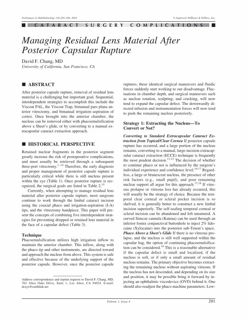
FIGURE 1. Nuclear fragments partially descendedthrough a posterior capsule defect.
TABLE 1. Early signs of possible posterior capsule ruptureor zonular dehiscence
1. Sudden deepening of the chamber, with momentaryexpansion of the pupil
2. Sudden, transitory appearance of a clear red reflexperipherally
3. Newly apparent inability to rotate a previously mobilenucleus
4. Excessive lateral mobility or displacement of the nucleus5. Excessive tipping of one pole of the nucleus6. Partial descent of the nucleus into a more posterior
position or plane
Chang
202 Techniques in Ophthalmology
is in these situations where the nucleus has only partiallydescended that a rescue technique may be possible.
The worst tactic for recovering a partially descendednucleus is to try to chase and spear it with the phaco tip.Lacking the normal capsular barrier, the downwardly di-rected irrigation flow will flush more vitreous forward,expanding the rent and propelling the nucleus away. At-tempting to phacoemulsify or aspirate the nucleus mayensnare vitreous into the phaco tip. Applying ultrasoundor moving the phaco tip after massive vitreous incarcera-tion into such a large diameter opening can easily createa giant retinal tear.5,11,20
Clearly, the safer alternative is to elevate the nucleusinto the pupillary plane or anterior chamber. However,there may be numerous obstacles to accomplishing this.First, the pupil or capsulorrhexis diameter may be verysmall. Indeed, these may have been the predisposing fac-tors for a capsular complication in the first place. Toosmall a pupil or capsulorrhexis may impede elevation ofthe nucleus and can make it particularly difficult for aspatula or OVD cannula to maneuver behind it. Anyvitreous that has prolapsed in front of the nucleus willfurther hinder attempts to inject OVD behind it. Contin-ued vitreous loss or prolapse deprives the nucleus ofmuch-needed remaining support.
Finally, even without a small pupil and vitreous loss,it may still be difficult to inject OVD behind a large,partially descended nucleus via a limbal incision. This isbecause any instrument inserted through the phacowound is approaching the nucleus from too steep anangle. For this reason, Charles Kelman, MD popularizedthe posterior assisted levitation, or “PAL” technique inwhich a metal spatula, inserted through a pars plana scle-rotomy, is used to levitate the nucleus into the anteriorchamber from below.21 Compared with the phaco inci-sion, the pars plana sclerotomy provides a much betterinstrument angle for getting behind the lens.
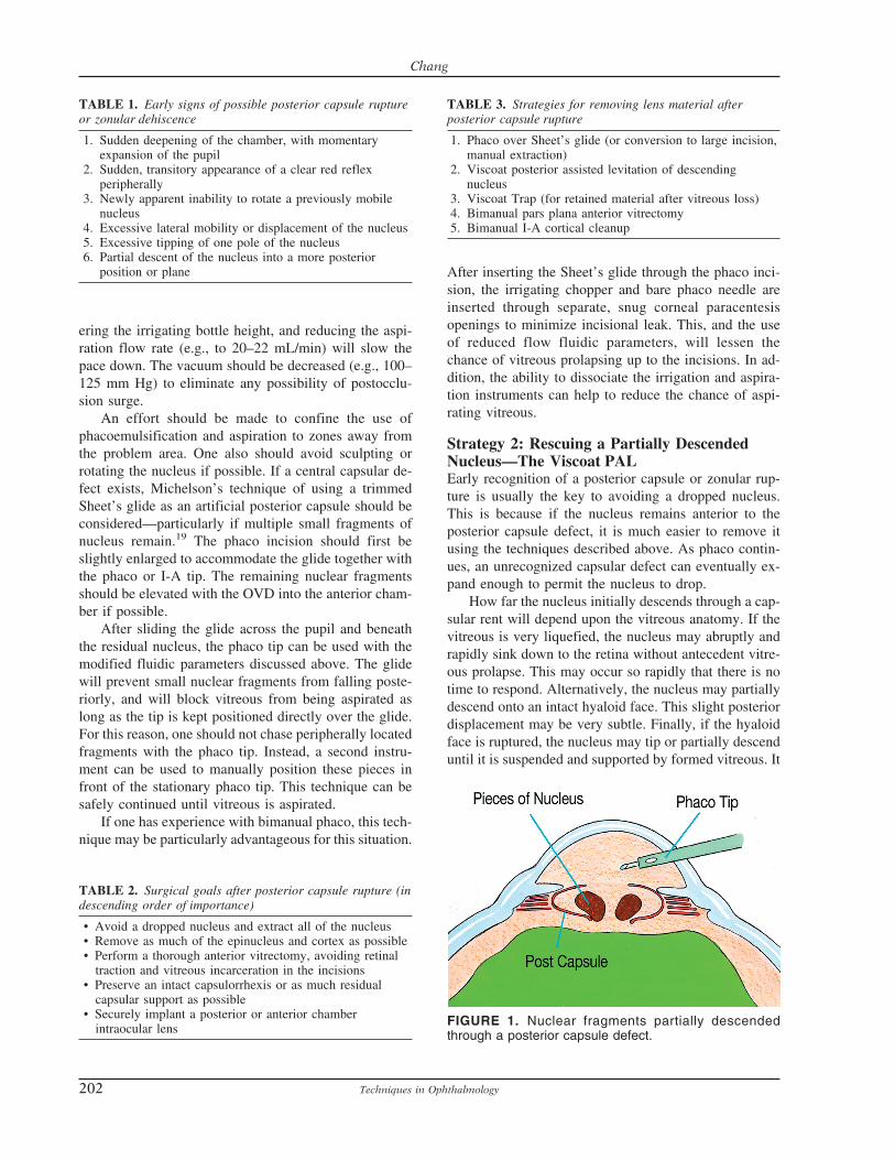
Richard Packard, MD, has suggested using Viscoat(Alcon, 3% sodium hyalvronate and 4% chondroitin sul-fate) instead of a spatula to levitate the nucleus via a parsplana approach. He and this author reported their successusing the Viscoat PAL technique in a series of 6 con-secutive cases with partially descended nuclei.22 A dis-posable microvitreoretinal (MVR) blade (Alcon, FortWorth, TX) is used to make a pars plana stab incisionlocated 3.5 mm behind the limbus. The Viscoat cannulais inserted far enough into the eye to be visualized andthen advanced or aimed behind the nucleus. The first stepis to inject a bolus of dispersive OVD behind the nucleusto immediately provide supplemental support. Periodicpalpation of the globe confirms that overinflation has notoccurred.
If the nucleus is subluxated laterally, directing OVDtoward the region beneath it will often float the nucleustoward a more central position. This is preferable to us-ing a metal spatula to reposition the nucleus because thetip, which cannot be visualized that far laterally, mightaccidentally contact the retina. One should not attempt tofloat the nucleus up using a massive injection of OVDalone. Unlike using liquid perfluorocarbon in a vitrecto-mized cavity, an excessive infusion of OVD may over-inflate the globe and cause vitreous expulsion throughthe sclerotomy or cataract incision.23
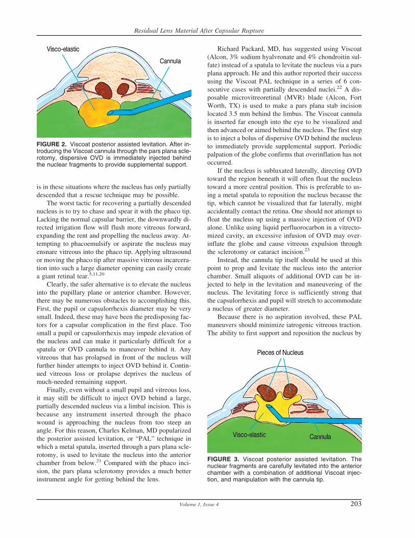
Instead, the cannula tip itself should be used at thispoint to prop and levitate the nucleus into the anteriorchamber. Small aliquots of additional OVD can be in-jected to help in the levitation and maneuvering of thenucleus. The levitating force is sufficiently strong thatthe capsulorrhexis and pupil will stretch to accommodatea nucleus of greater diameter.
Because there is no aspiration involved, these PALmaneuvers should minimize iatrogenic vitreous traction.The ability to first support and reposition the nucleus by
FIGURE 2. Viscoat posterior assisted levitation. After in-troducing the Viscoat cannula through the pars plana scle-rotomy, dispersive OVD is immediately injected behindthe nuclear fragments to provide supplemental support.
FIGURE 3. Viscoat posterior assisted levitation. Thenuclear fragments are carefully levitated into the anteriorchamber with a combination of additional Viscoat injec-tion, and manipulation with the cannula tip.
Residual Lens Material After Capsular Rupture
203Volume 1, Issue 4
injecting dispersive OVD prior to definitive manual levi-tation reduces the chance of touching the retina with ametal spatula tip. This is a major advantage of the Vis-coat PAL variation.
Once a fragment descends into the mid or posteriorvitreous cavity, or lands on the retina, it is dangerous toblindly fish for it with the OVD cannula or vitrectomyinstrument.24 One should abandon the dropped nucleusand concentrate on removing the epinucleus and cortex,while preserving as much capsular support as possible. Athorough anterior vitrectomy must be performed prior toinserting the IOL. Because the vitreoretinal specialistwill later use a 3-port fragmatome-vitrectomy approachto remove the retained nucleus, it is preferable to insertan IOL through the cataract incision at the time of theinitial surgery if possible.
Strategy 3: Managing Vitreous Loss andResidual Lens Material—The “Viscoat Trap”As previously discussed, any residual nucleus or epi-nucleus delivered into the anterior chamber using theViscoat PAL technique can be removed either by con-tinuing phaco or by converting to a manual large-incisionECCE method. At some point during this sequence, vit-reous may prolapse and be ensnared by the phaco or I-Atip. Because repeated vitreous aspiration and traction willrisk causing a retinal tear, surgeons should stop to per-form an anterior vitrectomy, with the goal being to laterresume removing residual nuclear or cortical remnants.
The most common practice is to place a separateself-retaining irrigating cannula though a limbal paracen-tesis, and to insert the sleeveless vitrectomy probethrough the phaco incision. However, there are multipledrawbacks to this approach. First, the phaco incision istoo large for the probe diameter. This leaking incision
results in poor chamber fluidics and allows both irriga-tion fluid and vitreous to prolapse externally alongsidethe vitrector shaft. Second, performing the vitrectomy inthe anterior chamber will tend to draw more posteriorlylocated vitreous forward. Finally, as more and more vit-reous exits the eye through either the cutting instrumentor the incision, the residual lens material that it is sup-porting will sink down toward the retina. It bears repeat-ing that once the posterior capsule is open, it is the re-sidual vitreous that is propping up and preventing theremaining nucleus and epinucleus from descending.
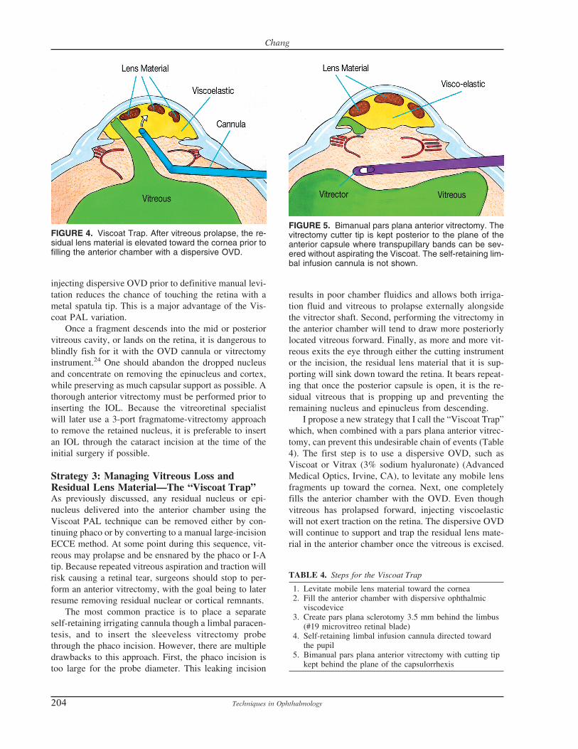
I propose a new strategy that I call the “Viscoat Trap”which, when combined with a pars plana anterior vitrec-tomy, can prevent this undesirable chain of events (Table4). The first step is to use a dispersive OVD, such asViscoat or Vitrax (3% sodium hyaluronate) (AdvancedMedical Optics, Irvine, CA), to levitate any mobile lensfragments up toward the cornea. Next, one completelyfills the anterior chamber with the OVD. Even thoughvitreous has prolapsed forward, injecting viscoelasticwill not exert traction on the retina. The dispersive OVDwill continue to support and trap the residual lens mate-rial in the anterior chamber once the vitreous is excised.
FIGURE 4. Viscoat Trap. After vitreous prolapse, the re-sidual lens material is elevated toward the cornea prior tofilling the anterior chamber with a dispersive OVD.
FIGURE 5. Bimanual pars plana anterior vitrectomy. Thevitrectomy cutter tip is kept posterior to the plane of theanterior capsule where transpupillary bands can be sev-ered without aspirating the Viscoat. The self-retaining lim-bal infusion cannula is not shown.
TABLE 4. Steps for the Viscoat Trap
1. Levitate mobile lens material toward the cornea2. Fill the anterior chamber with dispersive ophthalmic
viscodevice3. Create pars plana sclerotomy 3.5 mm behind the limbus
(#19 microvitreo retinal blade)4. Self-retaining limbal infusion cannula directed toward
the pupil5. Bimanual pars plana anterior vitrectomy with cutting tip
kept behind the plane of the capsulorrhexis
Chang
204 Techniques in Ophthalmology
Strategy 4: Bimanual Pars PlanaAnterior VitrectomyUnless it has already been created for a Viscoat PAL, apars plana sclerotomy is made 3.5 mm posterior to thelimbus, after making an overlying conjunctival peritomy.A disposable #19 MVR blade will create an adequatelysized opening for most anterior vitrectomy cutters. Aself-retaining irrigating cannula is placed through a lim-bal paracentesis, and angled toward the pupil. Thesleeveless vitrectomy shaft is inserted through the parsplana sclerotomy until the tip can be visualized in theretropupillary space. If it does not pass through the in-cision easily, it is better to slightly enlarge the openingrather than to force the entry.
Utilizing a high cutting rate, a thorough anterior vit-rectomy is performed while focusing posteriorly enoughwith the microscope to keep the tip under direct visual-ization. One should attempt to keep the vitrectomy tipbehind the pupil at all times. While any transpupillarybands of vitreous will still be severed, this will avoidremoving the dispersive OVD that fills the anteriorchamber. When properly performed, one will see that theanteriorly trapped lens fragments remain completely im-mobilized as the vitrectomy is being carried out below.This is because two separate chambers have been formedby the OVD partition, such that the anterior chamber isisolated from the vitrectomized posterior chamber.
Use of a pars plana sclerotomy is an underutilizedoption for performing an anterior vitrectomy. A majoradvantage is that the opening is properly sized to avoidincisional leak and vitreous prolapse, and to provide abetter fluidic seal. Unlike with a limbal incision, thevitrector need not transverse and invade the ViscoatTrap, and will not draw vitreous up into the anteriorchamber. Performing the vitrectomy posterior to the pu-pil and the plane of the capsulorrhexis also decreases the
chance of inadvertently cutting either structure. If thelatter is preserved, a foldable posterior chamber IOL maystill be implanted in the ciliary sulcus.3–5,7–9,13,14,17 Thesclerotomy can be closed with an interrupted 8–0 Vicrylsuture.
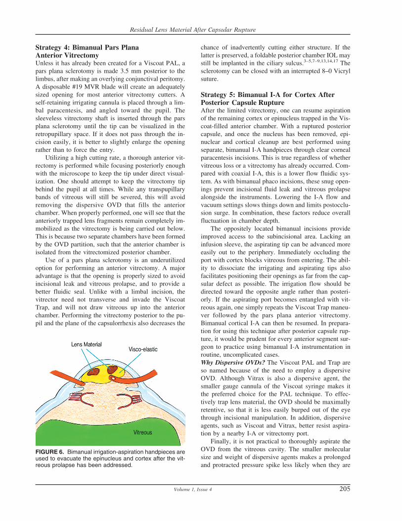
Strategy 5: Bimanual I-A for Cortex AfterPosterior Capsule RuptureAfter the limited vitrectomy, one can resume aspirationof the remaining cortex or epinucleus trapped in the Vis-coat-filled anterior chamber. With a ruptured posteriorcapsule, and once the nucleus has been removed, epi-nuclear and cortical cleanup are best performed usingseparate, bimanual I-A handpieces through clear cornealparacentesis incisions. This is true regardless of whethervitreous loss or a vitrectomy has already occurred. Com-pared with coaxial I-A, this is a lower flow fluidic sys-tem. As with bimanual phaco incisions, these snug open-ings prevent incisional fluid leak and vitreous prolapsealongside the instruments. Lowering the I-A flow andvacuum settings slows things down and limits postocclu-sion surge. In combination, these factors reduce overallfluctuation in chamber depth.
The oppositely located bimanual incisions provideimproved access to the subincisional area. Lacking aninfusion sleeve, the aspirating tip can be advanced moreeasily out to the periphery. Immediately occluding theport with cortex blocks vitreous from entering. The abil-ity to dissociate the irrigating and aspirating tips alsofacilitates positioning their openings as far from the cap-sular defect as possible. The irrigation flow should bedirected toward the opposite angle rather than posteri-orly. If the aspirating port becomes entangled with vit-reous again, one simply repeats the Viscoat Trap maneu-ver followed by the pars plana anterior vitrectomy.Bimanual cortical I-A can then be resumed. In prepara-tion for using this technique after posterior capsule rup-ture, it would be prudent for every anterior segment sur-geon to practice using bimanual I-A instrumentation inroutine, uncomplicated cases.Why Dispersive OVDs? The Viscoat PAL and Trap areso named because of the need to employ a dispersiveOVD. Although Vitrax is also a dispersive agent, thesmaller gauge cannula of the Viscoat syringe makes itthe preferred choice for the PAL technique. To effec-tively trap lens material, the OVD should be maximallyretentive, so that it is less easily burped out of the eyethrough incisional manipulation. In addition, dispersiveagents, such as Viscoat and Vitrax, better resist aspira-tion by a nearby I-A or vitrectomy port.
Finally, it is not practical to thoroughly aspirate theOVD from the vitreous cavity. The smaller molecularsize and weight of dispersive agents makes a prolongedand protracted pressure spike less likely when they are
FIGURE 6. Bimanual irrigation-aspiration handpieces areused to evacuate the epinucleus and cortex after the vit-reous prolapse has been addressed.
Residual Lens Material After Capsular Rupture
205Volume 1, Issue 4
not fully evacuated.25–27 Because of the larger molecularweight, residual amounts of maximally cohesive OVDs,such as Healon 5 or Healon GV (Pharmacia Corporation,Peapack, NJ) will usually produce the most severe andmost prolonged pressure spikes.
In conclusion, posterior capsule rupture whilenucleus is still present is a precipitous and intimidatingcomplication that tests a surgeon’s ability to operate un-der pressure. It is incumbent upon all cataract surgeons toprepare for this scenario by understanding the manage-ment principles and mentally rehearsing the maneuversin advance.
� REFERENCES1. Fastenberg DM, Schwartz PL, Shakin JL, et al. Management of
dislocated nuclear fragments after phacoemulsification. Am J Oph-thalmol. 1991;112:535–539.
2. Blodi BA, Flynn HW Jr, Blodi CF, et al. Retained nuclei aftercataract surgery. Ophthalmology. 1992;99:41–44.
3. Gilliland GD, Hutton WL, Fuller DG. Retained intravitreal lensfragments after cataract surgery. Ophthalmology. 1992;99:1263–1267.
4. Kim LMW, Flynn HW Jr, Smiddy WE, et al. Retained intravitreallens fragments after phacoemulsification. Ophthalmology. 1994;101:1827–1832.
5. Borne MJ, Tasman W, Regillo C, et al. Outcomes of vitrectomy forretained lens fragments. Ophthalmology. 1996;103:971–976.
6. Kapusuta MA, Chen JC, Lam W-C. Outcomes of dropped nucleusduring phacoemulsification. Ophthalmology. 1996;103:1184–1187; discussion by Brucker AJ, 1187.
7. Vilar NF, Flynn HW Jr, Smiddy WE, et al. Removal of retainedlens fragments after phacoemulsification reverses secondary glau-coma and restores visual acuity. Ophthalmology 1997;104:787–791; discussion by McDonald HR, 791–792.
8. Margherio RR, Margherio AR, Pendergast SD, et al. Vitrectomyfor retained lens fragments after phacoemulsification. Ophthalmol-ogy. 1997;104:1426–1432.
9. Stilma JS, van der Sluijs FA, van Meurs JC, et al. Occurrence ofretained lens fragments after phacoemulsification in The Nether-lands. J Cataract Refract Surg. 1997;23:1177–1182.
10. Yeo LMW, Chartesis DG, Bunce C, et al. Retained intravitreal lensfragments after phacoemulsification: a clinicopathological correla-tion. Br J Ophthalmol. 1999;83:1135–1138.
11. Lu H, Jiang YR, Grabow HB. Managing a dropped nucleus duringthe phacoemulsification learning curve. J Cataract Refract Surg.1999;25:1311–1312.
12. Rossetti A, Doro D. Retained intravitreal lens fragments afterphacoemulsification: Complications and visual outcome in vitrec-
tomized and nonvitrectomized eyes. J Cataract Refract Surg.2002;28:310–315.
13. Hansson LJ, Larsson J. Vitrectomy for retained lens fragments inthe vitreous after phacoemulsification. J Cataract Refract Surg.2002;28:1007–1011.
14. Scott IU, Flynn HW Jr, Smiddy WE, et al. Clinical features andoutcomes of pars plana vitrectomy in patients with retained lensfragments. Ophthalmology. 2003;110:1567–1572.
15. Akura J, Hatta S, Kaneda S, et al. Management of posterior capsulerupture during phacoemulsification using the dry technique.J Cataract Refract Surg. 2000;27:982–989.
16. Ah-Fat FG, Sharma MK, Majid MA, et al. Vitreous loss duringconversion from conventional extracapsular cataract extraction tophacoemulsification. J Cataract Refract Surg. 1998;24:801–805.
17. Aasuri MK, Kompella VB, Majji AB. Risk factors for and man-agement of dropped nucleus during phacoemulsification. J Cata-ract Refract Surg. 2001;27:1428–1432.
18. Guzek JP, Holm M, Cotter JB, et al. Risk factors for intraoperativecomplications in 1000 extracapsular cataract cases. Ophthalmol-ogy. 1987;94:461–466.
19. Michelson MA. Use of a Sheets’ glide as a pseudoposterior capsulein phacoemulsification complicated by posterior capsule rupture.Eur J Implant Surg. 1993;570–572.
20. Moore JK, Scott IU, Flynn HW Jr, et al. Retinal detachment in eyesundergoing pars plana vitrectomy for removal of retained lensfragments. Ophthalmology. 2003;110:709–713.
21. Rao SK, Chan WM, Leung AT, et al. Impending dropped nucleusduring phacoemulsification [Letter]. J Cataract Refract Surg.1999;25:1311–1312.
22. Chang DF, Packard RB. Posterior assisted levitation for nucleusretrieval using Viscoat after posterior capsule rupture. J CataractRefract Surg. 2003;29:1860–1865.
23. Verma L, Gogoi M, Tewari HK, et al. Comparative study of vit-rectomy for dropped nucleus with and without the use of perfluo-rocarbon liquid. Clinical, electrophysiological and visual field out-comes. Acta Ophthalmol Scand. 2001;79:354–358.
24. Horiguchi M, Kojima Y, Shimada Y. Removal of lens materialdropped into the vitreous cavity during cataract surgery using anoptical fiber-free intravitreal surgery system. J Cataract RefractSurg. 2003;29:1256–1259.
25. Burke S, Sugar J, Farber MD. Comparison of the effects of twoviscoelastic agents, Healon and Viscoat, on postoperative intraoc-ular pressure after penetrating keratoplasty. Ophthalmic Surg.1990;21:821–826.
26. Probst LE, Hakim OJ, Nichols BD. Phacoemulsification with as-pirated or retained Viscoat. J Cataract Refract Surg. 1994;20:145–149.
27. Torngren L, Lundgren B, Madsen K. Intraocular pressure devel-opment in the rabbit eye after aqueous exchange with ophthalmicviscosurgical devices. J Cataract Refract Surg. 2000;26:1247–1252.
Chang
206 Techniques in Ophthalmology