casereport - downloads.hindawi.comdownloads.hindawi.com/journals/cria/2017/7196340.pdf ·...
TRANSCRIPT
Case ReportEffect of Arm Positioning on Entrapment ofInfraclavicular Nerve Block Catheter
Eric Kamenetsky,1 Rahul Reddy,2 Mark C. Kendall,1 Antoun Nader,1 and Jessica J. Weeks1
1Department of Anesthesiology, Feinberg School of Medicine, Northwestern University, Chicago, IL, USA2Department of Anesthesiology, McGaw Medical Center, Northwestern University, Chicago, IL, USA
Correspondence should be addressed to Eric Kamenetsky; [email protected]
Received 9 December 2016; Accepted 18 January 2017; Published 28 February 2017
Academic Editor: Alparslan Apan
Copyright © 2017 Eric Kamenetsky et al. This is an open access article distributed under the Creative Commons AttributionLicense, which permits unrestricted use, distribution, and reproduction in any medium, provided the original work is properlycited.
Continuous brachial plexus nerve block catheters are commonly inserted for postoperative analgesia after upper extremitysurgery. Modifications of the insertion technique have been described to improve the safety of placing an infraclavicular brachialplexus catheter. Rarely, these catheters may become damaged or entrapped, complicating their removal. We describe a case ofinfraclavicular brachial plexus catheter entrapment related to differences in armpositioning during catheter placement and removal.Written authorization to obtain, use, and disclose information and images was obtained from the patient.
1. Introduction
Continuous brachial plexus nerve block catheters are com-monly used to prolong postoperative analgesia after painfulupper extremity procedures. Removal of these catheters istypically uncomplicated and often can be performed by thepatient after hospital discharge.When peripheral nerve blockcatheters become damaged or entrapped, their removal canbe challenging. Most reported cases of catheter entrapmentare associated with epidural catheters. In these cases, it isrecommended that the spine is flexed and continuous, gentletraction is placed on the catheter [1]. If these recommenda-tions are applied to peripheral nerve block catheters, then, ifresistance is met during removal, a patient’s extremity shouldbe positioned similar to when the catheter was inserted.We describe a unique case of infraclavicular brachial plexuscatheter damage and entrapment related to differences in armpositioning during placement and removal of the catheter.
2. Case Description
A healthy 47-year-old male underwent left wrist radioscaph-olunate fusion for posttraumatic arthrosis. An infraclavicularbrachial plexus nerve block was performed as the primary
anesthetic, with an indwelling catheter placed for postoper-ative analgesia. After sterile preparation and draping of theleft upper chest and positioning the left arm in an abductedand externally rotated position, a 2.5 cm linear array ultra-sound transducer (13–6MHz probe, SonoSite, S-Nerve�,Bothell, WA, USA) with sterile covering was used to visualizethe infraclavicular brachial plexus. A medial infraclavicularapproach was used as described by Bigeleisen and Wilson,with the needle puncture at the apex of the deltopectoralgroove [2]. Prior to local anesthetic injection, a distal evokedmotor response was obtained with a nerve stimulator, whichdisappeared at 0.4mA. After performing a block throughthe needle with 30mL 0.5% bupivacaine and 1 : 300,000epinephrine, the 18 g × 4 cm continuous nerve block nee-dle (Arrow International, Reading, PA, USA) was posi-tioned with the tip between the posterior and medial cordsof the brachial plexus. A 20 g × 60 cm continuous nerveblock catheter (StimuCath�, Arrow International, Reading,PA, USA) was advanced 5 cm beyond the needle tip withoutresistance and secured with adhesive bandages with the9 cm mark at the skin. A test dose of 5mL 1.5% lidocainewith 1 : 200,000 epinephrine was administered through thecatheter and an additional 10mL of 0.5% bupivacaine was
HindawiCase Reports in AnesthesiologyVolume 2017, Article ID 7196340, 4 pageshttps://doi.org/10.1155/2017/7196340
2 Case Reports in Anesthesiology
Cephalad
Caudad
Lateral
Clavicle
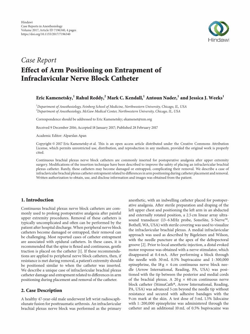
Figure 1: Wire fragment extending out of skin. During removal,the central stimulating wire and coil structure were noted to befractured. Yellow arrows: wire fragment.
subsequently injected. Perineural spread near the poste-rior cord was confirmed with ultrasound during injectionthrough the catheter.
The patient successfully underwent the surgical proce-dure with the peripheral nerve block and intraoperativesedation. He was discharged homewith the continuous nerveblock catheter infusing 0.2% ropivacaine at 5mL per hour,plus an additional patient-controlled dose of 2mL per houras needed. The patient reported excellent analgesia with 0/10pain on postoperative days (POD) 1 and 2 and there were nosigns of leakage or damage to the catheter. On POD 2, theentire 275mL 0.2% ropivacaine infusion was completed. OnPOD 3, the patient went to the surgical office for a scheduledfollow-up, at which time the surgeon attempted to remove thecatheter with the arm adducted. Prior to removal, the patientreported no residual effects of the nerve block. The surgeonwas able to remove the entire polyurethane catheter bodywithout difficulty, but during removal, the central stimulatingwire and coil structure were noted to be fractured, withapproximately 3 cm remaining above the skin level (Figure 1).The patient reported a transient paresthesia down the left armat the time of removal.
The patient was referred to the ambulatory surgerycenter for further evaluation by the anesthesiology team. Onphysical exam, the patient had normal strength and sensationin the lefthand.Hedid experience sharp, severe, nonradiatingpain near the clavicle upon abduction and external rotationof the arm, which resolved with adduction of the arm.Ultrasound evaluation did reveal the wire extending throughthe pectoralis major and minor muscles and coursing under
Transducer
Pectoralis minor muscle
Pectoralis major muscle
A
Arm position: AdductionCaud
ad
Cep
hala
d
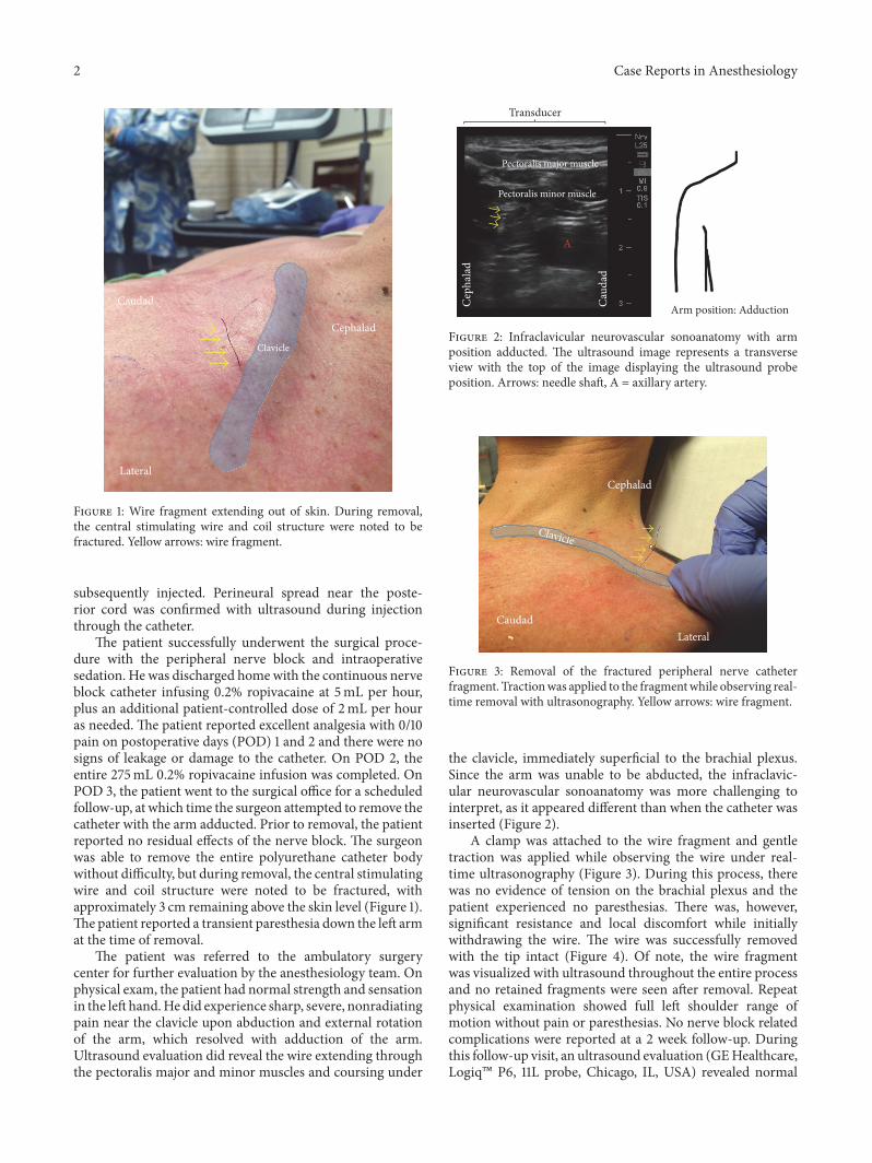
Figure 2: Infraclavicular neurovascular sonoanatomy with armposition adducted. The ultrasound image represents a transverseview with the top of the image displaying the ultrasound probeposition. Arrows: needle shaft, A = axillary artery.
Cephalad
CaudadLateral
ClavicleC lll
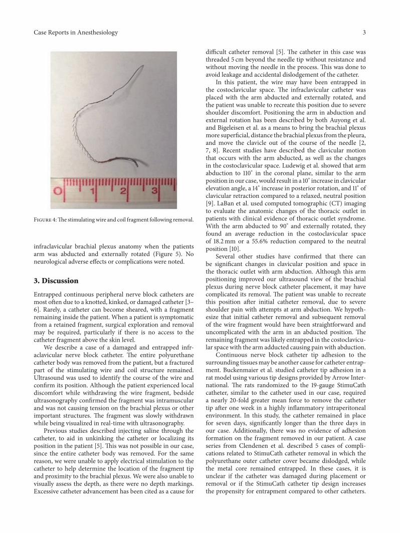
Figure 3: Removal of the fractured peripheral nerve catheterfragment. Tractionwas applied to the fragmentwhile observing real-time removal with ultrasonography. Yellow arrows: wire fragment.
the clavicle, immediately superficial to the brachial plexus.Since the arm was unable to be abducted, the infraclavic-ular neurovascular sonoanatomy was more challenging tointerpret, as it appeared different than when the catheter wasinserted (Figure 2).
A clamp was attached to the wire fragment and gentletraction was applied while observing the wire under real-time ultrasonography (Figure 3). During this process, therewas no evidence of tension on the brachial plexus and thepatient experienced no paresthesias. There was, however,significant resistance and local discomfort while initiallywithdrawing the wire. The wire was successfully removedwith the tip intact (Figure 4). Of note, the wire fragmentwas visualized with ultrasound throughout the entire processand no retained fragments were seen after removal. Repeatphysical examination showed full left shoulder range ofmotion without pain or paresthesias. No nerve block relatedcomplications were reported at a 2 week follow-up. Duringthis follow-up visit, an ultrasound evaluation (GEHealthcare,Logiq� P6, 11L probe, Chicago, IL, USA) revealed normal
Case Reports in Anesthesiology 3
Figure 4:The stimulatingwire and coil fragment following removal.
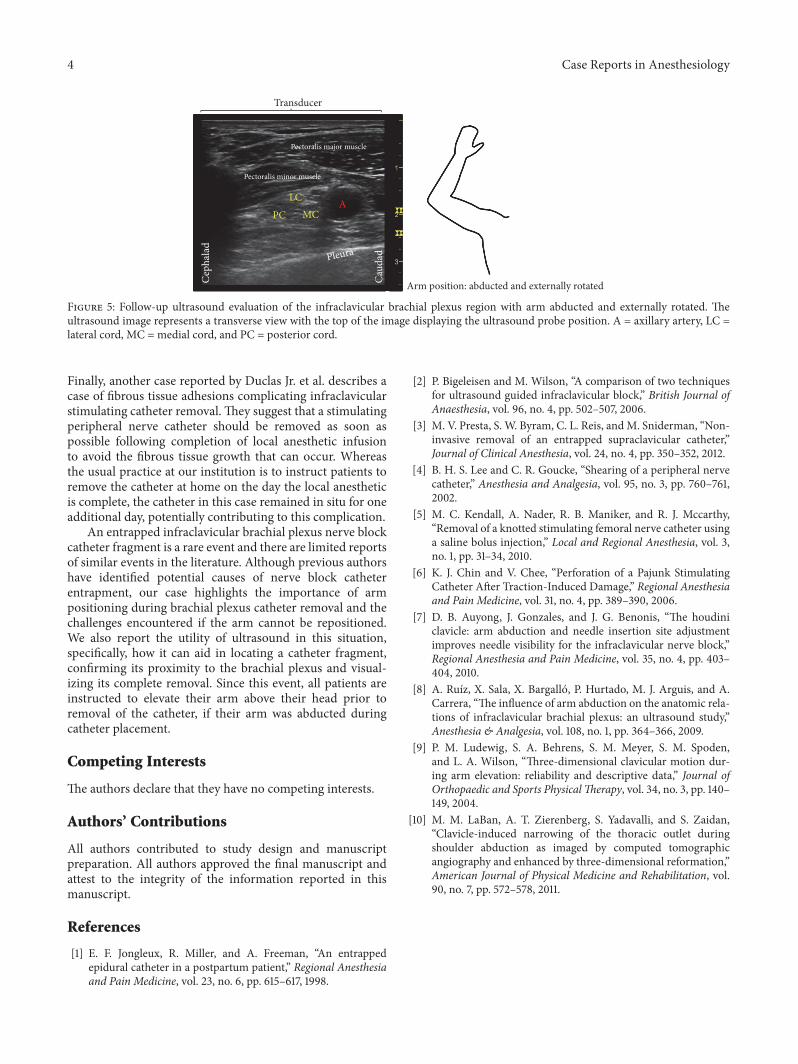
infraclavicular brachial plexus anatomy when the patientsarm was abducted and externally rotated (Figure 5). Noneurological adverse effects or complications were noted.
3. Discussion
Entrapped continuous peripheral nerve block catheters aremost often due to a knotted, kinked, or damaged catheter [3–6]. Rarely, a catheter can become sheared, with a fragmentremaining inside the patient. When a patient is symptomaticfrom a retained fragment, surgical exploration and removalmay be required, particularly if there is no access to thecatheter fragment above the skin level.
We describe a case of a damaged and entrapped infr-aclavicular nerve block catheter. The entire polyurethanecatheter body was removed from the patient, but a fracturedpart of the stimulating wire and coil structure remained.Ultrasound was used to identify the course of the wire andconfirm its position. Although the patient experienced localdiscomfort while withdrawing the wire fragment, bedsideultrasonography confirmed the fragment was intramuscularand was not causing tension on the brachial plexus or otherimportant structures. The fragment was slowly withdrawnwhile being visualized in real-time with ultrasonography.
Previous studies described injecting saline through thecatheter, to aid in unkinking the catheter or localizing itsposition in the patient [5]. This was not possible in our case,since the entire catheter body was removed. For the samereason, we were unable to apply electrical stimulation to thecatheter to help determine the location of the fragment tipand proximity to the brachial plexus. We were also unable tovisually assess the depth, as there were no depth markings.Excessive catheter advancement has been cited as a cause for
difficult catheter removal [5]. The catheter in this case wasthreaded 5 cm beyond the needle tip without resistance andwithout moving the needle in the process. This was done toavoid leakage and accidental dislodgement of the catheter.
In this patient, the wire may have been entrapped inthe costoclavicular space. The infraclavicular catheter wasplaced with the arm abducted and externally rotated, andthe patient was unable to recreate this position due to severeshoulder discomfort. Positioning the arm in abduction andexternal rotation has been described by both Auyong et al.and Bigeleisen et al. as a means to bring the brachial plexusmore superficial, distance the brachial plexus from the pleura,and move the clavicle out of the course of the needle [2,7, 8]. Recent studies have described the clavicular motionthat occurs with the arm abducted, as well as the changesin the costoclavicular space. Ludewig et al. showed that armabduction to 110∘ in the coronal plane, similar to the armposition in our case, would result in a 10∘ increase in clavicularelevation angle, a 14∘ increase in posterior rotation, and 11∘ ofclavicular retraction compared to a relaxed, neutral position[9]. LaBan et al. used computed tomographic (CT) imagingto evaluate the anatomic changes of the thoracic outlet inpatients with clinical evidence of thoracic outlet syndrome.With the arm abducted to 90∘ and externally rotated, theyfound an average reduction in the costoclavicular spaceof 18.2mm or a 55.6% reduction compared to the neutralposition [10].
Several other studies have confirmed that there canbe significant changes in clavicular position and space inthe thoracic outlet with arm abduction. Although this armpositioning improved our ultrasound view of the brachialplexus during nerve block catheter placement, it may havecomplicated its removal. The patient was unable to recreatethis position after initial catheter removal, due to severeshoulder pain with attempts at arm abduction. We hypoth-esize that initial catheter removal and subsequent removalof the wire fragment would have been straightforward anduncomplicated with the arm in an abducted position. Theremaining fragment was likely entrapped in the costoclavicu-lar space with the arm adducted causing pain with abduction.
Continuous nerve block catheter tip adhesion to thesurrounding tissuesmay be another cause for catheter entrap-ment. Buckenmaier et al. studied catheter tip adhesion in arat model using various tip designs provided by Arrow Inter-national. The rats randomized to the 19-gauge StimuCathcatheter, similar to the catheter used in our case, requireda nearly 20-fold greater mean force to remove the cathetertip after one week in a highly inflammatory intraperitonealenvironment. In this study, the catheter remained in placefor seven days, significantly longer than the three days inour case. Additionally, there was no evidence of adhesionformation on the fragment removed in our patient. A caseseries from Clendenen et al. described 5 cases of compli-cations related to StimuCath catheter removal in which thepolyurethane outer catheter cover became dislodged, whilethe metal core remained entrapped. In these cases, it isunclear if the catheter was damaged during placement orremoval or if the StimuCath catheter tip design increasesthe propensity for entrapment compared to other catheters.
4 Case Reports in Anesthesiology
Pectoralis major muscle
Pectoralis minor muscle
A
Pleura
Arm position: abducted and externally rotated
Transducer
PCLC
MCC
epha
lad
Caud
ad
Figure 5: Follow-up ultrasound evaluation of the infraclavicular brachial plexus region with arm abducted and externally rotated. Theultrasound image represents a transverse view with the top of the image displaying the ultrasound probe position. A = axillary artery, LC =lateral cord, MC = medial cord, and PC = posterior cord.
Finally, another case reported by Duclas Jr. et al. describes acase of fibrous tissue adhesions complicating infraclavicularstimulating catheter removal. They suggest that a stimulatingperipheral nerve catheter should be removed as soon aspossible following completion of local anesthetic infusionto avoid the fibrous tissue growth that can occur. Whereasthe usual practice at our institution is to instruct patients toremove the catheter at home on the day the local anestheticis complete, the catheter in this case remained in situ for oneadditional day, potentially contributing to this complication.
An entrapped infraclavicular brachial plexus nerve blockcatheter fragment is a rare event and there are limited reportsof similar events in the literature. Although previous authorshave identified potential causes of nerve block catheterentrapment, our case highlights the importance of armpositioning during brachial plexus catheter removal and thechallenges encountered if the arm cannot be repositioned.We also report the utility of ultrasound in this situation,specifically, how it can aid in locating a catheter fragment,confirming its proximity to the brachial plexus and visual-izing its complete removal. Since this event, all patients areinstructed to elevate their arm above their head prior toremoval of the catheter, if their arm was abducted duringcatheter placement.
Competing Interests
The authors declare that they have no competing interests.
Authors’ Contributions
All authors contributed to study design and manuscriptpreparation. All authors approved the final manuscript andattest to the integrity of the information reported in thismanuscript.
References
[1] E. F. Jongleux, R. Miller, and A. Freeman, “An entrappedepidural catheter in a postpartum patient,” Regional Anesthesiaand Pain Medicine, vol. 23, no. 6, pp. 615–617, 1998.
[2] P. Bigeleisen and M. Wilson, “A comparison of two techniquesfor ultrasound guided infraclavicular block,” British Journal ofAnaesthesia, vol. 96, no. 4, pp. 502–507, 2006.
[3] M. V. Presta, S. W. Byram, C. L. Reis, andM. Sniderman, “Non-invasive removal of an entrapped supraclavicular catheter,”Journal of Clinical Anesthesia, vol. 24, no. 4, pp. 350–352, 2012.
[4] B. H. S. Lee and C. R. Goucke, “Shearing of a peripheral nervecatheter,” Anesthesia and Analgesia, vol. 95, no. 3, pp. 760–761,2002.
[5] M. C. Kendall, A. Nader, R. B. Maniker, and R. J. Mccarthy,“Removal of a knotted stimulating femoral nerve catheter usinga saline bolus injection,” Local and Regional Anesthesia, vol. 3,no. 1, pp. 31–34, 2010.
[6] K. J. Chin and V. Chee, “Perforation of a Pajunk StimulatingCatheter After Traction-Induced Damage,” Regional Anesthesiaand Pain Medicine, vol. 31, no. 4, pp. 389–390, 2006.
[7] D. B. Auyong, J. Gonzales, and J. G. Benonis, “The houdiniclavicle: arm abduction and needle insertion site adjustmentimproves needle visibility for the infraclavicular nerve block,”Regional Anesthesia and Pain Medicine, vol. 35, no. 4, pp. 403–404, 2010.
[8] A. Ruız, X. Sala, X. Bargallo, P. Hurtado, M. J. Arguis, and A.Carrera, “The influence of arm abduction on the anatomic rela-tions of infraclavicular brachial plexus: an ultrasound study,”Anesthesia & Analgesia, vol. 108, no. 1, pp. 364–366, 2009.
[9] P. M. Ludewig, S. A. Behrens, S. M. Meyer, S. M. Spoden,and L. A. Wilson, “Three-dimensional clavicular motion dur-ing arm elevation: reliability and descriptive data,” Journal ofOrthopaedic and Sports PhysicalTherapy, vol. 34, no. 3, pp. 140–149, 2004.
[10] M. M. LaBan, A. T. Zierenberg, S. Yadavalli, and S. Zaidan,“Clavicle-induced narrowing of the thoracic outlet duringshoulder abduction as imaged by computed tomographicangiography and enhanced by three-dimensional reformation,”American Journal of Physical Medicine and Rehabilitation, vol.90, no. 7, pp. 572–578, 2011.

Submit your manuscripts athttps://www.hindawi.com
Stem CellsInternational
Hindawi Publishing Corporationhttp://www.hindawi.com Volume 2014
Hindawi Publishing Corporationhttp://www.hindawi.com Volume 2014
MEDIATORSINFLAMMATION
of
Hindawi Publishing Corporationhttp://www.hindawi.com Volume 2014
Behavioural Neurology
EndocrinologyInternational Journal of
Hindawi Publishing Corporationhttp://www.hindawi.com Volume 2014
Hindawi Publishing Corporationhttp://www.hindawi.com Volume 2014
Disease Markers
Hindawi Publishing Corporationhttp://www.hindawi.com Volume 2014
BioMed Research International
OncologyJournal of
Hindawi Publishing Corporationhttp://www.hindawi.com Volume 2014
Hindawi Publishing Corporationhttp://www.hindawi.com Volume 2014
Oxidative Medicine and Cellular Longevity
Hindawi Publishing Corporationhttp://www.hindawi.com Volume 2014
PPAR Research
The Scientific World JournalHindawi Publishing Corporation http://www.hindawi.com Volume 2014
Immunology ResearchHindawi Publishing Corporationhttp://www.hindawi.com Volume 2014
Journal of
ObesityJournal of
Hindawi Publishing Corporationhttp://www.hindawi.com Volume 2014
Hindawi Publishing Corporationhttp://www.hindawi.com Volume 2014
Computational and Mathematical Methods in Medicine
OphthalmologyJournal of
Hindawi Publishing Corporationhttp://www.hindawi.com Volume 2014
Diabetes ResearchJournal of
Hindawi Publishing Corporationhttp://www.hindawi.com Volume 2014
Hindawi Publishing Corporationhttp://www.hindawi.com Volume 2014
Research and TreatmentAIDS
Hindawi Publishing Corporationhttp://www.hindawi.com Volume 2014
Gastroenterology Research and Practice
Hindawi Publishing Corporationhttp://www.hindawi.com Volume 2014
Parkinson’s Disease
Evidence-Based Complementary and Alternative Medicine
Volume 2014Hindawi Publishing Corporationhttp://www.hindawi.com