case report stress fracture of the distal fibula in ... · pdf filecase report stress fracture...
TRANSCRIPT

Int J Clin Exp Med 2015;8(4):6303-6307www.ijcem.com /ISSN:1940-5901/IJCEM0005081
Case Report Stress fracture of the distal fibula in flatfoot patients: case report
Yu Cheng, Huilin Yang, Li Ni, Dawei Song, Hongtao Zhang
Department of Orthopedics, The First Affiliated Hospital of Soochow University, 188 Shizi Street, Suzhou 215006, Jiangsu, China
Received December 20, 2014; Accepted March 20, 2015; Epub April 15, 2015; Published April 30, 2015
Abstract: The increase in proportional loading of the fibula with progression of hindfoot deformity would lead to high fibular loads during rapid walking, resulting in insufficiency fractures. We report an unusual mechanism of such fracture in a textile worker resulting from valgus alignment from a stage III flatfoot deformity. The stress fracture was missed initially and only confirmed by CT examination. The patient responded well to nonoperative treatment and had an excellent recovery with no residual symptoms finally. Fracture of the distal fibula caused by rigid hindfoot valgus in stage III flatfoot deformity is a previously undescribed injury. We report a patient who presented with this injury, the possible mechanisms of such injury, its management and outcome.
Keywords: Stress fracture, flatfoot, fibular loading
Stress fractures are more unusual in the fibula than in the tibia because the fibula plays a minor role concerning the axial load of the lower extremity. However, in rigid flatfoot defor-mity with hindfoot valgus, the loads on the fibu-la may have increased. We present a unique case of fibular stress fracture in a flatfoot patient, which is missed at initial presentation. To our knowledge, no cases with distal fibular stress fractures attributable to flatfoot have been reported in the English literature.
Case report
A 44-year-old female presented to our clinic for lateral distal pain in the left leg of four days duration. The patient was employed as a work-er in the textile mill for 8 years and the job needed the patient to walk around to fill all the textile machinery with water repeatedly. After 2 weeks of continuous overtime work, the patient noticed a gradual swelling in her left leg just above the ankle. She was still able to stand on her leg, although with pain, and completed the work days. But four days later, she had to dis-continue work because of aggravated pain and a slight limp toward the left side. Physical examination revealed localized swelling and
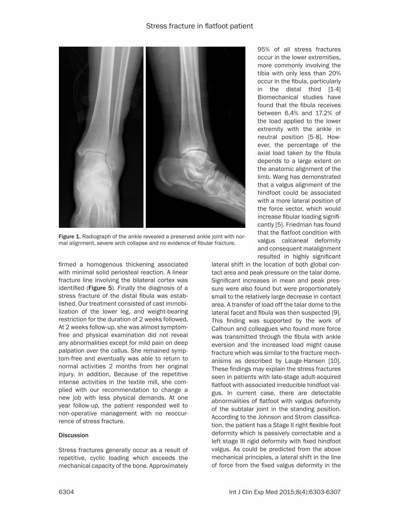
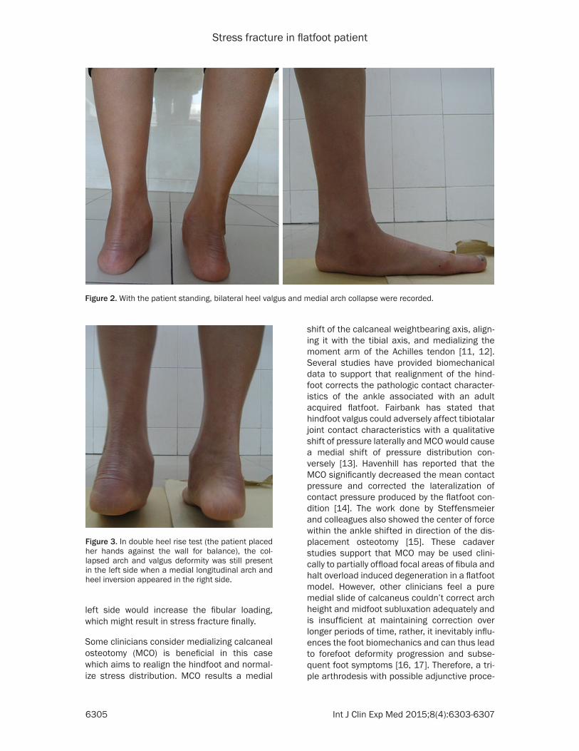
tenderness on the distal fibula about 3 cm prox-imal to the lateral malleolus. Radiographs of the ankle did not reveal any abnormalities (Figure 1) and the patient was given nonsteroi-dal anti-inflammatory medications for pain relief and was placed on sick leave. Unfortunately, two weeks later the patient returned to our clinic with no improvement of the symptoms. On meticulous physical exami-nation again, we found she had bilateral foot arch collapse with increased hindfoot valgus deformity (Figure 2). In contrast to the right reducible deformity, the left side showed inabil-ity to perform a single heel-rise test with a pas-sively uncorrectable subtalar joint (Figure 3). In consideration of these clinical signs, we sus-pected it was a lateral impingement caused by calcaneal valgus and decided to do an MRI examination of the left ankle. The MRI identified an abnormal segmental signal of the distal fibu-la above the ankle and a cortical defect sur-rounded by soft-tissue swelling (Figure 4). According to these findings, further diagnostic protocols were ordered. Laboratory tests includ-ing routine blood tests, as well as erythrocyte sedimentation rate, C-reactive protein, all part of the blood workup were in the normal ranges. Subsequent CT with 3D reconstruction con-
Stress fracture in flatfoot patient
6304 Int J Clin Exp Med 2015;8(4):6303-6307
lateral shift in the location of both global con-tact area and peak pressure on the talar dome. Significant increases in mean and peak pres-sure were also found but were proportionately small to the relatively large decrease in contact area. A transfer of load off the talar dome to the lateral facet and fibula was then suspected [9]. This finding was supported by the work of Calhoun and colleagues who found more force was transmitted through the fibula with ankle eversion and the increased load might cause fracture which was similar to the fracture mech-anisms as described by Lauge-Hansen [10]. These findings may explain the stress fractures seen in patients with late-stage adult-acquired flatfoot with associated irreducible hindfoot val-gus. In current case, there are detectable abnormalities of flatfoot with valgus deformity of the subtalar joint in the standing position. According to the Johnson and Strom classifica-tion, the patient has a Stage II right flexible foot deformity which is passively correctable and a left stage III rigid deformity with fixed hindfoot valgus. As could be predicted from the above mechanical principles, a lateral shift in the line of force from the fixed valgus deformity in the
firmed a homogenous thickening associated with minimal solid periosteal reaction. A linear fracture line involving the bilateral cortex was identified (Figure 5). Finally the diagnosis of a stress fracture of the distal fibula was estab-lished. Our treatment consisted of cast immobi-lization of the lower leg, and weight-bearing restriction for the duration of 2 weeks followed. At 2 weeks follow-up, she was almost symptom-free and physical examination did not reveal any abnormalities except for mild pain on deep palpation over the callus. She remained symp-tom-free and eventually was able to return to normal activities 2 months from her original injury. In addition, Because of the repetitive intense activities in the textile mill, she com-plied with our recommendation to change a new job with less physical demands. At one year follow-up, the patient responded well to non-operative management with no reoccur-rence of stress fracture.
Discussion
Stress fractures generally occur as a result of repetitive, cyclic loading which exceeds the mechanical capacity of the bone. Approximately
95% of all stress fractures occur in the lower extremities, more commonly involving the tibia with only less than 20% occur in the fibula, particularly in the distal third [1-4] Biomechanical studies have found that the fibula receives between 6.4% and 17.2% of the load applied to the lower extremity with the ankle in neutral position [5-8]. How- ever, the percentage of the axial load taken by the fibula depends to a large extent on the anatomic alignment of the limb. Wang has demonstrated that a valgus alignment of the hindfoot could be associated with a more lateral position of the force vector, which would increase fibular loading signifi-cantly [5]. Friedman has found that the flatfoot condition with valgus calcaneal deformity and consequent malalignment resulted in highly significant
Figure 1. Radiograph of the ankle revealed a preserved ankle joint with nor-mal alignment, severe arch collapse and no evidence of fibular fracture.
Stress fracture in flatfoot patient
6305 Int J Clin Exp Med 2015;8(4):6303-6307
left side would increase the fibular loading, which might result in stress fracture finally.
Some clinicians consider medializing calcaneal osteotomy (MCO) is beneficial in this case which aims to realign the hindfoot and normal-ize stress distribution. MCO results a medial
shift of the calcaneal weightbearing axis, align-ing it with the tibial axis, and medializing the moment arm of the Achilles tendon [11, 12]. Several studies have provided biomechanical data to support that realignment of the hind-foot corrects the pathologic contact character-istics of the ankle associated with an adult acquired flatfoot. Fairbank has stated that hindfoot valgus could adversely affect tibiotalar joint contact characteristics with a qualitative shift of pressure laterally and MCO would cause a medial shift of pressure distribution con-versely [13]. Havenhill has reported that the MCO significantly decreased the mean contact pressure and corrected the lateralization of contact pressure produced by the flatfoot con-dition [14]. The work done by Steffensmeier and colleagues also showed the center of force within the ankle shifted in direction of the dis-placement osteotomy [15]. These cadaver studies support that MCO may be used clini-cally to partially offload focal areas of fibula and halt overload induced degeneration in a flatfoot model. However, other clinicians feel a pure medial slide of calcaneus couldn’t correct arch height and midfoot subluxation adequately and is insufficient at maintaining correction over longer periods of time, rather, it inevitably influ-ences the foot biomechanics and can thus lead to forefoot deformity progression and subse-quent foot symptoms [16, 17]. Therefore, a tri-ple arthrodesis with possible adjunctive proce-
Figure 2. With the patient standing, bilateral heel valgus and medial arch collapse were recorded.
Figure 3. In double heel rise test (the patient placed her hands against the wall for balance), the col-lapsed arch and valgus deformity was still present in the left side when a medial longitudinal arch and heel inversion appeared in the right side.

Stress fracture in flatfoot patient
6306 Int J Clin Exp Med 2015;8(4):6303-6307
and improved treatment algorithms of this condition.
Conclusion
Stress fracture of the fibula may be related with the long-standing presence of an adult acquired flatfoot deformity. Increased proportional load-ing in the fibula due to lateralization of the load axis in fixed hindfoot valgus deformity is thought to contribute to this condition. However, wheth-er it’s an operative indication using calcaneal
dures should be a better long-term procedure to achieve full correction and prevent underly-ing disease process. However, in the absence of symptoms in the foot, the arthrodesis seems too radical and the significant degenerative effect on adjacent joints particularly the ankle, is concerned [18, 19]. With above arguments, we finally determined to use cast immobiliza-tion as initial treatment and the patient was instructed to return if pain reoccurs. As with other stress fractures, healing occurred once the offending loading force was removed.
Finally, the patient changed to a less physical-demanding job without symptoms recurrence at one year follow-up. However the long-term outcome is still uncertain.
The weight-bearing function of the fibula in flatfoot model does not seem to have been adequately studied. Neither the occurrence nor the fre-quency of fibular stress frac-ture in patients with flatfoot deformity has been docu-mented. Also definitive stud-ies would be necessary to assess whether treatment of the deformity reduces fibular stress and prevents overload degeneration. With these informations, it may lead to better understanding
Figure 4. MRI showed extensive bone marrow edema in the distal fibula with surrounding soft tissue edema.
Figure 5. CT showed a nondisplaced transverse hairline fracture of the distal fibula with callus formation.

Stress fracture in flatfoot patient
6307 Int J Clin Exp Med 2015;8(4):6303-6307
osteotomy or even arthrodesis to reduce the fibular weight-bearing stress is still in doubt. Finally, understanding the etiology of the distal fibular pain in patients with flatfoot deformity can potentially alter the physician to the possi-bility of stress fracture and aid in clinical diag-nosis and treatment designing.
Disclosure of conflict of interest
None.
Address correspondence to: Hongtao Zhang, De- partment of Orthopedics, The First Affiliated Hospital of Soochow University, 188 Shizi Street, Suzhou 215006, Jiangsu, China. E-mail: [email protected]
References
[1] Reeder MT, Dick BH, Atkins JK, Pribis AB, Mar-tinez JM. Stress fractures. Current concepts of diagnosis andtreatment. Sports Med 1996; 22: 198-212.
[2] Matheson GO, Clement DB, McKenzie DC Taunton JE, Lloyd-Smith DR, MacIntyre JG. Stress fractures in athletes. A study of 320 cases. Am J Sports Med 1987; 15: 46-58.
[3] Brukner P, Bradshaw C, Khan KM, White S, Crossley K. Stress fractures: a review of 180 cases. Clin J Sport Med 1996; 6: 85-9.
[4] Symeonides PP. High stress fractures of the fibula. J Bone Joint Surg Br 1980; 62: 192-3.
[5] Wang Q, Whittle M, Cunningham J, Kenwright J. Fibula and its ligaments in load transmission and ankle joint stability. Clin Orthop Relat Res 1996; 330: 261-70.
[6] Goh JC, Mech AM, Lee EH, Ang EJ, Bayon P, Pho RW. Biomechanical study on the load-bearing characteristics of the fibula and the effects of fibular resection. Clin Orthop Relat Res 1992; 279: 223-8.
[7] Lambert KL. The weight-bearing function of the fibula. A strain gauge study. J Bone Joint Surg Am 1971; 53: 507-13.
[8] Takebe K, Nakagawa A, Minami H, Kanazawa H, Hirohata K. Role of the fibula in weight-bear-ing. Clin Orthop Relat Res 1984; 184: 289-92.
[9] Friedman MA, Draganich LF, Toolan B, Brage ME. The effects of adult acquired flatfoot de-formity on tibiotalar joint contact characteris-tics. Foot Ankle Int 2001; 22: 241-6.
[10] Calhoun JH, Li F, Ledbetter BR, Viegas SF. A comprehensive study of pressure distribution in the ankle joint with inversion and eversion. Foot Ankle Int 1994; 15: 125-33.
[11] Guha AR, Perera AM. Calcaneal osteotomy in the treatment of adult acquired flatfoot defor-mity. Foot Ankle Clin 2012; 17: 247-58.
[12] Arangio GA, Salathe EP. A biomechanical anal-ysis of posterior tibial tendon dysfunction, me-dial displacement calcaneal osteotomy and flexor digitorum longus transfer in adult ac-quired flat foot. Clin Biomech (Bristol, Avon) 2009; 24: 385-90.
[13] Fairbank A, Myerson MS, Fortin P. The effect of calcaneal osteotomy on contact characteris-tics of the tibiotalar joint. The Foot 1995; 5: 137-42.
[14] Havenhill TG, Toolan BC, Draganich LF. Effects of a UCBL orthosis and a calcaneal osteotomy on tibiotalar contact characteristics in a cadav-er flatfoot model. Foot Ankle Int 2005; 26: 607-13.
[15] Steffensmeier SJ, Saltzman CL, Berbaum KS, Brown TD. Effects of medial and lateral dis-placement calcaneal osteotomies on tibiotalar joint contact stresses. J Orthop Res 1996; 14: 980-5.
[16] Niki H, Hirano T, Okada H, Beppu M. Outcome of medial displacement calcaneal osteotomy for correction of adult-acquired flatfoot. Foot Ankle Int 2012; 33: 940-6.
[17] Haddad SL, Myerson MS, Younger A,Anderson RB, Davis WH, Manoli A 2nd. Symposium: Adult acquired flatfoot deformity. Foot Ankle Int 2011; 32: 95-111.
[18] Francisco R, Chiodo CP, Wilson MG. Manage-ment of the rigid adult acquired flatfoot defor-mity. Foot Ankle Clin 2007;12:317-27.
[19] Deland JT. Adult-acquired flatfoot deformity. J Am Acad Orthop Surg 2008; 16: 399-406.