case report robot-assisted excision of a pararectal...
TRANSCRIPT
Case ReportRobot-Assisted Excision of a Pararectal Gastrointestinal StromalTumor in a Patient with Previous Ileal Neobladder
A. Ploumidis,1 A. Mottrie,1 A. F. Spinoit,1,2 M. Gan,1 V. Ficarra,1,3 and R. Andrianne4
1 Department of Urology, OLV Vattikuti Robotic Surgery Institute, Moorselbaan 164, 9300 Aalst, Belgium2Department of Urology, Ghent University Hospital, De Pintelaan 185, 9000 Ghent, Belgium3Department of Urology, University of Padova, Via Giustiniani, 2, 35100 Padova, Italy4Department of Urology, CHU Liege, Domaine Universitaire du Sart Tilman, 4000 Liege, Belgium
Correspondence should be addressed to A. Mottrie; [email protected]
Received 3 June 2014; Accepted 21 August 2014; Published 1 September 2014
Academic Editor: Elijah O. Kehinde
Copyright © 2014 A. Ploumidis et al. This is an open access article distributed under the Creative Commons Attribution License,which permits unrestricted use, distribution, and reproduction in any medium, provided the original work is properly cited.
Gastrointestinal stromal tumors (GISTs) are the most frequent mesenchymal tumors of the gastrointestinal tract with surgicalresection remaining the cornerstone of therapy. Pararectal lesions are considered to be technically difficult and pose in somecases a challenge. We report, to the best of our knowledge, the first robotic-assisted pararectal GIST excision. A 43-year-oldman was referred to our center with pararectal GIST recurrence, despite treatment with targeted therapy. Eleven years ago, heunderwent extensive abdominal surgery including cystoprostatectomy with ileal neobladder diversion due to GIST resection in therectoprostatic space. Robot-assisted surgical resection was successfully performed without the need for temporary colostomy. Thepostoperative course of the patient was uneventful, and the pathology report confirmed a GIST recurrence with negative surgicalmargins and pelvic lymph nodes free of any tumor. Robotic-assisted pelvic surgery can be extended to incorporate excision ofpararectal GISTs, as a safe, less invasive surgical alternative with promising oncological results and minimal injury to adjacentstructures.
1. Introduction
Gastrointestinal stromal tumors (GISTs) are the most fre-quent mesenchymal tumors of the gastrointestinal tract withan incidence of 3300–4350 cases per year in the United States.Themost common sites of occurrence are the stomach (60%)and the small intestine (30%), while about 5% originate in thecolon and rectum. Growing knowledge of the pathogenesis ofthe disease and targetedmolecular therapies have revolution-ized the treatment of rectal GISTs, though surgical excisionstill remains the mainstay of therapy [1, 2].
Rectal lesions are considered to be technically difficultand pose in some cases a challenge to the surgeon due tothe confined space of the pelvis combined with the inherentcapability of the tumor to adhere to adjacent structures oreven the pelvic floor [3, 4].However, advantages of the roboticplatform, such as the magnified visual field accompanied by
the wristed instrumentation, can facilitate radical resectionwith minim tissue trauma. We report to the best of ourknowledge, the first robotic-assisted pararectal GIST excisionin a patient with previous extensive abdominal surgery.
2. Case Report
A 43-year-old man was referred to our institution with apararectal recurrence of aGIST. In 2001, he underwent an ini-tial resection of a GIST localized in the rectoprostatic space.This procedure resulted in an open partial rectal resectionwith temporary colostomy and a cystoprostatectomy withcreation of an ileal neobladder. Since 2009, he was followedfor a pararectal lesion suspicious for recurrence, which wasconfirmed with biopsies. Despite the initial treatment withimatinib mesylate, progressive tumor growth occurred. Thepatient placed a high priority on his quality of life and was
Hindawi Publishing CorporationCase Reports in UrologyVolume 2014, Article ID 632852, 4 pageshttp://dx.doi.org/10.1155/2014/632852
2 Case Reports in Urology
reluctant to any open procedure that would likely end up ina temporary diverting colostomy. Therefore, the patient wasplanned for robot-assisted tumor enucleation in an attemptto minimize collateral damage.
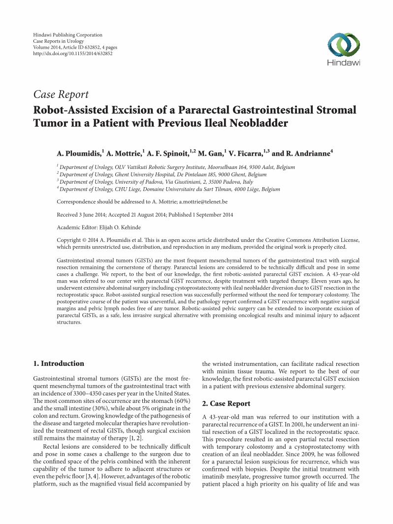
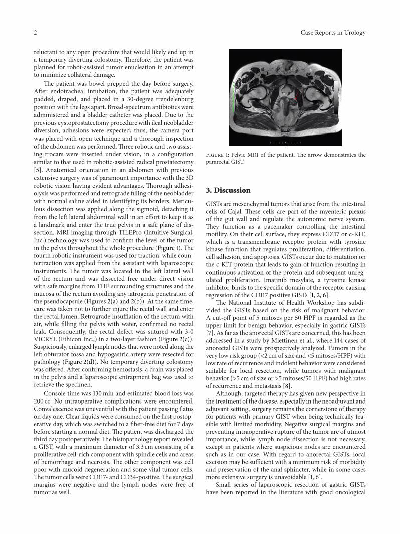
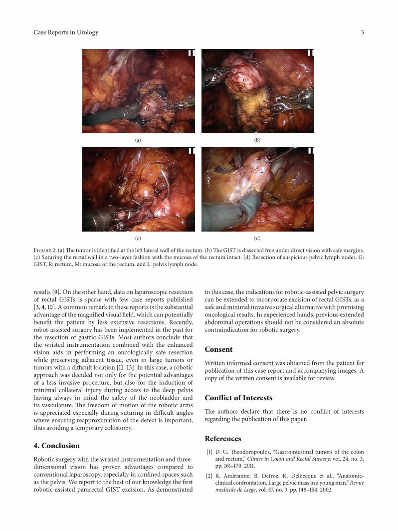
The patient was bowel prepped the day before surgery.After endotracheal intubation, the patient was adequatelypadded, draped, and placed in a 30-degree trendelenburgposition with the legs apart. Broad-spectrum antibiotics wereadministered and a bladder catheter was placed. Due to theprevious cystoprostatectomy procedure with ileal neobladderdiversion, adhesions were expected; thus, the camera portwas placed with open technique and a thorough inspectionof the abdomen was performed.Three robotic and two assist-ing trocars were inserted under vision, in a configurationsimilar to that used in robotic-assisted radical prostatectomy[5]. Anatomical orientation in an abdomen with previousextensive surgery was of paramount importance with the 3Drobotic vision having evident advantages. Thorough adhesi-olysis was performed and retrograde filling of the neobladderwith normal saline aided in identifying its borders. Meticu-lous dissection was applied along the sigmoid, detaching itfrom the left lateral abdominal wall in an effort to keep it asa landmark and enter the true pelvis in a safe plane of dis-section. MRI imaging through TILEPro (Intuitive Surgical,Inc.) technology was used to confirm the level of the tumorin the pelvis throughout the whole procedure (Figure 1). Thefourth robotic instrument was used for traction, while coun-tertraction was applied from the assistant with laparoscopicinstruments. The tumor was located in the left lateral wallof the rectum and was dissected free under direct visionwith safe margins from THE surrounding structures and themucosa of the rectum avoiding any iatrogenic penetration ofthe pseudocapsule (Figures 2(a) and 2(b)). At the same time,care was taken not to further injure the rectal wall and enterthe rectal lumen. Retrograde insufflation of the rectum withair, while filling the pelvis with water, confirmed no rectalleak. Consequently, the rectal defect was sutured with 3-0VICRYL (Ethicon Inc.,) in a two-layer fashion (Figure 2(c)).Suspiciously, enlarged lymph nodes that were noted along theleft obturator fossa and hypogastric artery were resected forpathology (Figure 2(d)). No temporary diverting colostomywas offered. After confirming hemostasis, a drain was placedin the pelvis and a laparoscopic entrapment bag was used toretrieve the specimen.
Console time was 130min and estimated blood loss was200 cc. No intraoperative complications were encountered.Convalescence was uneventful with the patient passing flatuson day one. Clear liquids were consumed on the first postop-erative day, which was switched to a fiber-free diet for 7 daysbefore starting a normal diet. The patient was discharged thethird day postoperatively.The histopathology report revealeda GIST, with a maximum diameter of 3.3 cm consisting of aproliferative cell-rich component with spindle cells and areasof hemorrhage and necrosis. The other component was cellpoor with mucoid degeneration and some vital tumor cells.The tumor cells were CD117- and CD34-positive.The surgicalmargins were negative and the lymph nodes were free oftumor as well.
Figure 1: Pelvic MRI of the patient. The arrow demonstrates thepararectal GIST.
3. Discussion
GISTs are mesenchymal tumors that arise from the intestinalcells of Cajal. These cells are part of the myenteric plexusof the gut wall and regulate the autonomic nerve system.They function as a pacemaker controlling the intestinalmotility. On their cell surface, they express CD117 or c-KIT,which is a transmembrane receptor protein with tyrosinekinase function that regulates proliferation, differentiation,cell adhesion, and apoptosis. GISTs occur due to mutation onthe c-KIT protein that leads to gain of function resulting incontinuous activation of the protein and subsequent unreg-ulated proliferation. Imatinib mesylate, a tyrosine kinaseinhibitor, binds to the specific domain of the receptor causingregression of the CD117 positive GISTs [1, 2, 6].
The National Institute of Health Workshop has subdi-vided the GISTs based on the risk of malignant behavior.A cut-off point of 5 mitoses per 50 HPF is regarded as theupper limit for benign behavior, especially in gastric GISTs[7]. As far as the anorectal GISTs are concerned, this has beenaddressed in a study by Miettinen et al., where 144 cases ofanorectal GISTs were prospectively analyzed. Tumors in thevery low risk group (<2 cm of size and <5 mitoses/HPF) withlow rate of recurrence and indolent behavior were consideredsuitable for local resection, while tumors with malignantbehavior (>5 cm of size or >5mitoses/50 HPF) had high ratesof recurrence and metastasis [8].
Although, targeted therapy has given new perspective inthe treatment of the disease, especially in the neoadjuvant andadjuvant setting, surgery remains the cornerstone of therapyfor patients with primary GIST when being technically fea-sible with limited morbidity. Negative surgical margins andpreventing intraoperative rupture of the tumor are of utmostimportance, while lymph node dissection is not necessary,except in patients where suspicious nodes are encounteredsuch as in our case. With regard to anorectal GISTs, localexcision may be sufficient with a minimum risk of morbidityand preservation of the anal sphincter, while in some casesmore extensive surgery is unavoidable [1, 6].
Small series of laparoscopic resection of gastric GISTshave been reported in the literature with good oncological
Case Reports in Urology 3
(a) (b)
(c) (d)
Figure 2: (a)The tumor is identified at the left lateral wall of the rectum. (b)The GIST is dissected free under direct vision with safe margins.(c) Suturing the rectal wall in a two-layer fashion with the mucosa of the rectum intact. (d) Resection of suspicious pelvic lymph nodes. G:GIST, R: rectum, M: mucosa of the rectum, and L: pelvis lymph node.
results [9]. On the other hand, data on laparoscopic resectionof rectal GISTs is sparse with few case reports published[3, 4, 10]. A common remark in these reports is the substantialadvantage of the magnified visual field, which can potentiallybenefit the patient by less extensive resections. Recently,robot-assisted surgery has been implemented in the past forthe resection of gastric GISTs. Most authors conclude thatthe wristed instrumentation combined with the enhancedvision aids in performing an oncologically safe resectionwhile preserving adjacent tissue, even in large tumors ortumors with a difficult location [11–13]. In this case, a roboticapproach was decided not only for the potential advantagesof a less invasive procedure, but also for the induction ofminimal collateral injury during access to the deep pelvishaving always in mind the safety of the neobladder andits vasculature. The freedom of motion of the robotic armsis appreciated especially during suturing in difficult angleswhere ensuring reapproximation of the defect is important,thus avoiding a temporary colostomy.
4. Conclusion
Robotic surgery with the wristed instrumentation and three-dimensional vision has proven advantages compared toconventional laparoscopy, especially in confined spaces suchas the pelvis. We report to the best of our knowledge the firstrobotic-assisted pararectal GIST excision. As demonstrated
in this case, the indications for robotic-assisted pelvic surgerycan be extended to incorporate excision of rectal GISTs, as asafe andminimal invasive surgical alternative with promisingoncological results. In experienced hands, previous extendedabdominal operations should not be considered an absolutecontraindication for robotic surgery.
Consent
Written informed consent was obtained from the patient forpublication of this case report and accompanying images. Acopy of the written consent is available for review.
Conflict of Interests
The authors declare that there is no conflict of interestsregarding the publication of this paper.
References
[1] D. G. Theodoropoulos, “Gastrointestinal tumors of the colonand rectum,” Clinics in Colon and Rectal Surgery, vol. 24, no. 3,pp. 161–170, 2011.
[2] R. Andrianne, B. Detroz, K. Delbecque et al., “Anatomic-clinical confrontation. Large pelvicmass in a youngman,”Revuemedicale de Liege, vol. 57, no. 3, pp. 148–154, 2002.
4 Case Reports in Urology
[3] T. Nakamura, A. Ihara, H. Mitomi et al., “Gastrointestinalstromal tumor of the rectum resected by laparoscopic surgery:report of a case,” Surgery Today, vol. 37, no. 11, pp. 1004–1008,2007.
[4] S.-C. Chang, T.-W. Ke, H.-C. Chiang, C. Wu, and W. T.-L. Chen, “Laparoscopic excision is an alterative method forrectal gastrointestinal stromal tumor,” Surgical Laparoscopy,Endoscopy and Percutaneous Techniques, vol. 20, no. 4, pp. 284–287, 2010.
[5] A. Mottrie, G. de Naeyer, P. Schatteman, E. Frumenzio, M.Rossanese, and V. Ficarra, “Robot-assisted radical prostatec-tomy: tips, tricks and pitfalls,”Minerva Urologica e Nefrologica,vol. 64, no. 2, pp. 89–96, 2012.
[6] R. Tielen, C. Verhoef, F. van Coevorden et al., “Surgicalmanagement of rectal gastrointestinal stromal tumors,” Journalof Surgical Oncology, vol. 107, no. 4, pp. 320–323, 2012.
[7] M. Miettinen, M. Sarlomo-Rikala, and J. Lasota, “Gastrointesti-nal stromal tumors: recent advances in understanding of theirbiology,” Human Pathology, vol. 30, no. 10, pp. 1213–1220, 1999.
[8] M. Miettinen, M. Furlong, M. Sarlomo-Rikala, A. Burke, L. H.Sobin, and J. Lasota, “Gastrointestinal stromal tumors, intra-mural leiomyomas, and leiomyosarcomas in the rectum andanus: a clinicopathologic, immunohistochemical, and molecu-lar genetic study of 144 cases,”The American Journal of SurgicalPathology, vol. 25, no. 9, pp. 1121–1133, 2001.
[9] Y.H. Chen, K.H. Liu, C.N. Yeh et al., “Laparoscopic resection ofgastrointestinal stromal tumors: safe, efficient, and comparableoncologic outcomes,” Journal of Laparoendoscopic & AdvancedSurgical Techniques, vol. 22, no. 8, pp. 758–763, 2012.
[10] R. Takahashi, S. Nagayama, Y. Mori et al., “A large cysticgastrointestinal stromal tumor of the rectum in the retrorectalspace,” International Journal of Clinical Oncology, vol. 15, no. 6,pp. 601–607, 2010.
[11] N. C. Buchs, P. Bucher, F. Pugin, M. E. Hagen, and P. Morel,“Robot-assisted oncologic resection for large gastric gastroin-testinal stromal tumor: a preliminary case series,” Journal ofLaparoendoscopic andAdvanced Surgical Techniques, vol. 20, no.5, pp. 411–415, 2010.
[12] E. Ortiz-Oshiro, P. B. Exposito, J. M. Sierra, J. D. Gonzalez, D.S. Barbosa, and J. A. Fernandez-Represa, “Laparoscopic androbotic distal gastrectomy for gastrointestinal stromal tumour:case report,” International Journal of Medical Robotics andComputer Assisted Surgery, vol. 8, no. 4, pp. 491–495, 2012.
[13] H. Moriyama, N. Ishikawa, M. Kawaguchi, K. Hirose, andG. Watanabe, “Robot-assisted laparoscopic resection for gas-tric gastrointestinal stromal tumor,” Surgical Laparoscopy,Endoscopy and Percutaneous Techniques, vol. 22, no. 3, pp. e155–e156, 2012.

Submit your manuscripts athttp://www.hindawi.com
Stem CellsInternational
Hindawi Publishing Corporationhttp://www.hindawi.com Volume 2014
Hindawi Publishing Corporationhttp://www.hindawi.com Volume 2014
MEDIATORSINFLAMMATION
of
Hindawi Publishing Corporationhttp://www.hindawi.com Volume 2014
Behavioural Neurology
EndocrinologyInternational Journal of
Hindawi Publishing Corporationhttp://www.hindawi.com Volume 2014
Hindawi Publishing Corporationhttp://www.hindawi.com Volume 2014
Disease Markers
Hindawi Publishing Corporationhttp://www.hindawi.com Volume 2014
BioMed Research International
OncologyJournal of
Hindawi Publishing Corporationhttp://www.hindawi.com Volume 2014
Hindawi Publishing Corporationhttp://www.hindawi.com Volume 2014
Oxidative Medicine and Cellular Longevity
Hindawi Publishing Corporationhttp://www.hindawi.com Volume 2014
PPAR Research
The Scientific World JournalHindawi Publishing Corporation http://www.hindawi.com Volume 2014
Immunology ResearchHindawi Publishing Corporationhttp://www.hindawi.com Volume 2014
Journal of
ObesityJournal of
Hindawi Publishing Corporationhttp://www.hindawi.com Volume 2014
Hindawi Publishing Corporationhttp://www.hindawi.com Volume 2014
Computational and Mathematical Methods in Medicine
OphthalmologyJournal of
Hindawi Publishing Corporationhttp://www.hindawi.com Volume 2014
Diabetes ResearchJournal of
Hindawi Publishing Corporationhttp://www.hindawi.com Volume 2014
Hindawi Publishing Corporationhttp://www.hindawi.com Volume 2014
Research and TreatmentAIDS
Hindawi Publishing Corporationhttp://www.hindawi.com Volume 2014
Gastroenterology Research and Practice
Hindawi Publishing Corporationhttp://www.hindawi.com Volume 2014
Parkinson’s Disease
Evidence-Based Complementary and Alternative Medicine
Volume 2014Hindawi Publishing Corporationhttp://www.hindawi.com