case report pneumothorax caused by an isolated midshaft clavicle...
TRANSCRIPT
Case ReportPneumothorax Caused by an Isolated Midshaft Clavicle Fracture
Najla Feriani, Hassen Ben Ghezala, and Salah Snouda
Faculty of Medicine of Tunis, Tunis El-Manar University, Regional Hospital of Zaghouan, Street of Republic, 1100 Zaghouan, Tunisia
Correspondence should be addressed to Hassen Ben Ghezala; [email protected]
Received 20 March 2016; Revised 28 March 2016; Accepted 29 March 2016
Academic Editor: Aristomenis K. Exadaktylos
Copyright © 2016 Najla Feriani et al. This is an open access article distributed under the Creative Commons Attribution License,which permits unrestricted use, distribution, and reproduction in any medium, provided the original work is properly cited.
Patients with isolated clavicle fractures are frequent in the emergency department. However, unusual clavicle fracturescomplications, such as pneumothorax, are rare. Previous reports indicated that all pneumothorax cases were treated via performingthoracostomy. Conservatively, the treatment of the clavicle fracture, like in our case, was successful. Despite the fact that isolatedclavicle fractures rarely cause complications and generally heal with immobilization, serious complications may occur requiringurgent treatment. It has been proven that physical examinations, with particular attention to the neurovascular and chestexaminations, and radiographs of the clavicle are necessary to prevent overlooking these potentially dangerous complications.
1. Introduction
Fractures of the clavicle are relatively common, representing2.6% to 4% of fractures among adults [1–3]. The majority ofthese fractures occur in the middle third of the shaft. Theyare the result of low-energy mechanisms such as a fall ontothe shoulder [2–5]. Pneumothorax, as a consequence of aclavicle fracture, is a rare but potentially lethal complication[6]. We report in this work an exceptional case of isolatedclavicle fracture causing a pneumothorax, which required theinsertion of a chest drain.
2. Case Report
A 30-year-old male was admitted to the emergency depart-ment of our hospital, after a sideslip of his car. During themedical examination, he complained of a pain in the leftshoulder. There was no relevant medical history. The patientsymptoms and behavior revealed no clinical distress. Therewere no neurovascular deficits in the right upper limb. Thelung breath sounds and percussion notes were normal on theright shoulder and low on the left one.
The patient denied any chest discomfort, dyspnea,or hemoptysis. He had moderate central and peripheralcyanosis. The respiratory rate was 24 breaths per minute. Apulse oximetry monitor showed an oxygen saturation level of92 percent.The arterial blood pressure was 130/70mmHg and
the pulse was 95 bpm. Any further physical examination wasunremarkable.The Glasgow coma scale was 15 with a normalneurological examination.
The initial laboratory data included serum hemoglobin of125 g/L and hematocrit of 43%. The white blood cell countwas normal at 9000 cells per microliter with a normal plateletcount of 326 billion platelets per liter of blood. Laboratorystudies included normal prothrombin time, activated partialthromboplastin time, normal platelet functional analysis,and negative disseminated intravascular coagulation screen.Serum electrolyte levels and renal function were normal.Arterial blood gas analysis showed a mild hypoxemia with aPaO2of 78mmHg and a SaO
2greater than 96%.The patient
had a normal acid-base balance; the pHwas 7.42 with a serumbicarbonate level at 22mEq/L. The blood gas carbon dioxidelevel was normal at 36mmHg.
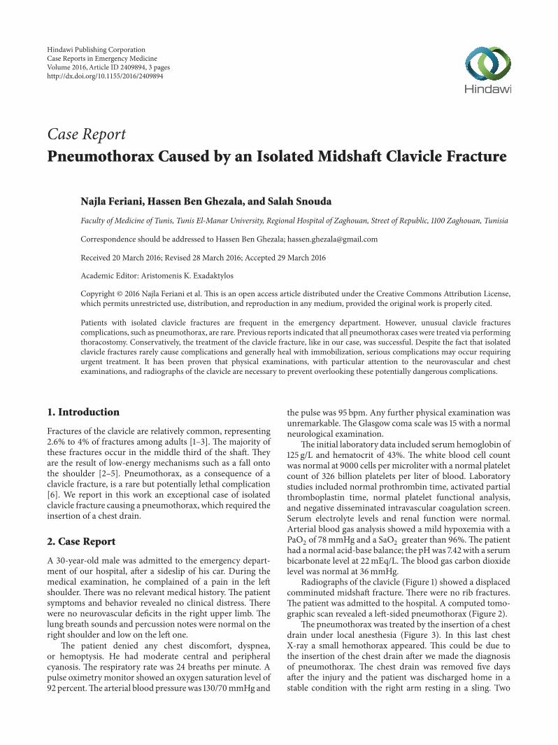
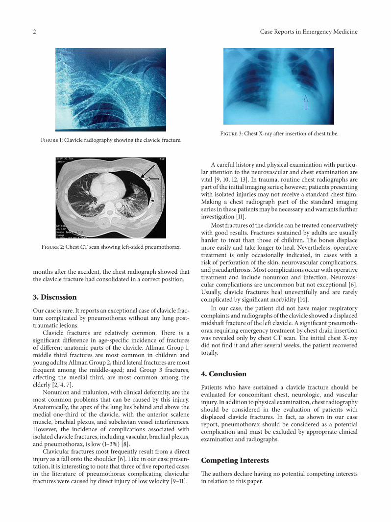
Radiographs of the clavicle (Figure 1) showed a displacedcomminuted midshaft fracture. There were no rib fractures.The patient was admitted to the hospital. A computed tomo-graphic scan revealed a left-sided pneumothorax (Figure 2).
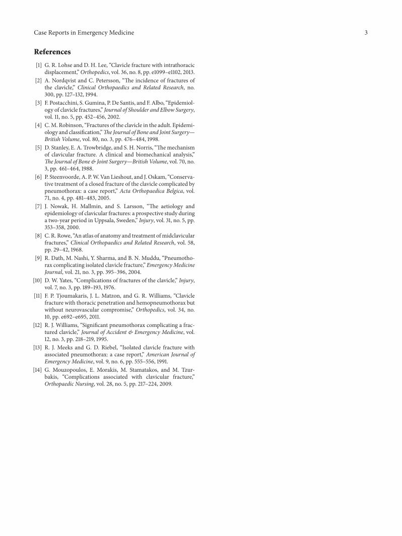
The pneumothorax was treated by the insertion of a chestdrain under local anesthesia (Figure 3). In this last chestX-ray a small hemothorax appeared. This could be due tothe insertion of the chest drain after we made the diagnosisof pneumothorax. The chest drain was removed five daysafter the injury and the patient was discharged home in astable condition with the right arm resting in a sling. Two
Hindawi Publishing CorporationCase Reports in Emergency MedicineVolume 2016, Article ID 2409894, 3 pageshttp://dx.doi.org/10.1155/2016/2409894
2 Case Reports in Emergency Medicine
Figure 1: Clavicle radiography showing the clavicle fracture.
Figure 2: Chest CT scan showing left-sided pneumothorax.
months after the accident, the chest radiograph showed thatthe clavicle fracture had consolidated in a correct position.
3. Discussion
Our case is rare. It reports an exceptional case of clavicle frac-ture complicated by pneumothorax without any lung post-traumatic lesions.
Clavicle fractures are relatively common. There is asignificant difference in age-specific incidence of fracturesof different anatomic parts of the clavicle. Allman Group 1,middle third fractures are most common in children andyoung adults; AllmanGroup 2, third lateral fractures aremostfrequent among the middle-aged; and Group 3 fractures,affecting the medial third, are most common among theelderly [2, 4, 7].
Nonunion and malunion, with clinical deformity, are themost common problems that can be caused by this injury.Anatomically, the apex of the lung lies behind and above themedial one-third of the clavicle, with the anterior scalenemuscle, brachial plexus, and subclavian vessel interferences.However, the incidence of complications associated withisolated clavicle fractures, including vascular, brachial plexus,and pneumothorax, is low (1–3%) [8].
Clavicular fractures most frequently result from a directinjury as a fall onto the shoulder [6]. Like in our case presen-tation, it is interesting to note that three of five reported casesin the literature of pneumothorax complicating clavicularfractures were caused by direct injury of low velocity [9–11].
Figure 3: Chest X-ray after insertion of chest tube.
A careful history and physical examination with particu-lar attention to the neurovascular and chest examination arevital [9, 10, 12, 13]. In trauma, routine chest radiographs arepart of the initial imaging series; however, patients presentingwith isolated injuries may not receive a standard chest film.Making a chest radiograph part of the standard imagingseries in these patientsmay be necessary andwarrants furtherinvestigation [11].
Most fractures of the clavicle can be treated conservativelywith good results. Fractures sustained by adults are usuallyharder to treat than those of children. The bones displacemore easily and take longer to heal. Nevertheless, operativetreatment is only occasionally indicated, in cases with arisk of perforation of the skin, neurovascular complications,and pseudarthrosis. Most complications occur with operativetreatment and include nonunion and infection. Neurovas-cular complications are uncommon but not exceptional [6].Usually, clavicle fractures heal uneventfully and are rarelycomplicated by significant morbidity [14].
In our case, the patient did not have major respiratorycomplaints and radiographs of the clavicle showed a displacedmidshaft fracture of the left clavicle. A significant pneumoth-orax requiring emergency treatment by chest drain insertionwas revealed only by chest CT scan. The initial chest X-raydid not find it and after several weeks, the patient recoveredtotally.
4. Conclusion
Patients who have sustained a clavicle fracture should beevaluated for concomitant chest, neurologic, and vascularinjury. In addition to physical examination, chest radiographyshould be considered in the evaluation of patients withdisplaced clavicle fractures. In fact, as shown in our casereport, pneumothorax should be considered as a potentialcomplication and must be excluded by appropriate clinicalexamination and radiographs.
Competing Interests
The authors declare having no potential competing interestsin relation to this paper.
Case Reports in Emergency Medicine 3
References
[1] G. R. Lohse and D. H. Lee, “Clavicle fracture with intrathoracicdisplacement,”Orthopedics, vol. 36, no. 8, pp. e1099–e1102, 2013.
[2] A. Nordqvist and C. Petersson, “The incidence of fractures ofthe clavicle,” Clinical Orthopaedics and Related Research, no.300, pp. 127–132, 1994.
[3] F. Postacchini, S. Gumina, P. De Santis, and F. Albo, “Epidemiol-ogy of clavicle fractures,” Journal of Shoulder and Elbow Surgery,vol. 11, no. 5, pp. 452–456, 2002.
[4] C.M. Robinson, “Fractures of the clavicle in the adult. Epidemi-ology and classification,”The Journal of Bone and Joint Surgery—British Volume, vol. 80, no. 3, pp. 476–484, 1998.
[5] D. Stanley, E. A. Trowbridge, and S. H. Norris, “Themechanismof clavicular fracture. A clinical and biomechanical analysis,”The Journal of Bone & Joint Surgery—British Volume, vol. 70, no.3, pp. 461–464, 1988.
[6] P. Steenvoorde, A. P.W.Van Lieshout, and J. Oskam, “Conserva-tive treatment of a closed fracture of the clavicle complicated bypneumothorax: a case report,” Acta Orthopaedica Belgica, vol.71, no. 4, pp. 481–483, 2005.
[7] J. Nowak, H. Mallmin, and S. Larsson, “The aetiology andepidemiology of clavicular fractures: a prospective study duringa two-year period in Uppsala, Sweden,” Injury, vol. 31, no. 5, pp.353–358, 2000.
[8] C. R. Rowe, “An atlas of anatomy and treatment ofmidclavicularfractures,” Clinical Orthopaedics and Related Research, vol. 58,pp. 29–42, 1968.
[9] R. Dath, M. Nashi, Y. Sharma, and B. N. Muddu, “Pneumotho-rax complicating isolated clavicle fracture,” EmergencyMedicineJournal, vol. 21, no. 3, pp. 395–396, 2004.
[10] D. W. Yates, “Complications of fractures of the clavicle,” Injury,vol. 7, no. 3, pp. 189–193, 1976.
[11] F. P. Tjoumakaris, J. L. Matzon, and G. R. Williams, “Claviclefracture with thoracic penetration and hemopneumothorax butwithout neurovascular compromise,” Orthopedics, vol. 34, no.10, pp. e692–e695, 2011.
[12] R. J. Williams, “Significant pneumothorax complicating a frac-tured clavicle,” Journal of Accident & Emergency Medicine, vol.12, no. 3, pp. 218–219, 1995.
[13] R. J. Meeks and G. D. Riebel, “Isolated clavicle fracture withassociated pneumothorax: a case report,” American Journal ofEmergency Medicine, vol. 9, no. 6, pp. 555–556, 1991.
[14] G. Mouzopoulos, E. Morakis, M. Stamatakos, and M. Tzur-bakis, “Complications associated with clavicular fracture,”Orthopaedic Nursing, vol. 28, no. 5, pp. 217–224, 2009.

Submit your manuscripts athttp://www.hindawi.com
Stem CellsInternational
Hindawi Publishing Corporationhttp://www.hindawi.com Volume 2014
Hindawi Publishing Corporationhttp://www.hindawi.com Volume 2014
MEDIATORSINFLAMMATION
of
Hindawi Publishing Corporationhttp://www.hindawi.com Volume 2014
Behavioural Neurology
EndocrinologyInternational Journal of
Hindawi Publishing Corporationhttp://www.hindawi.com Volume 2014
Hindawi Publishing Corporationhttp://www.hindawi.com Volume 2014
Disease Markers
Hindawi Publishing Corporationhttp://www.hindawi.com Volume 2014
BioMed Research International
OncologyJournal of
Hindawi Publishing Corporationhttp://www.hindawi.com Volume 2014
Hindawi Publishing Corporationhttp://www.hindawi.com Volume 2014
Oxidative Medicine and Cellular Longevity
Hindawi Publishing Corporationhttp://www.hindawi.com Volume 2014
PPAR Research
The Scientific World JournalHindawi Publishing Corporation http://www.hindawi.com Volume 2014
Immunology ResearchHindawi Publishing Corporationhttp://www.hindawi.com Volume 2014
Journal of
ObesityJournal of
Hindawi Publishing Corporationhttp://www.hindawi.com Volume 2014
Hindawi Publishing Corporationhttp://www.hindawi.com Volume 2014
Computational and Mathematical Methods in Medicine
OphthalmologyJournal of
Hindawi Publishing Corporationhttp://www.hindawi.com Volume 2014
Diabetes ResearchJournal of
Hindawi Publishing Corporationhttp://www.hindawi.com Volume 2014
Hindawi Publishing Corporationhttp://www.hindawi.com Volume 2014
Research and TreatmentAIDS
Hindawi Publishing Corporationhttp://www.hindawi.com Volume 2014
Gastroenterology Research and Practice
Hindawi Publishing Corporationhttp://www.hindawi.com Volume 2014
Parkinson’s Disease
Evidence-Based Complementary and Alternative Medicine
Volume 2014Hindawi Publishing Corporationhttp://www.hindawi.com