case report - hindawi publishing corporation · case report ... scheduled to perform a diagnostic...
TRANSCRIPT
Hindawi Publishing CorporationCase Reports in CardiologyVolume 2011, Article ID 413645, 3 pagesdoi:10.1155/2011/413645
Case Report
Inverted Takotsubo Cardiomyopathy Induced by DobutamineStress Echocardiography with Atypical Presentation
Christian Cadeddu,1 Silvio Nocco,1 Fabio Cadeddu,1 Martino Deidda,1 Pierpaolo Bassareo,1
Alessandra Serra,2 Mario Piga,2 and Giuseppe Mercuro1
1 Department of Cardiovascular and Neurological Sciences, University Hospital of Cagliari, Strada Statale 554,Km 4.500, Monserrato, 09042 Cagliari, Italy
2 Department of Nuclear Medicine, University Hospital of Cagliari, Strada Statale 554, Km 4.500, Monserrato, 09042 Cagliari, Italy
Correspondence should be addressed to Christian Cadeddu, [email protected]
Received 7 June 2011; Accepted 11 July 2011
Academic Editors: K.-R. Chiou, K. Shimada, and C. S. Snyder
Copyright © 2011 Christian Cadeddu et al. This is an open access article distributed under the Creative Commons AttributionLicense, which permits unrestricted use, distribution, and reproduction in any medium, provided the original work is properlycited.
A 48-year-old woman was scheduled by our lab to perform a standard dobutamine/atropine stress echocardiogram. Duringthe test, the patient referred to a slight chest discomfort and developed a progressive left ventricle akinesia of all midbasal LVsegments, thus mimicking a midbasal ballooning. ECG persisted without significant abnormalities and with no raise of Troponin I.Coronary angiography showed normal coronary arteries and ventriculography a severe EF reduction and apical hypercontractility.Echocardiography showed a progressive improvement with a complete recovery 48 hours later. This is a rare case of invertedtakotsubo syndrome induced by dobutamine stress echocardiography that occurred with atypical presentation.
1. Introduction
Takotsubo cardiomyopathy is increasingly recognised as asyndrome, usually provoked by severe mental stress andassociated with acute LV apical “ballooning” or dyskinesia[1]. Its clinical symptoms and signs resemble those of anacute coronary event in presence of normally appearingepicardial coronary arteries. Major criteria of this syndromeare (i) an echocardiogram showing decreased apical con-tractility with basal hyperkinesia and, occasionally, intraven-tricular pressure gradients, (ii) the absence of obstructivecoronary disease or angiographic evidence of acute plaquerupture, (iii) new ECG abnormalities (ST-segment elevationand/or T-wave inversion) or modest elevation in cardiactroponin, and (iv) the absence of pheochromocytoma andmyocarditis. Although the aetiology of Takotsubo syndromeremains obscure, the combination of severe anxiety andcatecholamine release appears to be the major trigger[2]. Cases of transient cardiomyopathy of mid and basalmyocardial segments have recently been described [3, 4].
We present a rare case of inverted Takotsubo syndromeinduced by dobutamine stress echocardiography with atypi-cal presentation.
2. Case Report
A 48-year-old female with a long history of atypical pre-cordial chest pain was referred to our echolaboratory fora dobutamine/atropine stress echocardiogram (DSE). Onemonth before she was admitted to a peripheral general hospi-tal for prolonged chest discomfort precipitated by emotionalstress. At arrival, resting ECG showed no abnormalitieswith no raise in troponin I or in other markers of cardiacnecrosis. Transthoracic echocardiography only showed amild impairment of LV diastolic function. At discharge, abicycle ergometric test demonstrated ST segment depressionin the inferolateral leads; during the test the patient didnot experience any chest pain. Therefore, the patient wasscheduled to perform a diagnostic DSE.
At our lab, the patient was clinically stable but with ananxious personality profile. The patient interview revealed anegative family history for cardiovascular diseases and thatshe had undergone an early menopause (age 45 years)without hormonal replacement therapy. Risk factors forcoronary artery disease included smoking and hyperc-holesterolaemia, but not overweight or suffering fromhypertension or diabetes. The patient underwent a full
2 Case Reports in Cardiology
(a) (b)
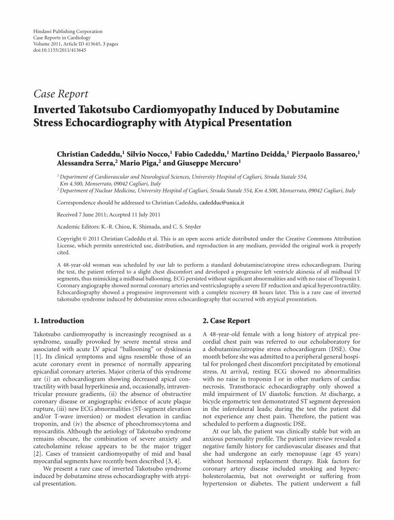
Figure 1: Ventriculography confirmed a global severe reduction of the systolic function (EF 28%) and apical hyperkinesis and severedysfunction of the basal segments; (a) diastole, (b) systole.
cardiovascular assessment (including physical examination,ECG, and transthoracic echocardiography), which revealedno cardiovascular or pulmonary diseases.
A standard DSE protocol was used with 10 mcg/kg/mindose increments at 3 min intervals. Resting echocardio-gram and blood pressure were normal. At 40 mcg/kg/minof dobutamine and following 0.5 mg of atropine (heartrate reached 154 bpm), we noticed anterior wall mid-basalhypokinesia without any ECG abnormalities. The test wasinterrupted and the recovery images showed a progressiveworsening of the kinetic pattern with first marked systolichypokinesia followed by akinesia of all LV mid-basal seg-ments and severe impairment of global systolic function(ejection fraction 25%; Clip 1 (supplementary material)shows the 4 chamber echocardiographic view of the leftventricle demonstrating the severe impairment of globalsystolic function (supplementary material available on line atdoi:10.1155/2011/413645)). ECG continued without show-ing any significant abnormalities and the patient referred tono chest pain. Blood pressure at this moment was 160/100,nitrates were administered i.v. and the patient was trans-ferred to the cardiac catheterisation laboratory to perform acoronary angiography with suspected dobutamine-inducedmyocardial infarction. Ventriculography confirmed a severereduction of the systolic function (EF 28%) and showedapical hyperkinesis and severe dysfunction of the midseg-ments with initial recovery of the basal segments (Figure 1);on the contrary, the angiographic study did not show anycoronary lesions, despite a slow flow in the left coronaryartery. The patient was then admitted to the intensive careunit, where the monitored plasma troponin I level remainedin the normal range (<0.1 pg/mL) in the next 24 hours.Serial echocardiograms showed a progressive recovery of thesystolic function with normal wall motion 48 hours later.
One month later, the patient underwent a cardiacMRI study which confirmed the complete recovery of the
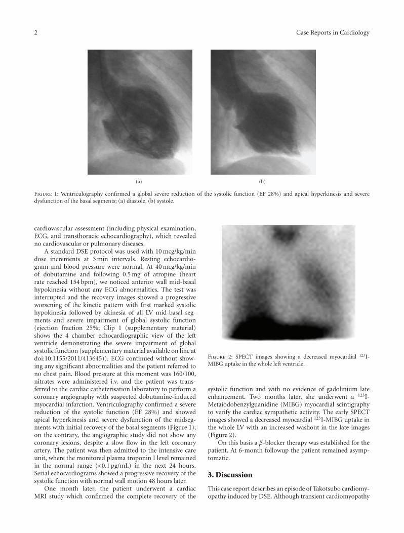
Figure 2: SPECT images showing a decreased myocardial 123I-MIBG uptake in the whole left ventricle.
systolic function and with no evidence of gadolinium lateenhancement. Two months later, she underwent a 123I-Metaiodobenzylguanidine (MIBG) myocardial scintigraphyto verify the cardiac sympathetic activity. The early SPECTimages showed a decreased myocardial 123I-MIBG uptake inthe whole LV with an increased washout in the late images(Figure 2).
On this basis a β-blocker therapy was established for thepatient. At 6-month followup the patient remained asymp-tomatic.
3. Discussion
This case report describes an episode of Takotsubo cardiomy-opathy induced by DSE. Although transient cardiomyopathy
Case Reports in Cardiology 3
induced by pharmacological stress has been reported previ-ously [5], this is a rare case of dobutamine-induced invertedTakotsubo (mid-basal dysfunction), which occurred with anatypical presentation. Atypical features included no chestpain, no ECG abnormalities, and a lack of increase in cardiactroponin.
Several aetiological factors have been proposed for Takot-subo cardiomyopathy, including microvascular dysfunction,multivessel epicardial coronary spasm, catecholamine car-diotoxicity, and neurogenic stunned myocardium [6]. Oneor more of these pathogenetic interpretations conformto our patient’s physical and clinical profile: an anxious,perimenopausal female who was a heavy smoker.
Furthermore, our patient suits the profile of an X syn-drome: a postmenopausal woman with ECG changes duringexercise, but without patent coronary stenosis. Consistentwith this, an impaired endothelial function has been demon-strated in peri- and postmenopausal women. In comparisonwith premenopausal individuals of the same age, post-menopausal women showed increased vascular resistance,worse vasodilator reserve, and higher basal and stimulatedplasma norepinephrine levels [7]. Moreover, reduced estro-gen levels following menopause might be involved both byindirect action on the nervous system and by direct actionon the heart [8].
In addition, several studies reported that Takotsubocardiomyopathy may be accompanied by coronary microcir-culatory abnormalities [9], which in turn correlate with theseverity of myonecrosis and ECG abnormalities.
The most obvious clinical finding is, however, that theTakotsubo syndrome has a predilection for women andshows a clear tendency to occur at times of very intense emo-tional stress, thereby suggesting a sympathetic-based aetiol-ogy. The close relationship with a high dobutamine dose,which was also observed elsewhere, supports a catechola-mine-related mechanism in our patient.
The reduction of 123I-MIBG uptake agrees with theresults of a study conducted with the same technique in 8patients in which it was objectively documented that theTakotsubo syndrome is produced by an impaired cardiacadrenergic activity [6]. In that study, the dysfunction waspresent in the acute phase, whereas the 123I-MIBG scintig-raphy documented a gradual normalization of sympatheticfunction after 3 months. Conversely, in the woman westudied, a global and severe uptake reduction was still presentafter the same time. This finding suggests that in some cases,like ours, the disturbance of cardiac innervation may bestructural and, at least in part, independent of the ultimatecause triggering neurogenic myocardial stunning.
In conclusion, the most recent data on the atypicalTakotsubo, given the variety of clinical profiles describedin these patients [10], suggest the need for further researchregarding the definition and pathophysiology of stressinduced cardiomyopathy.
References
[1] S. W. Sharkey, J. R. Lesser, A. G. Zenovich et al., “Acute andreversible cardiomyopathy provoked by stress in women from
the United States,” Circulation, vol. 111, no. 4, pp. 472–479,2005.
[2] J. Silberbauer, P. Hong, and G. W. Lloyd, “Takotsubo car-diomyopathy (left ventricular ballooning syndrome) inducedduring dobutamine stress echocardiography,” European Jour-nal of Echocardiography, vol. 9, no. 1, pp. 136–138, 2008.
[3] R. T. Hurst, J. W. Askew, C. S. Reuss et al., “Transientmidventricular ballooning ayndrome. A new variant,” Journalof the American College of Cardiology, vol. 48, no. 3, pp. 579–583, 2006.
[4] A. Sanchez-Recalde, C. Iborra, O. Costero et al., “Takotsubocardiomyopathy—a new variant and widening disease spec-trum. “Inverted Takotsubo” pattern related to catecholamine-toxicity,” International Journal of Cardiology, vol. 132, no. 3,pp. 437–438, 2009.
[5] J. Abraham, J. O. Mudd, N. Kapur, K. Klein, H. C. Champion,and I. S. Wittstein, “Stress cardiomyopathy after intravenousadministration of catecholamines and beta-receptor agonists,”Journal of the American College of Cardiology, vol. 53, no. 15,pp. 1320–1325, 2009.
[6] Y. J. Akashi, K. Nakazawa, M. Sakakibara, F. Miyake, H.Musha, and K. Sasaka, “123I-MIBG myocardial scintigraphyin patients with “takotsubo” cardiomyopathy,” Journal ofNuclear Medicine, vol. 45, no. 7, pp. 1121–1127, 2004.
[7] G. Mercuro, G. Longu, S. Zoncu, and A. Cherchi, “Impairedforearm blood flow and vasodilator reserve in healthy post-menopausal women,” American Heart Journal, vol. 137, no. 4,part 1, pp. 692–697, 1999.
[8] T. Ueyama, K. Kasamatsu, T. Hano, Y. Tsuruo, and F.Ishikura, “Catecholamines and estrogen are involved in thepathogenesis of emotional stress-induced acute heart attack,”Annals of the New York Academy of Sciences, vol. 1148, pp. 479–485, 2008.
[9] T. Yoshida, T. Hibino, N. Kako et al., “A pathophysiologic studyof tako-tsubo cardiomyopathy with F-18 fluorodeoxyglucosepositron emission tomography,” European Heart Journal, vol.28, no. 21, pp. 2598–2604, 2007.
[10] S. S. Abdelmoneim, S. V. Mankad, M. Bernier et al.,“Microvascular function in takotsubo cardiomyopathy withcontrast echocardiography: prospective evaluation and reviewof literature,” Journal of the American Society of Echocardiogra-phy, vol. 22, no. 11, pp. 1249–1255, 2009.

Submit your manuscripts athttp://www.hindawi.com
Stem CellsInternational
Hindawi Publishing Corporationhttp://www.hindawi.com Volume 2014
Hindawi Publishing Corporationhttp://www.hindawi.com Volume 2014
MEDIATORSINFLAMMATION
of
Hindawi Publishing Corporationhttp://www.hindawi.com Volume 2014
Behavioural Neurology
EndocrinologyInternational Journal of
Hindawi Publishing Corporationhttp://www.hindawi.com Volume 2014
Hindawi Publishing Corporationhttp://www.hindawi.com Volume 2014
Disease Markers
Hindawi Publishing Corporationhttp://www.hindawi.com Volume 2014
BioMed Research International
OncologyJournal of
Hindawi Publishing Corporationhttp://www.hindawi.com Volume 2014
Hindawi Publishing Corporationhttp://www.hindawi.com Volume 2014
Oxidative Medicine and Cellular Longevity
Hindawi Publishing Corporationhttp://www.hindawi.com Volume 2014
PPAR Research
The Scientific World JournalHindawi Publishing Corporation http://www.hindawi.com Volume 2014
Immunology ResearchHindawi Publishing Corporationhttp://www.hindawi.com Volume 2014
Journal of
ObesityJournal of
Hindawi Publishing Corporationhttp://www.hindawi.com Volume 2014
Hindawi Publishing Corporationhttp://www.hindawi.com Volume 2014
Computational and Mathematical Methods in Medicine
OphthalmologyJournal of
Hindawi Publishing Corporationhttp://www.hindawi.com Volume 2014
Diabetes ResearchJournal of
Hindawi Publishing Corporationhttp://www.hindawi.com Volume 2014
Hindawi Publishing Corporationhttp://www.hindawi.com Volume 2014
Research and TreatmentAIDS
Hindawi Publishing Corporationhttp://www.hindawi.com Volume 2014
Gastroenterology Research and Practice
Hindawi Publishing Corporationhttp://www.hindawi.com Volume 2014
Parkinson’s Disease
Evidence-Based Complementary and Alternative Medicine
Volume 2014Hindawi Publishing Corporationhttp://www.hindawi.com