case report amiodarone-induced pulmonary toxicity: … · amiodarone-induced pulmonary toxicity:...
TRANSCRIPT
144144 THE EWHA MEDICAL JOURNALTHE EWHA MEDICAL JOURNAL
Amiodarone-Induced Pulmonary Toxicity: Percutaneous Needle Aspiration Biopsy and Ultrastructural Findings
In Sook Kang, Jin Hwa Lee, Sun Hee Sung1, Seong Hoon ParkDepartments of Internal Medicine and 1Pathology, Ewha Womans University School of Medicine, Seoul, Korea
Introduction
Amiodarone, a bi-iodinated benzofuran derivative, is widely
used for arrhythmias. Many patients benefit from the effective-
ness of amiodarone in treating potentially life-threatening ar-
rhythmias. However, amiodarone-induced pulmonary toxicity
(APT) is a severe complication. Even after the discontinuation
of amiodarone, not only is APT difficult to manage, but there
is also a potentially life-threatening risk of recurrence of ar-
rhythmias. In this case, we describe multiorgan complications
secondary to amiodarone use, especially APT combined with
pneumonia with atypical pathogens and pulmonary hemorrhage
following procedures. Following treatment and stabilization, the
patient experienced sudden cardiac death while recovering from
pulmonary complications.
Case
A 65-year-old man presented with fever, worsening cough,
and dyspnea. He had lost 10 kg over the past 2 months. He
had a medical history of atrial fibrillation; myocardial infarction
caused by diffuse spasm of the left and right coronary arter-
ies; and aborted sudden cardiac death from ventricular tachy-
cardia, which was converted to sinus rhythm following electric
cardioversion. He was a 40 pack-year smoker and a chronic
alcoholic. Regular medications included aspirin, nicorandil,
diltiazem, valsartan, warfarin, and amiodarone. He had taken
400 mg/day amiodarone for 3 years (a total cumulative dose
of 232 g) to control atrial fibrillation and life-threatening ven-
tricular tachycardia. Because APT was suspected, amiodarone
was immediately discontinued. On examination, the patient had
a regular heart rate of 78 beats/min, blood pressure of 90/50
mm Hg, respiratory rate of 20 breaths/min, and body tem-
Case Report
Ewha Med J 2013;36(2):144-148http://dx.doi.org/10.12771/emj.2013.36.2.144pISSN 2234-3180 • eISSN 2234-2591
Amiodaronehasbeenwidelyusedforsupraventricularandventriculararrhythmiasandmanypatientsbenefitfromitseffectivenessintreatingpotentiallylife-threateningarrhythmias.However,thisdrugcancausemulti-organtoxicity,includingamiodarone-inducedpulmonarytoxicity(APT).Notonlydoesamiodaronehavealonghalf-lifebutalsoislipophilicandthereforecaneasilyaccumulateintissues.Hence,itisdifficulttomonitortherapeuticlevelsandsideeffects,makingitdifficulttopredicttoxicities.Inthiscase,wedescribemulti-organcomplicationssecondarytoamiodaroneuse,especiallyAPTcombinedwithpneumoniawithatypicalpathogensandpulmonaryhemorrhage.Thepatientreachedahighcumulativedoseofamiodaronedespitealowmaintenancedoseofamiodarone.ThiscasehighlightsanunusualpresentationofAPTwithmulti-organtoxicityandwereviewarticlesregardingtheassociationbetweenthecumulativedoseofamiodaroneandamiodarone-inducedtoxicities. (Ewha Med J 2013;36(2):144-148)
Received May 14, 2013 Accepted June 17, 2013
Corresponding authorSeong Hoon ParkDivision of Cardiology, Department of Internal Medicine, Ewha Womans University School of Medicine, 1071 Anyangcheon-ro, Yangcheon-gu, Seoul 158-710, Korea Tel: 82-2-2650-5018, Fax: 82-2-2650-5424E-mail: [email protected]
Key WordsAmiodarone; Lung, toxicity; Respiratory insufficiency

145THE EWHA MEDICAL JOURNAL
Amiodarone-Induced Pulmonary Toxicity
perature of 36.2oC. Breath sounds were clear without crackles
or wheezing and heart sounds were regular with normal S1,
S2 sound without murmur. Initial electrocardiography indicated
normal sinus rhythm. Carbon monoxide diffusion in the lung
was decreased at 11.4 mL·mm Hg-1min-1 (64% of the pre-
dictive value; reference 17.8 mL·mm Hg-1min-1). Laboratory
results were as follows: potassium level, 4.6 mmol/L (normal
range, 3.5 to 5.0 mmol/L); alanine transaminase level, 66 U/
L (normal range, ≤33 U/L), aspartate transaminase level, 74
U/L (normal range, <32 U/L); creatinine level, 1.2 mg/dL
(normal range, 0.6 to 1.2 mg/dL); C-reactive protein level,
23.32 mg/dL (normal range, 0 to 0.3 mg/dL); and erythro-
cyte sedimentation rate, 120. Chest radiography showed new
consolidations in the right upper lobe and reticulonodular opaci-
ties in both lung fields compared with previous normal chest
radiography (Fig. 1). Chest computed tomography (CT) revealed
multifocal subpleural consolidations with high attenuation in
parenchymal lesions in both lungs, which had a bronchiolitis
obliterans obstructive pneumonia pattern, as well as increased
attenuation of the liver, suggestive of amiodarone exposure (Fig.
2). On the basis of the chest CT findings, combined pneumonia
or tuberculosis infection was also suspected. Fungus (Aspergillus)
and methicillin-resistant Streptococcus aureus were isolated from
sputum. Parenteral amphotericin and teicoplanin were prescribed
for pneumonia. After discontinuing warfarin and aspirin for 7
days, percutaneous needle aspiration biopsy (PCNB) was per-
formed at the right upper lobe lesion for differential diagnosis
of atypical pathogens, amiodarone induced lung injury and ma-
lignancy. Immediately after PCNB, the patient had hemoptysis
of approximately 20~30 mL. After stabilization with supportive
care including intravenous injection of vitamin K and transfusion
of fresh frozen plasma, aspirin and warfarin were resumed.
The patient then complained of general weakness, and objec-
tively, his muscle strength was slightly decreased. Thyroid func-
Fig. 1. Chest X-ray showing new right up-per lobe consolidation with reticulonodu-lar opacities on both lung fields (A), when compared to a previous chest X-ray (B).
Fig. 2. Chest computed tomography scan showing subpleural consolidation with internal high attenuation (A), subpleural reticular opacity and interlobular septal thickening with patchy ground-glass opacities (B), and diffuse increase in the attenuation of the liver (C).

146 THE EWHA MEDICAL JOURNAL
Kang IS, et al
tion tests were abnormal: the triiodothyronine (T3) level was
normal at 73.3 ng/dL (reference range, 58 to 159 ng/dL); the
thyroxine (fT4) level was increased at 2.64 ng/dL (reference
range, 0.7 to 1.48 ng/dL); and the thyroid stimulating hormone
level was decreased at 0.03 μIU/mL (reference range, 0.35
to 4.94 μIU/mL). The thyroglobulin antibody level was mark-
edly elevated at 206.9 IU/mL (reference range, <115 IU/mL).
A thyroid scan revealed decreased 24-hour iodine uptake (Fig.
3). These results suggested amiodarone-induced thyrotoxicosis
without hyperthyroidism.
Mycobacterium avium was isolated from an initial sputum
culture; however the patient was not on antituberculosis medica-
tions beside macrolide for any possible atypical pathogens. Six
days after resuming aspirin and warfarin, the patient experienced
massive hemoptysis with acute respiratory distress syndrome
(ARDS), requiring endotracheal intubation, artificial ventilator
support, and urgent bronchial artery embolization. However,
ARDS failed to improve despite the management. Pathologic
analysis of PCNB sample showed chronic interstitial pneumonia
with fibrous thickening of alveolar septa, chronic inflammation,
and collections of intra-alveolar foamy macrophages in alveolar
spaces, which were positive on Oil-Red-O staining (Fig. 4).
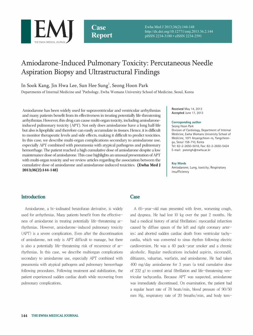
The ultrastructure of foamy macrophages revealed numerous
lysosomal inclusion bodies, which had electron-dense multi-
lamellated deposits (Fig. 5). These histological findings were
consistent with those of amiodarone-induced toxicities.
After the administration of high dose parenteral steroids,
ARDS gradually improved. The patient was successfully weaned
Fig. 4. Pathologic analysis of percutaneous needle aspiration biopsy sample showing chronic interstitial inflammation with fibrous thickening of alveolar septum with features of organizing pneumonia (A: H&E, ×200). Some alveolar spaces showing collection of many intra-alveolar foamy macrophages (B: H&E, ×400). A circle shows Oil Red O-positive lipid droplets in the alveolar macrophages.
Fig. 3. Thyroid scan showing decreased 24-hour iodine uptake as 0.8%.
147THE EWHA MEDICAL JOURNAL
Amiodarone-Induced Pulmonary Toxicity
from the artificial ventilator and his general condition continued
to improve. However, he faced sudden cardiac death, which was
probably caused by a ventricular arrhythmia.
Discussion
Amiodarone is similar in structure to the thyroid hormone.
It is lipophilic and therefore can easily accumulate in tissues.
Further, amiodarone has a long half-life of more than 50 days.
Hence, it is difficult to monitor therapeutic levels and side ef-
fects, and toxicities are unpredictable.
Amiodarone toxicity has been reported to affect the liver,
lung, eye, heart, thyroid, nerves, and other organs [1]. In par-
ticular, APT can present as interstitial pneumonia, lung fibrosis,
various types of pneumonia, and life-threatening ARDS. The
incidence of APT is approximately 5~10%. The risk factors for
APT remain unclear, but advanced age, pre-existing lung dis-
ease, and past thoracotomy are regarded as risk factors [2,3].
Studies have found no association between the dose of amio-
darone (both initial loading and maintenance dose) and APT
[2]. For example, patents who have taken a low dose of amio-
darone or those who have taken amiodarone for less than 1 year
frequently experienced APT [4,5]. Some authors recommend
using a low maintenance dose of <400 mg/day because of the
relatively low likelihood of developing APT at this dose [3,6].
However, many recent studies have found that a high cumula-
tive dose of amiodarone is closely associated with pulmonary
and thyroid toxicities, even with low maintenance doses of
amiodarone (1,7,8). One study reported that long-term use of a
low maintenance dose of amiodarone in the elderly, with a cu-
mulative high total dose of 101~151 g, increased the risk of APT
significantly (odds ratio [OR], 10.29; 95% confidence interval
[CI], 3.42 to 30.92) and reached a plateau at the cumulative
dose of >150 g (OR, 9.5; 95% CI 3.8 to 23.67) [7]. Simi-
larly, another study reported a significantly increased ATP risk
with cumulative doses of 140~230 mg [9]. In this case, a low
maintenance dose of 400 mg/day amiodarone for 3 years was
prescribed, which was quite a long period. As the result, the pa-
tient’s total cumulative dose was >200 g. Although there were
no pathognomonic findings for APT, pathology results from the
needle biopsy showed foamy macrophages with characteristic
lysosomal inclusions and typical electron-dense multilamellar
bodies. APT is a diagnosis of exclusion and frequent mani-
fest as chronic interstitial pneumonitis and pulmonary fibrosis
[2,3]. Given the patient’s medical history, clinical presentation,
pathologic findings, and especially the rapid improvement with
parenteral high-dose steroids, the diagnosis of APT was clear.
However, this case had some complex components. The patient
was a 40 pack-year current smoker and also suffered from lung
infections caused by multiple atypical organisms, so multimodal
therapy was important. When APT was suspected, the first step
was to discontinue amiodarone and use another antiarrhythmic
therapy while managing APT. The second step was to address
and treat other possible causes.
Fig. 5. Electron micrographs showing the ultrastructures of foamy macrophages with numerous round to oval membrane-bound cytoplasmic inclusions (A, ×10,000). Higher magnification of inclusions showing characteristic, closely spaced concentrically arranged lamellae (B, ×20,000).
148 THE EWHA MEDICAL JOURNAL
Kang IS, et al
Another rare presentation of APT is pulmonary alveolar
hemorrhage [10]. Although the patient in this case experienced
hemoptysis, the first episode occurred after PCNB and PCNB
pathology did not show evidence of hemophagocytic macro-
phages. The second episode of massive hemoptysis occurred 6
days after aspirin and warfarin treatment was resumed. Follow-
ing the massive hemoptysis, the patient developed ARDS, which
was controlled by bronchial artery embolization and high-dose
parenteral steroids. The patient’s overall respiratory condition
rapidly improved, not because of antibiotic treatment for the
atypical pneumonia, but rather in response to high-dose steroid
therapy. The association between hemoptysis and APT remains
questionable in this case.
The cumulative dose of amiodarone is also important in the
development of thyroid toxicities. One study showed that the
risk of thyroid dysfunction increases with an increasing cumula-
tive dose, and when the cumulative dose is >144 g, the adjusted
OR is 12.0 (95% CI, 6.1 to 27.3) [8]. Amiodarone-induced
hyperthyroidism was also noted in this case. New thyroid dys-
function was confirmed by laboratory tests, and a thyroid scan
showed decreased 24-hour iodine uptake.
Several mechanisms of amiodarone toxicities have been pro-
posed, but there are 2 major basic mechanisms [9,10]. The
first mechanism constitutes an immune-mediated hypersensitiv-
ity and the second is a direct drug-induced phospholipidosis.
Dose-independent acute onset toxicities usually have a rapidly
progressive course and can be unpredictable. This pattern might
be related to APT, which occurs after initial or low-dose amio-
darone use. While, insidious onset toxicities commonly progress
more slowly than immune-mediated hypersensitivity and typi-
cally present in patients who have taken amiodarone for >2
months [10]. Common symptoms include weight loss, chronic
aggravating cough, and dyspnea, conditions that were apparent
in the clinical presentation of this patient.
This case reiterates the importance of the cumulative dose of
amiodarone. Physicians should carefully evaluate patients who
have been taking low maintenance doses of amiodarone for a
long time, even if they are asymptomatic. This is particularly
important if the cumulative dose is >100 g, as the possibili-
ties of lung and thyroid toxicities should be weighed. In these
cases, we recommend that chest CT, pulmonary function tests,
thyroid function tests, and serial follow-up of liver enzymes be
performed.
References
1. StelfoxHT,AhmedSB,FiskioJ,BatesDW.Monitoringamioda-rone’stoxicities:recommendations,evidence,andclinicalprac-tice.Clin Pharmacol Ther 2004;75:110-122.
2. JackeviciusCA,TomA,EssebagV,EisenbergMJ,RahmeE,TuJV,etal.Population-levelincidenceandriskfactorsforpulmonarytoxic-ityassociatedwithamiodarone.Am J Cardiol2011;108:705-710.
3. AdamsGD,KehoeR,LeschM,GlassrothJ.Amiodarone-in-ducedpneumonitis:assessmentofriskfactorsandpossibleriskreduction.Chest1988;93:254-263.
4. JangWJ,ChonHR, JungJS,YooSH,KohKH,KohYM,etal.Amiodarone-inducedpulmonarytoxicitywithinashortperiodoftheinitiationofamiodaronetherapy:acasereport.Korean J Crit Care Med2011;26:117-121.
5. VeltriEP,ReidPR.Amiodaronepulmonarytoxicity:earlychang-esinpulmonaryfunctiontestsduringamiodaronerechallenge.J Am Coll Cardiol1985;6:802-805.
6. KerinNZ,RubenfireM.Detectionofamiodaronepulmonarytoxicity.J Am Coll Cardiol1989;13:261-262.
7. ErnawatiDK,StaffordL,HughesJD.Amiodarone-inducedpul-monarytoxicity. Br J Clin Pharmacol2008;66:82-87.
8. BouvyML,HeerdinkER,HoesAW,LeufkensHG.Amiodarone-inducedthyroiddysfunctionassociatedwithcumulativedose.Pharmacoepidemiol Drug Saf2002;11:601-606.
9. AshrafianH,DaveyP.IsamiodaroneanunderrecognizedcauseofacuterespiratoryfailureintheICU? Chest2001;120:275-282.
10. TanawuttiwatT,HarindhanavudhiT,HanifS,SahloulMZ.Amio-darone-inducedalveolarhaemorrhage:ararecomplicationofacommonmedication.Heart Lung Circ 2010;19:435-437.