case of presymptomatic aceruloplasminemia treated with deferasirox
TRANSCRIPT
Case Report
Case of presymptomatic aceruloplasminemia treatedwith deferasirox
Mayumi Tai,1,2 Nobuo Matsuhashi,1 Osamu Ichii,1 Tomohiro Suzuki,1 Yutaka Ejiri,1
Satoshi Kono,3 Tatsuhiro Terada,3 Hiroaki Miyajima3 and Masaru Harada2
1Department of Gastroenterology, Fukushima Rosai Hospital, Iwaki, 2The Third Department of Internal Medicine,University of Occupational and Environmental Health, Kitakyushu, and 3The First Department of Medicine,Hamamatsu University School of Medicine, Hamamatsu, Japan
Aceruloplasminemia is an autosomal recessive disease char-acterized by an abnormal iron metabolism. The absence offerroxidase activity caused by mutation of ceruloplasminleads to iron overload in the brain, liver and otherorgans. We report a 35-year-old man who was diagnosedwith aceruloplasminemia without neurological manifestationdespite the accumulation of iron in the brain and liver. To
prevent the development of neurodegenerative disorderrelated to iron toxicity, iron depletion therapy was performed.Iron chelator deferasirox was effective in reducing serum fer-ritin level and to prevent the progression of the disease.
Key words: aceruloplasminemia, copper, deferasirox, iron
INTRODUCTION
ACERULOPLASMINEMIA IS AN autosomal recessivedisorder caused by mutations of the ceruloplasmin
gene, first reported by Miyajima et al. in 1987.1 Theprevalence of aceruloplasminemia was estimated to beapproximately 1/2 million in non-consanguineous mar-riages.2 The deficiency of ceruloplasmin results in ironoverload in the brain, pancreas, liver, retina and otherorgans. This iron metabolic disorder is associatedwith the ferroxidase activity of ceruloplasmin, which isrelated to the oxidation of Fe2+ to Fe3+, allowing it to betransported by transferrin.3 Ceruloplasmin is associatedwith ferroportin. Ferroportin exports iron from the cellsand ceruloplasmin stabilizes ferroportin in the plasmamembrane of various types of cells.4 The expression offerroportin in the plasma membrane is regulated byhepcidin. Hepcidin is the principal regulator of systemiciron metabolism. Without ceruloplasmin or highhepcidinemia, ferroportin is internalized and degraded
in the lysosomes.5–7 Therefore, iron accumulates invarious cells in patients with aceruloplasminemia.8
Clinically, diabetes mellitus, retinal degeneration,neurological dysfunction, anemia, low serum ironand increased serum ferritin are typical features of thisdisease. Regarding the treatment of patients withaceruloplasminemia, several therapeutic opinions, suchas iron chelation therapy, have been reported with dif-ferent outcomes.9–12
We report a relatively young patient with acerulo-plasminemia without neurological dysfunction treatedwith deferasirox.
CASE REPORT
A35-YEAR-OLD MAN was diagnosed with osteomy-elitis of the lower limbs and antibiotic therapy had
been continued for 2 months at our orthopedics unit. Atthe time of admission, liver function test was normal.Abnormal liver function was detected at 1 month afterthe admission and drug-induced liver injury was sus-pected. After the cessation of antibiotics, liver dysfunc-tion had been improved. Then, he was introducedto our unit. He had been diagnosed with anemiaand insulin-dependent diabetes mellitus at the age of16 years. There was no family history of diabetes melli-tus and no medical history except his father. His father
Correspondence: Dr Mayumi Tai, Department of Gastroenterology,Fukushima Rosai Hospital, 3 Numaziri Tsuzuramachi, Uchigo IwakiCity, Fukushima 973-8403, Japan. Email: [email protected] 16 July 2013; revision 5 December 2013; accepted 10December 2013.
bs_bs_banner
Hepatology Research 2014 doi: 10.1111/hepr.12292
© 2013 The Japan Society of Hepatology 1
had presented with Parkinson’s disease in the pastseveral years. His parents were not consanguineous. Onexamination, he showed no evident neurological orophthalmoscopic abnormalities. Kayser–Fleischer ringwas not detected. Laboratory data at admission to ourunit is shown in Table 1. They include increased serumferritin (3530 ng/dL), low serum copper concentration(9 μg/dL), undetectable ceruloplasmin (<1.0 g/dL),hepcidin-25 (<0.7 ng/mL) and normal urinary copperexcretion (35 μg/L).
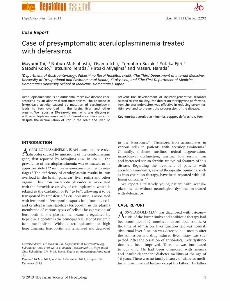
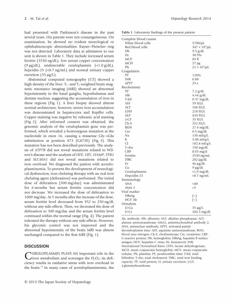
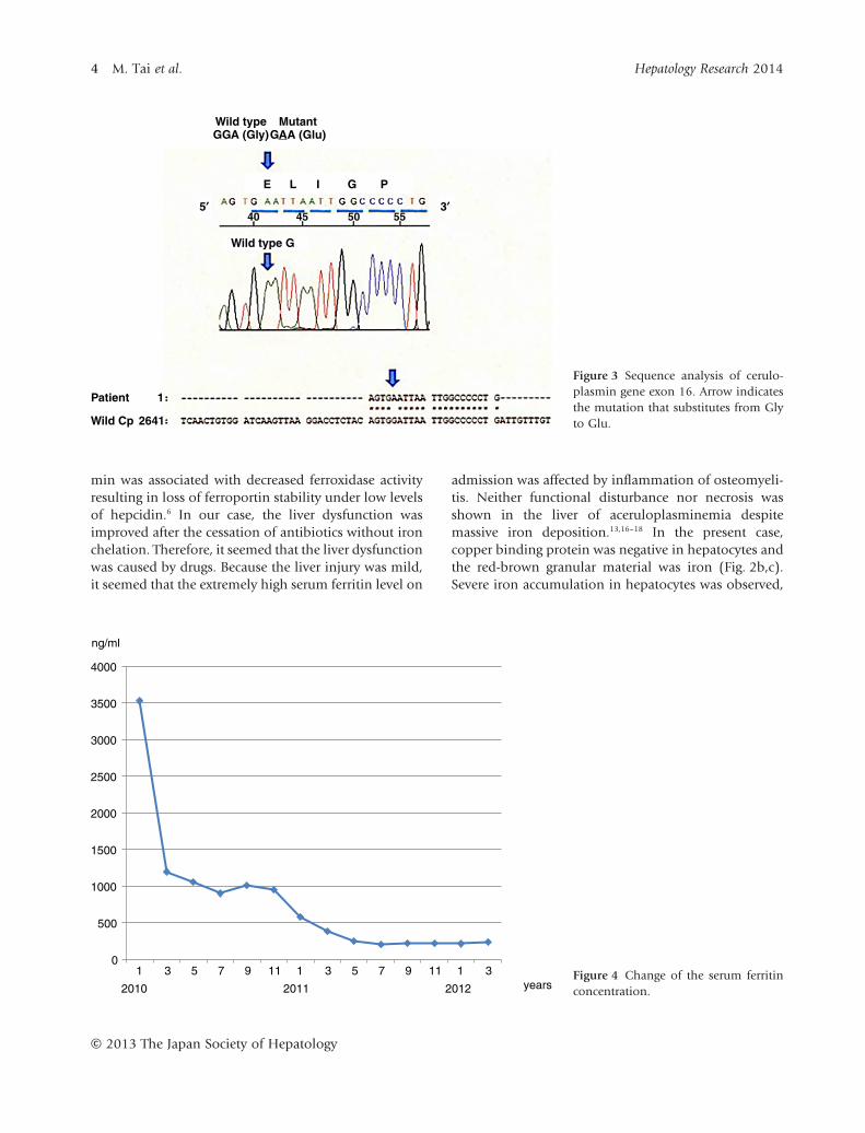
Abdominal computed tomography (CT) showed ahigh density of the liver. T1- and T2-weighted brain mag-netic resonance imaging (MRI) showed an abnormalhypointensity in the basal ganglia, hypothalamus anddentate nucleus, suggesting the accumulation of iron inthese regions (Fig. 1). A liver biopsy showed almostnormal architecture, however, severe iron accumulationwas demonstrated in hepatocytes and Kupffer cells.Copper staining was negative by rubeanic acid staining(Fig. 2). After informed consent was obtained, thegenomic analysis of the ceruloplasmin gene was per-formed, which revealed a homozygous mutation at thenucleotide in exon 16, causing a missense Gly→Glusubstitution at position 873 (G873E) (Fig. 3). Thismutation has not been described previously. The analy-sis of ATP7B did not reveal mutations related to Wil-son’s disease and the analysis of HFE, HJV, HAMP, TFR2and SLC40A1 did not reveal mutations related toiron overload. We diagnosed the patient with acerulo-plasminemia. To prevent the development of neurologi-cal dysfunction, iron chelating therapy with an oral ironchelating agent (deferasirox) was performed. The initialdose of deferasirox (500 mg/day) was administratedfor 6 months but serum ferritin concentration didnot decrease. We increased the dose of deferasirox to1000 mg/day. At 5 months after the increase of the dose,serum ferritin level decreased from 952 to 250 ng/dLwithout any side-effects. Then, we decreased the dose ofdeferasirox to 500 mg/day and the serum ferritin levelcontinued within the normal range (Fig. 4). The patienttolerated the therapy without any side-effects. However,his glycemic control was not improved and theabnormal hypointensity of the brain MRI was almostunchanged compared to the first MRI (Fig. 1).
DISCUSSION
CERULOPLASMIN PLAYS AN important role in theiron metabolism and scavenges the H2O2; its defi-
ciency results in oxidative stress with iron overload inthe brain.13 In many cases of aceruloplasminemia, the
Table 1 Laboratory findings of the present patient
Complete blood countsWhite blood cells 5700/μLRed blood cells 347 × 104/μLHb 9.5 g/dLHt 30.9%MCV 89 flMCH 27 pgPlt 21 × 104/μL
CoagulationPT 128%INR 0.88APTT 29 s
BiochemistryTP 7.2 g/dLAlb 4.44 g/dLT-bil 0.37 mg/dLAST 59 IU/LALT 106 IU/LLDH 218 IU/LALP 434 IU/Lγ-GT 35 IU/LCh-E 252 IU/LBUN 27.4 mg/dLCre 0.5 mg/dLNa 138 mEq/LK 5.08 mEq/LCl 102.8 mEq/LT-cho 150 mg/dLCRP 0.03 mg/dFerritin 3530 ng/mLTIBC 282 μg/dLFe 46 μg/dLCu 9 μg/dLCeruloplasmin <1.0 mg/dLHepcidin-25 <0.7 ng/mL
ImmunologyANA <40AMA 2 <5
Viral markerHBsAg (−)HCV Ab (−)
UrinalysisU-Cu 35 μg/LU-Cr 284.5 mg/dL
Ab, antibody; Alb, albumin; ALP, alkaline phosphatase; ALT,alanine aminotransferase; AMA2, antimitochondrial antibody 2;ANA, antinuclear antibody; APTT, activated partialthromboplastin time; AST, aspartate aminotransferase; BUN,blood urea nitrogen; Ch-E, cholinesterase; Cre, creatinine; CRP,C-reactive protein; Hb, hemoglobin; HBsAg, hepatitis B surfaceantigen; HCV, hepatitis C virus; Ht, hematocrit; INR,International Normalized Ratio; LDH, lactate dehydrogenase;MCH, mean corpuscular hemoglobin; MCV, mean corpuscularvolume; Plt, platelets; PT, prothrombin time; T-bil, totalbilirubin; T-cho, total cholesterol; TIBC, total iron bindingcapacity; TP, total protein; U, urinary excretion; γ-GT,γ-glutamyltransferase.
2 M. Tai et al. Hepatology Research 2014
© 2013 The Japan Society of Hepatology
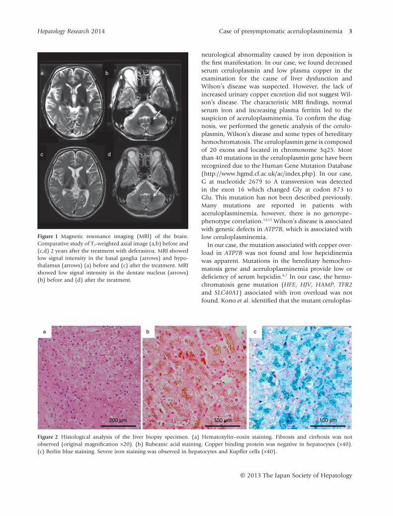
neurological abnormality caused by iron deposition isthe first manifestation. In our case, we found decreasedserum ceruloplasmin and low plasma copper in theexamination for the cause of liver dysfunction andWilson’s disease was suspected. However, the lack ofincreased urinary copper excretion did not suggest Wil-son’s disease. The characteristic MRI findings, normalserum iron and increasing plasma ferritin led to thesuspicion of aceruloplasminemia. To confirm the diag-nosis, we performed the genetic analysis of the cerulo-plasmin, Wilson’s disease and some types of hereditaryhemochromatosis. The ceruloplasmin gene is composedof 20 exons and located in chromosome 3q25. Morethan 40 mutations in the ceruloplasmin gene have beenrecognized due to the Human Gene Mutation Database(http://www.hgmd.cf.ac.uk/ac/index.php). In our case,G at nucleotide 2679 to A transversion was detectedin the exon 16 which changed Gly at codon 873 toGlu. This mutation has not been described previously.Many mutations are reported in patients withaceruloplasminemia, however, there is no genotype–phenotype correlation.14,15 Wilson’s disease is associatedwith genetic defects in ATP7B, which is associated withlow ceruloplasminemia.
In our case, the mutation associated with copper over-load in ATP7B was not found and low hepcidinemiawas apparent. Mutations in the hereditary hemochro-matosis gene and aceruloplasminemia provide low ordeficiency of serum hepcidin.6,7 In our case, the hemo-chromatosis gene mutation (HFE, HJV, HAMP, TFR2and SLC40A1) associated with iron overload was notfound. Kono et al. identified that the mutant ceruloplas-
a b
c d
Figure 1 Magnetic resonance imaging (MRI) of the brain.Comparative study of T2-weighted axial image (a,b) before and(c,d) 2 years after the treatment with deferasirox. MRI showedlow signal intensity in the basal ganglia (arrows) and hypo-thalamus (arrows) (a) before and (c) after the treatment. MRIshowed low signal intensity in the dentate nucleus (arrows)(b) before and (d) after the treatment.
a
200 μm 100 μm
b
100 μm
c
Figure 2 Histological analysis of the liver biopsy specimen. (a) Hematoxylin–eosin staining. Fibrosis and cirrhosis was notobserved (original magnification ×20). (b) Rubeanic acid staining. Copper binding protein was negative in hepatocytes (×40).(c) Berlin blue staining. Severe iron staining was observed in hepatocytes and Kupffer cells (×40).
Hepatology Research 2014 Case of presymptomatic aceruloplasminemia 3
© 2013 The Japan Society of Hepatology
min was associated with decreased ferroxidase activityresulting in loss of ferroportin stability under low levelsof hepcidin.6 In our case, the liver dysfunction wasimproved after the cessation of antibiotics without ironchelation. Therefore, it seemed that the liver dysfunctionwas caused by drugs. Because the liver injury was mild,it seemed that the extremely high serum ferritin level on
admission was affected by inflammation of osteomyeli-tis. Neither functional disturbance nor necrosis wasshown in the liver of aceruloplasminemia despitemassive iron deposition.13,16–18 In the present case,copper binding protein was negative in hepatocytes andthe red-brown granular material was iron (Fig. 2b,c).Severe iron accumulation in hepatocytes was observed,
Wild typeGGA (Gly)
Wild type G
Patient 1
Wild Cp 2641
40 45 50 55
E
5¢ 3¢
L I G P
MutantGAA (Glu)
Figure 3 Sequence analysis of cerulo-plasmin gene exon 16. Arrow indicatesthe mutation that substitutes from Glyto Glu.
0
500
1000
1500
2000
2500
3000
3500
4000
1 3 5 7 9 11 1 3 5 7 9 11 1 3
ng/ml
2010 2011 2012 yearsFigure 4 Change of the serum ferritinconcentration.
4 M. Tai et al. Hepatology Research 2014
© 2013 The Japan Society of Hepatology
however, few inflammatory cells were observed(Fig. 2c).
It is hypothesized that the difference in tissue injurybetween the brain and the liver is due to the manyantioxidant enzymes in the liver.13 In fact, the activity ofcatalase in the liver is more than 30-times higher thanthat in the brain.19 In the present case, the first clinicalmanifestation was adult-onset diabetes mellitus, butretinal degeneration was not present. The neurologicaldisease might have appeared several years later, if he hadnot been treated.
Iron chelating therapy for aceruloplasminemia hasbeen reported, but the clinical efficacy of the patienthas varied. In the present case, the treatment withdeferasirox led to a reduction of serum ferritin, but thehepatic deposition analyzed by CT scan did not change.In some cases, brain MRI showed a steady hyposignal ofthe iron deposition even after the treatment, despite adecrease of hepatic iron accumulation and serum ferri-tin.9,20,21 These data suggest that iron chelating therapymay be useful in limiting the liver iron deposition butnot in brain iron deposition. In another case, the treat-ment with desferrioxamine led to a reduction of serumferritin and neurological abnormalities as well asthe improvement of diabetes.11 A case of aceruloplas-minemia with neurological symptoms despite visibleiron deposition on the brain MRI was reported, inwhich deferasirox therapy improved neurologicalsymptoms.22 Deferasirox was effective in another casewith neurological symptoms.23 Therefore, early diagno-sis and treatment must be important and the ironremoval therapy may be effective to prevent the devel-opment of neurological dysfunction. Because it wasreported that several side-effects prohibit the long-termtreatment with deferasirox, it is important to monitoranemia and renal dysfunction and clinical symptoms,particularly gastrointestinal disturbances and skinrashes, when iron chelating therapy with deferasirox iscontinued.21,24,25
In conclusion, we report a case with aceruloplas-minemia in whom iron chelating therapy usingdeferasirox was useful to reduce serum ferritin concen-tration and to prevent progression of the phenotype.
ACKNOWLEDGMENT
WE THANK DR Hisao Hayashi (Aichi GakuinUniversity, School Of Pharmacy) for the genetic
analysis of Wilson’s disease and the hereditary hemo-chromatosis genes.
REFERENCES
1 Miyajima H, Nishimura Y, Mizoguchi K et al. Familialapoceruloplasmin deficiency associated with blepharo-spasm and retinal degeneration. Neurology 1987; 37: 761–7.
2 Miyajima H, Kohno S, Takahashi Y et al. Estimation of thegene frequency of aceruloplasminemia in Japan. Neurology1999; 53: 617–9.
3 Osaki S, Johnson DA, Frieden E. The possible significanceof the ferrous oxidase activity of ceruloplasmin in normalhuman serum. J Biol Chem 1966; 241: 2746–51.
4 De Domenico I, Ward DM, di Patti MC et al. Ferroxidaseactivity is required for the stability of cell surfaceferroportin in cells expressing GPI-ceruloplasmin. EMBO J2007; 26: 2823–31.
5 Pietrangelo A. Hereditary hemochromatosis: pathogenesis,diagnosis, and treatment. Gastroenterology 2010; 139: 393–408.
6 Kono S, Yoshida K, Tomosugi N et al. Biological effects ofmutant ceruloplasmin on hepcidin-mediated internaliza-tion of ferroportin. Biochim Biophys Acta 2010; 1802: 968–75.
7 Kaneko Y, Miyajima H, Pipemo A et al. Measurement ofserum hepcidin-25 levels as a potential test for diagnosinghemochromatosis and related disorders. J Gastroenterol2010; 45: 1163–71.
8 Harris ZL, Klomp LW, Gitlin JD. Aceruloplasminemia: aninherited neurodegenerative disease with impairment ofiron homeostasis. Am J Clin Nutr 1998; 67: 972S–7S.
9 Loreal O, Turlin B, Pigeon C et al. Aceruloplasminemia:new clinical, pathophysiological and therapeutic insights.J Hepatol 2002; 36: 851–6.
10 Mariani R, Arosio C, Pelucchi S et al. Iron chelation therapyin aeruloplasminemia: study of a patient with a novelmissense mutation. Gut 2004; 53: 756–8.
11 Miyajima H, Takahashi Y, Kamata T et al. Use ofdesferrioxamine in the treatment of aceruloplasminemia.Ann Neurol 1997; 41: 404–7.
12 Yonekawa M, Okabe T, Asamoto Y et al. A case of heredi-tary ceruloplasmin deficiency with iron deposition in thebrain associated with chorea, dementia, diabetes mellitusand retinal pigmentation: administration of fresh-frozenhuman plasma. Eur Neurol 1999; 42: 157–62.
13 Tajima K, Kawanami T, Nagai R et al. Hereditary cerulo-plasmin deficiency increases advanced glycation end prod-ucts in the brain. Neurology 1999; 53: 619–22.
14 Yoshida K. Mutations in the ceruloplasmin gene inaeruloplasminemia. Neurol Med 2004; 61: 146–50. (InJapanese.)
15 Yoshida K, Kaneko K. Aeruloplasminemia. Neurol Med2009; 71: 517–26. (In Japanese.)
16 Kawanami T, Kato T, Daimon M et al. Hereditary cerulo-plasmin deficiency: clinicopathological study of a patient.J Neurol Neurosurg Psychiatry 1996; 61: 506–9.
17 Wada S, Kimura K, Mizuno I et al. A case of acerulo-plasminemia associated with insulin-dependent diabetes
Hepatology Research 2014 Case of presymptomatic aceruloplasminemia 5
© 2013 The Japan Society of Hepatology
mellitus, cerebellar ataxia and retinal degeneration. FoliaEndocrinol 1996; 72: 543–50. (In Japanese.)
18 Daimon M. Hereditary ceruloplasmin deficiency. NipponRinsho 2008; 66: 563–67. (In Japanese.)
19 Tiedge M, Lortz S, Drinkgern J et al. Relation between anti-oxidant enzyme expression and antioxidative defense statusof insulin-producing cells. Diabetes 1997; 46: 1733–42.
20 Fasano A, Colosimo C, Miyajima H et al. Acerulo-plasminemia: a novel mutation in a family with markedphenotypic variability. Mov Disord 2008; 23: 751–5.
21 Finkenstedt A, Wolf E, Höfner E et al. Hepatic but notbrain iron is rapidly chelated by deferasirox in acerulo-plasminemia due to a novel gene mutation. J Hepatol 2010;53: 1101–7.
22 Skidmore FM, Dragon V, Foster P et al. Acerulo-plasminemia with progressive atrophy without brain ironoverload: treatment with oral chelation. J Neurol NeurosurgPsychiatry 2008; 79: 467–70.
23 McNeill A, Pandolfo M, Kuhn J et al. The neurologicalpresentation of ceruloplasmin gene mutations. Eur Neurol2008; 60: 200–5.
24 Suzuki Y, Yoshida K, Aburakawa Y et al. Effectivenessof oral iron chelator treatment with Deferasirox in anaceruloplasminemia patient with a novel ceruloplasmingene mutation. Intern Med 2013; 52: 1527–30.
25 Lee JW. Iron chelation therapy in the myelodysplastic syn-dromes and aplastic anemia: a review of experience inSouth Korea. Int J Hematol 2008; 88: 16–23.
6 M. Tai et al. Hepatology Research 2014
© 2013 The Japan Society of Hepatology