carson tahoe endocrinology carson city, nv kcom class of 1989 · steroid production pathway...
TRANSCRIPT
John Sutton, DO, FACOI, FACE, CCD
Carson Tahoe
Endocrinology
Carson City, NV
KCOM Class of 1989
Gonadal Physiology and Disease
3
No Disclosures
Gonadal Axis
Hypothalamic-pituitary-gonadal
Feedback mechanisms important
Without production of hormone end products,
axis should respond
Hypergonadotropic Hypogonadism
Elevated gonadotropins (LH/FSH)
Low gonadal hormones
Hypogonadotropic Hypogonadism
Low or inappropriately normal gonadotropins
Low gonadal hormones
24 Year Old Male
Female body habitus
Lack of secondary sex characteristics
Absent facial hair
Infertility
Physical exam: small testicles, Tanner I
Laboratory
Elevated LH, FSH, Low testosterone total,
free and bioavailable
Normal prolactin
Normal thyroid function
Pituitary is responding appropriately to an
end organ hormonal deficiency

Diagnosis
Hyper-
gonadotropic
Hypogonadism
Additional Testing
Karyotype: check for Klinefelter syndrome
47 XXY
Hypergonadotropic Hypogonadism
Germ cell arrest
Surgery, chemotherapy
Mumps
Alcohol
Immune
Intra-abdominal testicles
14 Year Old Male
Lack of secondary sex characterisitics
Gynecomastia
Laboratory
Low LH, FSH
Low total and low bioavailable (active)
testosterone
Normal prolactin
Normal thyroid function
Diagnosis
Hypo-
gonadotropic
Hypogonadism
Differential Diagnosis
Delayed puberty
Kallman syndrome: anosmia, deficiency in GnRH
Hyperprolactinemia
Hemochromatosis or infiltrative
Hypopituitarism
Neoplasm (Brain or pituitary)
Anorexia, excess exercise
Treatment
If fertility is in question, will require HCG or
GnRH administration
For restoring male hormone levels without
fertility: testosterone IM, transdermal patch
or gel, including axillary administration
Exogenous Testosterone suppress axis may
affect future fertility
Hyperprolactinemia
Hypothyroidism
Pituitary neoplasm
Non-fasting
Medications
TREATMENT: Medical
44 Year Old Male
Breast tenderness, decreased sex drive
Breast enlargement, normal genital exam
Laboratory
Normal estradiol, low free and total
testosterone
Normal B-HCG
Normal gonadotropins
High prolactin
Normal thyroid function
Diagnosis
Hypogonadotropic
Hypogonadism
due to
Hyperprolactinemia
Treatment
Evaluate MRI for pituitary/brain lesion
Treatment of pituitary lesion: medical with
Cabergoline or Bromocriptine as Dopamine
Agonist
Rarely require pituitary surgery
Visual field testing if large tumor &
compressing optic chiasm
Differential Diagnosis
Gynecomastia common in elderly, obese,
puberty
High estradiol may represent a testicular or
adrenal neoplasm
High HCG suggests testicular or pulmonary
neoplasm
Hypothyroidism
Differential Diagnosis
Hypothyroidism: promotes increased
prolactin, suppression of gonadotropins, low
male hormone, infertility, gynecomastia

16 Year Old Female
Lack of secondary sex characteristics
No menses, primary amenorrhea
Laboratory
Low LH, FSH
Low estradiol

Diagnosis
Hypo-
gonadotropic
Hypogonadism
Hypogonadotropic Hypogonadism
Pituitary tumors
Kallman syndrome (anosmia)
Anorexia Nervosa
Excessive exercise
Additional Findings with similar history but elevated LH & FSH
Short stature
Prepubertal genital exam
These patients have primary amenorrhea
with no history of menses
Secondary amenorrhea refers to absent
menses after menarche
Diagnosis
Hypergonadotropic
Hypogonadism
Turner Syndrome
Hypergonadotropic Hypogonadism
Menopause ovarian failure
Surgical removal of ovaries
Turner Syndrome 45 X0
Secondary Amenorrhea
Polycystic ovarian syndrome: adequate
estrogen, excess androgen
Uterine defects and trauma
Pregnancy, profound stress
Systemic illness
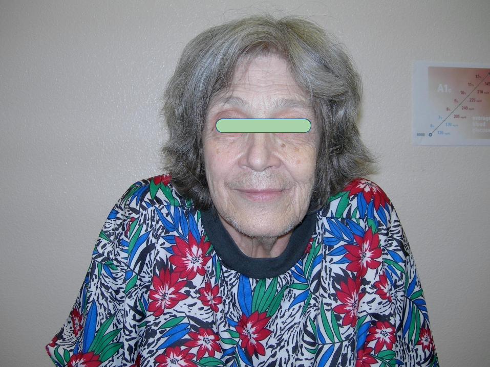

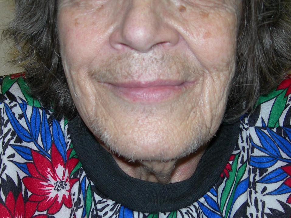
Hirsutism
Check DHEAS and Total testosterone:
Neoplasm considered with DHEAS twice
normal(adrenal) and Testosterone total >
200 ng/dl (ovarian) or as low as 150 ng/dl
Consider congenital adrenal hyperplasia,
Cushing Syndrome, virilizing tumors, PCO
Disease of theAdrenals
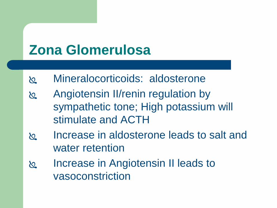
Zona Glomerulosa
Mineralocorticoids: aldosterone
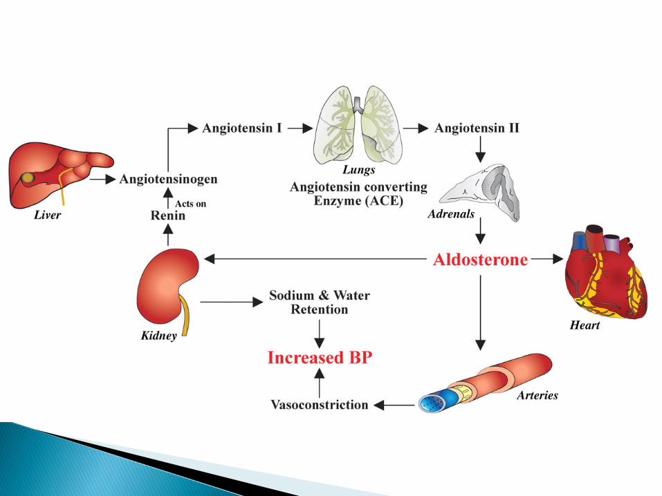
Angiotensin II/renin regulation by
sympathetic tone; High potassium will
stimulate and ACTH
Increase in aldosterone leads to salt and
water retention
Increase in Angiotensin II leads to
vasoconstriction
Zona Fasiculata and Reticularis
Glucocorticoids: Cortisol
Androgen: DHEAS
Regulated by ACTH
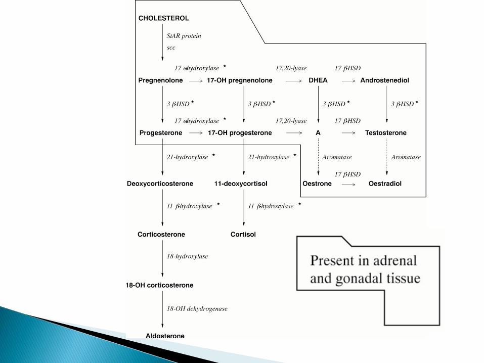
Steroid Production Pathway (steroidogenesis)
Baseline substrate = cholesterol
Precursors: DHEAS
17-OH progesterone
End products: estradiol, cortisol,
aldosterone
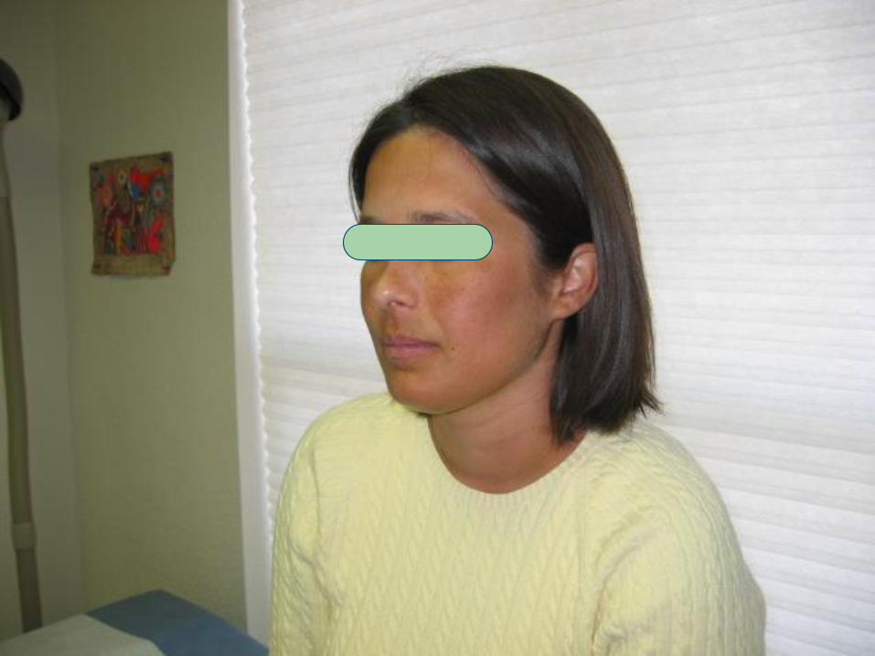
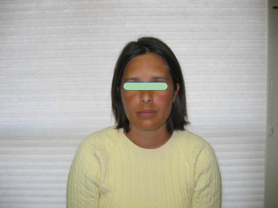
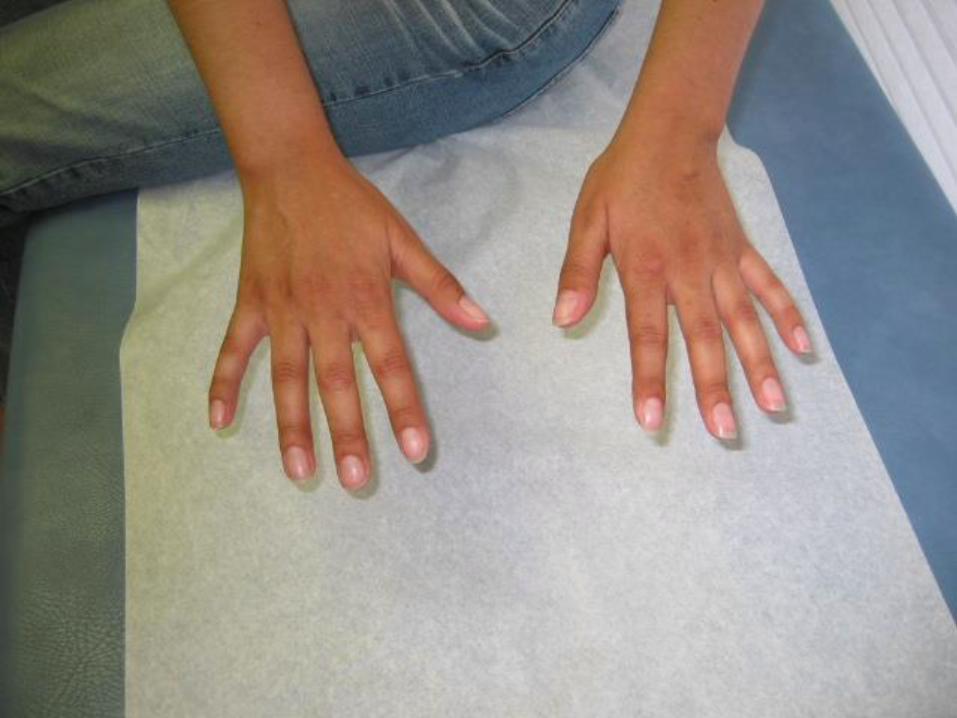
20 year old female
Weight loss, easy tanning, nausea, vomiting,
abdominal pain, weakness, dizzy
BP=70/30, tan, confusion
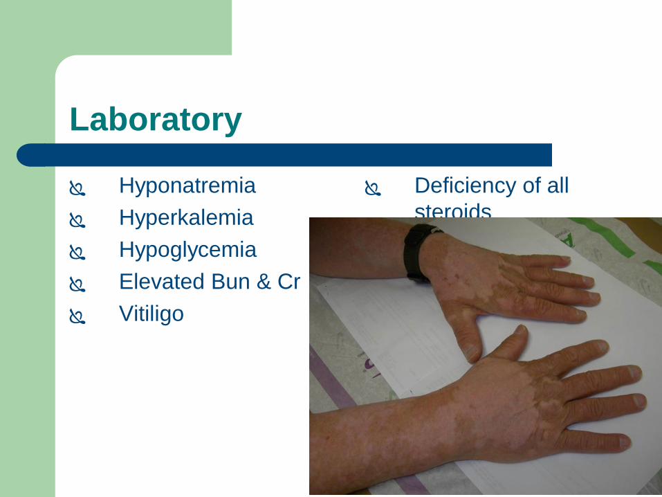
Laboratory
Hyponatremia
Hyperkalemia
Hypoglycemia
Elevated Bun & Cr
Vitiligo
Deficiency of all
steroids
Diagnosis
Primary
Adrenal
Insufficiency
Laboratory
AM cortisol, ACTH
Cosyntropin (ACTH) stimulation IV or IM Baseline, 30 min and 60 minute values for cortisol
If Aldosterone drawn with Cosyntropin stimulation, response blunted
**Cortisol Goal > 18 micrograms/dl with Cosyntropin Stimulation, assuming a normal baseline cortisol
Etiology
80 % Autoimmune/Idiopathic
20 % Tuberculosis
Other: Vascular, infectious, AIDS,
trauma, mets, meds,
congenital adrenal hyperplasia
Autoimmune Etiology
Addison Disease, adrenalitis
May be associated with other autoimmune
conditions, as in Hashimoto or vitiligo
50 year old female
Similar symptoms to index patient: low to
low normal BP, fatigue weakness
No change in skin color
COPD
Laboratory
Low sodium
Normal potassium, which suggests normal
aldosterone production
Low ACTH or inappropriately normal when
the end organ value is low
Be careful with the timing of the stimulation
test and blood draw for ACTH
Additional History
History of long term steroid use IV and oral
treatment
Recent change in pharmacy
Prednisone not renewed
Presents with fever and lung infiltrate
Diagnosis
Secondary
Adrenal
Insufficiency
Etiology
Steroid dependent
Tumor, infection, radiation, surgery, trauma
involving hypothalamic region or pituitary
Physical findings in AI
Generalized abdominal tenderness
Fever
Postural hypotension
Look for precipitating infection
Careful with consideration for surgical abdomen
Surgery could precipitate adrenal crisis if
adequate steroids are not on board
120
Adrenal Insufficiency
Electrolyte imbalance: Hyponatremia,
Hyperkalemia in primary adrenal insufficiency
Hypotension and medical crisis
Hyponatremia without hyperkalemia in
secondary adrenal insufficiency, less likely to
result in adrenal crisis
121
Electrolyte imbalance in AI
85 to 90 % of patients have hyponatremia
Mineralocorticoid deficiency results in sodium
loss and volume depletion and increased
Vasopressin secretion due to loss of cortisol
Hyperkalemia in 60 to 65 % of patients
Rare hypercalcemia
122
Imaging
Consider CT of the adrenals for primary
adrenal insufficiency ———small adrenals
MRI of the brain for secondary adrenal
insufficiency unless the cause is evident
Treatment at diagnosis in Crisis
IV hydrocortisone 100 mg q 6-8 hrs wean
as tolerated to daily oral dose of 25 mg
daily/divided
Saline and glucose
Supportive and correcting precipitating
factors
Primary adrenal insufficiency: Florinef as
aldosterone replacement
If steroids < 30 days in general medical
treat-ment, do not necessarily need to
wean slowly
Crisis Intervention
Surgery
Acute illness
Additional steroids IV and/or PO
Home illness: short course of double dose
steroids
Observe sodium, potassium and BP; Pt can
follow BP at home for crisis intervention
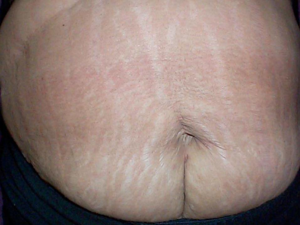
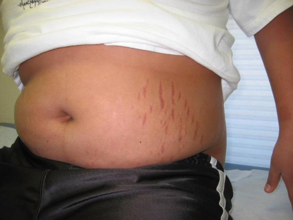
25 Year Old Female
Weight gain, hirsutism, diabetes,
osteoporosis
Centripetal obesity, striae, acne,
hypertension, capillary fragility, amenorrhea
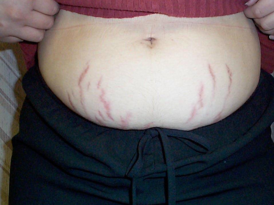
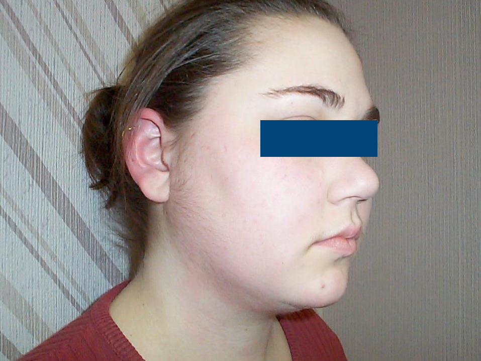
Diagnosis
Cushing
Syndrome
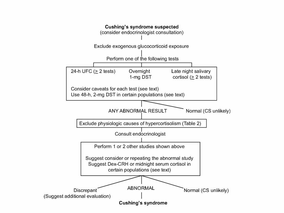
Laboratory
1 mg overnight dexamethasone suppression
testing; 1 mg Dex 11 pm with 8 am cortisol
next day---may identify subtle with normal
urine free cortisol---goal suppression < 1.8
24 hour urine free cortisol
Late night salivary cortisol
Hypokalemia, hyperglycemia
Some false positives
Pregnancy Striae
Differential Diagnosis
Cushing disease: Cushing syndrome due to
pituitary adenoma/high ACTH---dependent
ACTH Independent vs ACTH Dependent
Exogenous steroids
Adrenal adenoma or hyperplasia
Ectopic: lung tumor

Differential Diagnosis
Cushing disease and ectopic have higher
ACTH>>>>ACTH Dependent
Adrenal disease is ACTH independent
Clarification required with additional
dexamethasone testing including urinary
testing
Imaging
Cushing disease: MRI of the pituitary
Cushing syndrome: CT or MRI of (adenoma
vs hyperplasia) adrenals
Ectopic: localize source
Treatment
Pituitary: surgery, radiation, anti-adrenal
drugs
Ectopic: surgery, drugs
Adrenal: surgery, drugs
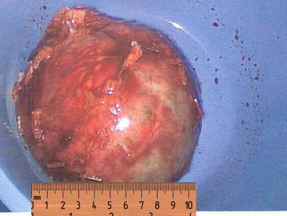
Adrenal Carcinoma
Metastatic at diagnosis
Presents with weight loss
Rapid onset
Typical excessive activity of steroidogenesis
pathway, not typical insufficiency
35 Year Old female
Hypertension
Hypokalemia
Thin
Metabolic alkalosis
Diagnosis
Hyper-
aldosteronism
Hyperaldosteronism
Biochemical work-up first
Low renin/high aldosterone is primary
High renin/high aldosterone is secondary
Elevated 24 hour urine aldosterone on high
sodium diet and off diuretics
Saline Suppression Testing
Hyperaldosteronism
Adrenal adenoma (Conn Syndrome) “APA”
Aldosterone Producing Adenoma
Idiopathic Hyperaldosteronism “IHA” with
bilateral disease
Secondary Hyperaldosteronism
Sodium restriction
Renal disease
High Potassium intake
Pregnancy
Diuretics
Localization testing
Cat Scan
Nuclear imaging with Iodocholesterol
Adrenal venous sampling—Gold Standard

Adrenal Venous Sampling Summary
RE: Male DOB: 1942
Aldosterone (ng/dL) Cortisol (ug/dL)
Basal:
Right adrenal vein 1 4.3
Left adrenal vein 221 10.2
Peripheral Arm 12 12.3
POST ACTH:
Right adrenal vein 4 108.1
Left adrenal vein 16430 >150
Peripheral Vena Cava 71 14.4
Peripheral Femoral Vein 46 18.9
Treatment
Aldosterone producing adenoma: surgery;
takes 6 months for final HTN results, may have
underlying essential HTN but hypokalemia
should resolve; Spironolactone in patients with
poor surgical risk
Bilateral adrenal disease: restrict sodium,
spironolactone use, no surgery
40 Year Old Male
Hypertension unresponsive to meds
Normal electrolytes
Thin
Headache, palpitations
Diagnosis
Pheochromocytoma
Laboratory
Check 24 hour urine fractionated
catecholamines, fractionated metanephrines,
VMA
24 hour urine testing off meds if possible
Some endocrinologists recommend serum
catecholamine/metanephrine testing
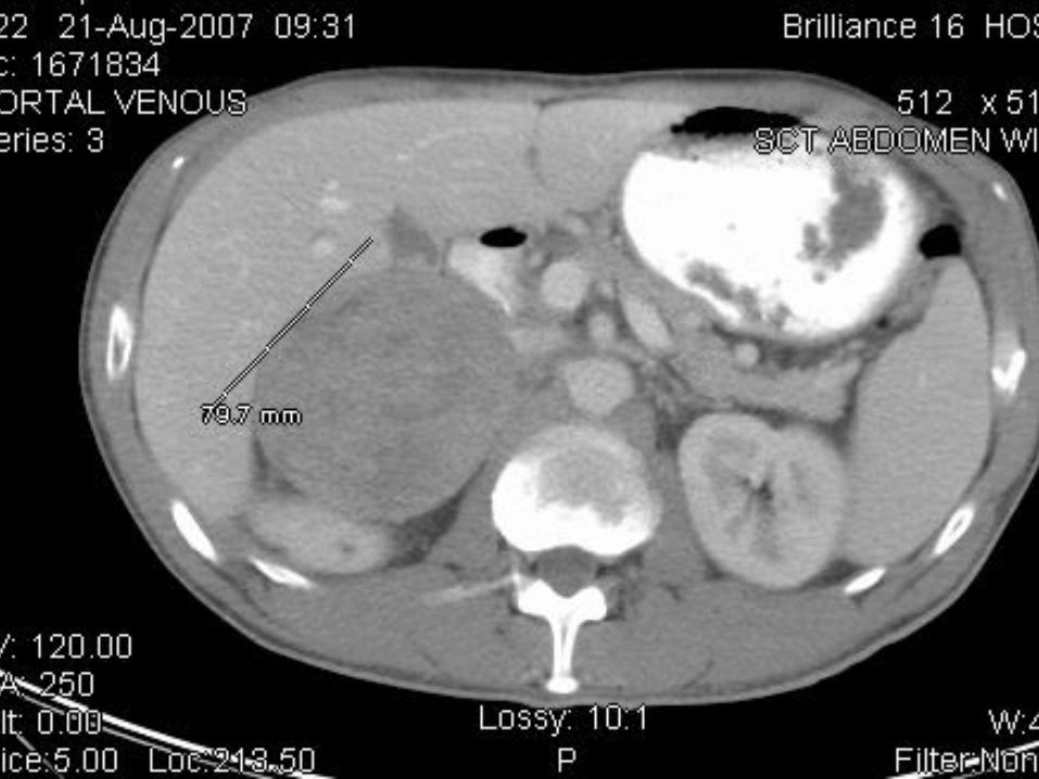
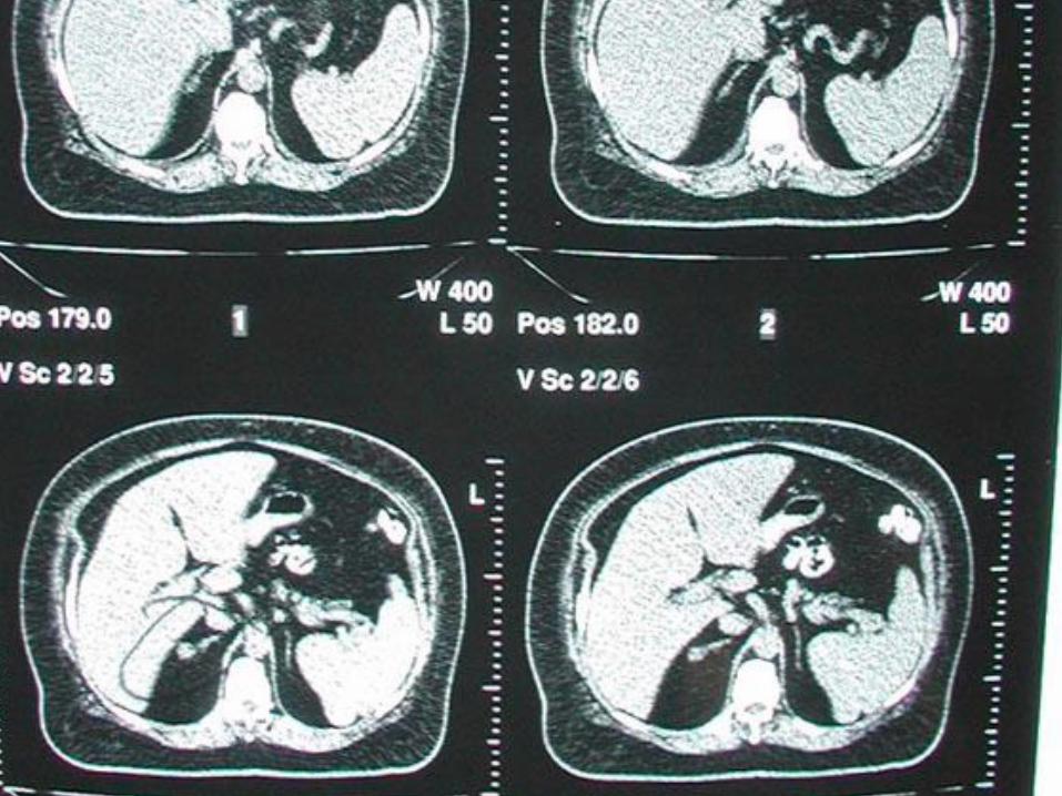
Imaging
MRI or CT: MRI may help with difference in
signal intensity T1/T2---bright signal in pheo
MIBG (Metalogobenzylguanidine) nuclear
imaging tracer concentrates in catecholamine
producing cells
Treatment
Alpha blockers preferred
Avoid Beta Blockers, which can precipitate a
pheo crisis without alpha blockade on board
Avoid adrenal biopsy of a lesion that is not
yet evaluated for pheochromocytoma

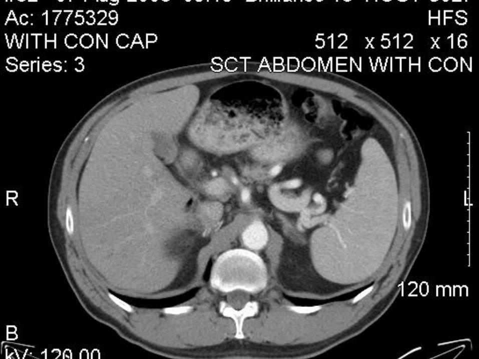
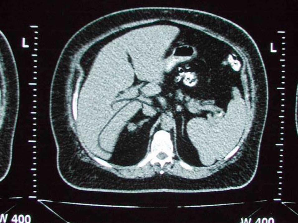
Incidental Adrenal Adenoma
Benign adenomas common
Avoid imaging until biochemical diagnosis
Evaluation important with coexisting HTN,
hypokalemia, hirsutism
Adrenal Incidentaloma
Lesions discovered “inadvertently in the course
of diagnostic testing or treatment for other
clinical conditions that are not related to the
suspicion of adrenal disease”

Prevalence
In autopsy series 2.1 %
More identified with better imaging
Prevalence of 4.3 % in patients with a
previous diagnosis of cancer
Higher with aging at 7 % in 70 +
More lesions in women—related to who is
being tested
Causes
Pathology: Cancer patients ¾ mets, No
history of cancer 2/3 benign
70 % non-functioning in patients without
endocrine symptoms
5-10 %-----Cushing Syndrome, subclinical
Natural History of lesion size
25 % of lesions larger than 6 cm represent
adrenal cancer
Up to 25 % of adrenal lesions may grow 1
cm, but the significance of size change is not
known
Adrenal Cancer rapid growth “doubling time”
Natural History of function
Up to 20 % may develop a functional
component
Development of function more common in
larger neoplasms (3 cm)---this evidence can
depend on study follow-up length and
methods
Less than 3 cm neoplasms rarely change in
function
Adrenal Incidentaloma Diagnosis
Function
Surgical resection vs non-surgical treatment
Malignant vs benign