care of wound
TRANSCRIPT
THE CARE OF WOUNDSA GUIDE FOR NURSES
THIRD EDITION
CAROL DEALEY PhD MA BSc (Hons) RGN RCNT
Research FellowUniversity Hospital Birmingham NHS Trust, andSchool of Health SciencesUniversity of Birmingham
© 2005 Blackwell Publishing Ltd
Editorial offices:Blackwell Publishing Ltd, 9600 Garsington Road,Oxford OX4 2DQ, UK
Tel: +44 (0)1865 776868Blackwell Publishing Inc., 350 Main Street, Malden,MA 02148-5020, USA
Tel: +1 781 388 8250Blackwell Publishing Asia Pty Ltd, 550 SwanstonStreet, Carlton, Victoria 3053, Australia
Tel: +61 (0)3 8359 1011
The right of the Author to be identified as theAuthor of this Work has been asserted in accordance with the Copyright, Designs andPatents Act 1988.
All rights reserved. No part of this publication may be reproduced, stored in a retrieval system, or transmitted, in any form or by any means, electronic, mechanical, photocopying, recording or otherwise, except as permitted by the UK Copyright, Designs and Patents Act 1988, without the prior permission of the publisher.
First published 2005 by Blackwell Publishing Ltd
Library of Congress Cataloging-in-Publication Data
Dealey, Carol.The care of wounds : a guide for nurses / Carol
Dealey. – 3rd ed.p. ; cm.
Includes bibliographical references and index.ISBN-13: 978-1-4051-1863-7 (pbk. : alk. paper)ISBN-10: 1-4051-1863-6 (pbk. : alk. paper)1. Wounds and injuries – Nursing.[DNLM 1. Wounds and Injuries – nursing.
2. Wound Healing – physiology. WO 700 D279c2005] I. Title.
RD93.95.D43 2005617.1 – dc22
2005001740
ISBN 10: 1-4051-1863-6ISBN 13: 978-14051-1863-7
A catalogue record for this title is available from theBritish Library
Set in 10/12pt Palatinoby SNP Best-set Typesetter Ltd., Hong KongPrinted and bound in Spainby GraphyCems, Navarra
The publisher’s policy is to use permanent paperfrom mills that operate a sustainable forestry policy,and which has been manufactured from pulpprocessed using acid-free and elementary chlorine-free practices. Furthermore, the publisher ensuresthat the text paper and cover board used have metacceptable environmental accreditation standards.
For further information on Blackwell Publishing,visit our website:www.blackwellpublishing.com
Contents
Contents iii
Preface vii
Chapter 1 The Physiology of Wound Healing 11.1 Introduction 11.2 Definitions associated with wounds 11.3 The structure of the skin 2
1.3.1 Dermis 21.3.2 Epidermis 2
1.4 Wound healing 31.4.1 Inflammation 41.4.2 Reconstruction 61.4.3 Epithelialisation 81.4.4 Maturation 9
1.5 Impaired wound healing 91.5.1 Hypertrophic scars 91.5.2 Keloids 101.5.3 Contractures 101.5.4 Acute to chronic wounds 10
1.6 Conclusion 11
Chapter 2 The Management of Patients with Wounds 132.1 Introduction 132.2 Physical care 13
2.2.1 Nutrition 132.2.2 Infection 192.2.3 Smoking 232.2.4 Diabetes mellitus 252.2.5 They physical effects of stress 262.2.6 Pain 272.2.7 Sleeping 302.2.8 Hypothermia 322.2.9 Steroids 332.2.10 Radiotherapy 34
2.3 Psychological care 352.3.1 Anxiety 352.3.2 Motivation and education 372.3.3 Body image 392.3.4 Other psychological problems 41
2.4 Spiritual care 43
Chapter 3 General Principles of Wound Management 563.1 Introduction 563.2 Wound assessment 56
3.2.1 Wound classification 563.2.2 The position of the wound 573.2.3 The environment of care 583.2.4 M = measure 583.2.5 E = exudate 613.2.6 A = appearance 623.2.7 S = suffering 693.2.8 U = undermining 703.2.9 R = re-evaluate 703.2.10 E = edge 71
3.3 Managing wounds 723.3.1 Moist wound healing 723.3.2 Wound bed preparation 723.3.3 Pain management 76
3.4 Documentation 773.5 Evaluating the dressing 78
Chapter 4 Wound Management Products 834.1 Introduction 834.2 The development of dressings through the ages 83
4.2.1 Early days 834.2.2 The Dark Ages and early Middle Ages 854.2.3 Late Middle Ages and Renaissance 864.2.4 The seventeenth, eighteenth and early nineteenth centuries 874.2.5 Mid-nineteenth and early twentieth century developments 884.2.6 The British Pharmaceutical Codices 89
4.3 Traditional techniques 904.4 The use of lotions 91
4.4.1 Antiseptics 914.4.2 Antibiotics 954.4.3 Honey 964.4.4 Saline 0.9% 974.4.5 Tap water 97
4.5 Clinical effectiveness of wound management products 974.5.1 Providing an effective environment 974.5.2 The handling qualities of an effective wound
management product 984.6 Modern wound management products 994.7 Advanced technologies 105
4.7.1 Growth factors 1054.7.2 Protease-modulating wound management products 1054.7.3 Hyaluronan-based products 1064.7.4 Hyperbaric oxygen 107
iv The Care of Wounds
4.7.5 Topical negative pressure 1084.7.6 Tissue culture 1104.7.7 Tissue engineering 110
4.8 Alternative therapies and wound management 112
Chapter 5 The Management of Patients with Chronic Wounds 1215.1 Introduction 1215.2 The prevention and management of pressure ulcers 121
5.2.1 The cost of pressure ulcers 1225.2.2 The aetiology of pressure ulcers 1235.2.3 Prevention of pressure ulcers 1285.2.4 Management of pressure ulcers 138
5.3 The management of leg ulcers 1435.3.1 The epidemiology of leg ulcers 1445.3.2 The cost of leg ulcers 1445.3.3 The causes of leg ulcers 1455.3.4 Venous ulceration 1455.3.5 Arterial ulcers 1525.3.6 Ulcers of mixed aetiology 1555.3.7 Malignant leg ulcers 1565.3.8 Leg ulceration in rheumatoid arthritis 157
5.4 Diabetic foot ulcers 1585.4.1 Aetiology 1585.4.2 Prevention 1595.4.3 Management 160
5.5 Fungating wounds 1625.5.1 Aetiology and incidence 1635.5.2 Management of fungating wounds 163
5.6 Lymphoedema 1655.6.1 Lymphoedema management 166
5.7 Conclusion 168
Chapter 6 The Management of Patients with Acute Wounds 1796.1 Introduction 1796.2 The care of surgical wounds 179
6.2.1 Management of surgical wounds 1796.2.2 Managing complications 186
6.3 Traumatic wounds 1946.3.1 Minor traumatic wounds 1946.3.2 The management of specific types of traumatic wounds 196
6.4 The burn injury 1996.4.1 Aetiology 2006.4.2 Incidence 2016.4.3 The severity of the injury 2016.4.4 Burn oedema 2036.4.5 First aid treatment of burns 2036.4.6 The management of burn injuries 205
Contents v
6.5 Radiation reactions 2096.5.1 Aetiology 2106.5.2 The classification of radiation reactions 2106.5.3 Preventive skin care 2106.5.4 Managing radiation reactions 2116.5.5 Care of the patient 212
Chapter 7 Clinically Effective Wound Care 2187.1 Introduction 2187.2 Evidence-based practice and clinical effectiveness 2187.3 Searching and appraising the literature 218
7.3.1 Appraising the literature on wound care 2217.4 Developing clinical guidelines 222
7.4.1 Guidelines in wound care 2227.5 The clinical audit cycle 224
7.5.1 Auditing clinical practice 2247.5.2 Dissemination of audit findings 225
7.6 Conclusions 225
Chapter 8 The Organisation of Wound Management 2288.1 Introduction 2288.1 Managing wounds in the community 228
8.2.1 Nurse prescribing 2288.2.2 Community leg ulcer clinics 229
8.3 Nurse specialists in wound care 2308.4 The management of pressure-redistributing equipment 232
8.4.1 Equipment stores 2328.4.2 Total bed management 232
8.5 Wound-healing centres 2338.6 Conclusions 234
Index 236
vi The Care of Wounds
Preface
Preface vii
This is an exciting time in wound care and there have been many new devel-opments in the last few years, including new approaches as well as systematicreviews and guidelines. Much of this is reflected in this third edition of The Careof Wounds and as a result, several of the chapters have been rewritten ratherthan updated. I also believe that many of those involved in caring for patientswith wounds are more sophisticated in their approach to the subject. I hopethat I have managed to reflect this in the text.
I would like to thank all those people who have encouraged me to producethis new edition and for their kind remarks about the usefulness of the lastedition. I hope they find it to be relevant to their clinical practice and that nursesespecially will find it useful when caring for patients with wounds.
Carol Dealey

Dedication
To my husband for his endless patience
The Physiology of Wound Healing 1
Chapter 1The Physiology of Wound Healing
1.1 INTRODUCTION
Wound healing is a highly complex process. It is important that the nurse hasan understanding of the physiological processes involved for several reasons.
� Understanding the physiology of skin assists in understanding the healingprocess.
� An understanding of the physiology of wound healing makes it possible torecognise the abnormal.
� Recognition of the stages of healing allows the selection of appropriate dressings.
� Understanding of the requirements of the healing process means that appro-priate nutrition can, as far as is possible, be given to the patient.
1.2 DEFINITIONS ASSOCIATED WITH WOUNDS
Any damage leading to a break in the continuity of the skin can be called awound. There are several causes of wounding.
� Traumatic – mechanical, chemical, physical� Intentional – surgery� Ischaemia – e.g. arterial leg ulcer� Pressure – e.g. pressure ulcer
In both traumatic and intentional injury there is rupture of the blood vessels,which results in bleeding, followed by clot formation. In wounds caused byischaemia or pressure, the blood supply is disrupted by local occlusion of themicrocirculation. Tissue necrosis follows and results in ulcer formation, pos-sibly with a necrotic eschar or scab.
Wounds in the skin or deeper have been labelled in various ways. Some ofthem can be described as follows.
Partial- and full-thickness wounds
� A partial-thickness wound is one where some of the dermis remains andthere are shafts of hair follicles or sweat glands.
� In a full-thickness wound all the dermis is destroyed and deeper layers mayalso be involved.
Healing by first and second intention
These definitions were first described by Hippocrates around 350 bc.
� Healing by first intention is when there is no tissue loss and the skin edgesare held in apposition to each other, such as a sutured wound.
� Healing by second intention means a wound where there has been tissue lossand the skin edges are far apart, such as a leg ulcer.
Open and closed wounds
These are the same as healing by second and first intention respectively.
1.3 THE STRUCTURE OF THE SKIN
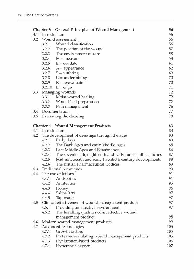
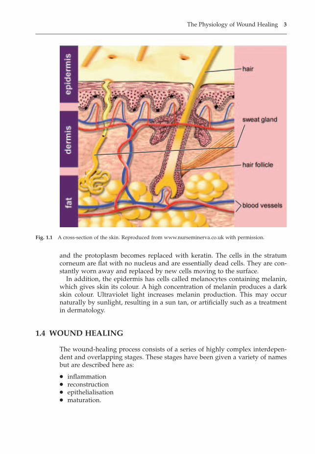
The skin is the largest and one of the most active organs of the body. It is com-posed of two layers – the epidermis and dermis – with the epidermis formingthe outer surface of the body and the dermis forming the deeper layer of theskin. The main structures of the skin can be found in the dermis. Figure 1.1shows a cross-section of the skin.
1.3.1 Dermis
Dermis is composed of connective tissue, both collagen and elastic fibres, whichis both elastic and resilient and provides support for the structures in thedermis. Within the dermis can be found blood vessels, lymph vessels, sensorynerve endings, sweat and sebaceous glands and hair follicles. The ducts of theglands and hair shafts pass through the epidermis to the skin surface. Sweatglands have their own ducts opening on the skin surface but sebaceous glandsopen onto the hair follicles. The base or bulb of hair follicles is sited deep intothe dermis. They are lined with epithelial cells and can play a role in the healingof partial-thickness wounds.
The surface of the dermis where it interlocks with the epidermis is irregular,with projections of cells called papillae. The base of the dermis is less clearlydefined as it blends into subcutaneous tissue, which contains both connectivetissue and adipose tissue and helps to anchor the skin to muscle and bone.
1.3.2 Epidermis
The epidermis comprises several layers of cells. The deepest layer is the stratumbasale and it is constantly producing new cells by cell division. These cells aregradually pushed towards the skin surface, taking about seven weeks to reachthe surface. The stratum spinosum contains bundles of keratin filaments, whichhold the skin together. The top three layers of epidermis are the stratum gran-ulosum, which produces the precursor to keratin, the stratum lucidum and thestratum corneum. As they move through the strata, the cells gradually flatten
2 The Care of Wounds
and the protoplasm becomes replaced with keratin. The cells in the stratumcorneum are flat with no nucleus and are essentially dead cells. They are con-stantly worn away and replaced by new cells moving to the surface.
In addition, the epidermis has cells called melanocytes containing melanin,which gives skin its colour. A high concentration of melanin produces a darkskin colour. Ultraviolet light increases melanin production. This may occur naturally by sunlight, resulting in a sun tan, or artificially such as a treatmentin dermatology.
1.4 WOUND HEALING
The wound-healing process consists of a series of highly complex interdepen-dent and overlapping stages. These stages have been given a variety of namesbut are described here as:
� inflammation� reconstruction� epithelialisation� maturation.
The Physiology of Wound Healing 3
Fig. 1.1 A cross-section of the skin. Reproduced from www.nurseminerva.co.uk with permission.
The stages last for variable lengths of time. Any stage may be prolongedbecause of local factors such as ischaemia or lack of nutrients. The factors thatcan delay healing are discussed in more detail in Chapter 2.
1.4.1 Inflammation
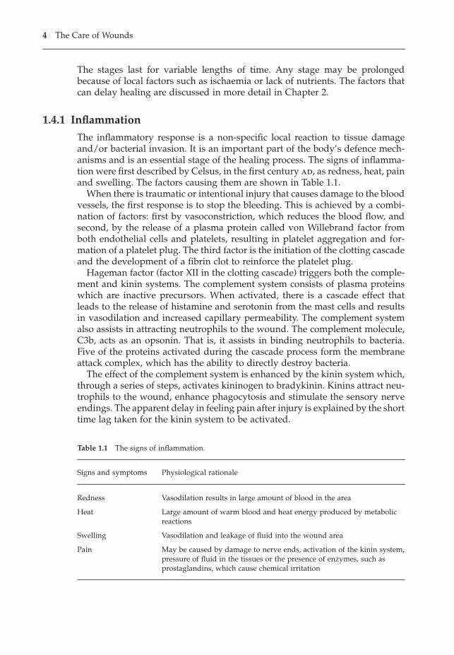
The inflammatory response is a non-specific local reaction to tissue damageand/or bacterial invasion. It is an important part of the body’s defence mech-anisms and is an essential stage of the healing process. The signs of inflamma-tion were first described by Celsus, in the first century ad, as redness, heat, painand swelling. The factors causing them are shown in Table 1.1.
When there is traumatic or intentional injury that causes damage to the bloodvessels, the first response is to stop the bleeding. This is achieved by a combi-nation of factors: first by vasoconstriction, which reduces the blood flow, andsecond, by the release of a plasma protein called von Willebrand factor fromboth endothelial cells and platelets, resulting in platelet aggregation and for-mation of a platelet plug. The third factor is the initiation of the clotting cascadeand the development of a fibrin clot to reinforce the platelet plug.
Hageman factor (factor XII in the clotting cascade) triggers both the comple-ment and kinin systems. The complement system consists of plasma proteinswhich are inactive precursors. When activated, there is a cascade effect thatleads to the release of histamine and serotonin from the mast cells and resultsin vasodilation and increased capillary permeability. The complement systemalso assists in attracting neutrophils to the wound. The complement molecule,C3b, acts as an opsonin. That is, it assists in binding neutrophils to bacteria.Five of the proteins activated during the cascade process form the membraneattack complex, which has the ability to directly destroy bacteria.
The effect of the complement system is enhanced by the kinin system which,through a series of steps, activates kininogen to bradykinin. Kinins attract neu-trophils to the wound, enhance phagocytosis and stimulate the sensory nerveendings. The apparent delay in feeling pain after injury is explained by the shorttime lag taken for the kinin system to be activated.
4 The Care of Wounds
Table 1.1 The signs of inflammation.
Signs and symptoms Physiological rationale
Redness Vasodilation results in large amount of blood in the area
Heat Large amount of warm blood and heat energy produced by metabolicreactions
Swelling Vasodilation and leakage of fluid into the wound area
Pain May be caused by damage to nerve ends, activation of the kinin system,pressure of fluid in the tissues or the presence of enzymes, such asprostaglandins, which cause chemical irritation
As the capillaries dilate and become more permeable, there is a flow of fluidinto the injured tissues. This fluid becomes the ‘inflammatory exudate’ and con-tains plasma proteins, antibodies, erythrocytes, leucocytes and platelets. Aswell as being involved in clot formation, platelets also release fibronectin andgrowth factors called platelet-derived growth factor (PDGF) and transforminggrowth factor alpha and beta (TGFa and TGFb). Their role is to promote cellmigration and growth at the wound site.
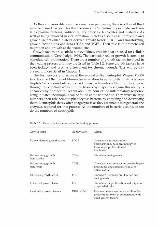
Growth factors are a subclass of cytokines, proteins that are used for cellularcommunication (Greenhalgh, 1996). The particular role of growth factors is tostimulate cell proliferation. There are a number of growth factors involved inthe healing process and they are listed in Table 1.2. Some growth factors havebeen isolated and used as a treatment for chronic wounds. This will be dis-cussed in more detail in Chapter 4.
The first leucocyte to arrive at the wound is the neutrophil. Wagner (1985)has described the role of fibronectin in relation to neutrophils. It attracts neu-trophils to the wound site, a process known as chemotaxis. Neutrophils squeezethrough the capillary walls into the tissues by diapedesis; again this ability isenhanced by fibronectin. Within about an hour of the inflammatory responsebeing initiated, neutrophils can be found at the wound site. They arrive in largenumbers, their role being to phagocytose bacteria by engulfing and destroyingthem. Neutrophils decay after phagocytosis as they are unable to regenerate theenzymes required for this process. As the numbers of bacteria decline, so toodo the numbers of neutrophils.
The Physiology of Wound Healing 5
Table 1.2 Growth factors involved in the healing process.
Growth factor Abbreviation Action
Platelet-derived growth factor PDGF Chemotactic for neutrophils,fibroblasts and, possibly, monocytesEncourages proliferation offibroblasts
Transforming growth TGFa Stimulates angiogenesisfactor alpha
Transforming growth TGFb Chemotactic for monocytes (macrophages)factor beta Encourages angiogenesis. Regulates
inflammation
Fibroblast growth factor FGF Stimulates fibroblast proliferation andangiogenesis
Epidermal growth factor EGF Stimulates the proliferation and migration of epithelial cells
Insulin-like growth factors IGF-I, IGF-II Promote protein synthesis and fibroblast proliferation. Work in combination with other growth factors

TGFb attracts monocytes to the wound where they differentiate intomacrophages. Fibronectin binds onto the surface receptors on the cells, promoting diapedesis and phagocytosis. Oxygen is vital to this process andmacrophages can be inactivated and their ability to undertake phagocytosisreduced if the partial oxygen pressure falls below 30mmHg (Cherry et al., 2000).Macrophages are larger than neutrophils and so are able to phagocytose largerparticles, such as necrotic debris, as well as bacteria. The lifespan of the neutrophil can be a few hours or a few days. When they die, they are alsophagocytosed by the macrophages.
T lymphocytes also migrate into the wound, although in smaller numbersthan macrophages (Martin & Muir, 1990). They influence macrophage phago-cytic activity by the production of several macrophage-regulating factors. Theyalso produce colony-stimulating factors that encourage the macrophage toproduce a range of enzymes and cytokines. These include prostaglandins whichmaintain vasodilation and capilliary permeability and can be produced ondemand to prolong the inflammatory response if required. A study by Martinand Muir (1990) found that both macrophages and lymphocytes are present inwounds from day 1, with macrophages peaking between days 3 and 6 and lym-phocytes between days 8 and 14.
Inflammation lasts about 4–5 days. It requires both energy and nutritionalresources. In large wounds the requirements may be considerable. If this stageis prolonged by irritation to the wound, such as infection, foreign body ordamage caused by the dressing, it can be debilitating to the patient as well asdelaying healing.
1.4.2 Reconstruction
The reconstruction phase is characterised by the development of granulationtissue. This consists of a loose matrix of fibrin, fibronectin, collagen andhyaluronic acid and other glycosaminoglycans. Within this matrix can be found macrophages and fibroblasts and the newly formed blood vessels.Macrophages play a major role in this phase of healing. They produce PDGFand fibroblast growth factor (FGF), which are chemotactic to fibroblasts, attract-ing them to the wound and stimulating them to divide and later to producecollagen fibres. Fibronectin also seems to play a role in enhancing fibroblastactivity (Orgill & Demling, 1988). Collagen has been seen in a new wound asearly as the second day. Collagen fibres are made up of chains of amino acids in a triple helix formation. There are a number of different types of col-lagen characterised by different formations of amino acids. Type III is presentin the healing wound in greater proportions than would normally be found in skin. Over time, this proportion reduces in favour of higher levels of type Icollagen.
Fibroblasts are key cells in this phase of healing (Harding et al., 2002). As wellas being responsible for the production of collagen, they also produce the extra-
6 The Care of Wounds
cellular matrix, which is seen visually as granulation tissue. As new extracel-lular matrix is synthesised, the existing matrix is degraded by enzyme systemssuch as matrix metalloproteinases (MMPs). There are a number of MMPs, inparticular MMP1, MMP2 and MMP9, involved in the healing process, althoughtheir role is imperfectly understood at present.
The activity of fibroblasts depends on the local oxygen supply. If the tissuesare poorly vascularised the wound will not heal well. The wound surface hasa relatively low oxygen tension, encouraging the macrophages to produce TGFband FGF, which instigate the process of angiogenesis, the growth of new bloodvessels. Undamaged capillaries beneath the wound, sprout buds, which growtowards the surface and loop over and back to the capillary. The loops form anetwork within the wound, supplying oxygen and nutrients. At this stage, ahigh oxygen tension promotes the continued growth of the capillary loopsbecause collagen is required in their formation (Cherry et al., 2000).
Some fibroblasts have a further role as they are involved in the process ofcontraction. The exact mechanism is not clearly understood and there are cur-rently two theories postulated: cell contraction and cell traction. The theory ofcell contraction is based on specialised fibroblasts known as myofibroblasts(Gabbiani et al., 1973), which have a contractile apparatus, similar to that insmooth muscle cells. In in vitro models, they have been shown to cause con-traction of the wound. Tomasek et al. (1989) found a higher level of contractileforces when a high level of myofibroblasts was present. The concept of cell traction was put forward by Stopak and Harris (1982), who demonstrated that fibroblasts could contract collagen gels by a physical pull, resulting in arearrangement of the extracellular matrix. It must be noted that all these studieswere undertaken in vitro and there is no certainty that they could be repeatedin vivo.
Whatever the actual process, contraction may start at around the fifth or sixthday. It considerably reduces the surface area of open wounds. Irvin (1987) sug-gests that contraction could be responsible for as much as 40–80% of the closure.It is certainly of considerable importance in large cavity wounds. However, inshallower wounds with a large surface area such as burns, contraction may leadto contractures. Myofibroblasts disappear after healing is completed.
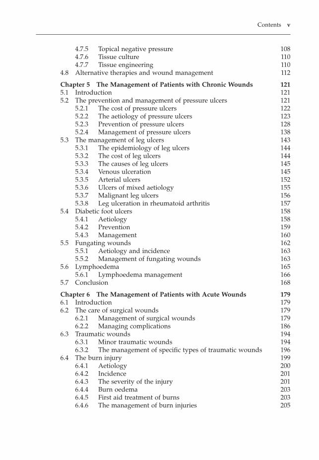
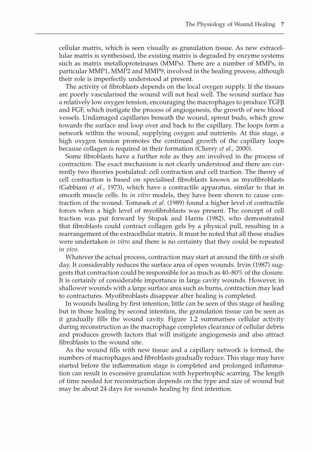
In wounds healing by first intention, little can be seen of this stage of healingbut in those healing by second intention, the granulation tissue can be seen asit gradually fills the wound cavity. Figure 1.2 summarises cellular activityduring reconstruction as the macrophage completes clearance of cellular debrisand produces growth factors that will instigate angiogenesis and also attractfibroblasts to the wound site.
As the wound fills with new tissue and a capillary network is formed, thenumbers of macrophages and fibroblasts gradually reduce. This stage may havestarted before the inflammation stage is completed and prolonged inflamma-tion can result in excessive granulation with hypertrophic scarring. The lengthof time needed for reconstruction depends on the type and size of wound butmay be about 24 days for wounds healing by first intention.
The Physiology of Wound Healing 7
1.4.3 Epithelialisation
This is the phase in which the wound is covered with epithelial cells.Macrophages release epidermal growth factor (EGF), which stimulates both theproliferation and migration of epithelial cells. Keratinocytes at the woundmargins and around hair follicle remnants synthesise fibronectin, which formsa temporary matrix along which the cells migrate. The cells move over thewound surface in a leapfrog fashion, the first cell remaining on the woundsurface and forming a new basement membrane. When cells meet, either in thecentre of the wound, forming islets of cells, or at the margin, they stop. This isknown as contact inhibition. Epithelial cells only move over viable tissue andrequire a moist environment (Winter, 1962). In sutured wounds, epithelial cellsalso migrate along the suture tracks. They are either pulled out with the suturesor gradually disappear.
Once the cells stop moving on the wound surface, they start to reconstitutethe basement membrane, which is essential in order for the epidermis to ‘fix’
8 The Care of Wounds
Production ofPDGF & FGFchemotactic to
fibroblasts
Production ofTGFB & FGF
instigatesangiogenesis
Phagocytosis
MACROPHAGE
Fig. 1.2 The process of reconstruction.
to the dermis. Until the basement membrane is fully reconstituted, it is easy forepithelial cells to be sheared off the wound surface by mechanical forces(Cherry et al., 2000).
Epithelialisation commences as early as the second day in closed wounds.However, in open wounds it is necessary for the wound cavity to be filled withgranulation tissue before it can commence. There is a very variable time spanfor this stage.
1.4.4 Maturation
During maturation the wound becomes less vascularised as there is a reduc-tion in the need to bring cells to the wound site. The collagen fibres are reor-ganised so that, instead of being laid down in a random fashion, they lie at rightangles to the wound margins. During this process, collagen is constantlydegraded and new collagen synthesised. The highest level of activity in thisprocess occurs between days 14 and 21 (Cherry et al., 2000). The scar tissuepresent is gradually remodelled and becomes comparable to normal tissue aftera long period of time. The scar gradually flattens to a thin white line. This maytake up to a year in closed wounds and very much longer in open wounds.
Tensile strength gradually increases. This is a way of describing the ability ofthe wound to resist rupture or dehiscence. Forester et al. (1969) found that atten days an apparently well-healed surgical incision has little strength. Duringmaturation it increases so that by three months the tensile strength is 50% thatof normal tissue. Further work by Forester et al. (1970) compared surgical inci-sions where the skin edges were held together by tape with those where sutureswere used. The findings showed that, when tape was used, the woundsregained 90% strength of normal tissue whereas sutured wounds only regained70% strength.
1.5 IMPAIRED WOUND HEALING
Although the majority of wounds heal without problem, impaired healing maysometimes occur. Some of the different types of impaired healing are describedhere. Their management will be discussed elsewhere.
1.5.1 Hypertrophic scars
Hypertrophic scars occur when there is an excessive fibrous tissue responseduring the healing process resulting in excessive deposition of collagen and athick wound scar (Munro, 1995). Cherry et al. (2000) suggest that the ratio oftype I to type III collagen is lower than in normal skin. Hypertrophic scars aremore common after traumatic injury, especially large burns. They occur shortlyafter the injury or surgery and remain limited to the area of the injury. Theywill generally flatten out with time, about one to two years.
The Physiology of Wound Healing 9
1.5.2 Keloids
Keloids are similar to hypertrophic scars in that they are also the result of anexcessive fibrous response but this time, the ratio of type I to type III collagenis much higher than in normal skin. Another difference is that keloids take sometime to form and may occur years after the initial injury. They can range in sizefrom small papules to large pendulous growths (Munro, 1995). Keloids morecommonly occur in individuals aged between 10 and 30 (Cosman et al., 1961)and in those with a darker skin (Placik & Lewis, 1992). Unfortunately, unlikehypertrophic scars, keloids do not gradually flatten out.
1.5.3 Contractures
Wound contraction is part of the normal healing process but occasionally con-traction will continue after re-epithelialisation has occurred, resulting in scarcontraction (Tredget et al., 1997). Engrav et al. (1987) describe how this type ofscar contracture can lead to joint contracture with subsequent loss of mobility,functional loss, delay in return to work and a poor cosmetic result, any of whichmay necessitate surgery.
1.5.4 Acute to chronic wounds
Wounds may be called ‘chronic’ because their underlying aetiology makeshealing a very long process. A good example is the venous leg ulcer. However,some chronic wounds may have originally been acute wounds that have failedto heal over a long period of time, perhaps years. The original factor delayinghealing may have been related to infection or local irritation, perhaps causedby a suture. Once these problems have been resolved, the wound still fails toheal, causing considerable misery to the patient.
The differences between acute and chronic wounds are still imperfectlyunderstood. However, work by Phillips et al. (1998) did shed some light on theproblem. They used cultured fibroblasts from human neonatal foreskin as aplated laboratory model and treated them with either chronic wound fluid(CWF) or bovine serum albumen (the control). They found that CWF inhibitedthe growth of the fibroblasts quite dramatically. The researchers concluded thatthis study gave some indication of how the microenvironment has a negativeeffect on the healing wound. As result of this work, other research groups havelooked at wound exudate in more detail.
Trengrove et al. (1999) used wound fluid from venous leg ulcers at both non-healing and healing stages to measure MMP levels. They found elevated levelsof MMPs at the non-healing stage, which decreased significantly as the ulcersstarted to heal (p = 0.01). The levels of MMPs in the healing ulcers were similarto those in acute wounds, thus suggesting that failure to heal may be linked toexcessive matrix degradation. Ladwig et al. (2002) collected wound fluid from56 pressure ulcers and found lower levels of MMP9 in those ulcers that wenton to heal well compared with those that healed poorly.
10 The Care of Wounds
Trengrove et al. (2000) undertook further studies of wound exudate from non-healing and healing leg ulcers. They found significantly higher concentrationsof a number of proinflammatory cytokines or growth factors in the non-healingulcers. They consider that wound healing is delayed in chronic wounds becauseof an impairment of inflammatory mediators rather than by any deficit ofgrowth factors.
Premature ageing of fibroblasts may also be a problem. Mendez et al. (1998)investigated the characteristics of fibroblasts cultured from chronic venousulcers and found signs of accelerated ageing or senescence in these cells. Senes-cent fibroblasts have reduced mobility, are less able to replicate, have abnormalprotein production and do not respond well to growth factors. A small studyof seven patients by Stanley and Osler (2001) compared the senescence rates infibroblasts from chronic venous ulcers with fibroblasts from punch biopsiestaken from the proximal thigh of the same patient. They found a significantlyhigher senescence rate in the fibroblasts from the leg ulcers (p = 0.0001).
1.6 CONCLUSION
This chapter has described ‘normal’ physiology. However, not all wounds healwithout complication or delay and some of the differences between acute andchronic wound healing have been discussed. But many factors can affect thehealing process and they will be considered in more detail in Chapter 2.
FURTHER READING
Cherry, G.W., Hughes, M.A., Ferguson, M.W.J., Leaper, D.J. (2000) Wound healing, in(eds) Morris, P.J., Wood, W.C., Oxford Textbook of Surgery, 2nd edn. Oxford University Press, Oxford.
Vander, A., Sherman, J., Luciano, D. (1998) Human Physiology, 7th edn. McGraw-Hill,Boston.
REFERENCES
Cherry, G.W., Hughes, M.A., Ferguson, M.W.J., Leaper, D.J. (2000) Wound healing, in(eds) Morris, P.J., Wood, W.C., Oxford Textbook of Surgery, 2nd edn. Oxford Uni-versity Press, Oxford.
Cosman, B., Crikelair, G.F., Ju, M.C. et al. (1961) The surgical treatment of keloids. Plasticand Reconstructive Surgery, 27, 335–358.
Engrav, L.H., Covey, M.H., Dutcher, K.D. et al. (1987) Impairment, time out of schooland time out of work after burns. Plastic and Reconstructive Surgery, 79, 927.
Forester, J.C., Zederfeldt, B.H., Hunt, T.K. (1969) A bioengineering approach to thehealing wound. Journal of Surgical Research, 9, 207.
Forester, J.C., Zederfeldt, B.H., Hunt, T.K. (1970) Tape-closed and sutured wounds: acomparison by tensiometry and scanning electron microscope. British Journal ofSurgery, 57, 729.
The Physiology of Wound Healing 11
Gabbiani, G., Hajno, G., Ryan, G.B. (1973) The fibroblast as a contractile cell: the myofibroblast, in (eds) Kulonen, E., Pikkarainen, J., The Biology of the Fibroblast.Academic Press, London.
Greenhalgh, D. (1996) The role of growth factors in wound healing. Journal of Trauma,41 (1), 159–167.
Harding, K.G., Morris, H.L., Patel, G.K. (2002) Healing chronic wounds. British MedicalJournal, 324, 160–163.
Irvin, T.T. (1987) The principles of wound healing. Surgery, 1, 1112–1115.Ladwig, G.P., Robson, M.C., Liu, R., Kuhn, M.A., Muir, D.F., Schultz, G.S. (2002) Ratios
of activated matrix metalloproteinase-9 to tissue inhibitor of matrix matellopro-teinase-1 in wound fluids are inversely correlated with healing in pressure ulcers.Wound Repair and Regeneration, 10 (1), 26.
Martin, C.W., Muir, I.F.K. (1990) The role of lymphocytes in wound healing. BritishJournal of Plastic Surgery, 43, 655–662.
Mendez, M.V., Stanley, A.C., Phillips, T.H., Murphy, M., Menzoian, J.O., Park, H.Y. (1998)Fibroblasts cultured from venous ulcers display cellular characteristics of senes-cence. Journal of Vascular Surgery, 28, 1040–1050.
Munro, K.J.G. (1995) Hypertrophic and keloid scars. Journal of Wound Care, 4 (3), 143–148.Orgill, D., Demling, R.H. (1988) Current concepts and approaches to wound healing.
Critical Care Medicine, 16 (9), 899–908.Phillips, T.J., Al-Amoudi, H.O., Leverkus, M., Park, H-Y. (1998) Effect of chronic wound
fluid on fibroblasts. Journal of Wound Care, 7 (10), 527–532.Placik, O., Lewis, V.L. (1992) Immunological associations of keloids. Surgery, Gynaecol-
ogy and Obstetrics, 175, 185–193.Stanley, A., Osler, T. (2001) Senescence and the healing rates of venous ulcers. Journal of
Vascular Surgery, 33, 1206–1210.Stopak, D., Harris, A.K. (1982) Connective tissue morphogenesis by fibroblast traction.
1. Tissue culture observations. Developments in Biology, 90 (2), 383–398.Tomasek, J.J., Haaksma, C.J., Eddy, R.T. (1989) Rapid contraction of collagen lattices by
myofibroblasts is dependent upon organised actin microfilaments. Journal of CellBiology, 170, 3410.
Tredget, E.E., Nedelec, B., Scott, P.G., Ghahary, A. (1997) Hypertrophic scars, keloids andcontractures. Surgical Clinics of North America, 77 (3), 701–730.
Trengrove, M.K., Stacey, M.C., McCauley, S. et al. (1999) Analysis of the acute and chronicwound environments: the role of proteases and their inhibitors. Wound Repair andRegeneration, 7 (6), 442–452.
Trengrove, N.J., Bielefeldt-Ohmann, H., Stacey, M.C. (2000) Mitogenic activity andcytokine levels in non-healing and healing chronic leg ulcers. Wound Repair andRegeneration, 8 (1), 13–25.
Wagner, B.M. (1985) Wound healing revisited: fibronectin and company. Human Pathol-ogy, 16 (11), 1081.
Winter, G.D. (1962) Formation of the scab and the rate of epithelialisation of superficialwounds in the skin of the domestic pig. Nature, 193, 293.
12 The Care of Wounds
The Management of Patients with Wounds 13
Chapter 2The Management of Patients with Wounds
2.1 INTRODUCTION
This chapter looks at assessment of the patient with a wound and how appro-priate care may be planned and evaluated. When caring for patients withwounds of all types, it is important to take a holistic approach, consideringphysical, psychological and spiritual care as they are inextricably linked. Thereare many factors that can affect the healing process. If they are taken intoaccount when taking a history and assessing the patient, it may be possible tomitigate some of the effects. Nursing intervention is not able to resolve everyproblem (for example, age) but where nursing intervention can be effective,appropriate strategies are suggested.
2.2 PHYSICAL CARE
2.2.1 Nutrition
The precise relationship between wound healing and nutrition remains uncer-tain (Williams & Barbul, 2003). There is increasing evidence that nutritionaldeficit impairs healing, such as the study by Wissing et al. (2001) which followed up patients with leg ulceration identified in a previous study. Thosepatients whose ulcers were still open had significantly lower nutritional statuscompared with those whose ulcers had healed. A number of other studies haveidentified the impact of malnutrition on the healing of surgical wounds, burnsand pressure ulcers (Andel et al., 2003; Haydock & Hill, 1986; Mathus-Vliegen,2004). The importance of nutrition in relation to pressure ulcer prevention andmanagement is highlighted by the development of nutrition guidelines by theEuropean Pressure Ulcer Advisory Panel (EPUAP, 2003).
Malnutrition is a pathological state that results from a relative or absolutedeficiency or excess of one or more essential nutrients. As protein or carbohy-drates are used in the largest quantities, they are usually the deficient nutrients.This is referred to as protein energy malnutrition or PEM. In her Notes onNursing, What it is and What it is Not, Florence Nightingale (1974) said: ‘Everycareful observer of the sick will agree in this, that thousands of patients areannually starved in the midst of plenty, from want of attention to the wayswhich alone make it possible for them to take food’. More than a century laterthis statement is still true. McWhirter and Pennington (1994) assessed the nutri-
tional status of 500 acutely ill patients and found 40% were undernourished onadmission to hospital. Gallagher-Allred et al. (1996) reviewed studies involving1327 patients which showed that 40–55% were malnourished and 12% wereseverely malnourished. Edington et al. (1996) surveyed community patients andfound that 10% of cancer patients and 8% of those with chronic diseases weremalnourished.
Overall, malnutrition is seldom recognised in hospital patients although ithas a major impact on morbidity and mortality (Pablo et al., 2003). Correia andWaitzberg (2003) undertook multivariate analysis of the impact of malnutritionon adult hospital patients and found mortality increased to 12.4%, comparedwith 4.7% in the well nourished. Hospital costs increased up to 308.9%. Olderpatients are at particular risk of malnutrition. Guigoz et al. (2002) identified mal-nutrition in 20% of hospitalised patients in a survey of more than 10000 Swisselderly people in the community, nursing homes and hospitals. Similar resultswere found in a Spanish study of hospital patients – 18.2% of patients hadsevere malnutrition (Cereceda et al., 2003). Fortunately, politicians have beenalerted to the importance of food and nutritional care in hospitals. The Councilof Europe Committee of Ministers passed a resolution in 2003 that each memberstate should have national recommendations that encompass all aspects ofnutritional care (Resolution RESAP(2003)3) (Committee of Ministers, 2003).
Nutritional status
The initial causes of malnutrition may be related to debilitating disease, especially of the gastrointestinal tract, old age, poverty or ignorance. Onceadmitted to hospital, other factors become relevant. An early study by Hamilton Smith (1972) found that patients were starved for up to 12 hours priorto surgery and for varying lengths of time afterwards. Chapman (1996) foundlittle had changed in over 20 years. She found that patients fasted for periodsranging from 4 to 29 hours. A long period of preoperative starvation serves tocompound the effects of trauma and surgery, both of which cause marked catab-olism. This catabolic state usually lasts between 6 and 18 hours. Following this,the basal metabolic rate rises, leading to increased energy requirements. Unlessadequate protein and carbohydrate are taken in to supply these needs, furthertissue breakdown occurs, resulting in muscle wasting and a negative nitrogenbalance. Lee (1979) suggests that the consequences of a negative nitrogenbalance include poor wound healing, impaired immunocompetence and susceptibility to infection.
Whilst some patients will return to a normal diet fairly quickly and so redressthe balance, others will receive only intravenous fluids. A litre of dextrose 5%contains approximately 150 calories. Normal saline does not contain any at all.These fluids obviously do not provide adequate calories to meet the body’srequirements.
Burn patients are particularly at risk and may continue to be so for as longas four weeks (Sutherland, 1985). Trauma, burns and pain increase the meta-bolic rate, further diminishing the patient’s nutritional status (Arturson, 1978).
14 The Care of Wounds
Zinc, in particular, is burned up in large amounts during emotional or physi-cal stress. Taylor (1999) studied 106 burn patients who received enhancedenteral nutrition (50% of energy and nitrogen requirements). There was a sig-nificantly greater incidence of infection and length of hospital stay when therewas a delay of 24 hours in commencing the enhanced nutrition treatment. Zhouet al. (2003) randomly allocated severely burned patients to additional enteralglutamine (an amino acid) and found a 19% improvement in wound healingcompared to controls, who had received standard feeds.
It is the responsibility of the nurse to see that patients have an adequate diet. Many patients have their mealtimes disrupted by medical ward rounds or being away from the ward undergoing investigations, although there isincreased awareness of the need to have protected mealtimes. Older et al. (1980)saw food being placed beyond the reach of a patient and then removed laterwithout the patient ever having the chance to actually eat any of it. Delmi et al.(1990), in their study of a group of elderly patients with fractured neck of femur,found that inadequate amounts of food were consumed. It should also be notedthat 80% of patients in the study were malnourished on admission. Lewis et al.(1993) studied the diet of a small group of elderly patients with leg ulcers andfound their intake was below the estimated average requirement for their agegroup and did not meet the requirements for healing their ulcers. A similarstudy by Sitton-Kent and Gilchrist (1993) of elderly hospitalised patients withchronic wounds found that they did not consume adequate levels of nutrientsand in some instances had inadequate quantities on their plates. Many thingscan affect the appetite such as anxiety, altered mealtimes, cultural differencesor malaise. It is obvious that a nutritional assessment of all patients should bemade on admission and at regular intervals afterwards.
The Better Food Programme (DoH, 2001c) was introduced to try and addresssome of the problems described above. The Essence of Care document (DoH,2001a) has provided best practice standards against which healthcare providerscan benchmark their practice. It includes statements such as the following.
� Patients/clients receive the care and assistance they require with eating anddrinking.
� Food that is provided by the service meets the needs of individualpatients/clients.
Age
The cell metabolic rate slows with advancing years. There is also an increasedrisk of malnutrition. Exton Smith (1971) divided the causes of this into primaryand secondary. Primary causes included ignorance, social isolation, physicaldisability, mental disturbance, iatrogenic disorder and poverty. Secondarycauses were impaired appetite, masticatory inefficiency, malabsorption, alco-holism, drugs and increased requirements.
The Management of Patients with Wounds 15
Disease
Many patients suffering from malignant disease have a reduced nutritionalstatus. Stubbs (1989) found that one in four cancer patients experienced alter-ations in taste perception that affected their appetite and eating habits.
Drugs
Several drugs affect the nutritional status of patients. Methotrexate has an anti-vitamin effect which means that the enzyme that would normally bind avitamin binds the drug instead. Methotrexate competes with folic acid andcauses it to be excreted, thereby inhibiting DNA synthesis and cell replication(Holmes, 1986). Neomycin reduces the absorption of vitamins K and D. Para-aminosalicylic acid (PAS) and colchicine reduce the absorption of vitamin B12.A number of drugs can cause loss of appetite, which may lead to a diminishednutritional status. Examples are phenformin, metformin, indomethacin, mor-phine, digoxin and cancer drugs.
It should also be noted that patients not deemed to be at risk of undernutri-tion may fail to eat adequately. Brown (1991) studied the intake of patients whowere considered to have no special dietary requirements. She found that 68%had intakes of less than 1000 kcal and large deficits of a range of vitamins andminerals. The deficit was caused by failure to eat the food provided. Adequatemonitoring of patients’ diets is essential as this group of patients are oftenmissed.
It is important to identify those who are malnourished in order that appro-priate steps can be taken to improve their nutritional status. A number ofscreening tools have been developed and some have been widely validated.One such is the Mini-Nutritional Assessment Tool (MNA), which has been usedto assess elderly patients with leg ulceration (Wissing & Unosson, 1999). Thefirst part of the MNA is a screening tool that identifies those who require moredetailed assessment. The second part allows the assessor to identify those atrisk of malnutrition and those who are actually malnourished, allowing thehealthcare professional to develop an appropriate plan of care.
The British Association for Parenteral and Enteral Nutrition (BAPEN)launched the MUST screening tool in 2003 (Elia & Stratton, 2004). It is a five-step tool that has been validated for use with adults of all ages in both hospi-tal and community settings. It allows the assessor to determine if a patient isat low, medium or high risk of malnutrition and provides appropriate man-agement guidelines, depending on whether the patient is in hospital, a carehome or the community. The guidance also provides information on how tocalculate height for a patient who cannot be measured in the usual way. Furtherinformation can be obtained from www.bapen.org.uk.
Hunt (1997) and her colleagues have devised a nutritional assessment toolthat considers various factors that can affect nutritional status. Patients areassessed according to their mental condition, weight, appetite, ability to eat, gutfunction, medical condition, including chronic wounds, and age. The tool pro-vides a score that indicates whether the patient is nutritionally at risk. Use of a
16 The Care of Wounds
screening tool can be helpful in identifying those less obviously at risk of poornutritional status than those discussed above.
� Nursing assessment �
On admission:
� identify those at special risk using an appropriate screening tool.� take a dietary history.� observe for obvious signs of obesity, emaciation or muscle wasting.
� Nursing intervention �
Problem: Reduced nutritional statusGoal: The patient will consume sufficient nutrients for his daily needs
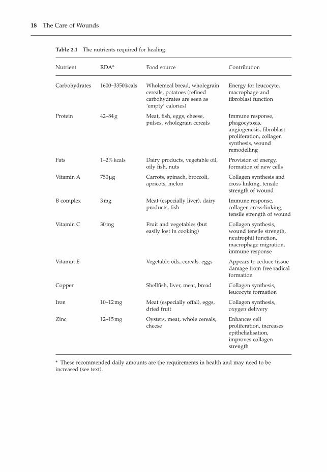
The nutritional needs for each individual vary according to their age, sex, activ-ity and the severity of any illness. If a patient has been assessed as having areduced nutritional status or falls into a high-risk category, then his nutritionalintake should be very carefully monitored. Each patient requires sufficientnutrients to support his basal metabolic rate, his level of activity and the meta-bolic response to trauma. Patients with heavily exuding wounds, such as fis-tulae or leg ulcers, may lose large amounts of protein without it being realised.Table 2.1 shows the nutrients required for wound healing and their sources.
The dietician will be able to help in assessing individual needs, so that veryspecific goals can be set. The goal set at the beginning of this section is of neces-sity broad but needs to be more clearly defined for each individual. If a patientis being cared for at home, the carer must also be involved. Many patients willeat better at home, where they can eat what they want, when they want to.
The elderly may have special problems or needs. Penfold and Crowther(1989) have provided helpful guidelines for assisting the elderly to maintain agood diet. One problem may be developing disability. The occupational thera-pist can give guidance on adapting cooking equipment. Another problem maybe lack of education as to what constitutes a ‘good’ diet. An even simplerproblem may be poorly fitting dentures. A new set of teeth may be all that isneeded to allow an elderly person to maintain an adequate nutritional status.
For many people, the short period of starvation during surgery followed bya rapid return to an adequate diet will not be harmful and the body will quicklyadapt. However, nurses need to be aware of the amount of food their patientsactually eat. These days the plated meal system is widely used in hospitals andthere has been little monitoring of the amount of food which patients actuallyeat. It is to be hoped that the benchmarking process discussed earlier will assistin resolving this problem. When assisting a patient to plan appropriate menus,it is helpful to bear in mind the sources of the nutrients particularly requiredfor wound healing (see Table 2.1).
Some critically ill patients will not have an adequate intake without artificialfeeding. This may take the form of a supplement or total nutrition, either byenteral or parenteral feeding. Enteral feeding is the more desirable way of pro-viding nutrition but if the gastrointestinal tract is not functioning, then total
The Management of Patients with Wounds 17
18 The Care of Wounds
Table 2.1 The nutrients required for healing.
Nutrient RDA* Food source Contribution
Carbohydrates 1600–3350kcals Wholemeal bread, wholegrain Energy for leucocyte,cereals, potatoes (refined macrophage andcarbohydrates are seen as fibroblast function‘empty’ calories)
Protein 42–84g Meat, fish, eggs, cheese, Immune response,pulses, wholegrain cereals phagocytosis,
angiogenesis, fibroblastproliferation, collagensynthesis, woundremodelling
Fats 1–2%kcals Dairy products, vegetable oil, Provision of energy,oily fish, nuts formation of new cells
Vitamin A 750mg Carrots, spinach, broccoli, Collagen synthesis andapricots, melon cross-linking, tensile
strength of wound
B complex 3mg Meat (especially liver), dairy Immune response,products, fish collagen cross-linking,
tensile strength of wound
Vitamin C 30mg Fruit and vegetables (but Collagen synthesis,easily lost in cooking) wound tensile strength,
neutrophil function,macrophage migration,immune response
Vitamin E Vegetable oils, cereals, eggs Appears to reduce tissuedamage from free radicalformation
Copper Shellfish, liver, meat, bread Collagen synthesis,leucocyte formation
Iron 10–12mg Meat (especially offal), eggs, Collagen synthesis,dried fruit oxygen delivery
Zinc 12–15mg Oysters, meat, whole cereals, Enhances cellcheese proliferation, increases
epithelialisation,improves collagenstrength
* These recommended daily amounts are the requirements in health and may need to beincreased (see text).
parenteral nutrition is necessary. Zainal (1995) discussed the issues around thefeeding of critically ill patients and stressed the importance of starting feedsearly. However, this must be done with caution as overfeeding the critically illcan cause major metabolic problems. Further problems may be caused by per-sisting with enteral feeding for a patient in septic shock with reduced splanch-nic blood flow. The nutrition team should be involved in managing thesepatients.
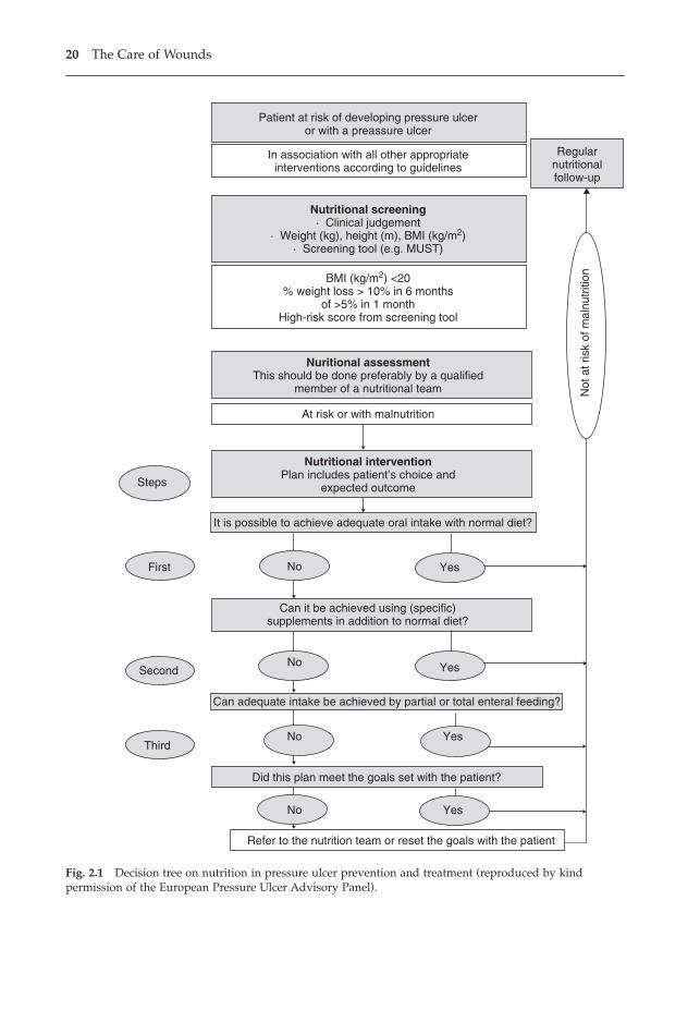
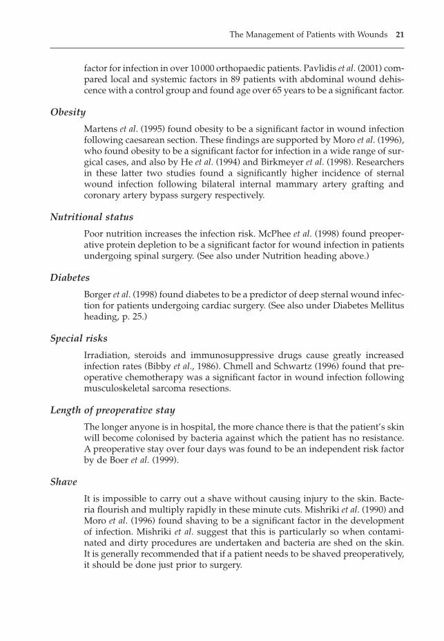
Decision trees can also be used in planning care. They can be particularlyhelpful in providing guidance for less experienced staff and for setting a stan-dard for care. The European Pressure Ulcer Advisory Panel has developed adecision tree to use alongside their nutrition guideline (Fig 2.1) which maps theessential elements of the guideline (Clark et al., 2004).
� Evaluation �
Evaluation may be achieved by regular weighing of the patient and reassess-ment using a nutritional screening tool. Gazzotti et al. (2003) used weighing andthe MNA in a randomised trial to determine the effectiveness of nutritional supplements in preventing malnutrition.
2.2.2 Infection
Consideration of infection must include both systemic and localised woundinfection. Systemic infection affects healing as the wound has to compete withmore widespread infection for white cells and nutrients. Wound healing maynot take place until after the body has dealt with the infection. Systemic infection is frequently associated with pyrexia which causes an increase in themetabolic rate, thus increasing catabolism or tissue breakdown.
All wounds are contaminated with bacteria, especially open wounds. Thisdoes not affect healing but clinical infection will certainly do so. Infection pro-longs the inflammatory stage of healing as the cells combat the large numbersof bacteria. It also appears to inhibit the ability of fibroblasts to produce colla-gen (Senter & Pringle, 1985).
Infection in a burn wound increases the metabolic rate and thereby increasesthe period of negative nitrogen balance. Kinney (1977) has shown that theremay be a loss of 20–30% of the initial body weight in the presence of majorsepsis. Infection also causes pain, which raises the metabolic rate (Arturson,1978).
A number of factors have been found to increase the risk of developing awound infection. They are identified below.
Age
Moro et al. (1996) used logistical regression to identify factors associated withincreased risk of surgical wound infection. They found that age greater than 85years was a significant factor. A study using multivariate analysis (de Boer et al., 1999) found age over 74 years to be the most important independent risk
The Management of Patients with Wounds 19
20 The Care of Wounds
Patient at risk of developing pressure ulceror with a preassure ulcer
In association with all other appropriateinterventions according to guidelines
Nutritional screening· Clinical judgement
· Weight (kg), height (m), BMI (kg/m2)· Screening tool (e.g. MUST)
BMI (kg/m2) <20 % weight loss > 10% in 6 months
of >5% in 1 monthHigh-risk score from screening tool
Nuritional assessmentThis should be done preferably by a qualified
member of a nutritional team
At risk or with malnutrition
Nutritional interventionPlan includes patient’s choice and
expected outcome
No
No
No
No
Yes
Yes
Yes
Steps
First
Second
Third
Refer to the nutrition team or reset the goals with the patient
Regularnutritionalfollow-up
It is possible to achieve adequate oral intake with normal diet?
Not
at r
isk
of m
alnu
triti
on
Can it be achieved using (specific)supplements in addition to normal diet?
Can adequate intake be achieved by partial or total enteral feeding?
Did this plan meet the goals set with the patient?
Yes
Fig. 2.1 Decision tree on nutrition in pressure ulcer prevention and treatment (reproduced by kindpermission of the European Pressure Ulcer Advisory Panel).
factor for infection in over 10000 orthopaedic patients. Pavlidis et al. (2001) com-pared local and systemic factors in 89 patients with abdominal wound dehis-cence with a control group and found age over 65 years to be a significant factor.
Obesity
Martens et al. (1995) found obesity to be a significant factor in wound infectionfollowing caesarean section. These findings are supported by Moro et al. (1996),who found obesity to be a significant factor for infection in a wide range of sur-gical cases, and also by He et al. (1994) and Birkmeyer et al. (1998). Researchersin these latter two studies found a significantly higher incidence of sternalwound infection following bilateral internal mammary artery grafting andcoronary artery bypass surgery respectively.
Nutritional status
Poor nutrition increases the infection risk. McPhee et al. (1998) found preoper-ative protein depletion to be a significant factor for wound infection in patientsundergoing spinal surgery. (See also under Nutrition heading above.)
Diabetes
Borger et al. (1998) found diabetes to be a predictor of deep sternal wound infec-tion for patients undergoing cardiac surgery. (See also under Diabetes Mellitusheading, p. 25.)
Special risks
Irradiation, steroids and immunosuppressive drugs cause greatly increasedinfection rates (Bibby et al., 1986). Chmell and Schwartz (1996) found that pre-operative chemotherapy was a significant factor in wound infection followingmusculoskeletal sarcoma resections.
Length of preoperative stay
The longer anyone is in hospital, the more chance there is that the patient’s skinwill become colonised by bacteria against which the patient has no resistance.A preoperative stay over four days was found to be an independent risk factorby de Boer et al. (1999).
Shave
It is impossible to carry out a shave without causing injury to the skin. Bacte-ria flourish and multiply rapidly in these minute cuts. Mishriki et al. (1990) andMoro et al. (1996) found shaving to be a significant factor in the developmentof infection. Mishriki et al. suggest that this is particularly so when contami-nated and dirty procedures are undertaken and bacteria are shed on the skin.It is generally recommended that if a patient needs to be shaved preoperatively,it should be done just prior to surgery.
The Management of Patients with Wounds 21
Type of surgery
Infection rates are much higher in some types of surgery than others. This isdiscussed in more detail in Chapter 6. The appearance of infected wounds willbe discussed in Chapter 3.
� Nursing assessment �
� Identify those at risk.� Assess wound (see Chapter 3).� Monitor temperature regularly.
Recently neural network analysis has been used to predict outcomes and iden-tify those at higher risk of developing an infection. Lammers et al. (2003) under-took a study of a cohort of 1142 uncomplicated traumatic wounds. Cliniciansundertaking the initial treatment of the wound were asked to estimate the like-lihood of subsequent infection. Staff blinded to this prediction followed thewounds until the sutures were removed. Independent predictors were identi-fied and used in the neural network analysis as input variables; infection wasthe output variable, in order to arrive at an equation for the analysis model.The researchers were able to use this as a diagnostic test for wound infectionin this group of wounds. This type of data analysis has potential for the futureas neural network analysis becomes more widely used.
� Nursing intervention �
Problem: Actual/potential risk of infectionGoal: Prevention or early detection
The prevention of infection is the responsibility of all healthcare professionals.There are both general and specific measures that can be taken. Most healthauthorities have infection control policies that provide guidelines both toprevent infection and to reduce the risk of cross-infection. The infection controlteam, especially infection control nurses, can give advice and support.
Much has been written on the prevention of infection. The UK Departmentof Health developed guidelines entitled Standard Principles for Preventing Infec-tions in Hospitals together with guidelines for preventing hospital-acquiredinfections (HAI) associated with the use of short-term indwelling urethralcatheters in acute care and with central venous catheters in acute care (DoH,2001b). It is intended that these guidelines are incorporated into local protocols.Within the first part of the guideline there are four standard principles.
� Hospital environmental hygiene.� Hand hygiene.� The use of personal protective equipment.� The use and disposal of sharps.
The spread of infection is mostly by people from people. Thus, the simplest andmost effective measure to prevent infection is good handwashing. A review byLarson and Kretzer of the period 1984–1994 found that researchers consistently
22 The Care of Wounds
reported that whilst the action of handwashing was carried out mostly at theappropriate times, the methods used were ineffective (Larson & Kretzer, 1995).Gould (1992) also supports this view but she suggests that there has been afailure to consider the reality of the situation in the clinical area. One examplecited is that compliance is unlikely if the designated cleanser makes hands sore. Adequate facilities for handwashing are also necessary although alcoholhandrub may be a useful alternative.
Tibballs (1996) observed doctors to obtain their handwashing rates and thenasked a sample for their estimated frequency of handwashing prior to patientcontact. There was a considerable difference between the estimated rate of 73%(range 50–90%) and the observed rate of 9%. It is also interesting to note thatan editorial in the British Medical Journal by the Handwashing Liaison Group(1999) provoked 21 letters to the editor from seven different countries, not allof them in support of handwashing.
The DoH guideline recommendations for handwashing include either the use of soap and water or the use of alcohol-based handrub. Hands that are obviously soiled or could be grossly contaminated must be washed in soap andwater. Handwashing is defined as having three stages: preparation, washingand rinsing, and drying. It is important to ensure that the soap or handrubtouches all the surfaces of the hands. The guidelines also recommend regularuse of an emollient hand cream in order to reduce skin dryness.
Identification of patients at risk of infection means that appropriate measurescan be taken. Some particularly vulnerable patients may require extra mea-sures. These may include the use of a single room with a positive pressure filtered air system, providing protective isolation, prophylactic drugs or special operating techniques such as a Charnley Howarth tent for orthopaedicprocedures.
A more controversial prevention method is the use of supplemental oxygen.Greif et al. (2000) randomly allocated 500 patients undergoing colorectal surgeryto one of two regimes of supplemental oxygen. They found that provision ofsupplemental oxygen during surgery and for two hours afterwards halved theinfection rate. This approach seems promising but Gottrup (2000) suggestedthat the optimal treatment period has not been finally determined.
� Evaluation �
Careful monitoring of vulnerable patients is essential. Monitoring a patient’stemperature is a useful means of evaluation as a rise in temperature is often thefirst indication of infection. The use of clinical audit will identify areas wherecross-infection may be a regular problem.
2.2.3 Smoking
Smoking causes vasoconstriction and is associated with Buerger’s disease, acondition causing intermittent claudication and gangrene. Smoking may alsoact as an appetite depressant. Smokers have been found to be deficient in vitamins B1, B6, B12 and C. Smoking reduces subcutaneous oxygen tension
The Management of Patients with Wounds 23
significantly for up to 30–45 minutes after each cigarette. Synthesis of type I col-lagen has also been found to be reduced in smokers (Jorgensen et al., 1998). Areview of the effects of smoking on wound healing by Siana et al. (1992) foundthat nicotine affected macrophage activity and reduced epithelialisation andwound contraction. There is increasing evidence that smoking is associatedwith poor wound healing and increased complications. However, most studiesundertaken in this area have looked at surgical wounds and there is little infor-mation regarding smoking and chronic wound healing (Sorensen, 2003).
Sorensen et al. (2002) studied the impact of smoking compared with non-smoking on 425 patients undergoing breast surgery. They found smoking wassignificantly associated with wound infection, skin flap necrosis and epider-molysis. The same group studied the impact of abstinence from smoking onhealing of experimental incisional wounds in healthy individuals. They com-pared never-smokers with smokers randomised to either continue smoking orabstain from smoking for a four-week period. They found a significantly higherincidence of wound infection in the smokers compared with the never-smokers(12% versus 2%, p < 0.05). They also found there was a significant reduction in infection in the abstinent smokers compared with continuous smokers(Sorensen et al., 2003). Manassa et al. (2003) undertook a retrospective study of132 patients undergoing abdominoplasty and compared outcomes for smokersand non-smokers. They found a significant incidence of wound complications,including wound dehiscence (47.9% versus 14.8%, p < 0.01).
In reviewing the evidence on smoking and problems in wound healing,Sorensen (2003) suggests that there is a need for research into the impact ofsmoking on chronic wound healing. Further research is also required to deter-mine the impact of abstinence on collagen production and delayed healing.
� Nursing assessment �
Early identification of patients who smoke, especially prior to surgery.
� Nursing intervention �
Problem: Patient who smokes about to undergo surgical interventionGoal: Patient to abstain from smoking for a four-week period, two weeks before andtwo weeks after operation
Nurses can play a significant role in both educating patients about the harmfuleffects of smoking on a healing wound and encouraging them to abstain overthe perioperative period. Prescription of nicotine patches may be helpful forsome patients and additional support from helplines and other agencies mayalso be beneficial.
� Evaluation �
Monitoring abstinence (or otherwise) may not be easy. It requires both honestyfrom the patient and respect from the nurse, whatever the outcome. Even ifthere are lapses, nurses should continue to encourage their patients to refrainfrom smoking.
24 The Care of Wounds
2.2.4 Diabetes mellitus
Both Type I and Type II diabetes have been shown to be associated with delayedhealing. King (2001) suggested that the most frequently quoted reason fordelayed healing is infection as high glucose levels encourage proliferation ofbacteria. McCampbell et al. (2002) found higher infection rates in 181 diabeticpatients with burn injuries compared with 190 non-diabetic burn patients.There are, however, a number of other problems that may be encounteredthrough each stage of the healing process in diabetics with deep wounds suchas surgical incisions.
� Signs of inflammation may be limited because a thickened basement membrane causes a rigidity that prevents vasodilation (Renwick et al., 1998).
� In addition, high glucose levels make erythrocytes, platelets and leucocytesmore adhesive and they tend to stick together, filling the vascular lumen(Alberti & Press, 1992).
� There is decreased phagocytosis and poor chemotactic response in neu-trophils although the precise reason is uncertain (King, 2001).
� Chbinou and Frenette (2004) found reduced levels of neutrophils,macrophages and angiogenesis in a diabetic animal model.
� Several studies have demonstrated that diabetics have abnormal fibroblastswith reduced capacity for proliferation and collagen synthesis. This resultsin abnormal cross-linking of collagen and reduced wound contraction,further prolonging the healing process (Hehenberger et al., 1998; Leaper &Harding, 1998; Loots et al., 1999).
� Also, diabetics deal with the stress of wounding (trauma or surgery) by pro-ducing increased levels of glucagons, cortisol and growth hormone, leadingto raised levels of blood glucose and an increased need for insulin. If this sit-uation is not corrected the patient can become catabolic. The body will startto break down proteins and fats, ultimately resulting in a state of negativenitrogen balance (Rosenberg, 1990).
� Nursing assessment �
Regular checks of blood glucose levels, the frequency depending on patientcondition.
� Nursing intervention �
Actual/potential problem: Unstable glycaemic control increases potential for poorwound healing and infection in diabetic patients with surgical or traumatic woundsGoal: Effective glycaemic control and uncomplicated wound healing
In planned procedures it is possible to ensure that the patient is adequately pre-pared and the diabetes well controlled. Obviously, this is not possible when apatient suffers traumatic injury. In either event, during any period of fastingthe greatest risk is from hypoglycaemia and it may be necessary to commencea dextrose intravenous infusion. In the immediate postoperative or postinjury
The Management of Patients with Wounds 25
period the patient is at considerable risk of hyperglycaemia as a result of thestress of the event.
Perkins (2004) discussed this problem in relation to critically ill patients, inparticular the danger of intensive insulin therapy resulting in hypoglycaemia.She proposed that effective interventions could only be achieved by multipro-fessional teamwork and agreement of planned actions or protocols. Such anagreement would need to consider the frequency of monitoring for bloodglucose and the level at which insulin therapy would be commenced. Perkinsdescribes the regime that was set for the critically ill trauma patient: two-hourlymonitoring with insulin therapy set to commence if blood glucose levels roseabove 7mmol/l. Insulin was to be administered according to a sliding scale inorder to ensure titration. Obviously, this degree of intervention is not necessaryor appropriate for every patient but the principle of team working and devel-oping agreed, planned action can be applied to any situation where diabeticcontrol is challenged because of stress.
� Evaluation �
Regular monitoring of blood glucose levels will determine the effectiveness ofcare.
2.2.5 The physical effects of stress
Stress has a physiological effect. Stimulated by the release of adrenalin, aprimary biochemical change in stress is an increased secretion of adrenocorti-cotrophic hormone (ACTH), which stimulates production of adrenal cortex hor-mones. In particular, ACTH regulates production of glucocorticoids, cortisoland hydrocortisone. Glucocorticoids cause the breakdown of body stores toglucose, raising the blood sugar, and reduce the mobility of granulocytes andmacrophages, impeding their migration to the wound. In effect, this suppressesthe immune system and reduces the inflammatory response. Glucocorticoidsalso increase protein breakdown and nitrogen excretion, which inhibits theregeneration of endothelial cells and delays collagen synthesis.
Kiecolt-Glaser and colleagues researched the effects of psychological stresson 13 women and found it significantly slowed the rate of healing when com-pared with a group matched for sex, age and income (Kiecolt-Glaser et al., 1995).There would also seem to be an increased risk of wound infection in a stressedpatient (Kiecolt-Glaser et al., 2002). An animal model study found a significantlyhigher incidence of opportunistic infection compared with the control group aswell as a 30% rate of delayed healing (Rojas et al., 2002). Cole-King and Harding(2001) measured stress levels in 53 patients with leg ulcers that had been presentfor no more than three months. They found that of the 16 patients identified ashaving clinical anxiety, 15 had delayed healing and that all 13 patients with clin-ical depression also had delayed healing.
A great number of factors can cause stress and they will be discussedthroughout the rest of this chapter.
26 The Care of Wounds

2.2.6 Pain
Pain and stress are closely related because pain can increase stress and stressincreases pain (Augustin & Maier, 2003). Hayward (1975) showed that preop-erative information to reduce stress and anxiety resulted in less postoperativepain. Fear of pain can cause much anxiety to patients. Pracek et al. (1995) foundthat procedural pain experienced by burn patients in the early stages of theiradmission could be a causal factor in their ability to adjust after discharge. Thegreater the pain levels, the poorer the adjustment.
There has been increasing recognition of the effect of pain on patients withchronic wounds, especially leg ulceration (Ebbeskog & Ekman, 2001). Nemethet al. (2003) surveyed leg ulcer patients for the prevalence of pain and foundthat approximately half of them suffered from pain to the extent that it impactedon their quality of life. Gibson and Kenrick (1998) graphically described theimpact that the pain from a chronic condition (peripheral vascular disease) canhave on the sufferer and the resulting sense of powerlessness.
There is a wealth of evidence that lack of adequate pain control is common.Carr (1997) described four barriers to effective pain control.
Lack of knowledge and inappropriate attitudes of healthcare professionals
A large study by the Royal College of Surgeons and College of Anaesthetists(1990) on pain after surgery found that nurses had insufficient commitment toproviding adequate pain control and a lack of relevant knowledge; as a resultup to 75% of patients experience moderate to severe postoperative pain. Field(1996) found that nurses consistently underestimated the pain suffered by theirpatients. Closs (1992) found that patients’ sleep was disturbed by pain. In herstudy of 100 surgical patients, 49 said the pain was worse at night.
It is not only patients undergoing surgery who experience unrelieved pain.A study was undertaken by Chan et al. (1990) to determine the prevalence ofchronic pain in diabetics. They found that chronic pain was more common inthose suffering from diabetes than in those who did not. The pain was mostcommonly reported to be in the lower limbs. The researchers noted that thereseemed to be little recognition of the problem or facilities to help resolve it.Hitchcock et al. (1994) surveyed over 200 individuals who suffered from chronicpain. They found that on average, the respondents suffered pain 80% of thetime and 50% reported that their prescribed analgesia was inadequate.
Patients expect pain and patients may minimise their pain
Yates et al. (1995) studied older patients in long-term residential care. Theyfound that these patients were resigned to having pain and expected that theywould just have to tolerate it. They also reported being reluctant to discuss theirpain for fear of being labelled a complainer. Carr and Thomas (1997) foundsimilar results when they interviewed postoperative patients. Ward et al. (1996)discussed cancer patients’ perceptions of pain and noted that many feared thatthey would become addicted to their analgesia. Others do not complain because
The Management of Patients with Wounds 27
they believe that ‘good’ patients should not complain. Also, pain to the cancerpatient indicates further progression of the disease and the patient may bereluctant to report increased pain.
The organisation may inhibit the provision of good pain relief
Fagerhaugh and Strauss (1977) considered the organisational structure withinwhich pain management takes place. They suggested that workload in the clin-ical area, lack of accountability and the complexity of the nurse–patient rela-tionship were all factors that resulted in poor pain management. The acute painteam can play a major role in improving the standards of assessment and organ-isation of analgesia which result in improved pain control (Harmer & Davies,1998). However, there can also be problems. Carr and Thomas (1997) suggestthat ward nurses still fail to recognise their responsibilities for pain manage-ment and may abdicate their role to the pain management team. They alsofound that nurses tended to assume that ‘high-tech’ equipment, such as patient-controlled analgesia, automatically abolished pain and therefore pain assess-ment was not necessary.
Parsons (1992) gave an overview of studies of cultural aspects of pain andconcluded that definitions of pain by both the sufferer and carer are shaped bycultural beliefs. In some cultures, free expression of feelings of pain is expectedwhereas in others it is unacceptable. There needs to be recognition of these cul-tural differences in order to manage pain successfully.
� Nursing assessment �
Holzman and Turk (1986) describe pain as a unique experience for each indi-vidual. It therefore follows that only the patient can describe its presence andseverity. Pedley (1996) reviewed some of the assessment tools that have beendeveloped. She recommends the use of a visual analogue scale such as that inFigure 2.2. However, elderly people do not always find such a concept easy touse. Verbal analogue scales may be more suitable. This type of scale usesdescriptions ranging from no pain through mild, moderate and severe tounbearable pain. Simons and Malaber (1995) considered the problem of patientswho cannot communicate by speech and developed a method of assessingbehaviour and body language.
� Nursing interventions �
Problem: Inadequate pain controlGoal: The patient will be able to express feelings of comfort and relief from pain
Pain management is a big topic and can only be addressed briefly here. Pain atdressing change will be addressed in Chapter 3. Modern systems of drug deliv-ery, such as slow-release drugs or intravenous pumps, provide constant painrelief that is more effective than the use of injections that have a bolus effect.Spinal infusion has been found to be effective for a small group of terminallyill patients for whom other methods failed (Hicks et al., 1994).
28 The Care of Wounds
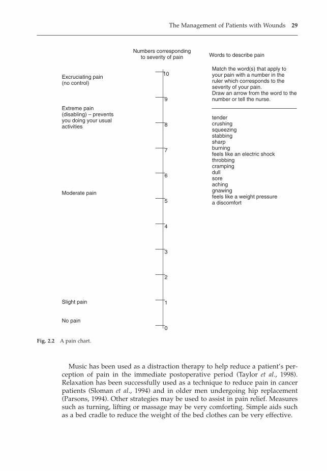
Music has been used as a distraction therapy to help reduce a patient’s per-ception of pain in the immediate postoperative period (Taylor et al., 1998).Relaxation has been successfully used as a technique to reduce pain in cancerpatients (Sloman et al., 1994) and in older men undergoing hip replacement(Parsons, 1994). Other strategies may be used to assist in pain relief. Measuressuch as turning, lifting or massage may be very comforting. Simple aids suchas a bed cradle to reduce the weight of the bed clothes can be very effective.
The Management of Patients with Wounds 29
Excruciating pain(no control)
Extreme pain(disabling) – preventsyou doing your usualactivities
Moderate pain
Slight pain
No pain
10
9
8
7
6
5
4
3
2
1
0
Numbers correspondingto severity of pain Words to describe pain
Match the word(s) that apply toyour pain with a number in theruler which corresponds to theseverity of your pain.Draw an arrow from the word to thenumber or tell the nurse.
tendercrushingsqueezingstabbingsharpburningfeels like an electric shockthrobbingcrampingdullsoreachinggnawingfeels like a weight pressurea discomfort
Fig. 2.2 A pain chart.
� Evaluation �
Regular use of a pain chart allows constant evaluation of the effectiveness ofpain relief.
2.2.7 Sleeping
Most people consider sleep to be important as it provides a sense of refresh-ment and well-being. In recent years there has been considerable research onsleep and its effects. Sleep deprivation causes people to become increasinglyirritable and irrational (Carter, 1985). They may complain of lassitude and lossof feelings of well-being. The sleep–activity cycle is part of the circadianrhythm. During wakefulness, the body is in a state of catabolism. Hormonessuch as catecholamine and cortisol are released. They encourage tissue degra-dation to provide energy for activity; in particular, protein degradation occursin muscle.
Growth hormone is secreted from the anterior pituitary during sleep andstimulates protein synthesis and the proliferation of a variety of cells, includ-ing fibroblasts and endothelial cells (Lee & Stotts, 1990). Rose et al. (2001)reviewed the impact of burn injury on the sleep patterns of children and theneed to aggressively treat growth hormone insufficiency. They speculate that improved sleep would improve growth hormone levels. However, Brandenberger et al. (2000) argue that the body is able to compensate duringthe day and the levels of growth hormone secreted over 24 hours remain muchthe same, regardless of any sleep deprivation.
There has been some reliance on animal studies to determine the impact ofsleep deprivation and the surrogate measure, noise, on wound healing.Wysocki (1996) measured the impact of noise on wound healing and found thatwounds healed more slowly compared with controls. Landis and Whitney(1997) studied the impact of 72 hours of sleep deprivation and found that therewas no difference between experimental and control groups. It should be notedthat these studies were undertaken on basically healthy animals and the find-ings may not be transferable to sick humans. Further studies, especially clini-cal studies, are needed to clarify the matter.
There is much evidence that sleep patterns are disturbed in hospital. Hill(1989) suggested that ward routines, such as early morning waking, prevent thepatient getting adequate sleep. Also, many patients are disturbed during thenight, especially in the intensive care unit. Woods (1972) observed cardiacsurgery patients and noted that they were disturbed as many as 56 times duringthe first postoperative night. Morgan and White (1983) found that intensive carenurses were aware of the importance of sleep but failed to recognise when theywere disturbing their patients unnecessarily.
Freedman et al. (1999) surveyed 203 patients immediately after dischargefrom different types of intensive care units and found that poor sleep qualitywas common to all units. They also found that sleep disruption was caused byhuman interventions, diagnostic testing and environmental noise. A further
30 The Care of Wounds
study from the same research centre investigated the impact of environmentalnoise on sleep. The sleep patterns of 22 medical patients in an intensive careunit were monitored for 24–48 hours continuously. Patients were found to sleepfor short periods throughout a 24-hour timeframe with a mean number of sleepperiods of 41+/-28 and the mean length of a sleep bout was 15+/-9 minutes.Overall, environmental noise was responsible for only 17% of sleep distur-bances (Freedman et al., 2001).
Other factors may also disturb sleep, as shown in a survey by Southwell andWistow (1995) of 454 patients and 129 nurses across a variety of wards in threehospitals. Half the patients had difficulty in sleeping through the nights anddid not get as much sleep as they wished. Many of the patients complained thatthe ward was too hot and the mattresses were uncomfortable and they dislikedhaving plastic covers on both mattresses and pillows. Pain and worry were alsolikely to make sleeping more difficult. A variety of factors were found to disturbsleep including: other patients making a noise, nurses attending other patients,telephones ringing, lights in the ward, nurses talking to each other or topatients, having treatment including medication, toilets flushing or commodesbeing used, nurses’ shoes making a noise. Patients on surgical wards reportedmore disturbances than other ward specialties. The authors of this study con-sidered that nurses should be more aware of the need to ensure that patientsget a good night’s sleep.
� Nursing assessment �
� Compare ‘normal’ with present patterns of sleep.� Assess the ward environment – is it conducive to sleeping?� Consider ward routines – do they allow the patient to follow any aspect of
their usual routines or are they too rigid?
� Nursing intervention �
Problem: Disruption of normal sleeping patternsGoal: Patients are able to sleep a number of hours at night and state that they feelwell rested
McMahon (1990) suggested four types of sleeplessness:
� difficulty getting to sleep.� waking regularly during the night.� waking early in the morning.� sleeping for the normal length of time but not waking refreshed.
Various strategies can be employed to help patients resolve their specific problems. The provision of a milky drink can be beneficial, especially if thepatient normally has one at bedtime. Some people may become hungry afterhaving supper between 1700 and 1800 hours. They may ask their visitors to bring them in a snack. Whilst there is no evidence that food can induce sleep, it is difficult to sleep when hungry. Most people have specific routinesthat they follow each night. As much as possible, this same routine should be
The Management of Patients with Wounds 31
followed in hospital which can introduce a feeling of normality into a strangesituation.
Some people find their sleep disrupted because of pain. This may be acutepain following trauma or surgery or a more chronic pain relating to a long-standing illness or condition. Adequate pain control is essential. Pain is a resolv-able problem (see also section 2.2.6). The position of patients may affect theircomfort so it is helpful to ensure that the patient is in a comfortable position,with a bell close to hand.
During the night, many fears that are suppressed during the day come to thesurface. Sleep may be disturbed because of a particular anxiety. Nighttime is aquieter time on the ward so the nurse may have an opportunity to sit and listenand allow patients to express their fears and anxieties. Once this has happened,the patient may be able to return to normal sleep patterns.
Hospital routines can disrupt normal sleep patterns. The lights of a ward maygo off late, around 2300 hours, and come on again at 0600 hours (Southwell &Wistow, 1995). Patients are woken for their drugs and a drink. It seems rea-sonable for more flexibility to be introduced, with a reduction of the 0600 druground to the minimum and an arrangement not to wake those who wouldprefer to sleep later. Jarmon et al. (2002) experimented with flexible morningand evening medication times and found that patients were able to sleep forlonger. Haddock (1994) found some patients benefited from using earplugs butthey are not suitable for everyone. It should be possible, with careful planning,to provide an environment that is conducive to sleep, and a comfortable patientwho is able to benefit from it.
� Evaluation �
Patient questionnaires are a useful way of establishing the success or otherwiseof the above plan.
2.2.8 Hypothermia
Anaesthesia for major surgery can lead to hypothermia as a result of decreasedmetabolic rate and impaired thermoregulation (Sellden, 2002). Recently, therehas been increased understanding of the impact that even mild levels of intra-operative hypothermia can have on postoperative recovery. Schmied et al.(1996) randomised 60 patients undergoing total hip arthroplasties to either nor-mothermia (36.6°C) or mild hypothermia (35°C). They found that blood losswas significantly higher in the hypothermic group compared with the nor-mothermic patients.
Kurz et al. (1996) proposed that mild hypothermia increased the risk ofwound infection because of vasoconstriction decreasing the level of tissueoxygen and impacting on neutrophil activity. To investigate this further, theyundertook a double-blind randomised study of 200 patients undergoing col-orectal surgery. Patients were randomised to either the normothermic group(37°C) or the hypothermic group (34.4°C). They found that patients in thehypothermic group were three times more likely to develop a postoperative
32 The Care of Wounds
wound infection and had significantly longer hospital stay. Their sutures werealso removed later than those in the normothermic group.
Mahoney and Odom (1999) reviewed the outcome costs for mildly hypother-mic patients compared with normothermic patients undergoing a range ofoperations. They were able to pool the findings of the studies and undertake ameta-analysis. They found that mildly hypothermic patients were more likelyto require blood transfusions and to develop wound infections and the cost ofthese adverse outcomes ranged between $2500 and $7000.
Plattner et al. (2000) tested an experimental warming bandage system andcompared it with conventional gauze with elastic adhesive in 40 normothermicpatients following elective abdominal surgery. The experimental bandage con-sisted of an adhesive shell and a foam frame surrounding a clear window. Aheated card was inserted into the frame approximately 1cm above the woundsurface and left in situ for two hours at a time. Oxygen tension was measuredvia a probe inserted 2–3cm laterally to the incision. Their results were unex-pected. They found that the oxygen tension was considerably lower in thosewith a conventional dressing because of the pressure exerted by the elasticstrapping. The warming device did not appear to benefit normothermicpatients particularly, although it had potential for use for hypothermic patients.
Perioperative hypothermia is associated with a higher incidence of woundinfection. This is of particular relevance in surgery where there is already a high risk of infection such as abdominal surgery. It is less likely to be impor-tant in surgery with a low risk of infection, such as neurosurgery. There are also procedures when cooling the patient is appropriate, such as during craniotomy.
2.2.9 Steroids
Glucocorticoids or corticosteroids are widely used in the treatment of inflam-matory diseases. Although they produce effective anti-inflammatory outcomes,this can have a serious impact on wound healing. A review by Anstead (1998)highlighted the fact that glucocorticoids affect every stage of the healingprocess. This includes overall effects such as the increased risk of infection anddehiscence in surgical wounds, although this is probably dose dependent.Grunbine et al. (1998) studied 73 patients who had had a steroid injection fol-lowing surgery to the foot or ankle and compared the outcome with those whodid not. The use of a single dose of steroids made no difference to healing rates.
Anstead (1998) summarised the effects of glucocorticoids on wound healingas follows.
� Inflammation is suppressed because of a reduction in the numbers of neu-trophils and macrophages and an impaired ability to digest phagocytosedmaterial.
� Wound contracture is poor as a result of inhibition of fibroblast proliferation.� Reduced wound strength, as collagen structure and cross-linking are
affected.
The Management of Patients with Wounds 33
� Epithelialisation is delayed and the cells are thin, producing a weak woundcovering.
When there is planned wounding, such as surgery, it may be beneficial toreduce the level of glucocorticoids, especially preoperatively. Pollack (1982)suggests that doses of prednisolone greater than 40mg/day have the greatesteffect on wound healing. However, it may not be possible to change dosagelevels and there has been considerable interest in the potential of vitamin A tocounteract the unwanted side effects of glucosteroids. Vitamin A has beenfound to restore a normal inflammatory response (Ehrlich et al., 1972) and alsoepithelial regeneration, fibroblast proliferation and collagen content (Talas et al.,2003). However, although there have been a number of animal studies, there isa need to test the use of vitamin A clinically using controlled trials to determinewhat might be an appropriate dose and also to ensure that its use does notadversely affect the beneficial systemic outcomes of glucocorticoids (Anstead,1998).
2.2.10 Radiotherapy
Radiation effectively destroys cancer cells as they are more radiosensitive thannormal cells. A dosage high enough to kill cancer cells does not affect the sur-rounding cells. However, if the dosage has to be increased there is increasedrisk of normal tissue necrosing. Radiation has the potential to impact on woundhealing and it would seem that the worst outcomes occur when radiotherapyis given before surgery.
Hillmann et al. (1997) found preoperative irradiation was an influencingfactor in postoperative complications for patients with Ewing’s sarcoma. Sassler et al. (1995) found major wound complications in patients undergoinghead and neck surgery following an initial regime of chemotherapy and irradiation. They reported a 77% incidence of complications in patients undergoing surgery within one year of the regime compared with a 20% incidence after one year. Lin et al. (2001) found that a history of radiotherapywas a factor in wound-healing complications of breast reconstruction. Similarresults were found by Wang et al. (2003) and O’Sullivan et al. (2002), when com-paring pre- and postoperative radiotherapy. Both studies found a significantlyhigher rate of wound-healing complications when radiotherapy was given preoperatively.
Radiation may affect the healing of an existing wound or it may causechanges to the skin so that any later wound will heal slowly. The skin may showsigns of damage from the radiation during treatment. This is known as a radi-ation reaction and will be discussed in Chapter 6.
Levenson et al. (1984) investigated the use of vitamin A supplements to coun-teract the effects of radiation on wound healing. In the animal model, theyfound that giving vitamin A supplements was effective. Good results wereobtained if the supplementation was started prior to radiotherapy or up to twodays after treatment.
34 The Care of Wounds
2.3 PSYCHOLOGICAL CARE
Nurses have always excelled at the physical care of patients. It is only recentlythat the emotional needs of patients have been considered. Many situationsmay cause psychological distress, which may be described as stress. The phys-iological effects of stress and its effect on wound healing have already beendescribed in section 2.2.5. Factors causing psychological distress may be definedas stressors. Those that may be particularly associated with wounded patientswill be discussed in this section. It should be noted that other factors, notaddressed here, can also act as stressors.
2.3.1 Anxiety
Lazarus and Averill (1972) stated that ‘Anxiety results when a person is unableto fully comprehend the world around him’. This could be considered in rela-tion to ill health. A further quotation from Frankenhaeuser (1967) adds to this:‘Information is necessary for comprehension, but the perception of this infor-mation can be modified by the expectations of the subject’. Many nurses willhave seen patients who have not heard or have misunderstood what has beensaid to them because of their degree of anxiety.
Admission to hospital, whether planned or unplanned, can be a very stress-ful experience.
� Nursing assessment �
Zigmond and Snaith (1983) designed a simple questionnaire, known as the Hos-pital Anxiety and Depression Score or HAD Score, that can identify the degreeof stress being suffered and can be completed by patients. The questionnairecomprises a series of questions such as whether an individual is worried or ableto relax and enjoy watching television. There is a choice of four answers to eachquestion, such as ‘most of the time’ or ‘seldom’. Most patients find it simple touse. Cole-King and Harding (2001) used the HAD Score to identify stress in 53patients with leg ulcers and found it to be a useful research tool.
� Nursing intervention �
Problem: Anxiety related to hospital admissionGoal: Patients will be able to express their specific anxieties
Many patients find their admission to hospital a very anxious time. Gammon(1998) compared the impact of hospital admission on patients with that of iso-lating a patient because of infection. All patients were assessed using the HADScore and two other assessment tools to measure self-esteem (the Self-EsteemScale) and a sense of control (the Health Illness (Powerlessness) Questionnaire)(Rosenburg, 1965; Roy, 1976). The isolated patients had significantly higherlevels of anxiety and depression and lower levels of self-esteem and sense ofcontrol than the routine admission patients. However, results for all patients
The Management of Patients with Wounds 35
showed them to be outside normal levels, indicating that they were all suffer-ing above normal levels of stress.
Communication is essential to address this problem. The initial assessmentshould provide both information and an opportunity for the patient to ask ques-tions and express their feelings and concerns. Not everyone will always be ableto discuss their anxieties immediately so there may be an ongoing process ofbuilding up a relationship over a period of time.
A study by McCabe (2004) investigated patients’ experiences of nurse–patientcommunication. She noted that all the patients in the study frequently statedthat nurses did not provide them with enough information and seemed to bemore interested in tasks than in talking to patients. This was then excused bysaying that the nurses were too ‘busy’. McCabe analysed the findings from herstudy in terms of patient-centred communication and task-centred communi-cation. Patient-centred communication involves attending behaviour and thepatients in the study described this in terms of: giving time and being there,open/honest communication, genuineness, empathy and friendliness andhumour. McCabe considered that nurses could have been using friendlinessand humour as a means of maintaining a superficial level of communication to avoid the need to address emotional or difficult issues. She also consideredthat the culture of the organisation tended to discourage patient-centred communication.
Active listening is not a very safe occupation. The consequences may be emo-tionally painful to the nurse because of the difficult questions that may beasked. Many may feel inadequate or too inexperienced. Koshy (1989) describesactive listening as ‘the process of receiving and assimilating ideas and infor-mation from verbal and non-verbal messages and responding appropriately’.Tschudin (1991) emphasises the importance of not making assumptions. It istoo easy for nurses to assume that they not only know the problem but alsohave the answers for dealing with it.
� Evaluation �
Repetition of the assessment will enable the nurse to identify any reduction instress levels.
Problem: Anxiety related to surgeryGoal: The patient’s anxiety will be reduced by adequate preoperative preparation
There is now much greater awareness of the importance of providing good preoperative information. A Department of Health circular (1990) makes it clearthat all patients have the right to understand their treatment and the risksinvolved. The role of the nurse is to ensure that each patient receives appro-priate preoperative information about the surgery and what to expect in thepostoperative period. Radcliffe (1993) has considered how a suitable strategycan be implemented. She suggests that oral information is reinforced withwritten leaflets, which can be a source of reference for both patients and relatives.
36 The Care of Wounds
Doering et al. (2000) assessed the impact of providing a videotape of a patientundergoing a hip replacement from time of admission to discharge, purely froma patient’s perspective. One hundred patients were randomly allocated to eitherthe preparation group (shown the videotape) or a control group. They wereassessed for levels of anxiety and pain for four days post surgery. Theresearchers found significantly lower levels of anxiety in the preparation groupas well as a lower intake of analgesia, although there were no differences in theamount of pain. They considered that this was an effective method of provid-ing information to patients.
� Evaluation �
The patient should be able to describe the likely course of events in the peri-operative and postoperative period.
Alternative therapies
Some alternative therapies have been successful in assisting patients to reduceanxiety. Marshall (1991) described the use of aromatherapy and relaxation tech-niques to reduce stress in dermatology patients. Dossey (1991) used case studiesto discuss the benefits of guided imagery. This is a method of relaxation thatencourages the patient to use their imagination to first identify the healthproblem and then to visualise how the treatment will work effectively. This mayinvolve favourite scenes, music or other audiotapes. Ultimately patients have tovisualise themselves in the final healed state. This technique should be used overa period of time, such as for two weeks prior to surgery, for it to be effective.
It is easy to dismiss such a concept as ‘mumbo-jumbo’ because it is alien tomany healthcare professionals. However, Holden-Lund (1988) randomly allo-cated cholecystectomy patients to either guided imagery or period of quiet andthose receiving guided imagery had significantly lower anxiety levels, cortisollevels and surgical wound erythema compared to the control group.
Therapeutic touch has been used in the USA for a number of years to reduceanxiety levels. Heidt (1981) used matched patient groups in a cardiovascularunit to receive therapeutic touch, casual touch or no touch. Those receiving ther-apeutic touch had significantly lower levels of anxiety compared with the othergroups.
2.3.2 Motivation and education
Bentley (2001) discussed the importance of working in partnership with chronicwound patients in their own care. Such an approach can help to reduce thesense of powerlessness and loss of control felt by many patients. Bentley usesa case example of a patient with both pressure ulcers and a chronic leg ulcer tosupport her arguments. Rivera et al. (2000) used a similar approach to demon-strate how a model of behaviour modification was utilised to motivate andeducate a patient with a diabetic foot ulcer. Such papers provide support forthe importance of patient education. Unfortunately, there is limited research
The Management of Patients with Wounds 37
evidence of effective outcomes, mainly because of poor methodological quality,as demonstrated by the Cochrane review of the benefits of patient education inpreventing diabetic foot ulceration (Valk et al., 2003). The authors concludedthat it appeared that patient education may reduce the incidence of ulcerationand amputation but randomised controlled trials were needed to provide con-clusive evidence.
Involving patients in their own care can reduce the risk of non-complianceand therefore it is useful to have a greater understanding of compliance andnon-compliance. Nyatanga (1997) reviewed psychosocial theories of non-com-pliance and divided them into the following categories.
� Perceptual theory – people interpret the world based on what they alreadyknow. They may have pre-existing theories about their treatment.
� Value clarification – patients may consider the choices in relation to their treatment and choose whether to comply. An example would be a manchoosing not to give up smoking despite knowing it increases his risk of legamputation.
� Attribution theory – this relates to the locus of control. Patients who feel incontrol of their treatment or who see a link between their treatment and thehealing of their wound are more likely to be compliant.
� Cultural theories – all individuals have a cultural understanding of theirwound, its meaning and the treatment. This may lead them to decide not tofollow all the treatment requirements, such as not wishing to take analgesia.
� Health belief model – patients may choose to engage in health-related behav-iour if they believe that the benefits in terms of health gain outweigh thecosts. For example, a leg ulcer patient may choose to wear a four-layerbandage that she finds hot and uncomfortable because she believes that itwill heal her ulcer.
More recently, it has been recognised that the term ‘compliance’ does not reflectthe move to increased patient autonomy and ‘concordance’ is now seen as amore accurate word to use. Moffatt (2004) has discussed concordance in rela-tion to leg ulcer management and emphasised the importance of understand-ing the reasons why patients may not be concordant with their treatment plan.Physical reasons, such as bodily pain that reduces tolerance to treatment, orpsychosocial issues, such as a patient’s expectations of care, can have an impacton concordance. It is important to understand the patient’s perspective whenplanning care so that they can truly become engaged in the healing process.
� Nursing assessment �
Simple questioning can determine the level of relevant knowledge that a patientpossesses.
� Nursing intervention �
Problem: A lack of understanding of the care needed to promote wound healingGoal: Patients demonstrate the ability to be self-caring and are able to explain theirplan of care
38 The Care of Wounds
In the climate of early discharge from hospital, many patients will return homewith a wound that still requires dressing. If it is practicable, it is helpful to teachpatients to undertake their own dressing. Monitoring and supervision by thedistrict nurse or practice nurse would still be necessary. A planned educationprogramme using short-term goals is the most effective. Information aboutallied care such as diet or exercise can also be incorporated into the programme.Patients with chronic wounds should be given information about their causesand possible prevention.
Some patients will have little motivation to carry on the plan of care oncethey have been discharged from hospital. Recognition of the reasons for the lackof motivation and good communication with the community staff may be ofsome help. A few patients will still fail to respond. It is necessary to accept thatevery patient has the right to choose not to comply with the care recommendedby the healthcare team.
� Evaluation �
The ability of the patient to undertake management of the wound will provideadequate evaluation of the effectiveness of the nursing care.
2.3.3 Body image
Body image is the mental picture that people have of themselves. Body imageis also closely associated with self-esteem. Shipes (1987) suggests that self-esteem can be defined as the sum total of all we believe about ourselves. Allpatients with wounds have an altered body image which can have a profoundeffect on the person’s self-esteem and motivation. Obvious types of woundsthat can have these effects are those resulting in disfigurement, such as burns,head and neck surgery, mastectomy, amputation and ostomies. Many patientswill also be suffering from anxiety about their prognosis. Chronic wounds canalso affect body image, as shown in a study by Ebbeskog and Ekman (2001)who found that leg ulcer patients were embarrassed by their ulcer and ban-dages and hid them with clothing. The resultant stress can be so overwhelm-ing that the patient may be unable to take in information, to share their feelingsor to commence rehabilitation.
� Nursing assessment �
Neil (2001) developed an assessment tool called the Stigma Scale to measurebody image in relation to the skin. Users are asked to judge a series of 11 state-ments using a five-point Likert scale. This is a promising approach but a majorlimitation is that, as yet, the scale has been validated only by educated, mainlywhite, women and further work is required.
� Nursing intervention �
Problem: Loss of self-esteem related to altered body imageGoal: Patients acknowledge change in body image and express their feelings aboutthis change
The Management of Patients with Wounds 39
In the early stages, following the circumstances that led to an altered bodyimage, some patients appear to be quite euphoric. This is due to simple reliefat having survived. After a while the patient’s attitude is likely to change.Common problems that can occur include:
� a sense of loss, similar to bereavement.� anxiety related to diagnosis, especially if it is cancer.� loss of sexual function, which may be related to type of surgery or trauma
or to either of the previous problems.� withdrawal from social relationships with family or significant others, pos-
sibly due to a malodorous wound or any of the previous problems.
The role of the nurse is to assist the patient to develop a reintegrated bodyimage (Burgess, 1994). This may be achieved in a variety of ways. Perhaps themost important is accepting patients as they are, at whatever stage they havereached. Allowing patients to express their feelings and providing them withmatter-of-fact information, such as an honest appraisal of the progress of thewound, is beneficial. It is also essential to include family and/or significantothers in the patient’s care and in any education programme. Good manage-ment of the wound should prevent odour or leakage, which helps to boost con-fidence. Burgess also suggests that if patients are having difficulty coping, itmay be necessary to emphasise the importance of the surgery for the health ofthe individual and the fact that it does not change them as a person.
As already discussed under Anxiety (section 2.3.1), preoperative informationand counselling are most important. Kelly (1989) studied 67 patients who hadundergone head and neck surgery. Generally, they said they were more anxiousbefore surgery than after but 42% of men and 21% of women would have likedmore information. Another study by Elspie et al. (1989) found that 41% ofpatients suffered psychological stress following major surgery for intraoralcancer.
In many areas, specialist nurses are employed to give help and support topatients, such as colorectal nurses or breast care nurses. They can build up arelationship with their patients which can give patients the confidence toexpress their feelings freely. In other circumstances, it may be a nurse whoalready has a good relationship with a patient who is able to provide thisservice.
� Evaluation �
Regular evaluation of progress is important for patients with an altered bodyimage. Learning to cope with the new image may take time and strategies mayhave to be changed along the way. This may be particularly true for thoseundergoing a series of plastic surgery operations who may have to cope witha constantly changing body image.
40 The Care of Wounds
2.3.4 Other psychological problems
Fear
Fear is a common human experience that may be transitory or longer lasting.Illness may release many fears: fear of hospitalisation, fear of illness, fear of alife-threatening condition, fear of loss of affection of loved ones, fear of themutilation of surgery. Such fear creates great stress within the sufferer. This maybe made worse by the healthcare team failing to recognise when patients areexperiencing fear and so not allowing them to express their feelings.
Grief
Grief is a normal process that allows adaptation to some major loss in a person’slife. The wounded patient may have to come to terms with skin damage fromburns, the loss of a limb or breast or other types of mutilating surgery (see alsoBody Image, section 2.3.3.). Kubler-Ross (1969) described various stages in thegrief process. She related them to dying but they can be applied to all types ofgrief. The stages are: denial, isolation, anger, bargaining, depression and accep-tance. Each person will progress through some or all of these stages at a dif-ferent rate and not necessarily in the same sequence. By listening to the patientand accepting without judgement, the nurse can assist in this process and thusreduce the amount of stress suffered. This may be particularly difficult duringthe stage of anger as the aggression expressed by the patient is often directedat the main caregivers. Understanding of the cause of the aggression will helpthe nurse deal with this stage of grief.
Powerlessness
Taylor and Cress (1987) describe powerlessness as the ‘perception of loss ofcontrol over what happens to oneself and one’s environment’. This is a feelingexperienced by many hospital patients as they are placed in the subservient‘patient role’. Even simple decisions such as when to eat or go to bed are takenaway from the individual. There is pressure to conform and be a ‘good’ patient.Stockwell (1972) describes very graphically the fate of the unpopular patientwho did not conform to the role the nurses desired from him. A ‘good’ patientwill submit without question to treatment and will not ask too many questions.Although it is to be hoped that nursing has moved forward since 1972, manypatients are still aware of their loss of status once they are in hospital. Somemay feel depressed because of their feelings of learned helplessness. Othersmay feel quite euphoric to have survived, which may also be misinterpreted asa lack of compliance.
In a society where independence is prized, dependence on others mayproduce feelings of anger and frustration. Many patients remark that they feela nuisance because they cannot care for themselves. It may also reduce feelingsof self-worth. A common attitude of elderly people when asked to participatein a research project is that they will do so – because it will help others. Such a
The Management of Patients with Wounds 41
contribution is important to them as they feel that, despite their physical limi-tations, they can still make a contribution to the good of society.
� Nursing assessment �
Although the precise problem may vary, the assessment is the same.
� Observe body language – does the patient look relaxed, tense, fidgety, with-drawn, hypoactive or hyperactive? Do they avoid eye contact?
� Conversation – does the patient talk excessively, not talk to anyone, ask questions?
� Do any of these terms describe the patient? Angry, confused, aggressive, con-fident, demanding, distrustful, anxious, fearful, critical, passive, depressed,euphoric, disorientated.
Katona and Katona (1997) proposed that a slightly different approach isrequired for assessing older people. They recommended the use of four simplequestions.
� Are you basically satisfied with your life?� Do you feel that your life is empty?� Are you afraid that something bad is going to happen to you?� Do you feel happy most of the time?
The patient would score a point for replying ‘no’ to the first and last questionsand for replying ‘yes’ to the middle two. Anyone with a score of two or moreis probably depressed.
� Nursing interventions �
Although nurse training is providing improved knowledge of psychologicalcare, it may be more appropriate for the patient to have further help from otherssuch as a clinical psychologist, a psychiatric trained nurse, a chaplain or atrained counsellor.
Problem: Fear due to separation from loved ones or related to unfamiliarityGoal: Patients identify the source(s) of their fear and are able to describe their feelings
Once the nurse recognises that the patient is very frightened, then strategiescan be developed to allow the patient the opportunity of expressing their spe-cific fears. Time may have to be set aside for ‘casual’ conversation, especially ifthe patient has few visitors. Assigning the same nurses to care for the patientcan build up confidence. Involving the patient and, possibly, the family in allaspects of planning may be helpful. Patients with any sort of sensory loss willneed orientation to the new surroundings.
Problem: Grieving related to loss of or disfigurement to a body partGoal: Patients will allow themselves to experience the grieving process
A variety of strategies can be adopted to help the patient move through thegrief process. Setting aside time to allow the patient to talk is very important.
42 The Care of Wounds
However, it may be constructive to set time constraints as it can be exhaustingfor both the patient and the nurse. Patients may prefer to know that they havethe undivided attention of the nurse for a set period of time each day, ratherthan an indeterminate amount of time occasionally. For some individuals, itmay be appropriate for others to assist the patient in recognising and talkingthrough their grief. In those situations, the nurse should be there to supportand encourage.
Problem: Powerlessness related to feelings of loss of control of the environmentGoal: The patent will express feelings of having a sense of control and will partici-pate in the planning of care
Many healthcare workers fail to recognise the degree to which the ‘system’takes charge of the individual once they pass through the doors of a hospital.Whilst in an emergency situation there may be some alteration of priorities,every patient is entitled to be treated with respect. Nurses can play an impor-tant role in assisting their patients to remain in control of as many areas of theirlives as possible. Patient education not only promotes compliance but alsoallows the patient to participate in care, thus having a degree of control. Dis-cussing with the patient when particular treatment should be given and involv-ing them in planning care will reduce feelings of powerlessness.
A study by Efraimsson et al. (2003) demonstrates graphically how this can gowrong. They undertook a case study of discharge planning for an elderlypatient. The patient and some of her family were invited to participate in thecase conference. Observation showed that the clinical staff talked about ratherthan with the patient, using technical language, and they failed to answer ques-tions posed by the patient or her children. In an interview with one of theresearchers later, the patient revealed that she felt like on object during the dis-charge planning meeting and that she felt powerless. It is pointless to involvepatients in the discharge planning process if we do not give them time andspace to express their own feelings and desires.
� Evaluation �
Evaluation of psychological care is not easy. Some indication can be obtainedby repeating the assessment and by talking to the patient.
2.4 SPIRITUAL CARE
In recent years there has been increasing awareness of the importance of spir-itual care within healthcare. Although much of it has centred around palliativecare, as is evidenced in the NICE guideline Improving Supportive and PalliativeCare for Adults with Cancer (NICE, 2004), it is seen to be wider than that, as dis-cussed in the paper NHS Chaplaincy: meeting the religious and spiritual needs ofpatients and staff (DoH, 2003). A survey by King et al. (1999) of 250 patientsadmitted to a London teaching hospital found that 79% of patients professedsome form of spiritual belief, although not all engaged in a religious activity. A
The Management of Patients with Wounds 43
survey undertaken of 1.75 million patients in American hospitals found thatemotional and spiritual care was seen as largely ineffective and as an area thatwas in considerable need of improvement (Clark et al., 2003).
NICE (2004) suggests that healthcare professionals frequently fail to recog-nise when patients need spiritual support. Spirituality is a concept that manynurses find difficult to define and they may feel unsure of what is required.Spirituality should not just be put into the framework of religion. Everyone,whether they believe in a God or not, has spiritual needs. Spirituality can bedefined as that within us that responds to the infinite realities of life.Narayanasamy (1996) listed some indicators of spirituality which providefurther clarity: a sense of purpose, hopefulness, creativity, joy, enthusiasm,courage, reverence, serenity, humour, providing meaning in struggle and suffering.
If the spiritual needs of individuals are not met or they experience a cata-strophic event in their lives, the result is spiritual pain or distress. This acts asa stressor and thus can impact on wound healing. Kohler (1999) defined spiri-tual distress as the lack of meaning in one’s life. In her study of French AIDSand cancer patients, Kohler found that nearly all of them expressed feelings of‘ill being’ and raised questions about the meaning of life, death, pain or illness.Kawa et al. (2003) studied palliative care patients in Japan and described spir-itual distress as consciousness of the gap between an individual’s aspirationsand their current situation. The patients in the study were distressed by the gapbetween their current situation and how they wanted to live or how theywanted to die or how they wanted to maintain relationships with others.Despite the cultural differences between the patient groups in these studies,there are obvious similarities in the factors causing their spiritual distress.
Cressey and Winbolt-Lewis (2000) worked with a group of chaplains and laypeople to determine areas of potential distress where the patient can be left withfeelings of isolation, fragmentation or despair. They are listed in Table 2.2. Inthe context of spiritual distress, acceptance also has significance. A patient mayneed reassurance that others, particularly family and friends, will accept themin the new role as a patient, especially if having to come to terms with a dis-figuring wound. Finlay et al. (2000) suggest that patients perceive that health-care professionals are too busy to discuss spiritual matters and so only disclosetheir distress when it becomes severe.
� Nursing assessment �
Peterman et al. (2002) have developed a validated assessment tool to measurespiritual well-being, called the Functional Assessment of Chronic IllnessTherapy – Spiritual Well-Being (FACIT-sp). This measures a sense of meaningand peace and also the role of faith in illness. However, it has only been vali-dated with cancer patients. King et al. (2001) took a different approach anddeveloped an assessment questionnaire that consistently differentiates betweenpatients with high and low spiritual beliefs.
Villines and Harrington (1998) suggested a number of signs indicative of spir-itual distress.
44 The Care of Wounds
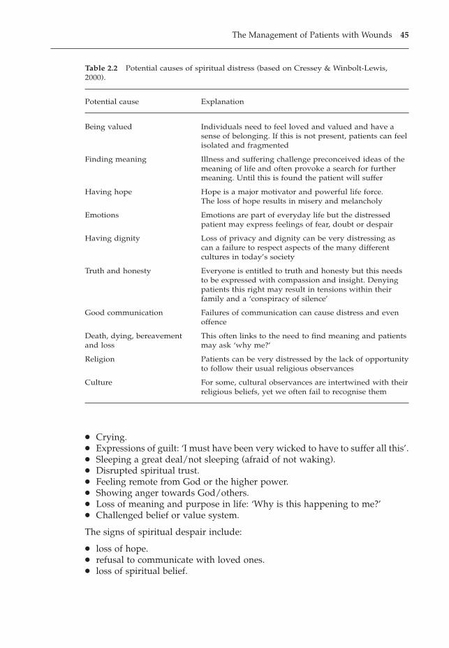
� Crying.� Expressions of guilt: ‘I must have been very wicked to have to suffer all this’.� Sleeping a great deal/not sleeping (afraid of not waking).� Disrupted spiritual trust.� Feeling remote from God or the higher power.� Showing anger towards God/others.� Loss of meaning and purpose in life: ‘Why is this happening to me?’� Challenged belief or value system.
The signs of spiritual despair include:
� loss of hope.� refusal to communicate with loved ones.� loss of spiritual belief.
The Management of Patients with Wounds 45
Table 2.2 Potential causes of spiritual distress (based on Cressey & Winbolt-Lewis,2000).
Potential cause Explanation
Being valued Individuals need to feel loved and valued and have asense of belonging. If this is not present, patients can feel isolated and fragmented
Finding meaning Illness and suffering challenge preconceived ideas of themeaning of life and often provoke a search for furthermeaning. Until this is found the patient will suffer
Having hope Hope is a major motivator and powerful life force.The loss of hope results in misery and melancholy
Emotions Emotions are part of everyday life but the distressedpatient may express feelings of fear, doubt or despair
Having dignity Loss of privacy and dignity can be very distressing ascan a failure to respect aspects of the many differentcultures in today’s society
Truth and honesty Everyone is entitled to truth and honesty but this needsto be expressed with compassion and insight. Denying patients this right may result in tensions within their family and a ‘conspiracy of silence’
Good communication Failures of communication can cause distress and evenoffence
Death, dying, bereavement This often links to the need to find meaning and patients and loss may ask ‘why me?’
Religion Patients can be very distressed by the lack of opportunity to follow their usual religious observances
Culture For some, cultural observances are intertwined with their religious beliefs, yet we often fail to recognise them
� death wish.� severe depression.
� Nursing interventions �
Problem: Spiritual distress related to separation from religious or cultural ties or froma challenged belief or value systemGoal: The patient will be able to identify the cause of spiritual distress and specifythe assistance required to alleviate it
Cavendish et al. (2003) suggest that provision of spiritual care is part of nursingcare, although nurses may not always be comfortable with the concept. Nursesare not always able to recognise the difference between spiritual needs and reli-gious needs. Cressey and Winbolt-Lewis (2000) describe spiritual care as con-veying to patients that they have value and are unconditionally regarded forwho they are, regardless of illness, colour or creed.
The chaplaincy team is very much part of the multiprofessional network andcan give much support to both the patients and staff by listening, comfortingand counselling when necessary. However, nurses need to have some under-standing of the range of spiritual care activities. There can be no standardisedspiritual care as there is with postoperative physical care. In this situation, eachpatient must be cared for in the light of their unique needs. Fish and Shelly(1985) proposed the following activities: listening, empathy, vulnerability,humility and commitment.
� Listening – this is active listening, giving the patient full attention and notingall the non-verbal as well as verbal cues.
� Empathy – this allows the nurse to share the feelings of the patient without losing objectivity. This is essential to enable the patient to consideralternatives.
� Vulnerability – as nurses enter into and share patients’ feelings, they becomevulnerable. This may be painful but can also be rewarding.
� Humility – this is not easy. Few people want to admit that they do not havethe answers in their particular field. A sense of humility will enable nursesto see that they can learn from their patients. Humility also allows nurses toaccept themselves and their patients in all their human frailty.
� Commitment – being with a patient through all the difficult times, sharing thepain as well as the joys, involves considerable commitment.
Several studies have investigated spiritual coping mechanisms. Dein andStygall (1997) undertook a review of religion and chronic illness and found thatreligion was a positive coping mechanism. A qualitative study using groundedtheory found that 13 Christian patients undergoing open heart surgery soughtcomfort through prayer. They used two strategies: praying themselves andenlisting others to pray for them, especially for the times when they wereunable to pray (Hawley, 1998). Narayanasamy (2002) interviewed 15 chroni-cally sick people (nine Christians, two Hindus and four with no religious affil-iation) and also found that prayer brought comfort as it gave both a sense of
46 The Care of Wounds
connectedness to God and also a sense of hope, strength and security. However,the author only found this sense of connectedness to God (or Hindu deities)amongst the Christians and Hindus in his sample.
� Evaluation �
Just as spiritual assessment is very difficult, so too is evaluation of the outcomesof care. Narayanasamy (1996) suggests that spiritual integrity is one outcomethat may be demonstrated as relief from spiritual pain or by restoration of thelife principle. Resolution of the signs of spiritual distress or despair would indi-cate a positive outcome, as would an improved FACIT-sp score.
REFERENCES
Alberti, K., Press, C. (1992) The biochemistry of the complications of diabetes mellitus,in (eds) Keen, H., Jarrett, J., Complications of Diabetes. Edward Arnold, London.
Andel, H., Kamolz, L.P., Horauf, K., Zimpfer, M. (2003) Nutrition and anabolic agentsin burned patients. Burns, 29 (6), 592–595.
Anstead, G.M. (1998) Steroids, retinoids and wound healing. Advances in Wound Care,11, 277–282.
Arturson, M.G.S. (1978) Metabolic changes following thermal injury. World Journal ofSurgery, 2, 203–213.
Augustin, M., Maier, K. (2003) Psychosomatic aspects of chronic wounds. Dermatologyand Psychosomatics, 4 (1), 5–13.
Bentley, J. (2001) Promoting patient partnership in wound care. British Journal of Com-munity Nursing, 6 (10), 493–500.
Bibby, B.A., Collins, B.J., Ayliffe, G.A.J. (1986) A mathematical model for assessing therisk of post-operative wound infection. Journal of Hospital Infection, 8, 31–39.
Birkmeyer, N.J., Charlesworth, D.C., Hernandez, F. et al. (1998) Obesity and risk ofadverse outcomes associated with coronary artery bypass surgery. Northern NewEngland Cardiovascular Disease Study Group. Circulation, 97 (17), 1689–1694.
Borger, M.A., Rao, V., Weisel, R.D. et al. (1998) Deep sternal wound infection: risk factorsand outcomes. Annals of Thoracic Surgery, 65 (4), 1050–1056.
Brandenberger, G., Gronfier, C., Chapotot, F., Simon, C., Piquard, F. (2000) Effect of sleepdeprivation on overall 24 h growth hormone secretion. Lancet, 356 (9239), 1408.
Brown, K. (1991) Improving intakes. Nursing Times, 87 (20), 64–68.Burgess, L. (1994) Facing the reality of head and neck cancer. Nursing Standard, 8 (32),
30–34.Carr, E. (1997) Overcoming barriers to effective pain control. Professional Nurse, 12 (6),
412–416.Carr, E., Thomas, V.J. (1997) Anticipating and experiencing post-operative pain: the
patients’ perspective. Journal of Clinical Nursing, 6, 191–201.Carter, D. (1985) In need of a good night’s sleep. Nursing Times, 81 (46), 24–26.Cavendish, R., Konecny, L., Mitzeliotis, C. et al. (2003) Spiritual care activities of nurses
using Nursing Interventions Classification (NIC) labels. International Journal ofNursing Terminologies and Classifications, 14 (4), 113–124.
Cereceda, F.C., Gonzalez, G.I., Antolin, J.F.M. et al. (2003) Detection of malnutrition onadmission to hospital. Nutricion Hospitalaria, 18 (2), 95–100.
The Management of Patients with Wounds 47
Chan, A.W., Macfarlane, I.A., Bowsher, D. (1990) Chronic pain in patients with diabetesmellitus: comparison with a non-diabetic population. Pain Clinic, 3 (3), 147–159.
Chapman, A. (1996) Current theory and practice: a study of pre-operative fasting.Nursing Standard, 10 (18), 33–36.
Chbinou, N., Frenette, J. (2004) Insulin-dependent diabetes impairs the inflammatoryresponse and delays angiogenesis following Achilles tendon injury. AmericanJournal of Physiology – Regulatory Integrative and Comparative Physiology, 286 (5),R952–957.
Chmell, M.J., Schwartz, H.S. (1996) Analysis of variables affecting wound healing aftermusculoskeletal sarcoma resections. Journal of Surgical Oncology, 61, 185–189.
Clark, M., Schols, J.M.G.A., Benati, G. et al. (2004) Pressure ulcers and nutrition: a newEuropean guideline. Journal of Wound Care, 13 (7), 267–272.
Clark, P.A., Drain, M., Malone, M.P. (2003) Addressing patients’ emotional and spiritualneeds. Joint Commission Journal on Quality and Safety, 29 (12), 659–670.
Closs, S.J. (1992) Patients’ night time pain, analgesic provision and sleep after surgery.International Journal of Nursing Studies, 29 (4), 381–392.
Cole-King, A., Harding, K.G. (2001) Psychological factors and delayed healing in chronicwounds. Psychosomatic Medicine, 63, 216–220.
Committee of Ministers (2003) Resolution ResAP(2003)3 on Food and Nutritional Care inHospitals. Council of Europe, Brussels.
Correia, M.L., Waitzberg, D.L. (2003) The impact of malnutrition on morbidity, mortal-ity, length of hospital stay and costs evaluated through a multivariate model analy-sis. Clinical Nutrition, 22 (3), 235–239.
Cressey, R.W., Winbolt-Lewis, M. (2000) The forgotten heart of care: a model of spiritualcare in the National Health Service. Accident and Emergency Nursing, 8, 170–177.
De Boer, A.S., Mintjes-de Groot, A.J., Severijnen, A.J., van den Berg, J.M.J., van Pelt, W.(1999) Risk assessment for surgical-site infections in orthopaedic patients. InfectionControl and Hospital Epidemiology, 20 (6), 402–407.
Dein, S., Stygall, J. (1997) Does being religious help or hinder coping with chronic illness?A critical review. Palliative Medicine, 11, 291–299.
Delmi, M., Rapin, C.H., Bengoa, J.M. (1990) Dietary supplementation in elderly patientswith fractured neck of femur. Lancet, 235, 1013–1016.
Department of Health (1990) Health Service Management – Patient Consent to Examinationand Treatment. NHS Management Executive, London.
Department of Health (2001a) The Essence of Care. Department of Health, London.Department of Health (2001b) Standard Principles for Preventing Infections in Hospitals.
Department of Health, London.Department of Health (2001c) Better hospital food panel launched. Department of
Health, London.Department of Health (2003) NHS Chaplaincy: meeting the religious and spiritual needs of
patients and staff. Department of Health, London.Doering, S., Katzlberger, F., Rumpold, G. et al. (2000) Videotape preparation of patients
before hip replacement surgery reduces stress. Psychosomatic Medicine, 62 (3),365–373.
Dossey, B. (1991) Awakening the inner healer. American Journal of Nursing, Aug, 31–34.Ebbeskog, B., Ekman, S.L. (2001) Elderly persons’ experiences of living with a venous
leg ulcer: living in a dialectal relationship between freedom and imprisonment.Scandinavian Journal of Caring Sciences, 15, 235–243.
Edington, J., Kon, P., Martyn, C.N. (1996) Prevalence of malnutrition in patients ingeneral practice. Clinical Nutrition, 15, 60.
48 The Care of Wounds
Efraimsson, E., Rasmussen, B.H., Gilje, F., Sandman, P.O. (2003) Expressions of powerand powerlessness in discharge planning: Journal of Clinical Nursing, 12, 707–716.
Ehrlich, H.P., Tarver, H., Hunt, T.K. (1972) Effects of Vitamin A and glucocorticoids uponinflammation and collagen synthesis. Annals of Surgery, 177, 222–227.
Elia, M., Stratton, R. (2004) On the ESPEN guidelines for nutritional screening 2002. Clin-ical Nutrition, 23 (1), 131–132.
Elspie, C.A., Freedlander, E., Campsie, L.M. et al. (1989) Psychological distress at follow-up after major surgery for intraoral cancer. Journal of Psychosomatic Research, 33 (4),441–448.
Exton-Smith, A.N. (1971) Nutrition of the elderly. British Journal of Hospital Medicine, 5,639–645.
European Pressure Ulcer Advisory Panel (EPUAP) (2003) Nutritional Guidelines for Pres-sure Ulcer Prevention and Treatment. EPUAP, Oxford.
Fagerhaugh, S.Y., Strauss, A. (1977) Politics of Pain Management: staff–patient interaction.Addison-Wesley, London.
Field, L. (1996) Are nurses still underestimating patients’ pain post-operatively? BritishJournal of Nursing, 5 (13), 778–784.
Finlay, I.G., Ballard, P.H., Jones, N., Searle, C., Roberts, S. (2000) A person to have around.What value do patients want from a hospice chaplain? Journal of Health Care Chap-laincy, 3 (2), 41–52.
Fish, S., Shelley, J.A. (1985) Spiritual Care: the nurse’s role. InterVarsity Press, DownersGrove, IL.
Frankenhaeuser, M. (1967) Some aspects of research in physiological psychology, in (ed.)Levi, L., Emotional Stress, Physiological and Psychological Reactions – medical, indus-trial and military implications. Karger, Basel.
Freedman, N.S., Kotzer, N., Schwab, R.J. (1999) Patient perception of sleep quality andetiology of sleep disruption in the intensive care unit. American Journal of Respira-tory and Critical Care Medicine, 159 (4 Pt 1), 1155–1162.
Freedman, N.S., Gazendam, J., Levan, L., Pack, A.I., Schwab, R.J. (2001) Abnormalsleep/wake cycles and the effect of environmental noise on sleep disruption in theintensive care unit. American Journal of Respiratory and Critical Care Medicine, 163,451–457.
Gallagher-Allred, C.R., Voss, A.C., Finn, S.C., McCamish, M.A. (1996) Malnutrition andclinical outcomes: a case for medical nutrition therapy. Journal of the AmericanDietetic Association, 96 (4), 361–366.
Gammon, J. (1998) Analysis of the stressful effects of hospitalisation and source isola-tion on coping and psychological constructs. International Journal of Nursing Prac-tice, 4 (2), 84–96.
Gazzotti, C., Arnaud-Battandier, F., Parello, M. et al. (2003) Prevention of malnutritionin older people during and after hospitalisation. Age and Ageing, 32 (3), 321–325.
Gibson, J.M.E., Kenrick, M. (1998) Pain and powerlessness: the experience of living withperipheral vascular disease. Journal of Advanced Nursing, 27, 737–745.
Gottrup, F. (2000) Prevention of surgical wound infections. New England Journal of Med-icine, 342 (3), 202–204.
Gould, D. (1992) Hygienic hand decontamination. Nursing Standard, 6 (32), 33–36.Greif, R., Akca, O., Horn, E.P., Kurz, A., Sessler, D.I. (2000) Supplemental peri-operative
oxygen to reduce the incidence of surgical wound infection. New England Journalof Medicine, 342 (3), 161–167.
The Management of Patients with Wounds 49
Grunbine, N., Dobrowolski, C., Bernstein, A. (1998) Retrospective evaluation of post-operative steroid injections on wound healing. Journal of Foot and Ankle Surgery,37 (2), 135–144.
Guigoz, Y., Lauque, S., Vellas, B.J. (2002) Identifying the elderly at risk for malnutrition.The Mini Nutritional Assessment. Clinics of Geriatric Medicine, 18 (4), 737–757.
Haddock, J. (1994) Reducing the effects of noise in hospital. Nursing Standard, 8 (43),25–28.
Hamilton Smith, A. (1972) Nil by Mouth. RCN Publications, London.Handwashing Liaison Group (1999) Hand washing; a modest measure with big effects.
British Medical Journal, 318, 686.Harmer, M., Davies, K.A. (1998) The effect of education, assessment and a standardised
prescription for post-operative pain management. The value of clinical audit inthe establishment of acute pain services. Anaesthesia, 53 (5), 424–430.
Hawley, G. (1998) Facing uncertainty and possible death: the Christian patients’ experi-ence. Journal of Clinical Nursing, 7 (5), 467–478.
Haydock, D.A., Hill, G.L. (1986) Impaired wound healing in surgical patients withvarying degrees of malnutrition. Journal of Enteral and Parenteral Nutrition, 10 (6),550–554.
Hayward, J. (1975) Information – a prescription against pain. RCN Publications, London.He, G.W., Ryan, W.H., Acuff, T.E. et al. (1994) Risk factors for operative mortality and
sternal wound infection in bilateral internal mammary artery grafting. Journal ofThoracic and Cardiovascular Surgery, 107 (1), 196–202.
Hehenberger, K., Heilborn, J.D., Brismar, K., Hansson, A. (1998) Inhibited proliferationof fibroblasts derived from chronic diabetic wounds and normal dermal fibrob-lasts treated with high glucose is associated with increased formation of l-lactate.Wound Repair and Regeneration, 6 (2), 135–141.
Heidt, P. (1981) Effect of therapeutic touch on anxiety level of hospitalised patients.Nursing Research, 30 (1), 32–37.
Hicks, F., Simpson, K.H., Tosh, G.C. (1994) Management of spinal infusions in palliativecare. Palliative Medicine, 8 (4), 325–332.
Hill, J. (1989) A good night’s sleep. Senior Nurse, 9 (5), 17–19.Hillmann, A., Ozaki, T., Rube, C. et al. (1997) Surgical complications after pre-operative
irradiation of Ewing’s sarcoma. Journal of Cancer Research and Clinical Oncology, 123(1), 57–62.
Hitchcock, L.S., Ferrell, B.R., McCaffery, M. (1994) The experience of chronic nonmalig-nant pain. Journal of Pain and Symptom Management, 9 (5), 312–318.
Holden-Lund, C. (1988) Effects of relaxation with guided imagery on surgical stress andwound healing. Research Nursing Health, 11 (4), 235–244.
Holmes, S. (1986) Nutritional needs of medical patients. Nursing Times, 82 (16), 34–36.
Holzman, A.D., Turk, D.C. (1986) Pain Management. Pergamon Press, Oxford.Hunt, A. (1997) Assessment and planning for older people. Nursing Times, 93 (21),
74–78.Jarmon, H., Jacobs, E., Walter, R., Witney, C., Zielinski, V. (2002) Allowing the patients
to sleep: flexible medication times in an acute hospital. International Journal ofNursing Practice, 8 (2), 75–80.
Jorgensen, L.N., Kallehave, F., Christensen, E., Siana, J.E., Gottrup, F. (1998) Less colla-gen production in smokers. Surgery, 123 (4), 450–455.
Katona, C.L., Katona, P.M. (1997) Geriatric depression scale can be used in older peoplein primary care. British Medical Journal, 315 (7117), 1236.
50 The Care of Wounds
Kawa, M., Kayama, M., Maeyama, E. et al. (2003) Distress of inpatients with terminalcancer in Japanese palliative care units: from the point of view of spirituality. Sup-portive Care in Cancer, 11 (7), 481–490.
Kelly, R. (1989) A study of patients who have had surgery in the head and neck region, Papergiven at the RCN Research Scotland Symposium, November.
Kiecolt-Glaser, J.K., Marucha, P.T., Malarkey, W.B., Mercado, A.M., Glaser, R. (1995)Slowing of wound healing by psychological stress. Lancet, 346 (8984), 1194–1196.
Kiecolt-Glaser, J.K., McGuire, L., Robles, T.F., Glaser, R. (2002) Emotions, morbidity andmortality: new perspectives from psychoneuroimmunology. Annual Review of Psy-chology, 53, 83–107.
King, L. (2001) Impaired wound healing in patients with diabetes. Nursing Standard, 15(38), 39–45.
King, M., Speck, P., Thomas, A. (1999) The effect of spiritual beliefs on outcome fromillness. Social Science and Medicine, 48 (9), 1291–1299.
King, M., Speck, P., Thomas, A. (2001) The Royal Free interview for spiritual and reli-gious beliefs: development and validation of a self-report version. PsychologicalMedicine, 31 (6), 1015–1023.
Kinney, J.M. (1977) The metabolic response to injury, in (eds) Richards, J.R., Kinney, J.M.,Nutritional Aspects of the Critically Ill. Churchill Livingstone, Edinburgh.
Kohler, C. (1999) The nursing diagnosis of spiritual distress, a necessary re-evaluation.Recherche en Soins Infirmiers, 56, 12–72.
Koshy, K.T. (1989) I only have ears for you. Nursing Times, 85 (30), 26–29.Kubler-Ross, E. (1969) On Death and Dying. Macmillan, London.Kurz, A., Sessler, D.I., Lenhardt, R. (1996) Perioperative normothermia to reduce the inci-
dence of surgical-wound infection and shortened hospitalisation. New EnglandJournal of Medicine, 334 (19), 1211–1215.
Lammers, R.L., Hudson, D.L., Seaman, M.E. (2003) Prediction of traumatic wound infec-tion with a neural network derived decision model. American Journal of EmergencyMedicine, 21 (1), 1–7.
Landis, C.A., Whitney, J.D. (1997) Effects of 72 hours sleep deprivation on wound healingin the rat. Research in Nursing and Health, 20, 259–267.
Larson, E., Kretzer, E.K. (1995) Compliance with handwashing and barrier precautions.Journal of Hospital Infection, 30 (Suppl), 88–106.
Lazarus, R.S., Averill, J.R. (1972) Emotion and cognition with special reference to anxiety,in (ed.) Spielberger, C.D., Anxiety: current trends in theory and research. AcademicPress, New York.
Leaper, D.J., Harding, K.G. (1998) Wounds: biology and management. Oxford UniversityPress, Oxford.
Lee, H.A. (1979) Why enteral nutrition? Research and Clinical Forums, 1, 15–24.Lee, K.A., Stotts, N.A. (1990) Support of the growth hormone-somatomedin system to
facilitate wound healing. Heart Lung, 19 (2), 157–163.Levenson, S.M., Gruber, C.A., Rettura, G., Gruber, D.K., Demetriou, A.A., Seifter, E.
(1984) Supplemental Vitamin A prevents acute radiation-induced deficit in woundhealing. Annals of Surgery, 200, 494–512.
Lewis, B.K., Hitchings, H., Bale, S., Harding, K.G. (1993) Nutritional status of elderlypatients with venous ulceration of the leg – report of a pilot study. Journal of HumanNutrition and Dietetics, 6, 509–515.
Lin, K.Y., Johns, F.R., Gibson, J., Long, M., Drake, D.B., Moore, M.M. (2001) An outcomestudy of breast reconstruction: presurgical identification of risk factors for com-plications. Annals of Surgical Oncology, 8 (7), 596–591.
The Management of Patients with Wounds 51
Loots, M.A., Lamme, E.N., Mekkes, J.R., Bos, J.D., Middelkoop, E. (1999) Cultured fibrob-lasts from chronic diabetic wounds on the lower extremity (non-insulin-depen-dent diabetes mellitus) show disturbed proliferation. Archives of DermatologicalResearch, 291 (2–3), 93–99.
Mahoney, C.B., Odom, J. (1999) Maintaining intraoperative normothermia: a meta-analy-sis of outcomes with costs. American Association of Nurse Anaesthetists Journal, 67(2), 307–308.
Manassa, E.H., Hertl, C.H., Olbrisch, R.R. (2003) Wound healing problems in smokersand non-smokers after 132 abdominoplasties. Plastic and Reconstructive Surgery,111 (6), 2082–2087.
Marshall, M. (1991) Stress management in dermatology patients. Nursing Standard, 5 (24),29–31.
Martens, M.G., Kolrud, B.L., Faro, S., Maccato, M., Hammill, H. (1995) Development ofwound infection or separation after caesarian delivery. Prospective evaluation of2431 cases. Journal of Reproductive Medicine, 40 (3), 171–175.
Mathus-Vliegen, E.M. (2004) Old age, malnutrition and pressure sores: an ill-fatedalliance. Journals of Gerontology Series A-Biological Sciences and Medical Sciences, 59(4), 355–360.
McCabe, C. (2004) Nurse–patient communication: an exploration of patients’ experi-ences. Journal of Clinical Nursing, 13, 41–49.
McCampbell, B., Wasif, N., Rabbitts, A., Staiano-Coico, L., Yurt, R.W., Schwartz, S. (2002)Diabetes and burns: a retrospective study. Journal of Burn Care and Rehabilitation,23 (3), 157–166.
McMahon, R. (1990) Sleep therapies. Surgical Nursing, 3 (5), 17–20.McPhee, I.B., Williams, R.P., Swanson, C.E. (1998) Factors influencing wound healing
after surgery for metastatic disease of the spine. Spine, 23 (6), 726–732.McWhirter, J.P., Pennington, C.R. (1994) Incidence and recognition of malnutrition in
hospital. British Medical Journal, 308 (6934), 945–948.Mishriki, S.F., Law, D.J.W., Jeffery, P.J. (1990) Factors affecting the incidence of postop-
erative wound infection. Journal of Hospital Infection, 16, 223–230.Moffatt, C.J. (2004) Perspectives on concordance in leg ulcer management. Journal of
Wound Care, 13 (6), 243–248.Morgan, H., White, B. (1983) Sleep deprivation. Nursing Mirror, 157 (14) (Suppl), 8–
11.Moro, M.L., Carrieri, M.P., Tozzi, A.E., Lana, S., Greco, D. (1996) Risk factors for surgi-
cal wound infections in clean surgery: a multicentre study. Italian PRINOS StudyGroup. Annals of Italian Chirugia, 67 (1), 13–19.
Narayanasamy, A. (1996) Spiritual care of chronically ill patients. British Journal ofNursing, 5 (7), 411–416.
Narayanasamy, A. (2002) Spiritual coping mechanisms in chronically ill patients. BritishJournal of Nursing, 11, 1461–1470.
National Institute for Clinical Excellence (NICE) (2004) Improving Supportive and Pallia-tive Care of Adults with Cancer. NICE, London.
Neil, J.A. (2001) The Stigma Scale: measuring body image and the skin. Plastic SurgicalNursing, 21 (2), 79–81, 87.
Nemeth, K.A., Harrison, M.B., Graham, I.D., Burks, S. (2003) Pain in pure and mixedaetiology venous leg ulcer: a three-phase point prevalence survey. Journal of WoundCare, 12 (9), 336–340.
Nightingale, F. (1974) Notes on Nursing. What it is and What it is not. Blackie and Son,Glasgow and London (first published 1859).
52 The Care of Wounds
Nyatanga, B. (1997) Psychosocial theories of patient non-compliance. Professional Nurse,12 (5), 331–334.
Older, M.W.J., Edwards, D., Dickerson, J.W.T. (1980) A nutrient survey in elderly womenwith femoral neck fracture. British Journal of Surgery, 67, 884.
O’Sullivan, B., Davis, A.M., Turcotte, R. et al. (2002) Peroperative versus postoperativeradiotherapy in soft-tissue sarcoma of the limbs: a randomised trial. Lancet, 359(9325), 2235–2241.
Pablo, A.M., Izaga, M.A., Alday, L.A. (2003) Assessment of nutritional status on hospi-tal admission: nutritional scores. European Journal of Clinical Nutrition, 57 (7),824–831.
Parsons, E.P. (1992) Cultural aspects of pain. Surgical Nurse, 5 (2), 14–16.Parsons, G. (1994) The benefits of relaxation in the control of pain. Nursing Times, 90 (19),
11–12.Pavlidis, T.E., Galatianos, I.N., Papaziogas, B.T. et al. (2001) Complete dehiscence of the
abdominal wound and incriminating factors. European Journal of Surgery, 167 (5),351–354.
Pedley, H. (1996) The nurse’s role in pain assessment and management in a coronarycare unit. Intensive and Critical Care Nursing, 12, 254–260.
Penfold, P., Crowther, S. (1989) Causes and management of neglected diet in the elderly.Care of the Elderly, 1 (1), 20–22.
Perkins, C. (2004) Improving glycaemic control in a metabolically stressed patient inICU. British Journal of Nursing, 13 (11), 652–657.
Peterman, A.H., Fitchett, G., Brady, M.J., Hernandez, L., Cella, D. (2002) Measuring spir-itual well-being in people with cancer: the functional assessment of chronic illnesstherapy-Spiritual Well-Being Scale (FACIT-sp). Annals of Behavioral Medicine, 24 (1),49–58.
Plattner, O., Akca, O., Herbst, F. et al. (2000) The influence of 2 surgical bandage systemson wound tissue oxygen tension. Archives of Surgery, 135 (7), 818–822.
Pollack, S.V. (1982) Wound healing IV. Systemic medications affecting wound healing.Journal of Dermatology and Surgical Oncology, 8, 667–671.
Pracek, J.T., Patterson, D.R., Montgomery, B.K., Heimbach, D.M. (1995) Pain, coping andadjustments in patients with burns: preliminary findings from a prospective study.Journal of Pain and Symptom Management, 10 (6), 446–455.
Radcliffe, S. (1993) Pre-operative information: the role of the ward nurse. British Journalof Nursing, 2 (6), 305–309.
Renwick, P., Vowden, K., Wilkinson, D., Vowden, P. (1998) The pathophysiology andtreatment of diabetic foot disease. Journal of Wound Care, 7 (2), 107–110.
Rivera, E., Walsh, A., Bradley, M. (2000) Using behaviour modification to promotewound healing. Home Healthcare Nurse, 18 (9), 579–587.
Rojas, I.G., Padgett, D.A., Sheridan, J.F., Marucha, P.T. (2002) Stress-induced suscepti-bility to bacterial infection during cutaneous wound healing. Brain, Behaviour andImmunity, 16 (1), 74–84.
Rose, M., Sanford, A., Thomas, C., Opp, M.R. (2001) Factors altering the sleep of burnedchildren. Sleep, 24 (1), 45–51.
Rosenberg, C.S. (1990) Wound healing in the patient with diabetes mellitus. NursingClinics of North America, 25 (1), 247–261.
Rosenburg, M. (1965) Society and the Adolescent Self Image. University Press, Princeton,NJ.
Roy, C. (1976) Decision making by the physically ill and adaptation to illness. Health-Illness (Powerlessness) Questionnaire, in US Department of Health, Education and
The Management of Patients with Wounds 53
Welfare, Instruments for Measuring Nursing Practice and Other Health Care Variables,Vol 1 and 11. US Department of Commerce, National Technical Information Ser-vices, Washington.
Royal College of Surgeons and College of Anaesthetists (1990) Commission on the Provi-sion of Surgical Services: Report of the Working Party on Pain after Surgery. RoyalCollege of Surgeons and College of Anaesthetists, London.
Sassler, A.M., Esclamado, R.M., Wolf, G.T. (1995) Surgery after organ preservationtherapy. Archives of Otolaryngology-Head and Neck Surgery, 121 (2), 162–165.
Schmied, H., Kurz, A., Sessler, D.I., Kozek, S., Reiter, A. (1996) Mild hypothermiaincreases blood loss and transfusion requirements during total hip arthroplasty.Lancet, 347 (8997), 289–292.
Sellden, E. (2002) Peri-operative amino acid administration and the metabolic responseto surgery. Proceedings of the Nutrition Society, 61 (3), 337–343.
Senter, H., Pringle, A. (1985) How Wounds Heal. Calmic Medical Division of the Well-come Foundation, London.
Shipes, E. (1987) Psycho-social issues, the person with an ostomy. Nursing Clinics of NorthAmerica, 22 (2), 291–302.
Siana, J.E., Frankild, S., Gottrup, F. (1992) The effect of smoking on tissue function.Journal of Wound Care, 1 (2), 37–41.
Simons, W., Malaber, R. (1995) Assessing pain in elderly patients who cannot respondverbally. Journal of Advanced Nursing, 22 (4), 663–669.
Sitton-Kent, L., Gilchrist, B. (1993) The intake of nutrients by hospitalised pensionerswith chronic wounds. Journal of Advanced Nursing, 18, 1962–1967.
Sloman, R., Brown, P., Aldana, E. et al. (1994) The use of relaxation for the promotion ofcomfort and pain relief in persons with advanced cancer. Contemporary Nurse, 3(1), 6–12.
Sorensen, L.T. (2003) Smoking and wound healing. EWMA Journal, 3 (1), 13–15.Sorensen, L.T., Horby, J., Friis, E., Pilsgaard, B., Jorgensen, T. (2002) Smoking as a risk
factor for wound healing and infection in breast cancer surgery. European Journalof Surgical Oncology, 28 (8), 815–820.
Sorensen, L.T., Karlsmark, T., Gottrup, F. (2003) Abstinence from smoking reduces inci-sional wound infection: a randomised, controlled trial. Annals of Surgery, 238 (1),1–5.
Southwell, M.T., Wistow, G. (1995) Sleep in hospitals at night: are patients’ needs beingmet? Journal of Advanced Nursing, 21, 1101–1109.
Stockwell, F. (1972) The Unpopular Patient. Royal College of Nursing, London.Stubbs, L. (1989) Taste changes in cancer patients. Nursing Times, 85 (3), 49–50.Sutherland, A.B. (1985) Nutrition and general factors influencing infection in burns.
Journal of Hospital Infection, 6 (suppl. B), 31–42.Talas, D.U., Nayci, A., Atis, S. et al. (2003) The effects of corticosteroids and vitamin A
on the healing of tracheal anastomoses. International Journal of Pediatric Otorhino-laryngology, 67 (2), 109–116.
Taylor, C.M., Cress, S.S. (1987) Nursing Diagnosis Cards. Springhouse Corporation, Penn-sylvania.
Taylor, L.K., Kuttler, K.L., Parks, T.A., Milton, D. (1998) The effect of music in thepostanaesthesia care unit on pain levels in women who have had abdominal hys-terectomies. Journal of Perianesthesia Nursing, 13 (2), 88–94.
Taylor, S.J. (1999) Early enhanced enteral nutrition in burned patients is associated withfewer infective complications and shorter hospital stay. Journal of Human Nutritionand Dietetics, 12 (2), 85–91.
54 The Care of Wounds
Tibballs, J. (1996) Teaching hospital medical staff to hand wash. Medical Journal of Aus-tralia, 164, 395–398.
Tschudin, V. (1991) Just four questions. Nursing Times, 87 (39), 46–47.Valk, G.D., Kriegsman, D.M.W., Assendelft, W.J.J. (2003) Patient education for prevent-
ing diabetic foot ulceration (Cochrane Methodology review). The Cochrane Library,Issue 4. John Wiley & Sons Ltd, Chichester.
Villines, B., Harrington, J. (1998) A spiritual needs protocol. American Journal of Nursing,98 (1), 28.
Wang, Z., Qiu, W., Mendenhall, W.M. (2003) Influence of radiation therapy on recon-structive flaps after radical resection of head and neck cancer. International Journalof Oral and Maxillofacial Surgery, 32 (1), 35–38.
Ward, S.E., Berry, P.E., Misiewicz, H. (1996) Concerns about analgesics among patientsand family caregivers in a hospice setting. Research in Nursing and Health, 19 (3),205–211.
Williams, J.Z., Barbul, A. (2003) Nutrition and wound healing. Surgical Clinics of NorthAmerica, 83 (3), 571–596.
Wissing, U., Unosson, M. (1999) The relationship between nutritional status and physi-cal activity, ulcer history and ulcer-related problems in patients with leg and footulcers. Scandinavian Journal of Caring Science, 13 (2), 123–128.
Wissing, U., Ek, A.C., Unosson, M. (2001) A follow-up study of ulcer healing, nutritionand life situation in elderly patients with leg ulcers. Journal of Nutrition, Health andAgeing, 5 (1), 37–42.
Woods, N. (1972) Patterns of sleep in post-cardiotomy patients. Nursing Research, 21,347–352.
Wysocki, A.B. (1996) The effect of intermittent noise exposure on wound healing.Advances in Wound Care, 9 (1), 35–39.
Yates, P., Dewar, A., Fentiman, B. (1995) Pain: the views of elderly people living in long-term residential care settings. Journal of Advanced Nursing, 21 (4), 667–674.
Zainal, G. (1995) Nutritional demands. Nursing Times, 91 (38), 57–59.Zhou, Y.P., Jiang, Z.M., Sun, Y.H., Wang, X.R., Ma, E.L., Wilmore, D. (2003) The effect of
supplemental enteral glutamine on plasma levels, gut function and outcome insevere burns: a randomised, double-blind, controlled clinical trial. Journal of Enteraland Parenteral Nutrition, 27 (4), 241–245.
Zigmond, A.S., Snaith, R.P. (1983) The Hospital Anxiety and Depression Scale. Acta Psy-chiatrica Scandinavica, 67, 361–370.
The Management of Patients with Wounds 55
56 The Care of Wounds
Chapter 3General Principles of Wound Management
3.1 INTRODUCTION
This chapter will discuss, in broad detail, the general principles of wound man-agement. Specific care of chronic and acute wounds will be considered in laterchapters. The products mentioned in this chapter will be described in moredetail in Chapter 4.
The ability to make an accurate assessment of a wound is an importantnursing skill. It should be carried out in conjunction with an assessment of thepatient as discussed in Chapter 2. The aim of the assessment is twofold. It willprovide baseline information of the state of the wound, so that progress can bemonitored, and will also ensure that an appropriate selection of wound man-agement products is made. Keast et al. (2004) consider that accurate and com-prehensive wound assessment requires meticulous and consistent clinicalobservation. To that end, they have proposed a mnemonic, ‘MEASURE’, toprovide a framework for assessment (see Table 3.1). In addition, wound classi-fication, the position of the wound and the environment of care also need to bedetermined. These factors provide the framework for wound assessment andwill be discussed next, followed by the MEASURE framework.
3.2 WOUND ASSESSMENT
3.2.1 Wound classification
Wounds can be classified as chronic, acute and postoperative wounds.Chronic wounds have been described as being of long duration or frequent
recurrence (Fowler, 1990). Typical examples are pressure ulcers and leg ulcers.Patients may have multifactorial problems that affect their ability to heal theirwounds.
Acute wounds are usually traumatic wounds. They may be cuts, abrasions,lacerations, burns or other traumatic wounds. They usually respond rapidly totreatment and heal without complication.
Postoperative wounds are intentional acute wounds. They may heal by firstintention, where the skin edges are held in approximation. Sutures, clips or tapemay be used. Some surgical wounds are left open to heal by second intention,usually to allow drainage of infected material. Donor sites are also openwounds.
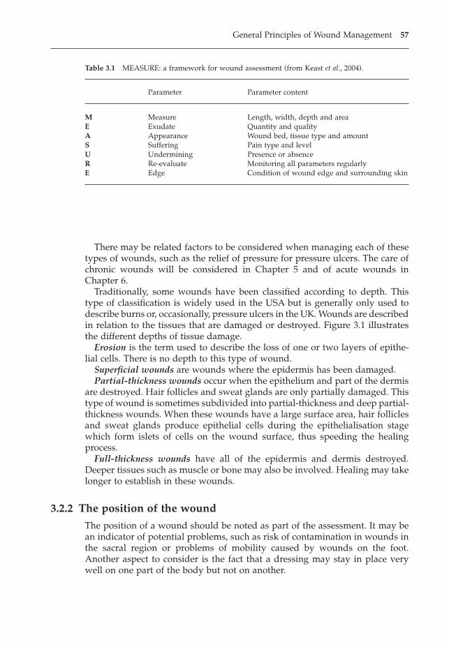
There may be related factors to be considered when managing each of thesetypes of wounds, such as the relief of pressure for pressure ulcers. The care ofchronic wounds will be considered in Chapter 5 and of acute wounds inChapter 6.
Traditionally, some wounds have been classified according to depth. Thistype of classification is widely used in the USA but is generally only used todescribe burns or, occasionally, pressure ulcers in the UK. Wounds are describedin relation to the tissues that are damaged or destroyed. Figure 3.1 illustratesthe different depths of tissue damage.
Erosion is the term used to describe the loss of one or two layers of epithe-lial cells. There is no depth to this type of wound.
Superficial wounds are wounds where the epidermis has been damaged.Partial-thickness wounds occur when the epithelium and part of the dermis
are destroyed. Hair follicles and sweat glands are only partially damaged. Thistype of wound is sometimes subdivided into partial-thickness and deep partial-thickness wounds. When these wounds have a large surface area, hair folliclesand sweat glands produce epithelial cells during the epithelialisation stagewhich form islets of cells on the wound surface, thus speeding the healingprocess.
Full-thickness wounds have all of the epidermis and dermis destroyed.Deeper tissues such as muscle or bone may also be involved. Healing may takelonger to establish in these wounds.
3.2.2 The position of the wound
The position of a wound should be noted as part of the assessment. It may bean indicator of potential problems, such as risk of contamination in wounds inthe sacral region or problems of mobility caused by wounds on the foot.Another aspect to consider is the fact that a dressing may stay in place verywell on one part of the body but not on another.
General Principles of Wound Management 57
Table 3.1 MEASURE: a framework for wound assessment (from Keast et al., 2004).
Parameter Parameter content
M Measure Length, width, depth and areaE Exudate Quantity and qualityA Appearance Wound bed, tissue type and amountS Suffering Pain type and levelU Undermining Presence or absenceR Re-evaluate Monitoring all parameters regularlyE Edge Condition of wound edge and surrounding skin
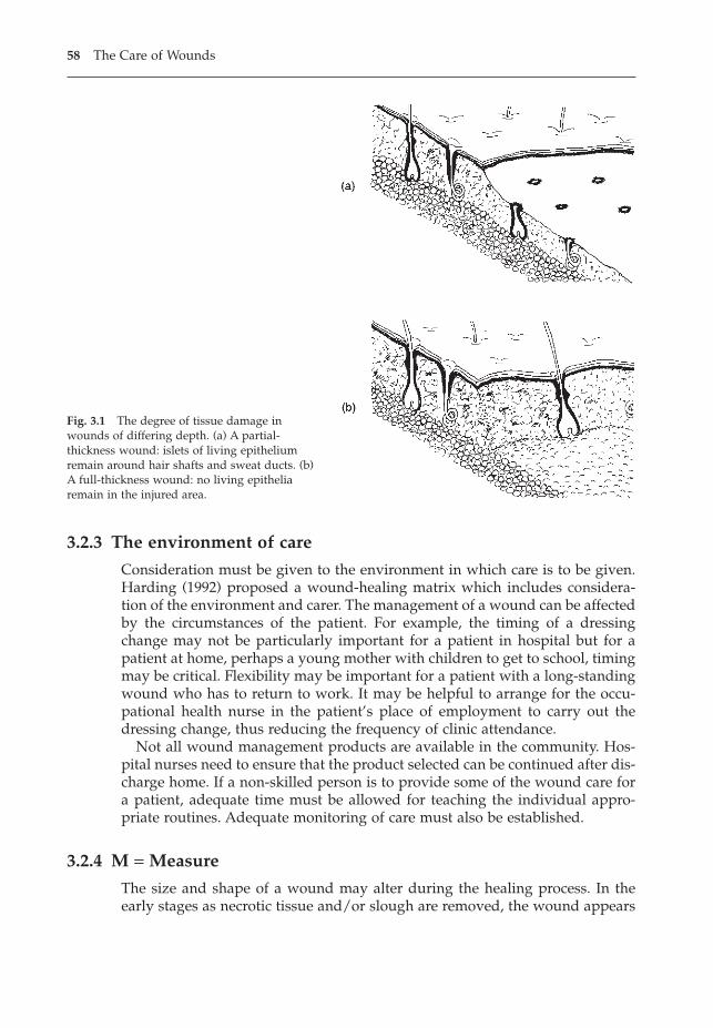
3.2.3 The environment of care
Consideration must be given to the environment in which care is to be given.Harding (1992) proposed a wound-healing matrix which includes considera-tion of the environment and carer. The management of a wound can be affectedby the circumstances of the patient. For example, the timing of a dressingchange may not be particularly important for a patient in hospital but for apatient at home, perhaps a young mother with children to get to school, timingmay be critical. Flexibility may be important for a patient with a long-standingwound who has to return to work. It may be helpful to arrange for the occu-pational health nurse in the patient’s place of employment to carry out thedressing change, thus reducing the frequency of clinic attendance.
Not all wound management products are available in the community. Hos-pital nurses need to ensure that the product selected can be continued after dis-charge home. If a non-skilled person is to provide some of the wound care fora patient, adequate time must be allowed for teaching the individual appro-priate routines. Adequate monitoring of care must also be established.
3.2.4 M = Measure
The size and shape of a wound may alter during the healing process. In theearly stages as necrotic tissue and/or slough are removed, the wound appears
58 The Care of Wounds
Fig. 3.1 The degree of tissue damage inwounds of differing depth. (a) A partial-thickness wound: islets of living epitheliumremain around hair shafts and sweat ducts. (b)A full-thickness wound: no living epitheliaremain in the injured area.
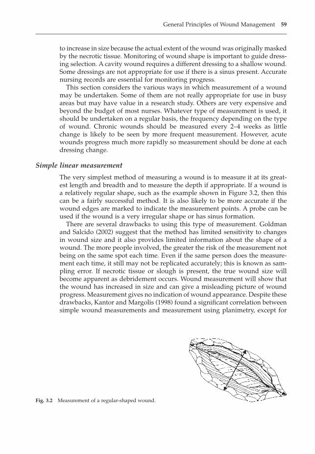
to increase in size because the actual extent of the wound was originally maskedby the necrotic tissue. Monitoring of wound shape is important to guide dress-ing selection. A cavity wound requires a different dressing to a shallow wound.Some dressings are not appropriate for use if there is a sinus present. Accuratenursing records are essential for monitoring progress.
This section considers the various ways in which measurement of a woundmay be undertaken. Some of them are not really appropriate for use in busyareas but may have value in a research study. Others are very expensive andbeyond the budget of most nurses. Whatever type of measurement is used, itshould be undertaken on a regular basis, the frequency depending on the typeof wound. Chronic wounds should be measured every 2–4 weeks as littlechange is likely to be seen by more frequent measurement. However, acutewounds progress much more rapidly so measurement should be done at eachdressing change.
Simple linear measurement
The very simplest method of measuring a wound is to measure it at its great-est length and breadth and to measure the depth if appropriate. If a wound isa relatively regular shape, such as the example shown in Figure 3.2, then thiscan be a fairly successful method. It is also likely to be more accurate if thewound edges are marked to indicate the measurement points. A probe can beused if the wound is a very irregular shape or has sinus formation.
There are several drawbacks to using this type of measurement. Goldmanand Salcido (2002) suggest that the method has limited sensitivity to changesin wound size and it also provides limited information about the shape of awound. The more people involved, the greater the risk of the measurement notbeing on the same spot each time. Even if the same person does the measure-ment each time, it still may not be replicated accurately; this is known as sam-pling error. If necrotic tissue or slough is present, the true wound size willbecome apparent as debridement occurs. Wound measurement will show thatthe wound has increased in size and can give a misleading picture of woundprogress. Measurement gives no indication of wound appearance. Despite thesedrawbacks, Kantor and Margolis (1998) found a significant correlation betweensimple wound measurements and measurement using planimetry, except for
General Principles of Wound Management 59
Fig. 3.2 Measurement of a regular-shaped wound.
wounds larger than 402 cm, and suggested that simple measurements could beused to monitor wound progress.
Simple measurement is best used on small, surgically induced cavities thatare regular in shape and should heal rapidly. More comprehensive data wouldbe obtained if used in conjunction with a nursing chart.
Measurement of surface area
Flanagan (2003a) reviewed the varying methods of wound measurement andfound that measuring true surface area and monitoring the percentage reduc-tion of wound surface area over time are the most useful methods. The mostfrequently used system for surface area measurement is that of tracing a wound.A variety of materials may be used, the commonest being acetate paper. Onepresentation of acetate paper is the lesion measure, samples of which are sup-plied by several dressing manufacturers. They are usually fairly small sheetswith a series of circles on them. The centre circle is 1cm in diameter and is sur-rounded by concentric circles that increase in size by 2cm increments. Thisgives some estimate of measurement in the tracing.
The surface area of a wound can be calculated quite accurately by placingthe tracing over squared paper and counting the number of whole squares orby using computerised planimetry. Successive tracings can be compared toshow any difference in wound size. If necrotic tissue or slough is present thenan initial increase in size will occur as debridement progresses.
More recently, sophisticated computerised methods have been developedthat can measure surface area. Lagan et al. (2000) compared planimetry anddigitising and found that digitising based on wound tracings gave a signifi-cantly higher level of repeatability than planimetry. Keast et al. (2004) describeda portable digital tablet (VisitrakTM) that can be used in conjunction with woundtracings. A tracing can be placed on the digital tablet and a special stylus usedto trace over the wound outline. The system calculates the surface area and per-centage reduction in wound size from previous measurements. Clinical evalu-ation of the digital tablet is in progress.
Another system is a specialised computer software package for use with adigital camera (Verge VideometerTM [VeV]). A photograph is taken and down-loaded onto a computer where the program calculates the wound surface area.Thawer et al. (2002) compared tracings and planimetry with VeV and foundexcellent intrarater and interrater reliability in 45 chronic wounds. However, itmust be noted that there is potential for inaccuracy when using a system involv-ing a digital camera if the wound is over a curved part of the body such as theleg.
Flanagan (2003a) noted that errors can occur when undertaking wound trac-ings, particularly in identifying the wound margin. She suggested that estab-lishing a simple protocol could help to improve accuracy. As with woundmeasurement, the tracing will show the increase or decrease in size without any explanation, as it does not provide information on wound appearance ordepth.
60 The Care of Wounds
If the acetate sheet is placed in direct contact with the wound, it will needcleansing with alcohol spray or similar. Some centres use plastic bags so theunderside can be discarded and the side with the tracing retained. Someacetates are provided with disposable backing paper.
Tracing is best used on fairly straightforward shallow wounds. Ideally itshould be used in combination with an assessment chart. Lucas et al. (2002) rec-ommend using both tracings and photography to monitor wound healing.
Measuring volume
When wounds are deep, it may be useful to measure wound volume. Theoreti-cally, it is possible to calculate the volume by measuring the wound depth(using a sterile swab) and multiplying by the surface area. However, there areinherent errors in this method (Goldman & Salcido, 2002). Deep wounds arerarely cuboid in shape, may not have a uniform depth, and sinus formation orundermining may be present. If the base of the wound is filled with necroticdebris, it must first be cleansed from the wound before undertaking any mea-surements (Keast et al., 2004). Various methods have been used to measurewound volume but they are not simple, valid or reliable (Keast et al., 2004).
The most practical method of monitoring changes in wound volume is tomeasure and record the depth at the deepest part of the wound and to note theamount of dressing required to fill the cavity.
3.2.5 E = Exudate
The amount of wound exudate varies during the healing process. In a normallyhealing wound, there is considerable exudate at the inflammatory stage andvery little at epithelialisation. A copious exudate may indicate a prolongedinflammatory stage or infection. In their review of the role of exudate in thehealing process, Vowden and Vowden (2003) consider the differences betweenacute and chronic wound exudate and conclude that chronic wound exudatehas the potential to become a barrier to healing. Unfortunately, it is not possi-ble to assess a wound and immediately determine if wound exudate is an actualbarrier to healing.
Keast et al. (2004) suggest that exudate can be measured in terms of quantity,quality and odour. Falanga (2000) proposed a scoring system for woundexudate quantity that provides some qualification with the aim of achievingstandardisation. Thus:
� 1 = minimal (dressings last up to one week).� 2 = moderate (dressings changed every 2–3 days).� 3 = heavy (dressings changed at least daily).
An alternative method to quantify exudate was used by Browne et al. (2004a)in a multicentre study assessing dressing performance in the presence of heavyexudate (called the WRAP Study). The research team used the TELER® systemof clinical indicators (Le Roux, 1993) to measure exudate leakage. A clinical indi-
General Principles of Wound Management 61
cator is an ordinal measuring scale with six reference points that measurechange. Each reference point is clinically significant, with 0 being the worst pos-sible outcome and 5 the best. Table 3.2 shows the clinical indicator for exudateleakage when the planned frequency of dressing change is every two days.Exudate is considered to leak if it strikes through the outer bandage or strap-ping. Although this system measures exudate leakage rather than exudateamount, it still has clear clinical relevance.
Falanga (2000) considered that there is little benefit in scoring the quality ofwound exudate. However, there may be some benefit in using clearly definedterminology to describe exudate quality. It may be of particular importance inpostsurgical wounds where there is a potential risk of postoperative bleeding.Therefore the terms ‘serous’, ‘serosanguinous’, ‘sanguinous’, ‘seropurulent’ and‘purulent’ may be useful.
Odour is much more difficult to measure in any objective way. Odour maybe indicative of infection but wound odour also increases as necrotic tissuedebrides by autolysis. Some dressings such as hydrocolloids may produce afoul-smelling odour when they are removed. It must also be remembered thatmany patients are distressed by wound odour and may feel embarrassment,disgust or shame (Hack, 2003). In a further paper on the WRAP project, Browneet al. (2004b) describe a clinical indicator to quantify odour (see Table 3.3).Browne et al. (2004b) used it in conjunction with a patient-focused indicator todetermine the impact of odour on the patient, thus making it an even morepowerful tool.
3.2.6 A = Appearance
The appearance of the wound gives an indication of the stage of healing that ithas reached or of any complication that may be present. Open wounds orwounds healing by second intention can be categorised as:
62 The Care of Wounds
Table 3.2 Exudate leakage using TELER indicator (fromBrowne et al. (2004a). Reproduced by kind permission of TELERLtd).
Code Exudate leakage
5 No leakage between routine/planned dressing change
4 Exudate leakage within 2 hours of next dressing change
3 Exudate leakage within 8 hours of next dressing change
2 Exudate leakage within 24 hours of dressing change
1 Exudate leakage within 8 hours of dressing change
0 Exudate leakage within 2 hours of dressing change
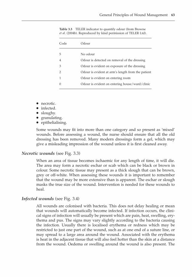
� necrotic.� infected.� sloughy.� granulating.� epithelialising.
Some wounds may fit into more than one category and so present as ‘mixed’wounds. Before assessing a wound, the nurse should ensure that all the olddressing has been removed. Many modern dressings form a gel, which maygive a misleading impression of the wound unless it is first cleaned away.
Necrotic wounds (see Fig. 3.3)
When an area of tissue becomes ischaemic for any length of time, it will die.The area may form a necrotic eschar or scab which can be black or brown incolour. Some necrotic tissue may present as a thick slough that can be brown,grey or off-white. When assessing these wounds it is important to rememberthat the wound may be more extensive than is apparent. The eschar or sloughmasks the true size of the wound. Intervention is needed for these wounds toheal.
Infected wounds (see Fig. 3.4)
All wounds are colonised with bacteria. This does not delay healing or meanthat wounds will automatically become infected. If infection occurs, the clini-cal signs of infection will usually be present which are pain, heat, swelling, ery-thema and pus. The signs may vary slightly according to the bacteria causingthe infection. Usually there is localised erythema or redness which may berestricted to just one part of the wound, such as at one end of a suture line, ormay spread to a large area around the wound. Associated with the erythemais heat in the adjacent tissue that will also feel hotter than the skin at a distancefrom the wound. Oedema or swelling around the wound is also present. The
General Principles of Wound Management 63
Table 3.3 TELER indicator to quantify odour (from Browne et al. (2004b). Reproduced by kind permission of TELER Ltd).
Code Odour
5 No odour
4 Odour is detected on removal of the dressing
3 Odour is evident on exposure of the dressing
2 Odour is evident at arm’s length from the patient
1 Odour is evident on entering room
0 Odour is evident on entering house/ward/clinic
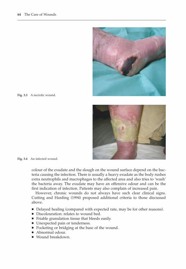
colour of the exudate and the slough on the wound surface depend on the bac-teria causing the infection. There is usually a heavy exudate as the body rushesextra neutrophils and macrophages to the affected area and also tries to ‘wash’the bacteria away. The exudate may have an offensive odour and can be thefirst indication of infection. Patients may also complain of increased pain.
However, chronic wounds do not always have such clear clinical signs.Cutting and Harding (1994) proposed additional criteria to those discussedabove.
� Delayed healing (compared with expected rate, may be for other reasons).� Discolouration: relates to wound bed.� Friable granulation tissue that bleeds easily.� Unexpected pain or tenderness.� Pocketing or bridging at the base of the wound.� Abnormal odour.� Wound breakdown.
64 The Care of Wounds
Fig. 3.3 A necrotic wound.
Fig. 3.4 An infected wound.
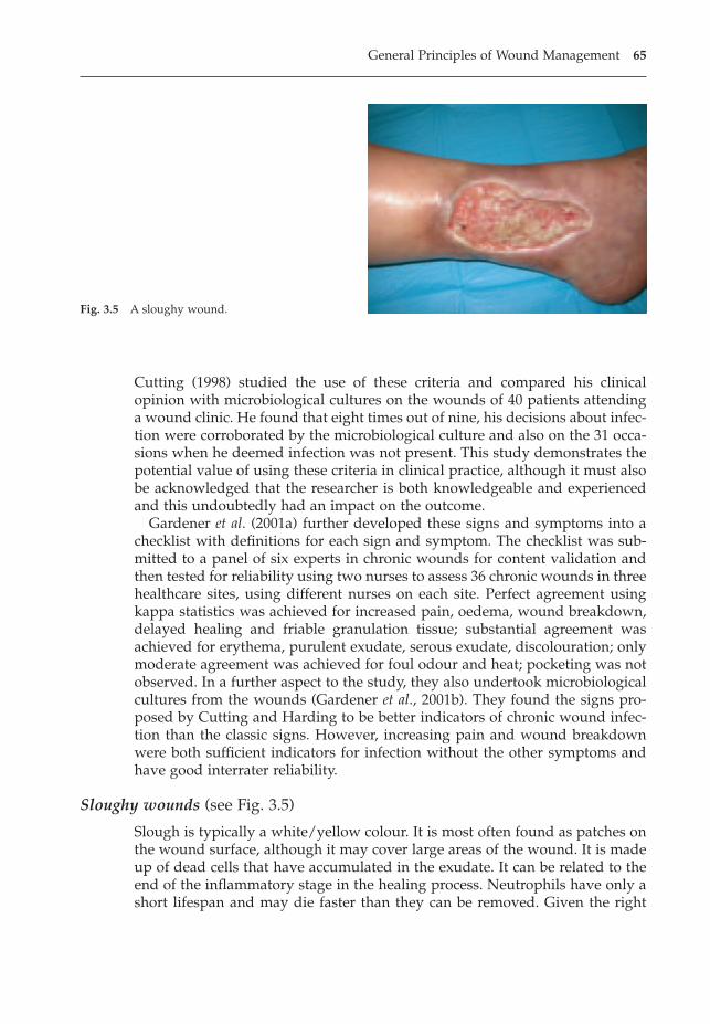
Cutting (1998) studied the use of these criteria and compared his clinicalopinion with microbiological cultures on the wounds of 40 patients attendinga wound clinic. He found that eight times out of nine, his decisions about infec-tion were corroborated by the microbiological culture and also on the 31 occa-sions when he deemed infection was not present. This study demonstrates thepotential value of using these criteria in clinical practice, although it must alsobe acknowledged that the researcher is both knowledgeable and experiencedand this undoubtedly had an impact on the outcome.
Gardener et al. (2001a) further developed these signs and symptoms into achecklist with definitions for each sign and symptom. The checklist was sub-mitted to a panel of six experts in chronic wounds for content validation andthen tested for reliability using two nurses to assess 36 chronic wounds in threehealthcare sites, using different nurses on each site. Perfect agreement usingkappa statistics was achieved for increased pain, oedema, wound breakdown,delayed healing and friable granulation tissue; substantial agreement wasachieved for erythema, purulent exudate, serous exudate, discolouration; onlymoderate agreement was achieved for foul odour and heat; pocketing was notobserved. In a further aspect to the study, they also undertook microbiologicalcultures from the wounds (Gardener et al., 2001b). They found the signs pro-posed by Cutting and Harding to be better indicators of chronic wound infec-tion than the classic signs. However, increasing pain and wound breakdownwere both sufficient indicators for infection without the other symptoms andhave good interrater reliability.
Sloughy wounds (see Fig. 3.5)
Slough is typically a white/yellow colour. It is most often found as patches onthe wound surface, although it may cover large areas of the wound. It is madeup of dead cells that have accumulated in the exudate. It can be related to theend of the inflammatory stage in the healing process. Neutrophils have only ashort lifespan and may die faster than they can be removed. Given the right
General Principles of Wound Management 65
Fig. 3.5 A sloughy wound.
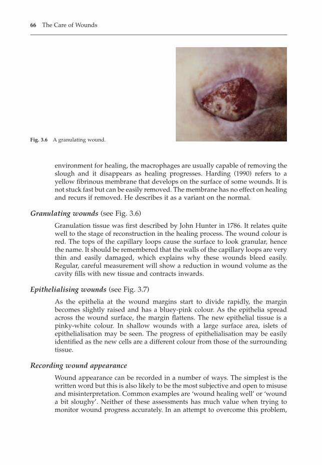
environment for healing, the macrophages are usually capable of removing theslough and it disappears as healing progresses. Harding (1990) refers to ayellow fibrinous membrane that develops on the surface of some wounds. It isnot stuck fast but can be easily removed. The membrane has no effect on healingand recurs if removed. He describes it as a variant on the normal.
Granulating wounds (see Fig. 3.6)
Granulation tissue was first described by John Hunter in 1786. It relates quitewell to the stage of reconstruction in the healing process. The wound colour isred. The tops of the capillary loops cause the surface to look granular, hencethe name. It should be remembered that the walls of the capillary loops are verythin and easily damaged, which explains why these wounds bleed easily.Regular, careful measurement will show a reduction in wound volume as thecavity fills with new tissue and contracts inwards.
Epithelialising wounds (see Fig. 3.7)
As the epithelia at the wound margins start to divide rapidly, the marginbecomes slightly raised and has a bluey-pink colour. As the epithelia spreadacross the wound surface, the margin flattens. The new epithelial tissue is apinky-white colour. In shallow wounds with a large surface area, islets ofepithelialisation may be seen. The progress of epithelialisation may be easilyidentified as the new cells are a different colour from those of the surroundingtissue.
Recording wound appearance
Wound appearance can be recorded in a number of ways. The simplest is thewritten word but this is also likely to be the most subjective and open to misuseand misinterpretation. Common examples are ‘wound healing well’ or ‘wounda bit sloughy’. Neither of these assessments has much value when trying tomonitor wound progress accurately. In an attempt to overcome this problem,
66 The Care of Wounds
Fig. 3.6 A granulating wound.
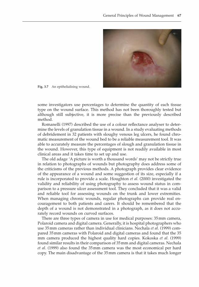
some investigators use percentages to determine the quantity of each tissuetype on the wound surface. This method has not been thoroughly tested butalthough still subjective, it is more precise than the previously describedmethod.
Romanelli (1997) described the use of a colour reflectance analyser to deter-mine the levels of granulation tissue in a wound. In a study evaluating methodsof debridement in 32 patients with sloughy venous leg ulcers, he found chro-matic measurement of the wound bed to be a reliable measurement tool. It wasable to accurately measure the percentages of slough and granulation tissue inthe wound. However, this type of equipment is not readily available in mostclinical areas and it takes time to set up and use.
The old adage ‘A picture is worth a thousand words’ may not be strictly truein relation to photographs of wounds but photography does address some ofthe criticisms of the previous methods. A photograph provides clear evidenceof the appearance of a wound and some suggestion of its size, especially if arule is incorporated to provide a scale. Houghton et al. (2000) investigated thevalidity and reliability of using photography to assess wound status in com-parison to a pressure ulcer assessment tool. They concluded that it was a validand reliable tool for assessing wounds on the trunk and lower extremities.When managing chronic wounds, regular photographs can provide real en-couragement to both patients and carers. It should be remembered that thedepth of a wound is not demonstrated in a photograph, as it does not accu-rately record wounds on curved surfaces.
There are three types of camera in use for medical purposes: 35mm camera,Polaroid camera and digital camera. Generally, it is hospital photographers whouse 35mm cameras rather than individual clinicians. Nechala et al. (1999) com-pared 35mm cameras with Polaroid and digital cameras and found that the 35mm camera produced the highest quality hard copies. Kokoska et al. (1999)found similar results in their comparison of 35mm and digital cameras. Nechalaet al. (1999) also found the 35mm camera was the most economical per hardcopy. The main disadvantage of the 35mm camera is that it takes much longer
General Principles of Wound Management 67
Fig. 3.7 An epithelialising wound.
to generate a photograph than the other camera types. Another disadvantageis that it is not possible to be certain of the quality or accuracy of an individualphotograph until the film is developed. It is also important to determine howindividual patients will be identified if using a film with large numbers of expo-sures. One issue many nurses have to consider if planning to purchase a camerais the need for an ongoing budget to pay for films and development.
Polaroid cameras have been very popular with nurses, especially in the com-munity. The major advantage of this camera is that the hard copy is producedimmediately and can be shown to the patient and immediately placed in thenotes (Nechala et al., 1999). Whilst the quality may not be as good as more pow-erful cameras, it is adequate for most needs, especially if a light lock lens is usedto enable close-ups. There is still an ongoing cost for the purchase of films.
Increasingly, digital cameras are being used in clinical practice. They aregradually improving in quality and becoming cheaper. After initial training hasbeen given, they have been found to be easy to use and focus and to transferto computer to provide digital images. There are no ongoing costs as the film-card can be reused many times. However, access to a computer with a largememory on the hard drive is necessary, as the images require a large storagecapacity. Once downloaded, the images can be utilised to produce hard copies,which can be displayed on the screen for teaching purchases or incorporatedinto a PowerPoint presentation. Galdino et al. (2000) monitored the costs of con-verting a large plastic surgery to digital photography rather than using the hos-pital photographic department. All the medical staff, including junior doctors,were supplied with their own camera and film-card. Over a five-year period,the authors estimated they would save $63950. The main disadvantages ofdigital cameras are the initial training to use them successfully and that it ispossible to manipulate the image and to falsely enhance the results (Nechala etal., 1999).
Hayes and Dodds (2003) successfully incorporated use of a digital camerainto clinical practice in a hospital vascular clinic. They utilise a shared electronicpatient record to communicate with primary care teams via the NHSnet, so thatall involved can see the digital images. They recommend using standardisedlighting and background and using the same patient position and that patientidentification codes, the date and a calibration scale should be included withinthe digital image.
Not all nurses have access to a camera. It may be possible to obtain one ona short-term loan if a dressing trial is being undertaken.
Several factors need to be considered if the purchase of a camera is planned.Nechala et al. (1999) considered that it was important to determine the reasonfor requiring a camera before making a choice of camera type. If high-qualityhard copies are required, a 35mm camera is the best option. If speed is impor-tant then a Polaroid camera should be chosen and if digital images are required,then a digital camera is best. Other questions to answer are as follows.
� Who will pay for films and developing?� Is the proposed camera capable of taking close-up pictures?
68 The Care of Wounds
� Who will be using the camera?� How can pictures be taken from the same angle and distance each time?� How will patient consent be recorded? Some hospitals have a consent form
for photography, others accept that implied consent is sufficient for routinephotography.
� How will the images be stored to protect patient confidentiality?
Photographs provide good visual evidence of wound appearance. If a camerais not readily available, photography is best only considered for complicatedor unusual wounds.
3.2.7 S = Suffering
Moffatt (2002) suggested that within chronic wound management, there is aneed to focus on the patient, particularly their pain control. Unfortunately, painassessment is rarely part of routine wound assessment despite the fact thatmany wounds are associated with pain (Krasner, 1995). Increased pain may bean indicator of infection and this must always be excluded in the first instance.Pain has been discussed in Chapter 2 and pain assessment should part of theoverall patient assessment. This section will focus on local wound factors inrelation to pain.
Dressing change and wound management activities such as debridementmay cause pain. An international survey of practitioners was undertaken across11 countries to investigate their views on wound pain (Moffatt et al., 2002).Dressing removal was identified as the time of greatest pain, with adherentdressings being seen as a major contributing factor. Szor and Bourguignon(1999) surveyed 32 patients with pressure ulcers for pain using the McGill PainQuestionnaire. They found that 28 (87.5%) had pain at dressing change and 12(42%) had continuous pain both at rest and during dressing changes. Sadly, onlytwo patients were receiving analgesia.
It is well recognised that burn patients suffer considerable pain at dressingchange (Latarjet, 2002). Nagy (1999) investigated the impact on nurses of havingto inflict pain on burn patients during dressing changes. She found that thecommonest strategy was distancing by the nurse, which had the side effect ofthe nurses becoming less concerned about controlling pain. But if nurses devel-oped a strategy of engaging with the pain, they saw it as a challenge to ensuregood pain control. Taal et al. (1999) undertook a validation study of a painanxiety scale specifically for burn patients. The scale included items such as ‘Ifind it impossible to relax when my burns are being treated’ and ‘I am fright-ened of the pain during and/or after the treatment’. They found high levels ofreliability and internal consistency and suggest that the scale could be used asa method of assessing therapeutic interventions.
Senecal (1999) stresses the importance of assessment and that it shouldinclude the type of pain and pain intensity. Krasner (1995) put forward a modelof chronic wound experience that divides wound pain into three types: non-cyclical acute wound pain, such as sharp debridement; cyclical acute wound
General Principles of Wound Management 69
pain, such as dressing changes; chronic wound pain, which is persistent andconstant. Pain intensity can be measured by means of a variety of visual ana-logue scales as discussed in Chapter 2. It may also be useful to identify anyfactors related to dressing change, such as dressing type, that may impact onpain levels. The frequency of reassessment should also be identified.
3.2.8 U = Undermining
In this part of their assessment framework, Keast et al. (2004) discuss assess-ment of the internal wound area. This is relevant in cavity wounds and it isimportant to identify any undermining, tunnelling or sinus tracts. A sterileswab can be used to probe under the wound edges to measure the extent ofundermining. If it is extensive it may be helpful to mark the area on the surfaceof the skin using a pen. This will facilitate future assessments and assist inevaluating any change.
3.2.9 R = Re-evaluate
The purpose of re-evaluating a wound is to check for any signs of complica-tions and to monitor progress towards achieving short-term goals, such asdebridement and the overall goal of healing. This may be achieved by check-ing the wound for complications such as infection at each dressing change andregularly assessing selected wound parameters such as reduction in woundsize. The frequency of assessment will vary according to wound type but formost chronic wounds, it should be every 1–2 weeks. Acute wounds may needto be assessed more frequently.
Flanagan (2003b) reviewed the methods for measuring wound healing andconcluded that percentage reduction in size was the most useful indicator ofwound progress as this can differentiate between healing and non-healingwounds in the first few weeks of treatment. Kantor and Margolis (2000) mea-sured percentage reduction in wound size for 104 patients with venous legulcers and found that percentage change in the first four weeks of treatmentpredicted the healing outcome at 24 weeks. In a small study of nine patientswith grade 4 pressure ulcers, Brown (2000) found that healing did not progressin a linear fashion and suggested that plotting wound-healing curves could beuseful; that is, multiple measurements of percentage reduction in wound areafrom baseline, measured over time.
This type of accurate measurement may not be necessary for everyday clini-cal practice but it has considerable value in research. Specialist centres may beable to invest in some of the digitised systems that can provide this type ofinformation very easily, as discussed in section 3.2.4, but they are not availableto all. In general clinical practice, wounds should be regularly assessed every1–4 weeks and progress towards treatment goals evaluated.
70 The Care of Wounds
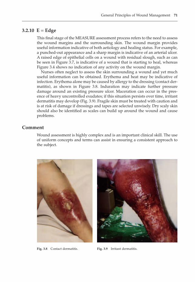
3.2.10 E = Edge
This final stage of the MEASURE assessment process refers to the need to assessthe wound margins and the surrounding skin. The wound margin providesuseful information indicative of both aetiology and healing status. For example,a punched-out appearance and a sharp margin is indicative of an arterial ulcer.A raised edge of epithelial cells on a wound with residual slough, such as canbe seen in Figure 3.7, is indicative of a wound that is starting to heal, whereasFigure 3.4 shows no indication of any activity on the wound margin.
Nurses often neglect to assess the skin surrounding a wound and yet muchuseful information can be obtained. Erythema and heat may be indicative ofinfection. Erythema alone may be caused by allergy to the dressing (contact der-matitis), as shown in Figure 3.8. Induration may indicate further pressuredamage around an existing pressure ulcer. Maceration can occur in the pres-ence of heavy uncontrolled exudates; if this situation persists over time, irritantdermatitis may develop (Fig. 3.9). Fragile skin must be treated with caution andis at risk of damage if dressings and tapes are selected unwisely. Dry scaly skinshould also be identified as scales can build up around the wound and causeproblems.
Comment
Wound assessment is highly complex and is an important clinical skill. The useof uniform concepts and terms can assist in ensuring a consistent approach tothe subject.
General Principles of Wound Management 71
Fig. 3.8 Contact dermatitis. Fig. 3.9 Irritant dermatitis.
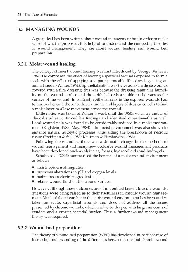
3.3 MANAGING WOUNDS
A great deal has been written about wound management but in order to makesense of what is proposed, it is helpful to understand the competing theoriesof wound management. They are moist wound healing and wound bed preparation.
3.3.1 Moist wound healing
The concept of moist wound healing was first introduced by George Winter in1962. He compared the effect of leaving superficial wounds exposed to form ascab with the effect of applying a vapour-permeable film dressing, using ananimal model (Winter, 1962). Epithelialisation was twice as fast in those woundscovered with a film dressing; this was because the dressing maintains humid-ity on the wound surface and the epithelial cells are able to slide across thesurface of the wound. In contrast, epithelial cells in the exposed wounds hadto burrow beneath the scab, dried exudate and layers of dessicated cells to finda moist layer to allow movement across the wound.
Little notice was taken of Winter’s work until the 1980s when a number ofclinical studies confirmed his findings and identified other benefits as well.Local wound pain was found to be considerably reduced in a moist environ-ment (Eaglstein, 1985; May, 1984). The moist environment was also shown toenhance natural autolytic processes, thus aiding the breakdown of necrotictissue (Freidman & Su, 1983; Kaufman & Hirshowitz, 1983).
Following these studies, there was a dramatic change in the methods ofwound management and many new occlusive wound management productshave been developed such as alginates, foams, hydrocolloids and hydrogels.
Schultz et al. (2003) summarised the benefits of a moist wound environmentas follows:
� assists epidermal migration.� promotes alterations in pH and oxygen levels.� maintains an electrical gradient.� retains wound fluid on the wound surface.
However, although these outcomes are of undoubted benefit to acute wounds,questions were being raised as to their usefulness in chronic wound manage-ment. Much of the research into the moist wound environment has been under-taken on acute, superficial wounds and does not address all the issuespresented by chronic wounds, which tend to be deeper, with larger amounts ofexudate and a greater bacterial burden. Thus a further wound managementtheory was required.
3.3.2 Wound bed preparation
The theory of wound bed preparation (WBP) has developed in part because ofincreasing understanding of the differences between acute and chronic wound
72 The Care of Wounds
exudate and the potentially harmful constituents of chronic wound fluid (seeChapter 1) and in part following clinical experience of using bio-engineeredskin products. Falanga (2000) proposed that it was inappropriate to utiliseexpensive, sophisticated products on a poorly prepared wound. As a minimum,a wound should have a well-vascularised wound bed, minimal bacterialburden and little or no exudate before an effective outcome can be achieved.Further debate has led to a consensus paper (Schultz et al., 2003), which hasprovided the following definition:
‘Wound bed preparation is the management of the wound to accelerateendogenous healing or to facilitate the effectiveness of other therapeutic measures.’
WBP has four aspects: debridement, management of exudate, resolution of bac-terial imbalance and undermined epidermal margin (Schultz et al., 2003).Falanga (2004) has utilised the work of Schultz et al. (2003) to develop a frame-work called TIME to provide a comprehensive approach to chronic wound care.The terms in the framework have been modified by the European Wound Man-agement Association WBP Advisory Board to maximise their use in differentlanguages (Box 3.1). Figure 3.10 shows how the TIME framework can be usedwithin a holistic approach to care.
Tissue management
This aspect of the TIME framework refers to the need to debride necrotic andsloughy tissue. Debridement may be achieved in a number of ways.
� Autolytic debridement utilises the ability of macrophages to phagocytosedebris and necrotic tissue. Hydrocolloids and hydrogels are widely used topromote autolytc debridement as they provide a moist environment thatenhances macrophage activity. Alginates are also used in the presence ofmoisture.
� Biosurgery or larval therapy has recently regained popularity and is widelyused in the UK. Sterile maggots of the fly Lucilia sericata secrete enzymes thatbreak down necrotic tissue to a semi-liquid form that the maggots can ingest,leaving only the healthy tissue (Thomas, 2001).
� Enzymatic debridement also ensures autolysis through the use of enzymessuch as elastase, collagenase and fibrinolysin. These enzymes cleave throughthe collagen holding necrotic tissue to the wound bed, thus debriding the
General Principles of Wound Management 73
Box 3.1 The TIME framework (based on Falanga, 2004).
T = Tissue managementI = Inflammation and infection controlM = Moisture balanceE = Epithelial (edge) advancement
Reproduced by kind permission of Radical Education Partnership.
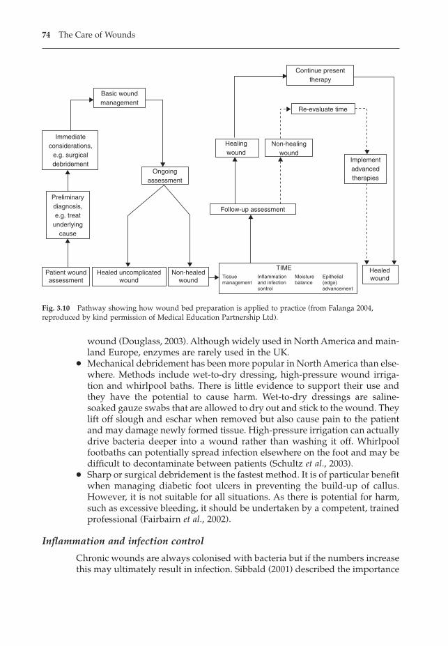
wound (Douglass, 2003). Although widely used in North America and main-land Europe, enzymes are rarely used in the UK.
� Mechanical debridement has been more popular in North America than else-where. Methods include wet-to-dry dressing, high-pressure wound irriga-tion and whirlpool baths. There is little evidence to support their use andthey have the potential to cause harm. Wet-to-dry dressings are saline-soaked gauze swabs that are allowed to dry out and stick to the wound. Theylift off slough and eschar when removed but also cause pain to the patientand may damage newly formed tissue. High-pressure irrigation can actuallydrive bacteria deeper into a wound rather than washing it off. Whirlpoolfootbaths can potentially spread infection elsewhere on the foot and may bedifficult to decontaminate between patients (Schultz et al., 2003).
� Sharp or surgical debridement is the fastest method. It is of particular benefitwhen managing diabetic foot ulcers in preventing the build-up of callus.However, it is not suitable for all situations. As there is potential for harm,such as excessive bleeding, it should be undertaken by a competent, trainedprofessional (Fairbairn et al., 2002).
Inflammation and infection control
Chronic wounds are always colonised with bacteria but if the numbers increasethis may ultimately result in infection. Sibbald (2001) described the importance
74 The Care of Wounds
Basic woundmanagement
Immediateconsiderations,
e.g. surgicaldebridement
Preliminarydiagnosis,e.g. treat
underlyingcause
Patient woundassessment
Healed uncomplicatedwound
Non-healedwound
Ongoingassessment
Healingwound
Non-healingwound
Follow-up assessment
Implementadvancedtherapies
Re-evaluate time
Continue presenttherapy
Healedwound
TIMETissuemanagement
Inflammationand infectioncontrol
Moisturebalance
Epithelial(edge)advancement
Fig. 3.10 Pathway showing how wound bed preparation is applied to practice (from Falanga 2004,reproduced by kind permission of Medical Education Partnership Ltd).
of maintaining a bacterial balance where the wound may be contaminated orcolonised but this does not impact on healing. He also discussed the issue ofdeep and superficial infections. If healing does not result from topical therapy,systemic antibiotics may be necessary, especially if a deep infection is present.Schultz et al. (2003) emphasise the importance of debridement as it can reducethe bacterial burden by removing devitalised tissue, which is a focus for bac-teria, and creating a more active wound. Using maggots for debridement is par-ticularly useful, as they have been found to ingest and destroy bacteria,including MRSA (Thomas, 2001). Topical antiseptics such as slow-release silverand iodine have been shown to be beneficial but should only be used for twoweeks (Edmonds et al., 2004; Moffatt et al., 2004). Topical antibiotics are not rec-ommended because of the risk of bacterial resistance.
Moisture balance
A major criticism of the moist wound-healing theory is that there is no infor-mation about what constitutes the correct level of moisture on the woundsurface. Wounds can produce widely varying quantities of exudate. Heavilyexuding wounds may cause maceration of the surrounding skin as well assoggy dressings and a very distressed patient. On the other hand, wounds withlittle or no exudate may become dessicated. A balance between the two isrequired. Moisture balance in the presence of heavy exudate can be achievedin a variety of ways.
� Use of highly absorbent dressings. There are a number of highly absorbentdressings available and selection depends on the type, position and size ofthe wound and the wound bed status. Alginates, foams and hydrofibre dress-ings are widely used; they absorb exudate and may also have moisturevapour transmission capability. Capillary action dressings are multilayeredand able to conduct exudate away from the wound surface.
� The use of appliances such as ‘bag systems’. This may be by means of a stomabag or a specialist wound appliance. These systems are useful in woundsthat constantly seep but they only contain the problem, rather than solvingit. However, they may be more effective and comfortable for the patient thanbulky dressings that frequently leak (Vowden & Vowden, 2003).
� Compression is an effective method of controlling wound exudate in legulceration as compression reduces chronic venous hypertension. It can beachieved by the use of compression bandages or by intermittent compres-sion therapy. This treatment is limited to leg ulcers where there is a venouscomponent and will be discussed further in Chapter 5.
� Topical negative pressure therapy (TNP) is increasingly being applied toheavily exuding wounds of all types. It has also been suggested that use ofTNP can reduce the bacterial burden of a wound (Morykwas et al., 1997).
Whatever method of moisture balance is used, it is important to care for theskin surrounding the wound. Excessive moisture can cause maceration or irri-tant dermatitis (Cutting & White, 2002). Various strategies may be used,
General Principles of Wound Management 75
depending on the method used to manage the exudate. Creams containing zincprovide an effective barrier and are frequently used with heavily exuding legulcers. They are soothing to macerated skin but would be inappropriate whereadhesive strapping is used close to the wound margin, such as with TNP. Skinsealants are particularly useful under adhesive dressings as they protect theskin from the adhesives as well as the exudate. Another strategy is to use ahydrocolloid dressing, first cutting a hole the shape of the wound in the dress-ing wafer and then applying it as a border around the wound, as is used in biosurgery.
Epithelial (edge) advancement
A healing wound not only has a wound bed filled with healthy granulatingtissue but also evidence of epithelialisation at the wound margins. For manywounds, achieving debridement of necrosis and slough and bacterial and mois-ture balance will stimulate the wound to heal. However, Falanga (2004) sug-gests that the problems of impaired MMP metabolism or cell senescence(discussed in Chapter 1) may prevent this last phase of healing, although thereis need for more research in this area. As shown in Figure 3.10, this may be thetime to re-evaluate the wound and to consider the implementation of advancedtherapies such as growth factors or tissue-engineered products (Schultz et al.,2003).
Comment
Both these theories of wound management have moved our understandingforward. It is too simplistic to suggest that moist wound-healing theory shouldbe used for the management of acute wounds and WBP for chronic wounds.Nurses should have an understanding of the underlying principles of both con-cepts and utilise them appropriately following a comprehensive patient andwound assessment.
3.3.3 Pain management
As discussed in section 3.2.7, pain management is often neglected in woundcare. Accurate assessment will determine the type and intensity of pain, allow-ing appropriate strategies to be developed. Senecal (1999) proposed incremen-tal steps to determine suitable analgesia and stressed the importance of regularadministration. However, many patients are reluctant to take regular analgesiaand may need to be encouraged to do so. Latarjet (2002) discussed the differ-ence between background pain and procedural pain in burn patients. He con-sidered that it is possible to achieve zero background pain but this is difficultduring dressing changes. If additional medication is given before dressingchanges, sufficient time must be allowed for the analgesia to take effect. If painis not controlled, it may be useful to involve the pain team.
76 The Care of Wounds
Briggs and Torra i Bou (2002) discussed additional strategies to reduce pain.They emphasise the importance of reducing anxiety and spending time explain-ing to the patient what will happen and the level of pain to expect. It is alsoimportant to select appropriate dressings that will maintain moisture balanceand minimise pain and trauma on removal. If dressings have adhered to thewound, then it may be necessary to soak them off. If dressings persistentlyadhere to the wound, an alternative dressing should be considered.
3.4 DOCUMENTATION
In order to judge the progress of a wound, it is essential to keep accuraterecords. Good documentation will help to ensure continuity of care. Poor docu-mentation has been implicated in frequent alterations of dressing type, accord-ing to the whim of individual nurses, as shown in a study by Rundgren et al.(1990). They followed the progress of 101 patients with a variety of woundsover a five-month period. They found that from week to week about 30% of thepatients were receiving a different treatment; 65% of the wounds did not healand were having treatment for the whole of the study period. They concludedthat this lack of continuity was related to poor documentation, impatience anda lack of understanding of the healing process.
The European Tissue Repair Society (ETRS) has produced several statementsin relation to wound management, including one on documentation (ETRS,2003). This statement is given in full below.
� Adequate and accurate documentation of all patients with wounds shouldtake place. This should include core information with additional informationrecorded depending on wound type.
� Core information should be recorded at least monthly and include:a) Wound size by:
i) Tracing.ii) Measurement.iii) Photography.
b) Wound bed colour (black, green, yellow, pink, red)i) Percentage of wound bed.ii) Photography.
c) Wound depth, using grading scale of initial assessment and detailingtissue at wound base
d) Surrounding skin:i) Healthy.ii) Unhealthy.
e) Exudate – composition and volume:i) Nil.ii) Normal.iii) Excessive.
f) Pain – continuous/dressing change/occasional
General Principles of Wound Management 77
g) An evaluation of treatment effect with the outcome graded as wound:i) Healing.ii) Static.iii) Deteriorating.
h) The reason for non-healing indicated, e.g. infection, and the reason fortreatment changes documented
3.5 EVALUATING THE DRESSING
Nurses should be prepared to objectively evaluate the dressings they use, par-ticularly if they are using new dressings, although traditional dressings shouldnot be exempt from evaluation. When evaluating a dressing, various aspectsneed to be considered.
� Patient comfort.� Ease of application.� Effectiveness.� Cost.
Patient comfort is of primary importance for any wound management product.It can be very distressing for a patient when the application of a dressing is painful. Eusol is a well-known example of a lotion which causes pain onapplication (see Chapter 4).
Other products may adhere to the wound and cause discomfort to patientswhen they move and pain when the dressing is removed. A dressing that failsto provide sufficient absorbency and allows leakage of exudate can cause con-siderable inconvenience, as well as promoting feelings of insecurity in thepatient. Although different nurses may carry out the dressing, the patient isalways present. Any evaluation should involve the patient. Many like to takean interest and can provide valuable information on new products.
Ease of application means that a dressing can be applied easily and so willstay in place. When using any new product, it may take a little practice todevelop the most effective method of application. The nurse should be preparedto try a dressing over a period of time on a variety of wounds (unless con-traindicated) and on different parts of the body. This will allow a more com-prehensive evaluation.
Effectiveness is most important. If a product does not promote healing, thenit does not matter if it is comfortable or easy to apply. Before a product becomesavailable for general use, it should have undergone stringent laboratory teststo check for safety. The ETRS (2003) has developed standards for clinical trialsin wound healing that can guide the development of study protocols. Nursesmay find particular difficulties in undertaking a clinical trial. Hunt (1983) sug-gested that these included a lack of control over the admission and dischargeof patients, staffing patterns, the large numbers of nurses involved in patientcare and the variations in patterns of care across healthcare providers. If nursesare to evaluate the effectiveness of any dressing they use, they need to be aware
78 The Care of Wounds
of any research that has been published and any relevant systematic reviews.This will be discussed further in Chapter 7.
Cost is an important factor in all aspects of care and should be consideredwhen evaluating any dressing. However, not only the unit cost is relevant. Theoverall costs should be considered. One example is a study by Thomas andTucker (1989) who compared the use of paraffin tulle and an alginate (SorbsanTM) and found a reduced overall cost using SorbsanTM, despite the factthat the unit cost is greater than that of tulle.
REFERENCES
Briggs, M., Torra i Bou, J.E. (2002) Pain at wound dressing changes: a guide to manage-ment, in European Wound Management Association (EWMA) Position Document,Pain at Wound Dressing Changes. MEP Ltd, London.
Brown, G.S. (2000) Reporting outcomes for stage IV pressure ulcer healing: a proposal.Advances in Skin and Wound Management, 13 (6), 277–283.
Browne, N., Grocott, P., Cowley, S. et al. (2004a) The TELER system in wound careresearch and post market surveillance. EWMA Journal, 4 (1), 26–32.
Browne, N., Grocott, P., Cowley, S. et al. (2004b) Woundcare research for appropriateproducts (WRAP) validation of the TELER method involving users. InternationalJournal of Nursing Studies, 41, 559–571.
Cutting, K. (1998) Identification of infection in granulating wounds by registered nurses.Journal of Clinical Nursing, 7, 539–546.
Cutting, K., Harding, K.G. (1994) Criteria for identifying wound infection. Journal ofWound Care, 3 (4), 198–201.
Cutting, K., White, T. (2002) Maceration of the skin and wound bed. 1 Its nature andcauses. Journal of Wound Care, 11 (7), 275–278.
Douglass, J. (2003) Wound bed preparation: a systematic approach to chronic wounds.British Journal of Community Nursing, 8 (6) (suppl), S26–S34.
Eaglstein, W.H. (1985) Experiences with biosynthetic dressings. Journal of the AmericanAcademy of Dermatology, 12, 434–440.
Edmonds, M., Foster, A.V.M., Vowden, P. (2004) Wound bed preparation for diabetic footulcers, in European Wound Management Association (EWMA) Position Docu-ment, Wound Bed Preparation in Practice. MEP Ltd, London.
European Tissue Repair Society (ETRS) (2003) ETRS Working Group Statements. ETRSBulletin, 10 (2&3), 10–13.
Fairbairn, K., Grier, J., Hunter, C., Preece, J. (2002) A sharp debridement proceduredevised by specialist nurses. Journal of Wound Care, 11 (10), 371–375.
Falanga, V. (2000) Classification for wound bed preparation and stimulation of chronicwounds. Wound Repair and Regeneration, 8 (5), 347–352.
Falanga, V. (2004) Wound bed preparation: science applied to practice, in EuropeanWound Management Association (EWMA) Position Document, Wound Bed Prepa-ration in Practice. MEP Ltd, London.
Flanagan, M. (2003a) Improving accuracy of wound measurement in clinical practice.Ostomy and Wound Management, 49 (10), 28–40.
Flanagan, M. (2003b) Wound measurement: can it help us monitor progression tohealing? Journal of Wound Care, 12 (5), 189–194.
General Principles of Wound Management 79
Fowler, E. (1990) Chronic wounds: an overview, in (ed.) Krasner, D., Chronic Wound Care:a clinical sourcebook for healthcare professionals. Health Management PublicationsInc., King of Prussia, PA.
Freidman, S., Su, D.W.P. (1983) Hydrocolloid occlusive dressing management of legulcers. Archives of Dermatology, 120, 1329–1331.
Galdino, G.M., Swier, P., Manson, P.N., Vander Kolk, C.A. (2000) Converting to digitalphotography: a model for a large group or academic practice. Plastic and Recon-structive Surgery, 106 (1), 119–124.
Gardener, S.E., Frantz, R.A., Troia, C. et al. (2001a) A tool to assess clinical signs andsymptoms of localised infection in chronic wounds: development and reliability.Ostomy and Wound Management, 47 (1), 40–47.
Gardener, S.E., Frantz, R.A., Doebbeling, B.N. (2001b) The validity of the clinical signsand symptoms used to identify localised chronic wound infection. Wound Repairand Regeneration, 9 (3), 178–186.
Goldman, R.J., Salcido, R. (2002) More than one way to measure a wound: an overviewof tools and techniques. Advances in Skin and Wound Care, 15 (5), 236–243.
Hack, A. (2003) Malodorous wounds – taking the patient’s perspective into account.Journal of Wound Care, 12 (8), 319–321.
Harding, K.G. (1990) Wound care: putting theory into practice, in (ed.) Krasner, D.,Chronic Wound Care: a clinical sourcebook for healthcare professionals. Health Man-agement Publications Inc., King of Prussia, PA.
Harding, K.G. (1992) The wound-healing matrix. Journal of Wound Care, 1 (3), 40–44.Hayes, S., Dodds, S. (2003) Digital photography in wound care. Nursing Times, 99 (42)
(Wound Care Suppl), 2–3.Houghton, P.E., Kincaid, C.B., Campbell, K.E., Woodbury, M.G., Keast, D.H. (2000)
Photographic assessment of the appearance of chronic pressure and leg ulcers.Ostomy and Wound Management, 46 (4), 20–30.
Hunt, J. (1983) Product evaluation. Nursing, 2 (12) (suppl), 6–7.Kantor, J., Margolis, D.J. (1998) Efficacy and prognostic value of simple wound mea-
surements. Archives of Dermatology, 134 (12), 1571–1574.Kantor, J., Margolis, D.J. (2000) A multicentre study of percentage change in venous leg
ulcer area as a prognostic index of healing at 24 weeks. British Journal of Derma-tology, 142 (5), 960–964.
Kaufman, C., Hirshowitz, B. (1983) Treatment of chronic leg ulcers with Opsite. Chirur-gica Plastica, 7, 211–215.
Keast, D.H., Bowering, C.K., Evans, A.W., Mackean, G.L., Burrows, C., D’Souza, L. (2004)MEASURE: a proposed framework for developing best practice recommenda-tions for wound assessment. Wound Repair and Regeneration, 12 (3) (suppl), S1–S17.
Kokoska, M.S., Currens, J.W., Hollenbeak, C.S., Thomas, J.R., Stack, B.C. (1999) Digitalvs 35-mm Photography: to convert or not to convert? Archives of Facial PlasticSurgery, 1 (4), 276–281.
Krasner, D. (1995) The chronic wound pain experience: a conceptual model. Ostomy andWound Management, 41, 20–25.
Lagan, K.M., Dusoir, A.E., McDonough, S.M., Baxter, G.D. (2000) Wound measurement:the comparative reliability of direct versus photographic tracings analysed byplanimetry versus digitising techniques. Archives of Physical Medicine and Rehabil-itation, 81 (8), 1110–1116.
Latarjet, J. (2002) The management of pain associated with dressing changes in patientswith burns. EWMA Journal, 2 (2), 5–9.
80 The Care of Wounds
Le Roux, A.A. (1993) TELER: the concept. Physiotherapy, 79 (11), 755–758.Lucas, C., Classen, J., Harrison, D., De, H. (2002) Pressure ulcer surface area measure-
ment using instant full-scale photography and transparency tracings. Advances inSkin and Wound Care, 15 (1), 17–23.
May, S.R. (1984) Physiology, immunology and clinical efficacy of an adherentpolyurethane wound dressing Opsite, in (ed.) Wise, D.L., Burn Wound Coverings,Vol.II. CRC Press, Boca Raton, FL.
Moffatt, C.J. (2002) Pain at wound dressing changes, in European Wound ManagementAssociation (EWMA) Position Document, Pain at Wound Dressing Changes. MEPLtd, London.
Moffatt, C.J., Franks, P.J., Hollinworth, H. (2002) Understanding wound pain andtrauma: an international perspective, in European Wound Management Asso-ciation (EWMA) Position Document, Pain at Wound Dressing Changes. MEP Ltd,London.
Moffatt, C., Morison, M.J., Pina, E. (2004) Wound bed preparation for venous leg ulcers,in European Wound Management Association (EWMA) Position Document,Wound Bed Preparation in Practice. MEP Ltd, London.
Morykwas, M.J., Argenta, L.C., Shelton-Brown, E.I., McGuirt, W. (1997) Vacuum-assistedclosure: a new method for wound control and treatment: animal studies and basicfoundation. Annals of Plastic Surgery, 38 (6), 553–562.
Nagy, S. (1999) Strategies used by burn nurses to cope with the infliction of pain onpatients. Journal of Advanced Nursing, 29 (6), 1427–1433.
Nechala, P., Mahoney, J., Farkas, L.G. (1999) Digital two-dimensional photogrammetry:a comparison of three techniques of obtaining digital photographs. Plastic andReconstructive Surgery, 103 (7), 1819–1825.
Romanelli, M. (1997) Objective measurement of venous ulcer debridement and granu-lation with a skin colour reflectance analyser. Wounds, 9 (4), 122–126.
Rundgren, A., Nordehammar, A., Bjornestol, A., Magnusson, H., Nelson, C. (1990) Pres-sure sores in hospitalised geriatric patients. Background factors, treatment, long-term follow-up. Care – Science and Practice, 8 (3), 100–103.
Schultz, G.S., Sibbald, R.G., Falanga, V. et al. (2003) Wound bed preparation: a system-atic approach to wound management. Wound Repair and Regeneration, 11 (2)(suppl), S1–S28.
Senecal, S. (1999) Pain management of wound care. Nursing Clinics of North America, 34(4), 847–860.
Sibbald, R.G. (2001) What is the bacterial burden of the wound bed and does it matter?in (eds) Cherry, G.W., Harding, K.G., Ryan, T.J., Wound Bed Preparation. RoyalSociety of Medicine Press, London.
Szor, J.K., Bourguignon, C. (1999) Description of pressure ulcer pain at rest and at dressing change. Journal of Wound, Ostomy and Continence Nursing, 26 (3), 115–120.
Taal, L.A., Faber, A.W., van Loey, N.E.E., Reynders, C.L.L., Hofland, H.W.C. (1999) Theabbreviated burn specific pain anxiety scale: a multicentre study. Burns, 25, 493–497.
Thawer, H.A., Houghton, P.E., Woodbury, G., Keast, D., Campbell, K. (2002) A compar-ison of computer-assisted and manual wound size measurement. Ostomy andWound Management, 48, 46–53.
Thomas, S. (2001) Sterile maggots and the preparation of the wound bed, in (eds) CherryG.W., Harding K.G., Ryan T.J., Wound Bed Preparation. Royal Society of MedicinePress, London.
General Principles of Wound Management 81

Thomas, S., Tucker, C.A. (1989) Sorbsan in the management of leg ulcers. Pharmaceuti-cal Journal, 243, 706–709.
Vowden, K., Vowden, P. (2003) Understanding exudate management and the role ofexudate in the healing process. British Journal of Nursing, 12 (20) (suppl), 4–13.
Winter, G.D. (1962) Formation of the scab and the rate of epithelialisation of superficialwounds in the skin of the domestic pig. Nature, 193, 293.
82 The Care of Wounds
Wound Management Products 83
Chapter 4Wound Management Products
4.1 INTRODUCTION
There are many wound management products available and much conflictingadvice on how they should be used. Many nurses have a great interest in thissubject and take a justifiable pride in the acquired skills that facilitate dressingchange. Recent developments have demonstrated a need to change or adapttraditional practices.
Wound management products include topical agents as well as dressings andalso what have been described as ‘advanced dressings’. A topical agent is onewhich is applied to a wound. A dressing is a covering on a wound that isintended to promote healing and protect from further injury. Advanced prod-ucts are much more sophisticated dressings compared to those in everydaypractice. The Department of Health divides dressings into primary and secondary. A primary dressing is that which is used in direct contact with thewound. A secondary dressing is superimposed over the primary dressing. Theuse of all these products will be discussed in this chapter.
4.2 THE DEVELOPMENT OF DRESSINGS THROUGH THE AGES
In L’Ingenue (1767) Voltaire described history as a ‘tableau of crimes and mis-fortunes’. A study of the dressings used through the ages suggests that theremay be some truth in this. Some of the treatments used on the wounded werebizarre, if not horrific, whilst others are still familiar today.
4.2.1 Early days
The earliest record of any dressing can be found on the Edwin Smith Papyrus.Edwin Smith was an American Egyptologist who bought the papyrus from atrader in Luxor in 1862. He was unable to translate it and its contents wereunknown until a complete translation was published in 1930 (Zimmerman &Veith, 1961). The papyrus is dated at around 1700 bc but it is a copy of origi-nal manuscripts that date back to around 3000–2500 bc. A variety of dressingsare mentioned including grease, honey, lint and fresh meat, which was valuedfor its haemostatic properties. Adhesive strapping was made by applying gumto strips of linen (Forrest, 1982).

As the power of Ancient Egypt waned, the Greek civilisation gradually devel-oped. Amongst the men who made their mark at this time was Hippocrates.He lived from about 460 to 377 bc. Hippocrates laid the basis for scientific medicine with his emphasis on careful observation. For the most part, Hippocrates considered that wounds should be kept clean and dry. He recom-mended tepid water, wine and vinegar for cleansing wounds. If a woundshowed signs of inflammation, he suggested applying a cataplasm or poulticeto the area around the wound to soften the tissues and to allow free drainageof pus (Zimmerman & Veith, 1961). Hippocrates also used propolis, a hardresinous material produced by bees, to help in the healing of sores and ulcers(Trevelyn, 1997). Hippocrates gave the first definition of healing by first inten-tion, where the skin edges are held in approximation to each other, and sec-ondary intention where there is tissue loss and the skin edges are far apart.
Some of these concepts can also be found in the writings of Sushruta, anIndian surgeon who lived some time between the sixth century bc and the sixthcentury ad. His surgical textbook, the ‘Sushruta Samhita’, was used as a basisby later writers. Sushruta described 14 different types of dressings made fromsilk, linen, wool and cotton (Zimmerman & Veith, 1961). He also placed greatemphasis on the importance of cleanliness. Meade (1968) describes Sushruta’srecommendations for the management of wounds involving the intestines. Firstblack ants were applied and then the intestines were washed in milk and lubri-cated with clarified butter before they were returned to their normal position.He differed from Hippocrates on the matter of the most appropriate diet forpatients. Sushruta considered meat, normally forbidden to Hindus, an impor-tant factor whereas Hippocrates recommended the restriction of food and gavehis patients only water to drink (Zimmerman & Veith, 1961).
The influence of Greek physicians continued on into the time of the RomanEmpire. Oil and wine were commonly applied to wounds. Reference to this wasmade by the Gospel writer Luke, when he was recording the parable of theGood Samaritan (Luke, Ch.10, v.34, New Testament). Luke describes the GoodSamaritan pouring oil and wine onto the wounds and then applying bandages.
Celsus compiled a history of the development of medicine from the time ofHippocrates to the first century ad, with great detail of the practices of his time.Although it is believed that Celsus was not a physician, he was the first to givea definition of inflammation. He listed the cardinal signs as redness, heat, painand swelling. He advocated the cleansing of wounds to remove foreign bodiesbefore suturing. He also expected wounds to suppurate; that is, to form pus(Meade, 1968). A Roman scholar, Pliny, described the use of propolis to softeninduration and reduce swelling. He also wrote that it healed sores when healingseemed impossible (Trevelyn, 1997).
It is, however, Galen who stands out as the person whose work had lastingimpact. Galen (129–199 ad) was surgeon to the gladiators in Pergamun and laterphysician to the Emperor Marcus Aurelius. He wrote many books, some ofwhich survived him and were seen as the ultimate in medical knowledge formany centuries. He is particularly known for his theory of ‘laudable pus’ (pusbonum et laudabile), which holds that the development of pus is necessary for
84 The Care of Wounds
healing and should, therefore, actively be promoted. Galen found the applica-tion of writing ink, cobwebs and Lemnian clay to wounds to be efficacious(Forrest, 1982). When reviewing his achievements, Duin and Sutcliffe (1992)considered that although in some ways Galen considerably expanded medicalknowledge, he also held it back for a thousand years.
4.2.2 The Dark Ages and early Middle Ages
After the fall of the Roman Empire, cultural influence moved eastwards andthe Arab doctors of Islam further developed medical knowledge. However,their wound care was based on Galenic teaching. A number of doctors, such asRhazes and Albucassis, translated Galen’s writings into Arabic but the mostfamous was Ali Ibn Al-Husain-al-Sina (980–1037), also known as Avicenna tothe Western world. Avicenna wrote the Canon Medicina, translating Galen’swork and adding his own commentary (Dealey, 2002). Ultimately it was trans-lated into both Hebrew and Latin and became the foremost medical textbookin the Middle East, North Africa and Europe up until the seventeenth century(Guthrie, 1945). Avicenna proposed treating pressure ulcers with white leadointment, covering the bed with salix leaves and preventing the patient fromsleeping on his back (Kanal, 1975). He also advised the use of astringents suchas cooked honey and myrrh to reduce the amount of exudate in wounds withtissue loss.
Medical knowledge swept across the Middle East, through North Africa andinto Spain and southern Italy with the spread of Islam and the Islamic Empire.Thus the teaching of the Arab doctors influenced many of the developments in Europe, including reinforcing the pre-eminence of Galen. It must also berecognised that during this time the Church supplied most of the healthcareprovision outside the home, resulting in the Church having control over manyaspects of medicine, such as giving support to Galenic teaching.
Salerno in southern Italy was the first European university to have a medicalschool, which was founded in the ninth century (Forrest, 1982). Unlike mostother universities of the time, Salerno was not under ecclesiastical control andso was able to incorporate surgery into the curriculum at a time when the clergywere prohibited from practising it (Zimmerman & Veith, 1961). Salerno was theleading centre for surgical training in the eleventh century and during this timethe Surgery of Roger was written. It was translated into 15 other languages andwas used up to the sixteenth century (Paterson, 1988). Roger advocated the useof lard in head wounds, either applied directly or soaking cloth in molten lardfor deep wounds. He did not recommend cleaning wounds but used dressingsmade from eggs and water, tow and salt and bandages of fine linen cloth (Paterson, 1988).
One of the expert surgeons who graduated from Salerno was Hugo of Lucca(1160–1257). He went on to found the School of Surgery at Bologna University(Dealey, 2003). Although none of his writings have survived, he was consid-ered to be a very innovative surgeon. His famous pupil, Theodoric (also knownat Teodrico Borgognoni) (1205–1296?), completed his Chirurgia or surgical text-
Wound Management Products 85
book in 1267, which he stated was based in Hugo’s teaching. Theodoric dis-agreed with the concept of ‘laudable pus’ as he considered that it prolongedhealing. He advocated cleaning a wound with wine, debriding it and remov-ing all foreign matter, approximating the wound edges and holding them inplace using compresses of lint soaked in warm wine and then binding them inplace. The dressing was then to be left in situ for 5–6 days, unless there wasexcessive heat or pain (Borgognoni, 1955). He proposed that chronic woundsshould be cleansed with honey mixed with wine and water of holm-oak or vineashes; alternatively, sea water could be used to cleanse and dry a wound. Un-fortunately, his ideas were discredited and gradually disappeared until thetwentieth century when they were again discovered in Italy (Popp, 1995).
One of the people influenced by Theodoric’s teaching was Henri de Mondeville (1260–1320). He was a French doctor who trained in Paris andMontpellier as a physician, before going to Bologna to learn surgery fromTheodoric (Gerster, 1910). He was highly respected as a man of profound eru-dition and moral character and became surgeon to King Phillippe le Bel ofFrance in 1301. De Mondeville also wrote a Chirurgie, which drew on the workof Avicenna for anatomy and Theodoric for wounds. Unfortunately, de Mondeville was not a tactful person and he commenced his treatise on woundsby first describing and then condemning the common practices of the day thatpromoted ‘laudable pus’. Unsurprisingly, he failed to influence his contempo-raries who tried to put pressure on him to abandon his treatment (Dealey, 2004).
A close successor to de Mondeville was Guy de Chauliac (1300–1368). He followed a similar pattern to de Mondeville, training in Toulouse, Montpellierand Paris before travelling to Bologna to study anatomy (Johnson, 1989). It waswhen de Chauliac was in Bologna that he became strongly influenced byGalen’s writing. This is reflected in his Grande Chirurgie in which he quotedGalen 890 times (Johnson, 1989). When writing about wound care, he rejectedthe ideas of Theodoric and de Mondeville and proposed treatments such as theuse of tents (drains) and meshes (packing) to hold wounds open and oily salvesto promote pus formation. De Chauliac’s book was highly successful, havingnumerous editions and translations, and it became the primary surgical text-book for 200 years. He had so great an influence that his critics consider thathe set back progress in wound management for up to six centuries, althoughnot every commentator takes such a harsh view (Johnson, 1989).
4.2.3 Late Middle Ages and Renaissance
Kirkpatrick and Naylor (1997) have described the contents of a surgical treatisedated 1446 and believed to be the work of Thomas Morstede (1380–1450) whowas a London surgeon. The work provides detailed information about the clas-sification of ulcers and their treatment. The step-by-step approach describeshow to enlarge the mouth of the ulcer, then the processes of mortification(debridement), mundification (cleansing) and fleshing (encouragement of granulation tissue). Recipes for the various topical applications are providedfor the reader. They include items such as sage leaves, wormwood, white
86 The Care of Wounds
Gascony wine, alum and honey for mundification. The recipe for a treatmentfor fleshing involved stirring the mixture for the length of time it took to saytwo Creeds (articles of belief for Christian faith).
Ambroise Paré (1510–1590) was the most famous surgeon of the Renaissanceperiod (Zimmerman & Veith, 1961). He rose from a poor background with little education to become surgeon to four successive French kings, ultimatelygaining the titles Premier Chirurgien and Conseiller du Roi. However, it is asa military surgeon that Paré is best known. With the discovery of gunpowder,warfare changed. Gunshot wounds were believed to be poisonous. In order totreat them, surgeons began to undertake more amputation of limbs. The stan-dard practice was to use boiling oil to cauterise the stump. Paré is famous forusing a mixture of egg yolks, oil of roses and turpentine instead. He is also wellknown for his saying ‘Je le pensait, Dieu le guarist’, which is generally trans-lated as ‘I dressed him, God healed him’ (Zimmerman & Veith, 1961). Paré proposed using a dressing composed of rock alum, verdigris, Roman vitriol,rose honey and vinegar, boiled together to form a paste, on traumatic wounds(Linker & Womack, 1969). He treated a severe pressure ulcer by encouragingthe patient, feeding him, providing pain relief and sleep-inducing medicationand pressure relief by means of a small pillow (Levine, 1992). Paré wrotenumerous books, all in the vernacular rather than Latin. Some of his later writ-ings are considered to be classics.
4.2.4 The seventeenth, eighteenth and early nineteenth centuries
Another doctor worthy of mention is the German surgeon Lorenz Heister(1683–1758). Heister was exceedingly well educated in languages and thehumanities as well as medicine and surgery at a time when most of his col-leagues had little education or training and charlatans abounded (Zimmerman& Veith, 1961). He developed his skills working as a military surgeon andbecame a professor of anatomy and surgery at the University of Helmstadt in 1719. He wrote a General System of Surgery in three parts on the doctrine and management of: I) Wounds of all kinds, II) Several operations, and III) Bandages (Heister, 1768). Heister used lint dressings dipped in alcohol or oil ofturpentine to treat haemorrhage or spread with a sound digestive ointment orbalsam to heal wounds. He suggested that wound edges should not be closed,in order to allow pus to drain. He believed that all bandages should be madefrom clean linen that had been softened by repeated use (Bishop, 1959). Hisbook was translated into numerous languages, including English, and ran toten editions.
Many of the developments in wound management seem to be associatedwith wounds from battles. One development was that of debridement, possibly originated by Henri Francois LeDran (1685–1770) in order to relievethe constriction of soft tissues, believed to be caused by inflammation follow-ing gunshot wounds (Helling & Daon, 1998). However, other surgeons acrossEurope suggested caution in its use. John Hunter (1728–1793), the famousEnglish surgeon, disagreed with the practice as he did not believe that wounds
Wound Management Products 87
should be made larger in order to remove extraneous matter. He thought thatsuppuration would bring it to the surface (Zimmerman & Veith, 1961). Duringthe Napoleonic wars, several French surgeons advocated the use of debride-ment, particularly in the care of gunshot wounds (Helling & Daon, 1998).However, its use was sporadic and most wounds were not explored and after the fall of Napoleon and his empire, the concept was almost entirely forgotten.
4.2.5 Mid-nineteenth and early twentieth century developments
The Crimean War led to a huge demand for dressings. Various types of dress-ings were produced in the workhouses, which were a source of cheap labour.Charpie was made from unravelled cloth. Oakum was old rope which had beenunpicked and teased into fluff; it became popular with doctors during theAmerican Civil War as they considered it to be very absorbent (Bishop, 1959).Tow was made from broken, ravelled flax fibres. Lint is linen that has beenscraped on one side. The production of this dressing was mechanised. All ofthese dressings were washed and reused many times so they gradually becamequite soft but they were not very absorbent.
Sheets of cotton wool retained by cotton bandages were used during theFranco-Prussian wars, but Joseph Sampson Gamgee (1828–1886) was theperson who further developed its use. He noted that cotton wool could be madeabsorbent by removing the oily matter within it. He then conceived the idea ofcovering the cotton wool with unbleached gauze to make dressing pads orGamgee tissue (Lawrence, 1987). Gamgee tissue is still available today.
In 1867, Lister introduced the use of carbolic acid and revolutionised surgeryand surgical wound management. When he realised that carbolic caused skinirritation, he developed a method of impregnated carbolic gauze that becamethe first antiseptic dressing (Bishop, 1959). The use of antiseptics and conceptof asepsis spread rapidly across Europe.
During the course of the First World War, severely wounded soldiers had towait several days before receiving more than a simple field dressing. As a result,many wounds became infected and gangrenous. Antiseptics were developed tohelp resolve this problem. In particular, two similar antiseptic solutions cameinto use: Eusol (Edinburgh University Solution of Lime) and Dakin’s solution.Other antiseptics such as iodine, carbolic acid and mercury and aluminiumchloride were also available.
Sinclair and Ryan (1993) have reviewed some of the medical literature of 1915 to identify thinking at that time on the use of antiseptics. Bond (1915) considered that it was important to apply a ‘germicide’ as early as possible towounds that were almost certain to be infected. British soldiers were advisedto carry tincture of iodine so that they could apply it immediately to anygunshot wounds (Mayo-Robson, 1915). However, Herzog (1915), writing of theGerman experience in the battlefield, reported that he had seen a number ofsoldiers suffering from dermatitis of the skin around the wound as a result ofthe indiscriminate use of iodine.
88 The Care of Wounds
Deparge, a Belgian surgeon of note, recognised that merely introducing antiseptics into war wounds was insufficient and he revived the concept ofwound debridement (Helling & Daon, 1998). He believed that such devastat-ing wounds, often covered in mud and dirt and receiving only minimal surgery,encouraged the development of soft tissue infections. He formulated a set ofprinciples for managing them that involved exploring the wound and excisionof all contaminated and contused tissue, followed by leaving the wound openand irrigating it with Dakin’s solution. He also favoured the use of delayedprimary closure or secondary closure if bacteria were present. Deparge’s use of debridement has had a major impact on trauma surgery (Helling & Daon,1998).
At this time, Lumiere devised a dressing called tulle gras, a gauze impreg-nated with paraffin. Sphagnum moss was also used as it was found to be twiceas absorbent as cotton wool. It could also be impregnated with antiseptics andsterilised. Eupad was a dressing designed for use on leg ulcers. It was a Eusolpreparation, made up with a mixture of boracic and bleach. Another populartype of dressing was Emplastrums which were made of white leather spreadwith a plaster mass, to which some type of medication was often added (Turner,1986).
During the Second World War an American neurosurgeon called EldridgeCampbell was with a field hospital in Italy. In 1943 there was a great deal of heavy fighting and many casualties. Some of the Italian doctors caring forthese patients proposed a method of wound care that involved cleansing anddebriding the wound and then suturing it. This was contrary to the currentpractice of the day, which recommended packing with Vaseline gauze andimmobilisation. Campbell was impressed by this method of healing and eventually traced its origins back to the thirteenth century and Theodoric. Indescribing this fascinating piece of medical history, Popp (1995) concludes that it demonstrates the problems of entrenched views that are not questioned,such as the teachings of Galen, and the dangers of summarily dismissing newideas.
4.2.6 The British Pharmaceutical Codices
The first British Pharmaceutical Codex was published in 1907. It provided infor-mation on all the drugs and medicinal preparations in common use through-out the British Empire. Turner (1986) has reviewed the dressings listed in theearliest British Pharmaceutical Codices and compared them with more recentlists of dressings in the British Pharmacopoeia. He found that the list from 1923contains much that is familiar today. Table 4.1 compares the 1923 list with the1980 list, showing that little had changed in the intervening years. Gauzes,cotton wool pads and bandages can be seen as very popular methods of woundcare. Turner suggests that it is only in the last 30 years that any attempt hasbeen made to design materials that are actually functional. Prior to that, dress-ings were made from materials that happened to come to hand.
Wound Management Products 89
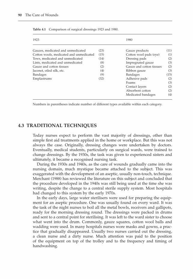
4.3 TRADITIONAL TECHNIQUES
Today nurses expect to perform the vast majority of dressings, other thansimple first aid treatments applied in the home or workplace. But this was notalways the case. Originally, dressing changes were undertaken by doctors.Eventually, medical students, particularly on surgical wards, were trained tochange dressings. By the 1930s, the task was given to experienced sisters andultimately, it became a recognised nursing task.
During the 1930s and 1940s, as the care of wounds gradually came into thenursing domain, much mystique became attached to the subject. This was exaggerated with the development of an aseptic, usually non-touch, technique.Merchant (1988) has reviewed the literature on this subject and concluded thatthe procedure developed in the 1940s was still being used at the time she waswriting, despite the change to a central sterile supply system. Most hospitalshad changed to this system by the early 1970s.
In the early days, large water sterilisers were used for preparing the equip-ment for an aseptic procedure. One was usually found on every ward. It wasthe task of the night nurses to boil all the metal bowls, receivers and gallipots,ready for the morning dressing round. The dressings were packed in drumsand sent to a central point for sterilising. It was left to the ward sister to choosewhat went into the drum. Commonly, gauze squares, cotton wool balls andwadding were used. In many hospitals nurses wore masks and gowns, a prac-tice that gradually disappeared. Usually two nurses carried out the dressing, a clean nurse and a dirty nurse. Much attention was paid to the position of the equipment on top of the trolley and to the frequency and timing of handwashing.
90 The Care of Wounds
Table 4.1 Comparison of surgical dressings 1923 and 1980.
1923 1980
Gauzes, medicated and unmedicated (23) Gauze products (11)Cotton wools, medicated and unmedicated (15) Cotton wool pads (eye) (1)Tows, medicated and unmedicated (14) Dressing pads (2)Lints, medicated and unmedicated (8) Impregnated gauze (3)Gauze and cotton tissues (2) Gauze and cotton tissues (2)Jaconet, oiled silk, etc. (4) Ribbon gauze (3)Bandages (9) Bandages (15)Emplastrums (32) Adhesive pads (2)
Foams (3)Contact layers (2)Absorbent cotton (2)Medicated bandages (4)
Numbers in parentheses indicate number of different types available within each category.
All wounds were redressed once or twice daily. The wound was thoroughlycleaned using cotton wool balls and forceps. The method of wiping across thewound surface varied from hospital to hospital. The Hippocratic principle ofkeeping wounds clean and dry became adapted to ‘allowing wounds to dryup’. Mostly gauze preparations were used but gradually all sorts of dubiouspractices crept in. There have been reports of Marmite, eggs and even toast usedon wounds (Dobrzanski et al., 1988; Johnson, 1987).
A wide variety of pharmaceutical preparations have also been appliedwithout any recognition of the need for evidence-based care. Murray (1988)found that within her health authority, an amazing selection of pharmaceuticalproducts were in use: 18 different cleansing agents, 53 substances left in contactwith open wounds and 24 products used for packing wounds. Millward (1989)found 19 different substances being used on pressure ulcers within one hospi-tal. Walsh and Ford (1989) have discussed the rituals in nursing, much of whichcan be applied to wound care. The common reasons for choosing a dressingcould be listed as:
� We always do it that way here.� Sister said so.� I have used this dressing for the last 30 years, why should I change?
Many older nurses will have been trained to use these ritualistic methods. It isonly recently that there has been a critical evaluation of these methods andchanges made to a more evidence-based approach.
4.4 THE USE OF LOTIONS
A variety of lotions are used in wound care, primarily for wound cleansing.The aims of wound cleansing are to remove any foreign matter such as gravelor soil, to remove any loose surface debris such as necrotic tissue and to removeany remnants of the previous dressing. A study by Thomlinson (1987) consid-ered the various ways in which swabs could be wiped across the woundsurface. The results showed that the action of cleansing did not reduce thenumber of bacteria on the wound surface but simply redistributed them.
4.4.1 Antiseptics
After saline, the commonest type of lotion in use is an antiseptic. An antisepticcan be defined as a non-toxic disinfectant, which can be applied to skin or livingtissues and has the ability to destroy vegetative compounds, such as bacteria,by preventing their growth. If antiseptics are simply used to wipe across thewound surface, they will have little effect. They need to be in contact with bac-teria for about 20 minutes before they actually destroy them (Russell et al., 1982).In some instances they can be applied in the form of soaks or incorporated intodressings, ointments or creams.
Wound Management Products 91
Research using experimental wounds in the animal model has demonstratedthat antiseptics have toxic effects that need to be weighed against any advan-tages obtained from their use. In the late 1980s and early 1990s there was con-siderable debate about antiseptics and their widespread use dramaticallyreduced. Since the introduction of the concept of wound bed preparation (dis-cussed in Chapter 3), antiseptics have made something of a comeback as theyare seen to have a place in achieving bacterial balance (Schultz et al., 2003).
Each of the common antiseptics will be listed in turn and their advantagesand disadvantages discussed.
Cetrimide
Cetrimide is useful for its detergent properties, particularly for the initialcleansing of traumatic wounds or the removal of scabs and crusts in skindisease. It should not be used in contact with the eye. It is rapidly inactivatedby organic material. Two dangers should be noted: it can cause skin irritationand sensitivity, and, it is very easy for it to become contaminated by bacteria,especially Pseudomonas aeruginosa. It is mostly only used in accident and emergency departments for initial cleansing of wounds rather than as a routinecleanser. It is available as a cream or as a lotion in combination with chlorhexi-dine. Morgan (1993) suggests that cetrimide should be used with caution inrestricted circumstances rather than as a general cleanser.
Chlorhexidine
Chlorhexidine is used in a variety of aqueous formulations. It is effectiveagainst Gram-positive and Gram-negative organisms. Brennan et al. (1986)found that it has a low toxicity to living cells. Tatnall et al. (1990) undertook asimilar study to identify the toxicity of several antiseptics when used on cul-tured keratinocytes (used for grafts). They found chlorhexidine to be the leasttoxic but considered that antiseptics should not be used over these graft sites.Kearney et al. (1988) found that chlorhexidine could maintain its antimicrobiallevels for a period of time when impregnated into a dressing. However, its efficacy is rapidly diminished in the presence of organic material such as pus or blood (Reynolds, 1982). Mengistu et al. (1999) tested chlorhexidine in different strengths against a range of Gram-negative bacteria and found that asignificant number were only inhibited by high concentrations of chlorhexidine.They concluded that it was more suitable for disinfection and hospital hygienerather than wound care.
Hydrogen peroxide
Hydrogen peroxide 3% (10 vols) has an oxidising effect that destroys anaero-bic bacteria. However, it loses its effect when it comes in contact with organicmaterial such as pus or cotton gauze. Lineaweaver et al. (1985) showed thathydrogen peroxide was cytotoxic to fibroblasts unless diluted to a strength of0.003%. This dilution is not effective against bacteria. O’Toole et al. (1996) found
92 The Care of Wounds
that even in concentrations 1000-fold less than 3% dilution, it inhibits keratinocyte migration and proliferation. Bennett et al. (2001) found that 3%hydrogen peroxide significantly reduced neodermal regeneration and fibrob-last proliferation compared to controls (untreated wounds) in an animal modelstudy. There is also a report of an incident where an air embolism occurred afterirrigation with hydrogen peroxide (Sleigh & Linter, 1985). Rees (2003) reviewedthe evidence on hydrogen peroxide and concluded that it was unsuitable foruse in cleaning wounds in A&E. Hydrogen peroxide is no longer widely usedas there is no evidence to demonstrate its efficacy and there are a number ofother more suitable alternatives.
Iodine
Iodine is a broad-spectrum antiseptic and is available as an alcohol and anaqueous solution. The aqueous solution is used in wound care, usually as povi-done iodine 10%, which contains 1% available iodine. It is used as a skin dis-infectant and to clean grossly infected wounds. McLure and Gordon (1992) andMichel and Zach (1997) found it to be effective against methicillin-resistantstaphylococcus aureus.
Several studies have questioned the value of using povidone iodine. It is cyto-toxic to fibroblasts unless diluted to 0.001%, retards epithelialisation and lowersthe tensile strength of the wound (Lineaweaver et al., 1985). Brennan and Leaper(1985) found that povidone iodine 5% damaged the microcirculation of thehealing wound but a 1% solution was innocuous. In contrast, Bennett et al.(2001) found that povidone iodine significantly increased fibroblast prolifera-tion and slightly increased neodermal regeneration and epithelialisation.Becker (1986) reported that when operating on contaminated head and neckcases, he irrigated 18 with povidone iodine and 17 with isotonic saline; 28% of wounds became infected, all of which had been irrigated with povidoneiodine.
Povidone iodine has been used within the wound bed preparation model ofwound management. Consequently, Selvaggi et al. (2003) reviewed and reap-praised the role of iodine and concluded that most of the evidence is confus-ing, especially as a mixture of in vitro and animal models have been used aswell as different preparations. They conclude that povidone iodine has beenshown to be an effective antibacterial that is superior to other products andseems to have no problem with resistance. Zhou et al. (2002) proposed that aslow-release form of iodine might overcome the toxicity problems. They testedcadexomer iodine using both in vitro methods and a variety of chronic wounds.They found no evidence of toxicity in the in vitro study. After 2–3 weeks of treatment, all 16 chronic wounds showed increased debridement, decreasedexudate, increased granulation tissue and reduced wound size.
Povidone iodine is available in ointment, spray and powder form andimpregnated into dressings. Iodine should not be used for patients with thyroiddisease or those who are sensitive to the product.
Wound Management Products 93
Potassium permanganate
Potassium permanganate 0.01% is mostly used on heavily exuding eczematousskin conditions, generally associated with leg ulceration. It is most easily usedin the form of tablets. One tablet dissolved in four litres of water provides a0.01% solution. The affected limb is generally placed in a container holding thefluid for approximately 15 minutes. It is mildly deodorising and has slight dis-infectant properties. It has been found to cause staining of the skin. There islittle evidence to demonstrate its efficacy (Anderson, 2003).
Proflavine
Proflavine has a mild bacteriostatic effect on Gram-positive organisms but noton Gram-negative bacteria. There has been little research to demonstrate itsvalue. Although it is available as a lotion it is mostly used as an aqueous cream.However, proflavine is not released from the cream into the wound so has noeffect on the bacteria. Foster and Moore (1997) compared the use of proflavine-soaked gauze packs with cellulose-based fibre dressing in surgical cavitywounds. They found that the proflavine packs were significantly more painfulto remove and patients required analgesia prior to removal.
Silver
Silver has been used as an antiseptic in wound care for many years in the formof silver nitrate. It is still used in this format in some burns units in NorthAmerica. However, it is extremely caustic, stains the skin black and prolongeduse causes hyponatraemia, hypokalaemia and hypocalcaemia. To overcomethese problems, a cream, silver sulphadiazine, was developed and has beenvery successful in controlling burn wound infections (Lansdown, 2004).However, the combination of cream and exudate makes dressing removalmessy so it is usually undertaken in a hydrotherapy unit, which can be a sourceof cross-infection (Tredget et al., 1998). As a result, there is increasing interest inthe use of silver-coated dressings of varying types.
Wright et al. (1998a) tested the bactericidal effect of silver in three modalities:liquid (silver nitrate), cream (silver sulphadiazine) and a silver-coated dressing.All products were effective at killing bacteria. The silver-coated dressing was the most efficacious against a wider range of bacteria and silver nitrate the least efficacious. Tredget et al. (1998) compared a silver-coated dressing,changed daily, with the standard procedure of covering a burn with gauze and moistening it with 0.5% silver nitrate every two hours. They found that the silver-coated dressing caused significantly less pain on removal and had substantially less infection. Also, the wound only required attention every 24 hours and could have been left for 48 hours, reducing dressing andnursing time costs. Silver dressings will be discussed in more detail in section4.6.
94 The Care of Wounds
Sodium hypochlorite
Sodium hypochlorite comes in several forms, the commonest being Eusol,Dakin’s solution and Milton. It was originally used on heavily infected woundsduring the First World War. Dakin suggested that to be effective, it should beused in large volumes (Thomas, 1990).
Several research studies have been undertaken which suggest that thehypochlorites may have few beneficial effects and do much harm. Bloomfieldand Sizer (1985) found they cause irritation to both the wound and the sur-rounding skin. They were found to have a cumulative effect, causing redness,pain and oedema, and to prolong the inflammatory stage of healing. They arecytotoxic to fibroblasts, unless diluted to a strength of 0.0005%, and retardepithelialisation (Lineaweaver et al., 1985). Brennan and Leaper (1985) foundthat they caused considerable damage to the microcirculation of the wound.The antiseptic effect is lost when the solution comes in contact with organicmaterial such as pus or gauze. A study describing the use of Eusol and liquidparaffin on leg ulcers was undertaken by Daltrey and Cunliffe (1981) who foundno significant evidence of antibacterial activity. Bennett et al. (2001) found that half-strength sodium hypochlorate had a mixed effect on wound repair,with no impairment of fibroblast proliferation but decreased vascular density.However, at that strength it was ineffective as an antimicrobial.
Carneiro and Nyawawa (2003) randomly allocated Eusol or phenytoinpowder to treat 102 leg ulcers. They found that there was a significant reduc-tion in pain, exudate levels and wound size in the phenytoin group comparedwith the Eusol group. This study was undertaken in Tanzania and many of theulcers were due to animal bites. These ulcers were found to heal fastest. Theauthors concluded that phenytoin was more suitable than Eusol, especially asit is cheap and easily applied.
Humzah et al. (1996) surveyed 124 plastic surgeons in the UK regarding theirviews on the use of Eusol. They had responses from 95 (77%) of the surgeons.Analysis of the responses found that 82% still used Eusol when it was avail-able and the majority (88%) used it for sloughy wounds. Since this survey wasundertaken, the use of Eusol has reduced even further. It is interesting to con-sider that where alternatives have replaced Eusol, its use has not been missedat all. Overall, Eusol is an outmoded product whose disadvantages far out-weigh any slight advantage there may be in its use.
4.4.2 Antibiotics
A range of antibiotics is available in topical form. They are potentially hazardous and they are not always absorbed into the wound. There is con-siderable risk of sensitisation to the patient as well as the development of re-sistant organisms. Systemic antibiotics are the treatment of choice when treatinginfected wounds because the infection may be too deep for topical antibioticsto penetrate.
Wound Management Products 95
D’Arcy (1972) recommends that any antibiotic that is used systemicallyshould not be applied to the skin. However, antibiotics that are not appropri-ate for systemic use may be developed for use on the skin or in wound care.This means that creams, gels, ointments or impregnated dressings containinggentamicin, tetracycline, fusidic acid or chlortetracycline hydrochloride shouldnot be used as these antibiotics are used systemically. Neomycin is no longerused systemically but topical use may cause systemic side effects such as ototoxicity.
One preparation which would seem to be of benefit in wound care ismupirocin. Mupirocin is used predominantly for treating methicillin-resistantStaphylococcus aureus (MRSA) either in skin infections or for nasal colonisation.Several studies have demonstrated its efficacy in treating MRSA in burnwounds (Deng et al., 1995; Rode et al., 1989; Trilla & Miro,1995). However,Cookson (1998) warns of the potential dangers of resistance and cites a numberof reported cases of mupirocin-resistant bacteria to support his arguments. Heproposes that prolonged and widespread use of mupirocin should be stoppedand a more judicious approach to its usage be adopted.
4.4.3 Honey
As was discussed earlier, honey has been used in wound care since ancienttimes and there has been a recent resurgence of interest in its use, particularlymanuka honey (Flanagan, 2000). A review by Molan (1999) discussed the roleof honey and considered that it had the following properties.
� Antibacterial action, as shown in laboratory studies and case studies,although, Kingsley (2001) did not find this in two case studies.
� Deodorising action, as demonstrated by Dunford and Hanano (2004) in astudy of 40 patients with recalcitrant venous leg ulcers.
� Debriding action, as found by Ahmed et al. (2003).� Anti-inflammatory action, as shown in histological studies.� Stimulation of wound healing, as shown in several studies (Dunford &
Hanano, 2004; Misirlioglu et al., 2003; Subrahmanyam, 1998; Vandeputte &Van Waeyenberge, 2003;).
� Pain relief was not listed by Molan (1999) but was demonstrated by Dunfordand Hanano in their study (Dunford & Hanano, 2004).
A systematic review by Moore et al. (2001) found that although honey appearedto be an effective treatment, many of the studies reviewed were of poor quality.There is a need for larger randomised controlled trials to determine the mosteffective use for honey. There is also an issue about frequency of dressingchange, as Molan (1999) suggested that up to three times daily may be neces-sary initially. This is very labour intensive and may not be acceptable to thepatient (Kingsley, 2001).
96 The Care of Wounds
4.4.4 Saline 0.9%
This is the only completely safe cleansing agent and is the treatment of choicefor use on most wounds. Manufacturers recommend that it is used in conjunc-tion with many of the modern wound management products. Saline is pre-sented in sachets, small plastic containers that allow the saline to be squirtedonto the wound and also in aerosols. These last two presentations are morewidely used in the community.
4.4.5 Tap water
Tap water is being used more frequently on a variety of wounds, in particularon areas already colonised such as wounds following rectal surgery or legulcers. Many patients may bath or shower prior to dressing change so thereseems to be little point in then ‘cleansing’ the wound. However, the bath orshower should be thoroughly cleaned afterwards to avoid cross-infection. Arecent systematic review found that using tap water to clean wounds did notdiffer from using sterile normal saline in respect of wound infection and healingrates (Fernandez et al., 2002). This would therefore suggest that either saline or water can be used, selection being based on practicality and individual circumstances.
4.5 CLINICAL EFFECTIVENESS OF WOUND MANAGEMENT PRODUCTS
Originally dressings were seen merely as coverings that could provide someprotection to the wound. The range of products currently available are muchmore sophisticated. There are so many products to choose from that it can causeconsiderable confusion. There is no single perfect dressing but an ‘identikit’ listof criteria can be established. A specific wound may not need all of the criterialisted. Selection can be assisted if the nurse has:
� assessed the wound and identified the specific objectives for the wound atthat time.
� an understanding of what can be reasonably expected from a dressing.� access to information regarding the characteristics and effectiveness of the
range of dressings available.
The characteristics of a clinically effective wound management product are considered below. Dressings are generally considered in relation to their performance and their handling qualities. Performance relates to the ability topromote healing.
4.5.1 Providing an effective environment
The qualities that will promote an effective environment for healing were dis-cussed in Chapter 3 under theories of wound management. They are:
Wound Management Products 97
� ability to maintain a moist environment.� antibacterial properties.� fluid-handling properties.
4.5.2 The handling qualities of an effective wound management product
These qualities can be listed as:
� easy to apply� conformability� easy to remove� comfortable to ‘wear’� does not require frequent dressing change.
Easy to apply
A major advantage of many of the modern products is that they are very simpleand quick to apply. Realistically, this has helped to promote their use with thenurses who regularly provide wound care.
Conformability
A dressing that conforms well to the shape of the wound is likely to assist inmaintaining a moist environment and also provide an effective barrier for bacteria.
Easy to remove
If a dressing is easy to remove it is less likely to damage the newly formed tissuein the wound. It is also less likely to be painful for the patient.
Comfortable to ‘wear’
Another advantage of many modern products is that they are comfortable forthe patient when they are in situ. This means that the patient is more likely towant to comply with the treatment regime. In any case, there is no need forpatients to suffer unnecessary pain or discomfort.
Does not require frequent dressing change
The majority of modern products can be left in place for several days, depend-ing on the wound and, particularly, the amount of exudate. This not only savesnursing time and reduces costs but also reduces the amount of interference withthe wound. Reduction in the frequency of dressing change helps to reduce theopportunities for a drop of temperature on the wound surface. This can poten-tially occur at each dressing change. Myers (1982) studied 420 patients andfound that, after wound cleansing, it took 40 minutes before the woundregained its original temperature. Furthermore, he found that it took three
98 The Care of Wounds
hours for mitotic activity to return to its normal rate. Patients also find less fre-quent dressing changes beneficial. Some patients find dressing change an ordealand others, especially community patients, an inconvenience which disruptstheir life.
Comment
It should be recognised that no one dressing provides the optimum environ-ment for the healing of all wounds. Equally, it may be necessary to use morethan one type of dressing during the healing of a wound. Many dressings willfulfil some of the criteria and they should be selected following careful assess-ment of the wound (see Chapter 3).
4.6 MODERN WOUND MANAGEMENT PRODUCTS
In order to make sense of all the dressings that are available, they will bedivided into different categories. Dressings can also be considered in terms oftheir suitability as a primary or secondary dressing on open wounds. In the UK,not all the dressings are freely available in the community as governmentrestrictions control which dressings can be prescribed. This may considerablyaffect continuity and quality of care between hospital and community.
This section aims to describe the different categories of wound managementproducts and some proprietary examples will be mentioned.
Absorbent pads
There are many versions of this type of dressing. Most are in the form of anabsorbent core, which is covered by a sleeve of gauze or synthetic material.These dressings are not suitable as a primary dressing on open wounds butmake an excellent secondary dressing, particularly when there is a heavyexudate. They are generally very cheap.
Absorbent cellulose dressings
Exu-dryTM and MesorbTM are one-piece multilayer dressings that are highlyabsorbent and able to wick exudate away from the wound surface. They maybe used in direct contact with the wound and are intended for use on heavilyexuding wounds of all types.
Adhesive island dressings
These dressings consist of a central pad which is covered with a wider band ofadhesive backing. They are lightweight and usually remain in position satis-factorily. There is little absorbent capacity in these dressings. They are widelyused on postsurgical wounds, which are healing by first intention, but are notsuitable for open wounds as a primary dressing.
Wound Management Products 99
Alginates
Alginate dressings contain calcium or sodium alginate, which is derived fromseaweed. There are several types of alginate including Algisite MTM, Algoster-ilTM, CurasorbTM, KaltostatTM, SorbalgonTM, SorbsanTM and TegagenTM. Thesedressings are interactive because as they react with the wound, their structurealters. As the dressing absorbs exudate, it changes from a fibrous structure toa gel. Some dressings allow removal in one piece, others have to be flushedfrom the wound. These dressings are available in a variety of formats: flat dress-ings, rope or ribbon, extra-absorbent versions and with an adhesive backing.They are appropriate for moderate or heavily exuding wounds and may requirea secondary dressing. They should not be used on wounds with no or lowexudate.
Antibacterials
MetrotopTM is a gel containing metronidazole. It reduces odour and anaerobicbacteria and is licensed for use as a deodoriser for fungating tumours. However,it has been used successfully on other wound types. Like all products of thistype, it should not be used indiscriminately.
Antibiotics
See section 4.2.2.
Antiseptics
See section 4.2.1.
Barrier film dressings
Barrier film dressings have developed to protect the skin. Some films, such asClinishieldTM and Skin PrepTM, were originally developed for use around stomasites. Others, such as CavilonTM and Comfeel Skin CareTM, have been morespecifically marketed for protecting the skin around a wound. The film is wipedon using an applicator such as an impregnated towel and dries to form a filmbarrier, protecting the skin from adhesives as well as moisture and resistant tourine and faeces.
Biosurgery
Biosurgery is another name for maggot or larval therapy. The often inadvertentuse of maggots in wounds has been recognised for centuries. Morgan (1995)chronicled the use of maggots by Maya Indians through to the 1930s. He considered that it fell into disrepute with the advent of antibiotics and asepticwound care and also noted the attendant aesthetic problems. However, larvaltherapy is enjoying a resurgence of popularity at present. Thomas et al. (1996)suggest this return in popularity might be in part because of the problemscaused by resistant bacteria such as MRSA.
100 The Care of Wounds
The larvae for biosurgery are packed in sterile containers directly from thesupplier. They may be ‘free range’ within a small plastic container or sealedwithin a porous bag. Before applying the larvae, a hydrocolloid dressing shouldbe applied around the wound to protect the skin from the proteolytic enzymesproduced by the maggots (Sherman, 1997). Once placed in the wound, the free-range maggots are retained by means of a small net, taped in place; those in abag are simply placed in the wound and held in place with tape. Once the larvaeare removed from the wound, they are disposed of in the same way as all useddressings. They are used to debride necrotic, sloughy and infected tissue froma wound.
Cadexomer bead dressings
These dressings are composed of hydrophilic beads containing iodine. As theyabsorb exudate, the beads swell and the iodine is released slowly into thewound. They come in the form of a powder (IodosorbTM) and an ointment (IodflexTM). This dressing is intended for heavily exuding necrotic, sloughy orinfected wounds. It should not be used for more than three months at a timeand is not suitable for people with iodine sensitivity or thyroid problems.
Capillary wound dressings
VacutexTM is the only dressing in this category. It is a non-interactive, three-layered dressing made from polyester filaments and polycotton fibres. Itabsorbs exudate into the middle layer and wicks it laterally in a capillary action.It may be cut to shape to fit a cavity and is suitable for all types of heavilyexuding wounds.
Foams
Foam dressings are made from polyurethane. They are either available as a flatfoam dressing, such as AllevynTM, BiatainTM, Cutinova HydroTM, LyofoamTM orTielleTM, or as a filler for cavity wounds, such as Allevyn Cavity Wound Dress-ingTM. Allevyn, Lyofoam and Tielle come in a range of presentations such asshaped dressings, varying absorbent capacity, tracheostomy dressings andadhesive dressings. Allevyn Cavity Wound is a preformed foam stent, whichcomes in two shapes and two sizes in each. Cavi-CareTM is also a foam stent butit has to be mixed with a catalyst and then poured into the wound cavity whereit sets in the shape of the wound. Foam dressings are best used on granulatingor epithelialising wounds with some exudate.
Hydrocapillary and multilayered absorbent dressings
The dressings within this category all have a slightly different action, depend-ing on their construction. They are Alione HydrocapillaryTM, TransorbentTM andVersivaTM. AlioneTM has a non-adherent wound contact layer, a hydrocapillarypad that absorbs exudate and distributes it horizontally and an outer low-friction film. TransorbentTM is a multilayer dressing containing a film, foam and
Wound Management Products 101
hydrogel. VersivaTM also comprises three layers: a wound contact layer of perforated hydrocolloid adhesive, a central absorbent layer of sodium carboxymethylcellulose fibres and an outer layer of polyurethane foam. Thesedressings can be left in place for several days. They are not suitable for drywounds.
Hydrocolloids
Hydrocolloids are a development from stoma products. They are interactivedressings consisting of a hydrocolloid base made from cellulose, gelatins andpectins and a backing made from a polyurethane film or foam. The technologyhas progressed since the original hydrocolloid dressings were introduced anda second generation of products is now available which have greaterabsorbency and hold exudate more effectively within the dressing. Examplesof hydrocolloid dressings include Comfeel PlusTM, Cutinova HydroTM, Granu-flexTM (known as DuodermTM outside the UK) and TegasorbTM. Several come in a wide range of sizes and shapes and variations such as a thinner than standard dressing. No secondary dressing is necessary. Cutinova HydroTM hasa greater absorptive capacity than other hydrocolloids. CombidermTM is anothervariation of hydrocolloid technology. It is composed of several layers includ-ing a thin hydrocolloid and an island pad containing polyacrylate granuleswhich hold the exudate within the pad. Cutinova CavityTM is suitable for cavitywounds; it is a flat sheet which can be folded or cut into a ribbon shape. Ini-tially it should only fill half the cavity because it swells as it absorbs exudateand moulds to the shape of the cavity. It is suitable for moderate to heavilyexuding cavity wounds.
Hydrocolloids can be used on a wide range of wounds but generally are mosteffective on the moderate to low exuding wounds.
Hydrofibres
Hydrofibre dressings are made from hydrocolloid fibres that gel in the presenceof exudate. They are highly absorbent. AquacelTM comes as a flat dressing in arange of sizes and also a ribbon for cavity wounds. They are not suitable foruse over dry necrotic wounds.
Hydrogels
These dressings are made from insoluble polymers and have a high watercontent. The amorphous gel, such as GranugelTM, Intrasite GelTM, Nu-GelTM orSterigelTM, may be used on a wide variety of wounds. They have the ability toeither absorb exudate or hydrate dry wounds such as necrotic eschar, thusencouraging debridement. They can be used on wounds with moderate to lowexudate and in small cavities. The gel sheets such as 2nd SkinTM or VigilonTM
are best used on granulating moderate to low exuding wounds. They all requirea secondary dressing.
102 The Care of Wounds
Low-adherent dressings
These types of dressing are low adherent rather than non-adherent. They havelittle if any absorbent capacity and are best on wounds with little exudate. Theymay be used to ‘carry’ a dressing such as an amorphous hydrogel and mostlyneed to be used in combination with an absorbent pad. They do not provide a moist wound environment. Examples of this type of dressing are MelolinTM,N-A UltraTM, ReleaseTM, TelfaTM and TricotexTM.
Low-adherent dressings (medicated)
InadineTM is a low-adherent dressing that is impregnated with water-solublepovidone iodine. It is suitable for moderate to low exuding infected wounds orfor prophylaxis in minor traumatic wounds. It is not suitable for patients withiodine sensitivity or thyroid problems. Activon TulleTM is a low-adherent dress-ing impregnated with manuka honey.
Non-medicated and medicated tulles
Tulles are also called paraffin gauze and were originally known as tulle gras.They are made of open weave cotton or rayon impregnated with soft paraffin.Although the paraffin makes the dressing less adherent, it readily becomesincorporated into granulation tissue. A pattern can be seen on the woundsurface when it is removed. It does not maintain a moist wound environmentand has no absorbent capacity. It is widely used on minor burns and traumaticinjuries. Examples are JelonetTM, ParanetTM, ParatulleTM and UnitulleTM. Asecondary dressing is required with these dressings.
Some types of tulles are impregnated with either antiseptics or antibiotics.The commonest type of antiseptic is chlorhexidine which is present in Bacti-grasTM, ChlorhexitulleTM and SerotulleTM. These dressings are useful for super-ficial infected wounds. Two tulles are impregnated with antibiotics (FucidinIntertulleTM and Sofra-TulleTM). The use of these dressings is not recommendedbecause of the problems of sensitivity and resistance of bacteria.
Paste bandages
These are cotton bandages impregnated with medicated paste. The use of pastebandages has largely been superseded by more modern products but they arestill used for leg ulceration, particularly when the surrounding skin is eczema-tous or inflamed. Whilst they are an effective form of treatment, many patientsdevelop allergies to the contents of the paste so it is wise to patch test the patientbefore applying a bandage. There are several types of bandage with differentpastes such as zinc paste with ichthammol or zinc paste with calamine. Asecondary bandage is required.
Silicone wound dressings
Silicone products have a number of uses. Silicone gel sheets such as Cica-CareTM, SilgelTM and Sil-K FilmTM help with scar management as they are able
Wound Management Products 103
to soften and flatten scars. Silicone N-ATM is similar to a low-adherent dressingas it is made from a knitted viscose material impregnated with silicone to alloweasy removal and is virtually non-adherent. MepitelTM is suitable for painfulwounds or where there is macerated or fragile skin as it is very easy to remove.Exudate passes through the dressing to the secondary pad. The outer pad maybe changed more frequently than the Mepitel, which can be left in place for upto ten days.
Silver and charcoal dressings
These dressings incorporate silver into their structure and some also containactivated charcoal cloth. Silver is an effective antibacterial and charcoal has beenfound to be effective in absorbing the chemicals released from malodorouswounds. Infected, necrotic or fungating wounds may have a very unpleasantodour. Actisorb Plus 25TM or Actisorb Plus 220TM are both in the form of a char-coal pad impregnated with silver; they are used with a primary dressing andrequire a covering pad. ArglaesTM contains alginate powder and a mixture ofinorganic polymer and ionic silver. As the alginate gels, the silver is graduallyreleased. Aquacel AgTM acts in a similar fashion. ActicoatTM, Acticoat 7TM andContreet AgTM are made from polyethylene or polyurethane coated with silver. Dressings such as CarbonetTM, ClinisorbTM or Lyofoam CTM are a combi-nation of dressing and charcoal. Most of these dressings require a secondarydressing.
TenderWet
TenderWetTM is a multilayer dressing with a central core of polyacrylate, whichis activated by soaking the dressing with an appropriate volume of Ringer’ssolution. The Ringer’s solution is delivered to the wound surface for up to 12hours and helps to soften necrotic tissue. It is able to absorb exudate but shouldbe changed daily.
Vapour-permeable films
There is a wide range of these film dressings available. They provide a moisthealing environment but have no absorbency. They should not be used oninfected wounds. The method of application varies according to make. Mostrequire a certain amount of skill and practice in application. Examples includeBioclusiveTM, CutifilmTM, MefilmTM, OpraflexTM, OpsiteTM and TegadermTM.
Vapour-permeable membranes
TegaporeTM allows exudate to pass through the dressing and can be left in placefor several weeks whilst the outer dressings are changed. It is non-adherent andwill protect delicate skin.
104 The Care of Wounds
4.7 ADVANCED TECHNOLOGIES
As with any other aspect of healthcare, new developments in wound care areregularly announced. Sometimes they are variations on an older form of treat-ment and sometimes they are new developments. This section seeks to identifythese innovations and consider the evidence to support their use.
4.7.1 Growth factors
Growth factors have been used for some time in wound care. In the USA, theuse of growth factors was primarily via the establishment of wound care centresthat provided a comprehensive programme of full assessment and plannedcare. Many of the patients treated in these centres have leg or foot ulcers suchas demonstrated in a randomised controlled trial undertaken by Knighton et al.(1990). They found a significantly higher rate of healing in those treated withgrowth factor solution compared with a blinded placebo.
The use of growth factors has not had the results that were initially expected.Spencer et al. (1996) suggested that whilst the results of clinical trials have sofar been disappointing, the interest in the use of growth factors has led to a greater understanding of the physiological make-up of different types ofchronic wound. Falanga (2000) suggested that part of the reason for this appar-ent failure may be because the concept of wound bed preparation (WBP) wasimperfectly understood at the time of many of the trials and there was a failureto prepare the wound adequately before applying the growth factor. It is inter-esting to note that the only growth factor product licensed in both the UK andthe USA is becaplermin (RegranexTM) (platelet-derived growth factor), specifi-cally for diabetic foot ulcers. It has been long understood that sharp debride-ment of neuropathic diabetic foot ulcers is an essential aspect of good practice(Brem et al., 2004). Ladin (2000) suggested that there is a direct correlationbetween debridement prior to becaplermin application and healing rates.
It is not just the preparation of the wound that needs to be considered; themethod of delivery of a growth factor to the wound surface may also affect theoutcome (Robson et al., 1998). There is increasing interest in the idea of usinggene transfer as a delivery system. Skin gene therapy has the potential to facil-itate delivery of a number of different growth factors (Guarini, 20003). Galeanoet al. (2003) have demonstrated that it is possible to deliver a virus vector-mediated vascular endothelial growth factor (VEGF) gene transfer to a burnwound, using an animal model. This model of delivery has considerable poten-tial and, combined with WBP, may reinvigorate the use of growth factors.However, there is still some way to go before this can become a clinical reality.
4.7.2 Protease-modulating wound management products
As discussed in Chapter 1, excessive levels of proteases such as matrix metal-loproteinases (MMPs) have been shown to be a factor in delayed healing ofchronic wounds. Relatively recent additions to the range of dressings are two
Wound Management Products 105
products designed to reduce the harmful effects of MMPs: PromogranTM andDermaxTM. These products are dissimilar in their structure and do not act in thesame way. PromogranTM is made from collagen and oxidised regenerated cel-lulose and is able to bind MMPs, thus negating their harmful effects. DermaxTM
is made from ethylene vinyl acetate mesh fabric impregnated with oak barkextract that downregulates MMP production, thus promoting healing. As yet,there is limited published evidence for their use.
Veves et al. (2002) undertook a randomised study comparing PromogranTM
with standard care in the USA (moistened gauze) in 276 patients with diabeticfoot ulcers. Overall, there was no statistical difference between the two groupsat 12 weeks. However, there was borderline significance in favour of Promo-granTM in a subset of patients with ulcers that had been present less than sixmonths. Vin et al. (2002) randomly allocated 73 patients with venous leg ulcersto either PromogranTM or AdapticTM (a low-adherent dressing impregnated withpetrolatum emulsion) and followed their progress for 12 weeks. Although moreulcers healed with PromogranTM than with AdapticTM, this did not achieve significance. There was, however, a significant reduction in ulcer size in the PromogranTM group. A cost modelling study was undertaken by Ghatnekar et al. (2002) to determine the cost-effectiveness of PromogranTM for treating diabetic foot ulcers, using costs from France, Germany, Switzerland and the UK.Based on the assumption that within the first three months of treatment, 26%of ulcers were treated with PromogranTM and 20.7% were treated with standardpractice, the authors calculated both recurring and non-recurring costs. Theyestimated that there was potential for cost savings in each country, there beingconsiderable variation due to overall treatment costs.
To date, there is only a small pilot study showing the clinical use of DermaxTM
in the public domain. Karim et al. (2002) studied the impact of using DermaxTM
on four non-healing chronic wounds over six weeks. They found an increase infibroblast activity and a decline in the levels of MMP2. Further clinical trials arein progress and the results are required before the value of this product can beclearly determined.
4.7.3 Hyaluronan-based products
Hyaluronan is an important component of the extracellular matrix and it playsa role in regulating the inflammatory response, stimulation of cell proliferationand angiogenesis and also epithelialisation (Taddeucci et al., 2004). Hyalofill-FTM is a non-woven fleece dressing made from HYAFF, an esterified benzyl esterof hyaluronic acid. There are as yet limited studies of its efficacy.
Boyce et al. (1997) undertook a randomised study of pilonidal sinus woundscomparing Hyalofill-FTM placed under a foam stent with the foam stent alone.An interim analysis was undertaken after 41 patients had been recruited thatshowed a dramatic reduction in wound size in the Hyalofill-FTM group duringthe first two weeks. Unfortunately, no statistical analysis was undertaken andthere have been no further reports on this study.
106 The Care of Wounds
Hollander et al. (2000) undertook two case studies to evaluate the use ofHyalofill-FTM in patients with traumatic wounds with healing difficulties andfound a rapid reduction in wound size within two weeks. They called forfurther randomised studies to determine the most effective use of this product.Vazquez et al. (2003) undertook a non-comparative pilot study of 36 patientswith diabetic foot ulcers to provide data for a future randomised, controlledtrial. They achieved a 75% healing rate within the 20-week study period. Themean healing time for this group was 10.0 ± 4.8 weeks. Unsurprisingly, theyfound that superficial ulcers healed more quickly than deep ulcers.
Two studies have been reported of the use of Hyalofill-FTM in the treatmentof venous leg ulcers. Colletta et al. (2003) undertook a non-comparative pilotstudy of non-healing venous ulcers, using the dressing in combination withcompression bandages. The 19 ulcers were followed for eight weeks. Theyfound that four ulcers healed completely with a mean healing time of 24.5 days.The mean percentage reduction of the remaining ulcers was 53% during thestudy period. The research team noticed a rapid reduction in ulcer size duringthe first two weeks of treatment. Taddeucci et al. (2004) compared Hyalofill-FTM
with paraffin gauze (standard treatment in Italy) for the treatment of 24 venousulcers. At eight weeks, they found a significant reduction in ulcer size and rateof epithelialisation in the experimental group. There was also a significantreduction in maceration in this group. Overall, the research team consideredthat the product had been shown to be effective in treating chronic venousulcers.
This product looks to be a useful tool but larger studies are needed to deter-mine its most effective use.
4.7.4 Hyperbaric oxygen
Hyperbaric oxygen chambers have been used for many years in recompressionof divers with the bends or decompression illness. More recently, they have alsobeen used for non-healing wounds. Hyperbaric oxygen (HBO) treatment hasbeen defined as “the patient breathing in 100% oxygen intermittently at a pointhigher than sea level pressure” (British Medical Association Board of Scienceand Education, 1993). It is delivered by placing a patient inside a pressurechamber, which may be designed for one or more people. The length of timeover which treatments are given varies according to the condition being treated.
There have been two systematic reviews of the use of HBO therapy forchronic wounds. Kranke et al. (2004) undertook a Cochrane review of five ran-domised studies using HBO. Four of the studies were of diabetic foot ulcers (atotal of 147 patients). The authors were able to pool the results of three studiesand found a reduction in the risk of major amputation with HBO therapy com-pared to controls. A numbers needed to treat analysis showed that four patientswould need to be treated in order to prevent one amputation in the long term.Kranke et al. (2004) also reviewed a study of 16 venous ulcer patients. Althoughthere was a significant reduction in ulcer size in the HBO group, the numbersare very small. They conclude that overall, the reviewed studies were of poor
Wound Management Products 107
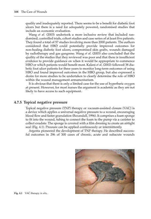
quality and inadequately reported. There seems to be a benefit for diabetic footulcers but there is a need for adequately powered, randomised studies thatinclude an economic evaluation.
Wang et al. (2003) undertook a more inclusive review that included ran-domised, controlled trials, cohort studies and case series of at least five patients.They found a total of 57 studies involving more than 2000 patients. The authorsconsidered that HBO could potentially provide improved outcomes for non-healing diabetic foot ulcers, compromised skin grafts, wounds damagedby radiotherapy and gas gangrene. Wang et al. (2003) also concluded that thequality of the studies that they reviewed was poor and that there is insufficientevidence to provide guidance on when it would be appropriate to commenceHBO or which patients would benefit most. Kalani et al. (2002) followed 38 dia-betic foot ulcer patients for three years to monitor long-term outcomes of usingHBO and found improved outcomes in the HBO group, but also expressed adesire for more studies to be undertaken to clearly determine the role of HBOwithin the wound management armamentarium.
It is obvious that there is only a limited case for the use of hyperbaric oxygenat present. However, for most nurses the argument is academic as they are notlikely to have access to such equipment.
4.7.5 Topical negative pressure
Topical negative pressure (TNP) therapy or vacuum-assisted closure (VAC) isa device which applies a universal negative pressure to a wound, encouragingblood flow and faster granulation (Baxandall, 1996). It comprises a foam spongeto fit into the wound, tubing to connect the foam to the pump via a canister tocollect exudate. The sponge is covered with a film dressing to create an airtightseal (Fig. 4.1). Pressure can be applied continuously or intermittently.
Argenta pioneered the development of TNP therapy. He described success-ful outcomes in 296 of 300 cases of chronic, acute and subacute wounds
108 The Care of Wounds
Fig. 4.1 VAC therapy in situ.
(Argenta & Morykwas, 1997). Subsequently, there have been numerous reportsof its use, many of which have been case series or case studies. Some of the benefits of TNP can be described as follows.
� Reduction of tissue oedema. Although this is particularly difficult tomeasure, Banwell (1999) suggests that this action could be very useful in themanagement of crush injuries or burns.
� Reduction of bacterial load. There are mixed reports of the impact of TNPon bacterial colonisation. Morykwas et al. (1997) found a significant reduc-tion in bacterial counts in the animal model. Banwell (1999) reported thesame outcome, whereas Moues et al. (2004) and Weed et al. (2004) found avariable outcome with reduction of some bacteria and an increase in others.However, both the latter studies still reported good healing rates. There arealso consistent reports of beneficial outcomes in infected wounds such as fordeep sternal wound infection following open heart surgery (Gustaffson et al.,2003; Luckraz et al., 2003).
� Management of chronic wound fluid. TNP can be a useful method of col-lecting and managing wound exudate. Banwell (1999) suggested that TNPmay be able to reduce the levels of MMPs within the wound.
� Removal of slough and promotion of granulation tissue. TNP has beendemonstrated to be effective in removal of slough (Gustaffson et al., 2003;Loree et al., 2004; Luckraz et al., 2003).
� Promotion of wound healing. Numerous cohort or uncontrolled studies haveconcluded that TNP is effective. However, a Cochrane review (Evans & Land,2004) was only able to identify two small randomised, controlled trials ofchronic wounds with a total of 34 patients up until November 2002. Thesestudies showed weak evidence of efficacy but the numbers are too small forany firm conclusions to be drawn.
� Improved take in skin grafts. Blackburn et al. (1998) reported using VACtherapy prior to skin grafting of large complex open wounds and found thatthey were able to get a minimum of a 95% take.
� Cost effectiveness of TNP. The unit costs of using TNP are high but a reviewby Neubauer and Ujlaky (2003) investigated the treatment costs using dataderived from three studies and concluded that TNP has the potential to makecost savings of up to $9000 per patient through a faster healing time.However, they also emphasise the need for further prospective studies inorder for firm conclusions to be obtained.
TNP therapy is gradually being used more widely, especially as a portableversion is now available. However, it can potentially be harmful if used inap-propriately and skill is needed in its application. Birchall et al. (2002) describehow one acute trust developed a centralised system for the use of TNP, alongwith guidelines to ensure appropriate use and a competency-based educationprogramme for nurses. The whole system was both clinically- and cost-effective as well as being more efficient.
Wound Management Products 109
4.7.6 Tissue culture
Tissue culture describes the process whereby a small full-thickness section of skin is harvested from a patient or donor and then cultured in the laboratory to form large sheets of cells (cultured keratinocytes). The sheets of cells are then grafted onto a granulating wound completely free of anynecrotic material. Autologous tissue (that taken from the patient) has beenfound to be more effective than allogenic tissue (that taken from a donor).However, it can take more than a month to prepare (Kakibuchi et al., 1996).Tissue culture has become well established since the early 1980s when the first clinical reports of its use appeared. Kumagai et al. (1997) followed up 38 patients who had received autologous cultured keratinocyte grafts for overtwo years. They concluded that cultured tissue has site specificity even after grafting and therefore, where possible, the maxim “closer is best”, which is applied to conventional skin grafting, should also be used for tissuecultures.
Wright et al. (1998b) describe the use of a polyurethane foam dressing as acarrier for cultured keratinocytes. They found it to be a useful vehicle but therewas slow proliferation of cells. They suggest that cells should be expandedusing the standard methods and then seeded onto the dressing. Myers et al.(1997) tested a hyaluronic membrane delivery system for cultured keratinocyteson an animal model and found that it gave a superior keratinocyte take. Theresearchers suggest that this system requires further investigation and clinicalevaluation. Caravaggi et al. (2003) used a similar product in the treatment ofdiabetic foot ulcers, comparing it with paraffin gauze (standard treatment inItaly). The results from 79 patients showed greater numbers of healed ulcers inthe experimental group but this difference failed to reach statistical significance,possibly because the study was underpowered. The ulcers were subdividedinto those on the plantar surface or dorsum. There were a significantly highernumber of healed ulcers in the experimental group on the dorsum of the footbut the precise significance of this is uncertain.
Generally, cultured keratinocytes have been used by plastic surgeons particu-larly for treating burn patients. However, they have also been used for treatingleg ulcers that have not responded to other forms of treatment. In reviewingthis, Kakibuchi et al. (1996) consider that it has not been an unqualified successas grafts have only shown a 10–20% take. Tissue culture is an important therapyfor burn patients and it may have potential for a wider use, but needs somefurther development.
4.7.7 Tissue engineering
Tissue engineering takes tissue culture a step forward. It uses human dermalfibroblasts and cultures them on a biosynthetic scaffold. The fibroblasts prolif-erate and secrete proteins and growth factors, resulting in the generation of athree-dimensional human dermis that can then be used to graft over woundsites. Two brands are currently available, ApligrafTM (also known as Graftskin)
110 The Care of Wounds
and DermagraftTM, and they can be described as cultured human skin equiva-lent (HSE).
Falanga et al. (1998) studied the effect of using a HSE (ApligrafTM) on patientswith venous ulcers in a multicentre trial. A total of 240 patients were ran-domised to HSE and compression or compression therapy alone. They founda significantly faster healing rate in the HSE group. In those patients (n = 120)whose ulcer had been open for more than a year, HSE was significantly moreeffective in achieving healing. Falanga and Sabolinski (1999) investigated theresults from this last group of patients in more detail. They found that 47% ofthose treated with HSE were healed by six months compared with 19% of con-trols. They suggest that HSE is particularly useful in the treatment of ulcers oflong duration. Omar et al. (2004) undertook a pilot study using DermagraftTM
for venous ulcers. Eighteen patients were randomised to either the HSE andcompression or compression alone and followed up for 12 weeks. Five patientsin the HSE group and one of the controls were healed at 12 weeks, but this didnot achieve significance. However, there was a significant reduction in ulcersize and in the linear rate of healing in the HSE group.
Brem et al. (2001) discussed the success of two centres in the use of HSE(ApligrafTM) for 33 patients with 54 venous ulcers that had been present formore than a year. They considered that their results were better than thosereported in an earlier trial (Falanga et al., 1998; Falanga & Sabolinski, 1999) asthey were able to achieve healing rates of 70% in six months. The authorssuggest that this was due to improved wound bed preparation, particularly inthe use of antiseptics to reduce wound exudate prior to commencement of HSEtherapy. They also meshed the HSE prior to application to allow excessiveexudate to escape. Schonfeld et al. (2001) undertook an economic analysis of theuse of HSE (ApligrafTM) in hard-to-heal venous ulcers using a semi-Markovmodel. They calculated that as ulcers would heal faster using HSE comparedwith compression alone, the overall treatment costs were lower ($20141 compared with $27493 over a 12-month period).
HSE has also been used to treat diabetic foot ulcers. Gentkow et al. (1996)carried out a randomised, controlled, single-blinded trial of 50 patients withdiabetic foot ulcers. Patients were allocated to four groups: three differentdosages of HSE (DermagraftTM) or a control group. They found that all three ofthe treatment regimens were significantly better than the control. Furthermore,after 14 months’ follow-up there were no recurrences in the HSE patients. Theauthors considered this fact to be significant, quoting recurrence rates fromother studies of 19.6–46% in times less than 12 months. Hanft and Surprenant(2002) compared HSE (DermagraftTM) with moistened gauze in 28 patients withdiabetic foot ulcers. They found that there was a significant difference in thehealing rate in the HSE group by week 12 and in the overall rate of healing. A larger multicentre randomised study of 245 diabetic foot ulcers comparedDermagraftTM with conventional therapy (Marston et al., 2003). A significantlyhigher number of patients in the HSE group were healed by 12 weeks. Thisgroup also experienced significantly fewer ulcer-related adverse events. Veveset al. (2001) reported a multicentre study of 208 neuropathic diabetic foot ulcers
Wound Management Products 111
comparing HSE (ApligrafTM) with moistened gauze. All patients had surgicaldebridement prior to treatment and adequate foot off-loading. There were sig-nificantly more ulcers healed in the HSE group at 12 weeks and the mediantime to healing was 65 days compared with 90 days for the controls (p = 0.0026).
Newton et al. (2002) studied the microvascular blood flow in seven full-thickness diabetic foot ulcers before and after application of DermagraftTM.They found that by eight weeks, there was a 72% increase of blood flow in fiveof the seven ulcers and that they were healed by 12 weeks. The other two ulcersreduced in size by 25%.
Allenet et al. (2000) used a Markov model to undertake an economic evalua-tion of the costs of using HSE (DermagraftTM) compared with standard treat-ment for diabetic foot ulcers over a 52-week period. The overall treatment costswere lower for the HSE because of a faster healing rate (53522FF compared with56687FF). Redekop et al. (2003) undertook a similar exercise comparingApligrafTM with general wound care for diabetic foot ulcers. They found thatthere was a 12% reduction in costs using HSE.
It would seem that the use of HSE has considerable potential in the treatmentof wounds that have failed to heal with other methods. Although the economicevaluations described above indicate that cost savings can be made using HSE,it should be remembered that this is only the case in those wounds that are nothealing. Other methods should be used first, before considering HSE.
4.8 ALTERNATIVE THERAPIES AND WOUND MANAGEMENT
Alternative or complementary therapies have been defined as those therapiesthat usually lie outside the official health sector (WHO, 1983). Trevelyn andBooth (1994) divided them into three categories in relation to their potentiallinks to nursing (see Box 4.1). Although there is much written in the literature
112 The Care of Wounds
Box 4.1 Categories of alternative therapies related to nursing.
Category 1: can be incorporated into nursing care
• Massage• Reflexology• Aromatherapy• Therapeutic touch
Category 2: can be used to some extent by nurses with relevant training
• Homeopathy• Herbal medicine
Category 3: not usually practised as part of nursing care
• Acupuncture• Osteopathy• Chiropractic
regarding these therapies, there is a paucity of research evidence to demonstratetheir place in patient care. Gates (1994) and Vickers (1997) have discussed thisproblem and suggested the need for a critical appraisal of the literature on thesubject.
One such review has been undertaken by Finch (1997), who appraised thetherapy of therapeutic touch, healing by laying on of hands, in relation towound healing. Its use in nursing was introduced in the USA in the 1970s. It isbased on the principle that human beings are energy fields and illness is theresult of an imbalance in the energy field. Therapeutic touch focuses on redi-recting the energy to restore balance. Finch reviewed five studies of therapeu-tic touch and wound healing carried out on healthy volunteers by the sameresearcher. She concluded that therapeutic touch was unreliable and generallyineffective when used to treat wounds. A Cochrane review by O’Mathuna andAshford (2004) considered the use of therapeutic touch in healing acutewounds. They concluded that there was insufficient evidence to show whetherit had a role in healing acute wounds. However, it has been shown to be beneficial in treating anxiety (see Chapter 2).
Aromatherapy involves the use of essential oils that are applied to the skin.A variety of oils can be used, depending on the effect required. Unfortunately,there are a number of inconsistencies found in the literature with a variety ofcontradictory properties being given to the same oil (Vickers, 1997). A study byKite et al. (1998) suggests that aromatherapy is effective in reducing anxiety andstress. They assessed 58 cancer patients using a HAD score before and after sixsessions of aromatherapy and found a significant reduction in anxiety anddepression at the end of the course of treatment. Asquith (1999) undertook areview of the literature and reported a number of case studies that found laven-der and tea tree oils beneficial in healing a variety of wounds. Asquith also iden-tified studies that improved other aspects of patient care such as sleep patterns.She concluded that there was little evidence to show a direct benefit to woundhealing.
There is no doubt that some patients find alternative therapies helpful.Whether these types of therapies have a role in wound healing remains ques-tionable. There is a real need for the development of high-quality researchmethodologies to explore the multifaceted nature of these treatments.
REFERENCES
Ahmed, A.K., Hoekstra, M.J., Hage, J.J., Karim, R.B. (2003) Honey-medicated dressing:transformation of an ancient remedy into a modern therapy. Annals of PlasticSurgery, 50 (2), 143–137.
Allenet, B., Paree, F., Lebrun, T. et al. (2000) Cost-effectiveness modelling of Dermagraftfor the treatment of diabetic foot ulcers in the French context. Diabetes and Metab-olism, 26 (2), 125–132.
Anderson, I. (2003) Should potassium permanganate be used in wounds? Nursing Times,99 (31) (suppl), 19.
Wound Management Products 113
Argenta, L.C., Morykwas, M.J. (1997) Vacuum-assisted closure: a new method for woundcontrol and treatment: clinical experience. Annals of Plastic Surgery, 38 (6), 563–576.
Asquith, S. (1999) The use of aromatherapy in wound care. Journal of Wound Care, 8 (6),318–320.
Banwell, P. (1999) Topical negative pressure therapy in wound care. Journal of WoundCare, 8 (2), 79–84.
Baxandall, T. (1996) Healing cavity wounds with negative pressure. Nursing Standard,11 (6), 49–51.
Becker, G.D. (1986) Identification and management of the patient at high risk of woundinfection. Head and Neck Surgery, 8, 205–210.
Bennett, L.L., Rosenblum, R.S., Perlov, C., Davidson, J.M., Barton, R.M., Nannet, L.B.(2001) An in vivo comparison of topical agents in wound repair. Plastic and Recon-structive Surgery, 108 (3), 675–685.
Birchall, L., Street, L., Clift, H. (2002) Developing a trust-wide centralised approach tothe use of TNP. Journal of Wound Care, 11 (8), 311–314.
Bishop, W.J. (1959) A History of Surgical Dressings. Robinson and Sons Ltd, Chesterfield.Blackburn, J.H., Boemi, L., Hall, W.W. et al. (1998) Negative-pressure dressings as a
bolster for skin grafts. Annals of Plastic Surgery, 40 (5), 453–457.Bloomfield, S.F., Sizer, T.J. (1985) Eusol BPC and other hypochlorite formulations used
in hospitals. Pharmaceutical Journal, 253, 153–157.Bond, C.J. (1915) The application of strong antiseptics to wounds. British Medical Journal,
March 6, 405–406.Borgognoni, T. (1955) The Surgery of Theodoric vol 1. Translated from the Latin by
Campbell, E., Colton, J. Appleton-Century-Crofts, New York.Boyce, D.E., Miller, L., Moore, K., Harding, K.G. (1997) An open comparative ran-
domised parallel-group clinical trial to evaluate the performance of HYAFFTM
wound dressing in the management of pilonidal sinus excision wounds: aninterim analysis, in (eds) Leaper, D.J., Cherry, G.W., Dealey, C., Lawrence, J.C.,Turner, T.D., Proceedings of the 6th European Conference on Advances in Wound Management. Macmillan Magazines Ltd, London.
Brem, H., Balledux, J., Sukkarieh, T., Carson, P., Falanga, V. (2001) Healing of venous legulcers of long duration with a bilayered living skin substitute: results from ageneral surgery and dermatology department. Dermatologic Surgery, 27 (11),915–919.
Brem, H., Sheehan, P., Boulton, A.J.M. (2004) Protocol for treatment of diabetic footulcers. American Journal of Surgery, 187 (5) (suppl), 1S–10S.
Brennan, S.S., Leaper, D.J. (1985) The effect of antiseptics on the healing wound: a studyusing the rabbit ear chamber. British Journal of Surgery, 72 (10), 780–782.
Brennan, S.S., Foster, M.E., Leaper, D.J. (1986) Antiseptic toxicity in wounds healing bysecond intention. Journal of Hospital Infection, 8 (3), 263–267.
British Medical Association Board of Science and Education (1993) Clinical HyperbaricMedicine Facilities in the UK. British Medical Association, London.
Caravaggi, C., Clerici, G., De Giglio, R. et al. (2003) HYAFF 11-based autologous dermaland epidermal grafts in the treatment of noninfected diabetic plantar and dorsalfoot ulcers. Diabetes Care, 26 (10), 2853–2859.
Carneiro, P.M., Nyawawa, E.T. (2003) Topical phenytoin versus EUSOL in the treatmentof non-malignant leg ulcers. East African Medical Journal, 80 (3), 124–129.
Colletta, V., Dioguardi, D., Di Lonardo, A., Maggio, G., Torasso, F. (2003) A trial to assessthe efficacy and tolerability of Hyalofill-F in non-healing venous leg ulcers. Journalof Wound Care, 12 (9), 357–361.
114 The Care of Wounds
Cookson, B. (1998) The emergence of mupirocin resistance: a challenge to infectioncontrol and antibiotic prescribing practice. Journal of Antimicrobial Chemotherapy.41 (1), 11–18.
Daltrey, D.C., Cunliffe, W.J. (1981) A double-blind study of the effects of Benzol perox-ide 20% and Eusol and liquid paraffin on the microbial flora of leg ulcers. ActaDermato-Venereologica, 61, 575–577.
D’Arcy, P.F. (1972) Drugs on the skin: a clinical and pharmaceutical problem. Pharma-ceutical Journal, 209, 491–492.
Dealey, C. (2002) Wound healing in Moorish Spain. EWMA Journal, 2 (1), 32–34.Dealey, C. (2003) Wound healing in medieval and renaissance Italy: was it art or science?
EWMA Journal, 3 (1), 15–17.Dealey, C. (2004) The contribution of French surgeons to wound healing in medieval and
renaissance Europe. EWMA Journal, 4 (1), 33–35.Deng, S., Sang, J., Cao, L. (1995) The effects of mupirocin on burn wounds with staphy-
lococcus aureus infection. Chung Hua Cheng Hsing Shao Shang Wai Ko Tsa Chih, 11(1), 45–48.
Dobrzanski, S., Duncan, S.E., Harkiss, A., Ball, A., Robertson, D. (1983) Topical applications in pressure sore therapy. British Journal of Pharmaceutical Practice, 5 (5),10.
Duin, N., Sutcliffe, J. (1992) A History of Medicine. Simon and Schuster, London.Dunford, C.E., Hanano, R. (2004) Acceptability to patients of a honey dressing for non-
healing venous ulcers. Journal of Wound Care, 13 (5), 193–197.Evans, D., Land, L. (2001) Topical negative pressure for treating chronic wounds
(Cochrane review). Cochrane Library, Issue 1. Update Software, Oxford.Falanga, V. (2000) Classification for wound bed preparation and stimulation of chronic
wounds. Wound Repair and Regeneration, 8 (5), 347–352.Falanga, V., Sabolinski, M. (1999) A bilayered living skin construct (Apligraf) accelerates
complete closure of hard-to-heal venous ulcers. Wound Repair and Regeneration, 7(4), 201–207.
Falanga, V., Marolis, D., Alvarez, O. et al. (1998) Rapid healing of venous ulcers and lackof clinical rejection with an allogeneic cultured human skin equivalent. Archives ofDermatology, 134, 293–300.
Fernandez, R., Griffiths, R., Ussia, C. (2002) Water for wound cleansing. Cochrane Database Systematic Review. 4: CD003861.
Finch, A. (1997) Therapeutic touch and wound healing. Journal of Wound Care, 6 (10),501–504.
Flanagan, M. (2000) Honey and the management of infected wounds. Journal of WoundCare, 9 (6), 287.
Forrest, R.D. (1982) Early history of wound treatment. Journal of the Royal Society of Medicine, 75, 198–205.
Foster, L., Moore, P. (1997) The application of a cellulose-based fibre dressing in surgi-cal wounds. Journal of Wound Care, 6 (10), 469–473.
Galeano, M., Deodato, B., Altavilla, D. et al. (2003) Effect of recombinant adeno-associated virus vector-mediated vascular endothelial growth factor gene transferon wound healing after burn injury. Critical Care Medicine, 31 (4), 1017–1025.
Gates, B. (1994) The use of complementary and alternative therapies in health care: aselective review of the literature and dicussion of the implications for nurse prac-titioners and health-care managers. Journal of Clinical Nursing, 3 (1), 43–47.
Gentkow, G.D., Iswaki, S.D., Hershon, K.S. et al. (1996) Use of Dermagraft, a culturedhuman dermis, to treat diabetic foot ulcers. Diabetes Care, 19 (4), 350–354.
Wound Management Products 115
Gerster, A.G. (1910) Surgical manners and customs in the times of Henry de Mondev-ille. Proceedings of the Charaka Club, 3, 70–90.
Ghatnekar, O., Willis, M., Persson, U. (2002) Cost-effectiveness of treating deep diabeticfoot ulcers with Promogran in four European countries. Journal of Wound Care, 11(2), 70–74.
Guarini, S. (2003) New gene therapy for the treatment of burn wounds. Critical Care Medicine, 31 (4), 1280–1281.
Gustafsson, R.I., Sjogren, J., Ingemansson, R. (2003) Deep sternal wound infection: asternal-sparing technique with vacuum-assisted closure therapy. Annals of ThoracicSurgery, 76 (6), 2048–2053.
Guthrie, D. (1945) A History of Medicine. Thomas Nelson, London.Hanft, J.R., Surprenant, M.S. (2002) Healing of chronic foot ulcers in diabetic patients
treated with human fibroblast-derived dermis. Journal of Foot and Ankle Injury, 41(5), 291–299.
Heister, L. (1768) A General System of Surgery in Three Parts, 6th edn. Translated intoEnglish by Heister L. Whiston, London.
Helling, T.S., Daon, E. (1998) In Flanders fields: the Great War, Antoine Deparge and theresurgence of debridement. Annals of Surgery, 228 (2), 173–181.
Herzog, W. (1915) German experiences. The dangers of tincture of iodine as a first-aiddressing. British Medical Journal, March 6, 441–442.
Hollander, D., Schmandra, T., Windolf, J. (2000) Using an esterified hyaluronan fleece topromote healing in difficult-to-treat wounds. Journal of Wound Care, 9 (10), 463–466.
Humzah, M.D., Marshall, J., Breach, N.M. (1996) Eusol: the plastic surgeon’s choice?Journal of the Royal College of Surgeons of Edinburgh, 41, 269–270.
Johnson, A. (1987) Wound care, packing cavity wounds. Nursing Times, 83 (36), 59–62.Johnson, P.C. (1989) Guy de Chauliac and the Grand Surgery. Surgery, Gynaecology and
Obstetrics, 169, 172–176.Kakibuchi, M., Hosokawa, K., Fujikawa, M., Yoshikawa, K. (1996) The use of cultured
epidermal cell sheets in skin grafting. Journal of Wound Care, 5 (10), 487–490.Kalani, M., Jorneskog, G., Naderi, N., Lind, F., Brismar, K. (2002) Hyperbaric oxygen
(HBO) therapy in treatment of diabetic foot ulcers: long term follow-up. Journal ofDiabetes and its Complications, 16, 153–158.
Kanal, H. (1975) Encyclopaedia of Islamic Medicine. General Egyptian Book Organisation,Cairo.
Karim, R.B., Ahmed, A.K.J., Brito, B.L.R. (2002) Balancing MMPs in chronic wounds: a pilotstudy with Dermax. Paper presented at the Dutch Association for Plastic SurgeryMeeting, Aalst, Belgium, November 23.
Kearney, J.N., Arain, T., Holland, K.T. (1988) Antimicrobial properties of antisepticimpregnated dressings. Journal of Hospital Infection, 11 (1), 68–76.
Kingsley, A. (2001) The use of honey in the treatment of infected wounds: case studies.British Journal of Nursing, 10 (22) (suppl), S13–S20.
Kirkpatrick, J.J.R., Naylor, I.L. (1997) Ulcer management in medieval England. Journal ofWound Care, 6 (7), 350–352.
Kite, S.M., Maher, E.J., Anderson, K. et al. (1998) Development of an aromatherapyservice at a cancer centre. Palliative Medicine, 12 (3), 171–180.
Knighton, D.R., Ciresi, K., Fiegal, V.D., Schumerth, S., Butler, E., Cerrs, F. (1990) Stimu-lation of repair in chronic non-healing cutaneous ulcers using platelet-derivedwound healing formula. Surgery, Gynaecology and Obstetrics, 170, 56–60.
Kranke, P., Bennett, M., Roeckl-Wiedmann, I., Debus, S. (2004) Hyperbaric oxygentherapy for chronic wounds. Cochrane Database for Systematic Reviews 2: CD004123.
116 The Care of Wounds
Kumagai, N., Oshima, H., Tanabe, M., Ishida, H., Uchikoshi, T. (1997) Favourable donorsite for epidermal cultivation for the treatment of burn scars with autologous cultured epithelium. Annals of Plastic Surgery, 38 (5), 506–513.
Ladin, D. (2000) Becaplermin gel (PDGF-BB) as topical wound therapy. Plastic SurgeryEducational Foundation DATA Committee. Plastic and Reconstructive Surgery, 105(3), 1230–1231.
Lansdown, A.B.G. (2004) A review of the use of silver in wound care: facts and fallac-ies. British Journal of Nursing, 13 (6) (suppl), S6–S19.
Lawrence, J.C. (1987) A century after Gamgee. Burns, 13 (1), 77–79.Levine, J.M. (1992) Historical Notes on Pressure Ulcers: The Cure of Ambrose Paré.
Decubites, 5(2), 23–26.Lineaweaver, W., Howard, R., Soucy, D. et al. (1985) Topical antimicrobial toxicity.
Archives of Surgery, 120, 267–270.Linker, R.W., Womack, N. (1969) Ten Books of Surgery by Ambroise Paré. University of
Georgia Press, Athens, GA.Loree, S., Dompmartin, A., Penven, K., Harel, D., Leroy, D. (2004) Is vacuum assisted
closure a valid technique for debriding leg ulcers? Journal of Wound Care, 13 (6),249–252.
Luckraz, H., Murphy, F., Bryant, S., Charman, S.C., Ritchie, A.J. (2003) Vacuum-assistedclosure as a treatment modality for infections after cardiac surgery. Journal of Thoracic and Cardiovascular Surgery, 125 (2), 301–305.
Marston, W.A., Hanft, J., Norwood, P., Pollack, R. (2003) The efficacy and safety of Dermagraft in improving the healing of diabetic foot ulcers. Diabetes Care, 26 (6),1701–1705.
Mayo-Robson, A.W. (1915) Hints on war surgery. British Journal of Dermatology, July 24,136.
McLure, A.R., Gordon J. (1992) In-vitro evaluation of povidone-iodine and chlorhexi-dine against methicillin resistant Staphylococcus aureus. Journal of Hospital Infec-tion, 21, 291–299.
Meade, R.H. (1968) An Introduction to the History of General Surgery. W.B. SaundersPhiladelphia, PA.
Mengistu, Y., Erge, W., Bellete, B. (1999) In vitro susceptibility of gram-negative bacterial isolates to chlorhexidine gluconate. East African Medical Journal, 76 (5),243–246.
Merchant, J. (1988) Aseptic technique reconsidered. Care, Science and Practice, 6 (3), 74–77.Michel, D., Zach, G.A. (1997) Antiseptic efficacy of disinfecting solutions in suspension
test in vitro against methicillin-resistant Staphylococcus aureus, Pseudomonasaeruginosa and Escherichia coli in pressure sore wounds after spinal cord injury.Dermatology, 195 (suppl 2), 36–41.
Millward, J. (1989) Assessment of wound management in a Care of the Elderly unit. Care,Science and Practice, 7 (2), 47–49.
Misirlioglu, A., Eroglu, S., Karacaoglan, N., Akan, M., Akoz, T., Yildirim, S. (2003) Theuse of honey as an adjunct in the healing of split-thickness skin graft donor site.Dermatologic Surgery, 29 (2), 168–172.
Molan, P.C. (1999) The role of honey in the management of wounds. Journal of WoundCare, 8 (8), 415–418.
Moore, O.A., Smith, L.A., Campbell, F., Seers, K., McQuay, H.J., Moore, R.A. (2001) Sys-tematic review of the use of honey as a wound dressing. BMC Complementary andAlternative Medicine, 1 (1), 2.
Morgan, D. (1993) Is there still a role for antiseptics? Journal of Tissue Viability, 3 (3), 80–84.
Wound Management Products 117
Morgan, D. (1995) Myiasis: the rise and fall of maggot therapy. Journal of Tissue Viabil-ity, 5 (2), 43–51.
Morykwas, M.J., Argenta, L.C., Shelton-Brown, E.I., McGuirt, W. (1997) Vacuum-assistedclosure: a new method for wound control and treatment: animal studies and basicfoundation. Annals of Plastic Surgery, 38, 553–562.
Moues, C.M., Vos, M.C., van den Bemd, G.J., Stijen, T., Hovius, S.E. (2004) Bacterial loadin relation to vacuum-assisted closure: a prospective, randomised trial. WoundRepair and Regeneration, 12 (1), 11–17.
Murray, Y. (1988) An investigation into the care of wounds in a health authority. Care,Science and Practice, 6 (4), 97–102.
Myers, J.A. (1982) Modern plastic surgical dressings. Health and Social Services Journal,92, 336–337.
Myers, S.R., Grady, J., Soranzo, C. et al. (1997) A hyaluronic acid membrane deliverysystem for cultured keratinocytes: clinical “take” rates in the porcine kerato-dermal model. Journal of Burn Care Rehabilitation, 18, 214–222.
Neubauer, G., Ujlaky, R. (2003) The cost-effectiveness of topical negative pressure versusother wound-healing therapies. Journal of Wound Care, 12 (10), 392–393.
Newton, D.J., Khan, F., Belch, J.J., Mitchell, M.R., Leese, G.P. (2002) Blood flow changesin diabetic foot ulcers treated with dermal replacement therapy. Journal of Foot andAnkle Surgery, 41 (4), 233–237.
Omar, A.A., Mavor, A.I.D., Jones, A.M., Homer-Vanniasinkam, S. (2004) Treatment ofvenous leg ulcers with DermagraftTM. European Journal of Endovascular Surgery, 27,666–672.
O’Mathuna, D.P., Ashford, R.L. (2004) Therapeutic touch for healing acute wounds(Cochrane Review). Cochrane Library, Issue 2. John Wiley, Chichester.
O’Toole, E.A., Goel, M., Woodley, D.T. (1996) Hydrogen peroxide inhibits human keratinocyte migration. Dermatology Surgery, 22 (6), 525–529.
Paterson, L.M. (1988) Military surgery: knights, sergeants and Ramon of Avignon’sversion of Chirugia of Roger of Salerno, in (eds) Harper-Bus, C., Harvey, R., TheIdeals and Practice of Medieval Knighthood. The Bodell Press, Woodbridge, Suffork.
Popp, A.J. (1995) Crossroads at Salerno: Eldridge Campbell and the writings ofTeodorico Borgognoni on wound healing. Journal of Neurosurgery, 83, 174–179.
Redekop, W.K., McDonnell, J., Verboom, P., Lovas, K., Kalo, Z. (2003) The cost effec-tiveness of Apligraf in the treatment of diabetic foot ulcers. Pharmacoeconomics, 21(16), 1171–1183.
Rees, J.E. (2003) Where have all the bubbles gone? An ode to hydrogen peroxide, thechampagne of all wound cleaners. Accident and Emergency Nursing, 11, 82–84.
Reynolds, J.E.F. (ed.) (1982) Martindale: The Extra Pharmacopoeia, 28th edn. Pharmaceuti-cal Press, London.
Robson, M.C., Mustoe, T.A., Hunt, T.K. (1998) The future of recombinant growth factorsin wound healing. American Journal of Surgery, 176 (suppl 2A), 80S–82S.
Rode, H., Hanslo, D., de Wet, P.M., Millar, A.J., Cywes, S. (1989) Efficacy of mupirocinin methicillin-resistant staphylococcus aureus burn wound infection. AntimicrobialAgents and Chemotherapy, 33 (8), 1358–1361.
Russell, A.D., Hugo, W.B., Ayliffe, G.A.J. (1982) Principles and Practice of Disinfection,Preservation and Sterilisation. Blackwell Scientific Publications, London.
Schonfeld, W.H., Villa, K.F., Fastenau, J.M., Mazonson, P.D., Falanga, V. (2001) An eco-nomic assessment of Apligraf (Graftskin) for the treatment of hard-to-heal venousleg ulcers. Wound Repair and Regeneration, 8 (4), 251–257.
118 The Care of Wounds
Schulz, G.S., Sibbald, R.G., Falanga, V. et al. (2003) Wound bed preparation: a systematicapproach to wound management. Wound Repair and Regeneration, 11 (2) (suppl),S1–S28.
Sherman, R.A. (1997) A new dressing design for use with maggot therapy. Plastic andReconstructive Surgery, 100 (2), 451–456.
Selvaggi, G., Monstrey, S., Van Landuyt, K., Hamdi, M., Blondeel, P. (2003) The role ofiodine in antisepsis and wound management: a reappraisal. Acta Chirurgica Belgica,103 (3), 241–247.
Sinclair, R.D., Ryan, T.J. (1993) A great war for antiseptics. Wound Management, 4 (1),16–18.
Sleigh, J.W., Linter, S.P.K. (1985) Hazards of hydrogen peroxide. British Medical Journal,291, 1706.
Spencer, M.J., Herrick, S.E., Shah, M. et al. (1996) Growth factor therapy: redressing thebalance, in (eds) Cherry, G.W., Gottrup, F., Lawrence, J.C., Moffatt, C.J., Turner,T.D., Proceedings of the 5th European Conference on Advances in Wound Management.Macmillan Magazines Ltd, London.
Subrahmanyam, M. (1998) A prospective randomised clinical and histological study ofsuperficial burn wound healing with honey and silver sulphadiazine. Burns, 24(2), 157–161.
Taddeucci, P., Pianigiani, E., Colletta, V., Torasso, F., Andreassi, L., Andreassi, A. (2004)An evaluation of Hyalofill-F plus compression bandaging in the treatment ofchronic venous ulcers. Journal of Wound Care, 13 (5), 202–204.
Tatnall, F.M., Leigh, I.M., Gibson, J.R. (1990) Comparative study of antiseptic toxicity on basal keratinocytes, transformed human keratinocytes and fibroblasts. SkinPharmacology, 3 (3), 157–163.
Thomas, S. (1990) Eusol revisited. Dressing Times, 3 (1), 3–4.Thomas, S., Jones, M., Shutler, S., Jones, S. (1996) Using larvae in modern wound
management. Journal of Wound Care, 5 (2), 60–69.Thomlinson, D. (1987) To clean or not to clean. Nursing Times, 83 (9), 71–75.Tredget, E.E., Shankowsky, H.A., Groeneveld, A., Burrell, R. (1998) A matched-pair, ran-
domised study evaluating the efficacy and safety of ActicoatTM silver coated dress-ing for the treatment of burn wounds. Journal of Burn Care and Rehabilitation, 19(6), 531–537.
Trevelyn, J. (1997) Spirit of the beehive. Nursing Times, 93 (7), 72–74.Trevelyn, J., Booth, B. (1994) Complementary Medicine for Nurses, Midwives and Health
Visitors, Macmillan Press, London.Trilla, A., Miro, J.M. (1995) Identifying high risk patients for staphylococcus aureus
infections: skin and soft tissue infections. Journal of Chemotherapy, 7 (suppl 3), 37–43.
Turner, T.D. (1986) Recent advances in wound management products, in (eds) Turner,T.D., Schmidt, R.J., Harding, K.G., Advances in Wound Management. John Wiley,Chichester.
Vandeputte, J., Van Waeyenberge (2003) Clinical evaluation of L-MesitranTM – a honey-based ointment. EWMA Journal, 3 (2), 8–11.
Vazquez, J.R., Short, B., Findlow, A.H., Nixon, B.P., Boulton, A.J.M., Armstrong, D.G.(2003) Outcomes of hyaluronan therapy in diabetic foot wounds. Diabetes Researchand Clinical Practice, 59, 123–127.
Veves, A., Falanga, V., Armstrong, D.G., Sabolinski, M.L. (2001) Graftskin, a human skin equivalent, is effective in the management of noninfected neuropathic
Wound Management Products 119
diabetic foot ulcers: a prospective multicentre clinical trial. Diabetes Care, 24 (2),290–295.
Veves, A., Sheehan, P., Pham, H.T. (2002) A randomised, controlled trial of Promogran(a collagen/oxidised regenerated cellulose dressing) vs standard treatment in themanagement of diabetic foot ulcers. Archives of Surgery, 137 (7), 822–827.
Vickers, A. (1997) Yes, but how do we know it’s true? Knowledge claims in massage andaromatherapy. Complementary Therapy in Nursing and Midwifery, 3 (3), 63–65.
Vin, F., Teot, L., Meaume, S. (2002) The healing properties of Promogran in venous legulcers. Journal of Wound Care, 11 (9), 335–341.
Walsh, M., Ford, P. (1989) Nursing Rituals, Research and Rational Actions. HeinnemanNursing, Oxford.
Wang, C., Schwaitzberg, S., Berliner, E., Zarin, D.A., Lau, J. (2003) Hyperbaric oxygenfor treating wounds: a systematic review of the literature. Archives of Surgery, 138(3), 272–279.
Weed, T., Ratcliff, C., Drake, D.B. (2004) Quantifying bacterial bioburden during nega-tive pressure therapy: does the wound VAC enhance bacterial clearance? Annalsof Plastic Surgery, 52 (3), 276–280.
World Health Organisation (WHO) (1983) Traditional Medicine and Health-Care Coverage.World health Organisation, Geneva.
Wright, J.B., Lam, K., Burrell, R.E. (1998a) Wound management in an era of increasingbacterial antibiotic resistance: a role for topical silver treatment. American Journalof Infection Control, 26 (6), 572–577.
Wright, K.A., Nadire, K.B., Busto, P., Tubo, R., McPherson, J.M., Wentworth, B.M. (1998b)Alternative delivery of keratinocytes using polyurethane membrane and theimplications for its use in the treatment of full-thickness burn injury. Burns, 24 (1),7–17.
Zhou, L.H., Nahm, W.K., Badiavas, E., Yufit, T., Falanga, V. (2002) Slow-release iodinepreparation and wound healing: in vitro effects consistent with lack of in vitro toxicity in human chronic wounds. British Journal of Dermatology, 146, 365–374.
Zimmerman, L.M., Veith, I. (1961) Great Ideas in the History of Surgery. Williams andWilkins, Baltimore, MD.
120 The Care of Wounds
The Management of Patients with Chronic Wounds 121
Chapter 5The Management of Patients with Chronic Wounds
5.1 INTRODUCTION
Fowler (1990) defined a chronic wound as one where there is tissue deficit asthe result of long-standing injury or insult or frequent recurrence. Despitemedical or nursing care, they do not heal easily. They are more likely to occurin the elderly or those with multisystem problems. This section will considerthe care of patients with pressure ulcers, leg ulcers, diabetic foot ulcers and fun-gating wounds.
Chronic wounds cause much discomfort and pain for the many people whohave to suffer them. A multidisciplinary approach is needed for their manage-ment and prevention. Nurses can play an important role in the team as theyusually have the most contact with the patient. An essential part of this role iscommunication and co-operation across the disciplines. Healing is not possiblefor all chronic wounds and in those cases, the goal is to assist the patient toachieve the maximum independence and function possible.
It cannot be denied that the treatment of chronic wounds is costly. Harding(1998) suggests that chronic wounds cost the National Health Service (NHS)about £1bn a year. The Department of Health calculates the costs for inpatienttreatments by category. Although there is no specific category for chronicwounds, they fit within the category of ‘skin ulcers’. The costs for skin ulcersfor the year 2000 were nearly £4.5 million and the average length of stay was21 days. In comparison, inpatient treatment for diabetes was just under £2.5million. These figures are very limited as they only include costs for inpatientswith skin ulcer as a primary diagnosis and they do not include communitycosts, which can be considerable as many of these patients are primarily caredfor in the community.
5.2 THE PREVENTION AND MANAGEMENT OF PRESSURE ULCERS
Pressure ulcers are also called pressure sores, bed sores and decubitus ulcers.A pressure ulcer can be described as localised damage to the skin caused bydisruption of the blood supply to the area, usually as a result of pressure, shearor friction or a combination of any of these. There has not been a national surveyof pressure ulcer prevalence in the UK but a series of surveys by O’Dea (1999)of hospital patients in 35 acute care hospitals, carried out between 1992 and
1998, provides some information. O’Dea found a gradual decline in pressureulcer prevalence rates from 18.6% in 1992 to 10% in 1997–1998. The fifth nationalsurvey in the USA was undertaken in 356 acute care hospitals in 1999 (Amlunget al., 2001). The researchers found a pressure ulcer prevalence of 14.8%, anincrease on previous surveys. They also found a nosocomial rate of 7.1%.Kaltenthaler et al. (2001) compared prevalence and incidence data of surveysundertaken in the UK, USA and Canada and found large variations in methods,patient groups and prevalence, making comparisons difficult. They noted oneunexpected finding – none of the studies undertaken during the period1976–1997 had a prevalence rate below 5%. They suggested that there may bea point beyond which it is difficult to reduce prevalence rates further.
Prevalence surveys have been undertaken in a number of other countries, withvariable results. Bours et al. (2002) report on a national survey in the Netherlandsthat showed a prevalence rate of 23.1% whereas a national survey in Icelandfound a prevalence rate of 8.9% (Thoroddsen, 1999). Asurvey across 18 rural hos-pitals in Australia identified a rate of 6% (Pearson et al., 2000) and in Sweden asimilar survey identified 3.75% of patients with pressure ulcers (Lingren et al.,2000). It is difficult to draw conclusions from this wide range of prevalence rates,mostly because of the variation in methods. However, the fact that they havebeen carried out at all demonstrates the increasing interest in the topic.
For many years pressure ulcers were seen as a failure of care, in particular asthe result of bad nursing. Florence Nightingale (1861) considered that goodnursing could prevent them whereas a very influential French doctor calledJean-Martin Charcot (1825–1893) believed that doctors could do nothing aboutpressure ulcers. As a result, pressure ulcers became a very emotive issue andwere only discussed by doctors as ‘a nursing problem’ and by nurses with com-ments like ‘we do not have pressure ulcers here’.
This attitude is changing. A document published by the Department ofHealth stated that pressure ulcers should be considered a key indicator of thequality of care provided by a hospital (DoH, 1993). There is a much greaterawareness that all healthcare professionals need to be involved in pressure ulcerprevention (Culley, 1998). A number of multidisciplinary societies have beenformed, such as the Tissue Viability Society and the European Pressure UlcerAdvisory Panel, with the intent of expanding knowledge and supporting goodpractice and there are now NICE guidelines available. As all topics for guide-line development have to be agreed by the Secretary of State for Health, thisindicates the level of interest in the Department of Health.
5.2.1 The cost of pressure ulcers
A recent cost-model for pressure ulcers undertaken in the Netherlands calcu-lated that the cost of treatment ranged from $362m to $2.8bn and potentiallyused 1% of the total Dutch healthcare budget (Severens et al., 2002). Bennett etal. (2004) also used a cost-model to calculate the cost of treating pressure ulcersin the UK. They found the costs to be even greater than those in the Nether-lands, as they ranged from £1.4bn to £2.1bn annually, which represented about4% of NHS expenditure. It is less easy to measure the cost to either the indi-
122 The Care of Wounds
vidual or the state in terms of loss of earnings and state support and no figuresare currently available to determine the overall cost.
Other studies have addressed the measurement of costs in other ways.Allman et al. (1999) measured hospital costs and length of stay in patients devel-oping pressure ulcers in a university teaching hospital. After adjusting the costsfor admission characteristics and hospital complications, they found that themean cost of patients who developed pressure ulcers was $1877 greater thanthose who did not and their length of stay was 4.0 days longer. Lapsley andVogels (1996) looked at the cost of pressure ulcers in patients who had under-gone surgery either for hip replacement or coronary artery bypass graft. Theyfound a significantly longer stay for those patients who developed pressureulcers compared with those who did not. Stordeur et al. (1998) also studiedpatients undergoing cardiovascular surgery. They found that total length of staywas longer by a mean of six days for those who developed pressure ulcers.
A classic study by Hibbs (1988) calculated that the cost of treating one patientwith a deep sacral pressure ulcer was £25905.58. This patient was in hospitalfor 180 days. A further calculation looked at the opportunity costs, whichdescribe what has been foregone because of specific circumstances. Forexample, because of the extended stay in hospital of this patient, the opportu-nity to carry out 16 routine hip or knee replacements was lost. Similar calcula-tions can also be made looking at standard days and standard costs. A critiqueof this work was undertaken by two economists, nine years later (Brookes &Thompson, 1997). They support Hibbs’ approach and regret that there havebeen few economic appraisals in relation to pressure sore prevention.
Another cost that has to be taken into consideration is that of litigation. Tingle(1997) described a number of legal cases where the patient or their families wereawarded damages ranging from £3500 to £12500. More recently, McKeeney(2002) described two cases in which the settlements were £32000 and £14000.In all of these cases there is a chronicle of incompetence and negligent care. Insome instances the pressure ulcer directly contributed to the patient’s death.Allied to this, there is often inadequate assessment and poor documentation.Compliance with clinical guidelines could, potentially, reduce the level of liti-gation, as discussed by Goebel and Goebel (1999) in an analysis of the situationin the USA. However, not all patients wish to comply with a guideline-derivedtreatment plan (McKeeney, 2002). Patients have a right to refuse treatment ifthey wish but it is incumbent upon the nurse to clearly explain the possiblerisks and record the matter in detail.
5.2.2 The aetiology of pressure ulcers
Pressure ulcers are caused by a combination of external factors and factorswithin the patient.
Extrinsic factors
There are three extrinsic factors that can cause pressure ulcers either on theirown or in any combination of the three. They are pressure, shear and friction.
The Management of Patients with Chronic Wounds 123
Pressure is the most important factor in pressure ulcer development. Whenthe soft tissue of the body is compressed between a bony prominence and ahard surface, causing pressures greater than capillary pressure, localisedischaemia occurs. The normal body response to such pressure is to shift posi-tion so the pressure is redistributed. When pressure is relieved a red areaappears over the bony prominence. This is called reactive hyperaemia and isthe result of a temporarily increased blood supply to the area, removing wasteproducts and bringing oxygen and nutrients. It is a normal physiologicalresponse.
Capillary pressure is generally described as being approximately 32mmHg,based on the research of Landis (1931). His research was carried out on young,healthy students. He found the average arteriolar pressure was 32mmHg butthe average pressure in the venules was 12mmHg. There is also a certain amountof tissue tension that resists deformation. It is not uncommon for interface pressures of around 30–40mmHg to be seen as ‘safe’ but this is not alwayscorrect. Ageing causes a reduction in the numbers of elastic fibres in the tissues,resulting in reduced tissue tension. In situations where the blood pressure is arti-ficially lowered, such as during some types of surgery, capillary pressure is alsolikely to be lower. In these circumstances, very little pressure is required to causecapillary occlusion. Ek et al. (1987) found that a pressure of only 11mmHg wasnecessary to cause capillary occlusion in some hemiplegic patients.
If unrelieved pressure persists for a long period of time, tissue necrosis willfollow. Prolonged pressure causes distortion of the soft tissues and results indestruction of tissue close to the bone. A cone-shaped ulcer is created, with thewidest part of the cone close to the bone and the narrowest on the body surface.Thus, the visible ulcer fails to reveal the true extent of tissue damage. The bonyprominences that are most vulnerable to pressure ulcer development are some-times referred to as the pressure areas. They include the sacrum, ischialtuberosities, trochanters, heels and elbows (see Fig. 5.1).
Some authors have discussed the possibility that pressure damage actuallyresults from repeated ischaemia-reperfusion injury (Houwing et al., 2000;Mustoe, 2004). In simple terms, this suggests that the precipitating factor inpressure ulcer development is repeated periods of ischaemia followed byrepeated periods of reperfusion that trigger a series of cellular events. Theseevents result in a lack of tissue perfusion (no-reflow phenomenon) that occursin reperfusion injury. Mustoe (2004) suggests that the elderly (or their agedcells) are less able to cope with this situation than those younger, so thatrepeated ischaemia-reperfusion injury results in a cycle of inflammation andprotease and oxidant injury leading to tissue necrosis. Two research groupshave been able to demonstrate this cycle of events using an animal model(Houwing et al., 2000; Peirce et al., 2000).
Gebhardt (1995) argues that pressure is rarely applied uniformly and that thesubsequent distortion leads to shearing. Shear forces can deform and disrupttissue and so damage the blood vessels. Shearing may occur if the patient slidesdown the bed. The skeleton and tissues nearest to it move but the skin on thebuttocks remains still. One of the main culprits of shearing is the backrest of
124 The Care of Wounds
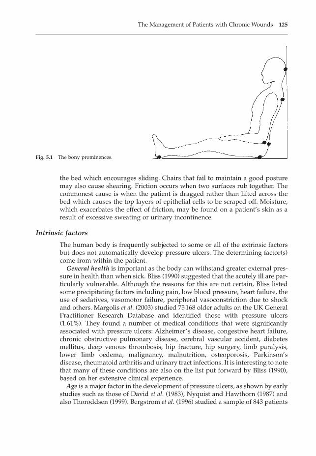
the bed which encourages sliding. Chairs that fail to maintain a good posturemay also cause shearing. Friction occurs when two surfaces rub together. Thecommonest cause is when the patient is dragged rather than lifted across thebed which causes the top layers of epithelial cells to be scraped off. Moisture,which exacerbates the effect of friction, may be found on a patient’s skin as aresult of excessive sweating or urinary incontinence.
Intrinsic factors
The human body is frequently subjected to some or all of the extrinsic factorsbut does not automatically develop pressure ulcers. The determining factor(s)come from within the patient.
General health is important as the body can withstand greater external pres-sure in health than when sick. Bliss (1990) suggested that the acutely ill are par-ticularly vulnerable. Although the reasons for this are not certain, Bliss listedsome precipitating factors including pain, low blood pressure, heart failure, theuse of sedatives, vasomotor failure, peripheral vasoconstriction due to shockand others. Margolis et al. (2003) studied 75168 older adults on the UK GeneralPractitioner Research Database and identified those with pressure ulcers(1.61%). They found a number of medical conditions that were significantlyassociated with pressure ulcers: Alzheimer’s disease, congestive heart failure,chronic obstructive pulmonary disease, cerebral vascular accident, diabetesmellitus, deep venous thrombosis, hip fracture, hip surgery, limb paralysis,lower limb oedema, malignancy, malnutrition, osteoporosis, Parkinson’sdisease, rheumatoid arthritis and urinary tract infections. It is interesting to notethat many of these conditions are also on the list put forward by Bliss (1990),based on her extensive clinical experience.
Age is a major factor in the development of pressure ulcers, as shown by earlystudies such as those of David et al. (1983), Nyquist and Hawthorn (1987) andalso Thoroddsen (1999). Bergstrom et al. (1996) studied a sample of 843 patients
The Management of Patients with Chronic Wounds 125
Fig. 5.1 The bony prominences.
in a variety of care settings over a period of four weeks for the incidence ofpressure ulcers. They found that those who developed pressure ulcers were sig-nificantly older than those who did not.
As people age, their skin becomes thinner and less elastic. In part, this isbecause the collagen in the dermis reduces in quantity and quality. Collagenprovides a buffer that helps to prevent disruption of the microcirculation(Krouskop, 1983). There may be wasting of the overall body mass, resulting inloose folds of skin. There is also an increased likelihood of chronic illness ordisease developing, many of which may also predispose to pressure ulcerdevelopment. Once an ulcer occurs it is much harder to heal in an older personthan a young one (see Chapter 2).
Reduced mobility can affect the ability to relieve pressure effectively, if at all.It also predisposes to shearing and friction if the patient is confined to bed orchair. General prevalence surveys, such as those of David et al. (1983), Nyquistand Hawthorn (1987) and Bergstrom et al. (1996), found reduced mobility to bea factor for many patients with pressure ulcers. In their study of 109 long-termcare facilities, Horn et al. (2002) also found reduced mobility to be a significantfactor for those who developed pressure ulcers. In a similar study of 2015patients in long-term care facilities, Baumgarten et al. (2003a) also found immo-bility to be a factor. A classic study by Versluysen (1986) studied 100 patientsover the age of 60 years with a fractured femur and found an incidence of 66%.Much attention has subsequently been paid to the management of this groupof patients and a study by Baumgarten et al. (2003b) of elderly patients with hipfacture has a pressure ulcer incidence rate of 8.8%.
Exton-Smith and Sherwin (1961) studied the number of movements made by50 elderly patients during the night. A strong relationship was found betweenthose with reduced movement and the development of pressure ulcers.Reduced movement during sleep may be associated with a variety of drugssuch as hypnotics, anxiolytics, antidepressants, opioid analgesics and antihist-amines. Berlowitz and Wilking (1989) studied a variety of factors in patientswith pressure ulcers and patients developing pressure ulcers and found thatreduced mobility was significantly associated with pressure ulcers in bothgroups. Brandeis et al. (1994) also found reduced mobility to be a significantfactor in a study involving nursing home patients.
Another aspect of reduced mobility is that of the patient undergoing majorsurgery. Operations may last many hours whilst the patient lies immobile onthe operating table. Mobility may be reduced in the immediate postoperativeperiod because of the effects of the anaesthetic, pain, analgesia, infusions ordrains. Today very sophisticated surgery is carried out, often on the olderpatient. The risks of pressure ulcer development associated with such surgeryare consequently increased. Schoonhoven et al. (2002a) observed 208 patientsundergoing surgery lasting at least four hours and found that 44 (21.2%) devel-oped pressure ulcers within two days of surgery. The research team also iden-tified possible risk indicators in these surgical patients and found that the onlypredictor of pressure ulcer development was length of surgery (Schoonhovenet al., 2002b). This outcome replicates the finding of Aronovitch (1999) who
126 The Care of Wounds
found that the incidence of pressure damage increased with the length ofsurgery.
Neurological deficit may be associated with reduced mobility, such as in apatient with paraplegia, but this is not always so. The diabetic may suffer fromneuropathy without loss of mobility. Neurological deficit may be associatedwith strokes, multiple sclerosis, diabetes and spinal cord injury or degenera-tion. Loss of sensation means the patient is unaware of the need to relieve pres-sure, even if he is able to do so. Dealey (1991a) found that neurological deficitwas a common factor in those patients with pressure ulcers in a survey of ateaching hospital. Kabagambe et al. (1994) compared ten patients with spinalcord injury with 11 healthy subjects. They found an impaired reactive hyper-aemia response in those with spinal cord injury.
Reduced nutritional status impairs the elasticity of the skin. In the long term,it will lead to anaemia and a reduction of oxygen to the tissues. Poor nutritionhas long been considered to be a factor in pressure ulcer development althoughresearch results are inconsistent (Langer et al., 2003; Mathus-Vliegen, 2001).Some studies, such as those of Berlowitz and Wilking (1989), Brandeis et al.(1994) and Williams et al. (2000), found impaired nutritional intake to be a factorin pressure ulcer development. Guenter et al. (2000) found that newly hospi-talised patients with grade 3 or 4 pressure ulcers had malnutrition. The factorsthat may lead to malnutrition are discussed in Chapter 2.
Body weight should also be considered. Very emaciated patients have no‘padding’ over bony prominences and so have less protection against pressure.On the other hand, very obese patients are difficult to move. Unless great careis taken, they may be dragged rather than lifted in the bed. Another problemof the obese patient is that moisture from sweating may become trappedbetween the rolls of fat, causing maceration. Both of these types of patient mayalso have a poor nutritional status.
Incontinence of urine can contribute to maceration of the skin and thusincrease the risk of friction. Constant washing because of either urinary or faecalincontinence removes natural body oils, drying the skin. Urinary incontinencemay be caused by the use of diuretics or sedatives. Diarrhoea may cause incon-tinence in the elderly or immobile patient and is a side effect of some antibi-otics. There is no clear evidence as to whether incontinence is a risk factor forpressure ulcer development or not. Baumgarten et al. (2003a) found faecalincontinence to be a risk factor whereas Reed et al. (2003) did not. Other studieshave found both urinary and faecal incontinence to be risk factors (Ferrell et al.,2000; Schue & Langemo, 1999).
Poor blood supply to the periphery lowers the local capillary pressure andcauses malnutrition in the tissues. It may be caused by disease, such as heartdisease, peripheral vascular disease or diabetes. Bergstrom et al. (1996) foundcardiovascular disease to be a significant predictor of pressure ulcer develop-ment. Drugs, such as beta-blockers and inotropic sympathomimetics, maycause peripheral vasoconstriction. These drugs may be used following cardiacsurgery when the patient is already suffering from reduced mobility. Blood flowmay also be affected during surgery. Sanada et al. (1997) measured blood flow
The Management of Patients with Chronic Wounds 127
during surgery in the skin over the iliac crest and sacrum. They found a 500%increase in flow in those patients who did not develop pressure sores but a dropin flow in those who later developed pressure sores. Spittle et al. (2001) mea-sured the incidence of pressure ulcers in patients who had undergone lowerlimb amputations for either ischaemia or neuroischaemia and found an inci-dence rate of 55% in those who had had major amputations and 20% in minoramputees. They suggest that the presence of peripheral vascular disease mayhave been a risk factor for these patients.
External factors
Dealey (1997) cited a number of external factors that can exacerbate the factorsdiscussed above. They include:
� inappropriate positioning, which may increase pressure or shear.� restrictions to movement, such as lying for long periods on a trolley.� lying for long periods in one position on hard surfaces, such as the X-ray
table.� poor lifting and handling techniques which increase the risk of friction and
shear.� poor hygiene that leaves the skin surface moist from urine, faeces or sweat.� drugs such as sedatives that make the patient drowsy and less likely to move.
5.2.3 Prevention of pressure ulcers
Whilst it is a truism, as far as pressure ulcers are concerned, prevention is betterthan cure. Waterlow (1988) suggested that 95% of all pressure ulcers could beprevented. Although this concept was based on opinion, it has been supportedby a study undertaken in Japan (Haglsawa & Barbenel, 1999) that observed theoutcome of providing preventive care for at-risk patients admitted to an inter-nal medicine ward. The researchers found an incidence rate of 4.4% in the 240patients they studied. They suggest that this may approach the lowest rateachievable in very ill patients.
In response to the development of evidence-based guidelines, most hospitaland primary care trusts use them as a framework from which a local policymay be developed. The most recent pressure ulcer prevention guidelines arethose published by the National Institute for Clinical Excellence (NICE, 2003).The guidelines can be divided into sections on risk assessment, pressure ulcerprevention, education and training and they will be used to provide a frame-work for this next section. They are also summarised in a useful poster that canbe downloaded from www.nice.org.uk.
Identify individuals vulnerable to or at elevated risk of pressure ulcers
Too often, risk assessment is just seen as using a risk calculator to determinethe patient’s score and then following a ‘recipe’ of care. The first part of theassessment guidance is listed in Box 5.1 and describes some of the issues about
128 The Care of Wounds
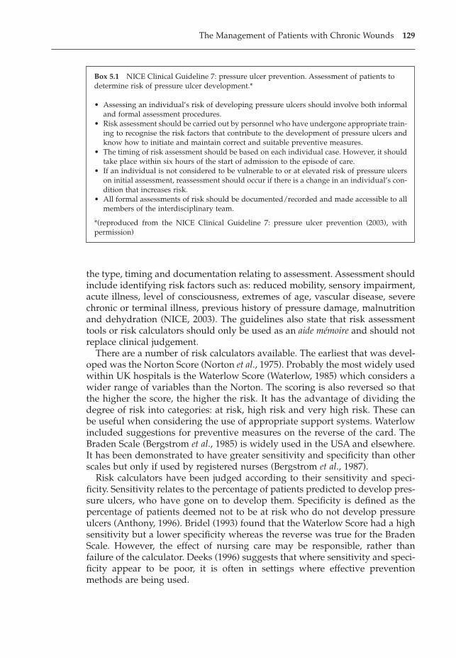
the type, timing and documentation relating to assessment. Assessment shouldinclude identifying risk factors such as: reduced mobility, sensory impairment,acute illness, level of consciousness, extremes of age, vascular disease, severechronic or terminal illness, previous history of pressure damage, malnutritionand dehydration (NICE, 2003). The guidelines also state that risk assessmenttools or risk calculators should only be used as an aide mémoire and should notreplace clinical judgement.
There are a number of risk calculators available. The earliest that was devel-oped was the Norton Score (Norton et al., 1975). Probably the most widely usedwithin UK hospitals is the Waterlow Score (Waterlow, 1985) which considers awider range of variables than the Norton. The scoring is also reversed so thatthe higher the score, the higher the risk. It has the advantage of dividing thedegree of risk into categories: at risk, high risk and very high risk. These canbe useful when considering the use of appropriate support systems. Waterlowincluded suggestions for preventive measures on the reverse of the card. TheBraden Scale (Bergstrom et al., 1985) is widely used in the USA and elsewhere.It has been demonstrated to have greater sensitivity and specificity than otherscales but only if used by registered nurses (Bergstrom et al., 1987).
Risk calculators have been judged according to their sensitivity and speci-ficity. Sensitivity relates to the percentage of patients predicted to develop pres-sure ulcers, who have gone on to develop them. Specificity is defined as thepercentage of patients deemed not to be at risk who do not develop pressureulcers (Anthony, 1996). Bridel (1993) found that the Waterlow Score had a highsensitivity but a lower specificity whereas the reverse was true for the BradenScale. However, the effect of nursing care may be responsible, rather thanfailure of the calculator. Deeks (1996) suggests that where sensitivity and speci-ficity appear to be poor, it is often in settings where effective preventionmethods are being used.
The Management of Patients with Chronic Wounds 129
Box 5.1 NICE Clinical Guideline 7: pressure ulcer prevention. Assessment of patients todetermine risk of pressure ulcer development.*
• Assessing an individual’s risk of developing pressure ulcers should involve both informaland formal assessment procedures.
• Risk assessment should be carried out by personnel who have undergone appropriate train-ing to recognise the risk factors that contribute to the development of pressure ulcers andknow how to initiate and maintain correct and suitable preventive measures.
• The timing of risk assessment should be based on each individual case. However, it shouldtake place within six hours of the start of admission to the episode of care.
• If an individual is not considered to be vulnerable to or at elevated risk of pressure ulcerson initial assessment, reassessment should occur if there is a change in an individual’s con-dition that increases risk.
• All formal assessments of risk should be documented/recorded and made accessible to allmembers of the interdisciplinary team.
*(reproduced from the NICE Clinical Guideline 7: pressure ulcer prevention (2003), with permission)
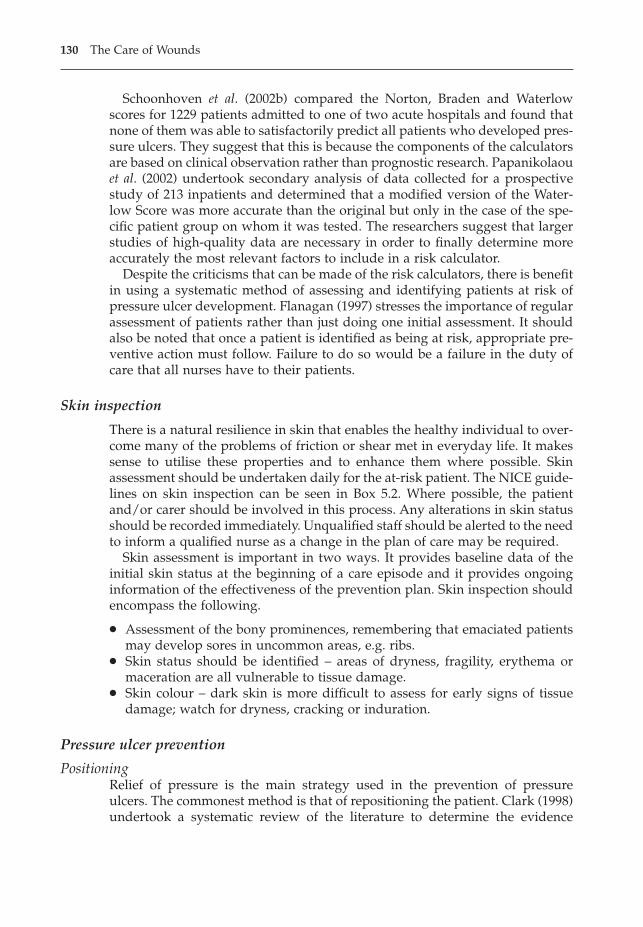
Schoonhoven et al. (2002b) compared the Norton, Braden and Waterlowscores for 1229 patients admitted to one of two acute hospitals and found thatnone of them was able to satisfactorily predict all patients who developed pres-sure ulcers. They suggest that this is because the components of the calculatorsare based on clinical observation rather than prognostic research. Papanikolaouet al. (2002) undertook secondary analysis of data collected for a prospectivestudy of 213 inpatients and determined that a modified version of the Water-low Score was more accurate than the original but only in the case of the spe-cific patient group on whom it was tested. The researchers suggest that largerstudies of high-quality data are necessary in order to finally determine moreaccurately the most relevant factors to include in a risk calculator.
Despite the criticisms that can be made of the risk calculators, there is benefitin using a systematic method of assessing and identifying patients at risk ofpressure ulcer development. Flanagan (1997) stresses the importance of regularassessment of patients rather than just doing one initial assessment. It shouldalso be noted that once a patient is identified as being at risk, appropriate pre-ventive action must follow. Failure to do so would be a failure in the duty ofcare that all nurses have to their patients.
Skin inspection
There is a natural resilience in skin that enables the healthy individual to over-come many of the problems of friction or shear met in everyday life. It makessense to utilise these properties and to enhance them where possible. Skinassessment should be undertaken daily for the at-risk patient. The NICE guide-lines on skin inspection can be seen in Box 5.2. Where possible, the patientand/or carer should be involved in this process. Any alterations in skin statusshould be recorded immediately. Unqualified staff should be alerted to the needto inform a qualified nurse as a change in the plan of care may be required.
Skin assessment is important in two ways. It provides baseline data of theinitial skin status at the beginning of a care episode and it provides ongoinginformation of the effectiveness of the prevention plan. Skin inspection shouldencompass the following.
� Assessment of the bony prominences, remembering that emaciated patientsmay develop sores in uncommon areas, e.g. ribs.
� Skin status should be identified – areas of dryness, fragility, erythema ormaceration are all vulnerable to tissue damage.
� Skin colour – dark skin is more difficult to assess for early signs of tissuedamage; watch for dryness, cracking or induration.
Pressure ulcer prevention
PositioningRelief of pressure is the main strategy used in the prevention of pressure ulcers. The commonest method is that of repositioning the patient. Clark (1998)undertook a systematic review of the literature to determine the evidence
130 The Care of Wounds
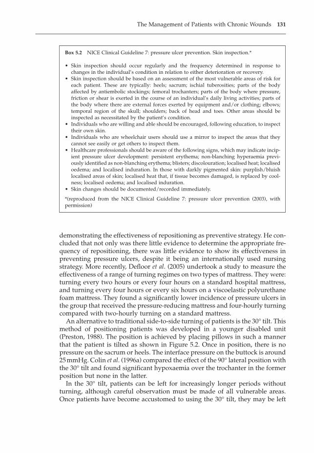
demonstrating the effectiveness of repositioning as preventive strategy. He con-cluded that not only was there little evidence to determine the appropriate fre-quency of repositioning, there was little evidence to show its effectiveness inpreventing pressure ulcers, despite it being an internationally used nursingstrategy. More recently, Defloor et al. (2005) undertook a study to measure theeffectiveness of a range of turning regimes on two types of mattress. They were:turning every two hours or every four hours on a standard hospital mattress,and turning every four hours or every six hours on a viscoelastic polyurethanefoam mattress. They found a significantly lower incidence of pressure ulcers inthe group that received the pressure-reducing mattress and four-hourly turningcompared with two-hourly turning on a standard mattress.
An alternative to traditional side-to-side turning of patients is the 30° tilt. Thismethod of positioning patients was developed in a younger disabled unit(Preston, 1988). The position is achieved by placing pillows in such a mannerthat the patient is tilted as shown in Figure 5.2. Once in position, there is nopressure on the sacrum or heels. The interface pressure on the buttock is around25mmHg. Colin et al. (1996a) compared the effect of the 90° lateral position withthe 30° tilt and found significant hypoxaemia over the trochanter in the formerposition but none in the latter.
In the 30° tilt, patients can be left for increasingly longer periods withoutturning, although careful observation must be made of all vulnerable areas.Once patients have become accustomed to using the 30° tilt, they may be left
The Management of Patients with Chronic Wounds 131
Box 5.2 NICE Clinical Guideline 7: pressure ulcer prevention. Skin inspection.*
• Skin inspection should occur regularly and the frequency determined in response tochanges in the individual’s condition in relation to either deterioration or recovery.
• Skin inspection should be based on an assessment of the most vulnerable areas of risk foreach patient. These are typically: heels; sacrum; ischial tuberosities; parts of the bodyaffected by antiembolic stockings; femoral trochanters; parts of the body where pressure,friction or shear is exerted in the course of an individual’s daily living activities; parts ofthe body where there are external forces exerted by equipment and/or clothing; elbows;temporal region of the skull; shoulders; back of head and toes. Other areas should beinspected as necessitated by the patient’s condition.
• Individuals who are willing and able should be encouraged, following education, to inspecttheir own skin.
• Individuals who are wheelchair users should use a mirror to inspect the areas that theycannot see easily or get others to inspect them.
• Healthcare professionals should be aware of the following signs, which may indicate incip-ient pressure ulcer development: persistent erythema; non-blanching hyperaemia previ-ously identified as non-blanching erythema; blisters; discolouration; localised heat; localisedoedema; and localised induration. In those with darkly pigmented skin: purplish/bluishlocalised areas of skin; localised heat that, if tissue becomes damaged, is replaced by cool-ness; localised oedema; and localised induration.
• Skin changes should be documented/recorded immediately.
*(reproduced from the NICE Clinical Guideline 7: pressure ulcer prevention (2003), with permission)
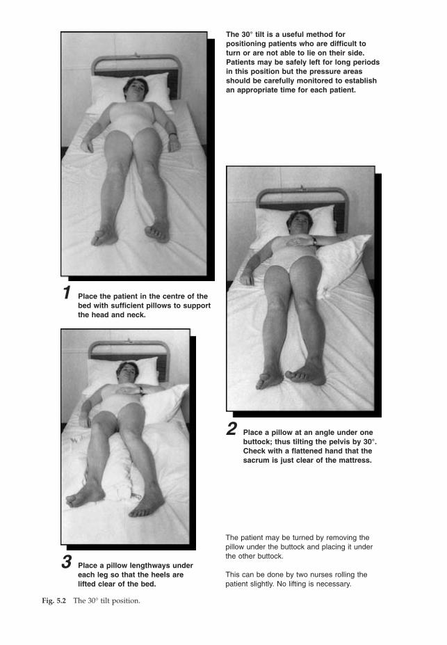
Fig. 5.2 The 30° tilt position.
The 30° tilt is a useful method forpositioning patients who are difficult toturn or are not able to lie on their side.Patients may be safely left for long periodsin this position but the pressure areasshould be carefully monitored to establishan appropriate time for each patient.
2 Place a pillow at an angle under onebuttock; thus tilting the pelvis by 30°.Check with a flattened hand that thesacrum is just clear of the mattress.
1 Place the patient in the centre of thebed with sufficient pillows to supportthe head and neck.
The patient may be turned by removing thepillow under the buttock and placing it underthe other buttock.
This can be done by two nurses rolling thepatient slightly. No lifting is necessary.
3 Place a pillow lengthways undereach leg so that the heels arelifted clear of the bed.
for up to eight hours without turning. Not only does this allow a patient tohave an undisturbed night’s sleep but also it is of great benefit for use in thecommunity. This method of positioning is not suitable for all patients, as shownby Young (2004) who randomised 46 elderly acutely ill patients to either 30° or90° lying. She found that many of the patients were unable to maintain the 30°position and questioned the value of the widespread advice to use it. It seemsreasonable to conclude that further evidence is needed to determine when itmay best be used. In the meantime, nurses should know how to use the posi-tion but should recognise that it probably has limited value for the acutely ill,conscious patient.
The use of correct positioning and repositioning techniques, as well as appro-priate methods for transferring patients from one surface to another, will reduceboth friction and shear forces. Incorrect positions in either bed or chair cancause patients to slide. Regular moving of patients puts nurses at risk of backinjury. The European Community directive on the moving and handling ofheavy loads states that all healthcare institutions must have a policy for manualhandling and staff should have regular training on correct methods of movingpatients. Hoists, slides and other aids for moving patients should be available.The RCN (2003) code of practice suggests that manual handling should be eliminated in all but exceptional or life-threatening situations. All nurses have a responsibility to take reasonable care for their own safety and that ofpatients and colleagues.
Seating� Chairs �
Most of the research in relation to pressure-redistributing equipment hasinvolved mattresses and beds rather than seating and yet sitting vulnerablepatients in armchairs is part of the routine rehabilitation process for many(McCafferty et al., 2000). In many instances, hospital armchairs have been pur-chased without any reference to clinical staff or the varied needs of patients(Collins, 1999; McCafferty et al., 2000). Once ill patients start to sit out of bed,they are perceived to be ‘mobile’ and so may be left in a chair for long periodsof time without being moved. The NICE guidelines suggest that patients at riskof pressure ulcer development should not be sat in a chair for more than twohours at a time (NICE, 2003).
Many hospital armchairs are in a poor state and fail to give any pressure reliefor maintain a good posture (Dealey et al., 1991). Chairs should be checked andreplaced or repaired in the same way as mattresses. Many chairs have a reclin-ing back of between 15° and 40° that puts the patient in a semi-reclining posture.This may make it more difficult for the patient to stand. Ideally, a chair shouldhave a recline of not more than 10°, enabling the patient to move more freely.Although cushions may be added to chairs to improve pressure relief, theyshould not make the chair so high that the patient’s feet cannot touch the floor.Collins (1999) undertook a comparative study of an armchair with an integralpressure-relieving cushion and standard hospital armchairs. She found a sig-
The Management of Patients with Chronic Wounds 133
nificantly lower incidence of pressure ulcers in the experimental group com-pared with the control group. Conventional seating is not suitable for every-one. Some patients have severe seating problems due to contractures, deformityor infirmity. Specialised seating must be considered for these people.
� Cushions �
Most of the research on cushions has been on those for use in wheelchairs.Wheelchairs have a canvas base, which Rithalia (1989) found exerted pressuresin the region of 226mmHg. It is essential that a cushion should always be usedin a wheelchair. For those people who become wheelchair bound because ofdisability, special assessment should be made to identify the cushion mostsuited to the specific needs of the patient (NICE, 2003). Many physiotherapistsand occupational therapists have developed specialist skills in assessment.There are many cushions available but there is insufficient evidence to demon-strate whether any cushion outperforms the others and so no specific recom-mendation can be made (NICE, 2003).
� Use of aids �
The NICE guidelines specifically state that water-filled gloves and doughnut-type devices should not be used (NICE, 2003).
Pressure-relieving devices (beds, mattresses and overlays)In 1995, an Effective Health Care Bulletin (1995) on pressure ulcer preventionrecommended that pressure-relieving foam mattresses should be used for at-risk patients rather than the standard mattress. Since then many hospitals havereplaced worn-out standard hospital mattresses with this type of mattress. Inthe NICE guidelines, this type of mattress is seen as a minimum provision forthose at risk of pressure ulcer development (NICE, 2003).
There is an ever-increasing range of pressure-redistributing equipment avail-able for use. They range from overlays to highly sophisticated beds and can bedivided into high-tech and low-tech categories (Cullum et al., 2001a). Low-techdevices conform to the shape of the body, thus ‘spreading the load’ and reduc-ing the pressure over bony prominences, whereas high-tech devices aredynamic systems with various modes of action. Examples of both high- andlow-tech devices are given in Box 5.3.
The NICE guidelines suggest that selection of a device should be based on aholistic assessment that includes:
� identified levels of risk.� skin assessment.� patient comfort.� general health state.� lifestyle and physical and mental abilities.� critical care needs.� acceptability of the proposed device to both patient and carer.
134 The Care of Wounds
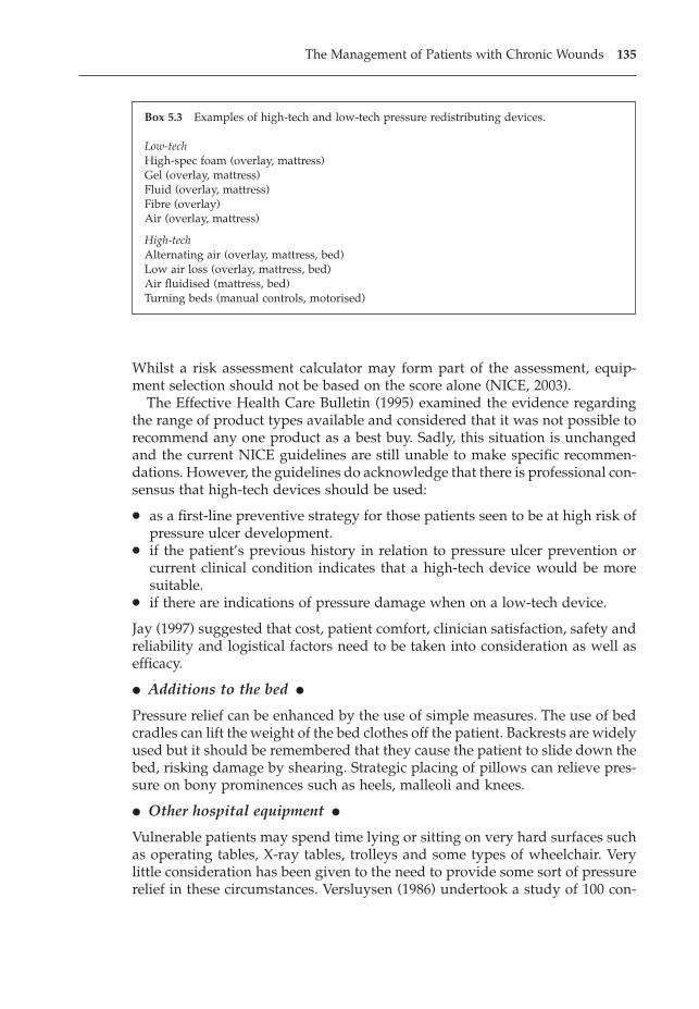
Whilst a risk assessment calculator may form part of the assessment, equip-ment selection should not be based on the score alone (NICE, 2003).
The Effective Health Care Bulletin (1995) examined the evidence regardingthe range of product types available and considered that it was not possible torecommend any one product as a best buy. Sadly, this situation is unchangedand the current NICE guidelines are still unable to make specific recommen-dations. However, the guidelines do acknowledge that there is professional con-sensus that high-tech devices should be used:
� as a first-line preventive strategy for those patients seen to be at high risk ofpressure ulcer development.
� if the patient’s previous history in relation to pressure ulcer prevention orcurrent clinical condition indicates that a high-tech device would be moresuitable.
� if there are indications of pressure damage when on a low-tech device.
Jay (1997) suggested that cost, patient comfort, clinician satisfaction, safety andreliability and logistical factors need to be taken into consideration as well asefficacy.
� Additions to the bed �
Pressure relief can be enhanced by the use of simple measures. The use of bedcradles can lift the weight of the bed clothes off the patient. Backrests are widelyused but it should be remembered that they cause the patient to slide down thebed, risking damage by shearing. Strategic placing of pillows can relieve pres-sure on bony prominences such as heels, malleoli and knees.
� Other hospital equipment �
Vulnerable patients may spend time lying or sitting on very hard surfaces suchas operating tables, X-ray tables, trolleys and some types of wheelchair. Verylittle consideration has been given to the need to provide some sort of pressurerelief in these circumstances. Versluysen (1986) undertook a study of 100 con-
The Management of Patients with Chronic Wounds 135
Box 5.3 Examples of high-tech and low-tech pressure redistributing devices.
Low-techHigh-spec foam (overlay, mattress)Gel (overlay, mattress)Fluid (overlay, mattress)Fibre (overlay)Air (overlay, mattress)
High-techAlternating air (overlay, mattress, bed)Low air loss (overlay, mattress, bed)Air fluidised (mattress, bed)Turning beds (manual controls, motorised)
secutive elderly patients admitted with a fractured femur. The interface pres-sures were measured on the casualty trolleys and operating tables. The casu-alty trolleys with a 5cm deep foam mattress showed pressures of 56–60mmHgat the sacrum and 150–160mmHg at the heels. The operating table had inter-face pressures of 75–80mmHg at the sacrum and 60–120mmHg at the heels.NICE (2003) states that all patients undergoing surgery who are identified asbeing at risk of pressure ulcer development should be placed on a high-specification foam theatre mattress or similar.
� Organisation of delivery of devices �
It is not practical for every clinical area to maintain their own stock of pressure-redistributing devices and many hospital and primary care trusts have someform of equipment store to ensure effective provision and use of devices. NICE(2003) states that there should be a co-ordinated approach to the acquisition,allocation and management of equipment and that prevention strategies orpolicies should have a specified standard time between assessment and allocation of equipment. NICE also provides guidance on auditing the use ofequipment (NICE, 2003).
Every patient at risk of pressure ulcer development should have an indi-vidualised written plan of care. It is important to document all assessments andthe care given. This will enable staff to monitor the effectiveness of the plan andto identify any early signs of tissue damage. It should also ensure effective useof equipment as patients may be moved to less sophisticated equipment whenthey no longer have need of the high-tech equipment or vice versa.
Additional prevention strategies not mentioned in the NICE guidelines� Skin care �
Traditionally, skin care involved rubbing patients’ pressure areas at regularintervals. A variety of lotions and potions were used. A review of the literatureby Buss et al. (1997) considered the effects of massage or rubbing on pressuresore prevention. They concluded that this practice could not be recommended.When the practice of rubbing was discontinued most nurses developed a reluc-tance to use any type of cream over the pressure areas. Whilst this is generallyappropriate, there may be exceptions in the case of very dry or very moist skin.Emollients should be considered when caring for patients with very dry skin,either in the bath or an emollient cream. Creams should be applied gently tothe affected area. If the skin is moist, the source of the moisture should be iden-tified and dealt with if possible. A barrier cream may be needed to protect theskin from the harmful effects of moisture.
Frequent cleansing using soap and water for incontinent patients can causeexcessive drying of the skin. Dealey and Keogh (1998) found a significantimprovement in skin status using a cleanser and barrier cream compared tosoap and water in elderly incontinent patients at risk of pressure sore devel-opment. Cooper and Gray (2001) undertook a similar study comparing soapand water with a combination cleanser and emollient in 93 elderly nursing
136 The Care of Wounds
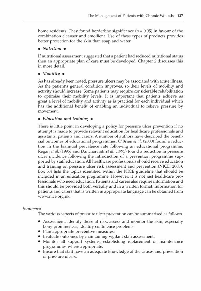
home residents. They found borderline significance (p = 0.05) in favour of thecombination cleanser and emollient. Use of these types of products providesbetter protection for the skin than soap and water.
� Nutrition �
If nutritional assessment suggested that a patient had reduced nutritional statusthen an appropriate plan of care must be developed. Chapter 2 discusses thisin more detail.
� Mobility �
As has already been noted, pressure ulcers may be associated with acute illness.As the patient’s general condition improves, so their levels of mobility andactivity should increase. Some patients may require considerable rehabilitationto optimise their mobility levels. It is important that patients achieve as great a level of mobility and activity as is practical for each individual whichhas the additional benefit of enabling an individual to relieve pressure by movement.
� Education and training �
There is little point in developing a policy for pressure ulcer prevention if noattempt is made to provide relevant education for healthcare professionals andassistants, patients and carers. A number of authors have described the benefi-cial outcomes of educational programmes. O’Brien et al. (2000) found a reduc-tion in the biannual prevalence rate following an educational programme.Regan et al. (1995) and Danchaivijitr et al. (1995) found a reduction in pressureulcer incidence following the introduction of a prevention programme sup-ported by staff education. All healthcare professionals should receive educationand training on pressure ulcer risk assessment and prevention (NICE, 2003).Box 5.4 lists the topics identified within the NICE guideline that should beincluded in an education programme. However, it is not just healthcare pro-fessionals who need education. Patients and carers also require information andthis should be provided both verbally and in a written format. Information forpatients and carers that is written in appropriate language can be obtained fromwww.nice.org.uk.
SummaryThe various aspects of pressure ulcer prevention can be summarised as follows.
� Assessment: identify those at risk, assess and monitor the skin, especiallybony prominences, identify continence problems.
� Plan appropriate preventive measures.� Evaluate outcomes by maintaining vigilant skin assessment.� Monitor all support systems, establishing replacement or maintenance
programmes where appropriate.� Ensure that staff have an adequate knowledge of the causes and prevention
of pressure ulcers.
The Management of Patients with Chronic Wounds 137
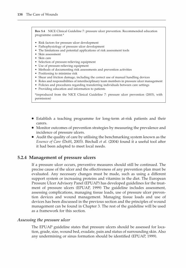
� Establish a teaching programme for long-term at-risk patients and theircarers.
� Monitor outcomes of prevention strategies by measuring the prevalence andincidence of pressure ulcers.
� Audit the quality of care by utilising the benchmarking system known as theEssence of Care (DoH, 2003). Birchall et al. (2004) found it a useful tool afterit had been adapted to meet local needs.
5.2.4 Management of pressure ulcers
If a pressure ulcer occurs, preventive measures should still be continued. Theprecise cause of the ulcer and the effectiveness of any prevention plan must beevaluated. Any necessary changes must be made, such as using a differentsupport system or increasing proteins and vitamins in the diet. The EuropeanPressure Ulcer Advisory Panel (EPUAP) has developed guidelines for the treat-ment of pressure ulcers (EPUAP, 1999) The guideline includes assessment,assessing complications, managing tissue loads, use of pressure ulcer preven-tion devices and wound management. Managing tissue loads and use ofdevices has been discussed in the previous section and the principles of woundmanagement can be found in Chapter 3. The rest of the guideline will be usedas a framework for this section.
Assessing the pressure ulcer
The EPUAP guideline states that pressure ulcers should be assessed for loca-tion, grade, size, wound bed, exudate, pain and status of surrounding skin. Alsoany undermining or sinus formation should be identified (EPUAP, 1999).
138 The Care of Wounds
Box 5.4 NICE Clinical Guideline 7: pressure ulcer prevention. Recommended educationprogramme content.*
• Risk factors for pressure ulcer development• Pathophysiology of pressure ulcer development• The limitations and potential applications of risk assessment tools• Skin assessment• Skin care• Selection of pressure-relieving equipment• Use of pressure-relieving equipment• Methods of documenting risk assessments and prevention activities• Positioning to minimise risk• Shear and friction damage, including the correct use of manual handling devices• Roles and responsibilities of interdisciplinary team members in pressure ulcer management• Policies and procedures regarding transferring individuals between care settings• Providing education and information to patients
*(reproduced from the NICE Clinical Guideline 7: pressure ulcer prevention (2003), with permission)
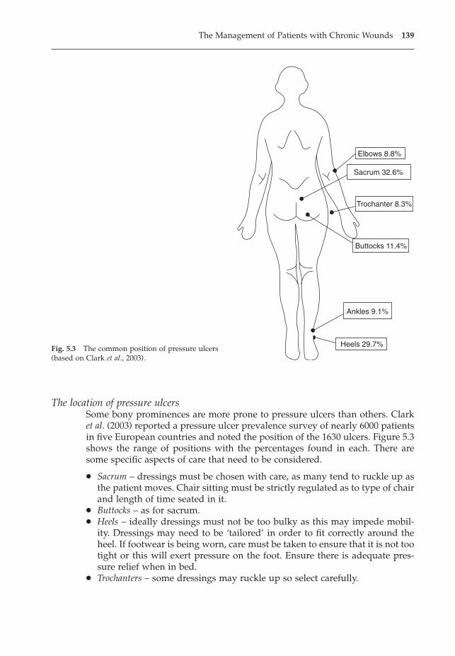
The location of pressure ulcersSome bony prominences are more prone to pressure ulcers than others. Clarket al. (2003) reported a pressure ulcer prevalence survey of nearly 6000 patientsin five European countries and noted the position of the 1630 ulcers. Figure 5.3shows the range of positions with the percentages found in each. There aresome specific aspects of care that need to be considered.
� Sacrum – dressings must be chosen with care, as many tend to ruckle up asthe patient moves. Chair sitting must be strictly regulated as to type of chairand length of time seated in it.
� Buttocks – as for sacrum.� Heels – ideally dressings must not be too bulky as this may impede mobil-
ity. Dressings may need to be ‘tailored’ in order to fit correctly around theheel. If footwear is being worn, care must be taken to ensure that it is not tootight or this will exert pressure on the foot. Ensure there is adequate pres-sure relief when in bed.
� Trochanters – some dressings may ruckle up so select carefully.
The Management of Patients with Chronic Wounds 139
Elbows 8.8%
Sacrum 32.6%
Trochanter 8.3%
Buttocks 11.4%
Ankles 9.1%
Heels 29.7%Fig. 5.3 The common position of pressure ulcers(based on Clark et al., 2003).
� Elbows – these pressure ulcers are usually caused by friction from movingabout the bed. Consider ways of reducing friction – use of a monkey pole,pads or semipermeable film dressings.
� Trunk – ulcers here are uncommon; try and identify source of pressure andremove or modify it.
Grading of pressure ulcersThe use of a recognised system of grading pressure ulcers can be helpful, as itprovides an objective description and gives a more accurate picture of theamount of tissue damage than comments such as ‘a deep ulcer’. It should be usedin conjunction with other descriptive tools such as measuring or tracing the ulcerand describing its appearance. The EPUAP uses the following grading system.
� Grade 1 – non-blanchable erythema of intact skin. Discolouration of the skin,warmth, oedema and induration may be used as indicators, especially fordark skin.
� Grade 2 – partial-thickness skin loss involving epidermis and/or dermis. Theulcer is superficial and may be seen as a blister, abrasion or crater.
� Grade 3 – full-thickness skin loss involving damage to or necrosis of subcu-taneous tissue that may extend down to but not through underlying fascia.
� Grade 4 – extensive destruction, tissue necrosis or damage to muscle, boneor supporting structures with or without full-thickness skin loss.
Figure 5.4 shows examples of each of these grades of pressure ulcer.There are a number of issues that need to be considered when utilising any
grading system: accurate identification of grade 1 ulcers, incontinence lesions,reverse grading and interrater reliability.
Grade 1 pressure ulcers are often found to account for approximately 50% ofall ulcers in prevalence surveys (Halfens et al., 2001). However, there are prob-lems with both over- and underreporting. Overreporting may occur if nursesfail to differentiate between blanching and non-blanching erythema. Alterna-tively, underreporting may occur because of the difficulties in identifying ery-thema on dark skin (Scanlon & Stubbs, 2004). Attempts have been made toprovide additional descriptors to the definition, as can be seen above. However,there is a need to undertake further research to identify a truly accurate methodof assessment that overcomes skin colour.
Incontinence lesions are sometimes mistaken for pressure ulcers (Defloor &Schoonhoven, 2004). Incontinence can cause erythema, excoriation and macer-ation of the skin and thus an incontinence lesion may have a similar appearanceto a grade 2 or 3 pressure ulcer. Care should be taken to determine the preciseposition of any lesion/ulcer in incontinent patients. If a lesion is not over a bonyprominence, it is more likely to be due to incontinence than pressure.
Reverse grading is the practice of describing a healing pressure ulcer asmoving from a grade 4, through grade 3 and then to grade 2 as the grade 4cavity heals and becomes shallower. The American National Pressure UlcerAdvisory Panel (NPUAP) debated this topic and produced a position statementthat can be found at www.npuap.org. This states that as pressure ulcers heal,
140 The Care of Wounds
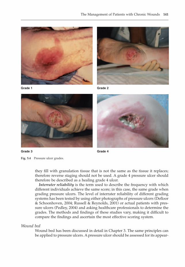
they fill with granulation tissue that is not the same as the tissue it replaces;therefore reverse staging should not be used. A grade 4 pressure ulcer shouldtherefore be described as a healing grade 4 ulcer.
Interrater reliability is the term used to describe the frequency with whichdifferent individuals achieve the same score; in this case, the same grade whengrading pressure ulcers. The level of interrater reliability of different gradingsystems has been tested by using either photographs of pressure ulcers (Defloor& Schoonhoven, 2004; Russell & Reynolds, 2001) or actual patients with pres-sure ulcers (Pedley, 2004) and asking healthcare professionals to determine thegrades. The methods and findings of these studies vary, making it difficult tocompare the findings and ascertain the most effective scoring system.
Wound bedWound bed has been discussed in detail in Chapter 3. The same principles canbe applied to pressure ulcers. A pressure ulcer should be assessed for its appear-
The Management of Patients with Chronic Wounds 141
Fig. 5.4 Pressure ulcer grades.
Grade 1 Grade 2
Grade 3 Grade 4
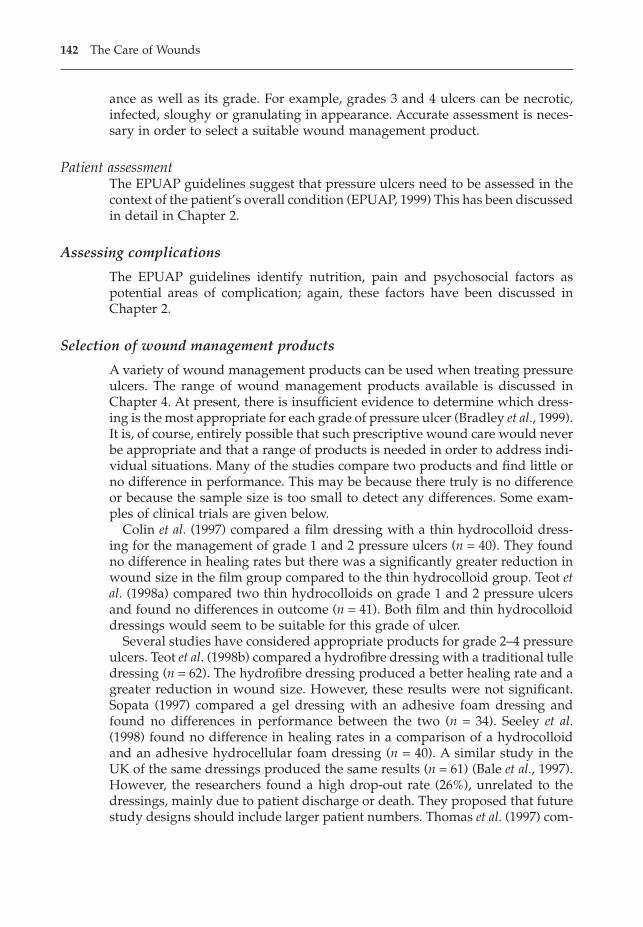
ance as well as its grade. For example, grades 3 and 4 ulcers can be necrotic,infected, sloughy or granulating in appearance. Accurate assessment is neces-sary in order to select a suitable wound management product.
Patient assessmentThe EPUAP guidelines suggest that pressure ulcers need to be assessed in thecontext of the patient’s overall condition (EPUAP, 1999) This has been discussedin detail in Chapter 2.
Assessing complications
The EPUAP guidelines identify nutrition, pain and psychosocial factors aspotential areas of complication; again, these factors have been discussed inChapter 2.
Selection of wound management products
A variety of wound management products can be used when treating pressureulcers. The range of wound management products available is discussed inChapter 4. At present, there is insufficient evidence to determine which dress-ing is the most appropriate for each grade of pressure ulcer (Bradley et al., 1999).It is, of course, entirely possible that such prescriptive wound care would neverbe appropriate and that a range of products is needed in order to address indi-vidual situations. Many of the studies compare two products and find little orno difference in performance. This may be because there truly is no differenceor because the sample size is too small to detect any differences. Some exam-ples of clinical trials are given below.
Colin et al. (1997) compared a film dressing with a thin hydrocolloid dress-ing for the management of grade 1 and 2 pressure ulcers (n = 40). They foundno difference in healing rates but there was a significantly greater reduction inwound size in the film group compared to the thin hydrocolloid group. Teot etal. (1998a) compared two thin hydrocolloids on grade 1 and 2 pressure ulcersand found no differences in outcome (n = 41). Both film and thin hydrocolloiddressings would seem to be suitable for this grade of ulcer.
Several studies have considered appropriate products for grade 2–4 pressureulcers. Teot et al. (1998b) compared a hydrofibre dressing with a traditional tulledressing (n = 62). The hydrofibre dressing produced a better healing rate and agreater reduction in wound size. However, these results were not significant.Sopata (1997) compared a gel dressing with an adhesive foam dressing andfound no differences in performance between the two (n = 34). Seeley et al.(1998) found no difference in healing rates in a comparison of a hydrocolloidand an adhesive hydrocellular foam dressing (n = 40). A similar study in theUK of the same dressings produced the same results (n = 61) (Bale et al., 1997).However, the researchers found a high drop-out rate (26%), unrelated to thedressings, mainly due to patient discharge or death. They proposed that futurestudy designs should include larger patient numbers. Thomas et al. (1997) com-
142 The Care of Wounds
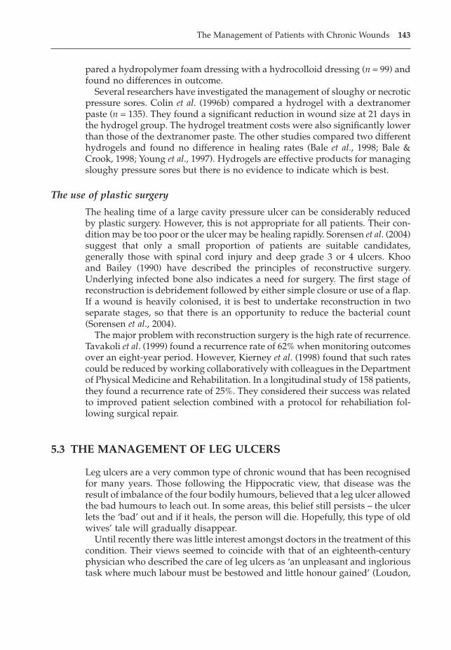
pared a hydropolymer foam dressing with a hydrocolloid dressing (n = 99) andfound no differences in outcome.
Several researchers have investigated the management of sloughy or necroticpressure sores. Colin et al. (1996b) compared a hydrogel with a dextranomerpaste (n = 135). They found a significant reduction in wound size at 21 days inthe hydrogel group. The hydrogel treatment costs were also significantly lowerthan those of the dextranomer paste. The other studies compared two differenthydrogels and found no difference in healing rates (Bale et al., 1998; Bale &Crook, 1998; Young et al., 1997). Hydrogels are effective products for managingsloughy pressure sores but there is no evidence to indicate which is best.
The use of plastic surgery
The healing time of a large cavity pressure ulcer can be considerably reducedby plastic surgery. However, this is not appropriate for all patients. Their con-dition may be too poor or the ulcer may be healing rapidly. Sorensen et al. (2004)suggest that only a small proportion of patients are suitable candidates, generally those with spinal cord injury and deep grade 3 or 4 ulcers. Khoo and Bailey (1990) have described the principles of reconstructive surgery.Underlying infected bone also indicates a need for surgery. The first stage ofreconstruction is debridement followed by either simple closure or use of a flap.If a wound is heavily colonised, it is best to undertake reconstruction in twoseparate stages, so that there is an opportunity to reduce the bacterial count(Sorensen et al., 2004).
The major problem with reconstruction surgery is the high rate of recurrence.Tavakoli et al. (1999) found a recurrence rate of 62% when monitoring outcomesover an eight-year period. However, Kierney et al. (1998) found that such ratescould be reduced by working collaboratively with colleagues in the Departmentof Physical Medicine and Rehabilitation. In a longitudinal study of 158 patients,they found a recurrence rate of 25%. They considered their success was relatedto improved patient selection combined with a protocol for rehabiliation fol-lowing surgical repair.
5.3 THE MANAGEMENT OF LEG ULCERS
Leg ulcers are a very common type of chronic wound that has been recognisedfor many years. Those following the Hippocratic view, that disease was theresult of imbalance of the four bodily humours, believed that a leg ulcer allowedthe bad humours to leach out. In some areas, this belief still persists – the ulcerlets the ‘bad’ out and if it heals, the person will die. Hopefully, this type of oldwives’ tale will gradually disappear.
Until recently there was little interest amongst doctors in the treatment of thiscondition. Their views seemed to coincide with that of an eighteenth-centuryphysician who described the care of leg ulcers as ‘an unpleasant and inglorioustask where much labour must be bestowed and little honour gained’ (Loudon,
The Management of Patients with Chronic Wounds 143
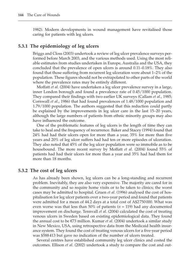
1982). Modern developments in wound management have revitalised thosecaring for patients with leg ulcers.
5.3.1 The epidemiology of leg ulcers
Briggs and Closs (2003) undertook a review of leg ulcer prevalence surveys per-formed before March 2003, and the various methods used. Using the most reli-able estimates from studies undertaken in Europe, Australia and the USA, theyconcluded that the prevalence of open ulcers is around 0.11–0.18%. They alsofound that those suffering from recurrent leg ulceration were about 1–2% of thepopulation. These figures should not be extrapolated to other parts of the worldwhere the prevalence rates may be entirely different.
Moffatt et al. (2004) have undertaken a leg ulcer prevalence survey in a large,inner London borough and found a prevalence rate of 0.45/1000 population.They compared their findings with two earlier UK surveys (Callam et al., 1985;Cornwall et al., 1986) that had found prevalences of 1.48/1000 population and1.79/1000 population. The authors suggested that this reduction could partlybe explained by the improvements in leg ulcer care in the last 15–20 years,although the large numbers of patients from ethnic minority groups may alsohave influenced the outcome.
One of the problematic features of leg ulcers is the length of time they cantake to heal and the frequency of recurrence. Baker and Stacey (1994) found that24% had had their ulcers open for more than a year, 35% for more than fiveyears and 20% of leg ulcer suffers had had ten or more episodes of ulceration.They also noted that 45% of the leg ulcer population were so immobile as to behousebound. The more recent survey by Moffatt et al. (2004) found 55% ofpatients had had their ulcers for more than a year and 35% had had them formore than 18 months.
5.3.2 The cost of leg ulcers
As has already been shown, leg ulcers can be a long-standing and recurrentproblem. Inevitably, they are also very expensive. The majority are cared for inthe community and so require home visits or to be taken to clinics; the worstcases may be admitted to hospital. Gruen et al. (1996) analysed the cost of hos-pitalisation for leg ulcer patients over a two-year period and found that patientswere admitted for a mean of 44.2 days at a total cost of A$2750000. What waseven worse was that less than 50% of patients (n = 119) had any documentedimprovement on discharge. Tennvall et al. (2004) calculated the cost of treatingvenous ulcers in Sweden based on existing epidemiological data. They foundthe annual cost to be €73 million. Kumar et al. (2004) undertook a similar studyin New Mexico, USA, using retrospective data from the Medicaid health insur-ance system. They found the cost of treating venous ulcers for a five-year periodwas $588413 but gave no indication of the number of ulcers treated.
Several centres have established community leg ulcer clinics and costed theoutcomes. Ellison et al. (2002) undertook a study to compare the cost and out-
144 The Care of Wounds
comes of leg ulcer care provision in two large health authorities. They foundthat in one health authority there was a 65% reduction in costs with the intro-duction of the clinics. Even so, 156 patients cost £212707.84 to treat over a three-month period in 1999.
None of the studies quoted above considers the cost to the patient but severalother researchers have investigated this cost. Persoon et al. (2004) undertook areview of the literature to determine the impact of leg ulceration on daily life.They included 37 studies, with both qualitative and quantitative methodol-ogies. The common problem identified in all studies was pain and a numberalso identified disturbed sleep associated with pain; for example, Hofman et al.(1997) interviewed 94 patients with venous ulcers and found that 64% hadsevere pain, 38.3% had continuous pain and 63.8% were woken by pain.Another problem identified by the reviewers is impaired mobility. Some of thequalitative studies also found that heavily exuding malodorous ulcers resultedin limitations in patients’ work and leisure activities and often resulted in poorself-esteem and social isolation. The reviewers conclude that leg ulceration hasa major impact on the life of the sufferer.
5.3.3 The causes of leg ulcers
There are a variety of causes of leg ulcers, the commonest being venous diseaseand arterial disease. O’Brien et al. (2000) surveyed a health district in Irelandand found that 81% of ulcers were venous in origin and 16.3% were due to arterial disease.
Increasingly, more complex aetiologies are being identified, perhaps becauseof increasing age and also improved management of the straightforward cases.A recent survey within London found uncomplicated venous ulceration wasstill the commonest cause (43%), but 35% of the ulcers in the survey hadcomplex aetiologies: lymphoedema (42%), mixed venous and diabetes (35%)and rheumatoid arthritis (26%) (Moffatt et al., 2004).
5.3.4 Venous ulceration
Aetiology
Chronic venous insufficiency is the main cause of venous leg ulcers. Initially,thrombosis or varicosity causes damage to the valves in the veins of the leg.The deep vein is surrounded by muscle. When the leg is exercised, the calfmuscle contracts and squeezes the veins, encouraging the flow of blood alongthe vein. This is often referred to as the calf muscle pump.
Normally blood flows from the superficial veins to the deep veins via a seriesof perforator vessels. The valves in the vessels ensure that blood moves fromthe capillary bed towards the heart. If some of the valves become damaged thenblood can flow in either direction. The backflow of blood towards the capillarybed leads to venous hypertension. As a result, the capillaries become distortedand more permeable. Larger molecules than normal are able to escape into the
The Management of Patients with Chronic Wounds 145
extravascular space, for example fibrinogen and red blood cells. The haemo-globin is first released from the red blood cells and then broken down, causingeczema and a brown staining in the gaiter area. Ultimately there is fibrosis ofthe underlying tissues, giving the leg a ‘woody’ feeling. This condition is calledlipodermatosclerosis. The slightest trauma to the leg and an ulcer will develop.Common examples of trauma are knocking the leg on the corner of a piece offurniture or a fall, injuring the lower leg.
The lymphatic system may also be affected. The lymphatics are responsiblefor removing protein, fat, cells and excess fluid from the tissues. Ryan (1987)has described how the superficial lymphatics in the dermis disappear. Thisresults in waste products accumulating in the tissues, which can cause fibrosisand further oedema.
Management
AssessmentFull assessment of the patient is essential as many factors can delay the healingof chronic wounds (see Chapter 2). A medical assessment may be necessary toensure accurate diagnosis. The leg ulcer guidelines developed by the RoyalCollege of Nursing (RCN, 1998) suggest a number of factors that may be speci-fically indicative of venous ulceration and those that are indicative of other aetiologies. They can be seen in Box 5.5. Other factors that may need to be particularly considered include nutritional status, mobility, sleeping, pain, psychological effects of leg ulceration and the patient’s understanding of thedisease process. A comprehensive assessment of the affected leg must be made,as it is important to rule out arterial disease. The treatments for these two typesof ulcer are not compatible.
146 The Care of Wounds
Box 5.5 Factors to include in assessment of patients with leg ulceration (based on RCN,1998).
Factors indicative of venous ulcerationFamily historyVaricose veinsProven DVT in affected legPhlebitis in affected legSuspected DVTSurgery/fractures to the legEpisodes of chest pain, haemoptysis, history of pulmonary embolus
Factors indicative of non-venous ulcerationFamily history of non-venous ulcersHeart disease, stroke, TIADiabetes mellitusIschaemic rest painCigarette smokingRheumatoid arthritisPeripheral vascular disease/intermittent claudication
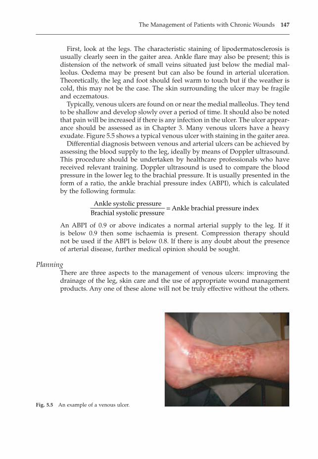
First, look at the legs. The characteristic staining of lipodermatosclerosis isusually clearly seen in the gaiter area. Ankle flare may also be present; this isdistension of the network of small veins situated just below the medial mal-leolus. Oedema may be present but can also be found in arterial ulceration. Theoretically, the leg and foot should feel warm to touch but if the weather iscold, this may not be the case. The skin surrounding the ulcer may be fragileand eczematous.
Typically, venous ulcers are found on or near the medial malleolus. They tendto be shallow and develop slowly over a period of time. It should also be notedthat pain will be increased if there is any infection in the ulcer. The ulcer appear-ance should be assessed as in Chapter 3. Many venous ulcers have a heavyexudate. Figure 5.5 shows a typical venous ulcer with staining in the gaiter area.
Differential diagnosis between venous and arterial ulcers can be achieved byassessing the blood supply to the leg, ideally by means of Doppler ultrasound.This procedure should be undertaken by healthcare professionals who havereceived relevant training. Doppler ultrasound is used to compare the bloodpressure in the lower leg to the brachial pressure. It is usually presented in theform of a ratio, the ankle brachial pressure index (ABPI), which is calculatedby the following formula:
An ABPI of 0.9 or above indicates a normal arterial supply to the leg. If it is below 0.9 then some ischaemia is present. Compression therapy should not be used if the ABPI is below 0.8. If there is any doubt about the presenceof arterial disease, further medical opinion should be sought.
PlanningThere are three aspects to the management of venous ulcers: improving thedrainage of the leg, skin care and the use of appropriate wound managementproducts. Any one of these alone will not be truly effective without the others.
Ankle systolic pressureBrachial systolic pressure
Ankle brachial pressure index=
The Management of Patients with Chronic Wounds 147
Fig. 5.5 An example of a venous ulcer.
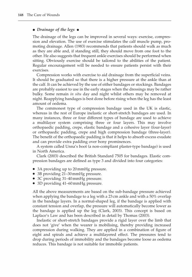
� Drainage of the legs �
The drainage of the legs can be improved in several ways: exercise, compres-sion and elevation. The use of exercise stimulates the calf muscle pump, pro-moting drainage. Allen (1983) recommends that patients should walk as muchas they are able and, if standing still, they should move from one foot to theother. He also suggests that frequent ankle exercises should be performed whensitting. Obviously exercise should be tailored to the abilities of the patient.Regular encouragement will be needed to ensure patients persist with theirexercises.
Compression works with exercise to aid drainage from the superficial veins.It should be graduated so that there is a higher pressure at the ankle than atthe calf. It can be achieved by the use of either bandages or stockings. Bandagesare probably easiest to use in the early stages when the dressings may be ratherbulky. Some remain in situ day and night whilst others may be removed atnight. Reapplying bandages is best done before rising when the leg has the leastamount of oedema.
The commonest type of compression bandage used in the UK is elastic,whereas in the rest of Europe inelastic or short-stretch bandages are used. Inmany instances, three or four different types of bandage are used to achieve a multilayer system comprising three or four layers. This may involveorthopaedic padding, crepe, elastic bandage and a cohesive layer (four-layer)or orthopaedic padding, crepe and high compression bandage (three-layer).The benefit of the orthopaedic padding is that it helps to absorb excess exudateand can provide extra padding over bony prominences.
A system called Unna’s boot (a non-compliant plaster-type bandage) is usedin North America.
Clark (2003) described the British Standard 7505 for bandages. Elastic com-pression bandages are defined as type 3 and divided into four categories:
� 3A providing up to 20mmHg pressure.� 3B providing 21–30mmHg pressure.� 3C providing 31–40mmHg pressure.� 3D providing 41–60mmHg pressure.
All the above measurements are based on the sub-bandage pressure achievedwhen applying the bandage to a leg with a 23cm ankle and with a 50% overlapin the bandage layers. In a normal-shaped leg, if the bandage is applied withconstant tension and overlap, the pressure will automatically become lower asthe bandage is applied up the leg (Clark, 2003). This concept is based onLaplace’s Law and has been described in detail by Thomas (2003).
Inelastic or short-stretch bandages provide a rigid layer over the limb thatdoes not ‘give’ when the wearer is mobilising, thereby providing increasedcompression during walking. They are applied in a combination of figure ofeight and spirals and achieve a multilayered effect. The pressures tend to drop during periods of immobility and the bandages become loose as oedemareduces. This bandage is not suitable for immobile patients.
148 The Care of Wounds
The level of compression needed for venous ulceration is around 40mmHg(Stemmer, 1969). Therefore, the high compression bandages are generally suit-able. However, these pressures are dependent on the size of the limb. A largelimb, swollen with oedema, would require a higher compression bandage thana thinner limb in order to achieve an adequate level of compression. There isalso a need to ensure protection over bony prominences as there is the poten-tial risk of pressure damage, especially if the bandage is poorly applied.
A systematic review of clinical trials of compression bandaging found thatmultilayered bandage systems were more effective than single-layer and highcompression better than low compression (Cullum et al., 2001b). There were noclear differences to dermine the most effective type of high compression. Sincethis review a number of studies have been published that provide further evidence. Two studies compared the four-layer bandage system with a short-stretch bandage and both found that the four-layer system gave a faster healingrate (Iglesias et al., 2004; Ukat et al., 2003). The research teams also found thefour-layer system was less expensive to use, mostly because the short-stretchbandages needed to be reapplied more frequently than the four-layer system.One study compared a three-layer paste bandage system with four-layer andfound the paste system gave a significantly faster healing rate (Meyer et al.,2003). A small study by Polignano et al. (2004) compared the four-layer systemwith Unna’s boot and found no difference in healing rates. Overall, the multi-layered systems such as the four-layer bandage would seem to provide the mosteffective compression.
Unfortunately, not all patients can tolerate high compression bandage systemsand a pragmatic approach to the problem may be necessary. Tubular bandagesmay be better tolerated by some patients and can be used to provide some com-pression. There is a shaped tubular bandage that can be most useful as it is rel-atively easy to apply. This type of compression may be the easiest to pull on forsomeone with arthritic hands. Bale and Harding (2003) studied 28 patientsunable to tolerate high compression and found that using three layers of grad-uated tubular bandage was effective in healing (n = 14) or reducing the ulcer.The tubular bandage was applied in gradually reduced lengths: layer 1 = toe toknee; layer 2 = toe to calf; layer 3 = toe to malleolus. The authors concluded thatwhilst using this type of bandage system cannot be recommended for routinecare, it is a possible alternative for those unable to tolerate therapeutic com-pression. It should be noted that the straight variety of tubular bandage doesnot give appropriate compression as pressure is higher at the calf than the ankle.
Another method of applying compression that can be used, particularly ifoedema is present, is pneumatic compression. It may be applied once or twicea day for periods of up to an hour. Initially the time should be shorter and gradually increased. Whilst this may be a useful form of treatment, it is diffi-cult in the community where there is limited access to such equipment. The useof pneumatic compression should be seen as an addition to the treatmentregime rather than an alternative. A Cochrane review found limited evidenceof the benefits of pneumatic compression and concluded that further researchis required to determine whether it increases healing rates (Mani et al., 2004).
The Management of Patients with Chronic Wounds 149
Elevation of the legs allows gravity to aid venous return. However, manypeople tend to place their feet on a low stool, which is of no benefit whatso-ever. To be effective, the feet should be higher than the heart. If there is an acuteexacerbation of the ulcer with oedema and heavy exudate, it may be worthadmitting the patient to hospital to allow a short period of bedrest. Bedrest withelevation of the foot of the bed can significantly reduce oedema and improvevenous return but it should not be considered as a long-term measure becausethe ulcer will merely break down again once the patient is up and about. It mayalso seriously affect the mobility of older patients. A more practical method isto raise the foot of the bed at home so that the legs are elevated at night. Thismay be achieved by the use of bricks or blocks of wood.
� Skin care �
Management of the ulcer involves cleansing and a suitable topical application.Consideration must be given to the presence of eczema, scaling on the legsaround the ulcer, any allergies to treatment and wound infection. Cleansing isan important factor as many patients may have been told in the past that theymust never get their ulcer wet. As a result, they may not have had a bath foryears. Footbaths are very useful as they allow the patient the opportunity togive the affected leg a good soak. Plain tap water is suitable for most patients.Attention needs to be paid to the adequate cleansing of the footbath after usein order to prevent cross-infection.
One skin complication associated with 37–44% of venous ulcers is gravita-tional or varicose eczema (Patel et al., 2001). It presents as skin excoriations with a diffuse edge, dryness, scaling and weeping and the patients may com-plain of itching, burning or stinging (Patel et al., 2001). Quartey-Papafio (1999)discussed the importance of being able to differentiate between gravitationaleczema and cellulitis and suggested that the most important sign in gravita-tional eczema is the presence of scaling or crusting. Potassium permanganate1 :10000 solution may be used for heavily weeping eczema for short periods,in conjunction with a steroid cream. Otherwise, emollients and topical steroidsmay be used and care taken to avoid any irritants that may aggravate theeczema.
When taking an initial history, any reported allergies should be noted. Manylong-term leg ulcer sufferers develop allergies to their treatment. Cameron(1998) reviewed the problems of allergic contact dermatitis or contact sensitiv-ity. Substances which have been found to cause sensitivities include lanolin,neomycin, framycetin, emulsifiers such as cetyl alcohol, rubber, parabens andcolophony. Cameron recommends avoiding the use of any products containingpotential irritants. Bland emollients such as a 50/50 mixture of soft white paraf-fin and liquid paraffin should be applied following cleansing of the leg. Thiscan be massaged gently into the skin, helping to lift the skin scales that rapidlybuild up on the leg. Machet et al. (2004) reviewed changes in sensitivitiesreported in France since 1990 and found an 8.5% increase in sensitivity rates.There was a slight decrease in sensitivity to lanolin and a marked increase insensitivity to balsam of Peru.
150 The Care of Wounds
� Wound management �
Management of the ulcer depends on the assessment and the factors previouslydiscussed. Selection of suitable dressings is discussed in Chapter 4. There havebeen of number of clinical trials of modern products on leg ulcers, many ofwhich have been of small size or inconclusive. Overall, there is insufficient evidence to recommend one product over another. Alginates, foams, hydro-colloids, hydrogels and low-adherent dressings may be particularly effective.Marston and Vowden (2003) suggest that dressing selection should be based onthe status of the ulcer and surrounding skin. It should be remembered thatwhilst the ulcer may be new to the nurse, the patient could have lived with itfor some time. There may be a credibility gap as the patient starts yet anothercourse of treatment which will ‘definitely resolve the problem’.
Paste bandages have been widely used in the treatment of leg ulcers in thepast, although less frequently now. They are cotton bandages impregnated withdifferent types of paste according to the manufacturer. They are particularlyuseful for sore, eczematous legs as they are soothing and will also lift off someof the scales that tend to form around the ulcer. Paste bandages do not providecompression but they enhance the effect of the compression bandages used overthe top. Paste bandages have to be applied in such a way as to allow for anyswelling of the leg. This may be achieved by making a pleat on each turn at thefront of the leg. An alternative method is to overlap each turn and cut thebandage. Paste bandages can be left in place for up to a week.
Skin grafts are used in some centres to promote faster healing. Either meshgrafts or pinch grafts may be used but the former requires hospital admission.Grafting should be used in conjunction with compression therapy. A Cochranereview assessed the benefits of skin grafts, including tissue culture and tissue-engineered products (artificial skin) (Jones & Nelson, 2001). The authors con-cluded that there was limited evidence of the benefits of using artificial skinand further research is needed. Margolis et al. (2000) suggested using a simpleprognostic model to identify those patients who would not heal with a simpledressing plus compression bandaging within 24 weeks. Those identified couldbe considered for more sophisticated treatment, such as artificial skin.
EvaluationRegular assessment of the ulcer may be done by the use of tracings or photographs which are essential to monitor the progress of the ulcer. If thereappears to be no progress over a period of 2–3 months, the ulcer should bereassessed and any ischaemia or infection ruled out. Patient assessment willidentify any relevant factors such as loss of appetite. Skin care should be con-tinued as the skin is likely to remain scaly.
Below-knee compression stockings are widely used as the ulcer improves andafter healing. Some manufacturers also produce compression socks for men thatlook like ordinary socks. For many patients, socks and stockings are easier toapply than bandages. Jones and Nelson (1998) have reviewed the use of com-pression stockings.
The Management of Patients with Chronic Wounds 151
The British Standards Institute has specified three classes of stockings avail-able on prescription which provide different ranges of compression at the ankle.
� Class I has pressures of 14–18mmHg.� Class II has pressures of 18–24mmHg.� Class III has pressures of 25–35mmHg.
It is very important that the patient should be correctly fitted for stockings.Class II may be appropriate for many patients. If Class III stockings are requiredbut the patient is unable or unwilling to pull them on, it may be better to usetwo layers of Class I stockings. Fentem (1986) has shown that this wouldprovide the same level of compression. A systematic review of the use of com-pression hosiery for preventing ulcer recurrence found only two trials to report(Nelson et al., 2004). The reviewers noted that both studies found that notwearing compression was strongly associated with leg ulcer recurrence,although this is circumstantial evidence as this condition was not being testedin either trial.
Some authors have discussed the benefits of surgery to correct venous reflux.Ghauri et al. (1998) found that the introduction of superficial venous surgeryreduced one-year recurrence rates from 23% to 9% within a vascular-led spe-cialist clinic service. They followed this with a randomised, controlled trial thatfound that surgery plus compression was significantly more effective than com-pression alone (Barwell et al., 2004).
Patients still need to be seen regularly once the ulcer is healed in order toprovide encouragement and to ensure that the preventive care is understood.They should also be given information on how to get further help if the ulcerrecurs. The sooner that appropriate care can be given, the sooner the ulcer willheal. Poore et al. (2002) monitored the impact of a healed ulcer clinic over a two-year period. Of the 110 patients studied, 14 patients did not attend after the firstyear, 75 (78%) remained healed and 21 (22%) had recurrence. The authors exam-ined the medical records to determine the actual length of time the healed ulcershad remained healed. The commonest length of time was three years (32%) witha range of 1.5 to 19 years.
Prevention is obviously better than cure. Ruckley et al. (2002) undertook across-sectional survey of adults aged 18–64 years randomly selected from 12general practices. They found a 9.4% prevalence of chronic venous insufficiencyin men and 6.6% in women. However, the rate rose steeply with age to 21.2%in men and 12% in women over 50 years. The research team suggests that asabout a third of the subjects had damage to the superficial veins, it would beworth advocating surgical treatment to prevent leg ulceration developing at alater date.
5.3.5 Arterial ulcers
Aetiology
Arterial ulcers are the result of inadequate tissue perfusion to the feet or legs.This is due to complete or partial blockage of the arterial supply to the legs and
152 The Care of Wounds
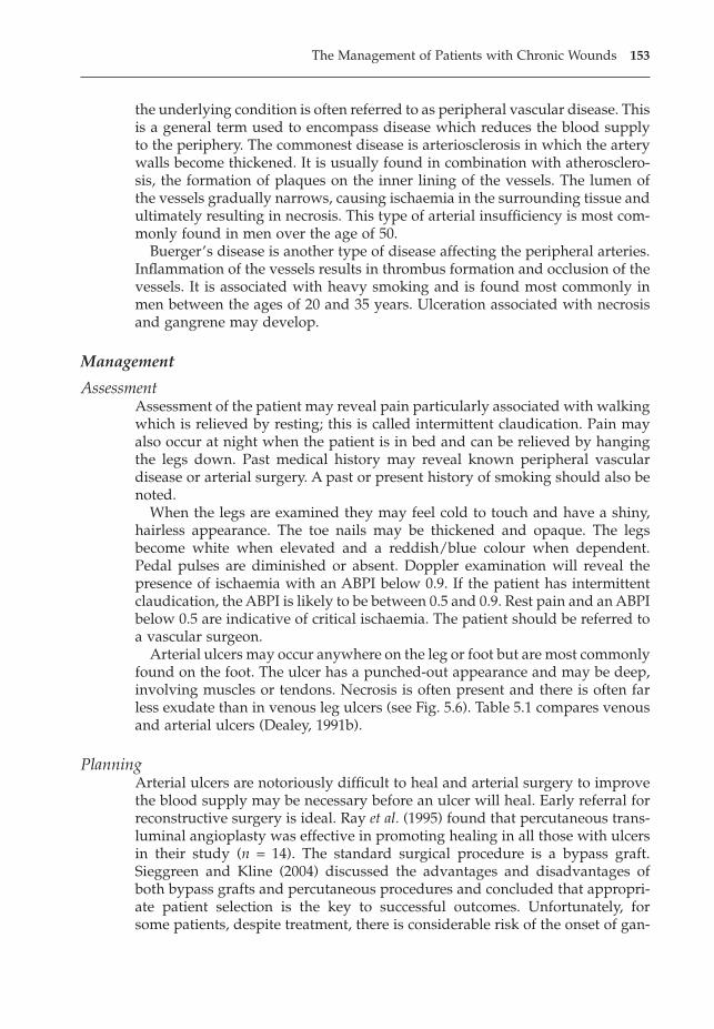
the underlying condition is often referred to as peripheral vascular disease. Thisis a general term used to encompass disease which reduces the blood supplyto the periphery. The commonest disease is arteriosclerosis in which the arterywalls become thickened. It is usually found in combination with atherosclero-sis, the formation of plaques on the inner lining of the vessels. The lumen ofthe vessels gradually narrows, causing ischaemia in the surrounding tissue andultimately resulting in necrosis. This type of arterial insufficiency is most com-monly found in men over the age of 50.
Buerger’s disease is another type of disease affecting the peripheral arteries.Inflammation of the vessels results in thrombus formation and occlusion of thevessels. It is associated with heavy smoking and is found most commonly inmen between the ages of 20 and 35 years. Ulceration associated with necrosisand gangrene may develop.
Management
AssessmentAssessment of the patient may reveal pain particularly associated with walkingwhich is relieved by resting; this is called intermittent claudication. Pain mayalso occur at night when the patient is in bed and can be relieved by hangingthe legs down. Past medical history may reveal known peripheral vasculardisease or arterial surgery. A past or present history of smoking should also benoted.
When the legs are examined they may feel cold to touch and have a shiny,hairless appearance. The toe nails may be thickened and opaque. The legsbecome white when elevated and a reddish/blue colour when dependent.Pedal pulses are diminished or absent. Doppler examination will reveal thepresence of ischaemia with an ABPI below 0.9. If the patient has intermittentclaudication, the ABPI is likely to be between 0.5 and 0.9. Rest pain and an ABPIbelow 0.5 are indicative of critical ischaemia. The patient should be referred toa vascular surgeon.
Arterial ulcers may occur anywhere on the leg or foot but are most commonlyfound on the foot. The ulcer has a punched-out appearance and may be deep,involving muscles or tendons. Necrosis is often present and there is often farless exudate than in venous leg ulcers (see Fig. 5.6). Table 5.1 compares venousand arterial ulcers (Dealey, 1991b).
PlanningArterial ulcers are notoriously difficult to heal and arterial surgery to improvethe blood supply may be necessary before an ulcer will heal. Early referral forreconstructive surgery is ideal. Ray et al. (1995) found that percutaneous trans-luminal angioplasty was effective in promoting healing in all those with ulcersin their study (n = 14). The standard surgical procedure is a bypass graft. Sieggreen and Kline (2004) discussed the advantages and disadvantages of both bypass grafts and percutaneous procedures and concluded that appropri-ate patient selection is the key to successful outcomes. Unfortunately, for some patients, despite treatment, there is considerable risk of the onset of gan-
The Management of Patients with Chronic Wounds 153
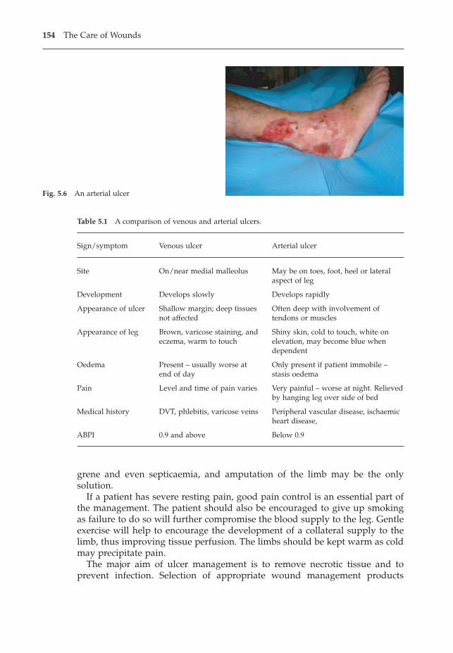
grene and even septicaemia, and amputation of the limb may be the only solution.
If a patient has severe resting pain, good pain control is an essential part ofthe management. The patient should also be encouraged to give up smokingas failure to do so will further compromise the blood supply to the leg. Gentleexercise will help to encourage the development of a collateral supply to thelimb, thus improving tissue perfusion. The limbs should be kept warm as coldmay precipitate pain.
The major aim of ulcer management is to remove necrotic tissue and toprevent infection. Selection of appropriate wound management products
154 The Care of Wounds
Fig. 5.6 An arterial ulcer
Table 5.1 A comparison of venous and arterial ulcers.
Sign/symptom Venous ulcer Arterial ulcer
Site On/near medial malleolus May be on toes, foot, heel or lateral aspect of leg
Development Develops slowly Develops rapidly
Appearance of ulcer Shallow margin; deep tissues Often deep with involvement ofnot affected tendons or muscles
Appearance of leg Brown, varicose staining, and Shiny skin, cold to touch, white oneczema, warm to touch elevation, may become blue when
dependent
Oedema Present – usually worse at Only present if patient immobile – end of day stasis oedema
Pain Level and time of pain varies Very painful – worse at night. Relievedby hanging leg over side of bed
Medical history DVT, phlebitis, varicose veins Peripheral vascular disease, ischaemic heart disease,
ABPI 0.9 and above Below 0.9
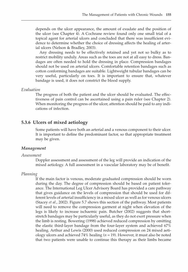
depends on the ulcer appearance, the amount of exudate and the position ofthe ulcer (see Chapter 4). A Cochrane review found only one small trial of atopical agent for arterial ulcers and concluded that there was insufficient evi-dence to determine whether the choice of dressing affects the healing of arter-ial ulcers (Nelson & Bradley, 2003).
Any dressing needs to be effectively retained and yet not so bulky as torestrict mobility unduly. Areas such as the toes are not at all easy to dress. Ban-dages are often needed to hold the dressing in place. Compression bandagesshould not be used on arterial ulcers. Comfortable retention bandages such ascotton conforming bandages are suitable. Lightweight tubular bandages can bevery useful, particularly on toes. It is important to ensure that, whateverbandage is used, it does not constrict the blood supply.
EvaluationThe progress of both the patient and the ulcer should be evaluated. The effec-tiveness of pain control can be ascertained using a pain ruler (see Chapter 2).When monitoring the progress of the ulcer, attention should be paid to any indi-cations of infection.
5.3.6 Ulcers of mixed aetiology
Some patients will have both an arterial and a venous component to their ulcer.It is important to define the predominant factor, so that appropriate treatmentmay be given.
Management
AssessmentDoppler assessment and assessment of the leg will provide an indication of themixed aetiology. A full assessment in a vascular laboratory may be of benefit.
PlanningIf the main factor is venous, moderate graduated compression should be wornduring the day. The degree of compression should be based on patient toler-ance. The International Leg Ulcer Advisory Board has provided a care pathwaythat gives guidance on the levels of compression that should be used for dif-ferent levels of arterial insufficiency in a mixed ulcer as well as for venous ulcers(Stacey et al., 2002). Figure 5.7 shows this section of the pathway. Most patientswill need to remove the compression garment at night when elevation of thelegs is likely to increase ischaemic pain. Butcher (2002) suggests that short-stretch bandages may be particularly useful, as they do not exert pressure whenthe limb is resting. Bowering (1998) achieved reduced compression by omittingthe elastic third-layer bandage from the four-layer system and achieved 67%healing. Arthur and Lewis (2000) used reduced compression on 24 mixed aeti-ology ulcers and achieved 74% healing (n = 19). However, it must also be notedthat two patients were unable to continue this therapy as their limbs became
The Management of Patients with Chronic Wounds 155
too ischaemic. Care and careful monitoring must be exercised if using com-pression for these patients. When arterial disease predominates, a vascularopinion must be sought. However, exercise and short periods of limb elevationcan be encouraged, within the limits of patient toleration.
EvaluationIf healing is very slow, further advice from a vascular surgeon should beobtained. Butcher (2002) emphasised the importance of regular reassessment tomonitor any changes in the level of arterial insufficiency. The RCN leg ulcerguidelines suggest that this should be every three months (RCN, 1998).
5.3.7 Malignant leg ulcers
A small number of leg ulcers may be malignant or become malignant. Voisardet al. (2001) reported on patients attending a vascular clinic between 1991 and1999 where they found six cases of squamous cell carcinoma and four with Marjolin’s ulcer. The authors observed that they did not see any existing legulcers become malignant in their series. Yang et al. (1996) monitored patientsattending a specialist leg ulcer clinic and found 43 patients with 55 malignantlesions, an incidence of 4.4%. In this group 75% were basal cell carcinoma and25% squamous cell carcinoma. They concluded that malignant changes arecommon in chronic leg ulcers. Over a 12-month period, Taylor (1998) found 12patients in a cohort of less than 200 patients attending a community leg ulcerclinic.
Hayes and Dodds (2003) discussed the identification and diagnosis of malig-nant ulcers and suggested that there are a number of clinical signs ofmalignancy.
� Irregular nodular appearance on the wound surface.� A raised or rolled edge.
156 The Care of Wounds
Assessment Diagnosis Recommendationsfor treatment
Non-invasive diagnostics ∑ Ankle-brachial pressure index (ABPI)∑ Confirmation of venous disease∑ Investigations to exclude other disorders
Mixed arterial and venousulcerArterial insufficiency(ABPI 0.5–0.8)
Mixed arterial and venousulcerSevere arterial insufficiency(ABPI <0.5)
Reduced compression(15–25 mmHg)Refer to vascular specialistparticularly if continuing restpain
Refer to vascular specialistNo compression
Fig. 5.7 Guidance on levels of compression for mixed aetiology ulcers from the treatment pathwaydeveloped by the International Leg Ulcer Advisory Board (adapted from Stacey et al., 2002).
� Raised granulation tissue on the ulcer bed with firm surrounding skin.� Islets of epithelialisation that do not persist.� Apparently healthy granulation tissue that is exuberant, translucent, shining
and rolls over the wound margins.
Yang et al. (1996) suggest that any ulcer that fails to heal when treated appro-priately should rouse suspicion. Walsh (2002) discussed the importance oftaking a biopsy from atypical ulcers or non-healing ulcers and of early referralfor specialist advice.
Treatment of malignant ulcers is by excision and skin grafting when caughtat a reasonably early stage. Amputation may be necessary for large ulcers.Although only a small percentage of leg ulcers are malignant, it is importantfor nurses to be aware of the possibility and alert to the need to obtain biopsiesof any atypical ulcers.
5.3.8 Leg ulceration in rheumatoid arthritis
It has been noted that patients with rheumatoid arthritis (RA) are more vul-nerable to leg ulceration and that the aetiology is frequently multifactorial(McRorie, 2000). In their leg ulcer survey, Moffatt et al. (2004) found that 35%of ulcers were multifactorial and of these, 26% were associated with rheuma-toid arthritis. This indicates a significant minority of patients who requirecareful assessment and management.
Oien et al. (2001) studied 20 patients with chronic leg ulcers and RA in orderto determine the ulcer aetiology. They found that three patients had vasculitisas well as both venous and arterial components; five had both vasculitis andvenous disease; two had vasculitis and diabetes; seven had venous diseasealone; one had vasculitis alone; one had arterial disease alone; one had neithervasculitis nor venous or arterial problems. The research team also noted thatthe ulcers were very painful.
Management
AssessmentA specialist rheumatology assessment as well as a vascular assessment isrequired for these patients. McRorie (2000) also suggests that nutritional assess-ment and assessment of footwear should be undertaken.
PlanningManagement of these patients is complex and depends on the underlying aeti-ologies. Compression can be used with caution where venous disease is thedominant factor, particularly if peripheral oedema is present. A small study of15 patients by Hafner et al. (2000) found that vein or arterial surgery was effec-tive in achieving healing in 9/15 patients and markedly improving the ulcersof 5/15. They suggest a prospective trial was needed to confirm these results.Oien et al. (2001) used pinch grafts to treat the patients in their study group.They achieved healing in 8/20 patients, the ulcers in these patients being much
The Management of Patients with Chronic Wounds 157
smaller (range 0.4–13.2cm2) than the unhealed ulcers (range 6.7–356.6cm2).They also found a significant reduction in pain in 18/20 patients following thepinch grafting.
EvaluationRegular monitoring of these patients is essential, especially as arterial diseasemay be accelerated in patients with RA (McRorie, 2000).
5.4 DIABETIC FOOT ULCERS
Ulceration of the foot is a serious complication of diabetes mellitus, which canlead to disability and possible amputation of the affected limb. Jeffcoate andHarding (2003) reviewed current data on surveys of foot ulcers and noted that5.3% (type 2 diabetes) and 7.4% (type 1 and 2 combined) have either an activeulcer or have previously had an ulcer. The lifetime risk for people with diabetesdeveloping an ulcer is about 15%. The incidence of major amputation is 0.5–5.0per 1000 people with diabetes and the perioperative mortality rates associatedwith amputation have been found to range from 9% to 15% (Jeffcoate &Harding, 2003).
Girod et al. (2003) evaluated the direct and indirect costs of treating foot ulcersin diabetic patients. They monitored the cost of 239 patients cared for by 80physicians and found the average monthly cost was €2287.96. This gives a totalmonthly cost for treating the 239 patients of €546822.44.
5.4.1 Aetiology
The underlying causes of diabetic ulcers are peripheral neuropathy and periph-eral vascular disease. Infection is an ever-present risk for the diabetic and canexacerbate the development of ulceration and increase the risk of amputation.
Peripheral neuropathy affects the peripheral sensory, motor and autonomicnerves of the leg. This has a twofold effect of causing a loss of sensation andcompromising the biomechanics of the foot. Muscle atrophy in the foot, par-ticularly over the arch of the foot, causes a transfer of body weight and reac-tive callus formation on the plantar surface. Ultimately, deformities of the foot,such as claw toes or Charcot foot, may occur along with alterations in gait.Walters et al. (1993) found foot ulceration was significantly associated with footdeformity. Poorly fitting shoes or a foreign body within the shoe can causeundetected injury, resulting in ulcer formation. The patient may be completelyunaware of the ulcer for some time.
Vascular disease in diabetics primarily affects the smaller arterioles withinthe foot. Intermittent claudication is unlikely, as larger arteries are not usuallyoccluded. As with all types of vascular disease, it is exacerbated by smoking.Gangrene of the toes can be caused by thrombosis in the artery supplying theaffected digit. Pressure from poorly fitting shoes is the commonest cause ofischaemic ulceration.
158 The Care of Wounds
Microvascular dilation plays a significant role in the healing of minorwounds. The ability of these vessels to dilate can be measured by using a laserDoppler to test the response to heat. A study by Sandeman et al. (1991) mea-sured the ability to respond to heat by vasodilation in insulin-dependent (ID)diabetics, non-insulin dependent (NID) diabetics and a control group of healthyindividuals. A significantly worse response was found in the NID diabetics thanin the other two groups.
5.4.2 Prevention
Given the grave implications of ulceration, prevention is very important. Themain methods used in prevention are patient education and adequate moni-toring of the patient by the healthcare team. Litchfield and Ramkissoon (1996)monitored the outcome of providing an educational footcare video for diabet-ics. They found that whilst most patients were aware of the need for good foot-care, there was a lack of compliance with the recommended care. NICE hasproduced guidelines for the prevention and management of foot problems indiabetics (NICE, 2004). The guidelines include guidance on the content of anyeducation programmes to be targeted at those with low risk of developing footulcers, those at high risk and those with foot ulcers. The guidelines also statethat effective prevention and care involves a partnership between patients andprofessionals (NICE, 2004).
Regular monitoring of diabetic feet is essential and should occur at leastannually. Assessment should include testing of foot sensation, palpation of footpulses, inspection for any foot deformity and inspection of footwear (NICE,2004). Assessment allows the classification of foot risk as follows:
� low current risk (normal sensation, palpable pulses).� increased risk (neuropathy or absent pulses or other risk factor).� high risk (neuropathy or absent pulses plus deformity or skin changes or
previous ulcer).� ulcerated foot (NICE, 2004).
Once patients have been identified as being at risk, they should be made awareof their responsibilities for prevention of foot problems. Feet should be washeddaily and dried carefully. They should be inspected for any red areas or swellingand cracked or broken skin. Toe nails should be cut straight across. Socks shouldbe changed daily and not wrinkle. Shoes should be well fitting. They should bechecked before wearing for any foreign bodies. New shoes should be properlyfitted and feet carefully observed for signs of rubbing. Patients should not wearsandals or go barefoot. Those with poor vision may need assistance. Those whosmoke should be encouraged to stop.
At each attendance at the diabetic clinic, the patient should have a full footcheck. This may involve the doctor, diabetic nurse specialist and chi-ropodist/podiatrist. Treatment of callus formation and management of anyfungal infections is usually carried out by the chiropodist/podiatrist. If thepatient has any deformity of the feet it may be helpful for the orthotist to assess
The Management of Patients with Chronic Wounds 159
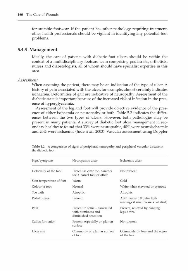
for suitable footwear. If the patient has other pathology requiring treatment,other health professionals should be vigilant in identifying any potential footproblems.
5.4.3 Management
Ideally, the care of patients with diabetic foot ulcers should be within thecontext of a multidisciplinary footcare team comprising podiatrists, orthotists,nurses and diabetologists, all of whom should have specialist expertise in thisarea.
AssessmentWhen assessing the patient, there may be an indication of the type of ulcer. Ahistory of pain associated with the ulcer, for example, almost certainly indicatesischaemia. Deformities of gait are indicative of neuropathy. Assessment of thediabetic state is important because of the increased risk of infection in the pres-ence of hyperglycaemia.
Assessment of the leg and foot will provide objective evidence of the pres-ence of either ischaemia or neuropathy or both. Table 5.2 indicates the differ-ences between the two types of ulcers. However, both pathologies may bepresent in many patients. A survey of diabetic foot ulcer management in sec-ondary healthcare found that 33% were neuropathic, 40% were neuroischaemicand 20% were ischaemic (Jude et al., 2003). Vascular assessment using Doppler
160 The Care of Wounds
Table 5.2 A comparison of signs of peripheral neuropathy and peripheral vascular disease inthe diabetic foot.
Sign/symptom Neuropathic ulcer Ischaemic ulcer
Deformity of the foot Present as claw toe, hammer Not presenttoe, Charcot foot or other
Skin temperature of foot Warm Cold
Colour of foot Normal White when elevated or cyanotic
Toe nails Atrophic Atrophic
Pedal pulses Present ABPI below 0.9 (false high readings if small vessels calcified)
Pain Present in some – associated Present, relieved by hangingwith numbness and legs downdiminished sensation
Callus formation Present, especially on plantar Not presentsurface
Ulcer site Commonly on plantar surface Commonly on toes and the edgesof foot of the foot
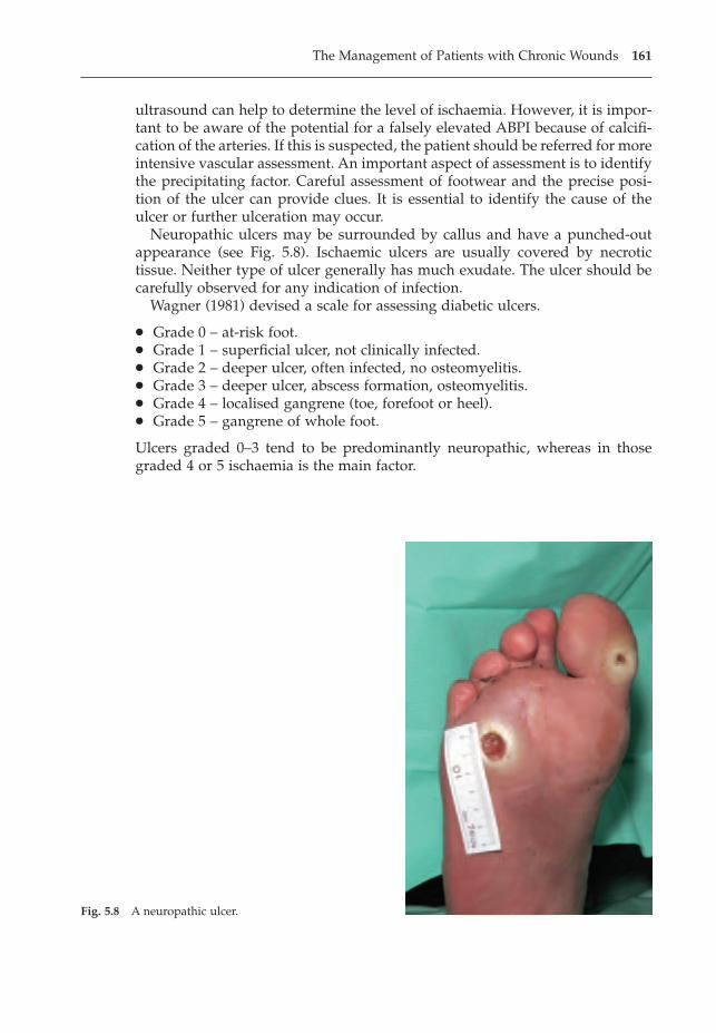
ultrasound can help to determine the level of ischaemia. However, it is impor-tant to be aware of the potential for a falsely elevated ABPI because of calcifi-cation of the arteries. If this is suspected, the patient should be referred for moreintensive vascular assessment. An important aspect of assessment is to identifythe precipitating factor. Careful assessment of footwear and the precise posi-tion of the ulcer can provide clues. It is essential to identify the cause of theulcer or further ulceration may occur.
Neuropathic ulcers may be surrounded by callus and have a punched-outappearance (see Fig. 5.8). Ischaemic ulcers are usually covered by necrotictissue. Neither type of ulcer generally has much exudate. The ulcer should becarefully observed for any indication of infection.
Wagner (1981) devised a scale for assessing diabetic ulcers.
� Grade 0 – at-risk foot.� Grade 1 – superficial ulcer, not clinically infected.� Grade 2 – deeper ulcer, often infected, no osteomyelitis.� Grade 3 – deeper ulcer, abscess formation, osteomyelitis.� Grade 4 – localised gangrene (toe, forefoot or heel).� Grade 5 – gangrene of whole foot.
Ulcers graded 0–3 tend to be predominantly neuropathic, whereas in thosegraded 4 or 5 ischaemia is the main factor.
The Management of Patients with Chronic Wounds 161
Fig. 5.8 A neuropathic ulcer.
PlanningAdequate control of the diabetic state is the primary goal when managingpatients with foot ulceration. Pain control may also be necessary. Managementof the ulcer depends on the causative factors. On the whole, neuropathic ulcersshould be referred to the chiropodist/podiatrist as the callus needs to beremoved before the ulcer can heal. Pressure must also be removed from theulcer and this can be achieved in several ways such as use of extra-depthinsoles, Scotchcast boot, Aircast boot, Total Contact Cast or even the use ofcrutches. Ischaemic ulcers may be resistant to treatment. Necrotic tissue mustbe debrided by use of appropriate wound management products (as discussedin Chapter 3). Any infection should be treated with systemic antibiotics and theapplication of suitable dressings (see Chapter 4). Compression bandages shouldnot be used but, if necessary, a simple retention bandage can be used to holdthe dressing in place.
Some patients may benefit from revascularisation and should have a promptreferral for a vascular opinion (NICE, 2004). Faries et al. (2004) discussed therole of arterial reconstruction and noted that in centres of excellence, 70% ofsurgical bypass grafts will still be patent after five years and the associated five-year limb salvage rate was greater than 80%.
EvaluationMeasurement of the ulcer by tracing may not provide adequate information asthe surface area may not appear to alter greatly. A photographic record may beof more value. Careful assessment for any indication of infection should bemade at each dressing change. Failure to respond to treatment can result inosteomyelitis. Surgical treatment is necessary to eradicate the infection. TheNICE guidelines state that patients should be referred to a multidisciplinaryfoot clinic within 24 hours if there is a new ulcer, new swelling or new dis-colouration of the foot (NICE, 2004).
Once the ulcer is healed, preventive measures need to be instigated.
5.5 FUNGATING WOUNDS
Malignant fungating wounds are particularly difficult wounds to manage. Theyare distressing for both the sufferer and the nurse. A major problem is that therehas been very little research into the management of this type of lesion. It isinteresting to note that what little research there is has predominantly beenundertaken within the UK (Schiech, 2002). A postal survey (Thomas, 1992) hasprovided some information about current practice in radiotherapy centresacross the UK. Thomas noted that several respondents commented on the lackof published data and the generally unscientific approach to the managementof fungating wounds. Little has changed in the years since that survey wasundertaken. It is undoubtedly a difficult area to research but it is an importantarea to tackle, not least in order to improve the quality of life for these patients.
162 The Care of Wounds
5.5.1 Aetiology and incidence
Fungating lesions occur when a cancerous mass invades the epithelium, thusulcerating through to the body surface. It has been estimated that between 5%and 10% of patients with metastatic cancer will develop a fungating wound(Haisfield-Wolfe & Rund, 1997). It most commonly occurs with cancer of thebreast but may also be found in cancers of the skin, head and neck, vulva andbladder (Dowsett, 2002). Fungating wounds not only develop at the site of theprimary tumour but if the nodes of the groin or axilla are affected, ulcerationmay also occur at that site.
Fungating lesions are often associated with neglect; that is, the patient delaysseeking medical help. This is commonly seen in patients with breast cancers. Atypical example is the lady who ‘ignores’ her breast lump for several years andonly seeks help when her family notice the offensive odour.
5.5.2 Management of fungating wounds
Management of the patient
This is a vital part of the management of these wounds. Many factors withinthe patient can affect the progress of the wound. Naylor (2001) discussed theissue of wound pain for patients with fungating wounds and the need toaddress the psychological, social and spiritual aspects of pain. Chapter 2 dis-cusses patient care in greater detail.
Management of the wound
Chapter 3 covers the general principles of wound management. This sectionwill address the specific problems related to fungating wounds.
AssessmentWhen assessing the wound, the following factors need to be considered.
� Fungating lesions are often necrotic, sloughy or infected.� There are usually copious amounts of exudate, which may have an offensive
odour.� Many of these wounds become malodorous as a result of bacterial invasion.
This causes distress to the patient, relatives and visitors and may be very dif-ficult to control.
� The position of the wound obviously depends on the type of cancer.However, it may spread along the trunk or limbs, sometimes in the form ofisolated nodules. Applying a dressing to protect such a spread-out lesion canbe very difficult and requires considerable nursing skill.
� Capillary bleeding may occur as the cancer increases in size and erodes bloodvessels. It may be sufficiently heavy or frequent as to cause anaemia.Removal of the old dressing must be done with great care in order to avoidloosening any clots.
The Management of Patients with Chronic Wounds 163
� Lymphoedema may be present with cancers of the breast, cervix or vulva.This is a chronic swelling of the adjacent limb(s) due to a failure of lymphdrainage. It may be associated with loss of function of the affected limb. (Seealso section 5.6.)
PlanningIt is essential to identify patient problems rather than nurse problems. Whilstin many instances they may be the same, they are not always. In her studies,Grocott (1998, 2000) reported the impact of fungating wounds on the patients,quoting patients using words such as ‘revulsion’ and describing the constantwashing of soiled clothing and bedclothes. She concluded that an efficientdressing system that would cope with the exudate was essential to allowpatients to cope with their disease (Grocott, 2000).
Once the specific problems have been identified, the treatment options haveto be planned in the light of the patient’s condition. If the expected outcome isvery poor, then totally palliative care with the minimum need to dress thewound must be the treatment of choice. For others, a more aggressive approachcan be used. A course of radiotherapy may be prescribed to help reduce the sizeof the lesion. It should be remembered that many patients find dressing changea major ordeal that leaves them feeling very tired.
As previously noted, copious exudate is a problem that concerns patients.Very absorbent dressings are necessary to provide comfort and dryness for thepatient. Alginates and hydrocellular foams can be effective in controllingexudate. Grocott (1997, 1998) has undertaken a longitudinal multiple case studydesign to monitor the outcomes when using different dressings to manageexudate. She considered that the factor of greatest importance is fitting con-formable dressing materials to the wound, thus reducing the risk of leakage.Dressing bulk can be reduced by the use of outer dressings with high moisturevapour transfer rates.
Odour causes great distress to patients. It is mostly due to bacterial invasion,although exuding necrotic wounds may also be offensive. A wound swab willidentify the invading bacteria, so that appropriate systemic antibiotics can beprescribed. Topical agents can also be used. Clark (2002) reviewed the use ofmetronidazole for malodorous wounds and concluded that there was a lack ofevidence to determine the best method of delivery, the dose and the optimumlength of treatment. However, it is widely used to reduce odour. Silver sul-phadiazine cream can be used for Pseudomonas aeruginosa infections. Honey andother types of silver dressings may be useful for odour reduction.
If the odour cannot be reduced or whilst action is being taken to reduceodour, other steps can be taken. The aim is to mask the smell which can beachieved in a variety of ways. Activated charcoal dressings can be effective inabsorbing odour (Lawrence et al., 1993) and are often used in conjunction withother dressings. Air fresheners can help and stoma therapists can give adviceon the use of deodorant solutions used by ostomists.
When aggressive treatment is suitable, wound debridement is an option.Removal of necrotic or sloughy tissue can reduce odour and exudate. The
164 The Care of Wounds
quickest method is surgical debridement which must be done by a skilledsurgeon because of the distorted anatomy and the risk of capillary bleeding.Surgical debridement is not a suitable option for patients with a history of cap-illary bleeding into the wound. A variety of wound management products canbe used, depending on the amount of exudate. Alginates can be used on heavilyexuding wounds. When there is moderate to low exudate, an amorphoushydrogel and occasionally hydrocolloids can be used. The position, size andspread of the lesion can affect dressing choice.
Capillary bleeding can be frightening for both the patient and the nurse.When there is a history of capillary bleeding, great care should be taken whenremoving the old dressing. If the dressing is adherent, it should be soaked withsaline before removal is attempted. It may also be necessary to remove thedressing slowly in stages. It is better to take a long time to remove a dressingthan to start bleeding which is difficult to control.
Adrenaline can be applied directly to the wound to control profuse bleeding.However, it should be used with caution, under medical supervision. Alginatedressings are useful when there is oozing and can be removed easily bywashing away with saline. If there is persistent bleeding, the haemoglobinshould be checked regularly. Blood transfusions may be necessary to treatanaemia.
If the lesion is being treated with radiotherapy, attention must be given todressing selection. A variety of dressings have been found to be suitable: char-coal dressings, alginates and an amorphous hydrogel. Dressing retention maybe a problem. Ideally, the dressing should not be too bulky because it makesthe wearing of clothes difficult and the patient becomes very self-conscious.Bandages and tubular net and tubular gauze are probably the most versatilemeans of dressing retention. Tape should be used with care as the skin maybecome sore with repeated dressing change. If the patient is undergoing acourse of radiotherapy to the lesion, it may be necessary for the outer dressingto be removed for treatment. Again, the skin may become sore. ‘Garments’made from tubular net allow easy access to the wound and will not furtherdamage the skin.
EvaluationEvaluation of the management of fungating wounds should always considerwhether the predetermined goals have been attained. Good documentation canbe used to maintain a record of effective care, thus providing guidelines for themanagement of other patients. Managing a fungating wound and providingcare for the patient require considerable nursing skills. More research is neededin order to be able to identify the most effective care.
5.6 LYMPHOEDEMA
Lymphoedema occurs when there is absence, obliteration or obstruction of thelymph vessels, which results in accumulation of fluid distal to the damage as
The Management of Patients with Chronic Wounds 165
the transporting capacity of the system is reduced. Although associated withcancer, lymphoedema can be either primary or secondary. To date, no cause of primary lymphoedema has been identified (BLS, 2001). Secondary lym-phoedema is primarily seen to be a consequence of cancer or cancer treatmentbut it may also be caused by low-grade infection or inflammation, trauma orfilariasis (Board & Harlow, 2002a). Filariasis is a parasitic invasion of the lymphsystem, enabled by mosquitoes, and is estimated to affect more than 120 millionpeople worldwide (WHO, 2001).
5.6.1 Lymphoedema management
Although there is much greater awareness of lymphoedema management thanthere used to be, some patients with lymphoedema may have been told thatnothing can be done to reduce the swelling. This is not true. Whilst it is notpossible to cure lymphoedema, it can be controlled. Ideally, patients should bereferred to a specialist treatment centre to ensure the most effective care. WithinEurope, especially Germany, Austria and the UK, there are a number of centresavailable that offer this treatment (Board & Harlow, 2002b).
AssessmentBoard and Harlow (2002a) list the clinical features that need to be consideredas part of an assessment.
� Identify the cause of the lymphoedema and any associated medical conditions,in conjunction with the medical team.
� Oedema – assess the extent of the oedema. Does it affect one or more limbs?Has it spread to the trunk? Measure limb size by taking measurements every4cm along both the affected and the unaffected limb.
� Skin status – the skin may become dry, scaly and rough or it may be taut andshiny. Eventually skinfolds and crevices occur because of the severe oedemaand will result in hyperkeratosis.
� Acute inflammatory episodes – the presence of lymphoedema makes an indi-vidual more vulnerable to infection from small breaks in the skin. Cellulitiscan spread rapidly through the affected limb (Mortimer, 2001).
� Lymphorrhoea – the leakage of lymph through small breaks in the skin; thiscan be seen as fluid constantly trickling down the affected limb.
� Mobility – has the oedema affected movements of the limb? What impact hasthis had on activities of daily living? Is there associated pain with the heav-iness of the limb?
ManagementBoard and Harlow (2002b) suggest that treatment should be offered in threecategories: intensive, in order to reduce moderate or severe oedema; mainte-nance, to provide long-term control of oedema; and palliative, to alleviatesymptoms in the final stage of life. Any treatment plan needs to be developedin partnership with the patient and it may also need to involve family members.Treatment comprises skin care, compression, massage and exercise.
166 The Care of Wounds
Skin care is a vital aspect. Williams and Venables (1996) describe the need fordaily washing and careful drying, especially between the digits. Emollientcreams should be applied to prevent the skin drying out and cracking and thedevelopment of hyperkeratosis. Care should be taken to prevent the swollenlimb getting burnt by the sun. Cuts and scratches can be a source of infectionand should be treated with antiseptic cream. Gloves should be worn for pro-tection when working in the kitchen or garden. Jeffs (1993) has reviewed theeffects of infection in lymphoedema. She suggests that Streptococcus is gener-ally the causative organism. The infection should be treated with appropriateantibiotics.
Compression can be provided by means of multilayer bandages or compres-sion hosiery, such as a sleeve (Board & Harlow, 2002b). In the initial stages oftreatment, bandages assist in reducing limb size. In severe cases the digits aswell as the hand and arm, or foot and leg, should be bandaged. Once the limbhas been reduced to a reasonable size, compression hosiery can be used to main-tain limb reduction. Intermittent pneumatic compression may be of use forsome patients, in addition to massage and not as an alternative. The most effec-tive type of compression is the multichamber sequential pump. The best effectis obtained by clearing fluid from the trunk before commencing treatment.
Massage is an important aspect of lymphoedema management and may bein the form of manual lymphatic drainage or simple massage. The formershould be undertaken by a healthcare professional who has received trainingin the technique but the latter may be taught to patients or their relatives.Massage encourages the flow of lymph away from the limb. Occasionally, theswelling may have spread beyond the limb into the trunk. Massage should starton an area of the trunk free of lymphoedema before moving to a lymphoede-matous area (Regnard et al., 1997). This creates a space for the lymph in theswollen limb to flow into. The massage then continues down the affected limb.The massage technique should be gentle, so that it does not stimulate bloodflow into the limb and increase congestion. It may need to be a little firmerwhen tissue fibrosis is present. Board and Harlow (2002b) recommend thatpatients/relatives be advised to spend 20 minutes each day undertakingmassage. Patients and relatives often find that undertaking this massage createsa sense of closeness that may be of particular benefit for those who are termi-nally ill.
Ko et al. (1998) monitored the effects of treatment for 299 patients over a 12-month period. They found a 59.1% size reduction in arms and 67.7% reductionin legs after initial treatment averaging 15.7 days. This was maintained in 88%of compliant patients. The non-compliant patients regained 33% of the initialreduction. McNeely et al. (2004) randomised 50 patients with lymphoedemaassociated with breast cancer to compression bandaging or compression ban-daging plus manual lymphatic drainage. They found that all patients achieveda significant reduction in arm volume after four weeks but those with mild lym-phoedema in the combination treatment group had a significantly larger reduc-tion in limb volume. However, these numbers are small and a larger study isneeded to confirm this finding.
The Management of Patients with Chronic Wounds 167
Exercise assists in improving drainage of lymph from the limb. Muscle move-ment alters tissue pressure and has a massaging effect on the lymph vessels.The best effect is obtained if the exercises are carried out when the patient iswearing compression bandages or hosiery. Exercise also prevents or reducesstiffness of the joints. Passive movements should be carried out if the limb isparalysed. All patients should be encouraged to move as much as possible butlifting and carrying heavy weights should be avoided. It is important to provideencouragement to the patient to persevere with all aspects of this plan as none of these treatments is effective in isolation. Significant reduction of aswollen limb can only be achieved when all aspects of the treatment plan areimplemented.
EvaluationReduction in limb size, improvements in skin status and greater limb mobilityare all desirable outcomes that can easily be measured.
5.7 CONCLUSION
The management of chronic wounds is complex and requires skilled care thatis best undertaken by a multiprofessional team. Fortunately, there is increasingaccess to specialist services that have made a major impact on improvement inpatient care.
REFERENCES
Allen, S. (1983) Hang the patient upside down from the ceiling – it works every time.General Practitioner, June 24, 40–41.
Allman, R.M., Goode, P.S., Burst, N., Bartolucci, A.A., Thomas, D.R. (1999) Pressureulcers, hospital complications and disease severity: impact on hospital costs andlength of stay. Advances in Wound Care, 12 (1), 22–30.
Amlung, S.R., Miller, W.L., Bosley, L.M. (2001) The 1999 national pressure ulcer preva-lence survey: a benchmarking approach. Advances in Skin and Wound Care, 14 (6),297–301.
Anthony, D. (1996) Receiver operating characteristic analysis. Nurse Researcher, 4 (2),75–88.
Aronovitch, S.A. (1999) Intraoperatively acquired pressure ulcer prevalence: a nationalstudy. Journal of Wound, Ostomy and Continence Nursing, 26 (3), 130–136.
Arthur, J., Lewis, P. (2000) When is reduced-compression bandaging safe and effective?Journal of Wound Care, 9 (10), 469–471.
Baker, S.R., Stacey, M.C. (1994) Epidemiology of chronic leg ulcers in Australia. Australiaand New Zealand Journal of Surgery, 64 (4), 258–261.
Bale, S., Crook, H. (1998) The preliminary results of a comparative study on the perfor-mance characteristics of a new hydrogel versus an existing hydrogel on necroticpressure ulcers, in (eds) Leaper, D., Cherry, G., Cockbill, S. et al., Proceedings of theEWMA/Journal of Wound Care Spring Meeting. Macmillan Magazines Ltd, London.
168 The Care of Wounds
Bale, S., Harding, K.G. (2003) Managing patients unable to tolerate therapeutic com-pression. British Journal of Nursing, 12 (19) (suppl), S4–S13.
Bale, S., Squires, D., Vernon, T., Walker, A., Benbow, M., Harding, K.G. (1997) A com-parison of two dressings in pressure sore management. Journal of Wound Care, 6(10), 463–466.
Bale, S., Banks, V., Haglestein, S., Harding, K.G. (1998) A comparison of two amorphoushydrogels in the debridement of pressure sores. Journal of Wound Care, 7 (2), 65–68.
Barwell, J.R., Davies, C.E., Deacon, J. et al. (2004) Comparison of surgery and compres-sion with compression alone in chronic venous ulceration (ESCHAR study): ran-domised controlled trial. Lancet, 363, 1854–1858.
Baumgarten, M., Margolis, D., Gruber-Baldini, A.L., Zimmerman, S., German, P., Hebel,J.R. (2003a) Pressure ulcers and the transition to long term care. Advances in Skinand Wound Care, 16 (6), 299–304.
Baumgarten, M., Margolis, D., Berlin, J.A. et al. (2003b) Risk factors for pressure ulcersamong elderly hip fracture patients. Wound Repair and Regeneration, 11 (2), 96–103.
Bennett, G., Dealey, C., Posnett, J. (2004) The cost of pressure ulcers in the UK. Age andAgeing, 33, 23–235.
Bergstrom, N., Braden, B., Brandt, J., Krall, K. (1985) Adequacy of descriptive scales forreporting diet intake in the institutionalised elderly. Journal of Nutrition for theElderly, 6 (1), 3–16.
Bergstrom, N., Braden, B., Laguzza, A. (1987) The Braden scale for predicting pressuresore risk. Nursing Research, 36 (4), 205–210.
Bergstrom, N., Braden, B., Kemp, M., Champagne, M., Ruby, E. (1996) Multi-site studyof incidence of pressure ulcers and the relationship between risk level, demo-graphic characteristics, diagnoses and prescription of preventive interventions.Journal of the American Geriatrics Society, 44, 22–30.
Berlowitz, D.R., Wilking, S.V.B. (1989) Risk factors for pressure sores: a comparison ofcross-sectional and cohort-derived data. Journal of the American Geriatrics Society,37, 1043–1050.
Birchall, L., Clark, N., Taylor, S. (2004) Implementing the Essence of Care benchmark forpressure ulcers. Nursing Times, 100 (32), 63–67.
Bliss, M. (1990) Editorial – preventing pressure sores. Lancet, 335, 1311–1312.BLS (2001) Clinical Definitions. BLS, Sevenoaks, Kent.Board, J., Harlow, W. (2002a) Lymphoedema 2: classification, signs, symptoms and diag-
nosis. British Journal of Nursing, 11 (6), 389–395.Board, J., Harlow, W. (2002b) Lymphoedema 3: the available treatments for lym-
phoedema. British Journal of Nursing, 11 (7), 438–450.Bours, G.J., Halfens, R.J., Abu-Saad, H.H., Grol, R.T. (2002) Prevalence, prevention and
treatment of pressure ulcers: descriptive study in 89 institutions. Research inNursing and Health, 25 (2), 99–110.
Bowering, C.K. (1998) Use of layered compression bandages in diabetic patients. Expe-rience in patients with lower leg ulceration, peripheral oedema, and features ofvenous and arterial disease. Advances in Wound Care, 11 (3), 129–135.
Bradley, M., Cullum, N., Nelson, E.A., Petticrew, M., Sheldon, T., Torgerson, D.J. (1999)Systematic reviews of wound care management: (2) dressings and topical agentsused in the healing of chronic wounds. Health Technology Assessment, 3 (17) Part 2,1–174.
Brandeis, G.H., Ooi, W.L., Hossain, M., Morris, J.N., Lipsitz, L.A. (1994) A longitudinalstudy of risk factors associated with the formation of pressure ulcers in nursinghomes. Journal of the American Geriatrics Society, 42, 388–393.
The Management of Patients with Chronic Wounds 169
Bridel, J. (1993) Assessing the risk of pressure sores. Nursing Standard, 7 (25), 32–35.Briggs, M., Closs, S.J. (2003) The prevalence of leg ulceration: a review of the literature.
EWMA Journal, 3 (2), 14–20.Brookes, R., Thomson, J. (1997) Pressure area care and estimating the cost of pressure
sores: critique 2. Journal of Wound Care, 6 (3), 135–137.Buss, I.C., Halfens, R.J., Abu-Saad, H.H. (1997) The effectiveness of massage in
preventing pressure sores: a literature review. Rehabilitation Nursing, 22 (5), 229–234.
Butcher, M. (2002) Managing mixed-aetiology leg ulcers. Practice Nursing, 13 (4), 161–166.
Callam, M.J., Ruckley, C.V., Harper, D.R., Dale, J.J. (1985) Chronic ulceration of the leg:extent of the problems and provision of care. British Medical Journal, 290,1855–1856.
Cameron, J. (1998) Skin care for patients with chronic leg ulcers. Journal of Wound Care,7 (9), 459–462.
Clark, J. (2002) Metronidazole gel in managing malodorous fungating wounds. BritishJournal of Nursing, 11 (6) (suppl), S54–S60.
Clark, M. (1998) Repositioning to prevent pressure sores – what is the evidence? NursingStandard, 13 (3), 58–64.
Clark, M. (2003) Compression bandages: principles and definitions. EWMA Position Doc-ument: Understanding Compression Therapy. Medical Education Partnership Ltd,London.
Clark, M., Bours, G., Defloor, T. (2003) The prevalence of pressure ulcers in Europe. Hos-pital Decisions, Winter, 123–127.
Colin, D., Abraham, P., Preault, L., Bregeon, C., Saumet, Jl. (1996a) Comparison of 90degrees and 30 degrees laterally inclined positions in the prevention of pressureulcers using transcutaneous oxygen and carbon dioxide pressures. Advances inWound Care, 9 (3), 35–38.
Colin, D., Kurring, P.A., Yvon, C. (1996b) Managing sloughy pressure ulcers. Journal ofWound Care, 5 (10), 444–446.
Colin, D., Dizien, O., Yvon, C. (1997) The clinical investigation of a transparent semi-permeable adhesive film dressing with an extra-thin hydrocolloid in the manage-ment of stage I and II pressure sores, in (eds) Leaper, D.J., Cherry, G.W., Dealey,C., Lawrence, J.C., Turner, T.D., Proceedings of the 6th European Conference onAdvances in Wound Management. Macmillan Magazines Ltd, London.
Collins, F. (1999) The contribution made by an armchair with integral pressure-reduc-ing cushion in the prevention of pressure sore incidence in the elderly, acutely illpatient. Journal of Tissue Viability, 9 (4), 133–137.
Cooper, P., Gray, D. (2001) Comparison of two skin care regimes for incontinence. BritishJournal of Nursing, 10 (6) (suppl), S6–S20.
Cornwall, J.V., Dore, C.J., Lewis, J.D. (1986) Leg ulcers: epidemiology and aetiology.British Medical Journal, 73, 693–696.
Culley, F. (1998) Nursing aspects of pressure sore prevention and therapy. British Journalof Nursing, 7 (15), 879–884.
Cullum, N., Nelson, E.A., Sheldon, T. (2001a) Systematic reviews of wound care man-agement (5): pressure-relieving beds, mattreses and cushions for the preventionand treatment of pressure sores. Health Technology Assessment, 5 (9), 1–75.
Cullum, N., Nelson, E.A., Fletcher, A.W., Sheldon, T.A. (2001b) Compression for venousleg ulcers (Cochrane Review). Cochrane Library, Issue 1. Update Software Ltd,Oxford.
170 The Care of Wounds
Danchaivijitr, S., Suthisanon, L., Jitreecheue, L., Tantiwatanapaibool, Y. (1995) Effects ofeducation on the prevention of pressure sores. Journal of the Medical Association ofThailand, 78 (suppl.1), S1–S6.
David, J.A., Chapman, R.G., Chapman, E.J., Lockett, B. (1983) An Investigation of theCurrent Methods Used in Nursing for the Care of Patients with Established PressureSores. Nursing Practice Research Unit, Northwick Park, Middlesex.
Dealey, C. (1991a) The size of the pressure sore problem in a teaching hospital. Journalof Advanced Nursing, 16, 663–670.
Dealey, C. (1991b) Causes of leg ulcers. Nursing, 4 (35), 23–24.Dealey, C. (1997) Managing Pressure Sore Prevention, Mark Allen Publishing Ltd,
Salisbury.Dealey, C., Keogh, A. (1998) A randomised study comparing the Triple Care cleanser and
cream system with soap and water for elderly incontinent patients, in (eds) Leaper,D., Dealey, C., Franks, P.J., Hofman, D., Moffatt, C.J., Proceedings of the 7th European Conference on Advances in Wound Management. EMAP Healthcare Ltd,London.
Dealey, C., Earwaker, T., Eden, L. (1991) Are your patients sitting comfortably? Journalof Tissue Viability, 1 (2), 36–39.
Deeks, J.J. (1996) Pressure sore prevention: using and evaluating risk assessment tools.British Journal of Nursing, 5 (5), 313–320.
Defloor, T., Schoonhoven, L. (2004) Interrater reliability of the EPUAP pressure ulcerclassification system using photographs. Journal of Clinical Nursing, 13 (8), 952–959.
Defloor, T., de Bacquer, D., Grypdonck, M.H. (2005) The effect of various combinationsof turning and pressure reducing devices on the incidence of pressure ulcers. Inter-national Journal of Nursing Studies, 42 (1), 37–46.
Department of Health (1993) Pressure Sores: a key quality indicator. Department of Health,London.
Department of Health and NHS Modernisation Agency (2003) The Essence of Care.Patient-focused Benchmarks for clinical governance. Department of Health, London.
Dowsett, C. (2002) Malignant fungating wounds: assessment and management. BritishJournal of Community Nursing, 7 (8), 394–400.
Effective Health Care (1995) The prevention and treatment of pressure sores. EffectiveHealth Care Bulletin, 2 (1), 1–16.
Ek, A.C., Gustavssen, G., Lewis, D.H. (1987) Skin blood flow in relation to external pres-sure and temperature in the supine position on a standard hospital mattress. Scan-dinavian Journal of Rehabilitation, 19, 121–126.
Ellison, D.A., Hayes, L., Lane, C., Tracey, A., McCollum, C.N. (2002) Evaluating the costand efficacy of leg ulcer care provided in two large UK health authorities. Journalof Wound Care, 11 (2), 47–51.
European Pressure Ulcer Advisory Panel (1999) Pressure ulcer treatment guidelines.EPUAP Review, 1 (2), 31–33.
Exton-Smith, A.N., Sherwin, R.W. (1961) The prevention of pressure sores: the signifi-cance of spontaneous bodily movement. Lancet, II, 1124–1126.
Faries, P.L., Teodorescu, V.J., Morrissey, N.J., Hollier, L.H., Marin, M.L. (2004) The roleof surgical revascularisation in the management of diabetic foot wounds. American Journal of Surgery, 187 (5) (suppl), 34S–37S.
Fentem, P.H. (1986) Elastic hosiery. Pharmacy Update, 5, 200–205.Ferrell, B.A., Josephson, K., Norvid, P., Alcorn, H. (2000) Pressure ulcers among patients
admitted to home care. Journal of the American Geriatrics Society, 49 (5), 1042–1047.
The Management of Patients with Chronic Wounds 171
Flanagan, M. (1997) Choosing pressure sore risk assessment tools. Professional Nurse, 12(6) (suppl), 3–7.
Fowler, E. (1990) Chronic wounds: an overview, in (ed.) Krasner, D., Chronic Wound Care:a clinical sourcebook for healthcare professionals. Health Management Publications,King of Prussia, PA.
Gebhardt, K. (1995) What causes pressure sores? Nursing Standard, 9 (suppl. 31), 48–51.Ghauri, A.S.K., Nyamekye, I., Grabs, A.J., Farndon, J.R., Whyman, M.R., Poskitt, K.R.
(1998) Influence of a specialised leg ulcer service and venous surgery on the out-comes of venous leg ulcers. European Journal of Vascular and Endovascular Surgery,16, 238–244.
Girod, I., Valensi, P., Laforet, C., Moreau-Defarges, T., Guillon, P., Baron, F. (2003) Aneconomic evaluation of the cost of diabetic foot ulcers: results of a retrospectivestudy on 239 patients. Diabetes and Metabolism, 29 (3), 269–277.
Goebel, R.H., Goebel, M.R. (1999) Clinical practice guidelines for pressure ulcer pre-vention can prevent malpractice lawsuits in older patients. Journal of Wound,Ostomy and Continence Nursing, 26 (4), 175–184.
Grocott, P. (1997) Evaluation of a tool to assess the management of fungating wounds.Journal of Wound Care, 6 (9), 421–424.
Grocott, P. (1998) Exudate management in fungating wounds. Journal of Wound Care, 7(9), 445–448.
Grocott, P. (2000) The palliative management of fungating malignant wounds. Journal ofWound Care, 9 (1), 4–9.
Gruen, R.L., Chang, S., MacLellan, D.G. (1996) Optimising the hospital management ofleg ulcers. Australia and New Zealand Journal of Surgery, 66 (3), 171–174.
Guenter, P., Malyszek, R., Bliss, D.Z. et al. (2000) Survey of nutritional status in newlyhospitalised patients with stage III or stage IV pressure ulcers. Advances in Skinand Wound Care, 13 (4), 164–168.
Hafner, J., Schneider, E., Burg, G., Cassina, P.C. (2000) Management of leg ulcers inpatients with rheumatoid arthritis or systemic sclerosis: the importance of concomitant arterial and venous disease. Journal of Vascular Surgery, 32 (2), 322–329.
Haglsawa, S., Barbenel, J. (1999) The limits of pressure sore prevention. Journal of theRoyal Society of Medicine, 92, 576–578.
Haisfield-Wolfe, M.A., Rund, C. (1997) Malignant cutaneous wounds: a managementprotocol. Ostomy and Wound Management, 43, 56–66.
Halfens, R.J.G., Bours, G.J.J.W., van Ast, W. (2001) Relevance of the diagnosis ‘stage 1pressure ulcer’: an empirical study of the clinical course of stage 1 ulcers in acutecare and long-term care hospital populations. Journal of Clinical Nursing, 10 (6),748–757.
Harding, K.G. (1998) The future of wound healing, in (eds) Leaper, D.J., Harding, K.G.,Wounds: biology and management. Oxford University Press, Oxford.
Hayes, S., Dodds, S. (2003) The identification and diagnosis of malignant leg ulcers.Nursing Times, 99 (31) (suppl), 8–10.
Hibbs, P. (1988) Pressure Area Care for the City and Hackney Health Authority. City andHackney Health Authority, London.
Hofman, D., Ryan, T.J., Arnold, F. et al. (1997) Pain in venous leg ulcers. Journal of WoundCare, 6 (5), 222–224.
Horn, S.D., Bender, S.A., Bergstrom, N. et al. (2002) Description of the national pressureulcer long-term care study. Journal of the American Geriatrics Society, 50, 1816–1825.
172 The Care of Wounds
Houwing, R., Overgoor, M., Kon, M., Jansen, G., van Asbeck, B.S., Haalboom, J.R. (2000)Pressure-induced skin lesions in pigs: reperfusion injury and the effects of vitaminE. Journal of Wound Care, 9 (1), 36–40.
Iglesias, C., Nelson, E.A., Cullum, N.A., Torgerson, D.J. (2004) VenUS 1: a randomisedcontrolled trial of two types of bandage for treating venous leg ulcers. Health Tech-nology Assessment, 8 (29), 1–106.
Jay, R. (1997) Other considerations in selecting a support surface. Advanced Wound Care,10 (7), 37–42.
Jeffcoate, W.J., Harding, K.G. (2003) Diabetic foot ulcers. Lancet, 361 (9368), 1545–1551.
Jeffs, E. (1993) The effect of acute inflammatory episodes on the treatment of lym-phoedema. Journal of Tissue Viability, 3 (2), 51–55.
Jones, J.E., Nelson, E.A. (1998) Compression hosiery in the management of venous legulcers. Journal of Wound Care, 7 (6), 293–296.
Jones, J.E., Nelson, E.A. (2001) Skin grafting for venous leg ulcers (Cochrane Review).Cochrane Library, Issue 1. Update Software Ltd, Oxford.
Jude, E.B., Oyibo, S.O., Millichip, M.M., Boulton, A.J.M. (2003) A survey of physicians’involvement in the management of diabetic foot ulcers in secondary health care.Practical Diabetes International, 20 (3), 89–92.
Kabagambe, M.K., Swain, I., Shakespeare, P. (1994) An investigation of the effects on themicrocirculation of the skin (reactive hyperaemia) in spinal cord injured patients.Journal of Tissue Viability, 4 (4), 110–123.
Kaltenthaler, E., Whitfield, M.D., Walter, S.J., Akehurst, R.L., Paisley, S. (2001) UK, USAand Canada: how do their pressure ulcer prevalence and incidence data compare?Journal of Wound Care, 10 (1), 530–535.
Khoo, C., Bailey, B.N. (1990) Reconstructive surgery, in (ed.) Bader, D., Pressure Sores –clinical practice and scientific approach. Macmillan Press, London.
Kierney, P.C., Engrav, L.H., Isik, F.F., Esselman, P.C., Cardenas, D.D., Rand, R.P. (1998)Results of 268 pressure sores in 158 patients managed jointly by plastic surgeryand rehabilitation medicine. Plastic and Reconstructive Surgery, 102 (3), 765–772.
Ko, D.S., Lerner, K., Klose, G., Cosimi, A.B. (1998) Effective treatment of lymphoedemaof the extremities. Archives of Surgery, 133 (4), 452–458.
Krouskop, M. (1983) A synthesis of the factors which contribute to pressure sore for-mation. Medical Hypothesis, 11, 255–267.
Kumar, R.N., Gupchup, G.V., Dodd, M.A. et al. (2004) Direct health care costs of 4common skin ulcers in New Mexico Medicaid fee-for-service patients. Advances inSkin and Wound Care, 17 (3), 143–149.
Landis, E.M. (1931) Micro-injection studies of capillary blood pressure in human skin.Heart, 15, 209–228.
Langer, G., Schloemer, G., Knerr, A., Kuss, O., Behrens, J. (2003) Nutritional interven-tions for preventing and treating pressure ulcers (Cochrane review). The CochraneLibrary, Issue 4. John Wiley & Sons Ltd, Chichester.
Lapsley, H.M., Vogels, R. (1996) Cost and prevention of pressure ulcer in an acute teach-ing hospital. International Journal for Quality in Health Care, 8 (1), 61–66.
Lawrence, J.C., Lilly, H.A., Kidson, A. (1993) Malodour and dressings containing activecharcoal, in (eds) Harding, K.G., Cherry, G., Dealey, C., Turner, T.D., Proceedings ofthe 2nd European Conference on Advances in Wound Management. Macmillan Maga-zines Ltd, London.
Lingren, M., Unosson, M., E.K., A.-C. (2000) Pressure sore prevalence within a publichealth services area. International Journal of Nursing Practice, 6 (6), 333–337.
The Management of Patients with Chronic Wounds 173
Litchfield, B., Ramkissoon, S. (1996) Foot-care education in patients with diabetes. Pro-fessional Nurse, 11 (8), 510–512.
Loudon, I.S.L. (1982) Leg ulcers in the eighteenth and early nineteenth centuries. Journalof the Royal College of General Practitioners, 32, 301–309.
Machet, L., Couhe, C., Perrinaud, A., Hoarau, C., Lorette, G., Vaillant, L. (2004) A highprevalence of sensitisation still persists in leg ulcer patients: a retrospective seriesof 106 patients tested between 2001 and 2002 and a meta-analysis of 1975–2003data. British Journal of Dermatology, 150 (5), 929–935.
Mani, R., Vowden, K., Nelson, E.A. (2004) Intermittant pneumatic compression for treat-ing venous leg ulcers (Cochrane review). Cochrane Library, Issue 4. John Wiley &Sons, Chichester.
Margolis, D.J., Berlin, J.A., Strom, B.L. (2000) Which venous leg ulcers will heal withlimb compression bandages? American Journal of Medicine, 109, 15–19.
Margolis, D., Knauss, J., Bilker, W., Baumgarten, M. (2003) Medical conditions as riskfactors for pressure ulcers in an out-patient setting. Age and Ageing, 32, 259–264.
Marston, W., Vowden, K. (2003) Compression therapy: a guide to safe practice. EWMAPosition Document: Understanding Compression Therapy. Medical Education Part-nership Ltd, London.
Mathus-Vleigen, E.M.H. (2001) Nutritional status, nutrition and pressure ulcers. Nutri-tion in Clinical Practice, 16, 286–291.
McCafferty, E., Watret, L., Brown, C. (2000) A multidisciplinary audit of patients’ seatingneeds. Professional Nurse, 15 (11), 715–718.
McKeeney, L. (2002) Legal issues for the prevention of pressure ulcers. Journal of Com-munity Nursing, 16 (7), 28–30.
McNeely, M.L., Magee, D.J., Lees, A.W., Bagnall, K.M., Haykowsky, M., Hanson, J. (2004)The addition of manual lymph drainage to compression therapy for breat cancerrelated lymphoedema: a randomised controlled trial. Breast Cancer Research andTreatment, 86 (2), 95–106.
McRorie, E.R. (2000) The management and assessment of leg ulcers in rheumatoid arthri-tis. Journal of Wound Care, 9 (6), 289–292.
Meyer, F.J., McGuinness, C.L., Lagatolla, N.R.F., Eastham, D., Burnand, K.G. (2003) Ran-domised clinical trial of three-layer paste and four-layer bandages for venous legulcers. British Journal of Surgery, 90 (8), 934–940.
Moffatt, C.J., Franks, P.J., Doherty, D.C., Martin, R., Blewitt, R., Ross, F. (2004) Prevalenceof leg ulceration in a London population. Quarterly Journal of Medicine, 97, 431–437.
Mortimer, P.S. (2001) Acute inflammatory episodes, in (eds) Twycross, R., Jenns, K.,Todd, J., Lymphoedema. Radcliffe Medical Press, Oxford.
Mustoe, T. (2004) Understanding chronic wounds: a unifying hypothesis on their patho-genesis and implications for therapy. American Journal of Surgery, 187 (5A),65S–70S.
National Institute for Clinical Excellence (2003) Clinical Guideline 7: pressure ulcer pre-vention. NICE, London.
National Institute for Clinical Excellence (2004) Clinical Guideline 10: type 2 diabetes: pre-vention and management of foot problems. NICE, London.
Naylor, W. (2001) Assessment and management of pain in fungating wounds. BritishJournal of Nursing, 10 (22) (suppl), S33–S52.
Nelson, E.A., Bradley, M.D. (2003) Dressings and topical agents for arterial leg ulcers.Cochrane Database of Systematic Reviews (1) CD001836.
174 The Care of Wounds
Nelson, E.A., Bell-Syer, S.E.M., Cullum, N.A. (2004) Compression for preventing recur-rence of venous ulcers (Cochrane Review). Cochrane Library, Issue 4. John Wiley &Sons, Chichester.
Nightingale, F. (1861) Notes on Nursing. Appleton Century, New York.Norton, D., Mclaren, R., Exton-Smith, A.N. (1975) An Investigation of Geriatric Nursing
Problems. Churchill Livingstone, Edinburgh.Nyquist, R., Hawthorn, P.J. (1987) The prevalence of pressure sores in an area health
authority. Journal of Advanced Nursing, 12, 183–187.O’Brien, J.F., Grace, P.A., Perry, I.J., Burke, P.E. (2000) Prevalence and aetiology of leg
ulcers in Ireland. Irish Journal of Medical Science, 169 (2), 110–112.O’Dea, K. (1999) The prevalence of pressure damage in acute care hospital patients in
the UK. Journal of Wound Care, 8 (4), 192–194.Oien, R.F., Hakansson, A., Hansen, B.U. (2001) Leg ulcers in patients with rheumatoid
arthritis – a prospective study of aetiology, wound healing and pain reductionafter pinch grafting. Rheumatology, 40, 816–820.
Papanikolaou, P., Clark, M., Lyne, P.A. (2002) Improving the accuracy of pressure ulcerrisk calculators: some preliminary evidence. International Journal of Nursing Studies,39 (2), 187–194.
Patel, G.K., Llewellyn, M., Harding, K.G. (2001) Managing gravitational eczema andallergic contact dermatitis. British Journal of Community Nursing, 6 (8), 394–406.
Pearson, A., Francis, K., Hodgkinson, B., Curry, G. (2000) Prevalence and treatment ofpressure ulcers in northern New South Wales. Australian Journal of Rural Health, 8(2), 103–110.
Pedley, G.E. (2004) Comparison of pressure ulcer grading scales: a study of clinical utility and inter-rater reliability. International Journal of Nursing Studies, 41, 129–140.
Peirce, S.M., Skalak, T.C., Rodeheaver, G.T. (2000) Ischaemia-reperfusion injury inchronic pressure ulcer formation: a skin model in the rat. Wound Repair and Regen-eration, 8 (1), 68–76.
Persoon, A., Heinen, M.M., van der Vleuten, C.J.M., de Rooij, M.J., van de Kerkhof,P.C.M., van Achterberg, T. (2004) Leg ulcers: a review of their impact on daily life.Journal of Clinical Nursing, 13, 341–354.
Polignano, R., Bonadeo, P., Gasbarro, S., Allegra, C. (2004) A randomised controlled trialof four-layer compression versus Unna’s boot for venous ulcers. Journal of WoundCare, 13 (1), 21–24.
Poore, S., Cameron, J., Cherry, G. (2002) Venous leg ulcer recurrence: prevention andhealing. Journal of Wound Care, 11 (5), 197–199.
Preston, K.W. (1988) Positioning for comfort and pressure relief: the 30 degree alterna-tive. Care – Science and Practice, 6 (4), 116–119.
Quartey-Papafio, C.M. (1999) Importance of distinguishing between cellulitis and vari-cose eczema. British Medical Journal, 318, 1672–1673.
Ray, S.A., Minty, I., Buckenham, T.M. (1995) Clinical outcome and restenosis followingPTA for ischaemic rest pain or ulceration. British Journal of Surgery, 82 (9),1217–1222.
Reed, R.L., Hepburn, K., Adelson, R., Center, B., McNight, P. (2003) Low serum albuminlevels, confusion, and faecal incontinence: are these risk factors for pressure ulcersin mobility impaired hospitalised adults? Gerontology, 49 (4), 255–259.
Regan, M.B., Byers, P.H., Mayrovitz, H.N. (1995) Efficacy of a comprehensive pressureulcer prevention programme in an extended care facility. Advances in Wound Care,8 (3), 49–55.
The Management of Patients with Chronic Wounds 175
Regnard, C., Allport, S., Stephenson, L. (1997) ABC of palliative care: mouthcare, skincare and lymphoedema. British Medical Journal, 315, 1002–1005.
Rithalia, S.V.S. (1989) Comparison of pressure distribution in wheelchair seat cushions.Care – Science and Practice, 7 (4), 87–89.
Royal College of Nursing (1998) Clinical Practice Guidelines. The management of patientswith venous leg ulcers. Royal College of Nursing, London.
Royal College of Nursing (2003) Safer Staff, Better Care: RCN manual handling trainingguidance and competencies. Royal College of Nursing, London.
Ruckley, C.V., Evans, C.J., Allan, P.L., Lee, A.J., Fowkes, F., Gerald, R. (2002) Chronicvenous insufficiency: clinical and duplex correlations. The Edinburgh Vein Studyof venous disorders in the general population. Journal of Vascular Surgery, 36 (3),52–525.
Russell, L.J., Reynolds, T.M. (2001) How accurate are pressure ulcer grades? An image-based survey of nurses’ performance. Journal of Tissue Viability, 11 (2), 67–75.
Ryan, T.J. (1987) The Management of Leg Ulcers, 2nd edn. Oxford University Press, Oxford.Sanada, H., Nagakawa, T., Yamamoto, M., Higashidani, K., Tsuru, H., Sugama, J. (1997)
The role of blood flow in pressure ulcer development during surgery. Advances inWound Care, 10 (6), 29–34.
Sandeman, D.D., Pym, C.A., Green, E.M. et al. (1991) Microvascular vasodilation in thefeet of newly diagnosed non-insulin dependent diabetics. British Medical Journal,302 (6785), 1122–1123.
Scanlon, E., Stubbs, N. (2004) Pressure ulcer risk assessment in patients with darkly pig-mented skin. Professional Nurse, 19 (6), 339–341.
Schiech, L. (2002) Malignant cutaneous wounds. Clinical Journal of Oncology Nursing, 6(5), 305–309.
Schoonhoven, L., Defloor, T., Grypdonck, M.H. (2002a) Incidence of pressure ulcers dueto surgery. Journal of Clinical Nursing, 11 (4), 479–487.
Schoonhoven, L., Haalboom, J.R.E., Bousema, T. et al. (2002b) Prospective cohort studyof routine use of risk assessment scales for prediction of pressure ulcers. BritishMedical Journal, 325 (7368), 797–801.
Schue, R.M., Langemo, D.K. (1999) Prevalence, incidence and prediction of pressureulcers on a rehabilitation unit. Journal of Wound, Ostomy and Continence Nursing,26 (3), 121–129.
Seeley, J., Jensen, J.L., Vigil, S. (1998) A 40-patient randomised clinical trial to comparethe performance of Allevyn Adhesive hydrocellular dressing and a hydrocolloidin the management of pressure ulcers, in (eds) Leaper, D., Dealey, C., Franks, P.J.,Hofman, D., Moffatt, C.J., Proceedings of the 7th European Conference on Advances inWound Management. EMAP Healthcare Ltd, London.
Severens, J.L., Habraken, J.M., Duivenvoorden, S., Frederiks, C.M.S. (2002) The cost ofillness of pressure ulcers in the Netherlands. Advances in Skin and Wound Care, 15(2), 72–77.
Sieggreen, M.Y., Kline, R.A. (2004) Arterial insufficiency and ulceration: diagnosis andtreatment options. Advances in Skin and Wound Care, 17 (5), 242–251.
Sopata, M. (1997) A comparative, prospective study of the treatment of stage II and IIIpressure sores in patients with advanced cancer: Aquagel versus Lyofoam A, in(eds) Leaper, D.J., Cherry, G.W., Dealey, C., Lawrence, J.C., Turner, T.D., Proceed-ings of the 6th European Conference on Advances in Wound Management. MacmillanMagazines Ltd, London.
Sorensen, J.L., Jorgensen, B., Gottrup, F. (2004) Surgical treatment of pressure ulcers.American Journal of Surgery, 188 (1) (suppl), 42–51.
176 The Care of Wounds
Spittle, M., Collins, R.J., Connor, H. (2001) The incidence of pressure sores followinglower limb amputations. Practical Diabetes International, 18 (2), 57–61.
Stacey, M., Falanga, V., Marston, W. et al. (2002) The use of compression therapy in thetreatment of venous leg ulcers: a recommended management pathway. EWMAJournal, 2 (1), 9–13.
Stemmer, R. (1969) Ambulatory elasto-compressive treatment of the lower extremitiesparticularly with elastic stockings. Der Kassenatzt, 9, 1–8.
Stordeur, S., Laurent, S., D’Hoore, W. (1998) The importance of repeated risk assessmentfor pressure sores in cardiovascular surgery. Journal of Cardiovascular Surgery(Torino), 39 (3), 343–349.
Tavakoli, K., Rutkowski, S., Cope, C. et al. (1999) Recurrence rates of ischial sores in para-and tetraplegics treated with hamstring flaps: an 8 year study. British Journal ofPlastic Surgery, 52, 476–479.
Taylor, A. (1998) The importance of tissue biopsy for non-healing leg ulcers. Journal ofTissue Viability, 8 (3), 32.
Tennvall, G.R., Andersson, K., Bjellerup, M., Hjelmgren, J., Oien, R. (2004) treatment ofvenous leg ulcers can be better and cheaper. Annual costs calculation based on aninquiry study. Lakartidningen, 101 (17), 1506–1513.
Teot, L., N’Guyen, C., Leglise, S., Gavroy, J.P., Handshuh, R. (1998a) Safety and efficacyof a new hydrocolloid in the treatment of stage I and II pressure sores, in (eds)Leaper, D., Cherry, G., Cockbill, S. et al., Proceedings of the EWMA/Journal of WoundCare Spring Meeting. Macmillan Magazines Ltd, London.
Teot, L., Richard, D., Votte, A. et al. (1998b) Comparison of an Aquacel dressing regimeand a traditional dressing regime in the management of pressure sores, in (eds)Leaper, D., Cherry, G., Cockbill, S. et al., Proceedings of the EWMA/Journal of WoundCare Spring Meeting. Macmillan Magazines Ltd, London.
Thomas, S. (1992) Current Practices in the Managment of Fungating Lesions and RadiationDamaged Skin. Surgical Materials Testing Laboratory, Bridgend.
Thomas, S. (2003) The use of the Laplace equation in the calculation of sub-bandagepressure. World Wide Wounds. Available online at: www.worldwidewounds.com
Thomas, S., Banks, V., Bale, S. et al. (1997) A comparison of two dressings in the man-agement of chronic wounds. Journal of Wound Care, 6 (8), 383–386.
Thoroddsen, A. (1999) Pressure sore prevalence: a national survey. Journal of ClinicalNursing, 8 (2), 170–179.
Tingle, J. (1997) Pressure sores: counting the legal cost of nursing neglect. British Journalof Nursing, 6 (13), 757–758.
Ukat, A., Konig, M., Vanscheidt, W., Munter, K.-C. (2003) Short-stretch versus multilayercompression for venous leg ulcers: a comparison of healing rates. Journal of WoundCare, 12 (4), 139–143.
Versluysen, M. (1986) How elderly patients with femoral fractures develop pressuresores in hospital. British Medical Journal, 292, 1311–1313.
Voisard, J.J., Lazareth, I., Baviera, E., Priollet, P. (2001) Leg ulcers and cancer. 6 casereports. Journal des Malades Vasculaires, 26 (2), 85–91.
Wagner, F.W. (1981) The dysvascular foot: a system for diagnosis and treatment. FootAnkle, 2, 64.
Walsh, R. (2002) Improving diagnosis of malignant leg ulcers in the community. BritishJournal of Nursing, 11 (9), 604–613.
Walters, D.P., Gatling, W., Hill, R.D., Mullee, M.A. (1993) The prevalence of foot defor-mity in diabetic subjects: a population study in an English community. PracticalDiabetes, 10 (3), 106–108.
The Management of Patients with Chronic Wounds 177
Waterlow, J. (1985) A risk assessment card. Nursing Times, 81 (48), 49–55.Waterlow, J. (1988) Prevention is cheaper than cure. Nursing Times, 84 (25), 69–70.Williams, A., Venables, J. (1996) Skin care in patients with uncomplicated lymphoedema.
Journal of Wound Care, 5 (5), 223–226.Williams, D.F., Stotts, N.A., Nelson, K. (2000) Patients with existing pressure ulcers
admitted to acute care. Journal of Wound and Ostomy Care Nursing, 27 (4), 216–226.
World Health Organisation (2001) Lymphatic filariasis. World Health Organisation.Available online at: www.who.int/inf-fs/en/fact102.html
Yang, D., Morrison, B.D., Vandongen, Y.K., Singh, A., Stacey, M.C. (1996) Malignancy inchronic leg ulcers. Medical Journal of Australia, 164 (12), 718–720.
Young, T. (2004) The 30° tilt position vs the 90° lateral and supine positions in reducingthe incidence of non-blanching erythema in a hospital inpatient population: a ran-domised trial. Journal of Tissue Viability, 14 (3), 88–96.
Young, T., Williams, C., Benbow, M., Collier, M., Banks, V., Jones, H. (1997) A study oftwo hydrogels used in the management of pressure sores, in (eds) Leaper, D.J.,Cherry, G.W., Dealey, C., Lawrence, J.C., Turner, T.D., Proceedings of the 6th European Conference on Advances in Wound Management. Macmillan Magazines Ltd,London.
178 The Care of Wounds
The Management of Patients with Acute Wounds 179
Chapter 6The Management of Patients with Acute Wounds
6.1 INTRODUCTION
Acute wounds can be defined as wounds of sudden onset and of short dura-tion. They include surgical wounds and traumatic wounds such as burns. Acutewounds can occur in people of all ages and generally heal easily without com-plication. This section will consider the specific care needed for patients withacute wounds.
6.2 THE CARE OF SURGICAL WOUNDS
Surgical wounds are, by their very nature, premeditated wounds. This allowsthe surgeon to attempt to reduce any risks of complication to a minimum.However, as increasingly sophisticated surgery is performed, often on relativelyelderly patients, complications are still a hazard. One aspect of nursing care isto monitor the progress of the wound, so that there is early identification of anyproblems.
6.2.1 Management of surgical wounds
Patient assessment
This is essential to identify any factors that might affect healing. This topic iscovered in more detail in Chapter 2.
Wound assessment
This should identify the method of closure, note the use of any drains andobserve for any indication of complication. Westaby (1985) described the mainaim of surgical wound closure as the restoration of function and physicalintegrity with the minimum deformity and without infection. The method ofwound closure is selected in order to achieve this aim and it will vary accord-ing to the surgery performed. There are three methods of closure: primaryclosure, delayed primary closure and healing by second intention.
Primary closure
Hippocrates (460–377 bc) was the first to describe this method of woundclosure. He called it healing by first intention. The skin edges are held in
approximation by sutures, clips, staples or tape. In these wounds the skin edgesseal very quickly, first with fibrin from clot formation and then as epitheliali-sation occurs. Within 48 hours the wound should be totally sealed, thus pre-venting the ingress of bacteria.
Nursing careThe care of wounds healing by first intention is generally straightforward. Asimple island dressing is commonly used to cover the wound at the end of theoperation. Several studies have shown that the dressing can be removed after 24–48 hours and need not be replaced, (Chrintz et al., 1989; Cruse & Foord, 1980; Weiss, 1983). Some surgeons prefer to cover the wound with a film dressing and leave it in place until the sutures are removed. Whichevermethod is used, normal hygiene can be resumed and the patient may bathe orshower.
Some more recent studies have been able to add further information. Wikbladand Anderson (1995) randomised 250 heart surgery patients to an island dress-ing, a hydrocolloid dressing or a hydroactive dressing. They found that overall,the island dressing outperformed the other dressings in terms of being lesspainful to remove and causing less redness to the wound site and skin changesto surrounding skin. Significantly more wounds healed under the standarddressing than the hydroactive (57% compared with 27%). Fifty percent of thewounds in the hydrocolloid group healed, there being no significant differencewith the standard dressing group. Despite requiring more dressing changesover five days, the island dressing was considerably cheaper than the comparators.
Allen (1996) also studied the care of sternal wounds following heart surgery.After an initial observation study of 100 patients to observe the impact of usinggauze immediately after surgery and removing it on the first postoperative day,it was concluded that there was actually an increased risk of infection in thesepatients. Subsequently three dressings were trialled successively: in week 1, 73patients received Duoderm Extra ThinTM; in week 2, 75 patients had OpraflexTM;and in week 3, 77 patients had Opsite Post-OpTM. The author found that Opsite Post-OpTM remained in place longer than the other dressings and bathing was easier. It was also easier to remove as it did not pull on the sutures.There was no real difference in infection rates compared with the observationalstudy.
Holm et al. (1998) compared a standard island (control) dressing with ahydrocolloid dressing on 73 patients following clean abdominal surgery, thedressings remaining in place until suture removal. Unfortunately, 29 patientswithdrew from the study, leaving a relatively small number to complete thestudy. The incidence of wound infection was higher in the control group (5/17)compared with the hydrocolloid group (1/26). There were no other differencesin respect of leakage, dressing detachment and cosmetic appearance. Theauthors noted that the patients seemed to find the hydrocolloid more comfort-able and mobilising was easier. This finding seems to support the results fromthe next study to be discussed.
180 The Care of Wounds
Briggs (1996) undertook a small study where 30 patients undergoing hysterectomy were randomised to either a dry dressing removed after 48 hoursor a film dressing left in place until suture removal. She found a significant difference in pain on day 3 after the dry dressing had been removed. Thosewith no dressing had sufficient pain to require analgesia.
A much larger study compared three dressings used on sternotomy wounds:an island dressing, a film dressing and a thin hydrocolloid dressing (Wynne etal., 2004). A total of 737 patients were randomised to one of the three dressings.The authors found no difference in the incidence of wound infection or healingrates. The island dressing was seen to be the most comfortable and cost effective of the three dressings. It seems reasonable to conclude that althoughthere may not be a need for a dressing over a surgical incision to prevent infection, use of a dressing is more comfortable for the patient. A simple islanddressing appears to be comfortable to wear and remove as well as being relatively cheap.
Surgical wounds should be monitored daily for any indication of complication.
Removal of sutures or other types of wound closure is usually carried outunder medical supervision.
Delayed primary closure
This method of closure is used when there has been considerable bacterial con-tamination. Initially, any body cavity is closed and the remaining tissue layersare left open to allow free drainage of pus. After about five days, these layersare closed and the wound will heal as any primarily closed wound. Wounddrains may be used to assist in the removal of any fluid remaining at the woundbase. Cohn et al. (2001) demonstrated that delayed primary closure was an effec-tive method of treatment for dirty abdominal surgical wounds compared withprimary closure. There was a significantly higher incidence of infection in theprimary closure group (11/23 – 48%) compared with the delayed primaryclosure group (3/26 – 12%).
Nursing careThe aim of management of these wounds is to allow free drainage of any pus.This may be achieved by loose packing of the cavity. As the wound will besutured at about day 5, the promotion of granulation is not a major aim. Ifribbon gauze is used, it should be kept moist and changed regularly to preventit drying out and adhering to the wound. Alginate rope may also be used, asit is very absorbent and can be removed without pain to the patient. Once thewound is sutured, it should be treated as a wound healing by first intention.
Healing by second intention
Healing by second intention describes a wound that is left open and heals bygranulation, contraction and epithelialisation. This method may be used for avariety of reasons.
The Management of Patients with Acute Wounds 181
� There may be considerable tissue loss, e.g. radical vulvectomy.� The surgical incision is shallow but has a large surface area, e.g. donor sites.� There may have been infection; for example, a ruptured appendix or an
abscess may have been drained and free drainage of any pus is essential.
Nursing careThe care of varying types of surgical wounds will be described here.
� Surgical cavities �
Surgical cavities are generally clean wounds with a healthy bed that would beexpected to heal without complication. Harding (1990) suggests that surgicalcavities should be boat-shaped in order to heal rapidly without prematuresurface healing. Simple wound measurement is usually sufficient to monitorhealing rates (see Chapter 3). These wounds should also be observed for indi-cations of infection.
Selection of a suitable dressing depends on the position of the wound andthe amount of exudate. Traditionally, ribbon gauze packing, often soaked inantiseptic solutions, has been used in these wounds. Ricci et al. (1998) comparediodine-soaked gauze with a foam stent in patients following pilonidal sinusexcision and found the foam stent to be more effective. Healing time was faster– median 33.5 days compared with 73 days. The patients with the foam stentwere able to return to work after 12 days compared with 23 days. Far fewerdressing changes were required: 20 foam stents compared with 868 gauzepacks. In addition, the foam stent group had pain-free dressing changeswhereas the gauze group found the dressing change painful and bleedingoccurred. This small study clearly demonstrates the problems found with gauzepacking.
Most of the studies have been small studies like the one described above. Vermeulen et al. (2004) undertook a systematic review of dressings and topicalagents used to manage surgical wounds healing by secondary intention. Theyconcluded that the studies were too small to provide guidance on the treatmentof choice. However, they found that foam had been most frequently studied asan alternative to gauze and that it seemed to be preferable in respect of painreduction, patient satisfaction and nursing time. Further large studies arerequired to provide clear evidence as to the most effective treatment regimes.
� Skin grafts �
Skin grafts are widely used in reconstructive surgery following trauma orburns. They may also be used to repair chronic wounds such as pressure soresor leg ulcers. Skin grafting is a technique that permits the transfer of a portionof skin from one part of the body to another. There are several ways of classi-fying skin grafts. They can be divided into:
� autografts – a graft of the patient’s own skin� allografts – a graft taken from another individual� xenografts – a graft taken from another species.
182 The Care of Wounds
Grafts can be described according to their thickness. This depends on theamount of dermis that is included in the graft. A full-thickness graft includesthe epidermis and all the dermis. A partial or split-thickness graft includes theepidermis and some dermis. This type of graft can be cut to varying thicknessdepending on need. The graft can also be meshed in order to cover a largersurface area.
Other types of graft are flaps or pedicle grafts, pinch grafts and tissue cul-tures. Flaps may include other tissue besides skin and one part of the graft isstill attached to original site. This provides a blood supply to the graft until anew blood supply has been established. It is particularly useful in areas wherethe blood supply is poor and for areas of the face. An example is a gluteal rota-tion flap to cover the cavity of an ischial pressure sore.
Pinch grafts are small pieces of skin that have been obtained by pinching thearea with forceps or lifting with a needle and slicing off with a knife. They havebeen used as a method of treating leg ulcers but there is limited evidence as totheir success in long-term healing of leg ulcers.
Tissue culture has been developed primarily in burns units, where repeatedgrafting from the same donor site may be necessary for patients with largesurface area burns. A small sample of skin about 2cm in diameter can be usedto culture epithelial sheets many times this size. One of the early studies usingthis method was carried out by Gallico et al. (1984) on two children who hadburn injuries affecting more than 95% of their body surface area. Tissue cultureprovided effective grafts for more than 50% of the body surface. When suchextensive burns occur, autografting is very limited because of the lack of appro-priate donor sites. Allografts are also used but they do not always take. Tissueculture can reduce the need for frequent surgery to take further grafts.
Grafts may be sutured or stapled in position or just laid in place. The graftmay be left exposed or covered with a dressing to help anchor it in place. Thegraft must be observed carefully for any indication of infection, oedema orhaematoma. It may also be necessary to immobilise the area so that the graftdoes not slip out of position. Tension over the graft must also be avoided as itmay damage the vulnerable blood supply.
Gauze dressings and paraffin gauze have been used to cover grafts but thereis no evidence to determine their effectiveness. Davey et al. (2003) described theuse of HyperfixTM in a paediatric burns unit over a 15-year period. It was usedfor over 700 grafts and found to be an effective covering, with only 18 patients(2%) requiring repeat grafts. Peanut oil was routinely applied to the strappingtwo hours before dressing change in order to facilitate its removal withoutdamage to the graft.
Some small studies have been undertaken to investigate the use of a siliconenet dressing. Vloemans (1994) used the silicone dressing on 45 split-thicknessskin grafts and found that no grafts were lost. However, the main advantagewas that dressing removal was painless, unlike the standard treatment of eithera non-adherent dressing or paraffin gauze. Platt et al. (1996) undertook a small,randomised trial comparing the use of a silicone net dressing with paraffingauze. There was no difference in graft take between the two groups. However,
The Management of Patients with Acute Wounds 183

the silicone net group had significantly less pain at dressing change and thedressing was easier to remove than the paraffin gauze. Although there havebeen no further published studies, there is widespread use of silicone net as afixative material.
A small study investigated the use of topical negative pressure therapy (TNP)as a fixative for split-thickness skin grafts (Moisidis et al., 2004). Twenty-twopatients were recruited and the donor sites divided into two halves and theneach half randomised to either TNP or standard treatment of silicone net,proflavine wool and foam sponge. A narrow bridge of several layers of hydro-colloid separated the two treatments. The authors found that there was no sig-nificant difference in graft take between the two treatments but quality of thegraft take was significantly better in the TNP group. However, this is a verysmall study and a larger study is required to determine a definitive outcome.
As the graft becomes vascularised, it turns approximately the same colour asthe donor site. In Caucasians, the ideal colour is pink. It is more difficult toassess the vascularity of a graft in darker skins. Coull and Wylie (1990) suggestthe use of a colour code along with an assessment chart to monitor the progressof skin flaps. Once a graft has taken, it still needs to be handled very carefullyas the tissues are still fragile. It should be protected against any extremes oftemperature and sunlight. Once the graft is healed, the skin should be mas-saged twice daily with a bland moisturising cream. This helps to improve thesuppleness of the skin, as it is likely to be less well lubricated than normal.
A graft may fail to take for a variety of reasons. If there is an inadequate bloodsupply to the graft bed the microcirculation will fail to grow into the graft andit will necrose from lack of oxygen. Equally, haematoma formation will causeseparation of the graft. If the graft slides out of position it will cause separationof some or all of the graft and lead to failure.
Infection, especially from Pseudomonas aeruginosa and beta-haemolytic strep-tococcus, will also cause failure. Infection will cause pain, odour, itching andredness around the edges of the graft and a low-grade fever (Francis, 1998). Itis most likely to occur between the second and fourth postoperative days.
� Donor sites �
Ideally, skin grafts are taken from a part of the body where the skin provides agood match for the recipient site. The colour, texture and hair-bearing proper-ties of the skin have to be considered. One of the commonest areas for a donorsite is the thigh, from which a large area of skin can be obtained.
Donor sites are often described as being more painful than the skin graft forwhich the removed skin has been used. This is probably, in part, because of thelarge number of exposed nerve endings. Initially, a donor site is a raw haem-orrhagic area. Pressure is needed to stop the bleeding and the wound shouldbe checked regularly in the immediate postoperative period. Analgesia is alsonecessary and may be needed for several days.
Traditionally, donor sites have been dressed with paraffin gauze, coveredwith ordinary gauze, wrapped in wool roll or gamgee and held in place withbandages (Wilkinson, 1997). The dressing is left in place for about ten days and
184 The Care of Wounds
then removed. This is often a very painful experience as the dressing has driedout and adhered to the wound. The patient may have to sit in the bath in orderto soak the dressing off. Damage to the newly formed tissue can occur as the dressing is pulled away. The newly epithelialising wound may be quite sore and dessicated. Aqueous cream or a similar emollient may be applied asa moisturiser.
A systematic review by Wiechula (2003) examined the use of dressings forthe management of donor sites. The outcomes of using all types of moist woundand non-moist products were compared using meta-analysis. It showed a sig-nificant benefit of moist wound products such as films, foams and hydrocol-loids compared with non-moist dressings such as paraffin gauze in terms ofhealing, pain and wound infection. The author also compared different typesof products with paraffin gauze. Hydrocolloids were significantly better interms of healing, pain and wound infection. Films also outperformed paraffingauze in terms of healing, pain and wound infection. There were insufficientstudies of good quality to determine the relative effectiveness of alginates.
Since this review was undertaken, two studies of interest have been pub-lished. Misirlioglu et al. (2003) compared honey-impregnated gauze with paraf-fin gauze, saline soaks and a hydrocolloid in 88 patients with donor sites. Theyfound the honey gauze performed significantly better than the paraffin gauzeand the saline soaks in respect of healing, pain and wound infection. There wasno difference in the results when compared with the hydrocolloid. A smallerstudy by Barnea et al. (2004) compared AcquacelTM with paraffin gauze on 23donor sites, each dressing being randomly applied to half of the site. TheAcquacelTM sites healed significantly faster and caused less pain to the patient.Dressing changes were also easier.
Donor site management seems to be one area where there is reasonably clearevidence that modern products outperform paraffin gauze. It is difficult tounderstand, therefore, why this outdated product is still in use.
Once the donor site has healed, the skin should be kept supple. Emollientsmay be of assistance and should be applied two or three times a day. The patientshould be advised to avoid extremes of temperature. If it is not possible to avoidexposing the site to sunlight, sun blockers should be used to cover the area.Donor sites remain susceptible to sunburn for up to a year after healing (Fowler& Dempsey, 1998). A tubular bandage may be applied to donor sites on a lowerlimb to provide support and to prevent hypertrophy of the scar.
Wound drains
Wound drains are inserted to provide a channel to the body surface for fluidthat might otherwise collect in the wound. The fluid may be blood, pus, serousexudate, bile or other body fluids. There are several different types of drains,which can be divided into open and closed drains. Open drains may be tubes,corrugated rubber or plastic, or soft tubes filled with ribbon gauze to providea wicking effect. Open drains originally drained into the dressing, causing con-siderable discomfort to the patient. They also increased the risk of infection as
The Management of Patients with Acute Wounds 185
the drain provided an open channel for bacteria. As a result of the classicresearch by Cruse and Foord (1973, 1980), which demonstrated the infectionhazard posed by open drains, they are rarely used these days.
Closed drains consist of the drain, connection tubing and the collecting recep-tacle. They usually provide a vacuum and so have a suction effect. Chest drainsare closed drains that work rather differently. The purpose of this type of systemis to allow air to escape from the chest cavity and bubble into the water in thecontainer. Closed drains are usually inserted through a stab wound adjacent tothe incision.
The use of drains is essential to prevent the collection of fluid in the wound.This is more important with some types of surgery than others. Several studieshave investigated the use of drains, some finding them of value and others not.Varley and Milner (1995) randomly allocated 177 patients undergoing surgeryfor proximal femoral fracture to receive wound drainage or no drainage. Twiceas many patients (13.2%) in the undrained group developed wound infectionscompared to the drained group (7%). However, this difference did not reachstatistical significance, possibly because the numbers of infections were small.Perkins et al. (1997) randomised 222 patients undergoing face-lift surgery todrains or no drains and found a significant reduction in the formation ofseromas in the group that received drainage. There was also a reduction ofhaematoma formation in this group but the difference did not reach statisticalsignificance. Holt et al. (1997) randomised 136 patients undergoing total kneearthroplasty to the use of drains or no drain. They found a significant differ-ence between the two groups, with the undrained requiring greater dressingreinforcement and developing a greater level of ecchymosis.
Conversely, the use of drains was shown not to have any particular advan-tage in the following studies. Maharaj et al. (2000) randomised 440 womenundergoing emergency caesarean section to either drainage or non-drainage.They found no significant difference between the two groups in terms of woundinflammation, wound infection, haematoma formation or length of stay.Purushotham et al. (2002) randomised women undergoing surgery for breastcancer to either drains or no drains and found no difference in surgical out-comes. However, they did find that the no drains group had a significantlyshorter hospital stay (3.75 days versus 5.23 days). Brown and Brookfield (2004)studied the impact of drain usage in a randomised study of 83 patients under-going extensive spinal surgery and found no difference between the two groupsin a range of measures including infection rates, haematoma and length of stay.
It seems reasonable to conclude that with improving surgical techniques andmaterials, drains do not need to be used as frequently as they have been in thepast. However, they are still of benefit in some types of surgery, especially sometypes of orthopaedic and plastic surgery procedures.
6.2.2 Managing complications
A variety of complications may occur following surgery. Only those related tothe wound will be discussed here.
186 The Care of Wounds
Haemorrhage
This may occur during surgery, in the immediate postoperative period and upto ten days afterwards. It is sometimes categorised as primary, intermediaryand secondary haemorrhage. The main cause of both primary and intermedi-ary haemorrhage is poor surgical technique due to failure to control bleedingduring surgery or poorly tied blood vessels. As the blood pressure returns tonormal levels, the clots and ties get pushed off the end of the blood vessel(s),resulting in bleeding. Secondary haemorrhage is invariably associated withinfection.
Taylor et al. (1987) studied the effects of haemorrhage on wound strength.They found that perioperative bleeding caused a weaker suture line, which wasassociated with impaired fibroblast function. The researchers suggest that non-absorbable or long-lasting sutures should be used if haemorrhage occurs duringsurgery.
The bleeding may be brisk and rapidly seen or more insidious. Blood maybe seen on the wound dressing or it may drain into a drainage bag. If the bleed-ing is internal, signs of shock may be the first indication of its presence. If thereis only a little bleeding, the blood may ooze into the superficial tissues and show as bruising around the suture line. Slow seeping of blood may lead tohaematoma formation when the blood collects in a ‘dead space’ around theoperative site and then clots.
If there is heavy bleeding, further surgery may be needed to find and controlthe bleeding point. In many cases the bleeding is monitored closely to see iffurther clotting will resolve the problem. When a haematoma forms it is apotential breeding ground for bacteria. It is sometimes possible to remove asuture in order to evacuate the haematoma.
Infection
Despite considerable improvement in standards of asepsis, postsurgical woundinfection still occurs. In a national survey undertaken in the UK, surgical woundinfection was found to account for 10.7% of all hospital-acquired infections(Emmerson et al., 1996). These findings are not dissimilar to those of nationalsurveys undertaken in Switzerland, which found an infection rate of 10.1% in1999 and 8.1% in 2002 (Sax, 2004). Measuring infection rates is one method usedin evaluating standards of care for surgical audit. Recently surveys have tendedto involve only those undergoing a particular type of surgery. For example,Harrington et al. (2004) undertook a survey of 4475 patients undergoing heartsurgery in five hospitals. Of these patients, 310 (6.9%) developed 346 infections:191 sternal infections, 89 saphenous vein site infections and 66 radial artery siteinfections. Using multivariate analysis, the study team also found obesity, dia-betes and age to be independent predictors of surgical site infection. Herruzoet al. (2004) monitored trauma patients who required more than two days inhospital and found a 2% clean wound infection rate.
However, the patients in both of these studies were not followed up after dis-charge and therefore the total numbers of infections may be underestimated. A
The Management of Patients with Acute Wounds 187
study by Jonkers et al. (2003) followed patients undergoing cardiac surgery for90 days and divided infections into sternal wound infections (SWI) and donorsite infections (DSI). They found that considerably more infections were diag-nosed at 30 days compared with those identified during hospital admission:SWI rose from 4.7% to 6.8% at 30 days and DSI rose from 1.5% to 4.6% duringthe same period. They had risen further by 90 days to 9% (SWI) and 7.3% (DSI).The authors concluded that accurate surveillance should include follow-upafter discharge.
Wilson et al. (1990) have produced a scoring system for infections that theyfound to be reproducible. The ASEPSIS wound scoring system, with thegrading for the severity of infection, is shown in Figure 6.1. The authors com-pared this system with other methods when assessing 1029 surgical patients.Further work by Wilson et al. (1998) reports a comparative study of four different methods of assessing wounds for infection. Patients were assessedduring their hospital stay and followed up after discharge. The authors foundthat the infection rate depended on the definition being used.
It is important to remember that it is not always possible to make direct com-parisons between different surveys because of the varying methods of data col-lection. The criteria used for defining wound infection may also vary. Croweand Cooke (1998) instigated a debate on case definitions of surgical woundinfections and listed several definitions used by a variety of researchers. Theyhoped that the ensuing debate would result in consensus on which definitionshould be used for national and international surveys.
When measuring the incidence of surgical wound infections, it is essential tounderstand the potential causes. The causes of infection can be divided intofactors related to the environment, the patient and the surgery; they are sum-marised in Box 6.1. The first two factors have already been discussed in Chapter2 so only those relating to the surgery will be considered here. One of the mostimportant aspects is the type of surgery being undertaken. Cruse and Foord(1973, 1980) categorised operations into clean, clean-contaminated, contami-nated and dirty, a method that has become widely recognised and used in otherstudies. Box 6.2 explains these categories and shows the infection rates foundby Cruse and Foord (1980). There is a quite dramatic difference in infection ratesbetween clean and dirty surgery. The clean wound infection rate is usually usedas a baseline for monitoring other factors that may affect infection rates. Kamp-Hopmans et al. (2003) also found different infection rates in different special-ties over a five-year period, with 1.8 per 100 operations in a thoracic surgeryward, 4.6/100 operations in an orthopaedic ward, 5.3/100 in a gynaecologyward and 5.4/100 in the general surgery and vascular wards.
Probably the most important factor to consider is surgical technique. Cruseand Foord (1980) found that meticulous attention to detail was essential to keepclean wound infection rates low. Israelsson (1998) surveyed 1013 patients whounderwent a midline laparotomy and found the individual surgeon’s infectionrates ranged from 0% to 27%. The length of surgical experience of the surgeonsdid not affect the complication rate in this study. Reilly (1997) audited the infec-tion rates within a district general hospital and found a direct correlation
188 The Care of Wounds
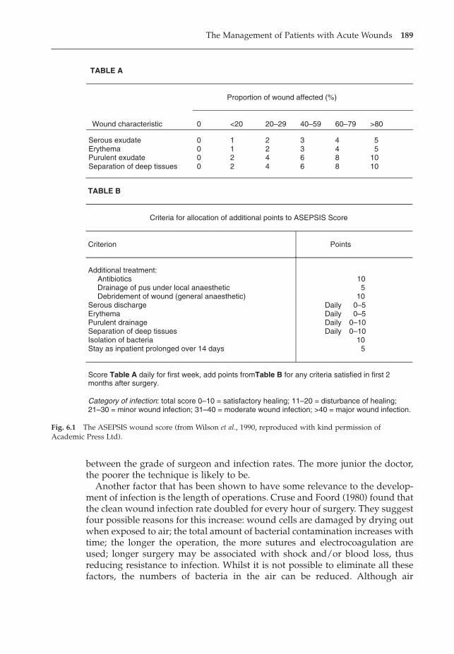
between the grade of surgeon and infection rates. The more junior the doctor,the poorer the technique is likely to be.
Another factor that has been shown to have some relevance to the develop-ment of infection is the length of operations. Cruse and Foord (1980) found thatthe clean wound infection rate doubled for every hour of surgery. They suggestfour possible reasons for this increase: wound cells are damaged by drying outwhen exposed to air; the total amount of bacterial contamination increases withtime; the longer the operation, the more sutures and electrocoagulation areused; longer surgery may be associated with shock and/or blood loss, thusreducing resistance to infection. Whilst it is not possible to eliminate all thesefactors, the numbers of bacteria in the air can be reduced. Although air
The Management of Patients with Acute Wounds 189
TABLE A
Proportion of wound affected (%)
Wound characteristic 0 <20 20–29 40–59 60–79 >80
Serous exudate 0 1 2 3 4 5Erythema 0 1 2 3 4 5Purulent exudate 0 2 4 6 8 10Separation of deep tissues 0 2 4 6 8 10
TABLE B
Criteria for allocation of additional points to ASEPSIS Score
Criterion Points
Additional treatment: Antibiotics 10Drainage of pus under local anaesthetic 5Debridement of wound (general anaesthetic) 10
Serous discharge Daily 0–5Erythema Daily 0–5Purulent drainage Daily 0–10Separation of deep tissues Daily 0–10Isolation of bacteria 10Stay as inpatient prolonged over 14 days 5
Score Table A daily for first week, add points fromTable B for any criteria satisfied in first 2months after surgery.
Category of infection: total score 0–10 = satisfactory healing; 11–20 = disturbance of healing;21–30 = minor wound infection; 31–40 = moderate wound infection; >40 = major wound infection.
Fig. 6.1 The ASEPSIS wound score (from Wilson et al., 1990, reproduced with kind permission ofAcademic Press Ltd).
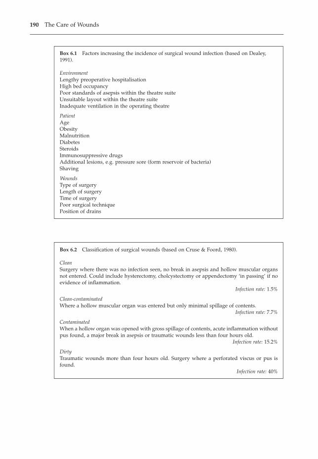
190 The Care of Wounds
Box 6.1 Factors increasing the incidence of surgical wound infection (based on Dealey,1991).
EnvironmentLengthy preoperative hospitalisationHigh bed occupancyPoor standards of asepsis within the theatre suiteUnsuitable layout within the theatre suiteInadequate ventilation in the operating theatre
PatientAgeObesityMalnutritionDiabetesSteroidsImmunosuppressive drugsAdditional lesions, e.g. pressure sore (form reservoir of bacteria)Shaving
WoundsType of surgeryLength of surgeryTime of surgeryPoor surgical techniquePosition of drains
Box 6.2 Classification of surgical wounds (based on Cruse & Foord, 1980).
CleanSurgery where there was no infection seen, no break in asepsis and hollow muscular organsnot entered. Could include hysterectomy, cholcystectomy or appendectomy ‘in passing’ if noevidence of inflammation.
Infection rate: 1.5%
Clean-contaminatedWhere a hollow muscular organ was entered but only minimal spillage of contents.
Infection rate: 7.7%
ContaminatedWhen a hollow organ was opened with gross spillage of contents, acute inflammation withoutpus found, a major break in asepsis or traumatic wounds less than four hours old.
Infection rate: 15.2%
DirtyTraumatic wounds more than four hours old. Surgery where a perforated viscus or pus isfound.
Infection rate: 40%
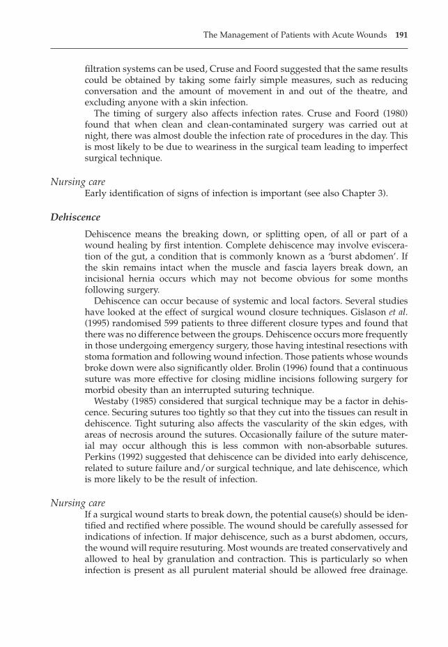
filtration systems can be used, Cruse and Foord suggested that the same resultscould be obtained by taking some fairly simple measures, such as reducing conversation and the amount of movement in and out of the theatre, andexcluding anyone with a skin infection.
The timing of surgery also affects infection rates. Cruse and Foord (1980)found that when clean and clean-contaminated surgery was carried out atnight, there was almost double the infection rate of procedures in the day. Thisis most likely to be due to weariness in the surgical team leading to imperfectsurgical technique.
Nursing careEarly identification of signs of infection is important (see also Chapter 3).
Dehiscence
Dehiscence means the breaking down, or splitting open, of all or part of awound healing by first intention. Complete dehiscence may involve eviscera-tion of the gut, a condition that is commonly known as a ‘burst abdomen’. Ifthe skin remains intact when the muscle and fascia layers break down, an incisional hernia occurs which may not become obvious for some months following surgery.
Dehiscence can occur because of systemic and local factors. Several studieshave looked at the effect of surgical wound closure techniques. Gislason et al.(1995) randomised 599 patients to three different closure types and found thatthere was no difference between the groups. Dehiscence occurs more frequentlyin those undergoing emergency surgery, those having intestinal resections withstoma formation and following wound infection. Those patients whose woundsbroke down were also significantly older. Brolin (1996) found that a continuoussuture was more effective for closing midline incisions following surgery formorbid obesity than an interrupted suturing technique.
Westaby (1985) considered that surgical technique may be a factor in dehis-cence. Securing sutures too tightly so that they cut into the tissues can result indehiscence. Tight suturing also affects the vascularity of the skin edges, withareas of necrosis around the sutures. Occasionally failure of the suture mater-ial may occur although this is less common with non-absorbable sutures.Perkins (1992) suggested that dehiscence can be divided into early dehiscence,related to suture failure and/or surgical technique, and late dehiscence, whichis more likely to be the result of infection.
Nursing careIf a surgical wound starts to break down, the potential cause(s) should be iden-tified and rectified where possible. The wound should be carefully assessed forindications of infection. If major dehiscence, such as a burst abdomen, occurs,the wound will require resuturing. Most wounds are treated conservatively andallowed to heal by granulation and contraction. This is particularly so wheninfection is present as all purulent material should be allowed free drainage.
The Management of Patients with Acute Wounds 191
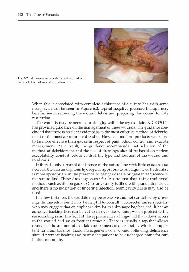
When this is associated with complete dehiscence of a suture line with somenecrosis, as can be seen in Figure 6.2, topical negative pressure therapy may be effective in removing the wound debris and preparing the wound for lateresuturing.
The wounds may be necrotic or sloughy with a heavy exudate. NICE (2001)has provided guidance on the management of these wounds. The guidance con-cluded that there is no clear evidence as to the most effective method of debride-ment or the most appropriate dressing. However, modern products were seento be more effective than gauze in respect of pain, odour control and exudatemanagement. As a result, the guidance recommends that selection of themethod of debridement and the use of dressings should be based on patientacceptability, comfort, odour control, the type and location of the wound andtotal costs.
If there is only a partial dehiscence of the suture line with little exudate andnecrosis then an amorphous hydrogel is appropriate. An alginate or hydrofibreis more appropriate in the presence of heavy exudate or greater dehiscence ofthe suture line. These dressings cause far less trauma than using traditionalmethods such as ribbon gauze. Once any cavity is filled with granulation tissueand there is no indication of lingering infection, foam cavity fillers may also beused.
In a few instances the exudate may be excessive and not controlled by dress-ings. In this situation it may be helpful to consult a colorectal nurse specialistwho may suggest that an appliance similar to a drainage bag be used. It has anadhesive backing that can be cut to fit over the wound, whilst protecting thesurrounding skin. The front of the appliance has a hinged lid that allows accessto the wound and saves frequent removal. There is usually a tap that allowsdrainage. The amount of exudate can be measured accurately which is impor-tant for fluid balance. Good management of a wound following dehiscenceshould promote healing and permit the patient to be discharged home for carein the community.
192 The Care of Wounds
Fig. 6.2 An example of a dehiscent wound withcomplete breakdown of the suture line.
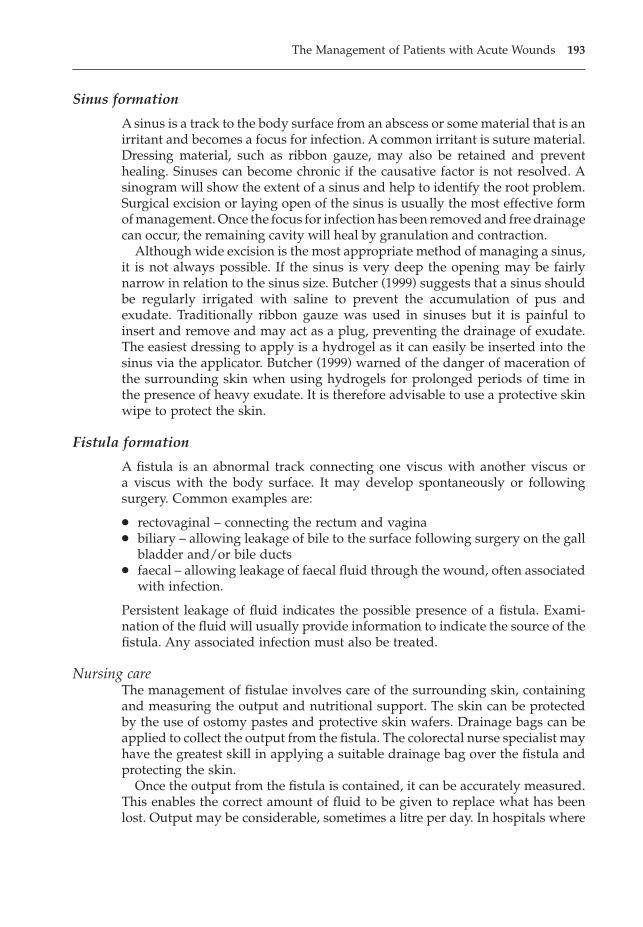
Sinus formation
A sinus is a track to the body surface from an abscess or some material that is anirritant and becomes a focus for infection. A common irritant is suture material.Dressing material, such as ribbon gauze, may also be retained and preventhealing. Sinuses can become chronic if the causative factor is not resolved. Asinogram will show the extent of a sinus and help to identify the root problem.Surgical excision or laying open of the sinus is usually the most effective formof management. Once the focus for infection has been removed and free drainagecan occur, the remaining cavity will heal by granulation and contraction.
Although wide excision is the most appropriate method of managing a sinus,it is not always possible. If the sinus is very deep the opening may be fairlynarrow in relation to the sinus size. Butcher (1999) suggests that a sinus shouldbe regularly irrigated with saline to prevent the accumulation of pus andexudate. Traditionally ribbon gauze was used in sinuses but it is painful toinsert and remove and may act as a plug, preventing the drainage of exudate.The easiest dressing to apply is a hydrogel as it can easily be inserted into thesinus via the applicator. Butcher (1999) warned of the danger of maceration ofthe surrounding skin when using hydrogels for prolonged periods of time inthe presence of heavy exudate. It is therefore advisable to use a protective skinwipe to protect the skin.
Fistula formation
A fistula is an abnormal track connecting one viscus with another viscus or a viscus with the body surface. It may develop spontaneously or followingsurgery. Common examples are:
� rectovaginal – connecting the rectum and vagina� biliary – allowing leakage of bile to the surface following surgery on the gall
bladder and/or bile ducts� faecal – allowing leakage of faecal fluid through the wound, often associated
with infection.
Persistent leakage of fluid indicates the possible presence of a fistula. Exami-nation of the fluid will usually provide information to indicate the source of thefistula. Any associated infection must also be treated.
Nursing careThe management of fistulae involves care of the surrounding skin, containingand measuring the output and nutritional support. The skin can be protectedby the use of ostomy pastes and protective skin wafers. Drainage bags can beapplied to collect the output from the fistula. The colorectal nurse specialist mayhave the greatest skill in applying a suitable drainage bag over the fistula andprotecting the skin.
Once the output from the fistula is contained, it can be accurately measured.This enables the correct amount of fluid to be given to replace what has beenlost. Output may be considerable, sometimes a litre per day. In hospitals where
The Management of Patients with Acute Wounds 193
there is a nutrition team, they can also be involved in the care of these patients.When it is possible, enteral feeding should be given. If the fistula is high in thegastrointestinal tract then it will be necessary to give parenteral nutrition.
Nienhuijs et al. (2003) reported on the management and outcomes for 17patients with enterocutaneous fistulae. They followed the treatment describedabove until spontaneous healing but ten patients had skin erosion and requiredfrequent dressing changes. Consequently topical negative pressure (TNP) wasapplied to these wounds. Patients were given TNP for a median of 25 days(range 14–161 days). Four patients went on to heal spontaneously after amedian of 73 days, five patients had surgery to correct the fistula and onepatient died, due to extensive metastasis. The four patients who healed spon-taneously were discharged home with TNP therapy. Despite the high costs ofusing TNP, the authors calculated that the cost was cheaper than conventionaltreatment (€290394 versus €428311). They noted that as these figures were cal-culated retrospectively, they should be treated with caution. Undoubtedly, thepatients in the study were very satisfied with the use of TNP and preferred itto the conventional treatment that they had been given previously.
Evaluation
Regular monitoring of surgical wounds is essential in order to identify anypotential complications as early intervention may prevent further problems.Therefore good documentation is essential. Birchall and Taylor (2003) describedthe development of a benchmarking tool based on The Essence of Care (DoH,2001). The project involved determining best practice (in the absence of anynational guidelines) and improving wound assessment documents and thenbenchmarking practice against it. The motivation for undertaking this workwas an unrelated audit that had identified that out of 40 surgical wounds, onlyone had a documented assessment. Projects such as this improve awareness ofthe need to maintain accurate records of each wound and its progress.
6.3 TRAUMATIC WOUNDS
Traumatic injuries can range from a simple cut to a major crushing injury. Majortraumatic injury is beyond the remit of this book. It requires surgical interven-tion and specialised nursing care. Most nurses will be required to care for minortraumatic wounds from time to time. Their care will be considered here.
6.3.1 Minor traumatic wounds
Assessment
Initial assessment should identify any life-threatening problems such as airwayobstruction, haemorrhage or shock. Vital signs should be recorded. Any of theseproblems should be addressed before treating the wound. If possible, a historyshould be obtained of when, where and how the injury occurred. A medical
194 The Care of Wounds
history can highlight any factors that may affect healing of the wound or thetype of treatment prescribed to manage the wound.
The wound should be assessed for any bleeding. Haemorrhage may beresolved by pressure or require surgical intervention. The presence of anyforeign bodies should be noted and the extent and severity of the injury. Cleans-ing of the wound may be necessary before a full assessment can be made. Looseparticles may easily be washed off but other debris may be more difficult toremove. Bianchi (2000) reviewed the limited evidence available as to the bestmethods of cleansing traumatic wounds. She concluded that pain assessmentand management were essential prior to the procedure, especially for children.Unfortunately, there is insufficient evidence to determine the best methods ofcleansing and cleansers to use. Some accident and emergency departments useClingfilm to cover a wound until seen by the doctor. This has the advantage ofkeeping the wound warm and moist and allowing easy observation.
Nursing intervention
Medical assessment and prescription may be necessary before the wound canbe dressed. Whilst many nurses are competent to dress minor injuries the fol-lowing guidelines should be considered. Patients should be examined by adoctor if:
� the nature and extent of the injury are uncertain.� there is persistent bleeding.� suturing is required.� a foreign body is present.� tetanus prophylaxis may be necessary.� the injury occurred to a hospital patient.� the nurse is uncertain of the appropriate management.� it is required by nursing policies.
Prior to dressing, the wound should be thoroughly cleaned using saline. Anyloose devitalised tissue should be removed. In some instances it may be nec-essary to shave the area around the wound, if there are hairs that may interferewith the healing process. An appropriate dressing can then be applied. Tetanusprophylaxis will be required if the patient has not had a complete course oftetanus toxoid or if no booster dose has been given for more than five years.Depending on the cause of the wound, the doctor may also prescribe a courseof antibiotics.
If the person is not a patient in hospital, they and any carer will require infor-mation about the management of the wound. Information should also be givenconcerning whom to contact if any complication occurs. Potential complicationssuch as fever, swelling around the wound, excessive pain or offensive dischargeshould be described to the patient. Ideally, this information should also be givenin written form so that the patient has it for future reference.
Traumatic injuries occur primarily to the young and the elderly. Whilst mostyoung people will heal easily, this is not necessarily true for the older people.
The Management of Patients with Acute Wounds 195
They may need to be admitted to hospital for further care. Wijetunge (1992) hasprovided a useful overview of a management plan for soft tissue injuries.
Evaluation
The aim of any treatment is uncomplicated healing of the wound with restora-tion of function and minimal scarring (Brunner & Suddarth, 1988). Patients mayreturn to be reviewed by those who made the original assessment or they maybe referred to the family practitioner. The majority of these wounds will healwithout complication.
6.3.2 The management of specific types of traumatic wounds
Abrasions
An abrasion is a superficial injury where the skin is rubbed or torn and it maybe extremely sore. It may occur as a result of falling on a gritty surface. Abra-sions should be cleaned carefully to ensure that there are no foreign bodiesembedded in the wound. In the case of extensive abrasions a general anaes-thetic may be required in order to allow adequate cleansing. Failure to removeall the debris may result in unsightly ‘tattooing’ (Evans & Jones, 1996).
Selection of a suitable dressing depends on the extent and depth of the injury.These wounds are often very sore. Occlusive dressings have been found toreduce the pain, possibly because they keep the nerve endings from drying out.If the abrasion is superficial a film dressing or a thin hydrocolloid can beapplied. Deeper wounds, with a heavier exudate, can be treated with hydro-colloids, foams or alginates.
Lacerations
A laceration is a wound that penetrates the skin and has a torn and jagged edge.They can be caused by blunt injury which produces shear forces or by a sharpobject such as glass (Singer et al., 1997). The aim of treatment is to achieveuncomplicated healing with a functional and cosmetically acceptable scar.
It is important to remove all debris from the wound and any devitalisedtissue. This procedure is sometimes called surgical toilet. The best way tomanage these wounds is to bring the skin edges together to heal by first inten-tion. This may be achieved by the use of sutures, adhesive strips or tissue adhe-sive. The choice of material depends on the position of the wound, its extentand the condition of the damaged skin. Suturing is recommended around jointsor on the hand where movement is involved. Farion et al. (2004) reviewed theuse of tissue adhesives for traumatic lacerations in both children and adults.They found no difference in cosmesis between adhesives and standard closuresbut tissue adhesives could be applied more quickly and were less painful touse. The authors of the review also noted that there was a small but significantincreased rate of dehiscence with tissue adhesives compared with standardclosure methods.
196 The Care of Wounds
One of the commonest positions for a laceration is the pretibial area. Daviset al. (2004) undertook an audit of A&E departments in a heavily populatedregion of the UK and found that the mean incidence of estimated patients withpretibial laceration was 5.2/1000 A&E attendances. The extent of the injury canvary and Dunkin et al. (2003) proposed a method of classifying these wounds.
I Laceration.II Laceration or flap with minimal haematoma and/or skin edge necrosis.III Laceration or flap with moderate to severe haematoma and/or necrosis.IV Major degloving injury.
Dunkin et al. (2003) have also provided a useful algorithm for the managementof each type of laceration. Fairly obviously, types III and IV require surgicalintervention. Type I lacerations should have the skin edges opposed and thenheld together with short, wide adhesive strips. The wound is covered with anon-adherent dressing and a crepe bandage applied from toe to knee. It maybe appropriate to place a wool bandage under the crepe bandage in order toprovide additional protection. Patients should be encouraged to mobilise andto elevate their leg when sitting in order to reduce any oedema. Type II is alsomanaged with use of adhesive strips and supportive bandages, after anyhaematoma has been evacuated, the non-viable wound edges trimmed and thewound edges apposed. Both these type of lacerations should heal without com-plication. Failure to do so indicates that the injury is more serious than origi-nally thought (Dunkin et al., 2003).
If there is any risk of infection as a result of severe contamination of thewound at the time of injury, primary closure is not appropriate. Conservativetreatment with antibiotics and dressings such as an amorphous hydrogel ispreferable. An iodine-impregnated low-adherent dressing may also be used fora short period of time. Once the wound is clean, skin grafting is the fastestmethod of wound closure.
Finger tip injuries
Crushing finger tip injuries are very common in children, mainly caused byfingers being caught in house or car doors. Buckles (1985) suggested that 14%of children below the age of 13 years suffer from this type of injury. Cockerilland Sweet (1993) described the procedure of trephining to remove a haematomafrom the nail bed. They also suggest that an X-ray may be necessary to iden-tify any fracture. Prophylactic antibiotics may be needed if a fracture is present.
The use of adhesive tapes over the finger tip can be an effective way ofholding the wound edges together and then protecting the finger by applyinga tubular bandage. Collier (1996) suggests that it is important to use dressingsthat will not stick to the wound as adherent dressings will cause pain andfurther trauma at dressing change. Given that many of the patients are chil-dren, this is an important consideration. Suitable dressings might be hydrocol-loids or adhesive foams which can be retained without bulky dressings orstrapping.
The Management of Patients with Acute Wounds 197
Mammalian and human bites
Patients presenting with mammalian bites account for 1% of visits to A&Edepartments (Medeiros & Saconato, 2004). As many as 74% of bites are fromdogs; other animals causing problems include cats, rats, squirrels and, occa-sionally, snakes. As well as animal bites, patients also present with human bites,usually as a result of physical violence such as a punch to the mouth whichcauses injury to the knuckles (Higgins et al., 1997).
The major problem with any type of bite injury is the risk of infection or thetransmission of diseases such as HIV or hepatitis B from human bites. Higginset al. (1997) stress the importance of not underestimating these wounds. Afterreviewing the evidence for the use of prophylactic antibiotics after mammalianbites, Medeiros and Saconato (2004) concluded that there was some evidencethat they were beneficial after human bites but not after dog or cat bites. Theycalled for further research on the subject.
Careful cleansing is essential in order to assess the extent of the injury. Salineor an antimicrobial such as povidone iodine should be used. Surgical debride-ment or exploration may be necessary to remove devitalised tissue, skin tagsor any foreign bodies. Large cuts and facial wounds need suturing. Delayedprimary closure may be used for some wounds. Chen et al. (2000) monitoredoutcome for 145 mammalian bite wounds that were treated by primary closure.They found there was an infection rate of 5.5% and considered that this was an acceptable risk for wounds where a good cosmetic result was essential.However, they also noted that appropriate selection of patients was important.
Puncture wounds should be left open because of the risk of infection. Thewound should be protected. An iodine-impregnated low-adherent dressingmay be suitable, unless there is a moderate to heavy exudate. Higgins et al.(1997) suggest that patients with human puncture wounds should be admittedfor surgical exploration and irrigation. So, too, should those with bites involv-ing joints or tendons or any indication of spreading infection. Patients with bitewounds should be followed up to ensure that there has been uncomplicatedhealing and the patient has regained full function of the affected area.
Skin tears
Skin tears are caused either by friction or by a combination of friction and shear-ing associated with thin, fragile skin. Thomas et al. (1999) suggest that up to 1.5million people living in care institutions in the USA suffer a skin tear each year.A survey of 154 nursing home residents revealed that they occurred mostly onthe arms in the very elderly who were frail, had limited mobility, poor appetiteand ecchymosis (McGough-Csarny & Kopac, 1998). Skin tears can be classifiedaccording to severity using a classification system devised by Payne and Martin(1993), given in Table 6.1.
There is limited evidence to determine the best method for managing skintears. Sutures are not an option as the skin is too fragile and they would causefurther damage. Adhesive strips may be used for category I tears but care mustbe taken to ensure that they do not cause damage on removal.
198 The Care of Wounds
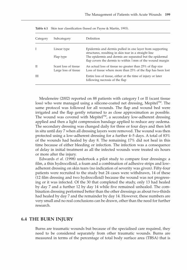
Meuleneire (2002) reported on 88 patients with category I or II (scant tissueloss) who were managed using a silicone-coated net dressing, MepitelTM. Thesame protocol was followed for all wounds. The flap and wound bed were irrigated and the flap gently returned to as close approximation as possible. The wound was covered with MepitelTM, a secondary low-adherent dressingapplied and then a light compression bandage applied to reduce any oedema.The secondary dressing was changed daily for three or four days and then leftin situ until day 7 when all dressing layers were removed. The wound was thenprotected using a low-adherent dressing for a further 4–5 days. A total of 83%of the wounds had healed by day 8. The remaining 17% did not heal in thistime because of either bleeding or infection. The infection was a consequenceof delay in initial treatment as all the infected wounds were treated six hoursor more after the injury.
Edwards et al. (1998) undertook a pilot study to compare four dressings: afilm, a thin hydrocolloid, a foam and a combination of adhesive strips and low-adherent dressing on skin tears (no indication of severity was given). Fifty-fourpatients were recruited to the study but 24 cases were withdrawn, 14 of these(12 film dressing and two hydrocolloid) because the wound was not progress-ing or it was infected. Of the 30 that completed the study, only 13 had healedby day 7 and a further 12 by day 14 while five remained unhealed. The com-bination dressing performed better than the other dressings as about two-thirdshad healed by day 7 and the remainder by day 14. However, these numbers arevery small and no real conclusions can be drawn, other than the need for furtherresearch.
6.4 THE BURN INJURY
Burns are traumatic wounds but because of the specialised care required, theyneed to be considered separately from other traumatic wounds. Burns are measured in terms of the percentage of total body surface area (TBSA) that is
The Management of Patients with Acute Wounds 199
Table 6.1 Skin tear classification (based on Payne & Martin, 1993).
Category Subcategory Definition
I Linear type Epidermis and dermis pulled in one layer from supporting structures, resulting in skin tear in a straight line
Flap type The epidermis and dermis are separated but the epidermal flap covers the dermis to within 1mm of the wound margin
II Scant loss of tissue An actual loss of tissue no greater than 25% of flap sizeLarge loss of tissue Loss of tissue where more than 25% of the flap has been lost
III Entire loss of tissue, either at the time of injury or later following necrosis of the flap
affected and they are divided into complex and non-complex burns. In the UK,there are national criteria for the referral of complex burns to a specialist burncare unit (NBCRC, 2001). They include:
� patients aged under five years or over 60 years.� full-thickness burns involving more than 10% TBSA in adults or 5% in
children less than 16 years of age.� burns of the face, hands, perineum, feet or flexures of the body such as neck
or axilla.� circumferential deep dermal or full-thickness burns of torso, limbs or neck.� inhalation injuries.� suspicion of non-accidental injury.
The burn care required by these patients is beyond the remit of this book.
6.4.1 Aetiology
A burn is an injury caused by excessive heat. It primarily damages the skin,causing tissue destruction and coagulation of the blood vessels of the affectedarea. Burn damage consists of three areas or zones (Hettiaratchy & Dziewulski,2004a). The central zone is the zone of coagulation and at this point there is coag-ulation of the tissue proteins and thus irreversible damage and tissue loss. Thezone of stasis surrounds the zone of coagulation and there is decreased tissueperfusion in this zone. However, if tissue perfusion is increased promptly it ispossible to prevent the tissue damage extending into this zone. The third andouter zone is the zone of hyperaemia and here tissue perfusion is increased. Gen-erally this area will recover unless the burn becomes severely infected or thereis prolonged hypoperfusion.
Burns can be divided into four categories depending on their cause.
� Thermal – caused by flame, hot water or steam, other hot liquids and hot sur-faces. Smoke inhalation injuries may be associated with fire casualties.
� Chemical – caused by spillage of strong acids, alkalis or other corrosive sub-stances. These are usually industrial injuries. Damage to vital organs canoccur if the chemicals are absorbed into the blood supply.
� Electrical – caused by an electrical current passing through the body. Theinternal damage may be considerably greater than is obvious from the skinappearance. Such burns may also be associated with thermal injury.
� Radiation – due to overexposure to industrial ionising radiation or followingradiotherapy treatment. (Reactions to radiotherapy are covered in moredetail in section 6.5.)
Although burn injuries are usually described as accidental, it has been sug-gested that some individuals are more likely to suffer from them than others(Hettiaratchy & Dziewulski, 2004b).
� Young children – 20% of all burn injuries are in young children, predominantlyboys. Most of these injuries (70%) are due to scalds.
200 The Care of Wounds
� Children aged 5–14 years – 10% of burns occur in this group and may be dueto illicit activity such as playing with fireworks or petrol or misuse of electrical equipment.
� Adults of working age – around 60% of burns occur in this group, generally asa result of flame injury. About a third of the burns in this group are due towork-related injury.
� Elderly people – 10% of burns occur in this group, often as a result of infir-mity or disability.
� Additional factors – within all age groups there are other predisposing factorsthat can increase the risk of burn injury. They include medical conditionssuch as epilepsy, psychiatric illness, alcoholism and social conditions suchas poor housing, including faulty wiring and overcrowding.
6.4.2 Incidence
It is estimated that around 250000 people suffer burn injury each year in theUK (NBCRC, 2001). Out of this group, around 175000 people attend A&E as aresult of their injury and for 13000 it is sufficiently severe as to require admis-sion to hospital. About 1000 people, half of whom are children under 12 years,have such severe burns that they require fluid resuscitation. It should also benoted that 300 people on average will die from their burns each year.
The figures for burn injury in the UK are similar to those in other developedcounties (Hettiaratchy & Dziewulski, 2004b). However, in developing countriesthey are likely to be much higher; for example, it is thought that 2 million ofthe 500 million people in India have suffered a burn injury. Mortality rates arealso likely to be higher in countries with a high level of poverty. Poor housing,overcrowding and the use of open fires for cooking are likely factors in the highincidence of fires in developing countries.
Staff should be aware of the possibility of non-accidental injury to children.Gordon and Goodwin (1997) suggest that around 10% of paediatric burns arethe result of child abuse. This view is supported by work by Andronicus et al.(1998) who reviewed 507 paediatric burn cases. They found that 8% of injurieswere considered to be as a result of abuse or neglect. The staff believed that thefamily’s social or emotional situation was likely to have been a major factor incausing the burn injury of a further 6% of children. Andronicus et al. (1998)provide a list of features that may indicate abuse or neglect. They include adelay between the incident and presentation, an inconsistent history of events,a history of previous accidents and presentation for treatment by someone otherthan the parent. Most A&E departments have specific procedures to follow ifan injury is suspected to be non-accidental.
6.4.3 The severity of the injury
The skin is the organ of the body that usually suffers the greatest damage froma burn injury. Depending on the extent of the injury, several layers may be
The Management of Patients with Acute Wounds 201
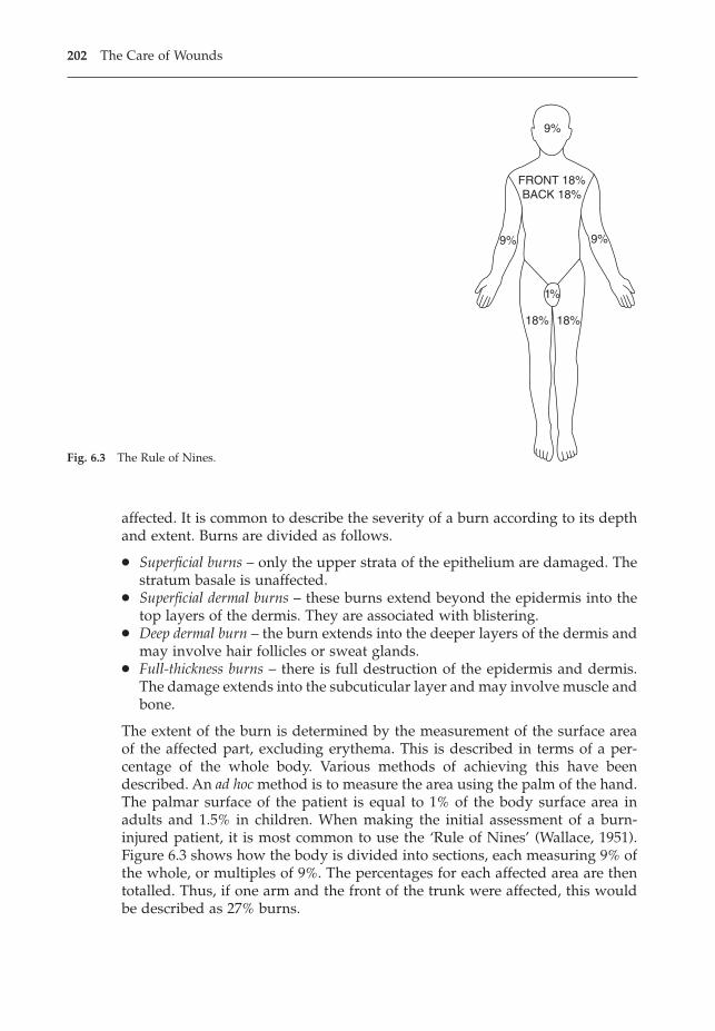
affected. It is common to describe the severity of a burn according to its depthand extent. Burns are divided as follows.
� Superficial burns – only the upper strata of the epithelium are damaged. Thestratum basale is unaffected.
� Superficial dermal burns – these burns extend beyond the epidermis into thetop layers of the dermis. They are associated with blistering.
� Deep dermal burn – the burn extends into the deeper layers of the dermis andmay involve hair follicles or sweat glands.
� Full-thickness burns – there is full destruction of the epidermis and dermis.The damage extends into the subcuticular layer and may involve muscle andbone.
The extent of the burn is determined by the measurement of the surface areaof the affected part, excluding erythema. This is described in terms of a per-centage of the whole body. Various methods of achieving this have beendescribed. An ad hoc method is to measure the area using the palm of the hand.The palmar surface of the patient is equal to 1% of the body surface area inadults and 1.5% in children. When making the initial assessment of a burn-injured patient, it is most common to use the ‘Rule of Nines’ (Wallace, 1951).Figure 6.3 shows how the body is divided into sections, each measuring 9% ofthe whole, or multiples of 9%. The percentages for each affected area are thentotalled. Thus, if one arm and the front of the trunk were affected, this wouldbe described as 27% burns.
202 The Care of Wounds
18%18%
9%
9%
9%
1%
FRONT 18%BACK 18%
Fig. 6.3 The Rule of Nines.
However, the Rule of Nines may overestimate the extent of the injury. Oncethe initial emergency treatment has been carried out, a reassessment of theextent of the injury is usually made. A more accurate picture can be obtainedusing a Lund and Browder chart (see Fig. 6.4). Lund and Browder (1944) devel-oped a system for assessing burn injury that not only divides the body intosmaller areas but also considers the age of the patient. Body proportions alterduring childhood, so that the front of the head is 8.5% of the whole in a childof one year but only 4.5% of the whole in a 15 year old. Patient managementmay need to be adapted once this reassessment has been made.
6.4.4 Burn oedema
Almost immediately after injury, oedema starts to collect beneath the damagedtissue. This is typically maximal within 24 hours of the injury but can last forup to three or four days. As plasma continues to leak into the tissues, there isa risk of hypovolaemia developing. Without treatment, burns shock candevelop and is potentially fatal. If the burn is on the face, neck or chest, theswelling from the oedema may cause obstruction of the airway. Patients withfacial burns are admitted for 24 hours as a precaution. Treatment must ensurethat the effects of burn oedema are minimised.
6.4.5 First aid treatment of burns
The British Burn Association has published recommendations for the first aidtreatment of burns (Lawrence, 1987). Box 6.3 is based on these recommenda-tions. The principal treatment is to remove the injured person from the sourceof heat and pour cold water over the affected area. Lawrence and Wilkins (1986)demonstrated that the subcutaneous temperature continues to rise after theburn injury occurs. Thus, if a burn injury at 100°C lasts for ten seconds, theaffected tissue will take three minutes to return to normal body temperature.Application of cold water within ten seconds of the injury can reduce the ‘burntime’ to 30 seconds.
There are occasions when cold water is not readily available and other alter-natives must be used. Lawrence (1996) tested a mousse containing a mixture of
The Management of Patients with Acute Wounds 203
Box 6.3 The first aid treatment of burns (based on Lawrence, 1987).
• Remove the person from the source of heat.• Turn off the electricity in the case of electric burns.• Apply copious amounts of water to the affected area.• Do NOT try to remove clothing if the burns are extensive.• Put wet compress on any exposed areas.• Wrap Clingfilm around wet compress to hold in place.• Seek qualified help quickly, especially if the burn is extensive.• If no tap water, use bottled/mineral water or milk.• Do not use solutions such as bleach, butter or oil.
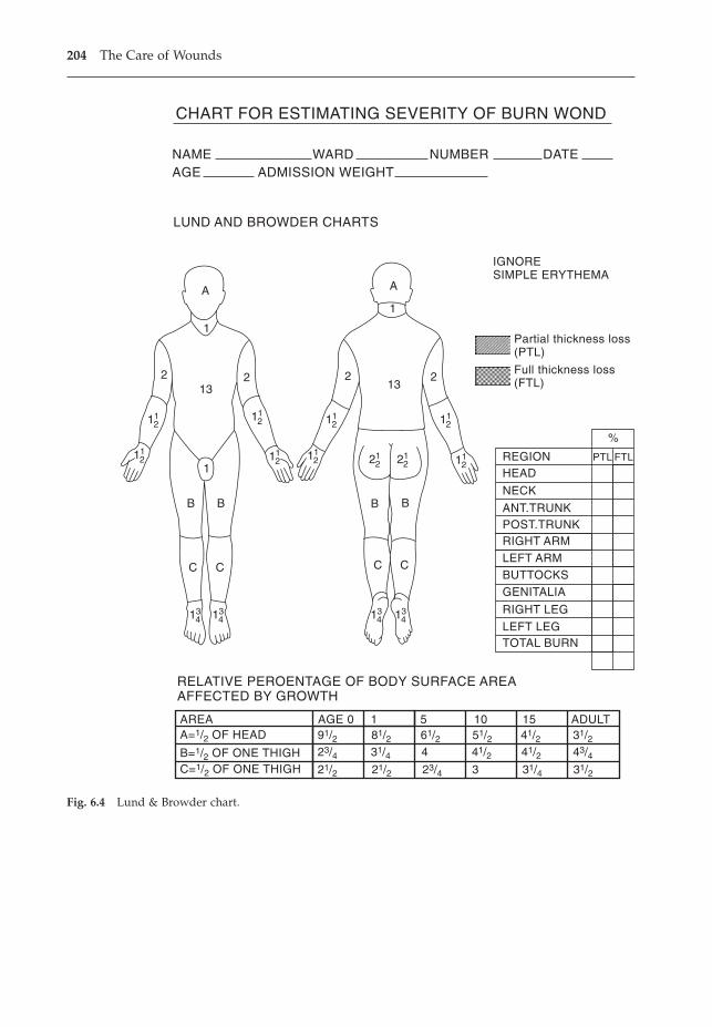
204 The Care of Wounds
1
1
132 2
A
B B
CC
1
132 2
A
B B
CC
CHART FOR ESTIMATING SEVERITY OF BURN WOND
NAMEAGE ADMISSION WEIGHT
WARD NUMBER DATE
LUND AND BROWDER CHARTS
IGNORESIMPLE ERYTHEMA
Partial thickness loss(PTL)
Full thickness loss(FTL)
%
PTL FTLREGIONHEAD
NECK
ANT.TRUNKPOST.TRUNKRIGHT ARMLEFT ARMBUTTOCKSGENITALIA
RIGHT LEG
LEFT LEGTOTAL BURN
RELATIVE PEROENTAGE OF BODY SURFACE AREAAFFECTED BY GROWTH
AREA AGE 0 1 5 10 15 ADULTA=1/2 OF HEAD 91/2
23/421/2 21/2 23/4 31/4 31/23
31/4 41/2 41/2 43/4481/2 61/2 51/2 41/2 31/2
B=1/2 OF ONE THIGHC=1/2 OF ONE THIGH
134 13
4 134 13
4
112
112
112 11
2 112
112
112 11
2 212 21
2
Fig. 6.4 Lund & Browder chart.
paraffin oils and waxes in the laboratory and found that it gave local coolingfor at least ten minutes. The same product was tested at a fire service collegeon 108 casualties with a variety of burns or scalds (Dunn, 1996). All subjectsfound immediate pain relief after application of the mousse. When the moussefell off, a thin film remained over the burn site, obviating the need for a dress-ing unless there was skin loss. This product would appear to be a useful firstaid treatment but further clinical evidence is required to support its use. Analternative is a cooling gel pad that can both cool the wound and relieve someof the initial wound pain (Hudspith & Rayatt, 2004).
6.4.6 The management of burn injuries
When considering the management of burns, the extent of the injury must bedefined as the treatment varies drastically between complex and non-complexburns. Non-complex burns may be treated in the outpatient department butanyone with complex burns must be admitted to hospital.
Complex burns
Depending on the circumstances, it may be necessary to provide initial resus-citation before transferring a patient to a burn care unit. Gordon and Goodwin(1997) describe the importance of the primary ABCDEF assessment of thepatient.
� A = airway, B = breathing. Check airway. Endotracheal intubation may beneeded if there are deep burns to face, neck or mouth or any indication ofrespiratory distress.
� C = circulation. Check pulse. May be absent or show signs of dysrhythmiasfollowing electric burn.
� D = disability or neurological deficit. Identify any associated trauma. Signs ofmental confusion or disorientation may be an indication of pre-existing con-ditions, hypoxia or unrecognised injury.
� E = exposure and evaluation. All jewellery or clothes should be removed fromthe burn-injured area because of the risk of constriction when burn oedemadevelops. This also allows for a rapid assessment of the full extent of theinjury, using the Rule of Nines. The patient should be kept warm; a spaceblanket is ideal.
� F = fluid resuscitation. Establish an intravenous infusion – this is essential forall burns greater than 15% TBSA in adults and 10% in children. Bloodsamples may be taken at the same time. Catheterise the bladder if the burnsare 25% or more TBSA; urinary output will indicate inadequate rehydrationas well as renal function.
Analgesia may also be required, especially for those with superficial burns asthey are very painful (Hettiaratchy & Papini, 2004).
It is also important to obtain a full history of cause of the injury from patient,relative or ambulance crew. This may provide information indicating potential
The Management of Patients with Acute Wounds 205
complications such as smoke inhalation. Inhalational analgesia or systemicanalgesia may be given depending on the condition of the patient.
When the patient’s condition has been stabilised by the above measures,urgent transfer to the burn care unit can take place. Once in the care of a burnsteam, a more detailed assessment of the patient and the extent of the burn injuryusing a Lund and Browder chart is undertaken. Essentially, the extent ofdamage is assessed visually. However, Mileski et al. (2003) found that a Dopplerflowmeter could be a useful tool to measure tissue perfusion, thus allowingeasier differentiation between the different zones of the burn injury. They foundthat serial measurements had a positive predictive value of 81% for identifyingnon-healing wounds.
Non-complex burns
Managing minor burns requires assessment of both the patient and the wound,as in any other type of wound. Assessment of the patient has been discussedin Chapter 2 and wound assessment and management in Chapter 3. Specificaspects of the care of minor burns are discussed here.
Patient assessmentBurn aetiology. Discover the cause of the injury. Non-accidental injury mustbe considered if the history seems inconsistent with the burn appearance. Therisk of infection is always present in the burn-injured patient.Pain. Analgesia may be required for pain.Nutrition. Nutritional status should be identified. Advice on nutrition maybe necessary as there will be increased nutritional demands on the body.Sleep. Pain or anxiety may affect sleep patterns.Early psychological care. Many patients will be very frightened. It is impor-tant to provide reassurance and explanation to the patient and the family.Later psychological care. A burn injury can be extremely disfiguring. Bodyimage may be profoundly altered, causing distress and loss of self-esteem.Many patients may have fears of disfigurement or disability. Some may fearloss of loved ones as a result of the scarring. Specific fears should be identifiedand addressed.
Wound assessmentInitial assessment of a burn injury includes the extent and depth of the burn.The use of a Lund and Browder chart has already been described. The zone ofhyperaemia should not be included when calculating the burn area. Identify-ing the depth of a burn is not always easy to do, especially as a burn injurymay have varying depths. Table 6.2 shows how it is possible to differentiatebetween the different wound depths.
A burn injury differs from other types of wounds in several importantrespects (Bayley, 1990). There are frequently large areas of devitalised tissue.The wound may have a large surface area and take some time to heal. As aresult, the burn wound is rapidly colonised by bacteria. There is considerable
206 The Care of Wounds
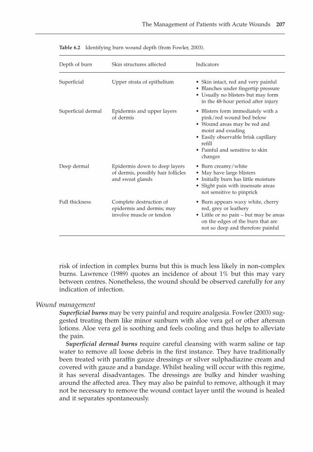
risk of infection in complex burns but this is much less likely in non-complexburns. Lawrence (1989) quotes an incidence of about 1% but this may varybetween centres. Nonetheless, the wound should be observed carefully for anyindication of infection.
Wound managementSuperficial burns may be very painful and require analgesia. Fowler (2003) sug-gested treating them like minor sunburn with aloe vera gel or other aftersunlotions. Aloe vera gel is soothing and feels cooling and thus helps to alleviatethe pain.
Superficial dermal burns require careful cleansing with warm saline or tapwater to remove all loose debris in the first instance. They have traditionallybeen treated with paraffin gauze dressings or silver sulphadiazine cream andcovered with gauze and a bandage. Whilst healing will occur with this regime,it has several disadvantages. The dressings are bulky and hinder washingaround the affected area. They may also be painful to remove, although it maynot be necessary to remove the wound contact layer until the wound is healedand it separates spontaneously.
The Management of Patients with Acute Wounds 207
Table 6.2 Identifying burn wound depth (from Fowler, 2003).
Depth of burn Skin structures affected Indicators
Superficial Upper strata of epithelium • Skin intact, red and very painful• Blanches under fingertip pressure• Usually no blisters but may form
in the 48-hour period after injury
Superficial dermal Epidermis and upper layers • Blisters form immediately with aof dermis pink/red wound bed below
• Wound areas may be red andmoist and exuding
• Easily observable brisk capillary refill
• Painful and sensitive to skinchanges
Deep dermal Epidermis down to deep layers • Burn creamy/whiteof dermis, possibly hair follicles • May have large blistersand sweat glands • Initially burn has little moisture
• Slight pain with insensate areasnot sensitive to pinprick
Full thickness Complete destruction of • Burn appears waxy white, cherryepidermis and dermis; may red, grey or leatheryinvolve muscle or tendon • Little or no pain – but may be areas
on the edges of the burn that arenot so deep and therefore painful
Modern products are easier to use and are also more comfortable for patients,especially when being removed. There is limited evidence to determine bestpractice but what evidence is available seems to favour modern products.Thomas et al. (1995) undertook a randomised study comparing a hydrocolloidalone, a hydrocolloid with silver sulphadiazine cream and a medicated paraf-fin gauze dressing in 50 patients with 54 non-complex burns. The hydrocolloidgroup required significantly fewer dressings than the other two groups. Thehydrocolloid group also had significantly faster healing than the hydrocolloidwith cream group. The researchers concluded that hydrocolloid dressings aloneare useful for the management of non-complex burns.
Two studies by Subrahmanyam (1991, 1998) investigated the use of honey forpartial-thickness burns compared with silver sulphadiazine. The 1991 studycompared these products on 104 patients with less than 40% partial-thicknessburns. Patients were randomly assigned to one of the two treatments. Theauthor found a significantly lower infection rate in the honey group. The studyin 1998 followed the same procedure as the previous study but monitoredhealing outcomes of 50 patients (25/group). The results showed that 84% ofwounds in the honey group showed evidence of granulation and epithelialisa-tion by day 7 compared with 72% in the silver sulphadiazine group. By day 21all patients in the honey group (n = 25) and 21/25 in the silver sulphadiazinegroup were healed, a significant difference in favour of the honey group (p =0.05).
Other dressings that can be used on minor burns include flat foam dressings,silicone net dressings and hydrogels. The foam dressings are comfortable andcan be held in place with tape or a tubular net. Hydrogels can give a coolingsensation when applied, which may be comforting to the patient. Silicone netdressings are widely used as they can be left in place for 7–14 days and the secondary dressing changed when necessary.
Whatever dressing is used, the patient and wound should be reviewed atbetween 48–72 hours when the local oedema has subsided (Fowler, 2003). Thisreview is especially important for patients who are being managed at home andattending the outpatient department.
Deep dermal and full-thickness burns generally require early excision ofnecrotic tissue and skin grafting for speedy healing.
Burns of the hands are best treated with silver sulphadiazine. Once the creamis applied, the hand should be covered with a plastic bag. This will allow freemovement and help to prevent contractures. The hand should be kept elevatedfor the first 48–72 hours to reduce swelling (Fowler, 1996). There are several dis-advantages with the use of plastic bags. They cause considerable maceration ofthe whole hand as large quantities of exudate accumulate in the bag. The bagbecomes heavy and pulls the wrist into flexion. Once full of fluid, the bag tendsto leak or tear, necessitating dressing change. Terrill et al. (1991) compared theuse of Gore-TexTM bags with plastic bags. Gore-TexTM has the property of watervapour permeability. Although there was no difference in healing times, therewas a considerable reduction in skin maceration and accumulation of exudatewhen using Gore-TexTM. Improved hand function was also noted. Witchell and
208 The Care of Wounds
Crossman (1991) compared the use of these bags over silver sulphadiazine withparaffin gauze for children with burns of the hands. They found that use ofGore-TexTM bags increased child activity and reduced the length of time ofdressing change. More significantly, there were considerably fewer unsched-uled visits to the accident and emergency department because of problems withthe dressing.
Evaluation
Evaluation of the burn wound involves monitoring of the wound as it pro-gresses towards healing and of the healed wound. Any non-complex burn thatfails to heal within three weeks should be referred to a specialist unit. Once thewound is healed, special care needs to be taken of the newly formed epithelia.Bayley (1990) suggests cleansing with a mild soap and then applying water-based cream several times a day. The patient should be warned to avoid anypossibility of sunburn. Fowler (2003) recommends use of total sunblock for thefirst year and then gradually reducing to factor 15.
Potential problems that can develop are contractures and hypertrophic scar-ring. Myofibroblasts contract and shorten the wound, causing the collagenfibres to become coiled. The scar develops a hard, red, raised appearance. If theburn injury is over a joint, as the myofibroblasts contract this causes flexion ofthe joint. Unless measures are taken to splint the joint, it will become contracted.A programme of exercise and splinting can help to counteract this problem.
Hypertrophic scarring is often managed by the use of pressure garments thatthe patients will have to wear for about a year. However, a randomised studyby Chang et al. (1995) found that pressure garments made no difference to thedegree of scarring. An alternative treatment, a silicone gel sheet, has been used,initially in Australia. Poston (2000) undertook a review of the use of silicone gelsheeting in the management of hypertrophic and keloid scars and concludedthat it appears to improve the appearance of these scars, but there is insuffi-cient evidence to determine its usefulness in restoring mobility and functionwhen scarring is associated with contractures.
A few patients will develop permanent pigmentation changes. This is possi-bly because of damage to the melanocytes in the basal layer of the epidermis.It can present as either apigmentation or hyperpigmentation. It is not possibleto predict when it will occur.
6.5 RADIATION REACTIONS
Radiation treatment or radiotherapy is the mainstay of cancer treatment. It hasbeen estimated that nearly 20% of people in Western countries will receiveradiotherapy at some point in their lives (Porock et al., 1999). Radiation reac-tion is the reaction of the skin to the effects of radiotherapy and is limited tothe treatment field or its exit point. Strictly speaking, a radiation reaction is nota wound. However, the skin reaction is akin to a superficial burn and has the
The Management of Patients with Acute Wounds 209
potential for ulceration. There is limited research into the management of whatcan be a very painful problem.
6.5.1 Aetiology
Ionising radiation or radiotherapy is the mainstay of cancer treatment. Treat-ment is usually given in a series of doses, ranging from daily to weekly,although a small number of patients will receive just one dose. A course of treat-ment may last up to eight weeks. Radiation reactions are most likely to occurwhen the treatment field is close to the body surface, such as the head or neck,or if it includes the axillae, under the breasts, perineum or groin. The reactionis dose dependent; that is, the more frequent the treatment or the higher thedose, the more likely the reaction. A reaction may occur during a course of treat-ment or after it is completed. Within six weeks of completion of treatment, allbut the most severe reactions have disappeared.
6.5.2 The classification of radiation reactions
The severity of radiation reactions is generally classified using the gradingsystem developed by the Radiation Therapy Oncology Group (RTOG) (Cox etal., 1995) and recommended by the College of Radiographers in the UK.
0 No change over baseline.1 Follicular, faint or dull erythema.2a Tender or bright erythema with/without dry desquamation.2b Patchy, moist desquamation, moderate oedema.3 Confluent wet desquamation, pitting oedema.4 Ulceration, haemorrhage, necrosis.
It is considered that 90–95% of patients undergoing radiotherapy will havesome degree of reaction (Porock et al., 1999). However, in a study of 126 womentreated for breast cancer, less than 10% had moist desquamation (Porock &Kristjanson, 1999). Thus it seems reasonable to assume that most patients arelikely to have erythema, possibly associated with soreness and itching. In areview of the subject, Porock et al. (1999) noted that women being treated forbreast cancer were particularly vulnerable if they smoked, were overweightand/or had large breast size or had required axillary lymphocoele drainageprior to treatment, whereas younger patients were more vulnerable than olderpatients in those having treatment to the sternum.
6.5.3 Preventive skin care
There is a body of evidence available to guide practice, in the form of best prac-tice statements (NHS Quality Improvement Scotland, 2004), a summary of keyresearch findings for breast cancer (National Breast Cancer Centre, 2004) andthe clinical guidelines issued by the UK College of Radiographers (2001). Box6.4 provides a summary of the recommended skin care. Unfortunately, despite
210 The Care of Wounds
this guidance, there is considerable variation in the advice given to patients con-cerning how the skin within the treatment field should be managed whilstradiotherapy is in progress (Faithfull et al., 2002).
Wells and colleagues reviewed the findings of their study of 357 patientsundergoing radiotherapy and concluded that levels of skin discomfort werelower than they had expected (Wells et al., 2004). They concluded that consis-tent advice, skin hygiene and regular assessment were major factors in reduc-ing problems. It is interesting to postulate that a reason for previous reports ofsevere skin problems may have been the advice given some years ago thatpatients should not wash the treatment area. Happily, that advice is now seenas outdated.
6.5.4 Managing radiation reactions
The basic principles of managing radiation reactions are as follows.
RTOG score 1 and 2a
A simple moisturiser such as aqueous cream can be soothing applied to the areatwice daily. If aqueous cream is not effective an alternative cream may help.There is much anecdotal evidence of the benefits of using aloe vera gel andHeggie et al. (2002) undertook a study of 225 patients undergoing treatment forbreast cancer comparing aloe vera gel with aqueous cream. They found thataqueous cream was significantly better than aloe vera gel in reducing drydesquamation and pain. However, Wells et al. (2004) did not find this to be thecase when comparing aqueous cream with either sucralfate cream or no cream.They concluded that patients should be given the choice to use a cream or not.
RTOG score 2b and 3
There is insufficient evidence to suggest a definitive treatment for wet desqua-mation. The main goal is to choose a dressing that is comfortable for the patientand promotes healing. Depending on the level of exudate, this could include
The Management of Patients with Acute Wounds 211
Box 6.4 Summary of recommendations for skin care during radiotherapy treatment.
• Wash treatment site daily using mild unperfumed soap.• Avoid having a very hot shower, especially if the water jets are powerful.• Pat area dry with soft towel, avoiding friction.• Do not apply deodorants, perfume or aftershave to the treatment site.• Apply a mild emollient twice daily, ensuring emollient is at room temperature.• For men having radiotherapy to face or neck, use an electric razor rather than a wet shave.• If the axilla is part of the treatment site, do not shave during treatment.• If swimming, wash off chlorinated water as it is drying to skin and apply emollient.• Wear loose clothing over treatment site to avoid friction.• Avoid sun exposure where possible and use sunblock if it is unavoidable.
hydrocolloids, hydrogels and silicone dressings (National Breast Care Centre,2004).
RTOG score 4
There is no information in the literature about the management of this grade ofradiation reaction but it would seem logical to treat them following the basicprinciples of wound management as described in Chapter 3.
6.5.5 Care of the patient
The most important aspect of care of any patient undergoing radiotherapy mustbe communication. Written information can be used to reinforce verbal expla-nations. This provides the patient with a permanent record that can be sharedwith others. Although UK radiotherapy centres provide written information fortheir patients, not all of this is evidence based (Faithfull et al., 2002). As hospi-tals seek to improve the information given to patients, it is hoped that this situation will improve.
REFERENCES
Allen, P. (1996) Sternal wound dressings – a research study into risk factors. BritishJournal of Theatre Nursing, 6 (7), 38–39.
Andronicus, M., Oates, R.K., Peat, J., Spalding, S., Martin, H. (1998) Non-accidentalburns in children. Burns, 24 (6), 552–558.
Barnea, Y., Amir, A., Leshem, D. et al. (2004) Clinical comparative study of Aquacel andparaffin gauze dressing for the treatment of split-skin donor site treatment. Annalsof Plastic Surgery, 53 (2), 132–136.
Bayley, E.W. (1990) Wound healing in the patient with burns. Nursing Clinics of NorthAmerica, 25 (1), 205–222.
Bianchi, J. (2000) The cleansing of superficial traumatic wounds. British Journal ofNursing, 9 (19) (suppl.), S28–S36.
Birchall, L., Taylor, S. (2003) Surgical wound benchmark tool and best practice guide-lines. British Journal of Nursing, 12 (17), 1013–1023.
Briggs, M. (1996) Surgical wound pain: a trial of two treatments. Journal of Wound Care,5 (10), 456–460.
Brolin, R.E. (1996) Prospective, randomised evaluation of midline fascial closure ingastric bariatric operations. American Journal of Surgery, 172, 328–331.
Brown, M.D., Brookfield, K.F.W. (2004) A randomised study of closed wound suctiondrainage for extensive lumbar spine surgery. Spine, 29 (10), 1066–1068.
Brunner, L.S., Suddarth, D.S. (1988) Textbook of Medical-Surgical Nursing. J.B. Lippincott,Philadelphia, PA.
Buckles, E. (1985) Wound care in accident and emergency. Nursing, 2 (42) (suppl), 3–5.Butcher, M. (1999) Management of wound sinuses. Journal of Wound Care, 8 (9), 451–454.Chang, P., Laubenthal, K.N., Lewis, R.W. et al. (1995) A prospective randomised study
of the efficacy of pressure garment therapy in patients with burns. Journal of BurnCare Rehabilitation, 16, 473–475.
212 The Care of Wounds

Chen, E., Hornig, S., Shepherd, S.M., Hollander, J.E. (2000) Primary closure of mam-malian bites. Academic Emergency Medicine, 7 (2), 157–161.
Chrintz, H., Vibits, H., Cordtz, T.O. et al. (1989) Need for surgical wound dressing. BritishJournal of Surgery, 76, 204–205.
Cockerill, J., Sweet, A. (1993) Nursing management of common accident wounds. BritishJournal of Nursing, 2 (11), 578–582.
Cohn, S.M., Gianotti, G., Ong, A.W. et al. (2001) Prospective randomised trial of twowound management strategies for dirty abdominal wounds. Annals of Surgery, 233(3), 409–413.
College of Radiographers (2001) Summary of Intervention for Acute Radiotherapy InducedSkin. Reaction in Cancer Patients: a clinical guideline. College of Radiographers,London.
Collier, M. (1996) Trauma injury nursing in A&E. Nursing Times, 93 (20), 74–79.Coull, A., Wylie, K. (1990) Regular monitoring: the way to ensure flap healing. Profes-
sional Nurse, 6 (1), 18–21.Cox, J.D., Stetz, J., Pajak, T.F. (1995) Toxicity criteria of the Radiation Therapy Oncology
Group (RTOG) and the European Organisation for Research and Treatment ofCancer (EORTC). International Journal of Radiation Oncology, Biology and Physics, 31,1341–1346.
Crowe, M.J., Cooke, E.M. (1998) Review of case definitions for nosocomial infection –towards consensus. Journal of Hospital Infection, 39, 3–11.
Cruse, P.J.E., Foord, R. (1973) A five-year prospective study of 23 649 surgical wounds.Archives of Surgery, 107, 206–210.
Cruse, P.J.E., Foord, R. (1980) The epidemiology of wound infection, a ten-year prospective study of 62 939 wounds. Surgical Clinics of North America, 60 (1), 27–40.
Davey, R.B., Sparnon, A.L., Lodge, M. (2003) Technique of split skin graft fixation usingHypafix: a 15-year review. Australian and New Zealand Journal of Surgery, 73 (11),958–962.
Davis, A., Chester, D., Allison, K., Davison, P. (2004) A survey of how a region’s A&Eunits manage pretibial lacerations. Journal of Wound Care, 13 (1), 5–7.
Dealey, C. (1991) Managing surgical wounds. Nursing, 4 (43), 29–32.Department of Health (2001) The Essence of Care. Department of Health, London.Dunkin, C.S.J., Elfleet, D., Ling, C.A., La Hause Brown, T. (2003) A step by step guide
to classifying and managing pretibial injuries. Journal of Wound Care, 12 (3), 109–111.
Dunn, R.J. (1996) Practical application of a first-aid treatment for burns and scalds.Journal of Wound Care, 5 (6), 265–266.
Edwards, H., Gaskill, D., Nash, R. (1998) Treating skin tears in nursing home residents:a pilot study comparing four types of dressings. International Journal of NursingPractice, 4 (1), 25–32.
Emmerson, A.M., Enstone, J.E., Griffin, M. et al. (1996) The second national prevalencesurvey of infection in hospitals – overview of the results. Journal of Hospital Infec-tion, 32, 175–190.
Evans, R.C., Jones, N.L. (1996) The management of abrasions and bruises. Journal ofWound Care, 5 (10), 465–468.
Faithfull, S., Hilton, M., Booth, K. (2002) Survey of information leaflets on advice foracute radiation skin reactions in UK radiotherapy centres: a rationale for a systematic review of the literature. European Journal of Oncology Nursing, 6 (3),176–178.
The Management of Patients with Acute Wounds 213
Farion, K., Osmond, M.H., Hartling, L. et al. (2004) Tissue adhesives for traumatic lac-erations in children and adults (Cochrane review). The Cochrane Library, Issue 4.John Wiley & Sons Ltd, Chichester.
Fowler, A. (1996) Superficial partial thickness burns of the hands. Nursing Standard, 11(6), 56–61.
Fowler, A. (2003) The management of non-complex wounds within the community.Nursing Times, 99 (25) (suppl), 5–7.
Fowler, A., Dempsey, A. (1998) Split-thickness skin graft donor sites. Journal of WoundCare, 7 (8), 399–402.
Francis, A. (1998) Nursing management of skin graft sites. Nursing Standard, 12 (33), 41–44.Gallico, G.G., O’Connor, N.E., Compton, C.C., Kehinde, O., Green, H. (1984) Permanent
cover of large burn wounds with autologous cultured human epithelium. NewEngland Journal of Medicine, 311, 448–451.
Gislason, H., Gronbech, J.E., Soreide, O. (1995) Burst abdomen and incisional hernia aftermajor gastrointestinal operations – comparison of three closure techniques. Euro-pean Journal of Surgery, 161, 349–354.
Gordon, M., Goodwin, C.W. (1997) Initial assessment, management and stabilisation.Nursing Clinics of North America, 32 (2), 237–249.
Harding, K.G. (1990) Wound care: putting theory into clinical practice, in (ed.) Krasner,D., Chronic Wound Care. Health Management Publications, King of Prussia, PA.
Harrington, G., Russo, P., Spelman, D. et al. (2004) Surgical-site infection rates and riskfactor analysis in coronary artery bypass graft surgery. Infection Control and Hospital Epidemiology, 25 (6), 472–476.
Heggie, S., Bryant, G.P., Tripcony, L. et al. (2002) A phase III study on the efficacy of aloevera gel on irradiated breast tissue. Cancer Nursing, 25 (6), 442–451.
Herruzo, C.R., Lopez, G.R., Diez, S.J., Lopez, A.M.J., Banegas, B.J.R. (2004) Surgical siteinfection of 7301 traumatologic inpatients (divided in two sub-cohorts, study andvalidation): modifiable determinants and potential benefit. European Journal of Epidemiology, 19 (2), 163–169.
Hettiaratchy, S., Dziewulski, P. (2004a) ABC of burns: pathophysiology and types ofburns. British Medical Journal, 328, 1427–1430.
Hettiaratchy, S., Dziewulski, P. (2004b) ABC of burns: introduction. British MedicalJournal, 328, 1366–1368.
Hettiaratchy, S., Papini, R. (2004) ABC of burns: initial management of a major burn: I overview. British Medical Journal, 328, 1555–1557.
Higgins, M.A.G., Evans, R.C., Evans, R.J. (1997) Managing animal bite wounds. Journalof Wound Care, 6 (8), 377–380.
Holm, C., Pederson, J.S., Gronbaek, F., Gottrup, F. (1998) Effects of occlusive and conventional gauze dressings on incisional healing after abdominal operations.European Journal of Surgery, 164 (3), 179–183.
Holt, B.T., Parks, N.L., Engh, G.A., Lawrence, J.M. (1997) Comparison of closed-suctiondrainage and no drainage after total knee arthroplasty. Orthopaedics, 20 (12), 1121–1124.
Hudspith, J., Rayatt, S. (2004) ABC of burns: first aid and treatment of minor burns.British Medical Journal, 328, 1487–1489.
Israelsson, L.A. (1998) The surgeon as a risk factor for complications of midline inci-sions. European Journal of Surgery, 164 (5), 353–359.
Jonkers, D., Elenbaas, T., Terporten, P., Nieman, F., Stobberingh, E. (2003) Prevalence of90-days postoperative wound infections after cardiac surgery. European Journal ofCardiothoracic Surgery, 23 (1), 97–102.
214 The Care of Wounds
Kamp-Hopmans, E.M., Blok, H.E.M., Troelstra, A. et al. (2003) Surveillance for hospitalacquired infections on surgical wards in a Dutch university hospital. InfectionControl and Hospital Epidemiology, 24 (8), 584–590.
Lawrence, J.C. (1987) British Burn Association recommended first aid for burns andscalds. Burns, 13 (2), 153.
Lawrence, J.C. (1989) Treating minor burns. Nursing Times, 85, 69–73.Lawrence, J.C. (1996) A first-aid preparation for burns and scalds. Journal of Wound Care,
5 (6), 262–264.Lawrence, J.C., Wilkins, M.D. (1986) The epidemiology of burns. Burncare Symposium
at Birmingham University, April.Lund, C.C., Browder, N.C. (1944) Estimations of areas of burns. Surgery, Gynecology and
Obstetrics, 79, 352.Maharaj, D., Bagratee, J.S., Moodley, J. (2000) Drainage at caesarian section – a ran-
domised prospective study. South African Journal of Surgery, 38 (1), 9–12.McGough-Csarny, J., Kopac, C.A. (1998) Objectives 1: risk factor identification. Skin tears
in institutionalised elderly: an epidemiological study. Ostomy and Wound Manage-ment, 44 (3A) (suppl), 14S–25S.
Medeiros, I., Saconato, H. (2004) Antibiotic prophylaxis for mammalian bites (Cochranereview). The Cochrane Library, Issue 4. John Wiley & Sons Ltd, Chichester.
Meuleneire, F. (2002) Using a soft silicone-coated net dressing to manage skin tears.Journal of Wound Care, 11 (10), 365–369.
Mileski, W.J., Atiles, L., Purdue, G. et al. (2003) Serial measurements increase the ac-curacy of laser Doppler assessment of burn wounds. Journal of Burn Care and Rehabilitation, 24 (4), 187–191.
Misirlioglu, A., Eroglu, S., Karacaoglan, N., Akan, M., Akoz, T., Yildirim, S. (2003) Useof honey as an adjunct in the healing of split-thickness skin graft donor site. Dermatologic Surgery, 29 (2), 168–172.
Moisidis, E., Heath, T., Boorer, C., Ho, K., Deva, A.K. (2004) A prospective, blinded, ran-domised, controlled clinical trial of topical negative pressure use in skin grafting.Plastic and Reconstructive Surgery, 114 (4), 917–922.
National Breast Cancer Centre (2004) Skin Care during Radiotherapy for Breast Cancer: ASummary of Key Research Findings. National Breast Cancer Centre, Sydney, Australia.
National Burn Care Review Committee (NBCRC) (2001) Standards and Strategy for BurnCare: a review of burn care in the British Isles. NBCRC, London.
National Institute for Clinical Excellence (2001) Guidance on the Use of Debriding Agentsand Specialist Wound Care Clinics for Difficult to Heal Surgical Wounds. NICE,London.
NHS Quality Improvement Scotland (2004) Best Practice Statement: Skincare of PatientsReceiving Radiotherapy. NHS QIS, Edinburgh.
Nienhuijs, S.W., Manupassa, R., Strobbe, L.J.A., Rosman, C. (2003) Can topical negativepressure be used to control complex enterocutaneous fistulae? Journal of WoundCare, 12 (9), 343–345.
Payne, R.L., Martin, M.C. (1993) Defining and classifying skin tears: need for a commonlanguage. A critique and revision of the Payne–Martin classification system forskin tears. Ostomy and Wound Management, 39 (5), 16–26.
Perkins, P. (1992) Wound dehiscence: causes and care. Nursing Standard, 6 (34) (suppl),12–14.
Perkins, S.W., Williams, J.D., Macdonald, K., Robinson, E.B. (1997) Prevention of seromasand haematomas after face-lift surgery with the use of postoperative vacuumdrains. Archives of Otolaryngology, Head and Neck Surgery, 123 (7), 743–745.
The Management of Patients with Acute Wounds 215
Platt, A.J., Phipps, A., Judkins, K. (1996) A comparative study of silicone net dressingand paraffin gauze dressing in skin-grafted sites. Burns, 22 (7), 543–545.
Porock, D., Kristjanson, L. (1999) Skin reactions during radiotherapy for breast cancer:the use and impact of topical agents and dressings. European Journal of Cancer Care,8 (3), 143–153.
Porock, D., Nikoletti, S., Kristjanson, L. (1999) Management of radiation skin reactions:literature review and clinical application. Plastic Surgical Nursing, 19 (4), 185–191.
Poston, J. (2000) The use of silicone gel sheeting in the management of hypertrophic andkeloid scars. Journal of Wound Care, 9 (1), 10–16.
Purushothom, A.D., McLatchie, E., Young, D. et al. (2002) Randomised clinical trial ofno wound drains and early discharge in the treatment of women with breastcancer. British Journal of Surgery, 89, 286–292.
Reilly, J. (1997) Under surveillance. Nursing Times, 93 (23), 57–60.Ricci, E., Aloesio, R., Cassino, R. et al. (1998) Foam dressing versus gauze soaks in the
treatment of surgical wounds healing by secondary intention, in (eds) Leaper, D.,Cherry, G., Cockbill, S. et al., Proceedings of the EWMA/Journal of Wound Care SpringMeeting. Macmillan Magazines Ltd, London.
Sax, H. (2004) Nationwide surveillance of nosocomial infection in Switzerland – methodsand results of the Swiss Nosocomial Infection Prevalence Studies (SNIP) in 1999and 2002. Therapeutische Umschau, 61 (3), 197–203.
Singer, A.J., Hollander, J.E., Quinn, J.V. (1997) Evaluation and management of traumaticlacerations. New England Journal of Medicine, 337 (16), 1142–1148.
Subrahmanyam, M. (1991) Topical application of honey in treatment of burns. BritishJournal of Surgery, 78, 497–498.
Subrahmanyam, M. (1998) A prospective randomised clinical and histological study ofsuperficial burn wound healing with honey and silver sulphadiazine. Burns, 24(2), 157–161.
Taylor, D.E.M., Whamond, J.S., Penhallow, J.E. (1987) Effects of haemorrhage on woundstrength and fibroblast function. British Journal of Surgery, 74, 316–319.
Terrill, P.J., Kedwards, S.M., Lawrence, J.C. (1991) The use of Gore-Tex bags for handburns. Burns, 17 (2), 161–165.
Thomas, D.R., Goode, P.S., LaMaster, K., Tennyson, T., Parnell, L.K.S. (1999) A compar-ison of an opaque foam dressing versus a transparent film dressing in the man-agement of skin tears in institutionalised subjects. Ostomy and Wound Management,45 (6), 22–28.
Thomas, S.S., Lawrence, J.C., Thomas, A. (1995) Evaluation of hydrocolloids and topicalmedication in minor burns. Journal of Wound Care, 4 (5), 218–220.
Varley, G.W., Milner, S.A. (1995) Wound drains in proximal femoral fracture surgery: arandomised prospective trial of 177 patients. Journal of the Royal College of Surgeonsof Edinburgh, 40, 416–418.
Vermeulen, H., Ubbink, D., Goossens, A., de Vos, R., Legemate, D. (2004) Dressings and topical agents for surgical wounds healing by secondary intention. CochraneDatabase of Systematic Reviews (2) CD003554.
Vloemans, A.F. (1994) Fixation of skin grafts with a new silicone rubber dressing(Mepitel). Scandinavian Journal of Plastic and Reconstructive Surgery and HandSurgery, 28 (1), 75–76.
Wallace, A.B. (1951) The exposure treatment of burns. Lancet, i: 501–504.Weiss, Y. (1983) Simplified management of operative wounds by early exposure. Inter-
national Surgery, 68, 237–240.
216 The Care of Wounds
Wells, M., Macmillan, M., Raab, G. et al. (2004) Does aqueous or sucralfate cream affectthe severity of erythematous radiation skin reactions? A randomised controlledtrial. Radiotherapy and Oncology, 73, 153–162.
Westaby, S. (1985) Wound closure and drainage, in (ed.) Westaby, S., Wound Care. WilliamHeinemann-Medical Books Ltd, London.
Wiechula, R. (2003) The use of moist wound healing dressings in the management ofsplit-thickness skin graft donor sites: a systematic review. International Journal ofNursing Practice, 9 (suppl.), S9–S17.
Wijetunge, D. (1992) An A & E approach. Nursing Times, 88 (46), 70–76.Wikblad, K., Anderson, B. (1995) A comparison of three wound dressings in patients
undergoing heart surgery. Nursing Research, 44 (5), 312–316.Wilkinson, B. (1997) Hard graft. Nursing Times, 93 (16), 63–68.Wilson, A.P.R., Weavill, C., Burridge, J., Kelsey, M.C. (1990) The use of the wound scoring
method ‘ASEPSIS’ in postoperative wound surveillance. Journal of Hospital Infec-tion, 16, 297–300.
Wilson, A.P.R., Helder, N., Theminimulle, S.K., Scott, G.M. (1998) Comparison of woundscoring methods for use in audit. Journal of Hospital Infection, 39, 119–126.
Witchell, M., Crossman, C. (1991) Dressing burns in children. Nursing Times, 87 (36),63–66.
Wynne, R., Botti, M., Stedman, H., Holsworth, L. (2004) Effect of three wound dressingson infection, healing comfort and cost in patients with sternotomy wounds: a randomised trial. Chest, 125 (1), 43–50.
The Management of Patients with Acute Wounds 217
218 The Care of Wounds
Chapter 7Clinically Effective Wound Care
7.1 INTRODUCTION
Most nurses want to give their patients high-quality care. In the last few yearsthere has been considerable emphasis on the importance of ensuring that clini-cal practice is based on research. Evidence-based care is a major component ofclinical governance, the framework used by the UK Department of Health toensure high standards of care within the NHS (DoH, 1997).
Alongside the move to identify clinically effective practice has been thedevelopment of clinical guidelines. In turn this links to clinical audit. Researchis an essential component underpinning each one. This chapter will address theissues in relation to developing clinically effective practice in wound care.
7.2 EVIDENCE-BASED PRACTICE AND CLINICAL EFFECTIVENESS
Providing clinically effective evidence based care involves:
� identifying current research and ‘best’ practice.� utilising appropriate national guidelines, protocols or standards where
available.� where guidelines are not available, critically appraising the available litera-
ture and evaluating its value to the proposed care setting.� implementing guidelines or protocols, changing practice where necessary.� undertaking clinical audits to measure outcomes and ensure patient care is
clinically effective.� sharing this knowledge with others within the clinical care setting and
elsewhere.
7.3 SEARCHING AND APPRAISING THE LITERATURE
With the advent of electronic databases, literature searches have become moresophisticated. It is no longer adequate for individuals to simply use those arti-cles that they have stored in the filing cabinet. They are likely to be out of dateand coincide with the point of view of the individual. Electronic databases suchas MEDLINE or CINAHL can assist in rapidly identifying relevant information
on a specific topic. It is also important to have access to library facilities in orderto retrieve the relevant journal papers.
Wound care research can be found in a wide range of journals as evidencedin the reference lists within this book. There are also a number of specialist jour-nals that focus wholly on tissue viability or very specific types of wounds. Box7.1 lists some examples. Hand searching these journals may also provide furtherinformation.
Other sources of information include the Cochrane Library, in particular theCochrane Wounds Group, which is based at the University of York in the UK.The Cochrane Wounds Group is responsible for overseeing systematic reviewsthat follow the same specific methodology and format for presentation of the findings as any other Cochrane review on a wide range of topics relatingto wounds. Another source of evidence is the Health Technology AssessmentProgramme in the UK. This programme has funded both primary (randomisedcontrolled trials) and secondary (systematic reviews) research in wound care.The relevant reports are freely available on the website. It is also sometimesuseful to be aware of research studies that are in progress. This information can be found on the Controlled Trials website, which also contains the meta Register of Clinical Trials. Details of all these resources can be found inTable 7.1.
Once a literature search is completed, the identified studies need to be criti-cally appraised. All healthcare professionals should be able to critically reviewa piece of research in order to judge the value of a study and whether the find-ings have implications for their clinical practice. Unfortunately, not everyonehas had training to develop this skill. In the UK, the Critical Appraisal SkillsProgramme (CASP) has been developed to assist large numbers of healthcareprofessionals in developing these skills. There are also useful proformas toguide appraisal of studies using different research methods. The broad issues
Clinically Effective Wound Care 219
Box 7.1 Examples of different kinds of wound care journals.
General wound care journalsAdvances in Skin and Wound CareInternational Wound JournalJournal of Tissue ViabilityJournal of Wound CareOstomy and Wound Management
Journals that cover specific types of woundsBurnsDiabetic Foot JournalJournal of Burncare RehabilitationJournal of Hospital InfectionSurgery
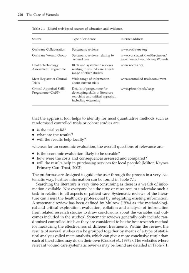
that the appraisal tool helps to identify for most quantitative methods such asrandomised controlled trials or cohort studies are:
� is the trial valid?� what are the results?� will the results help locally?
whereas for an economic evaluation, the overall questions of relevance are:
� is the economic evaluation likely to be useable?� how were the costs and consequences assessed and compared?� will the results help in purchasing services for local people? (Milton Keynes
Primary Care Trust, 2002)
The proformas are designed to guide the user through the process in a very sys-tematic way. Further information can be found in Table 7.1.
Searching the literature is very time-consuming as there is a wealth of infor-mation available. Not everyone has the time or resources to undertake such atask in relation to all aspects of patient care. Systematic reviews of the litera-ture can assist the healthcare professional by integrating existing information.A systematic review has been defined by Mulrow (1994) as ‘the methodologi-cal and critical exploration, evaluation, collation and analysis of informationfrom related research studies to draw conclusions about the variables and out-comes included in the studies’. Systematic reviews generally only include ran-domised controlled trials as they are considered to be the best research methodfor measuring the effectiveness of different treatments. Within the review, theresults of several studies can be grouped together by means of a type of statis-tical analysis called meta-analysis, which can give a more conclusive result thaneach of the studies may do on their own (Cook et al., 1997a). The websites whererelevant wound care systematic reviews may be found are detailed in Table 7.1.
220 The Care of Wounds
Table 7.1 Useful web-based sources of education and evidence.
Source Type of evidence Internet address
Cochrane Collaboration Systematic reviews www.cochrane.org
Cochrane Wound Group Systematic reviews relating to www.york.ac.uk/healthsciences/wound care gsp/themes/woundcare/Wounds
Health Technology RCTs and systematic reviews www.ncchta.org.Assessment Programme relating to wound care + wide
range of other studies
Meta-Register of Clinical Wide range of information www.controlled-trials.com/mrctTrials about current trials
Critical Appraisal Skills Details of programme for www.phru.nhs.uk/caspProgramme (CASP) developing skills in literature
searching and critical appraisal,including e-learning
7.3.1 Appraising the literature on wound care
The literature pertaining to all aspects of wound care is very large, as demon-strated by the many examples that have been given in other chapters of thisbook. It is therefore important for the nurse to be aware of how to identify boththe strengths and the weaknesses of the wound care evidence. Some of theissues are addressed here.
Study size
Early studies of modern dressings tended to compare them with gauze. As thedifferences between the two groups were so great, only relatively smallnumbers of patients were needed in a study. However, the differences are muchsmaller when comparing two modern products and therefore much largernumbers are needed. It may be difficult for one centre to recruit sufficientpatients, so several centres may be required. This may introduce other variablesif there are variations in local management patterns (Freak, 1995). High-qualityresearch will indicate that it has sought to reduce this problem by using a powercalculation to identify the required sample size and also addressing the issueof standardisation between centres.
Endpoints
Another issue to consider when appraising the wound care literature is end-points. In other words, at what point a patient should be considered to havecompleted a study. Freak (1995) suggests that complete healing is not alwaysthe most suitable endpoint, although Bradley et al. (1999) argue that assessingthe proportion of wounds healed is the most objective assessment currentlyavailable. They also propose that if reduction in wound size is used as an end-point then both the percentage and absolute change in surface area should bemeasured.
Length of study
There has been considerable debate regarding the length of time that is rea-sonable for a trial of effectiveness of treatment, especially if time to healing isused as an endpoint. Lengthy studies are costly to undertake and there is a riskthat patients may be lost to follow-up during the time of the study. Nelson(1998) suggests that healing rates for leg ulcers should be measured at intervalsover time, probably more than 12 weeks. However, other authors have notalways supported this view. Kantor and Margolis (2000) studied 104 venous legulcers and found that percentage change in area at four weeks was an indica-tor of healing at 24 weeks. Similarly, Sheehan et al. (2003), using 203 patientsfound the percentage change in diabetic foot ulcer area at four weeks is a robustindicator of healing at 12 weeks. There is obviously some disparity in the expected healing time between the two studies quoted, indicating theimportance of not trying to extrapolate the findings directly to another wound type.
Clinically Effective Wound Care 221
7.4 DEVELOPING CLINICAL GUIDELINES
Clinical guidelines are recommendations on the appropriate treatment and careof people with specific diseases and conditions (NICE, 2004a). They have anumber of uses: to guide the care of individuals; to develop standards againstwhich clinical practice can be measured; for education and training; and to helppatients make informed choices (NICE, 2004a). Generally, guidelines consist ofa series of statements around a particular topic with a rationale for each state-ment. The rationale relates to the evidence, usually obtained from systematicreviews, underpinning the statement. Many guidelines use a hierarchy tomeasure the quality of the evidence that they use and then grade the recom-mendation, as demonstrated in the NICE guidelines. For example, within thehierarchy of evidence, level I indicates evidence obtained from meta-analysisof randomised controlled trials or at least one randomised controlled trial; a statement based on this evidence would be graded as a grade A statement.Table 7.2 shows the grading system used in the NICE guidelines on preventionand management of foot problems in patients with type 2 diabetes (NICE,2004b).
Preparation and publication of a guideline are not enough to ensure that itis implemented. Any national guideline tends to be written in broad terms. Itneeds to be adapted to include more operational detail for local use and mayinclude policies and protocols. The implementation team should be multidis-ciplinary, reflecting all key personnel including representatives from purchas-ing authorities (Effective Health Care, 1994). Richens et al. (2004) undertook areview of the literature on guideline implementation and made several usefulobservations based on their findings.
� Guidelines and any local adaptation of guidelines should contain recom-mendations that are clear, specific and relevant to both practitioners andpractice.
� Ownership and adoption of guidelines are more likely to occur if there is apartnership between the guideline developers, users and implementers.
� Guideline implementation can be improved by use of dedicated ‘changeagents’ to work with individuals, teams and organisations in the clinicalsetting.
� The use of clinical audit is an essential aspect of guideline implementation.� Those responsible for implementing guidelines need to be aware of organi-
sational factors, especially linking in to the organisation’s strategy andresource commitments.
7.4.1 Guidelines in wound care
There are a great number of guidelines available that relate to various aspectsof wound care. A survey undertaken by the Tissue Viability Society in 2002found that there appeared to be a major shift over a five-year period from theuse of locally developed guidelines to the use of national and international
222 The Care of Wounds
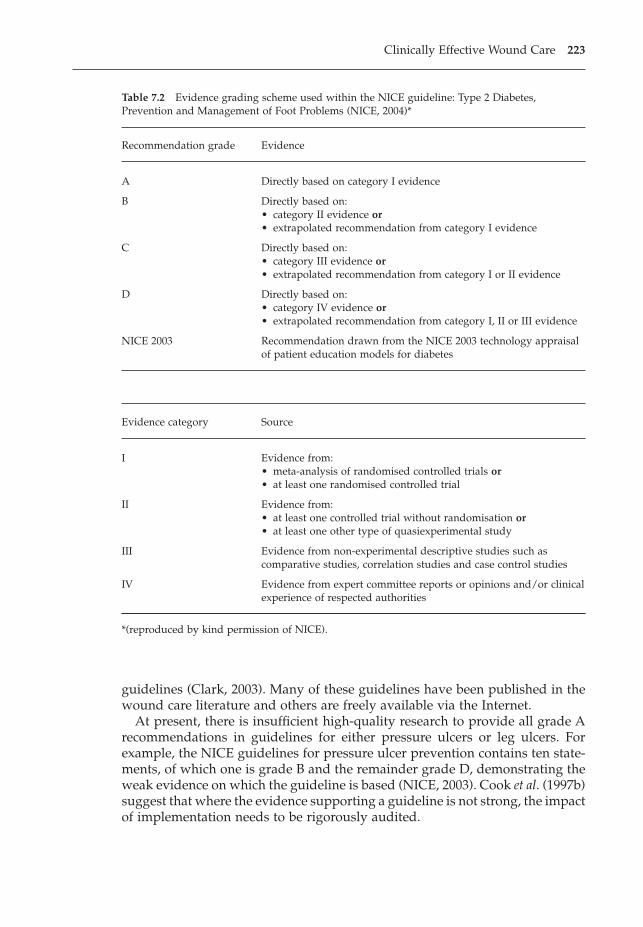
guidelines (Clark, 2003). Many of these guidelines have been published in thewound care literature and others are freely available via the Internet.
At present, there is insufficient high-quality research to provide all grade Arecommendations in guidelines for either pressure ulcers or leg ulcers. Forexample, the NICE guidelines for pressure ulcer prevention contains ten state-ments, of which one is grade B and the remainder grade D, demonstrating theweak evidence on which the guideline is based (NICE, 2003). Cook et al. (1997b)suggest that where the evidence supporting a guideline is not strong, the impactof implementation needs to be rigorously audited.
Clinically Effective Wound Care 223
Table 7.2 Evidence grading scheme used within the NICE guideline: Type 2 Diabetes,Prevention and Management of Foot Problems (NICE, 2004)*
Recommendation grade Evidence
A Directly based on category I evidence
B Directly based on:• category II evidence or• extrapolated recommendation from category I evidence
C Directly based on:• category III evidence or• extrapolated recommendation from category I or II evidence
D Directly based on:• category IV evidence or• extrapolated recommendation from category I, II or III evidence
NICE 2003 Recommendation drawn from the NICE 2003 technology appraisal of patient education models for diabetes
Evidence category Source
I Evidence from:• meta-analysis of randomised controlled trials or• at least one randomised controlled trial
II Evidence from:• at least one controlled trial without randomisation or• at least one other type of quasiexperimental study
III Evidence from non-experimental descriptive studies such ascomparative studies, correlation studies and case control studies
IV Evidence from expert committee reports or opinions and/or clinical experience of respected authorities
*(reproduced by kind permission of NICE).
7.5 THE CLINICAL AUDIT CYCLE
Audit has been defined as:
‘a clinically-led initiative which seeks to improve the quality and outcome ofpatient care through structured peer review whereby clinicians examine theirpractices and results against agreed explicit standards and modify their prac-tice where indicated’ (NHS Executive, 1998).
Burnett and Winyard (1998) suggest that clinical audit is at the heart of clinicaleffectiveness. Some guidelines also include an audit tool against which it is pos-sible to measure the effectiveness of implementation. Examples can be foundin the NICE guidelines on pressure ulcer prevention and the diabetic foot(NICE, 2003, 2004b).
One of the benefits of audit is that it can be used to measure the outcomes ofeveryday practice to provide baseline information before introducing clinicalguidelines. A repeat of the audit enables the guideline team to monitor anyhealth gains associated with the implementation of more effective practice aswell as monitoring whether the guidelines are being adhered to. Clinical staffare more likely to be willing to adhere to guidelines if they can see a beneficialoutcome. Providing information for staff about performance levels is an essen-tial aspect of the audit process.
7.5.1 Auditing clinical practice
Audit provides data both for initial baseline information and to measure theoutcomes of guideline implementation. In many instances this information isobtained by means of prevalence surveys and incidence monitoring. This is par-ticularly true of pressure ulcers where there has been much debate on the mosteffective methods for collecting this information and on its value.
Prevalence can be defined as: ‘The number of persons with a specific diseaseor condition as a proportion of a given population measured at a specific pointin time’ (Dealey, 1997), whereas incidence is defined as: ‘The number of peopledeveloping a specific condition as a proportion of the local population mea-sured over a period of time’ (Dealey, 1997).
An example of a baseline pressure ulcer audit is one undertaken acrossFrance by Barrois et al. (1997). They found an overall prevalence of 8.7% as wellas considerable detail about the characteristics of pressure ulcers in France. Thisprovided a very informative baseline prior to introducing a range of preven-tion strategies. Incidence rates are often used to evaluate the effectiveness ofprevention policies. Data collection for incidence measurement is more com-plicated than for prevalence surveys and subject to inaccuracies (Bridel et al.,1996). However, Dealey (1997) suggests that the main benefit of incidence ratemeasurement is to demonstrate improvements over time.
Lindholm et al. (1998) and Dealey (1998) undertook prevalence surveys of legulceration prior to instigating education and training and the implementation
224 The Care of Wounds
of management guidelines. Audit of the effectiveness of this strategy concen-trated on changes in practice, nursing knowledge and healing rates (Dealey,1998). Feedback of the results to the staff provided encouragement that adher-ence to the guidelines was ensuring increased healing rates. Stevens et al. (1997)also used healing rates to monitor the outcomes of a project establishing legulcer guidelines within two acute hospitals and the surrounding community.Over time they were also able to measure recurrence rates and compare themfavourably with the rate from another study previously reported in the literature.
Audit of surgical wounds is usually undertaken as a surveillance of woundinfection rates. This may be in the form of either prevalence or incidence. Preva-lence surveys have been used in national surveys to provide a snapshot of thesituation (Briggs, 1996). Incidence rates can be used to monitor the performanceof individual surgeons. Frequently this type of surveillance measures the inci-dence of surgical wound infection (SWI) in clean surgery. Incidence may beused with case control studies or with cohort studies. Case control studies ret-rospectively compare a group of patients with SWI with a control group inorder to determine any risk factors associated with SWI. Cohort studies areprospective and follow a group of patients over time to see who develops aSWI. Potential risk factors are determined at the beginning of the studies andthen monitored for the outcome. For example, in their ten-year study, Cruseand Foord (1980) considered diabetes to be a potential risk factor and so mon-itored the clean wound infection rate for diabetics and found it to be consider-ably higher than that for non-diabetics. The findings from this type of audit canbe used to improve patient care.
7.5.2 Dissemination of audit findings
As well as sharing audit results with those directly involved, it is important todiscuss them with a wider audience. This may be in the form of a report for theexecutive board of a hospital trust or the local purchasing authority or it maybe a conference presentation or a publication. There are a number of benefits.
� Internal dissemination allows other areas in the organisation to benefit fromthe audit findings.
� Healthcare professionals working in similar clinical areas can apply thelessons learned from the audit.
� Those involved in the audit will develop skills in writing and undertakingpresentations.
7.6 CONCLUSIONS
At present, the quality of the evidence available to support clinically effectivewound care is very variable. Systematic reviews can be used to determine thegaps so that future research can be directed to the area where it is most needed.
Clinically Effective Wound Care 225
REFERENCES
Barrois, B., Allaert, F., Urbinelli, R., Colin, D. (1997) National survey in France: pressuresores in hospital institutions in 1994, in (eds) Cherry, G.W., Gottrup, F., Lawrence,J.C., Moffat, C.J., Turner, T.D., Proceedings of the 5th European Conference on Advancesin Wound Management. Macmillan Magazines Ltd, London.
Bradley, M., Cullum, N., Sheldon, T. (1999) The debridement of chronic wounds: a sys-tematic review. Health Technology Assessment, 3 (17), Part 1, 1–78.
Bridel, J., Banks, S., Mitton, C. (1996) The admission prevalence and hospital-acquiredincidence of pressure sores within a large teaching hospital during April 1994 toMarch 1995, in (eds) Cherry, G.W., Gottrup, F., Lawrence, J.C., Moffat, C.J., Turner,T.D., Proceedings of the 5th European Conference on Advances in Wound Management.Macmillan Magazines Ltd, London.
Briggs, M. (1996) Epidemiological methods in the study of surgical wound infection.Journal of Wound Care, 5 (4), 186–191.
Burnett, A.C., Winyard, G. (1998) Clinical audit at the heart of clinical effectiveness.Journal of Quality and Clinical Practice, 18 (1), 3–19.
Clark, M. (2003) Barriers to the implementation of clinical guidelines. Journal of TissueViability, 13 (2), 62–72.
Cook, D.J., Mulrow, C.D., Haynes, R.B. (1997a) Systematic reviews: synthesis of best evi-dence for clinical decisions. Annals of Internal Medicine, 126 (5), 376–380.
Cook, D.J., Greengold, N.L., Ellrodt, A.G., Weingarten, S.R. (1997b) The relation betweensystematic reviews and practice guidelines. Annals of Internal Medicine, 127 (3),210–216.
Cruse, P.J.E., Foord, R. (1980) The epidemiology of wound infection: a 10-year prospec-tive study of 62 939 wounds. Surgical Clinics of North America, 60, 27–40.
Dealey, C. (1997) Managing Pressure Sore Prevention. Mark Allen Publishing Ltd, Salisbury.Dealey, C. (1998) The importance of education in effecting change in leg ulcer manage-
ment, in (eds) Leaper, D., Dealey, C., Franks, P.J., Hofman, D., Moffatt, C.J., Pro-ceedings of the 7th European Conference on Advances in Wound Management. EMAPHealthcare Ltd, London.
Department of Health (1997) The New NHS: Modern, Dependable. Department of Health,London.
Effective Health Care (1994) Implementing clinical practice guidelines. Effective HealthCare Bulletin no. 8. University of Leeds, Leeds, pp. 1–12.
Freak, L. (1995) Evaluating clinical trials. Journal of Wound Care, 4 (3), 114–116.Kantor J., Margolis D. (2000) A multicentre study of percentage change in venous leg
ulcer area as a prognostic index of healing at 24 weeks. British Journal of Derma-tology, 142, 960–964.
Lindholm, C., Tammelin, A., Bergsten, A., Berglund, E. (1998) The Uppsala experience:implications for developing educational strategies, in (eds) Leaper, D., Dealey, C.,Franks, P.J., Hofman, D., Moffatt, C.J., Proceedings of the 7th European Conference onAdvances in Wound Management. EMAP Healthcare Ltd, London.
Milton Keynes Primary Care Trust (2002) Critical Appraisal Skills Programme. MiltonKeynes Primary Care Trust, Milton Keynes.
Mulrow, C.D. (1994) Rationale for systematic reviews. British Medical Journal, 309 (6954),597–599.
National Institute for Clinical Excellence (2003) Clinical Guideline 7: the use of pressure-relieving devices (beds, mattresses and overlays) for the prevention of pressure ulcers inprimary and secondary care. NICE, London.
226 The Care of Wounds

National Institute for Clinical Excellence (2004a) About Clinical Guidelines. Availableonline at: www.nice.org.uk.
National Institute for Clinical Excellence (2004b) Clinical Guideline 10: type 2 diabetes: pre-vention and management of foot problems. NICE, London.
Nelson, E.A. (1998) The evidence in support of compression bandaging. Journal of WoundCare, 7 (3), 148–150.
Richens, Y., Rycroft-Malone, J., Morrell, C. (2004) Getting guidelines into practice: a lit-erature review. Nursing Standard, 18 (50), 33–40.
Sheehan, P., Jones, P., Caselli, A., Giurini, J.M., Veves, A. (2003) Percentage change inwound area of diabetic foot ulcers over a 4-week period is a robust predictor ofcomplete healing in a 12 week prospective trial. Diabetes Care, 26 (6), 1879–1882.
Stevens, J., Franks, P.J., Harrington, M. (1997) A community/hospital leg ulcer service.Journal of Wound Care, 6 (2), 62–68.
Clinically Effective Wound Care 227
228 The Care of Wounds
Chapter 8The Organisation of Wound Management
8.1 INTRODUCTION
The organisation of the delivery of wound care has undergone great change inthe last few years. This chapter explores some of the aspects of wound caredelivery that have had a great impact on nurses and nursing, both in the hos-pital and in the community. Overall, these changes have ensured that patientsreceive more effective care.
8.2 MANAGING WOUNDS IN THE COMMUNITY
In most healthcare systems there are some restrictions on which wound man-agement products are available in the community. In the UK, available productsare listed on the Drug Tariff. Today this list is regularly reviewed and new prod-ucts added to it but this was not always the case. Less than ten years ago, therewere considerable restrictions on the range of products available, particularlydressings for cavity wounds and compression bandages. Dealey (1997) reporteda survey undertaken by the Tissue Viability Society to gain information abouthow cavity wounds were managed in the community. The overall response ratewas poor but the results showed that nearly one-fifth of the dressings used wereobtained by illegal means such as obtaining a prescription for one product anddoing a ‘swap’ with the pharmacist for another, more suitable product. Happilya great deal has changed since then. As well as an increase in the products avail-able, nurses are now allowed to prescribe from a limited formulary.
8.2.1 Nurse prescribing
Nurse prescribing has followed a rocky road since Julia Cumberlege first putforward the idea in a report for the Department of Health (DHSS, 1986). Theconcept was seen as a way of reducing time wasting for both community nursesand general practitioners (GPs) and reducing delays in treatment. Typically anurse would see a patient with a leg ulcer at home and decide on a particularcourse of treatment. The nurse would have to go to the surgery to see the GPand request a prescription for the treatment. The patient, or a representative,would then collect the prescription from the surgery. In the case of house-bound, elderly patients, nurses would sometimes obtain the prescription anddeliver it to the pharmacist for the patient.
Initially it was only qualified district (community) nurses and health visitorswho were able to prescribe, once they had undertaken the relevant three-monthtraining course. Since 2000, this has widened and there are now a variety ofways in which a wide range of nurses can potentially undertake prescribingusing the Nurse Prescriber’s Formulary.
� Independent prescriber – person who is responsible for both assessment andtreatment of a range of specific conditions. This group includes doctors anddentists as well as nurses but there are obviously greater limitations to therange of conditions that nurses can treat. However, they do include skin andwound care and minor injuries.
� Supplementary prescriber – this type of prescribing is most frequently of benefitwhen caring for patients with long-term conditions. A doctor will draw upa plan of care including the range of medication that can be used and whento refer the patient back to the doctor (Dimond, 2003). The supplementaryprescriber can vary the dosage, keeping within the prescribed treatmentrange. This type of prescribing means that supplementary prescribers canlegally prescribe from a wider range of medications than can be found in thenurse’s formulary and this is generally more useful within specialist multi-disciplinary teams (Hay et al., 2004).
� Patient Group Directions (PGD) – these are documents that make it legal forspecific medicines to be given to patients without an individual prescription.However, they only relate to the supply or administration of a medicine,rather than its prescription (Baird, 2003). Each PGD has to be developedlocally. Although initially intended for situations such as the treatment ofmass casualties, they have also been used for walk-in centres and nurse-ledclinics.
Thus far, nurse prescribing would seem to be relatively successful in the UKand around 23000 district nurses and health visitors have been trained to pre-scribe so far. Latter and Courtney (2004) reviewed the literature for evaluationsof nurse prescribing and found that, overall, the outcome was positive with evi-dence of improvements in time saving and convenience. White and Biggs (2004)conducted a postal survey of health visitors and district nurses in three trusts.They found variation in the level of confidence as a prescriber and in the fre-quency of prescribing. There was also a strong association between prescribingmore than three times a week and wound care products and elastic hosiery,indicating wound care has particularly benefited from nurses’ ability to pre-scribe. However, as discovered by other writers (Banning, 2004; Lewis-Evans& Jester, 2004), the authors of this study found that the nurses desired greatereducation in applied pharmacology, both in the initial education programmeand as part of their ongoing professional development.
8.2.2 Community leg ulcer clinics
Another development has been that of nurse-led leg ulcer clinics in the com-munity. These clinics have gradually increased in number during the last ten
The Organisation of Wound Management 229
years, encouraged by the work of Moffatt et al. (1992) and the development ofleg ulcer courses for nurses, such as the N18 validated by the now defunctEnglish National Board (Moffatt & Karn, 1994). Many clinics have been estab-lished under the auspices of clinical nurse specialists in tissue viability or legulcer nurse specialists. The rationale for establishing leg ulcer clinics is the pos-sibility of concentrating resources and skilled nurses at the clinic to ensure thatall patients have a full assessment, including a Doppler assessment, and accessto appropriately applied compression bandages. However, it is the outcomesof care that are most important.
Simon et al. (1996) found a 38% reduction in costs after establishing leg ulcerclinics in a health authority alongside a 13-week healing rate that increasedfrom less than 25% to 42% in the first 12 months. They compared these out-comes with a control group in a neighbouring health authority where therewere no clinics and found no improvement from baseline in this group. In alater phase of this project, the project team monitored the continuing progressof the clinics as well as new clinics established in the neighbouring healthauthority (Ellison et al., 2002). They found the new clinics showed a similarimprovement in outcome but the longer established clinics did not totally main-tain their initial level of improvement, most probably because of the loss of theleg ulcer nurse specialist between the two phases of the project.
Morrell et al. (1998) undertook a randomised, controlled trial to compare out-comes for patients randomised to a leg ulcer clinic compared with usual careat home by a district nurse. They found a trend towards greater healing in theclinic group (p = 0.03) and a slightly greater cost per patient compared with thecontrols. The authors concluded that clinics provide better outcomes thanhomecare. However, in a critique by Thurlby and Griffiths (2002), the authorssuggest that the study was biased towards the clinic group as there was poorprovision of compression bandages with less than half the homecare patientsreceiving them. It is also possible that the nurses were less skilled in leg ulcercare.
Despite these criticisms, there seems to be a general view that leg ulcer clinicsare an effective method of proving care for patients with leg ulcers. Somepatients find the social aspect important as many have poor mobility thatrestricts their lifestyle. A study by Liew et al. (2000) found an improvement inquality of life over an average of eight weeks of clinic attendance. In particu-lar, patients had a significant reduction of pain, better sleep and greater mobil-ity. Overall, leg ulcer clinics have more supporters than critics, although it maybe that involvement of a nurse specialist is crucial in maintaining beneficial outcomes.
8.3 NURSE SPECIALISTS IN WOUND CARE
Specialist nurses in wound care have a variety of titles but the most commonin the UK is clinical nurse specialist in tissue viability (TVN). They straddle
230 The Care of Wounds
both hospital and community as TVN may be found in both community andhospital trusts. It is a relatively new nursing specialty developed in the late1980s and the numbers have grown rapidly. The precise number is difficult todetermine as there is no formal register but Finnie (2004) suggests that thereare now around 500 in post as well as a few nurse consultants.
Education is an essential prerequisite for the recognition of any specialty.Fletcher (1998) found that there are a variety of courses available within theUK. They comprise short courses at diploma level as well as degree and Masterslevel programmes. Fairbairn (2001) discussed the potential for a clinical doc-torate for TVN as an alternative to undertaking a PhD following the more tra-ditional route. He suggested that this option is more flexible, allowing thestudent to utilise aspects of existing work. A more recent development has beenthat of a competency framework devised for TVN in Scotland but it is intendedfor wider use (Finnie & Wilson, 2003). It involves six domains: clinical problemsolving, professional practice, teamwork, reflective practice, empowerment andleadership.
Austin (2002) undertook a survey of TVN in one region of the UK and foundseveral clearly defined aspects of the role.
� Clinical role – all the TVN had direct clinical contact with patients, receivingreferrals predominantly from nurses.
� Educator role – this is an important aspect of the role. The survey found thatTVN mostly taught nurses but also taught other disciplines such as doctors,allied health professionals and social workers.
� Leadership role – all respondents had a remit that extended across their organisation, involving advice on purchasing or commissioning resources(94%), standards or guideline development (93%), audit of care provision(83%).
� Research role – 57% had participated in research, predominantly product eval-uations. This aspect of the role generally requires further development (Gray,2004).
� Management role – respondents indicated that this involved managing a team(39%) and/or holding the service budget (42%) or influencing the budgetexpenditure.
Austin’s survey provides a picture of TVN activity that is fairly representative.She also noted that the role is poorly understood by others. Flanagan (1996)described some of the difficulties that the TVN may face. They include unreal-istic objectives or targets, role ambiguity, responsibilities across large geo-graphical areas or multiple sites, constant pressure to reduce the cost of thetissue viability service and often having to work with insufficient resourceswith limited professional support. Despite the constraints, the role of the TVNstill presents the post holder with considerable opportunities to enhancenursing practice and the personal satisfaction of seeing wounds healing andpatients’ quality of life improve.
The Organisation of Wound Management 231
8.4 THE MANAGEMENT OF PRESSURE-REDISTRIBUTINGEQUIPMENT
As noted above, many TVN have a major role in relation to the managementof equipment, especially pressure-redistributing equipment. Most trusts havesome type of budget for equipment but there is considerable variety in how itis used and the ways in which distribution and maintenance are organised.Several authors have reported that ad hoc ordering of rental equipment orunplanned purchasing on a ward-by-ward basis is both expensive and ineffi-cient and results in ineffective care (Birchall, 2001; Fox & Delve, 1994; Newton2001). One alternative is an equipment store or equipment library.
8.4.1 Equipment stores
This method of control and delivery of pressure-redistributing equipment ismost commonly found in the hospital setting but there are also a few withinprimary care trusts. Birchall (2001) described the development of an equipmentstore across an acute trust of 1100 beds on two sites, taking two years to becomefully established. The store contained a mixture of purchased and rental equip-ment. The cost savings from the previous ad hoc rental system covered the costof the equipment, including maintenance and repair, cost of the staff runningthe store, an annual foam mattress replacement programme and a five-yearreplacement programme for the alternating air mattresses.
Newton (2001) found similar problems in her trust. In particular, there wereproblems with staff attitudes as they were careless with equipment, unwillingto share equipment with other wards, kept patients on dynamic systems forlonger than necessary and tended to order rental equipment as an easy option,rather than seeking out trust-owned equipment. As a result, rental expenditurewas spiralling out of control. After implementing an equipment library, Newtonfound a marked change in staff attitudes and a more appropriate and cost-effective use of equipment.
Both Birchall (2001) and Newton (2001) implemented improved educationprogrammes for staff that covered not only the appropriate use of equipmentbut also how to set it up. The use of a central database is also an essential aspectof this type of service as it enables the store staff to determine the whereaboutsof every piece of equipment, making it easier to monitor usage.
8.4.2 Total bed management
An alternative to the establishment of an equipment store is the use of total bedmanagement (TBM). This concept means that a trust and a contractor enter intopartnership, with the contractor supplying the trust with all its tissue viability,manual handling and bed therapy needs (Preece, 1999). Obviously, this type ofcontract means that the equipment is supplied at a reduced cost. Preece (1999)described similar problems as those described above. In addition, there wasalso a major problem with existing bed frame stock as 492 bed frames failed at
232 The Care of Wounds
least one of five assessment criteria; also 389 of them were a manual handlingrisk. An audit of the foam mattresses showed that 75% needed replacement.There was also an excessive use of low air loss systems. The implementation of TBM enabled the trust to replace the worn-out bed frames with electric bed frames, which they found reduced both the manual handling risks and also the need for low air loss beds. The foam mattresses were replaced with high-specification foam mattresses and a suitable range of other pressure-redistributing equipment was included in the contract.
8.5 WOUND-HEALING CENTRES
Wound care falls into the remit of a great many specialist areas: vascularsurgery, dermatology, diabetology, plastic surgery, geriatrics and so on. This hasresulted in considerable fragmentation of services and lack of ownership inmany areas. Bennett (2000) suggested that unless wound care is able to estab-lish its own unique national profile, it is in danger of being subsumed into otherspecialties such as those listed above. There is also little awareness of the ben-efits of providing a multiprofessional specialist centre for non-healing wounds.The one exception seems to be diabetic foot ulcers as the NICE guidelines statethat patients should have access to a multidisciplinary footcare team (NICE,2004); even then, it only refers to the one wound type. There are a number ofcentres of excellence caring for one wound type, such as leg ulcers or diabeticfoot ulcers, but very few that provide care for wounds of all aetiologies.
Four wound-healing centres will be discussed: two in the UK and two inDenmark. All of these centres are based within the national healthcare systemsof the two countries and there is no charge associated with attendance. This is incontrast with the wound-healing centres in the USA that are commercially run.
Despite the fact that nurses have led much of the work in improving woundcare provision, it is doctors who have pioneered the concept of wound-healingcentres. In the case of the two centres in the UK, one centre was established bya general practitioner, although it is based in a department of surgery in a uni-versity hospital, and the other by a geriatrician (Bennett, 2000; Harding, 2001)and in Denmark, the two wound-healing centres were established by a surgeon(Gottrup, 2004). Although the centres all function differently, they have anumber of common features:
� The staff working in wound-healing centres include physicians from differ-ent specialisms, as well as a variety of other healthcare professionals, includ-ing nurses. The range of professionals varies from centre to centre.
� They are based within teaching hospitals and provide education pro-grammes of varying types.
� They treat both inpatients and outpatients, referred from both the primaryand secondary healthcare sectors, although the number of inpatient bedsmay be limited.
� All the wound-healing centres are involved in research programmes.
The Organisation of Wound Management 233
Beyond this common ground, the centres all function differently, due to the cir-cumstances in which they were developed. The centres in Denmark have beenfully integrated into the national health service, allowing for country-widereferrals. Gottrup (2004) suggests that for Denmark, with its 5.2 million inhab-itants, only one or two clinics are necessary. Obviously, larger or more denselypopulated countries would require more centres to provide the same levels ofcare provision.
Wound-healing centres have a great deal to offer in terms of specialised carefor problem wounds, educational opportunities to increase the numbers of clinical staff with specialist knowledge and an important research function toassist in developing the evidence base for this important subject.
8.6 CONCLUSIONS
Wound care is increasingly being recognised as an important aspect of nursingcare and it is becoming ever more sophisticated. Despite this, there are still areasof poor practice and patients who receive less than optimum care. It is hopedthat these variations in practice will decrease as more evidence and more guide-lines become available to guide the practitioner.
REFERENCES
Austin, L. (2002) A survey of tissue viability nurses’ role and background in one region.Journal of Wound Care, 11 (9), 347–350.
Baird, A. (2003) Nurse prescribing – how did we get here? Journal of Community Nursing,17 (4), 4–10.
Banning, M. (2004) Nurse prescribing, nurse education and related research in theUnited Kingdom: a review of the literature. Nurse Education Today, 24, 420–427.
Bennett, G. (2000) Is there a role for the multidisciplinary approach within tissue via-bility in the next millennium? Journal of Tissue Viability, 10 (2), 43–44.
Birchall, L. (2001) Centralising supply of a trust’s pressure-relieving equipment. Journalof Wound Care, 10 (6), 214–219.
Dealey, C. (1997) A survey of the management of cavity wounds in the community.Journal of Tissue Viability, 7 (3), 75–76.
Department of Health and Social Security (1986) Neighbourhood Nursing – a focus for care.Report of the Community Nursing Review. HMSO, London.
Dimond, B. (2003) The introduction of nurse prescribing 2: final Crown Report. BritishJournal of Nursing, 12 (16), 980–983.
Ellison, D.A., Hayes, L., Lane, C., Tracey, A., McCollum, C.N. (2002) Evaluating the costand efficacy of leg ulcer care provided in two large UK health authorities. Journalof Wound Care, 11 (2), 47–51.
Fairbairn, G. (2001) The role of a clinical doctorate in the advancement of practice. BritishJournal of Nursing, 10 (15) (suppl), S4–S5.
Finnie, A. (2004) We must act to move tissue viability forward. British Journal of Nursing,13 (6) (suppl), S3.
234 The Care of Wounds
Finnie, A., Wilson, A. (2003) Development of a tissue viability nursing competencyframework. British Journal of Nursing, 12 (6) (suppl), S38–S44.
Flanagan, M. (1996) The role of the clinical nurse specialist in tissue viability. BritishJournal of Nursing, 5 (11), 676–681.
Fletcher, J. (1998) A survey of courses available that are relevant to the field of tissue via-bility, in (eds) Leaper, D., Dealey, C., Franks, P.J., Hofman, D., Moffatt, C., Pro-ceedings of the 7th European Conference on Advances in Wound Management. EMAPHealthcare Ltd, London.
Fox, P., Delve, M. (1994) Equipped to care. Nursing Times, 90 (30), 46–47.Gottrup, F. (2004) A specialised wound-healing centre concept: importance of a multi-
disciplinary department structure and surgical treatment facilities in the treatmentof chronic wounds. American Journal of Surgery, 187 (5) (suppl), 38S–43S.
Gray, D. (2004) Specialists must accept challenge of improving clinical outcomes. BritishJournal of Nursing, 13 (6) (suppl), S4.
Harding, K.G. (2001) Meet the innovators in wound care. Journal of Wound Care, 10 (5),180.
Hay, A., Bradley, E., Nolan, P. (2004) Supplementary nurse prescribing. Nursing Standard,18 (4), 33–39.
Latter, S., Courtenay, M. (2004) Effectiveness of nurse prescribing: a review of the litera-ture. Journal of Clinical Nursing, 13 (1), 26–32.
Lewis-Evans, A.B., Jester, R. (2004) Nurse prescribers’ experiences of prescribing. Journalof Clinical Nursing, 13 (7), 796–805.
Liew, I.H., Law, K.A., Sinha, S.N. (2000) Do leg ulcer clinics improve patients’ quality oflife? Journal of Wound Care, 9 (9), 423–426.
Moffatt, C.J., Karn, A. (1994) Answering the call for more education. Development of an ENB course in leg ulcer management. Professional Nurse, 9 (10), 708–712.
Moffatt, C.J., Franks, P.J., Oldroyd, M. et al. (1992) Community clinics for leg ulcers andimpact on healing. British Medical Journal, 305, 1389–1392.
Morrell, C.J., Walters, S.J., Dixon, S. et al. (1998) Cost effectiveness of community leg ulcerclinics: randomised controlled trial. British Medical Journal, 316, 1487–1491.
National Institute for Clinical Excellence (2004) Clinical Guideline 10: type 2 diabetes: pre-vention and management of foot problems. NICE, London.
Newton, H. (2001) Developing an equipment library: the solution to increasing demand.British Journal of Nursing, 10 (22) (suppl), S59–S64.
Preece, J. (1999) Total bed management: the way forward in pressure sore prevention.British Journal of Nursing, 8 (22), 1524–1529.
Simon, D.A., Freak, L., Kinsella, J. et al. (1996) Community leg ulcer clinics: a compara-tive study in two health authorities. British Medical Journal, 312, 1648–1651.
Thurlby, K., Griffiths, P. (2002) Community leg ulcer clinics vs home visits: which is moreeffective? British Journal of Community Nursing, 7 (5), 260–264.
White, A., Biggs, K. (2004) Benefits and challenges of nurse prescribing. Journal ofAdvanced Nursing, 45 (6), 559–567.
The Organisation of Wound Management 235
236 The Care of Wounds
Page numbers in italics refer to figures or tables.
British Pharmaceutical Codices, 89, 90Buerger’s disease, 23, 153burn injuries, 9, 199–209
aetiology and incidence, 200–201classification and assessment, 201–03, 204, 206,
207first aid treatment, 203, 205management, 205–09
complex burns, 205–06non-complex, 206–09
non-accidental, 200, 201and oedema, 203
cadexomer bead dressings, 101capillary bleeding, 163, 165
see also haemorrhagecapillary wound dressings, 75, 101carbolic acid, 88Carbonet, 104carcinomas
and leg ulcers, 156–7see also fungating wounds
catheter use, and infection control, 22Cavi-Care, 101Cavilon, 100cell traction, 7cetrimide, 92charcoal dressings, 104, 164Charnley Howarth tent, 23chlorhexidine, 92, 103Chlorhexitulle, 103chronic wounds, 10–11
defined, 56diabetic foot ulcers, 158–62, 161economic costs, 121, 122–3and infection control, 64–5, 74–5pressure ulcers, 121–43types
fungating wounds, 162–5leg ulcers, 143–58
CicaCare, 103clinical guidelines development, 222–3
see also NICE guidelinesclinical trials, 78–9, 219–21Clinishield, 100Clinisorb, 104Cochrane reviews, 219, 220collagen, 6–9, 24, 209Combiderm, 102Comfeel Skin Care, 100communication needs see patient communication and
educationcommunity wound management regimes, 228–34
Index
absorbent pads and cellulose dressings, 99Acquacel, 185Acticoat, 104Actisorb Plus, 104active listening, 36Activon Tulle, 103adhesive island dressings, 99adrenaline, 165advanced technologies, 105–12
growth factors, 105hyaluronan-based products, 106–07hyperbaric oxygen, 23, 107–08protease-modulating products, 105–06tissue culture, 110tissue engineering, 110–11topical negative pressure (TNP) therapy, 75,
108–09age factors and risk, 15, 19, 21, 125–6air filtration systems, 189, 191alginates, 73, 75, 100, 165Algisite M, 100Alione Hydrocapillary, 101Allevyn, 101aloe vera gel, 211alternative therapies, 37, 112–13, 211antibacterial dressings, 100antibiotics, 75
topical forms, 95–6antiseptics, 75, 88–9
types, 92–5anxiety management, 35–7Apligraf (Graftskin), 110–12Aquacel, 102Aquacel Ag, 104Arglaes, 104aromatherapy, 37, 113arterial leg ulcers, 151–5, 154artificial skin, 110–11, 151, 183ASEPSIS wound scores, 188, 189assessment see wound assessmentauditing clinical practice, 224–5autolytic debridement, 73
Bactigras, 103‘bag systems’, 75, 192barrier film dressings, 100Better Food Programme (DoH 2001), 15Biatain, 101Bioclusive, 104biosurgery see larval therapybites, 198body image problems, 39–40, 206body temperature, and wound healing, 32–3
Index 237
complement system, 4compression, 75, 148–50, 167
bandage systems, 138–9pneumatic, 149socks and stockings, 151–2see also TNP (topical negative pressure) therapy
computer-assisted wound measurement, 60concordance (compliance), 38contact dermatitis, 71, 71contact inhibition, 8continuity of care, 42contractures, 7, 10Contreet Ag, 104Critical Appraisal Skills Programme (CASP), 219–20,
220Curasorb, 100cushions, 134cutifilm, 104Cutinova Cavity, 102Cutinova Hydro, 102cytokines, 5
Dakin’s solution, 88–9, 95debridement, 73–4
history of use, 86, 87–8, 89types
autolytic, 73enzymatic, 73–4larval therapy, 73, 75, 100–101mechanical, 74surgical, 74, 164–5
dehiscence, 191–2delayed primary closures, 56, 181depression, 26, 35–6Dermagraft, 111–12dermatitis, 71, 71Dermax, 106dermis, 2, 3diabetes mellitus, 25–6
complicationsfoot ulcers, 158–62, 161infection risk, 21, 225leg ulcers, 145neuropathic vs. ischaemic ulcers, 160–61, 160pain, 27
diet see nutrition managementdigital photography, 60, 67–8discharge planning
availability of dressings, 58involving patients, 43
documentation, 77–8see also measurement of wounds
donor sites, 184–5Doppler ultrasound/flowmeters, 147, 160–61, 206dressings
background history, 83–90dark ages and middle ages, 85–6late middle ages and renaissance, 86–7seventeenth to early nineteenth centuries, 87–8mid-nineteenth and twentieth centuries, 88–9traditional techniques, 90–91
costs, 79environmental considerations, 97–8evaluation, 78–9, 97–9handling qualities, 98–9and moisture balance, 75timing and frequency, 58types, 92–6, 99–104see also advanced technologies
education see patient communication and education;training and education
EGF (epidermal growth factor), 5enteral feeding, 17–19enzyme debridement, 73–4epidermis, 2–3, 3epithelialisation (wound healing), 8–9epithelialising wounds, 66, 67equipment stores, 136, 232erosion, 57erythema, 63, 71European Pressure Ulcer Advisory Panel, 122
decision ‘tree’ nutrition guidelines, 19, 20on grading and assessment, 138, 140
European Tissue Repair Society (ETRS), on documentation,77–8
Eusol, 78, 88–9, 95evaluation of treatments, 70
donor sites, 184–5dressing efficacy, 78–9foot ulcers, 162fungating wounds, 165leg ulcers, 144, 151, 155, 156, 158primary closure dressings, 180–81skin grafts, 182–4surgical cavities, 182see also clinical trials; measurement of wounds; wound
assessmentevidence-based practice, 218–25
clinical audit cycles, 224–5clinical guidelines development, 222–3, 223literature searches and appraisal, 218–21
Exu-dry, 99exudate
assessment and classification, 61–2, 62burn injuries, 208and chronic wounds, 10–11
FGF (fibroblast growth factor), 5, 6, 8fibroblasts, 6–7, 8
and burns, 209and chronic wounds, 10–11and diabetes, 25
finger tip injuries, 197fistula formation, 193–4foam dressings and cavity fillers, 101, 192foam stents, 182food standards in hospitals, 15
see also nutrition managementfootwear, 159–60friction, 125full-thickness wounds, 1, 57fungating wounds, 162–5
aetiology, 163management, 163–5
Galen (Roman surgeon), 84–5glucocorticoids, 26, 33–4Gore-Tex bags, 208–09Granuflex (Duoderm), 102Granugel, 102granulating wounds, 66, 66granulation tissue, 7grief reactions, 41growth factors, 5–6, 5, 11, 105guided imagery, 37
haemorrhage, 187Hageman factor, 4
238 The Care of Wounds
handwashing, 22–3healing see wound healinghealing by second intention, 2, 56, 181–2heart surgery, 180high-pressure wound irrigation, 74honey, 83–7, 96, 164, 185, 208Hospital Anxiety and Depression (HAD) Score, 35hospital trolleys, 135–6hospital-acquired infections (HAIs) see infection controlHyalofill-F, 106–07hyaluronan-based products, 106–07hydrocapillary and multilayered absorbent pads, 101–02hydrocolloids, 73, 102, 185, 196, 208
odour, 62hydrofibres, 102hydrogels, 73, 102, 197, 208hydrogen peroxide, 92–3hyperbaric oxygen, 23, 107–08hypertrophic scars, 9, 209hypothermia, 32–3
IGF-I/II (insulin-like growth factors), 5Inadine, 103incontinence lesions, 140infection control, 19–23, 63–5, 64, 74–5
during and following surgery, 187–91interventions and assessment, 22–3, 74–5
air filtration systems, 189, 191antibiotics/antiseptics, 91–5, 95–6handwashing, 22–3larval debridement, 73, 75, 100–101protective isolation, 23supplemental oxygen, 23, 107–08use of honey, 83–7, 96, 164, 185, 208
key risk factors, 19, 21–2inflammation (wound healing), 4, 4–6, 5insulin therapy, 26Intrasite Gel, 102iodine and products, 88–9, 93, 101, 103, 197Iodosorb/flex, 101irritant dermatitis, 71, 71ischaemic ulcers, vs. neuropathic ulcers, 160
Jelonet, 103
Kaltostat, 100keloid scars, 10keratinocyte grafts, 110kinin system, 4–5
Laplace’s Law, 148larval therapy, 73, 75, 100–101leg ulcers, 143–58
aetiology and epidemiology, 144–5economic costs, 144–5management, 146–51, 153–5, 157–8
community clinics, 229–30evaluation, 144, 151, 155, 156, 158
typesarterial ulceration, 152–5malignant, 156–7mixed aetiologies, 155–6, 156ulcers and rheumatoid arthritis, 157–8venous ulceration, 145–52
lifting and handling patients, 131–3lint dressings, 88listening skills, 36locus of control, 41–2, 43lotion use (wound cleansing), 91–7
low-adherent dressings, 103lymphoedema, 164, 165–8Lyofoam/C, 101, 104
maceration, 71maggots see larval therapymargins of wounds, 71massage, 167mattresses, 135
and TBM (total bed management) systems, 232–3and turning regimes, 131–3
maturation processes (wound healing), 9measurement of wounds, 58–61
and documentation, 77–8frequency, 59linear measures, 59–60surface area, 60–61volume of deep cavities, 61recording methods, 66–9
colour reflectance analyser, 67photographs, 60, 67–8use of percentages, 66–7, 70
MEDLINE, 218–19Mefilm, 104melanocytes, 3, 209Melolin, 103Mepitel, 104, 199Mesorb, 99methotrexate, and nutritional status, 16metronidazole, 164Metrotop, 100microvascular dilation assessment, 159MMPs (matrix metalloproteinases), 7, 10, 105–06mobility and exercise, 126, 137, 168moist wound healing theory, 72motivation and education, 37–9MRSA (methicillin-resistant Staphyloccus aureus), 96mupirocin, 96music, and pain management, 29MUST screening tool, 16
N-A Ultra, 103necrotic wounds, 63, 64neomycin, 16, 96neutrophils, 5–6, 65–6NICE guidelines, 222, 223
foot care in diabetics, 159, 233palliative care, 43–4pressure ulcer prevention, 129, 133, 134, 138
non-compliance, 38Norton Score, 129–30Nu-Gel, 102nurse prescribing, 228–9nurse specialists, 230–31nutrition management, 13–19
interventions and assessment, 17–19, 20daily nutrient requirements, 18enteral feeds, 17, 19monitoring and evaluation, 19screening tools, 16–17
key risk factors, 14–17
obesityand infection risk, 21, 190suture techniques, 191
odour, 62fungating wounds, 164
oedema, burn injuries, 103operating tables, 135–6
Index 239
Opraflex, 104Opsite/Post-Op, 104, 180osteomyelitis, 162oxygen see subcutaneous oxygen tension; supplemental
oxygen regimes
pain management, 27–30, 76–7barriers to control, 27–8interventions and assessment, 28–9, 29, 69–70, 76–7
dressing changes, 69–70positioning, 32
monitoring and evaluation, 29, 30palliative care, psychological and spiritual needs, 43–7papillae, 2paraffin gauze, 103, 183–4, 184–5, 207Paranet, 103Paratulle, 103partial-thickness wounds, 1, 57PAS (para-aminosalicylic acid), and nutritional status, 16paste bandages, 103, 151patient communication and education, 37–9
use of leaflets and videotapes, 36–7Patient Group Directives (PGD), 229PDGF (platelet-derived growth factor), 5, 5, 6, 8peanut oil, 183pedicle grafts, 183percutaneous transluminal angioplasty, 153–4photographic records, 67–8
and computer-assisted measurements, 60plastic surgery, 143pneumatic compression, 149positioning, 130–33
30 degree tilt, 131–3, 132pain relief, 32
potassium permanganate, 94powerlessness, 41–2, 43prayer, 46–7pressure ulcers, 121–43
aetiology, 123–8costs, 122–3grading, 140–41, 141locations, 139–40, 139prevention, 128–38
equipment and aids, 133–6positioning, 130–33skin care, 136–7
treatment and management, 138–43and plastic surgery, 143
pressure-relieving devices, 134–5, 135cushions, 134management of equipment, 232–3
primary closures, 179–81proflavine, 94Promogran, 106prostagladins, 6protease-modulating wound management products, 105–06psychological problems
anxiety and fear, 35–7, 41, 42grief reactions, 41, 42–3loss of power/control, 41–2, 43motivation and concordance, 37–9poor body image, 39–40, 206
radiation reactions, 34, 165, 209–12classification, 210skin care regimes, 210–11, 211
reconstruction processes (wound healing), 6–7, 8recording wound observations, 66–9, 77–8
colour reflectance analyser, 67
importance of documentation, 77–8photographs, 67–8use of percentages, 66–7, 70
Regranex, 105Release, 103religious needs, 43–7rheumatoid arthritis, and leg ulcers, 157–8Ringer’s solution, 104RTOG (Radiation Therapy Oncology Group) scores, 210,
211–12
saline solutions, 97scar tissues, remodelling processes, 9seating, 133–4self-care, motivation and education needs, 39Serotulle, 103shaving, and infection risk, 21shear forces, 123–4silicone wound dressings, 103–04, 183–4, 208, 209silver and silver-coated dressings, 94, 164, 208sinus formation, 70, 1932nd Skin, 102skin care regimes, 136–7
burns, 209leg ulcers, 150radiotherapy, 210–11
skin flaps, 183skin gene therapy, 105skin grafts, 151, 182–4skin inspection, 130, 132Skin Prep, 100skin structure, 2–3, 3skin tears, 198–9, 199sleep problems, 30–32sloughy wounds, 65–6, 65smell see odoursmoking, 23–4, 154sodium hypochlorite, 95Sorbsan, 100spiritual care, 43–7Sterigel, 102steroids, 26, 33–4Stigma Scale, 39stress management, 26
see also anxiety managementsubcutaneous oxygen tension, 6, 7
and smoking, 23–4supplemental oxygen regimes, 23
and hyperbaric oxygen therapy, 107–08surgical cavities, 182surgical debridement, 74, 164–5surgical wounds, 56, 179–94
cavities, 182closure types, 179–82complications, 186–94
dehiscence, 191–2, 192fistula formation, 193–4haemorrhage, 187infection, 187–91, 189–90sinus formation, 193
donor sites, 185–6grafts, 182–4use of drains, 185–6
suturesand dehiscence, 191and epithelialisation processes, 8lacerations, 196
SWI (surgical wound infection) studies, 187–91, 225

240 The Care of Wounds
tap water, 97TBM (total bed management), 232–3Tegaderm, 104Tegagen, 100Tegapore, 104Tegasorb, 102TELERr system, 61–2, 63Telfa, 103TenderWet, 104TGFs (transforming growth factors), 5–6, 5Theodoric, 85–6therapeutic touch, 37, 113Tielle, 101TIME framework, 73–6, 74tissue culture, 110, 151, 183tissue engineering, 110–11, 151Tissue Viability Society, 122, 228TNP (topical negative pressure) therapy, 75, 108–9, 184, 194training and education
equipment use, 232pressure ulcer prevention, 137
Transorbent, 101–2traumatic wounds, 194–9
general management principles, 194–6types
abrasions, 196bites, 198finger tip injuries, 197lacerations, 196–7skin tears, 198–9, 199
Tricotex, 103tulles, 89, 103
unitulle, 103Unna’s boot, 148
Vacutex, 101vapour-permeable films and membranes, 104varicose eczema, 150venous leg ulcers, 10–11, 145–52, 154
aetiology, 145–6management, 146–51
compression, 75, 148–9leg elevation, 150skin care, 150skin grafts and surgery, 151–2
risk factors, 146Versiva, 101–2Vigilon, 102vitamin A, 18, 34
Waterlow Score, 129–30wet-to-dry dressings, 74
wheelchair userscushions, 134skin inspection, 131
wound assessment, 56–71classification and definitions, 1–2, 56–7, 58
epithelialising, 66, 67granulating, 66, 66infected, 63–5, 64necrotic, 63, 64sloughy, 65–6, 66
environmental considerations, 58position, 57–8recording observations, 66–9use of MEASURE framework, 56, 57, 58–71see also burn injuries; chronic wounds; radiation
reactions; surgical wounds; traumatic woundswound bed preparation (WBP) theory, 72–6wound closures see surgical woundswound drains, 185–6wound healing
key stages, 3–9inflammation, 4–6reconstruction, 6–7, 8epithelialisation, 8–9maturation, 9
key theories and requirementsenvironmental considerations, 58, 97–8moist wound healing, 72nutritional needs, 18wound bed preparation, 72–6
management frameworks, 72–6problems and impairments, 9–10
chronic wounds, 10–11, 123–8contractures and scarring, 7, 9–10surgical wounds, 186–94
risk factorsdiabetes, 25–6, 225hypothermia, 32–3infections, 19, 21–2, 63–5, 74–5, 187–91malnutrition, 13–17, 18, 20, 127positioning, 130–33radiotherapy, 34, 165, 209–12sleep problems, 30–32smoking, 23–4, 154steroid use, 26, 33–4stress, 26surgery, 187–91, 225
see also advanced technologies; dressingswound-healing centres, 233–4WRAP project, and odour indicators, 62
zinc, nutrition needs, 15, 18zinc creams and paste, 76, 103