cardioplegia and cardiac surgery: pharmacological arrest ... · pdf fileassociate editor: m....
TRANSCRIPT
Pharmacology & Therapeutics 127 (2010) 41–52
Contents lists available at ScienceDirect
Pharmacology & Therapeutics
j ourna l homepage: www.e lsev ie r.com/ locate /pharmthera
Associate editor: M. Madhani
Cardioplegia and cardiac surgery: Pharmacological arrest and cardioprotectionduring global ischemia and reperfusion
David J. Chambers ⁎, Hazem B. FallouhCardiac Surgical Research/Cardiothoracic Surgery, The Rayne Institute (King's College London), Guy's and St Thomas' NHS Foundation Trust, St Thomas' Hospital, London SE1 7EH, UK
⁎ Corresponding author. Cardiac Surgical Research, THospital, London SE1 7EH, UK. Tel.: +44 20 7188 0958;
E-mail addresses: [email protected], david.c(D.J. Chambers).
0163-7258/$ – see front matter © 2010 Elsevier Inc. Aldoi:10.1016/j.pharmthera.2010.04.001
a b s t r a c t
a r t i c l e i n f oKeywords:
CardioplegiaDepolarizationPolarizationIschemiaReperfusionSince the start of cardiac surgery in the 1950s, multiple techniques have been used to protect the heartduring the surgical requirement for elective global ischemia (and the still, relaxed, bloodless field that thisprovides the surgeon for repair of the lesion). Most of these techniques have been discarded. The currentgold standard, established over 30 years ago, is hyperkalemic (moderately increased extracellularpotassium) cardioplegia; this technique revolutionized cardiac surgery, allowing significant surgicaladvancement with relative safety. Hyperkalemic cardioplegia induces a rapid depolarized arrest that isreadily reversible. Recent patient demographic changes, with surgeons operating on older, sicker patientswho have more severe and diffuse disease, potentially requires a more prolonged elective ischemia; hence,an improved myocardial protection would be of benefit. Several areas of study have demonstrated that a newconcept of myocardial protection—‘polarized’ arrest—may provide this additional protection. Manypharmacological agents have been shown (in experimental studies), to have the ability to induce apolarized arrest and to provide improved protection. However, the often-overlooked requirements of effectreversibility and systemic safety have meant that these agents usually remain experimental in nature. Thisreview attempts to highlight the cellular components that can be targeted, within the excitation–contractioncoupling cascade, to induce cardiac arrest, and to provide an explanation for the mechanism of action ofthese agents. In this context, the agents are discussed in terms of their clinical potential for use duringcardiac surgery, with particular reference to the safety aspects of the agents.
he Rayne Institute, St Thomas'fax: +44 20 7188 [email protected]
l rights reserved.
© 2010 Elsevier Inc. All rights reserved.
Contents
1. Introduction . . . . . . . . . . . . . . . . . . . . . . . . . . . . . . . . . . . . . . . . . . . . . . 412. Surgical cardioprotection: a short history . . . . . . . . . . . . . . . . . . . . . . . . . . . . . . . . . 423. The induction of arrest . . . . . . . . . . . . . . . . . . . . . . . . . . . . . . . . . . . . . . . . . 424. Inhibition of the fast sodium channels . . . . . . . . . . . . . . . . . . . . . . . . . . . . . . . . . . 435. Inhibition of calcium-activated mechanisms . . . . . . . . . . . . . . . . . . . . . . . . . . . . . . . 446. Inhibition of multiple cellular targets . . . . . . . . . . . . . . . . . . . . . . . . . . . . . . . . . . . 447. Additional protective strategies: the potential of endogenous mechanisms . . . . . . . . . . . . . . . . . 448. Conclusion . . . . . . . . . . . . . . . . . . . . . . . . . . . . . . . . . . . . . . . . . . . . . . . 45
4142424447494949
References . . . . . . . . . . . . . . . . . . . . . . . . . . . . . . . . . . . . . . . . . . . . . . . . . . 4650
1. Introduction
During cardiac surgery, the majority of surgeons prefer a relaxed,still (non-beating) heart with a blood-free operating field. The easiest
way to achieve this is to induce a global ischemia to the heart by cross-clamping the aorta (and thereby preventing coronary artery perfu-sion), with systemic blood circulation transferred to a heart–lungmachine. Although convenient for the surgeon, global ischemia of theheart is detrimental; considerable research has been conducted inexploring ways to reduce the damaging effects of surgically-inducedischemia. It is important to realise that ischemia is a progressiveprocess; as the ischemic duration increases, the cellular andmolecularchanges become more severe such that, without timely reperfusion,they will eventually lead to cell death. Reversible changes occur over
42 D.J. Chambers, H.B. Fallouh / Pharmacology & Therapeutics 127 (2010) 41–52
short periods (seconds to a few minutes) of ischemia, withreperfusion resulting in full recovery (albeit potentially prolonged).However, at some unknown point after a longer ischemic duration,the changes lead to an irreversible injury that will not benefit fromreperfusion. In fact, ‘reperfusion injury’ can occur that may exacerbatethe ischemic injury. Hence, it is important during cardiac surgery toinitiate cardioprotective procedures so that any ischemic injury isminimized by extending the period of reversible injury, and delayingthe onset of irreversible injury for as long as possible.
2. Surgical cardioprotection: a short history
The first open-heart surgery operation (Lewis & Taufic, 1953), in1952, used whole-body systemic hypothermia (∼28 °C) and brief(∼6 min) circulatory arrest. At that time, it was known thathypothermia was a protective mechanism that reduced organoxygen requirement, particularly to the brain. The subsequentdevelopment of cardiopulmonary bypass (Gibbon, 1954; Chambers& Hearse, 2001) prevented injury to the brain and other systemicorgans, but the extended periods of global ischemia required torepair complex cardiac pathologies leading to increased mortality(often as high as 65%). A common phenomenon was the ‘stoneheart’, the result of maintained and irreversible contractureprecipitated by ATP utilization during ischemia (Katz & Tada,1972). In an attempt to ameliorate this problem, Melrose et al.(1955) introduced the concept of ‘elective reversible cardiac arrest’using an intracoronary artery infusion of a high concentration ofpotassium citrate (77 mmol/L) added to blood. This ‘pharmacolog-ical arrest’ (now termed cardioplegia) induced a cell membranedepolarization, preventing conduction of the action potential andresulted in diastolic cardiac arrest. Reversibility of arrest was easilyachieved by washout of the solution. However, this elevatedpotassium (hyperkalemia) citrate solution was subsequently foundto induce focal myocardial necrosis and caused the death of manypatients, resulting in the use of hyperkalemic solutions beingabandoned for almost 20 years. Over this time period, surgeonsused various techniques to protect the heart during ischemia,including continuous or intermittent normothermic perfusion,electrically induced ventricular fibrillation or topical (and profound)hypothermia; surgical results were generally good, but mortalityrates were high (around 10–20%).
Interestingly, the concept of pharmacological arrest had beenmaintained throughout this period by surgeons in Germany, usingcardioplegia developed by Bretschneider (1964) and now known asHTK solution. This solution was sodium-poor, calcium-free andcontained procaine, a sodium channel blocker; it induced arrest bymaintaining a polarized cell membrane and was used routinely withconsiderable success (Preusse, 1993; Chambers & Hearse, 2001).Unfortunately, however, these advances were not widely known untilmuch later (Bretschneider et al., 1975) due to original publications inthe German literature.
In the mid-1970s, to address the continued influence of ischemicinjury during cardiac surgery, cardioplegic solutions were reintro-duced into surgical practice. These solutions (developed in both theUSA and in the UK) were based on an ‘extracellular-type’ ionicformulation and had moderately elevated potassium chloride con-centrations (Hearse et al., 1981b; Chambers & Braimbridge, 1993).Although earlier experimental studies had suggested that the focalnecrosis seen in patients resulted from the high citrate concentra-tions, rather than the high potassium, this was not subsequentlyconfirmed (Tyers et al., 1975). At St. Thomas' Hospital in London,David Hearse (a biochemist) in collaboration with Mark Braimbridge(a cardiac surgeon) developed the St. Thomas' Hospital cardioplegicsolutions. These solutions (Hearse et al., 1976; Jynge et al., 1981) werecharacterized and optimized for each component (based on plasmaconcentrations) and had potassium concentrations of either 20 or
16 mmol/L (in St. Thomas' Hospital cardioplegic solution No. 1 or 2,respectively), elevated magnesium concentrations of 16 mmol/L, andnormal ionized calcium concentrations. St. Thomas' Hospital cardio-plegia was first used surgically in 1975 (Braimbridge et al., 1977);within 2–3 years, the use of crystalloid buffer-based cardioplegiabecame the predominant cardioprotective technique throughout theworld, with the St. Thomas' solution being the most widely usedcrystalloid solution (Robinson et al., 1995). Further development ofcardioplegic solutions (from Buckberg's group) used blood as thevehicle for the arresting and protective agents (Follette et al., 1978;Buckberg et al., 1993); blood cardioplegia is now the most commonlyused form of hyperkalemic cardioplegia (Robinson et al., 1995;Karthik et al., 2004).
3. The induction of arrest
Cardioplegic arrest remains the current gold standard forcardioprotection during cardiac surgery, and involves the use of ahyperkalemic (elevated potassium) extracellular solution (eithercrystalloid or blood-based). The principle by which hyperkalemiainduces arrest is by establishing a new resting membrane potentialwhich is at a more positive value (ie. is depolarized from normal)and is, therefore, termed ‘depolarized’ arrest. Despite its almostuniversal usage, depolarized arrest has disadvantages that makehyperkalemia, potentially, a less than optimal means of inducingarrest. This is important in the present climate of cardiac surgeryand the changing population of patients undergoing operations;cardiologists have developed the technique of percutaneouscoronary intervention (PCI), which implants vascular stents intodiseased coronary arteries, to a sophisticated level. New anti-thrombotic drugs (such as clopidogrel) in combination withconventional aspirin therapy, together with drug eluting stents(that inhibit vascular cell proliferation, and hence stent occlusion),mean that patients with one or two vascular lesions and less diffusecoronary disease who used to receive coronary artery bypasssurgery benefit from these recently developed, less invasive,techniques compared to cardiac surgery (Taggart et al., 2008). Onthe other hand, cardiac surgery remains preferential for either leftmain coronary artery disease or three (or more) vessels with morediffuse disease and impaired ventricle, because it offers significantlyfewer adverse events and prolonged patency (Taggart et al., 2008).This severity of ischemic heart disease, coupled with the increasingage at which patients are undergoing cardiac surgical operations forother conditions such as degenerative valvular disease (Chambers,2005), means that alternative and more beneficial ways of inducingarrest to enhance cardioprotection are constantly being sought. Thisis especially relevant to the increase in surgical intervention forpatients with impaired ventricles associated with left ventricularhypertrophy and heart failure or urgent revascularization after acutecoronary syndrome (ACS) and non-ST-segment elevation myocar-dial infarction (NSTEMI), where it is generally acknowledged thatcurrent methods of myocardial protection are inadequate. Severelyhypertrophic hearts have limited metabolic and contractile reserve(Ingwall, 2009), which makes them more susceptible to ischemiaand reperfusion injury. Manipulation of the protective strategy forhypertrophic hearts induced by aortic stenosis, by varying thecardioplegia infusion temperature, demonstrated that intermittentantegrade cold blood cardioplegia was better than warm bloodcardioplegia (Ascione et al., 2002); however, it was concluded thatboth cardioplegic techniques conferred sub-optimal protection.Cardioplegic techniques have also been examined in patientsundergoing urgent CABG surgery for unstable angina and NSTEMI(Onorati et al., 2005). This study demonstrated that a combinationof antegrade and retrograde warm (34 °C) blood cardioplegiaadministration was superior to antegrade alone, but was alsoequivalent to off-pump surgery with no ischemia. Similarly, in
43D.J. Chambers, H.B. Fallouh / Pharmacology & Therapeutics 127 (2010) 41–52
patients undergoing emergency CABG surgery for ACS (Rastan et al.,2006), it was shown that beating heart strategies improved hospitaloutcome and long-term survival compared to cardioplegic arrest(using either HTK solution or hyperkalemic blood cardioplegia).Despite these results, the majority of cardiac surgeons continue touse ischemic cardioplegic arrest in these patients (Ricci & Salerno,2006); manipulating the conditions of delivery and administrationare, essentially, cosmetic changes; real change requires innovationin the concept of myocardial protection to achieve the standard ofmyocardial protection required for these patients. This reviewdescribes the potential disadvantages of depolarized arrest andhighlights alternative agents that may possibly be applicable in aclinical setting to induce alternative means of arrest, discussingtheir potential advantages and disadvantages.
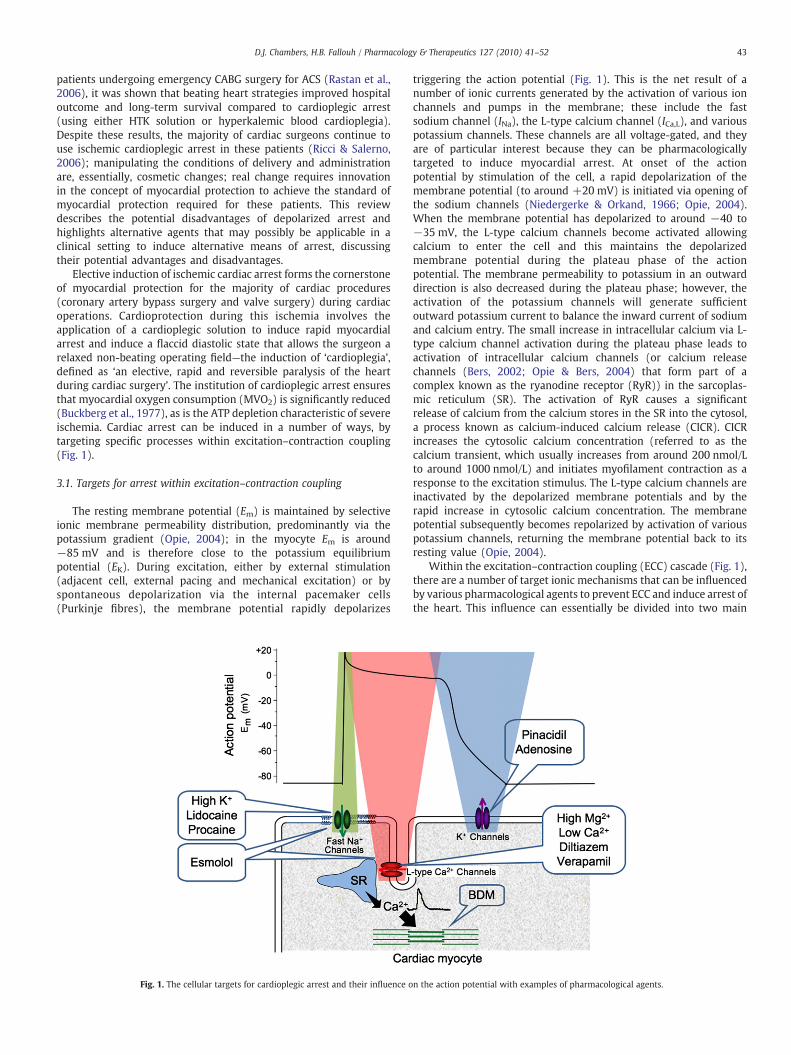
Elective induction of ischemic cardiac arrest forms the cornerstoneof myocardial protection for the majority of cardiac procedures(coronary artery bypass surgery and valve surgery) during cardiacoperations. Cardioprotection during this ischemia involves theapplication of a cardioplegic solution to induce rapid myocardialarrest and induce a flaccid diastolic state that allows the surgeon arelaxed non-beating operating field—the induction of ‘cardioplegia’,defined as ‘an elective, rapid and reversible paralysis of the heartduring cardiac surgery’. The institution of cardioplegic arrest ensuresthat myocardial oxygen consumption (MVO2) is significantly reduced(Buckberg et al., 1977), as is the ATP depletion characteristic of severeischemia. Cardiac arrest can be induced in a number of ways, bytargeting specific processes within excitation–contraction coupling(Fig. 1).
3.1. Targets for arrest within excitation–contraction coupling
The resting membrane potential (Em) is maintained by selectiveionic membrane permeability distribution, predominantly via thepotassium gradient (Opie, 2004); in the myocyte Em is around−85 mV and is therefore close to the potassium equilibriumpotential (EK). During excitation, either by external stimulation(adjacent cell, external pacing and mechanical excitation) or byspontaneous depolarization via the internal pacemaker cells(Purkinje fibres), the membrane potential rapidly depolarizes
Fig. 1. The cellular targets for cardioplegic arrest and their influence o
triggering the action potential (Fig. 1). This is the net result of anumber of ionic currents generated by the activation of various ionchannels and pumps in the membrane; these include the fastsodium channel (INa), the L-type calcium channel (ICa,L), and variouspotassium channels. These channels are all voltage-gated, and theyare of particular interest because they can be pharmacologicallytargeted to induce myocardial arrest. At onset of the actionpotential by stimulation of the cell, a rapid depolarization of themembrane potential (to around +20 mV) is initiated via opening ofthe sodium channels (Niedergerke & Orkand, 1966; Opie, 2004).When the membrane potential has depolarized to around −40 to−35 mV, the L-type calcium channels become activated allowingcalcium to enter the cell and this maintains the depolarizedmembrane potential during the plateau phase of the actionpotential. The membrane permeability to potassium in an outwarddirection is also decreased during the plateau phase; however, theactivation of the potassium channels will generate sufficientoutward potassium current to balance the inward current of sodiumand calcium entry. The small increase in intracellular calcium via L-type calcium channel activation during the plateau phase leads toactivation of intracellular calcium channels (or calcium releasechannels (Bers, 2002; Opie & Bers, 2004) that form part of acomplex known as the ryanodine receptor (RyR)) in the sarcoplas-mic reticulum (SR). The activation of RyR causes a significantrelease of calcium from the calcium stores in the SR into the cytosol,a process known as calcium-induced calcium release (CICR). CICRincreases the cytosolic calcium concentration (referred to as thecalcium transient, which usually increases from around 200 nmol/Lto around 1000 nmol/L) and initiates myofilament contraction as aresponse to the excitation stimulus. The L-type calcium channels areinactivated by the depolarized membrane potentials and by therapid increase in cytosolic calcium concentration. The membranepotential subsequently becomes repolarized by activation of variouspotassium channels, returning the membrane potential back to itsresting value (Opie, 2004).
Within the excitation–contraction coupling (ECC) cascade (Fig. 1),there are a number of target ionic mechanisms that can be influencedby various pharmacological agents to prevent ECC and induce arrest ofthe heart. This influence can essentially be divided into two main
n the action potential with examples of pharmacological agents.
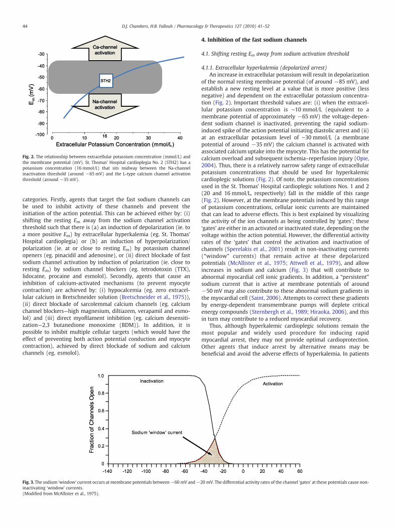
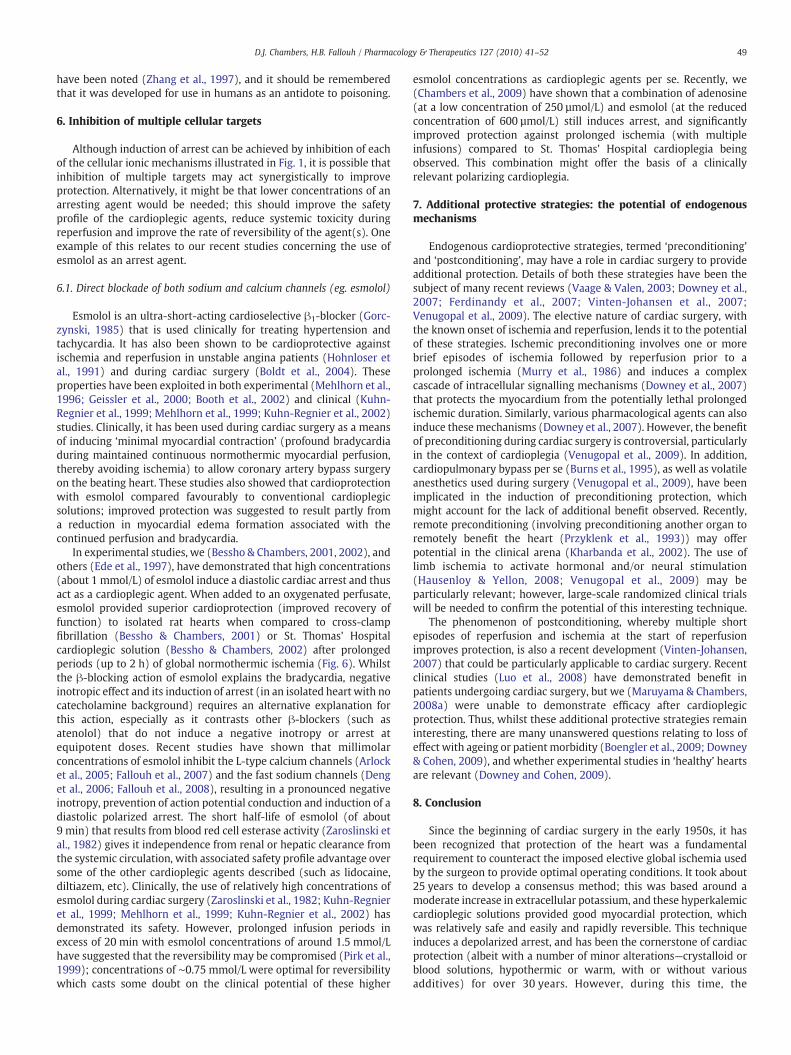
Fig. 2. The relationship between extracellular potassium concentration (mmol/L) andthe membrane potential (mV). St. Thomas' Hospital cardioplegia No. 2 (STH2) has apotassium concentration (16 mmol/L) that sits midway between the Na-channelinactivation threshold (around −65 mV) and the L-type calcium channel activationthreshold (around −35 mV).
44 D.J. Chambers, H.B. Fallouh / Pharmacology & Therapeutics 127 (2010) 41–52
categories. Firstly, agents that target the fast sodium channels canbe used to inhibit activity of these channels and prevent theinitiation of the action potential. This can be achieved either by: (i)shifting the resting Em away from the sodium channel activationthreshold such that there is (a) an induction of depolarization (ie. toa more positive Em) by extracellular hyperkalemia (eg. St. Thomas'Hospital cardioplegia) or (b) an induction of hyperpolarization/polarization (ie. at or close to resting Em) by potassium channelopeners (eg. pinacidil and adenosine), or (ii) direct blockade of fastsodium channel activation by induction of polarization (ie. close toresting Em) by sodium channel blockers (eg. tetrodotoxin (TTX),lidocaine, procaine and esmolol). Secondly, agents that cause aninhibition of calcium-activated mechanisms (to prevent myocytecontraction) are achieved by: (i) hypocalcemia (eg. zero extracel-lular calcium in Bretschneider solution (Bretschneider et al., 1975)),(ii) direct blockade of sarcolemmal calcium channels (eg. calciumchannel blockers—high magnesium, diltiazem, verapamil and esmo-lol) and (iii) direct myofilament inhibition (eg. calcium desensiti-zation—2,3 butanedione monoxime (BDM)). In addition, it ispossible to inhibit multiple cellular targets (which would have theeffect of preventing both action potential conduction and myocytecontraction), achieved by direct blockade of sodium and calciumchannels (eg. esmolol).
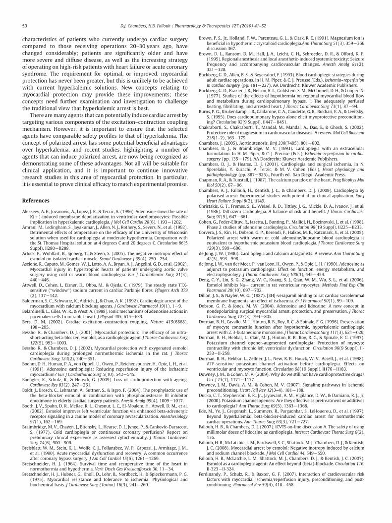
Fig. 3. The sodium ‘window’ current occurs at membrane potentials between−60 mV and−inactivating ‘window’ currents.(Modified from McAllister et al., 1975).
4. Inhibition of the fast sodium channels
4.1. Shifting resting Em away from sodium activation threshold
4.1.1. Extracellular hyperkalemia (depolarized arrest)An increase in extracellular potassium will result in depolarization
of the normal resting membrane potential (of around −85 mV), andestablish a new resting level at a value that is more positive (lessnegative) and dependent on the extracellular potassium concentra-tion (Fig. 2). Important threshold values are: (i) when the extracel-lular potassium concentration is ∼10 mmol/L (equivalent to amembrane potential of approximately −65 mV) the voltage-depen-dent sodium channel is inactivated, preventing the rapid sodium-induced spike of the action potential initiating diastolic arrest and (ii)at an extracellular potassium level of ∼30 mmol/L (a membranepotential of around −35 mV) the calcium channel is activated withassociated calcium uptake into the myocyte. This has the potential forcalcium overload and subsequent ischemia–reperfusion injury (Opie,2004). Thus, there is a relatively narrow safety range of extracellularpotassium concentrations that should be used for hyperkalemiccardioplegic solutions (Fig. 2). Of note, the potassium concentrationsused in the St. Thomas' Hospital cardioplegic solutions Nos. 1 and 2(20 and 16 mmol/L, respectively) fall in the middle of this range(Fig. 2). However, at the membrane potentials induced by this rangeof potassium concentrations, cellular ionic currents are maintainedthat can lead to adverse effects. This is best explained by visualizingthe activity of the ion channels as being controlled by ‘gates’; these‘gates’ are either in an activated or inactivated state, depending on thevoltage within the action potential. However, the differential activityrates of the ‘gates’ that control the activation and inactivation ofchannels (Sperelakis et al., 2001) result in non-inactivating currents(“window” currents) that remain active at these depolarizedpotentials (McAllister et al., 1975; Attwell et al., 1979), and allowincreases in sodium and calcium (Fig. 3) that will contribute toabnormal myocardial cell ionic gradients. In addition, a “persistent”sodium current that is active at membrane potentials of around−50 mV may also contribute to these abnormal sodium gradients inthe myocardial cell (Saint, 2006). Attempts to correct these gradientsby energy-dependent transmembrane pumps will deplete criticalenergy compounds (Sternbergh et al., 1989; Hiraoka, 2006), and thisin turn may contribute to a reduced myocardial recovery.
Thus, although hyperkalemic cardioplegic solutions remain themost popular and widely used procedure for inducing rapidmyocardial arrest, they may not provide optimal cardioprotection.Other agents that induce arrest by alternative means may bebeneficial and avoid the adverse effects of hyperkalemia. In patients
20 mV. The differential activity rates of the channel ‘gates’ at these potentials cause non-
45D.J. Chambers, H.B. Fallouh / Pharmacology & Therapeutics 127 (2010) 41–52
currently requiring cardiac surgery, who are older, sicker and whohave more severe and diffuse disease, improved myocardial protec-tion is likely to be of significance. An important characteristic of aneffective cardioplegic solution is a good systemic safety profile, withminimal adverse effects during acute (or even chronic) reperfusion.Hyperkalemic solutions fulfil these characteristics; they are relativelysafe, with little post-operative systemic toxic effects and with rapidcardiac reversibility. Key criteria to take into consideration withregard to developing alternative cardioplegic solutions, and themechanism of action of their constituent agents, are:
1. Arrest: a rapid diastolic arrest is required to keep the myocardiumrelaxed and to minimize cellular use of high-energy phosphatecompounds (ATP and phosphocreatine).
2. Myocardial protection: should delay the onset of irreversible injurycaused by global ischemia and limit the extent of reperfusioninjury.
3. Reversibility: arrest should be readily and rapidly reversible onwashout for quick resumption of heart function, to enable earlyweaning off cardiopulmonary bypass.
4. Low toxicity: the cardioplegic agent should have no toxic effects onthe heart or other organs after cessation of cardiopulmonarybypass. This is especially important if high concentrations of thepharmacological agent are required to achieve arrest.
Hyperkalemic cardioplegia meets all these criteria, which explainsits current monopoly of use in cardiac surgery. Any future alternativecardioplegic solution would be expected to offer a similar safetyprofile for arrest, reversibility and low toxicity, but with improvedmyocardial protection.
An alternative to inducing depolarized arrest by hyperkalemia is toinduce a hyperpolarized or polarized arrest. This can be achieved byusing specific agents that target the cellular components indicated inFig. 1 to induce arrest at membrane potentials closer to the normal‘resting’membrane potential. A number of benefits should ensue: theseinclude a reduced ionic imbalance leading to reduced energyutilization, and hence improved protection from a cellular perspective.
4.1.2. Hyperpolarization/polarization (potassium channel openers)The adenosine triphosphate-sensitive potassium channel (KATP-
channel) appears to have a role as a ‘metabolic sensor’, linkingcytosolic energy metabolism to membrane electrical activity inresponse to stress such as ischemia–reperfusion (Opie, 2004; Jahangir& Terzic, 2005), shortening the action potential and reducingintracellular calcium influx. Opening of KATP-channels allows anincreased potassium flux through the sarcolemma that will act to shiftEm towards the potassium equilibrium potential (EK), which isnormally around −90 mV in myocytes (Opie, 2004). This will inducea more negative Em (hyperpolarization) and act to keep Em away fromthe activation threshold of the fast sodium channel (INa). High(millimolar) concentrations of KATP-channel opening drugs caninduce arrest bymaintaining hyperpolarization. KATP-channel openersare also potent vasodilators, inducing hyperpolarization on vascularsmooth muscle cells (Quast et al., 1994), with some evidence that a‘hyperpolarizing’ cardioplegia protects endothelial function comparedto a hyperkalemic ‘depolarizing’ cardioplegia (He & Yang, 1997).Studies from Damiano's group (Maskal et al., 1995; Lawton et al.,1996a; Jayawant et al., 1997), using a variety of KATP-channel openers(aprikalim, pinacidil and nicorandil) to induce a hyperpolarizedarrest, have demonstrated either improved or equivalent myocardialprotection to that of hyperkalemic depolarized arrest. However, thesestudies are not without controversy; we (Walgama et al., 2000a;Walgama et al., 2000b) showed that pinacidil was unable to inducecomplete arrest during aerobic perfusion, even at very highconcentrations (1 mmol/L) over extended periods (30 mins). Inaddition, Em measurements indicated that pinacidil alone did notmaintain Em below the sodium channel activation threshold during
ischemia (ie. no hyperpolarization), and required the addition of asodium channel blocker (procaine, at 1 mmol/L) for complete arrestand improved protection. These drugs also increase the risk ofsignificant post-ischemic arrhythmias (Lawton et al., 1996b); fur-thermore, the systemic clearance of potassium channel openers isprolonged (Ward et al., 1984) which risks a residual hypotensiveeffect in patients post-bypass. These limitations make the concept ofusing a cardioplegia based on KATP-channel openers as unlikely forfuture clinical application.
KATP-channel openers may, however, have a role as an additiveprotective agent by enhancing the cardioprotection of hyperkalemiccardioplegic solutions. Short pretreatment (at least 3 min) with thesedrugs before hyperkalemic arrest was shown to improve post-ischemic function (Sugimoto et al., 1994; Dorman et al., 1998; Hebbaret al., 1998), possibly via a preconditioning mechanism (Menasche etal., 1996). Similar improvements were obtained when potassiumchannel openers were used as additives to hyperkalemic cardioplegicsolutions (Hosoda et al., 1994; Qiu et al., 1995; Dorman et al., 1997),but these studies remain controversial since others (Galinanes et al.,1992; Ducko et al., 2000) have been unable to show any benefit.Supplementing hyperkalemic solutions with KATP-channel openerswas shown to prevent increases in intracellular calcium seen withhyperkalemic solutions alone (Lopez et al., 1996; Dorman et al., 1997),which may account for any beneficial effects observed. Such benefitsmay also be associated with the location of the KATP-channels;improved protection has been observed with mitochondrial-specificKATP-channel openers such as diazoxide (McCully & Levitsky, 2003).
Using isolated rabbit sino-atrial (SA) node cells, adenosine (anendogenous purine nucleoside) was shown to induce complete arrest(at 50 µmol/L) and a hyperpolarization of ∼12 mV via increasedmembrane potassium permeability (Belardinelli et al., 1988). Itappears to act via an adenosine receptor-activated potassium channeland hence could have similar effects to KATP-channel openers. Thiscardioplegic property has been extended to whole hearts, withadenosine used either as an arrest agent (albeit at a high concentra-tion of 10 mmol/L) or as an additive (at 1 mmol/L) to hyperkalemicsolutions (Schubert et al., 1989; de Jong et al., 1990; Boehm et al.,1991); addition of adenosine reduced the time to arrest and improvedpost-ischemic recovery of function compared to hyperkalemiccardioplegia alone. As an additive to hyperkalemic solutions(16 mmol/L potassium), adenosine (1 mmol/L) did not influence thelevel of depolarization induced by potassium, but did slow the rate ofdepolarization which reduced the associated intracellular calciumloading (Alekseev et al., 1996). Additionally, it was shown that1 mmol/L adenosine (but not 0.1 mmol/L) prevented potassium-induced intracellular calcium loading and that this was associatedwith a PKC-dependent mechanism but not a KATP-channel dependentmechanism (Jovanovic et al., 1997). Supplementing St. Thomas'Hospital cardioplegia with adenosine (at concentrations rangingfrom 0.1 to 20 mmol/L) enhanced protection and increased myocar-dial content of high-energy phosphates and metabolites at the end ofischemia (Katayama et al., 1997). Interestingly, these effects weretemperature-dependent, with hypothermia reducing the benefit; thiswould limit its usefulness clinically in this context.
Clinical studies using adenosine as an adjunct to hyperkalemiccardioplegic solutions have had equivocal results. Adenosine (atconcentrations ranging from 1 to 250 µmol/L) added to warmantegrade blood cardioplegia was well tolerated up to 25 µmol/L,but higher concentrations induced hypotension (Fremes et al., 1996).In further studies, adding adenosine (at 15, 50 or 100 µmol/L) towarm antegrade blood cardioplegia showed no benefit to patientoutcome in terms of mortality, function or infarction (Cohen et al.,1998). However, a similar study with adenosine added to cold bloodcardioplegia (at higher concentrations of 0.5 or 2.0 mmol/L) showed atendency to reduce adverse events (Mentzer et al., 1999). The clinicalstudies leave many unanswered questions regarding the efficacy of
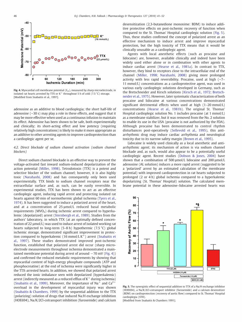
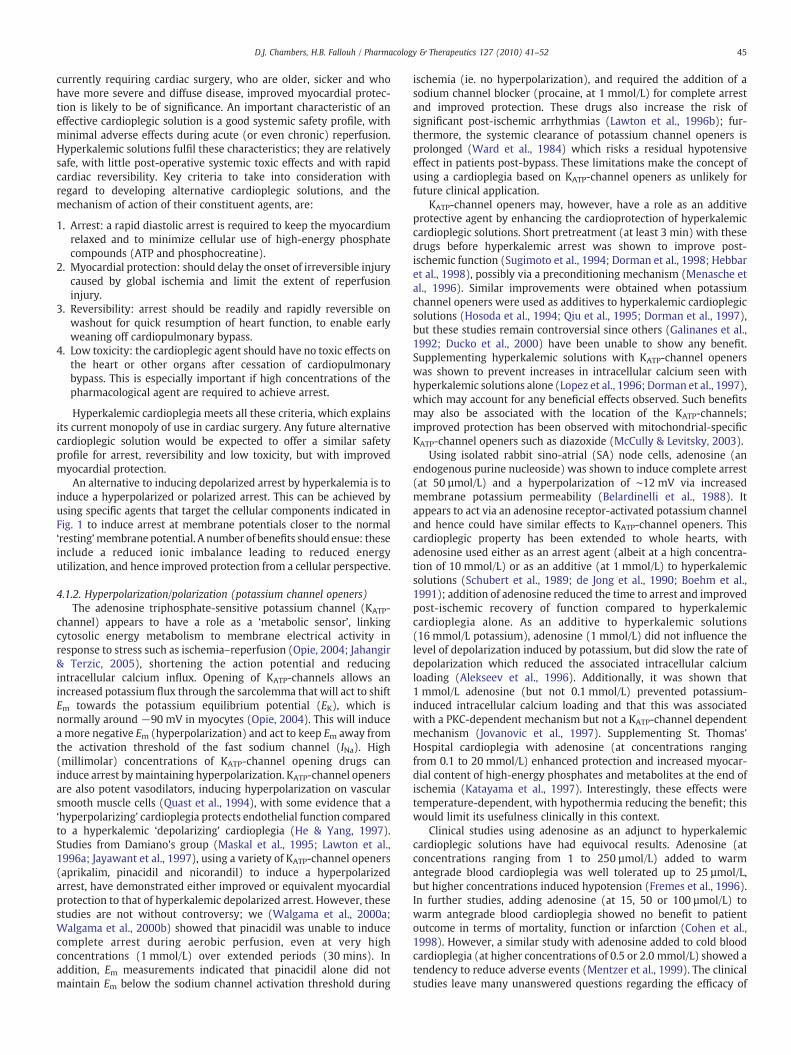
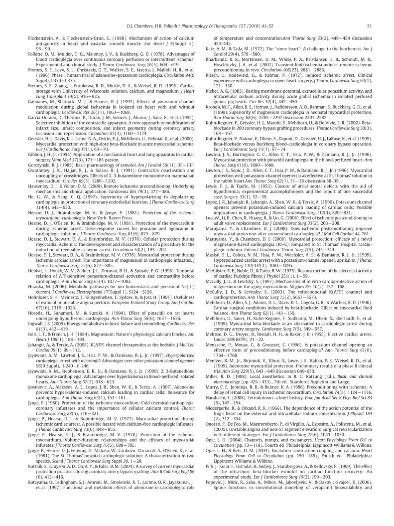
Fig. 4.Myocardial cell membrane potential (Em), measured by sharp microelectrode, inisolated rat hearts arrested by TTX or K+ throughout 5 h of cold (7.5 °C) storage.[Modified from Snabaitis et al., 1997].
Fig. 5. The synergistic effect of sequential addition to TTX of a Na/H exchange inhibitor(HOE694), a Na/K/2Cl-cotransport inhibitor (furosemide) and a calcium desensitizer(BDM) on cardioprotection (recovery of aortic flow) compared to St. Thomas' Hospitalcardioplegia (STH).[Modified from Snabaitis & Chambers 1991].
46 D.J. Chambers, H.B. Fallouh / Pharmacology & Therapeutics 127 (2010) 41–52
adenosine as an additive to blood cardioplegia; the short half-life ofadenosine (∼30 s) may play a role in these effects, and suggest that itmay bemore effectivewhen used as a continuous infusion tomaintainits effect. Adenosine has been shown to be safe, both experimentallyand clinically; its short-acting effect and low potency (requiringrelatively high concentrations) is likely tomake it more appropriate asan additive to other arresting agents to improve cardioprotection thana cardioplegic agent per se.
4.2. Direct blockade of sodium channel activation (sodium channelblockers)
Direct sodium channel blockade is an effective way to prevent thevoltage-activated fast inward sodium-induced depolarization of theaction potential (Miller, 1998). Tetrodotoxin (TTX) is a potent andselective blocker of the sodium channel; however, it is also highlytoxic (Narahashi, 2008) and has consequently only been usedexperimentally. TTX binds to sodium channel receptors near theextracellular surface and, as such, can be easily reversible. Inexperimental studies, TTX has been shown to act as an effectivecardioplegic agent, inducing rapid arrest and protecting isolated rathearts against 60 min of normothermic global ischemia (Tyers et al.,1974). It has been suggested to induce a polarized arrest of the heart,and at a concentration of 25 µmol/L reduced basal metabolicrequirements (MVO2) during ischemic arrest compared to hyperka-lemic (depolarized) arrest (Sternbergh et al., 1989). Studies from theauthors' laboratory, in which TTX (at an optimally defined concen-tration of 22 µmol/L) was used to induce arrest of isolated working rathearts subjected to long-term (5–8 h) hypothermic (7.5 °C) globalischemic storage, demonstrated significant improvement in protec-tion compared to hyperkalemic (16 mmol/L K+) arrest (Snabaitis etal., 1997). These studies demonstrated improved post-ischemicfunction, established that polarized arrest did occur (sharp micro-electrode measurements throughout ischemia demonstrated a main-tained membrane potential during arrest of around −70 mV (Fig. 4))and confirmed the reduced metabolic requirements by showing thatmyocardial content of high-energy phosphate compounds (ATP andphosphocreatine) at the end of ischemia were significantly higher inthe TTX-arrested hearts. In addition, we showed that polarized arrestreduced the ionic imbalance seen with depolarized (hyperkalemic)arrest (indirectly measured as a reduced efflux of K+ during ischemia)(Snabaitis et al., 1999). Moreover, the importance of Na+ and Ca2+
overload in the development of myocardial injury was shown(Snabaitis & Chambers, 1999) by the sequential addition to the TTX(polarizing) solution of drugs that induced Na/H exchange inhibition(HOE694), Na/K/2Cl-cotransport inhibition (furosemide) and calcium
desensitization (2,3-butanedione monoxime: BDM) to induce addi-tive protective effects on post-ischemic recovery of function whencompared to the St. Thomas' Hospital cardioplegic solution (Fig. 5).Thus, these studies confirmed the concept of polarized arrest as aneffective mechanism to induce arrest and improve myocardialprotection, but the high toxicity of TTX means that it would beclinically unusable as a cardioplegic agent.
Agents with local anesthetic effects (such as procaine andlidocaine) are, however, available clinically and indeed have beenwidely used either alone or in combination with other agents toinduce cardiac arrest (Hearse et al., 1981a). In contrast to TTX,however, they bind to receptors close to the intracellular end of thechannel (Miller, 1998; Narahashi, 2008) giving more prolongedactivity with less rapid reversibility. Procaine, used at high (∼7–11 mmol/L) concentrations as a cardioprotective agent, was used invarious early cardioplegic solutions developed in Germany, such asthe Bretschneider and Kirsch solutions (Kirsch et al., 1972; Bretsch-neider et al., 1975). However, later systematic characterization of bothprocaine and lidocaine at various concentrations demonstratedsignificant detrimental effects when used at high (1–20 mmol/L)concentrations (Hearse et al., 1981b). The original St. Thomas'Hospital cardioplegic solution No. 1 includes procaine (at 1 mmol/L)as a membrane stabilizer, but it was removed from the No. 2 solutionto enable its use in the USA (procaine is not authorized by the FDA).Although procaine has been demonstrated to control rhythmdisturbances post-operatively (Sellevold et al., 1995), this anti-arrhythmic drug may induce cardiac arrhythmia and neurologicaltoxicity due to its narrow safety margin (Brown et al., 1995).
Lidocaine is widely used clinically as a local anesthetic and anti-arrhythmic agent; its mechanism of action is via sodium channelblockade and, as such, would also appear to be a potentially usefulcardioplegic agent. Recent studies (Dobson & Jones, 2004) haveshown that a combination of 500 µmol/L lidocaine and 200 µmol/Ladenosine (AL solution) induces a more rapid arrest (suggested to bea ‘polarized’ arrest by an estimated calculation of the membranepotential) with improved cardioprotection in rat hearts subjected toprolonged (2 or 4 h) global ischemia compared to a hyperkalemicdepolarizing (St. Thomas' Hospital) solution. The calculated mem-brane potential in these adenosine–lidocaine arrested hearts was
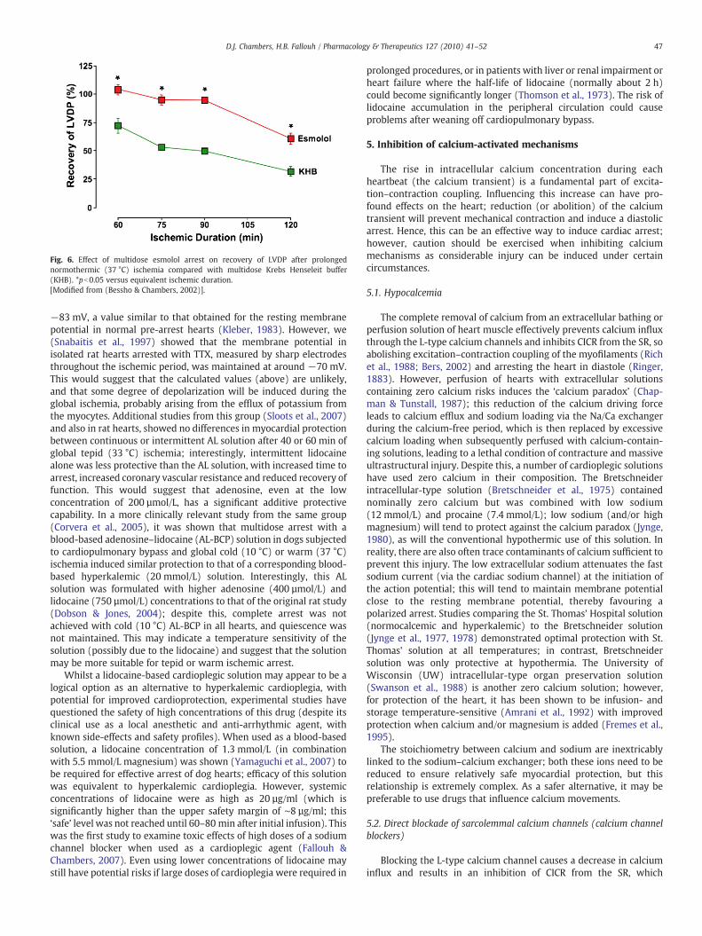
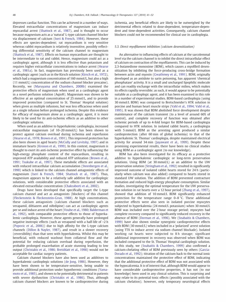
Fig. 6. Effect of multidose esmolol arrest on recovery of LVDP after prolongednormothermic (37 °C) ischemia compared with multidose Krebs Henseleit buffer(KHB). *pb0.05 versus equivalent ischemic duration.[Modified from (Bessho & Chambers, 2002)].
47D.J. Chambers, H.B. Fallouh / Pharmacology & Therapeutics 127 (2010) 41–52
−83 mV, a value similar to that obtained for the resting membranepotential in normal pre-arrest hearts (Kleber, 1983). However, we(Snabaitis et al., 1997) showed that the membrane potential inisolated rat hearts arrested with TTX, measured by sharp electrodesthroughout the ischemic period, was maintained at around −70 mV.This would suggest that the calculated values (above) are unlikely,and that some degree of depolarization will be induced during theglobal ischemia, probably arising from the efflux of potassium fromthe myocytes. Additional studies from this group (Sloots et al., 2007)and also in rat hearts, showed no differences in myocardial protectionbetween continuous or intermittent AL solution after 40 or 60 min ofglobal tepid (33 °C) ischemia; interestingly, intermittent lidocainealone was less protective than the AL solution, with increased time toarrest, increased coronary vascular resistance and reduced recovery offunction. This would suggest that adenosine, even at the lowconcentration of 200 µmol/L, has a significant additive protectivecapability. In a more clinically relevant study from the same group(Corvera et al., 2005), it was shown that multidose arrest with ablood-based adenosine–lidocaine (AL-BCP) solution in dogs subjectedto cardiopulmonary bypass and global cold (10 °C) or warm (37 °C)ischemia induced similar protection to that of a corresponding blood-based hyperkalemic (20 mmol/L) solution. Interestingly, this ALsolution was formulated with higher adenosine (400 µmol/L) andlidocaine (750 µmol/L) concentrations to that of the original rat study(Dobson & Jones, 2004); despite this, complete arrest was notachieved with cold (10 °C) AL-BCP in all hearts, and quiescence wasnot maintained. This may indicate a temperature sensitivity of thesolution (possibly due to the lidocaine) and suggest that the solutionmay be more suitable for tepid or warm ischemic arrest.
Whilst a lidocaine-based cardioplegic solution may appear to be alogical option as an alternative to hyperkalemic cardioplegia, withpotential for improved cardioprotection, experimental studies havequestioned the safety of high concentrations of this drug (despite itsclinical use as a local anesthetic and anti-arrhythmic agent, withknown side-effects and safety profiles). When used as a blood-basedsolution, a lidocaine concentration of 1.3 mmol/L (in combinationwith 5.5 mmol/L magnesium) was shown (Yamaguchi et al., 2007) tobe required for effective arrest of dog hearts; efficacy of this solutionwas equivalent to hyperkalemic cardioplegia. However, systemicconcentrations of lidocaine were as high as 20 µg/ml (which issignificantly higher than the upper safety margin of ∼8 µg/ml; this‘safe’ level was not reached until 60–80 min after initial infusion). Thiswas the first study to examine toxic effects of high doses of a sodiumchannel blocker when used as a cardioplegic agent (Fallouh &Chambers, 2007). Even using lower concentrations of lidocaine maystill have potential risks if large doses of cardioplegia were required in
prolonged procedures, or in patients with liver or renal impairment orheart failure where the half-life of lidocaine (normally about 2 h)could become significantly longer (Thomson et al., 1973). The risk oflidocaine accumulation in the peripheral circulation could causeproblems after weaning off cardiopulmonary bypass.
5. Inhibition of calcium-activated mechanisms
The rise in intracellular calcium concentration during eachheartbeat (the calcium transient) is a fundamental part of excita-tion–contraction coupling. Influencing this increase can have pro-found effects on the heart; reduction (or abolition) of the calciumtransient will prevent mechanical contraction and induce a diastolicarrest. Hence, this can be an effective way to induce cardiac arrest;however, caution should be exercised when inhibiting calciummechanisms as considerable injury can be induced under certaincircumstances.
5.1. Hypocalcemia
The complete removal of calcium from an extracellular bathing orperfusion solution of heart muscle effectively prevents calcium influxthrough the L-type calcium channels and inhibits CICR from the SR, soabolishing excitation–contraction coupling of the myofilaments (Richet al., 1988; Bers, 2002) and arresting the heart in diastole (Ringer,1883). However, perfusion of hearts with extracellular solutionscontaining zero calcium risks induces the ‘calcium paradox’ (Chap-man & Tunstall, 1987); this reduction of the calcium driving forceleads to calcium efflux and sodium loading via the Na/Ca exchangerduring the calcium-free period, which is then replaced by excessivecalcium loading when subsequently perfused with calcium-contain-ing solutions, leading to a lethal condition of contracture and massiveultrastructural injury. Despite this, a number of cardioplegic solutionshave used zero calcium in their composition. The Bretschneiderintracellular-type solution (Bretschneider et al., 1975) containednominally zero calcium but was combined with low sodium(12 mmol/L) and procaine (7.4 mmol/L); low sodium (and/or highmagnesium) will tend to protect against the calcium paradox (Jynge,1980), as will the conventional hypothermic use of this solution. Inreality, there are also often trace contaminants of calcium sufficient toprevent this injury. The low extracellular sodium attenuates the fastsodium current (via the cardiac sodium channel) at the initiation ofthe action potential; this will tend to maintain membrane potentialclose to the resting membrane potential, thereby favouring apolarized arrest. Studies comparing the St. Thomas' Hospital solution(normocalcemic and hyperkalemic) to the Bretschneider solution(Jynge et al., 1977, 1978) demonstrated optimal protection with St.Thomas' solution at all temperatures; in contrast, Bretschneidersolution was only protective at hypothermia. The University ofWisconsin (UW) intracellular-type organ preservation solution(Swanson et al., 1988) is another zero calcium solution; however,for protection of the heart, it has been shown to be infusion- andstorage temperature-sensitive (Amrani et al., 1992) with improvedprotection when calcium and/or magnesium is added (Fremes et al.,1995).
The stoichiometry between calcium and sodium are inextricablylinked to the sodium–calcium exchanger; both these ions need to bereduced to ensure relatively safe myocardial protection, but thisrelationship is extremely complex. As a safer alternative, it may bepreferable to use drugs that influence calcium movements.
5.2. Direct blockade of sarcolemmal calcium channels (calcium channelblockers)
Blocking the L-type calcium channel causes a decrease in calciuminflux and results in an inhibition of CICR from the SR, which
48 D.J. Chambers, H.B. Fallouh / Pharmacology & Therapeutics 127 (2010) 41–52
depresses cardiac function. This can be achieved in a number of ways.Elevated extracellular concentrations of magnesium can inducemyocardial arrest (Shattock et al., 1987), and is thought to occurbecause magnesium acts as a ‘natural’ L-type calcium channel blockervia displacement of calcium (Iseri & French, 1984). However, theseeffects are species-dependent; rat myocardium is very sensitivewhereas rabbit myocardium is relatively insensitive, possibly reflect-ing differential sensitivity of the calcium channel to magnesium(Shattock et al., 1987). Effects on human myocardium are thought tobe intermediate to rat and rabbit. Hence, magnesium could act as acardioplegic agent, although it is less effective than potassium andrequires higher extracellular concentrations to induce arrest (Hearseet al., 1981a). In fact, magnesium has previously been used as acardioplegic agent (such as in the Kirsch solution (Kirsch et al., 1972),which had a magnesium concentration of 160 mmol/L, but also a high(11 mmol/L) concentration of the sodium channel blocker procaine).Recently, we (Maruyama and Chambers, 2008b) examined theprotective effects of magnesium when used as a cardioplegic agentin a novel perfusion solution (Aqix®). Magnesium was shown to beeffective at an optimal concentration of 25 mmol/L, and to provideimproved protection (compared to St. Thomas' Hospital solution)when given as multiple infusions, but was less efficacious when usedas a single infusion before prolonged ischemia. Despite this evidencefor efficacy of magnesium alone as a cardioplegic agent, it is morelikely to be used for its anti-ischemic effects as an additive to othercardioplegic solutions.
When used as an additive to hyperkalemic cardioplegia, increasedextracellular magnesium (of 10–20 mmol/L) has been shown toprotect against calcium overload during ischemia and reperfusion(Hearse et al., 1978; Brown et al., 1991). This improved protection hasalso been shown in aged hearts (McCully and Levitsky, 1997) and inimmature hearts (Kronon et al., 1999). In this context, magnesium isthought to exert its anti-ischemic protective effect by influencing thehigh-energy phosphate content of the myocardium, resulting inimproved ATP availability and reduced ATP utilization (Brown et al.,1991; Tsukube et al., 1997). These metabolic effects are associatedwith reduced intracellular calcium accumulation (Steenbergen et al.,1990) which is linked to the calcium channel antagonistic effects ofmagnesium (Iseri & French, 1984; Shattock et al., 1987). Thus,magnesium appears to be a relatively safe additive for cardioplegicsolutions, with considerable protective effects associated with anelevated extracellular concentration (Chakraborti et al., 2002).
Drugs have been developed that specifically target the L-typecalcium channel and act as antagonists (blockers) of this channel(Fleckenstein & Fleckenstein-Grun, 1988). At high concentrations,these calcium antagonists (calcium channel blockers such asverapamil, diltiazem and nifedipine) can act as cardioplegic agentsper se and induce arrest of the heart (Vouhe et al., 1980; Balderman etal., 1992), with comparable protective effects to those of hyperka-lemic cardioplegia. However, these agents generally have prolongednegative inotropic effects (such as verapamil with a half-life of N2 h(Popovic et al., 2006)) due to high affinity for the L-type calciumchannels (Dillon & Nayler, 1987), and result in a slower recovery(reversibility) than that seen with hyperkalemia. Whilst this may bebeneficial, with reduced washout from collateral flow and thepotential for reducing calcium overload during reperfusion, theprobable prolonged exacerbation of acute stunning leading to lowoutput (Christakis et al., 1986; Breisblatt et al., 1990) would not beacceptable for cardiac surgery patients.
Calcium channel blockers have also been used as additives tohyperkalemic cardioplegic solutions (de Jong, 1986). However, theyhave been shown to be temperature-sensitive, being unable toprovide additional protection under hypothermic conditions (Yama-moto et al., 1985), and shown to be potentially detrimental in patientswith severe dysfunction (Christakis et al., 1986). Thus, althoughcalcium channel blockers are known to be cardioprotective during
ischemia, any beneficial effects are likely to be outweighed by thedetrimental effects related to dose-dependent, temperature-depen-dent and time-dependent activities. Consequently, calcium channelblockers could not be recommended for clinical use in cardioplegia.
5.3. Direct myofilament inhibition (calcium desensitization)
An alternative to influencing effects of calcium at the sarcolemmallevel via the calcium channel is to inhibit the direct intracellular effectof calcium on contraction of the myofilaments. This can be induced by2,3-butanedione monoxime (BDM), which causes a myofibril desen-sitization by inhibiting the force-producing cross-bridge formationbetween actin and myosin (Gwathmey et al., 1991). BDM, originallydeveloped as an antidote to sarin poisoning, has apparent ‘chemicalphosphatase’ activity. It is a small and uncharged lipophilic moleculeand can readily exchange with the intracellular milieu, which makesits effects rapidly reversible; as such, it would appear to be potentiallysuitable as a cardioplegic agent, and it has been examined in this rolein a number of experimental studies. BDM-based cardioplegia (using30 mmol/L BDM) was compared to Bretschneider's HTK solution inporcine and human heart muscle strips (Vahl et al., 1994; Vahl et al.,1995). It was shown that BDM abolished force development despitemaintenance of the calcium transient (to a level of around 60% ofcontrol), and complete recovery of function was obtained afterischemic periods of up to 4-fold longer for BDM-based cardioplegiacompared to HTK solution. In isolated rabbit hearts, a cardioplegiawith 5 mmol/L BDM as the arresting agent produced a similarcardioprotection (after 60 min of global ischemia) to that of thehyperkalemic St. Thomas' cardioplegia, despite maintained electricalactivity for around 34 min (Jayawant et al., 1999). Despite thesepromising experimental results, there have been no clinical studiesusing BDM as a cardioplegic agent (to our knowledge).
BDM has also been investigated for its efficacy as a protectiveadditive to hyperkalemic cardioplegic or long-term preservationsolutions. Using BDM (at 30 mmol/L) as an additive to the UWpreservation solution (Stringham et al., 1992) resulted in significantlyimproved outcome of isolated rabbit hearts stored for 24 h (partic-ularly when calcium was also added) compared to hearts stored instandard UW solution. The addition of BDM prevented contractureformation and reduced high-energy phosphate consumption. Similarstudies, investigating the optimal temperature for the UW preserva-tion solution in rat hearts over a 12 hour period (Zhang et al., 1997),showed that addition of 10 mmol/L BDM significantly enhancedprotection for the temperature range 0–8 °C. These additionalprotective effects were also seen in isolated porcine myocytessubjected to hyperkalemia (24 mmol/L potassium) when 30 mmol/LBDM was included over the 2 hour storage period; myocytes hadcomplete recovery compared to significantly reduced recovery in theabsence of BDM (Dorman et al., 1996). We (Snabaitis & Chambers,1999) have also shown similar synergistic cardioprotective effectswith BDM (30 mmol/L) when included in a ‘polarized’ arrest solution(using TTX to induce arrest via sodium channel blockade). Isolatedworking rat hearts were subjected to 8 h storage; significantadditional improvement in recovery was observed when BDM wasincluded compared to the St. Thomas' Hospital cardioplegic solution.In this study, we (Snabaitis & Chambers, 1999) also confirmed acalcium-chelating effect of BDM previously seen by others (Garcia-Dorado et al., 1992); titration of the calcium back to the normal ionicconcentrations maintained the protective effect of BDM, indicatingthat the additional protective effect of BDM was not associated withthe hypocalcemia. It is of interest that, although BDMwould appear tohave considerable cardioprotective properties, it has not (to ourknowledge) been used in any clinical solution. This is surprising andmay relate to its potential toxic effects (possibly associated with thecalcium chelation); however, only temporary neurological effects
49D.J. Chambers, H.B. Fallouh / Pharmacology & Therapeutics 127 (2010) 41–52
have been noted (Zhang et al., 1997), and it should be rememberedthat it was developed for use in humans as an antidote to poisoning.
6. Inhibition of multiple cellular targets
Although induction of arrest can be achieved by inhibition of eachof the cellular ionic mechanisms illustrated in Fig. 1, it is possible thatinhibition of multiple targets may act synergistically to improveprotection. Alternatively, it might be that lower concentrations of anarresting agent would be needed; this should improve the safetyprofile of the cardioplegic agents, reduce systemic toxicity duringreperfusion and improve the rate of reversibility of the agent(s). Oneexample of this relates to our recent studies concerning the use ofesmolol as an arrest agent.
6.1. Direct blockade of both sodium and calcium channels (eg. esmolol)
Esmolol is an ultra-short-acting cardioselective β1-blocker (Gorc-zynski, 1985) that is used clinically for treating hypertension andtachycardia. It has also been shown to be cardioprotective againstischemia and reperfusion in unstable angina patients (Hohnloser etal., 1991) and during cardiac surgery (Boldt et al., 2004). Theseproperties have been exploited in both experimental (Mehlhorn et al.,1996; Geissler et al., 2000; Booth et al., 2002) and clinical (Kuhn-Regnier et al., 1999; Mehlhorn et al., 1999; Kuhn-Regnier et al., 2002)studies. Clinically, it has been used during cardiac surgery as a meansof inducing ‘minimal myocardial contraction’ (profound bradycardiaduring maintained continuous normothermic myocardial perfusion,thereby avoiding ischemia) to allow coronary artery bypass surgeryon the beating heart. These studies also showed that cardioprotectionwith esmolol compared favourably to conventional cardioplegicsolutions; improved protection was suggested to result partly froma reduction in myocardial edema formation associated with thecontinued perfusion and bradycardia.
In experimental studies, we (Bessho & Chambers, 2001, 2002), andothers (Ede et al., 1997), have demonstrated that high concentrations(about 1 mmol/L) of esmolol induce a diastolic cardiac arrest and thusact as a cardioplegic agent. When added to an oxygenated perfusate,esmolol provided superior cardioprotection (improved recovery offunction) to isolated rat hearts when compared to cross-clampfibrillation (Bessho & Chambers, 2001) or St. Thomas' Hospitalcardioplegic solution (Bessho & Chambers, 2002) after prolongedperiods (up to 2 h) of global normothermic ischemia (Fig. 6). Whilstthe β-blocking action of esmolol explains the bradycardia, negativeinotropic effect and its induction of arrest (in an isolated heart with nocatecholamine background) requires an alternative explanation forthis action, especially as it contrasts other β-blockers (such asatenolol) that do not induce a negative inotropy or arrest atequipotent doses. Recent studies have shown that millimolarconcentrations of esmolol inhibit the L-type calcium channels (Arlocket al., 2005; Fallouh et al., 2007) and the fast sodium channels (Denget al., 2006; Fallouh et al., 2008), resulting in a pronounced negativeinotropy, prevention of action potential conduction and induction of adiastolic polarized arrest. The short half-life of esmolol (of about9 min) that results from blood red cell esterase activity (Zaroslinski etal., 1982) gives it independence from renal or hepatic clearance fromthe systemic circulation, with associated safety profile advantage oversome of the other cardioplegic agents described (such as lidocaine,diltiazem, etc). Clinically, the use of relatively high concentrations ofesmolol during cardiac surgery (Zaroslinski et al., 1982; Kuhn-Regnieret al., 1999; Mehlhorn et al., 1999; Kuhn-Regnier et al., 2002) hasdemonstrated its safety. However, prolonged infusion periods inexcess of 20 min with esmolol concentrations of around 1.5 mmol/Lhave suggested that the reversibility may be compromised (Pirk et al.,1999); concentrations of ∼0.75 mmol/L were optimal for reversibilitywhich casts some doubt on the clinical potential of these higher
esmolol concentrations as cardioplegic agents per se. Recently, we(Chambers et al., 2009) have shown that a combination of adenosine(at a low concentration of 250 µmol/L) and esmolol (at the reducedconcentration of 600 µmol/L) still induces arrest, and significantlyimproved protection against prolonged ischemia (with multipleinfusions) compared to St. Thomas' Hospital cardioplegia beingobserved. This combination might offer the basis of a clinicallyrelevant polarizing cardioplegia.
7. Additional protective strategies: the potential of endogenousmechanisms
Endogenous cardioprotective strategies, termed ‘preconditioning’and ‘postconditioning’, may have a role in cardiac surgery to provideadditional protection. Details of both these strategies have been thesubject of many recent reviews (Vaage & Valen, 2003; Downey et al.,2007; Ferdinandy et al., 2007; Vinten-Johansen et al., 2007;Venugopal et al., 2009). The elective nature of cardiac surgery, withthe known onset of ischemia and reperfusion, lends it to the potentialof these strategies. Ischemic preconditioning involves one or morebrief episodes of ischemia followed by reperfusion prior to aprolonged ischemia (Murry et al., 1986) and induces a complexcascade of intracellular signalling mechanisms (Downey et al., 2007)that protects the myocardium from the potentially lethal prolongedischemic duration. Similarly, various pharmacological agents can alsoinduce these mechanisms (Downey et al., 2007). However, the benefitof preconditioning during cardiac surgery is controversial, particularlyin the context of cardioplegia (Venugopal et al., 2009). In addition,cardiopulmonary bypass per se (Burns et al., 1995), as well as volatileanesthetics used during surgery (Venugopal et al., 2009), have beenimplicated in the induction of preconditioning protection, whichmight account for the lack of additional benefit observed. Recently,remote preconditioning (involving preconditioning another organ toremotely benefit the heart (Przyklenk et al., 1993)) may offerpotential in the clinical arena (Kharbanda et al., 2002). The use oflimb ischemia to activate hormonal and/or neural stimulation(Hausenloy & Yellon, 2008; Venugopal et al., 2009) may beparticularly relevant; however, large-scale randomized clinical trialswill be needed to confirm the potential of this interesting technique.
The phenomenon of postconditioning, whereby multiple shortepisodes of reperfusion and ischemia at the start of reperfusionimproves protection, is also a recent development (Vinten-Johansen,2007) that could be particularly applicable to cardiac surgery. Recentclinical studies (Luo et al., 2008) have demonstrated benefit inpatients undergoing cardiac surgery, but we (Maruyama & Chambers,2008a) were unable to demonstrate efficacy after cardioplegicprotection. Thus, whilst these additional protective strategies remaininteresting, there are many unanswered questions relating to loss ofeffect with ageing or patient morbidity (Boengler et al., 2009; Downey& Cohen, 2009), and whether experimental studies in ‘healthy’ heartsare relevant (Downey and Cohen, 2009).
8. Conclusion
Since the beginning of cardiac surgery in the early 1950s, it hasbeen recognized that protection of the heart was a fundamentalrequirement to counteract the imposed elective global ischemia usedby the surgeon to provide optimal operating conditions. It took about25 years to develop a consensus method; this was based around amoderate increase in extracellular potassium, and these hyperkalemiccardioplegic solutions provided good myocardial protection, whichwas relatively safe and easily and rapidly reversible. This techniqueinduces a depolarized arrest, and has been the cornerstone of cardiacprotection (albeit with a number of minor alterations—crystalloid orblood solutions, hypothermic or warm, with or without variousadditives) for over 30 years. However, during this time, the
50 D.J. Chambers, H.B. Fallouh / Pharmacology & Therapeutics 127 (2010) 41–52
characteristics of patients who currently undergo cardiac surgerycompared to those receiving operations 20–30 years ago, havechanged considerably; patients are significantly older and havemore severe and diffuse disease, as well as the increasing strategyof operating on high-risk patients with heart failure or acute coronarysyndrome. The requirement for optimal, or improved, myocardialprotection has never been greater, but this is unlikely to be achievedwith current hyperkalemic solutions. New concepts relating tomyocardial protection may provide these improvements; theseconcepts need further examination and investigation to challengethe traditional view that hyperkalemic arrest is best.
There aremany agents that can potentially induce cardiac arrest bytargeting various components of the excitation–contraction couplingmechanism. However, it is important to ensure that the selectedagents have comparable safety profiles to that of hyperkalemia. Theconcept of polarized arrest has some potential beneficial advantagesover hyperkalemia, and recent studies, highlighting a number ofagents that can induce polarized arrest, are now being recognized asdemonstrating some of these advantages. Not all will be suitable forclinical application, and it is important to continue innovativeresearch studies in this area of myocardial protection. In particular,it is essential to prove clinical efficacy tomatch experimental promise.
References
Alekseev, A. E., Jovanovic, A., Lopez, J. R., & Terzic, A. (1996). Adenosine slows the rate ofK(+)-induced membrane depolarization in ventricular cardiomyocytes: Possibleimplication in hyperkalemic cardioplegia. J Mol Cell Cardiol 28(6), 1193−1202.
Amrani, M., Ledingham, S., Jayakumar, J., Allen, N. J., Rothery, S., Severs, N., et al. (1992).Detrimental effects of temperature on the efficacy of the University of Wisconsinsolution when used for cardioplegia at moderate hypothermia. Comparison withthe St. Thomas Hospital solution at 4 degrees C and 20 degrees C. Circulation 86(5Suppl), II280−II288.
Arlock, P., Wohlfart, B., Sjoberg, T., & Steen, S. (2005). The negative inotropic effect ofesmolol on isolated cardiac muscle. Scand Cardiovasc J 39(4), 250−254.
Ascione, R., Caputo, M., Gomes, W. J., Lotto, A. A., Bryan, A. J., Angelini, G. D., et al. (2002).Myocardial injury in hypertrophic hearts of patients undergoing aortic valvesurgery using cold or warm blood cardioplegia. Eur J Cardiothorac Surg 21(3),440−446.
Attwell, D., Cohen, I., Eisner, D., Ohba, M., & Ojeda, C. (1979). The steady state TTX-sensitive (“window”) sodium current in cardiac Purkinje fibres. Pflugers Arch 379(2), 137−142.
Balderman, S. C., Schwartz, K., Aldrich, J., & Chan, A. K. (1992). Cardioplegic arrest of themyocardium with calcium blocking agents. J Cardiovasc Pharmacol 19(1), 1−9.
Belardinelli, L., Giles, W. R., &West, A. (1988). Ionic mechanisms of adenosine actions inpacemaker cells from rabbit heart. J Physiol 405, 615−633.
Bers, D. M. (2002). Cardiac excitation–contraction coupling. Nature 415(6868),198−205.
Bessho, R., & Chambers, D. J. (2001). Myocardial protection: The efficacy of an ultra-short-acting beta-blocker, esmolol, as a cardioplegic agent. J Thorac Cardiovasc Surg122(5), 993−1003.
Bessho, R., & Chambers, D. J. (2002). Myocardial protection with oxygenated esmololcardioplegia during prolonged normothermic ischemia in the rat. J ThoracCardiovasc Surg 124(2), 340−351.
Boehm, D. H., Human, P. A., von Oppell, U., Owen, P., Reichenspurner, H., Opie, L. H., et al.(1991). Adenosine cardioplegia: Reducing reperfusion injury of the ischaemicmyocardium? Eur J Cardiothorac Surg 5(10), 542−545.
Boengler, K., Schulz, R., & Heusch, G. (2009). Loss of cardioprotection with ageing.Cardiovasc Res 83(2), 247−261.
Boldt, J., Brosch, C., Lehmann, A., Suttner, S., & Isgro, F. (2004). The prophylactic use ofthe beta-blocker esmolol in combination with phosphodiesterase III inhibitorenoximone in elderly cardiac surgery patients. Anesth Analg 99(4), 1009−1017.
Booth, J. V., Spahn, D. R., McRae, R. L., Chesnut, L. C., El-Moalem, H., Atwell, D. M., et al.(2002). Esmolol improves left ventricular function via enhanced beta-adrenergicreceptor signaling in a canine model of coronary revascularization. Anesthesiology97(1), 162−169.
Braimbridge, M. V., Chayen, J., Bitensky, L., Hearse, D. J., Jynge, P., & Cankovic-Darracott,S. (1977). Cold cardioplegia or continuous coronary perfusion? Report onpreliminary clinical experience as assessed cytochemically. J Thorac CardiovascSurg 74(6), 900−906.
Breisblatt, W. M., Stein, K. L., Wolfe, C. J., Follansbee, W. P., Capozzi, J., Armitage, J. M.,et al. (1990). Acute myocardial dysfunction and recovery: A common occurrenceafter coronary bypass surgery. J Am Coll Cardiol 15(6), 1261−1269.
Bretschneider, H. J. (1964). Survival time and recuperative time of the heart innormothermia and hypothermia. Verh Dtsch Ges Kreislaufforsch 30, 11−34.
Bretschneider, H. J., Hubner, G., Knoll, D., Lohr, B., Nordbeck, H., & Spieckermann, P. G.(1975). Myocardial resistance and tolerance to ischemia: Physiological andbiochemical basis. J Cardiovasc Surg (Torino) 16(3), 241−260.
Brown, P. S., Jr., Holland, F. W., Parenteau, G. L., & Clark, R. E. (1991). Magnesium ion isbeneficial in hypothermic crystalloid cardioplegia.Ann Thorac Surg 51(3), 359−366discussion 367.
Brown, D. L., Ransom, D. M., Hall, J. A., Leicht, C. H., Schroeder, D. R., & Offord, K. P.(1995). Regional anesthesia and local anesthetic-induced systemic toxicity: Seizurefrequency and accompanying cardiovascular changes. Anesth Analg 81(2),321−328.
Buckberg, G. D., Allen, B. S., & Beyersdorf, F. (1993). Blood cardioplegic strategies duringadult cardiac operations. In H. M. Piper, & C. J. Preusse (Eds.), Ischemia–reperfusionin cardiac surgery (pp. 181−227). AA Dordrecht: Kluwer Academic Publishers.
Buckberg, G. D., Brazier, J. R., Nelson, R. L., Goldstein, S. M., McConnell, D. H., & Cooper, N.(1977). Studies of the effects of hypothermia on regional myocardial blood flowand metabolism during cardiopulmonary bypass. I. The adequately perfusedbeating, fibrillating, and arrested heart. J Thorac Cardiovasc Surg 73(1), 87−94.
Burns, P. G., Krukenkamp, I. B., Caldarone, C. A., Gaudette, G. R., Bukhari, E. A., & Levitsky,S. (1995). Does cardiopulmonary bypass alone elicit myoprotective precondition-ing? Circulation 92(9 Suppl), II447−II451.
Chakraborti, S., Chakraborti, T., Mandal, M., Mandal, A., Das, S., & Ghosh, S. (2002).Protective role of magnesium in cardiovascular diseases: A review.Mol Cell Biochem238(1–2), 163−179.
Chambers, J. (2005). Aortic stenosis. Bmj 330(7495), 801−802.Chambers, D. J., & Braimbridge, M. V. (1993). Cardioplegia with an extracellular
formulation. In H. M. Piper, & C. J. Preusse (Eds.), Ischemia–reperfusion in cardiacsurgery (pp. 135−179). AA Dordrecht: Kluwer Academic Publishers.
Chambers, D. J., & Hearse, D. J. (2001). Cardioplegia and surgical ischemia. In N.Sperelakis, Y. Kurachi, A. Terzic, & M. V. Cohen (Eds.), Heart physiology andpathophysiology (pp. 887−925)., Fourth ed. San Diego: Academic Press.
Chapman, R. A., & Tunstall, J. (1987). The calcium paradox of the heart. Prog Biophys MolBiol 50(2), 67−96.
Chambers, A. J., Fallouh, H., Kentish, J. C., & Chambers, D. J. (2009). Cardioplegia bypolarised arrest: Experimental studies with potential for clinical application. Eur JHeart Failure Suppl 8(2), ii149.
Christakis, G. T., Fremes, S. E., Weisel, R. D., Tittley, J. G., Mickle, D. A., Ivanov, J., et al.(1986). Diltiazem cardioplegia. A balance of risk and benefit. J Thorac CardiovascSurg 91(5), 647−661.
Cohen, G., Feder-Elituv, R., Iazetta, J., Bunting, P., Mallidi, H., Bozinovski, J., et al. (1998).Phase 2 studies of adenosine cardioplegia. Circulation 98(19 Suppl), II225−II233.
Corvera, J. S., Kin, H., Dobson, G. P., Kerendi, F., Halkos, M. E., Katzmark, S., et al. (2005).Polarized arrest with warm or cold adenosine/lidocaine blood cardioplegia isequivalent to hypothermic potassium blood cardioplegia. J Thorac Cardiovasc Surg129(3), 599−606.
de Jong, J. W. (1986). Cardioplegia and calcium antagonists: A review. Ann Thorac Surg42(5), 593−598.
de Jong, J. W., van der Meer, P., van Loon, H., Owen, P., & Opie, L. H. (1990). Adenosine asadjunct to potassium cardioplegia: Effect on function, energy metabolism, andelectrophysiology. J Thorac Cardiovasc Surg 100(3), 445−454.
Deng, C. Y., Lin, S. G., Zhang, W. C., Kuang, S. J., Qian, W. M., Wu, S. L., et al. (2006).Esmolol inhibits Na+ current in rat ventricular myocytes. Methods Find Exp ClinPharmacol 28(10), 697−702.
Dillon, J. S., & Nayler, W. G. (1987). [3H]-verapamil binding to rat cardiac sarcolemmalmembrane fragments; an effect of ischaemia. Br J Pharmacol 90(1), 99−109.
Dobson, G. P., & Jones, M. W. (2004). Adenosine and lidocaine: A new concept innondepolarizing surgical myocardial arrest, protection, and preservation. J ThoracCardiovasc Surg 127(3), 794−805.
Dorman, B. H., Cavallo, M. J., Hinton, R. B., Roy, R. C., & Spinale, F. G. (1996). Preservationof myocyte contractile function after hypothermic, hyperkalemic cardioplegicarrest with 2, 3-butanedionemonoxime. J Thorac Cardiovasc Surg 111(3), 621−629.
Dorman, B. H., Hebbar, L., Clair, M. J., Hinton, R. B., Roy, R. C., & Spinale, F. G. (1997).Potassium channel opener-augmented cardioplegia: Protection of myocytecontractility with chronic left ventricular dysfunction. Circulation 96(9 Suppl), II-253−II-259.
Dorman, B. H., Hebbar, L., Zellner, J. L., New, R. B., Houck, W. V., Acsell, J., et al. (1998).ATP-sensitive potassium channel activation before cardioplegia. Effects onventricular and myocyte function. Circulation 98(19 Suppl), II176−II183.
Downey, J. M., & Cohen, M. V. (2009). Why do we still not have cardioprotective drugs?Circ J 73(7), 1171−1177.
Downey, J. M., Davis, A. M., & Cohen, M. V. (2007). Signaling pathways in ischemicpreconditioning. Heart Fail Rev 12(3–4), 181−188.
Ducko, C. T., Stephenson, E. R., Jr., Jayawant, A. M., Vigilance, D. W., & Damiano, R. J., Jr.(2000). Potassium channel openers: Are they effective as pretreatment or additivesto cardioplegia? Ann Thorac Surg 69(5), 1363−1368.
Ede, M., Ye, J., Gregorash, L., Summers, R., Pargaonkar, S., LeHouerou, D., et al. (1997).Beyond hyperkalemia: beta-blocker-induced cardiac arrest for normothermiccardiac operations. Ann Thorac Surg 63(3), 721−727.
Fallouh, H. B., & Chambers, D. J. (2007). ICVTS on-line discussion A. The safety of usingmillimolar doses of lidocaine as cardioplegia. Interact Cardiovasc Thorac Surg 6(2),176.
Fallouh, H. B., McLatchie, L. M., Bardswell, S. C., Shattock, M. J., Chambers, D. J., & Kentish,J. C. (2008). Myocardial arrest by esmolol: Negative inotropy induced by calciumand sodium channel blockade. J Mol Cell Cardiol 44, S49−S50.
Fallouh, H. B., McLatchie, L. M., Shattock, M. J., Chambers, D. J., & Kentish, J. C. (2007).Esmolol as a cardioplegic agent: An effect beyond (beta)-blockade. Circulation 116,II-323−II-324.
Ferdinandy, P., Schulz, R., & Baxter, G. F. (2007). Interaction of cardiovascular riskfactors with myocardial ischemia/reperfusion injury, preconditioning, and post-conditioning. Pharmacol Rev 59(4), 418−458.
51D.J. Chambers, H.B. Fallouh / Pharmacology & Therapeutics 127 (2010) 41–52
Fleckenstein, A., & Fleckenstein-Grun, G. (1988). Mechanism of action of calciumantagonists in heart and vascular smooth muscle. Eur Heart J 9(Suppl H),95−99.
Follette, D. M., Mulder, D. G., Maloney, J. V., & Buckberg, G. D. (1978). Advantages ofblood cardioplegia over continuous coronary perfusion or intermittent ischemia.Experimental and clinical study. J Thorac Cardiovasc Surg 76(5), 604−619.
Fremes, S. E., Levy, S. L., Christakis, G. T., Walker, S. E., Iazetta, J., Mallidi, H. R., et al.(1996). Phase 1 human trial of adenosine–potassium cardioplegia. Circulation 94(9Suppl), II370−II375.
Fremes, S. E., Zhang, J., Furukawa, R. D., Mickle, D. A., & Weisel, R. D. (1995). Cardiacstorage with University of Wisconsin solution, calcium, and magnesium. J HeartLung Transplant 14(5), 916−925.
Galinanes, M., Shattock, M. J., & Hearse, D. J. (1992). Effects of potassium channelmodulation during global ischaemia in isolated rat heart with and withoutcardioplegia. Cardiovasc Res 26(11), 1063−1068.
Garcia-Dorado, D., Theroux, P., Duran, J. M., Solares, J., Alonso, J., Sanz, E., et al. (1992).Selective inhibition of the contractile apparatus. A new approach to modification ofinfarct size, infarct composition, and infarct geometry during coronary arteryocclusion and reperfusion. Circulation 85(3), 1160−1174.
Geissler, H. J., Davis, K. L., Laine, G. A., Ostrin, E. J., Mehlhorn, U., Hekmat, K., et al. (2000).Myocardial protection with high-dose beta-blockade in acute myocardial ischemia.Eur J Cardiothorac Surg 17(1), 63−70.
Gibbon, J. H., Jr. (1954). Application of a mechanical heart and lung apparatus to cardiacsurgery.Minn Med 37(3), 171−185 passim.
Gorczynski, R. J. (1985). Basic pharmacology of esmolol. Am J Cardiol 56(11), 3F−13F.Gwathmey, J. K., Hajjar, R. J., & Solaro, R. J. (1991). Contractile deactivation and
uncoupling of crossbridges. Effects of 2, 3-butanedione monoxime on mammalianmyocardium. Circ Res 69(5), 1280−1292.
Hausenloy, D. J., & Yellon, D. M. (2008). Remote ischaemic preconditioning: Underlyingmechanisms and clinical application. Cardiovasc Res 79(3), 377−386.
He, G. W., & Yang, C. Q. (1997). Superiority of hyperpolarizing to depolarizingcardioplegia in protection of coronary endothelial function. J Thorac Cardiovasc Surg114(4), 643−650.
Hearse, D. J., Braimbridge, M. V., & Jynge, P. (1981). Protection of the ischemicmyocardium: cardioplegia. New York: Raven Press.
Hearse, D. J., O'Brien, K., & Braimbridge, M. V. (1981). Protection of the myocardiumduring ischemic arrest. Dose–response curves for procaine and lignocaine incardioplegic solutions. J Thorac Cardiovasc Surg 81(6), 873−879.
Hearse, D. J., Stewart, D. A., & Braimbridge, M. V. (1976). Cellular protection duringmyocardial ischemia: The development and characterization of a procedure for theinduction of reversible ischemic arrest. Circulation 54(2), 193−202.
Hearse, D. J., Stewart, D. A., & Braimbridge, M. V. (1978). Myocardial protection duringischemic cardiac arrest. The importance of magnesium in cardioplegic infusates. JThorac Cardiovasc Surg 75(6), 877−885.
Hebbar, L., Houck, W. V., Zellner, J. L., Dorman, B. H., & Spinale, F. G. (1998). Temporalrelation of ATP-sensitive potassium-channel activation and contractility beforecardioplegia. Ann Thorac Surg 65(4), 1077−1082.
Hiraoka, M. (2006). Metabolic pathways for ion homeostasis and persistent Na(+)current. J Cardiovasc Electrophysiol 17(Suppl 1), S124−S126.
Hohnloser, S. H., Meinertz, T., Klingenheben, T., Sydow, B., & Just, H. (1991). Usefulnessof esmolol in unstable angina pectoris. European Esmolol Study Group. Am J Cardiol67(16), 1319−1323.
Hosoda, H., Sunamori, M., & Suzuki, A. (1994). Effect of pinacidil on rat heartsundergoing hypothermic cardioplegia. Ann Thorac Surg 58(6), 1631−1636.
Ingwall, J. S. (2009). Energymetabolism in heart failure and remodelling. Cardiovasc Res81(3), 412−419.
Iseri, L. T., & French, J. H. (1984). Magnesium: Nature's physiologic calcium blocker. AmHeart J 108(1), 188−193.
Jahangir, A., & Terzic, A. (2005). K(ATP) channel therapeutics at the bedside. J Mol CellCardiol 39(1), 99−112.
Jayawant, A. M., Lawton, J. S., Hsia, P. W., & Damiano, R. J., Jr. (1997). Hyperpolarizedcardioplegic arrest with nicorandil: Advantages over other potassium channel openers96(9 Suppl), II-240−II-246.
Jayawant, A. M., Stephenson, E. R., Jr., & Damiano, R. J., Jr. (1999). 2, 3-Butanedionemonoxime cardioplegia: Advantages over hyperkalemia in blood-perfused isolatedhearts. Ann Thorac Surg 67(3), 618−623.
Jovanovic, A., Alekseev, A. E., Lopez, J. R., Shen, W. K., & Terzic, A. (1997). Adenosineprevents hyperkalemia-induced calcium loading in cardiac cells: Relevance forcardioplegia. Ann Thorac Surg 63(1), 153−161.
Jynge, P. (1980). Protection of the ischemic myocardium: Cold chemical cardioplegia,coronary infustates and the importance of cellular calcium control. ThoracCardiovasc Surg 28(5), 310−321.
Jynge, P., Hearse, D. J., & Braimbridge, M. V. (1977). Myocardial protection duringischemic cardiac arrest. A possible hazard with calcium-free cardioplegic infusates.J Thorac Cardiovasc Surg 73(6), 848−855.
Jynge, P., Hearse, D. J., & Braimbridge, M. V. (1978). Protection of the ischemicmyocardium. Volume-duration relationships and the efficacy of myocardialinfusates. J Thorac Cardiovasc Surg 76(5), 698−705.
Jynge, P., Hearse, D. J., Feuvray, D., Mahalu, W., Cankovic-Darracott, S., O'Brien, K., et al.(1981). The St. Thomas' hospital cardioplegic solution: A characterization in twospecies. Scand J Thorac Cardiovasc Surg Suppl 30, 1−28.
Karthik, S., Grayson, A. D., Oo, A. Y., & Fabri, B. M. (2004). A survey of current myocardialprotection practices during coronary artery bypass grafting. Ann R Coll Surg Engl 86(6), 413−415.
Katayama, O., Ledingham, S. J., Amrani, M., Smolenski, R. T., Lachno, D. R., Jayakumar, J.,et al. (1997). Functional and metabolic effects of adenosine in cardioplegia: role
of temperature and concentration.Ann Thorac Surg 63(2), 449−454 discussion454–445.
Katz, A. M., & Tada, M. (1972). The “stone heart”: A challenge to the biochemist. Am JCardiol 29(4), 578−580.
Kharbanda, R. K., Mortensen, U. M., White, P. A., Kristiansen, S. B., Schmidt, M. R.,Hoschtitzky, J. A., et al. (2002). Transient limb ischemia induces remote ischemicpreconditioning in vivo. Circulation 106(23), 2881−2883.
Kirsch, U., Rodewald, G., & Kalmar, P. (1972). Induced ischemic arrest. Clinicalexperience with cardioplegia in open-heart surgery. J Thorac Cardiovasc Surg 63(1),121−130.
Kleber, A. G. (1983). Resting membrane potential, extracellular potassium activity, andintracellular sodium activity during acute global ischemia in isolated perfusedguinea pig hearts. Circ Res 52(4), 442−450.
Kronon, M. T., Allen, B. S., Hernan, J., Halldorsson, A. O., Rahman, S., Buckberg, G. D., et al.(1999). Superiority of magnesium cardioplegia in neonatal myocardial protection.Ann Thorac Surg 68(6), 2285−2291 discussion 2291–2282.
Kuhn-Regnier, F., Geissler, H. J., Marohl, S., Mehlhorn, U., & De Vivie, E. R. (2002). Beta-blockade in 200 coronary bypass grafting procedures. Thorac Cardiovasc Surg 50(3),164−167.
Kuhn-Regnier, F., Natour, E., Dhein, S., Dapunt, O., Geissler, H. J., LaRose, K., et al. (1999).Beta-blockade versus Buckberg blood-cardioplegia in coronary bypass operation.Eur J Cardiothorac Surg 15(1), 67−74.
Lawton, J. S., Harrington, G. C., Allen, C. T., Hsia, P. W., & Damiano, R. J., Jr. (1996).Myocardial protection with pinacidil cardioplegia in the blood-perfused heart. AnnThorac Surg 61(6), 1680−1688.
Lawton, J. S., Sepic, J. D., Allen, C. T., Hsia, P. W., & Damiano, R. J., Jr. (1996). Myocardialprotection with potassium-channel openers is as effective as St. Thomas' solution inthe rabbit heart.Ann Thorac Surg 62(1), 31−38 discussion 38–39.
Lewis, F. J., & Taufic, M. (1953). Closure of atrial septal defects with the aid ofhypothermia; experimental accomplishments and the report of one successfulcase. Surgery 33(1), 52−59.
Lopez, J. R., Jahangir, R., Jahangir, A., Shen, W. K., & Terzic, A. (1996). Potassium channelopeners prevent potassium-induced calcium loading of cardiac cells: Possibleimplications in cardioplegia. J Thorac Cardiovasc Surg 112(3), 820−831.
Luo,W., Li, B., Chen, R., Huang, R., & Lin, G. (2008). Effect of ischemic postconditioning inadult valve replacement. Eur J Cardiothorac Surg 33(2), 203−208.
Maruyama, Y., & Chambers, D. J. (2008). Does ischemic postconditioning improvemyocardial protection after conventional cardioplegia? J Mol Cell Cardiol 44, 761.
Maruyama, Y., & Chambers, D. J. (2008). Myocardial protection: efficacy of a novelmagnesium-based cardioplegia (RS-C) compared to St Thomas' Hospital cardio-plegic solution. Interact Cardiovasc Thorac Surg 7(5), 745−749.
Maskal, S. L., Cohen, N. M., Hsia, P. W., Wechsler, A. S., & Damiano, R. J., Jr. (1995).Hyperpolarized cardiac arrest with a potassium-channel opener, aprikalim. J ThoracCardiovasc Surg 110(4 Pt 1), 1083−1095.
McAllister, R. E., Noble, D., & Tsien, R.W. (1975). Reconstruction of the electrical activityof cardiac Purkinje fibres. J Physiol 251(1), 1−59.
McCully, J. D., & Levitsky, S. (1997). Mechanisms of in vitro cardioprotective action ofmagnesium on the aging myocardium. Magnes Res 10(2), 157−168.
McCully, J. D., & Levitsky, S. (2003). The mitochondrial K(ATP) channel andcardioprotection. Ann Thorac Surg 75(2), S667−S673.
Mehlhorn, U., Allen, S. J., Adams, D. L., Davis, K. L., Gogola, G. R., & Warters, R. D. (1996).Cardiac surgical conditions induced by beta-blockade: Effect on myocardial fluidbalance. Ann Thorac Surg 62(1), 143−150.
Mehlhorn, U., Sauer, H., Kuhn-Regnier, F., Sudkamp, M., Dhein, S., Eberhardt, F., et al.(1999). Myocardial beta-blockade as an alternative to cardioplegic arrest duringcoronary artery surgery. Cardiovasc Surg 7(5), 549−557.
Melrose, D. G., Dreyer, B., Bentall, H. H., & Baker, J. B. (1955). Elective cardiac arrest.Lancet 269(6879), 21−22.
Menasche, P., Mouas, C., & Grousset, C. (1996). Is potassium channel opening aneffective form of preconditioning before cardioplegia? Ann Thorac Surg 61(6),1764−1768.
Mentzer, R. M., Jr., Birjiniuk, V., Khuri, S., Lowe, J. E., Rahko, P. S., Weisel, R. D., et al.(1999). Adenosine myocardial protection: Preliminary results of a phase II clinicaltrial.Ann Surg 229(5), 643−649 discussion 649–650.
Miller, R. D. (1998). Local anesthetics. In B. G. Katzung (Ed.), Basic and clinicalpharmacology (pp. 425−433)., 7th ed. Stamford: Appleton and Lange.
Murry, C. E., Jennings, R. B., & Reimer, K. A. (1986). Preconditioning with ischemia: Adelay of lethal cell injury in ischemic myocardium. Circulation 74(5), 1124−1136.
Narahashi, T. (2008). Tetrodotoxin: a brief history. Proc Jpn Acad Ser B Phys Biol Sci 84(5), 147−154.
Niedergerke, R., & Orkand, R. K. (1966). The dependence of the action potential of thefrog's heart on the external and intracellular sodium concentration. J Physiol 184(2), 312−334.
Onorati, F., De Feo, M., Mastroroberto, P., di Virgilio, A., Esposito, A., Polistena, M., et al.(2005). Unstable angina and non-ST segment elevation: Surgical revascularizationwith different strategies. Eur J Cardiothorac Surg 27(6), 1043−1050.
Opie, L. H. (2004). Channels, pumps, and exchangers. Heart Physiology From Cell toCirculation (pp. 73−118)., Fourth ed Philadelphia: Lippincott Williams & Wilkins.
Opie, L. H., & Bers, D. M. (2004). Excitation–contraction coupling and calcium. HeartPhysiology From Cell to Circulation (pp. 159−185)., Fourth ed Philadelphia:Lippincott Williams & Wilkins.
Pirk, J., Kolar, F., Ost'adal, B., Sedivy, J., Stambergova, A., & Kellovsky, P. (1999). The effectof the ultrashort beta-blocker esmolol on cardiac function recovery: Anexperimental study. Eur J Cardiothorac Surg 15(2), 199−203.
Popovic, J., Mitic, R., Sabo, A., Mikov, M., Jakovljevic, V., & Dakovic-Svajcer, K. (2006).Spline functions in convolutional modeling of verapamil bioavailability and
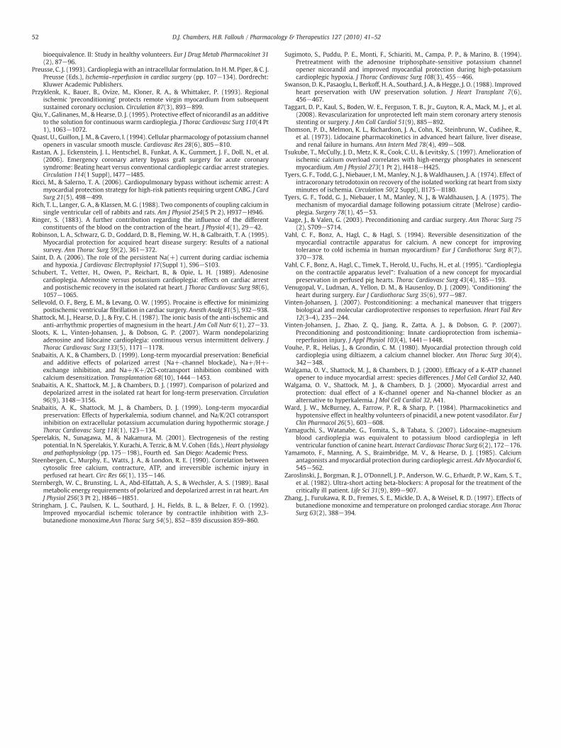
52 D.J. Chambers, H.B. Fallouh / Pharmacology & Therapeutics 127 (2010) 41–52
bioequivalence. II: Study in healthy volunteers. Eur J Drug Metab Pharmacokinet 31(2), 87−96.
Preusse, C. J. (1993). Cardioplegia with an intracellular formulation. In H. M. Piper, & C. J.Preusse (Eds.), Ischemia–reperfusion in cardiac surgery (pp. 107−134). Dordrecht:Kluwer Academic Publishers.
Przyklenk, K., Bauer, B., Ovize, M., Kloner, R. A., & Whittaker, P. (1993). Regionalischemic ‘preconditioning’ protects remote virgin myocardium from subsequentsustained coronary occlusion. Circulation 87(3), 893−899.
Qiu, Y., Galinanes, M., & Hearse, D. J. (1995). Protective effect of nicorandil as an additiveto the solution for continuous warm cardioplegia. J Thorac Cardiovasc Surg 110(4 Pt1), 1063−1072.
Quast, U., Guillon, J. M., & Cavero, I. (1994). Cellular pharmacology of potassium channelopeners in vascular smooth muscle. Cardiovasc Res 28(6), 805−810.
Rastan, A. J., Eckenstein, J. I., Hentschel, B., Funkat, A. K., Gummert, J. F., Doll, N., et al.(2006). Emergency coronary artery bypass graft surgery for acute coronarysyndrome: Beating heart versus conventional cardioplegic cardiac arrest strategies.Circulation 114(1 Suppl), I477−I485.
Ricci, M., & Salerno, T. A. (2006). Cardiopulmonary bypass without ischemic arrest: Amyocardial protection strategy for high-risk patients requiring urgent CABG. J CardSurg 21(5), 498−499.
Rich, T. L., Langer, G. A., & Klassen, M. G. (1988). Two components of coupling calcium insingle ventricular cell of rabbits and rats. Am J Physiol 254(5 Pt 2), H937−H946.
Ringer, S. (1883). A further contribution regarding the influence of the differentconstituents of the blood on the contraction of the heart. J Physiol 4(1), 29−42.
Robinson, L. A., Schwarz, G. D., Goddard, D. B., Fleming, W. H., & Galbraith, T. A. (1995).Myocardial protection for acquired heart disease surgery: Results of a nationalsurvey. Ann Thorac Surg 59(2), 361−372.
Saint, D. A. (2006). The role of the persistent Na(+) current during cardiac ischemiaand hypoxia. J Cardiovasc Electrophysiol 17(Suppl 1), S96−S103.
Schubert, T., Vetter, H., Owen, P., Reichart, B., & Opie, L. H. (1989). Adenosinecardioplegia. Adenosine versus potassium cardioplegia: effects on cardiac arrestand postischemic recovery in the isolated rat heart. J Thorac Cardiovasc Surg 98(6),1057−1065.
Sellevold, O. F., Berg, E. M., & Levang, O. W. (1995). Procaine is effective for minimizingpostischemic ventricular fibrillation in cardiac surgery. Anesth Analg 81(5), 932−938.
Shattock, M. J., Hearse, D. J., & Fry, C. H. (1987). The ionic basis of the anti-ischemic andanti-arrhythmic properties of magnesium in the heart. J Am Coll Nutr 6(1), 27−33.
Sloots, K. L., Vinten-Johansen, J., & Dobson, G. P. (2007). Warm nondepolarizingadenosine and lidocaine cardioplegia: continuous versus intermittent delivery. JThorac Cardiovasc Surg 133(5), 1171−1178.
Snabaitis, A. K., & Chambers, D. (1999). Long-term myocardial preservation: Beneficialand additive effects of polarized arrest (Na+-channel blockade), Na+/H+-exchange inhibition, and Na+/K+/2Cl-cotransport inhibition combined withcalcium desensitization. Transplantation 68(10), 1444−1453.
Snabaitis, A. K., Shattock, M. J., & Chambers, D. J. (1997). Comparison of polarized anddepolarized arrest in the isolated rat heart for long-term preservation. Circulation96(9), 3148−3156.
Snabaitis, A. K., Shattock, M. J., & Chambers, D. J. (1999). Long-term myocardialpreservation: Effects of hyperkalemia, sodium channel, and Na/K/2Cl cotransportinhibition on extracellular potassium accumulation during hypothermic storage. JThorac Cardiovasc Surg 118(1), 123−134.
Sperelakis, N., Sunagawa, M., & Nakamura, M. (2001). Electrogenesis of the restingpotential. In N. Sperelakis, Y. Kurachi, A. Terzic, & M. V. Cohen (Eds.), Heart physiologyand pathophysiology (pp. 175−198)., Fourth ed. San Diego: Academic Press.
Steenbergen, C., Murphy, E., Watts, J. A., & London, R. E. (1990). Correlation betweencytosolic free calcium, contracture, ATP, and irreversible ischemic injury inperfused rat heart. Circ Res 66(1), 135−146.
Sternbergh, W. C., Brunsting, L. A., Abd-Elfattah, A. S., & Wechsler, A. S. (1989). Basalmetabolic energy requirements of polarized and depolarized arrest in rat heart. AmJ Physiol 256(3 Pt 2), H846−H851.
Stringham, J. C., Paulsen, K. L., Southard, J. H., Fields, B. L., & Belzer, F. O. (1992).Improved myocardial ischemic tolerance by contractile inhibition with 2,3-butanedione monoxime.Ann Thorac Surg 54(5), 852−859 discussion 859–860.
Sugimoto, S., Puddu, P. E., Monti, F., Schiariti, M., Campa, P. P., & Marino, B. (1994).Pretreatment with the adenosine triphosphate-sensitive potassium channelopener nicorandil and improved myocardial protection during high-potassiumcardioplegic hypoxia. J Thorac Cardiovasc Surg 108(3), 455−466.
Swanson, D. K., Pasaoglu, I., Berkoff, H. A., Southard, J. A., & Hegge, J. O. (1988). Improvedheart preservation with UW preservation solution. J Heart Transplant 7(6),456−467.
Taggart, D. P., Kaul, S., Boden, W. E., Ferguson, T. B., Jr., Guyton, R. A., Mack, M. J., et al.(2008). Revascularization for unprotected left main stem coronary artery stenosisstenting or surgery. J Am Coll Cardiol 51(9), 885−892.
Thomson, P. D., Melmon, K. L., Richardson, J. A., Cohn, K., Steinbrunn, W., Cudihee, R.,et al. (1973). Lidocaine pharmacokinetics in advanced heart failure, liver disease,and renal failure in humans. Ann Intern Med 78(4), 499−508.
Tsukube, T., McCully, J. D., Metz, K. R., Cook, C. U., & Levitsky, S. (1997). Amelioration ofischemic calcium overload correlates with high-energy phosphates in senescentmyocardium. Am J Physiol 273(1 Pt 2), H418−H425.
Tyers, G. F., Todd, G. J., Niebauer, I. M., Manley, N. J., &Waldhausen, J. A. (1974). Effect ofintracoronary tetrodotoxin on recovery of the isolated working rat heart from sixtyminutes of ischemia. Circulation 50(2 Suppl), II175−II180.
Tyers, G. F., Todd, G. J., Niebauer, I. M., Manley, N. J., & Waldhausen, J. A. (1975). Themechanism of myocardial damage following potassium citrate (Melrose) cardio-plegia. Surgery 78(1), 45−53.
Vaage, J., & Valen, G. (2003). Preconditioning and cardiac surgery. Ann Thorac Surg 75(2), S709−S714.
Vahl, C. F., Bonz, A., Hagl, C., & Hagl, S. (1994). Reversible desensitization of themyocardial contractile apparatus for calcium. A new concept for improvingtolerance to cold ischemia in human myocardium? Eur J Cardiothorac Surg 8(7),370−378.
Vahl, C. F., Bonz, A., Hagl, C., Timek, T., Herold, U., Fuchs, H., et al. (1995). “Cardioplegiaon the contractile apparatus level”: Evaluation of a new concept for myocardialpreservation in perfused pig hearts. Thorac Cardiovasc Surg 43(4), 185−193.
Venugopal, V., Ludman, A., Yellon, D. M., & Hausenloy, D. J. (2009). ‘Conditioning’ theheart during surgery. Eur J Cardiothorac Surg 35(6), 977−987.
Vinten-Johansen, J. (2007). Postconditioning: a mechanical maneuver that triggersbiological and molecular cardioprotective responses to reperfusion. Heart Fail Rev12(3–4), 235−244.
Vinten-Johansen, J., Zhao, Z. Q., Jiang, R., Zatta, A. J., & Dobson, G. P. (2007).Preconditioning and postconditioning: Innate cardioprotection from ischemia–reperfusion injury. J Appl Physiol 103(4), 1441−1448.
Vouhe, P. R., Helias, J., & Grondin, C. M. (1980). Myocardial protection through coldcardioplegia using diltiazem, a calcium channel blocker. Ann Thorac Surg 30(4),342−348.
Walgama, O. V., Shattock, M. J., & Chambers, D. J. (2000). Efficacy of a K-ATP channelopener to induce myocardial arrest: species differences. J Mol Cell Cardiol 32, A40.
Walgama, O. V., Shattock, M. J., & Chambers, D. J. (2000). Myocardial arrest andprotection: dual effect of a K-channel opener and Na-channel blocker as analternative to hyperkalemia. J Mol Cell Cardiol 32, A41.
Ward, J. W., McBurney, A., Farrow, P. R., & Sharp, P. (1984). Pharmacokinetics andhypotensive effect in healthy volunteers of pinacidil, a new potent vasodilator. Eur JClin Pharmacol 26(5), 603−608.
Yamaguchi, S., Watanabe, G., Tomita, S., & Tabata, S. (2007). Lidocaine–magnesiumblood cardioplegia was equivalent to potassium blood cardioplegia in leftventricular function of canine heart. Interact Cardiovasc Thorac Surg 6(2), 172−176.
Yamamoto, F., Manning, A. S., Braimbridge, M. V., & Hearse, D. J. (1985). Calciumantagonists andmyocardial protection during cardioplegic arrest. AdvMyocardiol 6,545−562.
Zaroslinski, J., Borgman, R. J., O'Donnell, J. P., Anderson, W. G., Erhardt, P. W., Kam, S. T.,et al. (1982). Ultra-short acting beta-blockers: A proposal for the treatment of thecritically ill patient. Life Sci 31(9), 899−907.
Zhang, J., Furukawa, R. D., Fremes, S. E., Mickle, D. A., & Weisel, R. D. (1997). Effects ofbutanedione monoxime and temperature on prolonged cardiac storage. Ann ThoracSurg 63(2), 388−394.