cancer of face, especially region of eye: method of treatment
TRANSCRIPT
CANCER OF FACE, ESPECIALLY REGION OF EYE METHOD OF TREATMENT
HOLLIS L. ALBRIGHT, M.D.
BOSTON
C ANCER of epidermoid origin in
general resoIves itseIf into two types : (I) the acanthoma, with aduh
squamous ceIIs, varying degrees of horni- fication and pear1 formation, and (2) basa1 cell carcinoma, the so-caIIed “rodent uIcer.” Whereas the Iatter is rareIy seen within the ora cavity, both types frequent arise in the skin of the face. Basal ceil cancer has been termed reIativeIy non- malignant1 mainIy because of its tendency to Iocal recurrence and invasion rather than spreading by metastasis. The disturbing nature of this basa1 ceI1 growth may be seen in the IocaI persistence and progressive uIcerating spread of the Iesion over a period of years, especiaIIy in the vicinity of the orbit, nasofacia1 angIes and auricle.
Squamous ceI1 cancer, the acanthoma, likewise shows late rather than earIy spread, but is more rapidIy destructive, extends more wideIy and metastasizes oftener than the basa1 cell Iesion. Except in the case of direct extension of growths primary in the ora1, nasa1, orbita or aura1 cavities, to invoIve the skin of the face, most cancerous Iesions of the face wil1 fall into these two groups.
Of the methods empIoyed in eradicating cancer of the skin of the face, excision with or without pIastic repair of the defect is the commonest. The initiaI success of x-ray therapy as an agent in the fight against cancer was obtained from its striking destruction of cancer of the skin.2
In rgo3 GoIdberg and London first applied surface radium to 2 cases of basa1
1 MACCALLUM, W. G. “A Textbook of Pathology,” 4th edition, p, 1034.
*Roentgen therapv obtained its first cancer cure when Tar-Stenbeck “of Stockhohn, in 1898 treated a basa1 cell carcinoma of the nose that had recurred twice after coagulation with the red hot iron. ForrseII foIlowed that patient unti1 1920. There was no recurrence.
176
cel1 carcinoma of the face with good cos- metic result. At present, adequate x-ray and radium treatment have been accepted with surgery as proved methods for the eradication of skin cancer. Preference is heId here for none of these three agents to the excIusion of the other two.
It has come strikingIy to the author’s attention that a practical method of treat- ment has been evolved which Iends itseIf to fairIy universa1 appIicabiIity for skin cancer, especiahy that of the face. This is perhaps the onIy region where minima1 cosmetic deformity should receive due consideration.
A proved working method of treating maIignant skin Iesions up to 3 cm. in diameter is presented here, based largely on the experience of the Radiumhemmet, StockhoIm,3 where as is we11 known, foIlow-up examinations have been recorded on over go per cent of the cancer patients treated since 1915. The method aIIows effective destruction of the Iesion with regeneration of the ectoderma1 and meso- derma1 tissue layers at the site of the destroyed growth and preservation of the surrounding norma tissue. By this method an epidermicidal dosage of 7-10 S. E. D. is given, to ensure destruction of the tumor, rather than to empIoy a dosage of sub- epidermicida1, seIective intensity. A stable scar usuahy resuIts, with minima1 deform- ity and better cosmetic resuh.
Lesions up to 3 cm. in diameter, in&d- ing those of the Iip, are impIanted with radium eIement needIes, either six needIes each of IO mgm. strength or eight needIes each of 3 mgm. strength, using 0.5 mm. pIatinum filtration. Some therapists em- pIoy heavier hItration, but most use at
s Observed from JuIy to October, 1934, inclusive.
NEW SERIES VOL. XxX111, No. z AIbright-Cancer of Face American Journal of Surgery I77
Ieast the equivalent of 0.5 mm. pIatinum, for this adequateIy screens the aIpha and absorbs approximately 98.5 per cent of the soft beta rays coming from radium C.
TABLE OF FILTERS WHICH EQUAL 0.5 MM. PLATINUM* MilKmeters
C&Ad........................... 0.56 Lead........................... 0.97 SiIver.. 1.02 Copper......................... 1.20 Brass........................... 1.26 Steel........................... 1.37 Zinc............................ 1.51 Aluminum.. 3.96
* Computed from Heversy and Parnth.
One treatment of usually not more than four hours is required. This gives a tota dosage of 6 needIes X IO mgm. each x 4 hrs., or 240 mihigram hours. This is foIIowed by hyperemia, IocaI sweIIing, Ieading to uIceration in ten days, at which time beginning re-epitheIiaIization is evident. By the end of three to four weeks epithelial- ization shouId be compIete. The end resuIt should be a soft, stabIe scar, often whitened due to Ioss of normaI epitheIia1 pigmenta- tion. Any persistent induration after six weeks must be strongly suspected as tumor, requiring additional treatment of Iess intensity.
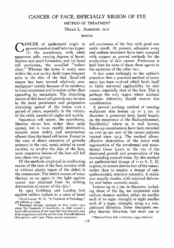
This genera1 method of treating cancer of the skin of the face, incIuding cancer of the Iip (Fig. I) has been standardIy em- pIoyed at the Radiumhemmet, StockhoIm, since 1926. Lesions Iarger than 3 cm. in diameter have been best controIIed by either surface x-irradiation (i.e. 7 to IO S. E. D. via x-ray with light filtration of 1-2 mm. aluminum, and careful screening of the surrounding tissues, or via the 5 gm. teleradium bomb at 3 cm. distance, giving a depth dosage of 2 cm.) ; or by wide exci- sion with pedicIe ffap repair. The ulcera- tion and necrosis produced by adequate needIing of the growth is so extensive as to Iead to much sIoughing and a less satis- factory resuIt.
To iIIustrate this method the foIIowing case is reported, wherein severa probIems arising in the management of Iesions cIose to the eye are handIed.
CASE I. J. F. M. a rugged, we11 preserved white maIe of sixty-nine years was admitted to the PaImer MemoriaI HospitaI on January
FIG. I. Cancer of lower Iip, interstitiaIIy is radiated, showing radium needIes in situ.
IO, 1936, compIaining of a recurring, uIcerated growth in the region of the inner canthus of the left eye, intermittentIy present for the past twenty years. Seventeen years ago the Iesion had been excised but recurred two years Iater, as Iarge as previousIy. Fourteen years ago four x-ray treatments had been given, with recurrence two years later. Since that time the growth had been cauterized severa times, and aIso had been treated with the eIectric needle. For the past two weeks the Iesion had become uIcerated and had shown increased growth. There had been moderateIy increased weeping from the Ie!‘t eye.
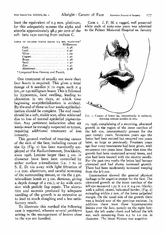
Examination showed the genera1 physica findings to be negative except for the face. The uIcerated growth at the inner canthus of the Ieft eye measured I .23 X 0.3 X 0.4 cm. (thick), with a roIIed, raised, indurated border, (Fig. 2) extending within 2 mm. of the Iower palpebral margin. On the nasa1 side of the growth there was a heaIed scar of the previous excision. In addition there were three hyperkeratotic Iesions over the face, namely on the right fore- head, the Ieft malar cheek and behind the Ieft ear, each measuring from 0.3 to 1.0 cm. in diameter. The bIood Hinton was negative.
178 ~~~ sERIEs voL. XXXIII, No. 3 AIbright-Cancer of Face AUGUST, 1936
Radium treatment was begun by applying In three days there was a good radium reac- a surface plaque of 5 mgm. strength and tion with hyperemia and beginning ulceration. 0.1 mm. platinum f&ration to each hyperkera- There was very slight injection of the bIood
FIG. 2. Case J. F. M. showing site of basal cell cancer at inner canthus of left eye.
tosis for six hours, giving a total dosage of 30 mgm. hrs. to each area.
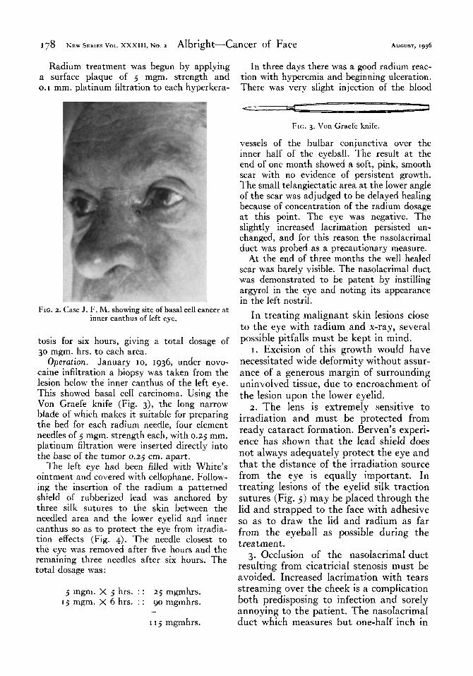
Operation. January IO, 1936, under novo- Caine infihration a biopsy was taken from the lesion beIow the inner canthus of the left eye. This showed basa1 ceI1 carcinoma. Using the Von Graefe knife (Fig. 3), the Iong narrow bIade of which makes it suitabIe for preparing the bed for each radium needle, four eIement needIes of 5 mgm. strength each, with 0.25 mm.
pIatinum filtration were inserted directly into the base of the tumor 0.25 cm. apart.
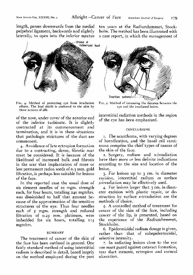
The Ieft eye had been filled with White’s ointment and covered with cellophane. FoIIow- ing the insertion of the radium a patterned shieId of rubberized Iead was anchored by three silk sutures to the skin between the needIed area and the Iower eyelid and inner canthus so as to protect the eye from irradia- tion effects (Fig. 4). The needIe cIosest to the eye was removed after five hours and the remaining three needIes after six hours. The tota dosage was:
5 mgm. X 5 hrs. : : 25 mgmhrs. 15 mgm. X 6 hrs. : : go mgmhrs.
I I 5 mgmhrs.
FIG. 3. Von Graefe knife.
vesseIs of the bulbar conjunctiva over the inner haIf of the eyeball. The resuIt at the end of one month showed a soft, pink, smooth scar with no evidence of persistent growth. The smaI1 teIangiectatic area at the Iower angle of the scar was adjudged to be deIayed heaIing because of concentration of the radium dosage at this point. The eye was negative. The sIightIy increased Iacrimation persisted un- changed, and for this reason the nasoIacrima1 duct was probed as a precautionary measure.
At the end of three months the we11 heaIed scar was barely visible. The nasoIacrima1 duct was demonstrated to be patent by instiIIing argyro1 in the eye and noting its appearance in the left nostril.
In treating malignant skin Iesions cIose to the eye with radium and x-ray, several possible pitfalls must be kept in mind.
I. Excision of this growth wouId have necessitated wide deformity without assur- ance of a generous margin of surrounding uninvolved tissue, due to encroachment of the Iesion upon the Iower eyeIid.
2. The Iens is extremeIy sensitive to irradiation and must be protected from ready cataract formation. Berven’s experi- ence has shown that the Iead shieId does not always adequateIy protect the eye and that the distance of the irradiation source from the eye is equaIIy important. In treating Iesions of the eyeIid siIk traction sutures (Fig. 5) may be pIaced through the Iid and strapped to the face with adhesive so as to draw the Iid and radium as far from the eyebaIl as possibIe during the treatment.
3. OccIusion of the nasoIacrima1 duct resuIting from cicatricial stenosis must be avoided. Increased Iacrimation with tears streaming over the cheek is a complication both predisposing to infection and soreIy annoying to the patient. The nasoIacrima1 duct which measures but one-half inch in
NEW SERIES VOL. XxX111, No. 2 Albright-Cancer of Face American Journal of Surgery 179
Iength, passes downwards from the media1 ten years at the Radiumhemmet, Stock- paIpebra1 Iigament, backwards and sIightIy hoIm. The method has been iIIustrated with IateraIIy, to open into the inferior meatus a case report, in which the management of
FIG. 4. Method of protecting eye from irradiation effects. The lead shield is anchored to the skin bv three sutures of silk.
I
of the nose, under cover of the anterior end of the inferior turbinate. It is sIightIy contracted at its commencement and termination, and it is in these situations that pathoIogic strictures of the duct are commonest.
4. Avoidance of Iate ectropion formation due to a contracting, dense, fibrotic scar must be considered. It is because of the IikeIihood of increased buIk and fibrosis in the scar that impIantation of more or Iess permanent radon seeds of 0.3 mm. goId Mtration, is perhaps Iess suitabIe for Iesions of the face.
In the reported case the usua1 dose of six eIement needIes of IO mgm. strength each, for four hours, totaIIing 240 mgmhrs. was diminished to haIf that amount be- cause of the approximation of the sensitive structures of the eye. Thus four needIes each of 5 mgm. strength and reduced Mtration of 0.25 mm. pIatinum, were imbedded for six hours, totaIIing I 15 mgm hrs.
SUMMARY
The treatment of cancer of the skin of the face has been outIined in generaI. One fairIy standard method of using interstitia1 radium is described in detail, based IargeIy on the method employed during the past
FIG. 5. Method of increasing the distance between the eye and the irradiated Iesion.
interstitia1 radiation methods in the region of the eye has been emphasized.
CONCLUSIONS
I. The acanthoma, with varying degrees of hornification, and the basa1 ceI1 carci- noma comprise the chief types of cancer of the skin of the face.
2. Surgery, radium and x-irradiation have their more or Iess definite indications according to the size and Iocation of the Iesion.
3. For Iesions up to 3 cm. in diameter excision, interstitia1 radium or surface x-irradiation may be effectiveIy used.
4. For Iesions Iarger that 3 cm. in diam- eter excision with pIastic repair, or de- struction by surface x-irradiation are the methods of choice.
5. A controIIed method of treatment for cancer of the skin of the face, incIuding cancer of the Iip, is presented, based on the experience of the Radiumhemmet, Stockholm.
6. Epidermicida radium dosage is given, rather than that of subepidermicida1, seIective intensity.
7. In radiating lesions cIose to the eye one must guard against cataract formation, tear duct stenosis, ectropion and cornea1 mceration.