buonocore memorial lecture adhesion to enamel and · pdf filethe fundamental principle of...
TRANSCRIPT
©Operative Dentistry, 2003, 28-3, 215-235
Buonocore Memorial Lecture
SUMMARY
Bonding to tooth tissue canbe achieved through an“etch&rinse,” “self-etch” or“glass-ionomer” approach.In this paper, the basicbonding mechanism toenamel and dentin of thesethree approaches is demon-strated by means of ultra-morphological and chemi-cal characterization oftooth-biomaterial interfa-cial interactions. Further-
more, bond-strength testing and measurement of mar-ginal-sealing effectiveness (the two most commonly
employed methodologies to determine “bonding effec-tiveness” in the laboratory) are evaluated upon theirvalue and relevance in predicting clinical performance.A new dynamic methodology to test biomaterial-toothbonds in a fatigue mode is introduced with a recentlydeveloped micro-rotary fatigue-testing device.Eventually, today’s adhesives will be critically weight-ed upon their performance in diverse laboratory stud-ies and clinical trials. Special attention has been givento the benefits/drawbacks of an etch&rinse versus aself-etch approach and the long-term performance ofthese adhesives. Correlating data gathered in the lab-oratory with clinical results clearly showed that labo-ratory research CAN predict clinical effectiveness.Although there is a tendency to simplify bonding pro-cedures, the data presented confirm that conventionalthree-step etch&rinse adhesives still perform most
Buonocore Memorial Lecture
Adhesion to Enamel and Dentin:Current Status andFuture Challenges
B Van Meerbeek • J De Munck • Y YoshidaS Inoue • M Vargas • P Vijay
K Van Landuyt • P Lambrechts • G Vanherle
Michael Buonocore
*Bart Van Meerbeek, DDS, PhD, professor, Leuven BIOMATResearch Cluster, Department of Conservative Dentistry,School of Dentistry, Oral Pathology and Maxillo-facial Surgery,Catholic University of Leuven, Belgium
Jan De Munck, DDS, PhD-student, Leuven BIOMAT ResearchCluster, Department of Conservative Dentistry, School ofDentistry, Oral Pathology and Maxillo-facial Surgery, CatholicUniversity of Leuven, Belgium
Yasuhiro Yoshida, DDS, PhD, associate professor, Department ofBiomaterials, Okayama University Graduate School ofMedicine and Dentistry, Japan
Satoshi Inoue, DDS, PhD, lecturer, Division for GeneralDentistry, Hokkaido University Dental Hospital, Japan
Marcos Vargas, DDS, MS, associate professor, Department ofOperative Dentistry, University of Iowa, USA
Padmini Vijay, DDS, MS-student, Leuven BIOMAT ResearchCluster, Department of Conservative Dentistry, School of
Dentistry, Oral Pathology and Maxillo-facial Surgery, CatholicUniversity of Leuven, Belgium
Kirsten Van Landuyt, DDS, PhD-student, Leuven BIOMATResearch Cluster, Department of Conservative Dentistry,School of Dentistry, Oral Pathology and Maxillo-facial Surgery,Catholic University of Leuven, Belgium
Paul Lambrechts, DDS, PhD, full professor, Leuven BIOMATResearch Cluster, Department of Conservative Dentistry,School of Dentistry, Oral Pathology and Maxillo-facial Surgery,Catholic University of Leuven, Belgium
Guido Vanherle, MD, DDS, emeritus full professor, Leuven BIO-MAT Research Cluster, Department of Conservative Dentistry,School of Dentistry, Oral Pathology and Maxillo-facial Surgery,Catholic University of Leuven, Belgium
*Reprint request: Catholic University of Leuven, Kapucijnenvoer7, B-3000, Leuven, Belgium; e-mail:[email protected]
Bart Van Meerbeek
216 Operative Dentistry
favorably and are most reliable in the long-term.Nevertheless, a self-etch approach may have the bestfuture perspective. Clinically, when adhesives nolonger require an “etch&rinse” step, the applicationtime, and probably more importantly, the technique-sensitivity are substantially reduced. Especially“mild,” two-step self-etch adhesives that bond througha combined micromechanical and chemical interactionwith tooth tissue closely approach conventional three-step systems in bonding performance.
INTRODUCTION
Adhesive dentistry evolves rapidly. Two main incen-tives drive this evolution. Adhesive techniques com-bined with using tooth-colored restorative materials arefrequently requested by patients. They want us torestore their teeth not only anatomically and function-ally, but also esthetically and, thus, nearly invisibly.From our perspective, today’s operative dentistryshould primarily involve “minimally invasive”(Degrange & Roulet, 1997) or “minimum intervention”(Tyas, Anusavice & Frencken, 2000) care. This meansthat only the lost or diseased tooth tissue is replaced bythe restorative material that is directly bonded to theremaining sound tissue. Also, the more recent approachof promoting “maintenance and repair” (Bouschlicher,Reinhardt & Vargas, 1997; Denehy, Bouschlicher &Vargas, 1998; Roeters, 2000; Wilson, Setcos & Brunton,2001), rather than replacing entire restorations(exhibiting marginal discolorations and/or defects) hasfurther boosted the use of adhesive techniques indiverse applications of everyday clinical practice.
Major drawbacks of this approach are that adhesivetechniques still accompany a higher placement com-plexity and technique sensitivity (risk of makingmanipulation errors). Also, though the retention ofadhesive restorations for a reasonable time is no longera clinical problem, maintaining the margins of adhesiverestorations sealed against leakage phenomenaremains the major factor that shortens clinical longevity.
The fundamental principle of adhesion to tooth sub-strate is based upon an exchange process by which inor-ganic tooth material is exchanged for synthetic resin(Van Meerbeek & others, 2001a). This process involvestwo phases. One phase consists of removing calciumphosphates by which microporosities are exposed atboth the enamel and dentin tooth surface. The other so-called hybridization phase involves infiltration and sub-sequent in situ polymerization of resin within the cre-ated surface microporosities. This results is micro-mechanical interlocking that is primarily based onmechanisms of diffusion. While micro-mechanicalinterlocking is believed to be a prerequisite to achievinggood bonding within clinical circumstances, the poten-tial benefit of additional chemical interaction betweenfunctional monomers and tooth substrate components
has recently gained new attention. This paper sketchesthe current status of adhesives in terms of bondingeffectiveness measured in the laboratory and in clinicalpractice. Special attention is given to the potential rolesof both micro-mechanical and chemical bonding mecha-nisms through correlating morphologic and chemicalinterfacial characteristics of tooth-biomaterial interac-tions using diverse kinds of adhesives.
MECHANISMS OF ADHESIONTO ENAMEL AND DENTIN
Using contemporary adhesives, the substance exchangebetween biomaterial and tooth tissue is carried out inone, two or three clinical application steps, respectively.Besides the number of application steps, adhesives canfurther be classified based on the underlying adhesionstrategy in “etch&rinse,” “self-etch” and “(resin-modi-fied) glass-ionomer adhesives” (Van Meerbeek & others,2001a) (Figure 1). The degree of substance exchangesubstantially differs among these adhesives. In general,the exchange intensity induced by etch&rinse adhe-sives exceeds that of self-etch adhesives, though amongthe latter, systems that rather intensively interact withtooth tissue also exist, even when applied in only a sin-gle step.
Etch&Rinse Approach
This adhesion strategy involves at least two steps and,in its most conventional form, three steps with succes-sive application of the conditioner or acid etchant, fol-lowed by the primer or adhesion promoting agent, andeventually, application of the actual bonding agent oradhesive resin (Figure 1). The simplified two-step ver-sion combines the second and third step but still fol-lows a separate “etch&rinse” phase.
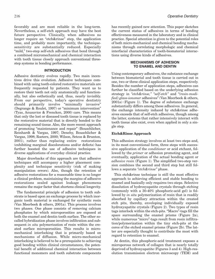
This etch&rinse technique is still the most effectiveapproach to achieving efficient and stable bonding toenamel and basically only requires two steps. Selectivedissolution of hydroxyapatite crystals through etching(commonly with a 30-40% phosphoric-acid gel) is fol-lowed by in situ polymerization of resin that is readilyabsorbed by capillary attraction within the createdetch pits, thereby, enveloping individually exposedhydroxyapatite crystals (Figure 2). Two types of resintags interlock within the etch-pits. “Macro”-tags fill thespace surrounding the enamel prisms (Figure 2a),while numerous “micro”-tags result from resin infiltra-tion/polymerization within the tiny etch-pits at thecores of the etched enamel prisms (Figure 2b). The lat-ter are especially thought to contribute the most withregard to retention to enamel.
At dentin, this phosphoric-acid treatment exposes amicroporous network of collagen that is nearly totallydeprived of hydroxyapatite (Figures 3 and 4). High-res-olution transmission electron microscopy (TEM) and
217Van Meerbeek & Others: Adhesion to Enamel and Dentin
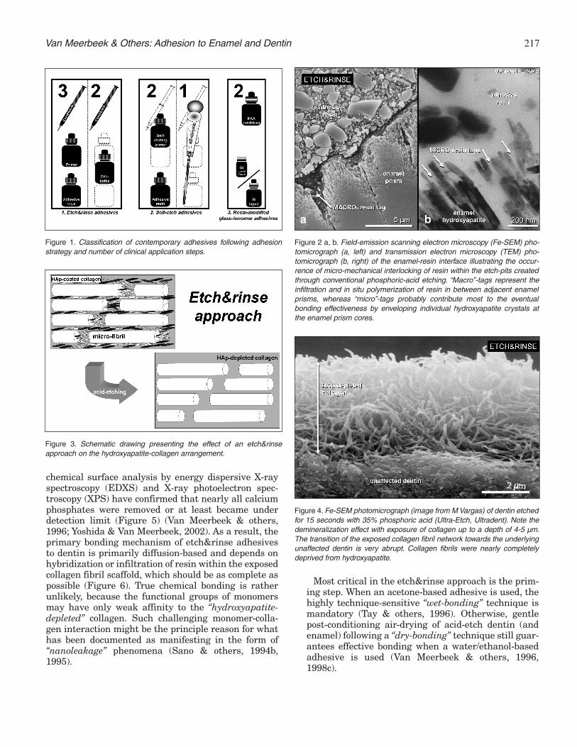
chemical surface analysis by energy dispersive X-rayspectroscopy (EDXS) and X-ray photoelectron spec-troscopy (XPS) have confirmed that nearly all calciumphosphates were removed or at least became underdetection limit (Figure 5) (Van Meerbeek & others,1996; Yoshida & Van Meerbeek, 2002). As a result, theprimary bonding mechanism of etch&rinse adhesivesto dentin is primarily diffusion-based and depends onhybridization or infiltration of resin within the exposedcollagen fibril scaffold, which should be as complete aspossible (Figure 6). True chemical bonding is ratherunlikely, because the functional groups of monomersmay have only weak affinity to the “hydroxyapatite-depleted’’ collagen. Such challenging monomer-colla-gen interaction might be the principle reason for whathas been documented as manifesting in the form of“nanoleakage” phenomena (Sano & others, 1994b,1995).
Most critical in the etch&rinse approach is the prim-ing step. When an acetone-based adhesive is used, thehighly technique-sensitive “wet-bonding” technique ismandatory (Tay & others, 1996). Otherwise, gentlepost-conditioning air-drying of acid-etch dentin (andenamel) following a “dry-bonding” technique still guar-antees effective bonding when a water/ethanol-basedadhesive is used (Van Meerbeek & others, 1996,1998c).
Figure 1. Classification of contemporary adhesives following adhesionstrategy and number of clinical application steps.
Figure 2 a, b. Field-emission scanning electron microscopy (Fe-SEM) pho-tomicrograph (a, left) and transmission electron microscopy (TEM) pho-tomicrograph (b, right) of the enamel-resin interface illustrating the occur-rence of micro-mechanical interlocking of resin within the etch-pits createdthrough conventional phosphoric-acid etching. “Macro”-tags represent theinfiltration and in situ polymerization of resin in between adjacent enamelprisms, whereas “micro”-tags probably contribute most to the eventualbonding effectiveness by enveloping individual hydroxyapatite crystals atthe enamel prism cores.
a b
Figure 3. Schematic drawing presenting the effect of an etch&rinseapproach on the hydroxyapatite-collagen arrangement.
Figure 4. Fe-SEM photomicrograph (image from M Vargas) of dentin etchedfor 15 seconds with 35% phosphoric acid (Ultra-Etch, Ultradent). Note thedemineralization effect with exposure of collagen up to a depth of 4-5 µm.The transition of the exposed collagen fibril network towards the underlyingunaffected dentin is very abrupt. Collagen fibrils were nearly completelydeprived from hydroxyapatite.
218 Operative Dentistry
Glass-Ionomer Approach
Glass-ionomers remain as the only materials that areself-adhesive to tooth tissue, in principle, without anysurface pre-treatment (Figure 7). Although this is cer-
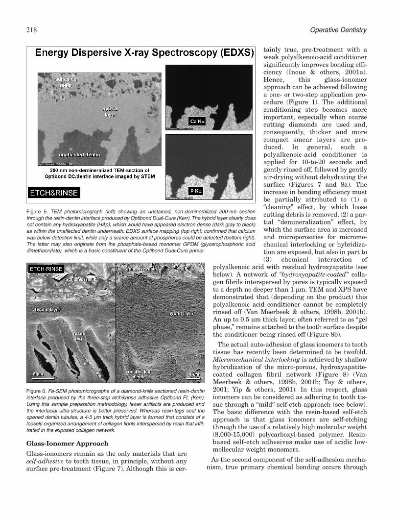
tainly true, pre-treatment with aweak polyalkenoic-acid conditionersignificantly improves bonding effi-ciency (Inoue & others, 2001a).Hence, this glass-ionomerapproach can be achieved followinga one- or two-step application pro-cedure (Figure 1). The additionalconditioning step becomes moreimportant, especially when coarsecutting diamonds are used and,consequently, thicker and morecompact smear layers are pro-duced. In general, such apolyalkenoic-acid conditioner isapplied for 10-to-20 seconds andgently rinsed off, followed by gentlyair-drying without dehydrating thesurface (Figures 7 and 8a). Theincrease in bonding efficiency mustbe partially attributed to (1) a“cleaning” effect, by which loosecutting debris is removed, (2) a par-tial “demineralization” effect, bywhich the surface area is increasedand microporosities for microme-chanical interlocking or hybridiza-tion are exposed, but also in part to(3) chemical interaction of
polyalkenoic acid with residual hydroxyapatite (seebelow). A network of “hydroxyapatite-coated” colla-gen fibrils interspersed by pores is typically exposedto a depth no deeper than 1 µm. TEM and XPS havedemonstrated that (depending on the product) thispolyalkenoic acid conditioner cannot be completelyrinsed off (Van Meerbeek & others, 1998b, 2001b).An up to 0.5 µm thick layer, often referred to as “gelphase,” remains attached to the tooth surface despitethe conditioner being rinsed off (Figure 8b).
The actual auto-adhesion of glass ionomers to toothtissue has recently been determined to be twofold.Micromechanical interlocking is achieved by shallowhybridization of the micro-porous, hydroxyapatite-coated collagen fibril network (Figure 8) (VanMeerbeek & others, 1998b, 2001b; Tay & others,2001; Yip & others, 2001). In this respect, glassionomers can be considered as adhering to tooth tis-sue through a “mild” self-etch approach (see below).The basic difference with the resin-based self-etchapproach is that glass ionomers are self-etchingthrough the use of a relatively high molecular weight(8,000-15,000) polycarboxyl-based polymer. Resin-based self-etch adhesives make use of acidic low-mollecular weight monomers.
As the second component of the self-adhesion mecha-nism, true primary chemical bonding occurs through
Figure 5. TEM photomicrograph (left) showing an unstained, non-demineralized 200-nm sectionthrough the resin-dentin interface produced by Optibond Dual-Cure (Kerr).The hybrid layer clearly doesnot contain any hydroxyapatite (HAp), which would have appeared electron dense (dark gray to black)as within the unaffected dentin underneath. EDXS surface mapping (top right) confirmed that calciumwas below detection limit, while only a scarce amount of phosphorus could be detected (bottom right).The latter may also originate from the phosphate-based monomer GPDM (glycerophosphoric aciddimethacrylate), which is a basic constituent of the Optibond Dual-Cure primer.
Figure 6. Fe-SEM photomicrographs of a diamond-knife sectioned resin-dentininterface produced by the three-step etch&rinse adhesive Optibond FL (Kerr).Using this sample preparation methodology, fewer artifacts are produced andthe interfacial ultra-structure is better preserved. Whereas resin-tags seal theopened dentin tubules, a 4-5 µm thick hybrid layer is formed that consists of aloosely organized arrangement of collagen fibrils interspersed by resin that infil-trated in the exposed collagen network.
219Van Meerbeek & Others: Adhesion to Enamel and Dentin
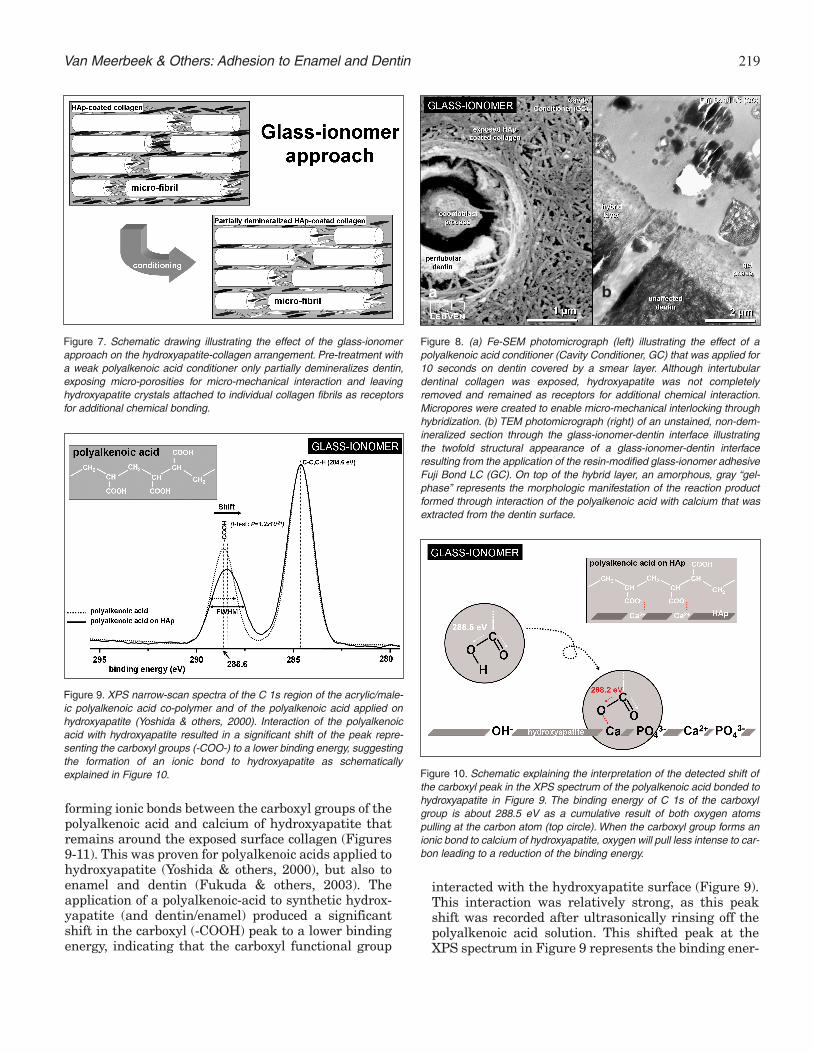
forming ionic bonds between the carboxyl groups of thepolyalkenoic acid and calcium of hydroxyapatite thatremains around the exposed surface collagen (Figures9-11). This was proven for polyalkenoic acids applied tohydroxyapatite (Yoshida & others, 2000), but also toenamel and dentin (Fukuda & others, 2003). Theapplication of a polyalkenoic-acid to synthetic hydrox-yapatite (and dentin/enamel) produced a significantshift in the carboxyl (-COOH) peak to a lower bindingenergy, indicating that the carboxyl functional group
interacted with the hydroxyapatite surface (Figure 9).This interaction was relatively strong, as this peakshift was recorded after ultrasonically rinsing off thepolyalkenoic acid solution. This shifted peak at theXPS spectrum in Figure 9 represents the binding ener-
Figure 7. Schematic drawing illustrating the effect of the glass-ionomerapproach on the hydroxyapatite-collagen arrangement. Pre-treatment witha weak polyalkenoic acid conditioner only partially demineralizes dentin,exposing micro-porosities for micro-mechanical interaction and leavinghydroxyapatite crystals attached to individual collagen fibrils as receptorsfor additional chemical bonding.
Figure 8. (a) Fe-SEM photomicrograph (left) illustrating the effect of apolyalkenoic acid conditioner (Cavity Conditioner, GC) that was applied for10 seconds on dentin covered by a smear layer. Although intertubulardentinal collagen was exposed, hydroxyapatite was not completelyremoved and remained as receptors for additional chemical interaction.Micropores were created to enable micro-mechanical interlocking throughhybridization. (b) TEM photomicrograph (right) of an unstained, non-dem-ineralized section through the glass-ionomer-dentin interface illustratingthe twofold structural appearance of a glass-ionomer-dentin interfaceresulting from the application of the resin-modified glass-ionomer adhesiveFuji Bond LC (GC). On top of the hybrid layer, an amorphous, gray “gel-phase” represents the morphologic manifestation of the reaction productformed through interaction of the polyalkenoic acid with calcium that wasextracted from the dentin surface.
a b
Figure 9. XPS narrow-scan spectra of the C 1s region of the acrylic/male-ic polyalkenoic acid co-polymer and of the polyalkenoic acid applied onhydroxyapatite (Yoshida & others, 2000). Interaction of the polyalkenoicacid with hydroxyapatite resulted in a significant shift of the peak repre-senting the carboxyl groups (-COO-) to a lower binding energy, suggestingthe formation of an ionic bond to hydroxyapatite as schematicallyexplained in Figure 10. Figure 10. Schematic explaining the interpretation of the detected shift of
the carboxyl peak in the XPS spectrum of the polyalkenoic acid bonded tohydroxyapatite in Figure 9. The binding energy of C 1s of the carboxylgroup is about 288.5 eV as a cumulative result of both oxygen atomspulling at the carbon atom (top circle). When the carboxyl group forms anionic bond to calcium of hydroxyapatite, oxygen will pull less intense to car-bon leading to a reduction of the binding energy.
220 Operative Dentistry
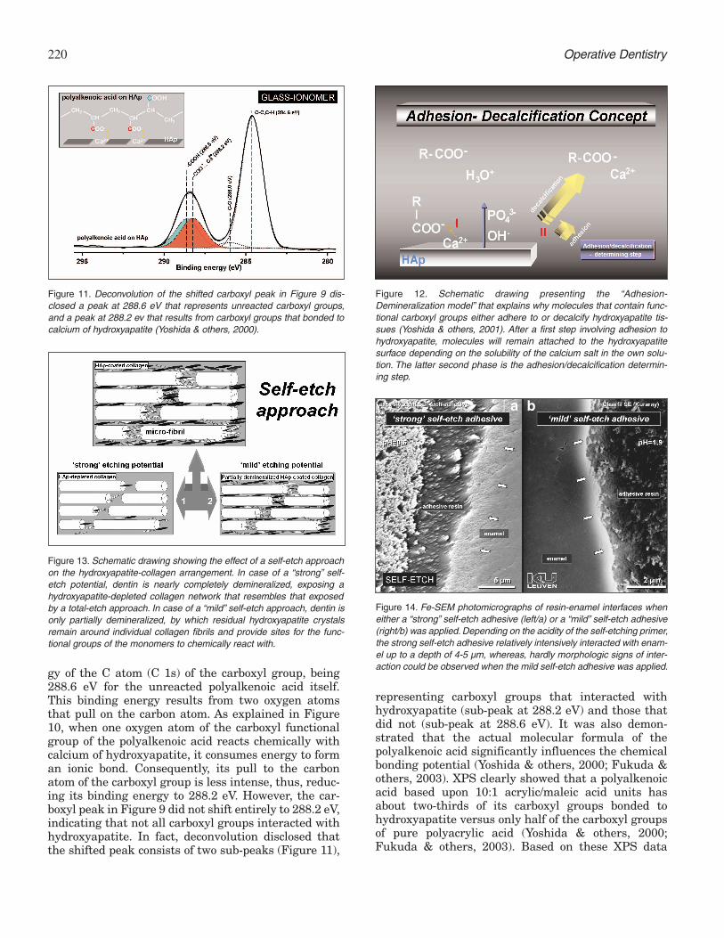
gy of the C atom (C 1s) of the carboxyl group, being288.6 eV for the unreacted polyalkenoic acid itself.This binding energy results from two oxygen atomsthat pull on the carbon atom. As explained in Figure10, when one oxygen atom of the carboxyl functionalgroup of the polyalkenoic acid reacts chemically withcalcium of hydroxyapatite, it consumes energy to forman ionic bond. Consequently, its pull to the carbonatom of the carboxyl group is less intense, thus, reduc-ing its binding energy to 288.2 eV. However, the car-boxyl peak in Figure 9 did not shift entirely to 288.2 eV,indicating that not all carboxyl groups interacted withhydroxyapatite. In fact, deconvolution disclosed thatthe shifted peak consists of two sub-peaks (Figure 11),
representing carboxyl groups that interacted withhydroxyapatite (sub-peak at 288.2 eV) and those thatdid not (sub-peak at 288.6 eV). It was also demon-strated that the actual molecular formula of thepolyalkenoic acid significantly influences the chemicalbonding potential (Yoshida & others, 2000; Fukuda &others, 2003). XPS clearly showed that a polyalkenoicacid based upon 10:1 acrylic/maleic acid units hasabout two-thirds of its carboxyl groups bonded tohydroxyapatite versus only half of the carboxyl groupsof pure polyacrylic acid (Yoshida & others, 2000;Fukuda & others, 2003). Based on these XPS data
Figure 11. Deconvolution of the shifted carboxyl peak in Figure 9 dis-closed a peak at 288.6 eV that represents unreacted carboxyl groups,and a peak at 288.2 ev that results from carboxyl groups that bonded tocalcium of hydroxyapatite (Yoshida & others, 2000).
Figure 12. Schematic drawing presenting the “Adhesion-Demineralization model” that explains why molecules that contain func-tional carboxyl groups either adhere to or decalcify hydroxyapatite tis-sues (Yoshida & others, 2001). After a first step involving adhesion tohydroxyapatite, molecules will remain attached to the hydroxyapatitesurface depending on the solubility of the calcium salt in the own solu-tion. The latter second phase is the adhesion/decalcification determin-ing step.
Figure 13. Schematic drawing showing the effect of a self-etch approachon the hydroxyapatite-collagen arrangement. In case of a “strong” self-etch potential, dentin is nearly completely demineralized, exposing ahydroxyapatite-depleted collagen network that resembles that exposedby a total-etch approach. In case of a “mild” self-etch approach, dentin isonly partially demineralized, by which residual hydroxyapatite crystalsremain around individual collagen fibrils and provide sites for the func-tional groups of the monomers to chemically react with.
Figure 14. Fe-SEM photomicrographs of resin-enamel interfaces wheneither a “strong” self-etch adhesive (left/a) or a “mild” self-etch adhesive(right/b) was applied. Depending on the acidity of the self-etching primer,the strong self-etch adhesive relatively intensively interacted with enam-el up to a depth of 4-5 µm, whereas, hardly morphologic signs of inter-action could be observed when the mild self-etch adhesive was applied.
a b
221Van Meerbeek & Others: Adhesion to Enamel and Dentin
(Yoshida & others, 2000, 2001; Yoshioka & others,2002), the authors proposed an “Adhesion-Decalcification model” (AD-model) that explains whycertain acids adhere to tooth tissue more than theydecalcify it (Figure 12). This largely depends on thesolubility of the formed calcium salt at the hydroxyap-atite surface in its own acidic solution. The more solu-ble the calcium salts of the acids (or the adhesive
monomer/polymer), the less it will adhere to the min-eral substrate. As the calcium salts of polyalkenoicacids could hardly be dissolved, they have an adequatechemical bonding potential to hydroxyapatite-basedtissues.
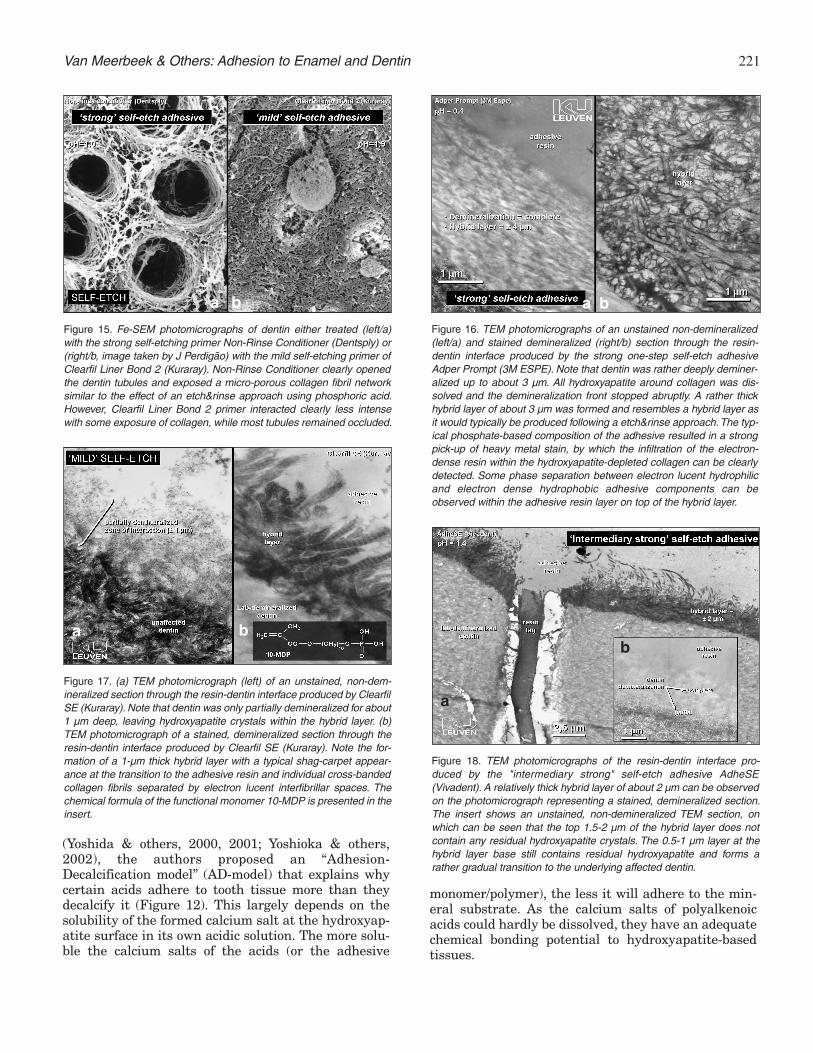
Figure 15. Fe-SEM photomicrographs of dentin either treated (left/a)with the strong self-etching primer Non-Rinse Conditioner (Dentsply) or(right/b, image taken by J Perdigão) with the mild self-etching primer ofClearfil Liner Bond 2 (Kuraray). Non-Rinse Conditioner clearly openedthe dentin tubules and exposed a micro-porous collagen fibril networksimilar to the effect of an etch&rinse approach using phosphoric acid.However, Clearfil Liner Bond 2 primer interacted clearly less intensewith some exposure of collagen, while most tubules remained occluded.
Figure 16. TEM photomicrographs of an unstained non-demineralized(left/a) and stained demineralized (right/b) section through the resin-dentin interface produced by the strong one-step self-etch adhesiveAdper Prompt (3M ESPE). Note that dentin was rather deeply deminer-alized up to about 3 µm. All hydroxyapatite around collagen was dis-solved and the demineralization front stopped abruptly. A rather thickhybrid layer of about 3 µm was formed and resembles a hybrid layer asit would typically be produced following a etch&rinse approach.The typ-ical phosphate-based composition of the adhesive resulted in a strongpick-up of heavy metal stain, by which the infiltration of the electron-dense resin within the hydroxyapatite-depleted collagen can be clearlydetected. Some phase separation between electron lucent hydrophilicand electron dense hydrophobic adhesive components can beobserved within the adhesive resin layer on top of the hybrid layer.
Figure 17. (a) TEM photomicrograph (left) of an unstained, non-dem-ineralized section through the resin-dentin interface produced by ClearfilSE (Kuraray). Note that dentin was only partially demineralized for about1 µm deep, leaving hydroxyapatite crystals within the hybrid layer. (b)TEM photomicrograph of a stained, demineralized section through theresin-dentin interface produced by Clearfil SE (Kuraray). Note the for-mation of a 1-µm thick hybrid layer with a typical shag-carpet appear-ance at the transition to the adhesive resin and individual cross-bandedcollagen fibrils separated by electron lucent interfibrillar spaces. Thechemical formula of the functional monomer 10-MDP is presented in theinsert.
Figure 18. TEM photomicrographs of the resin-dentin interface pro-duced by the "intermediary strong" self-etch adhesive AdheSE(Vivadent). A relatively thick hybrid layer of about 2 µm can be observedon the photomicrograph representing a stained, demineralized section.The insert shows an unstained, non-demineralized TEM section, onwhich can be seen that the top 1.5-2 µm of the hybrid layer does notcontain any residual hydroxyapatite crystals. The 0.5-1 µm layer at thehybrid layer base still contains residual hydroxyapatite and forms arather gradual transition to the underlying affected dentin.
a b
a
b
a b a b
222 Operative Dentistry
Typical of some glass ionomers is the morphologicmanifestation of a “gel-phase” at the interface, as wasshown correlatively by transmission electronmicroscopy (Figure 8) and atomic force microscopy(Van Meerbeek & others, 1998b, 2001b; Yoshida & oth-ers, 1999). Correlating TEM and XPS data elucidatedthat this gel phase represents the formation of a calci-um polycarboxylate salt resulting from either thepolyalkenoic acid conditioner or the glass ionomermaterial itself (Van Meerbeek & others, 2001b). Thisphase has been shown to be stable and strong, inter-mediary between the shallow 0.5-1 µm hybrid layerand the glass-ionomer matrix. In microtensile bondstrength testing, the interface typically fractured wellabove the gel phase within the matrix of the glass-ionomer material (Van Meerbeek & others, 2001b).AFM surface analysis confirmed that this gel phase isstronger than the actual glass-ionomer matrix (VanMeerbeek & others, 2001b). The actual function andcontribution of this phase to the bond integrity needsto be further elucidated.
Self-Etch Approach
Probably, in regard to user-friendliness and technique-sensitivity, clinically, the most promising approach isself-etch. It no longer needs an “etch&rinse” phase,which not only lessens clinical application time, butalso significantly reduces technique-sensitivity or therisk of making errors during application and manipu-lation. Another important advantage of the self-etchapproach is that infiltration of resin occurs simultane-ously with the self-etching process, by which the risk ofdiscrepancy between both processes is low or non-exis-
tent. However, little is known about the long-termeffects of incorporating dissolved hydroxyapatite crys-tals and residual smear layer remnants within thebond. How much of the primer/adhesive solvent is keptwithin the interfacial structure should also be investi-gated. Such solvent surplus will directly weaken thebond integrity, provide channels for nanoleakage ormay affect polymerization of the infiltrated monomers.The resultant interfacial structure also becomes morehydrophilic and, thus, more prone to hydrolytic degra-dation (Tay & others, 2002a; Tay, Pashley &Yoshiyama, 2002b).
A self-etch approach involves either a two- or one-step application procedure (Figure 1). The self-etcheffect should be ascribed to monomers to which one ormore carboxylic or phosphate acid groups are grafted(Van Meerbeek & others, 2001a). Depending on etchingaggressiveness, they can be subdivided into “strong”and “mild” self-etch adhesives (Figure 13).
“Strong” self-etch adhesives usually have a pH of 1 orbelow (Table 1). This high acidity results in rather deepdemineralization effects. At enamel, the resulting acid-etch pattern resembles a phosphoric-acid treatmentfollowing an etch&rinse approach (Figure 14a) (Inoue& others, 2000; Pashley & Tay, 2001). At dentin, colla-gen is exposed and nearly all hydroxyapatite is dis-solved (Figures 15a and 16). Consequently, the under-lying bonding mechanism of “strong” self-etch adhe-sives is primarily diffusion-based, similar to theetch&rinse approach. Such low-pH self-etch adhesiveshave often been documented with rather low bondstrength values, especially at dentin, and quite a high
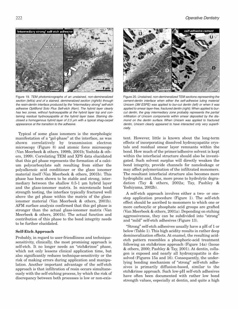
Figure 19. TEM photomicrographs of an unstained, non-demineralizedsection (left/a) and of a stained, demineralized section (right/b) throughthe resin-dentin interface produced by the “intermediary strong” self-etchadhesive OptiBond Solo Plus Self-etch (Kerr). The hybrid layer clearlyhas two zones, without hydroxyapatite at the hybrid layer top and con-taining residual hydrxoxyapatite at the hybrid layer base. Staining dis-closed a homogenous hybrid layer of 2.5 µm with a typical shag-carpetappearance at the transition to the adhesive.
Figure 20. Unstained, non-demineralized TEM sections representing thecement-dentin interface when either the self-adhesive luting materialUnicem (3M ESPE) was applied to bur-cut dentin (left) or when it wasapplied to smear layer-free, fractured dentin (right).When applied to bur-cut dentin, the gray intermediary zone probably represents the partialinfiltration of Unicem components within smear deposited by the dia-mond on the dentin surface. When Unicem was applied to fractureddentin, Unicem clearly appeared to have interacted only very superfi-cially.
a b a b
223Van Meerbeek & Others: Adhesion to Enamel and Dentin
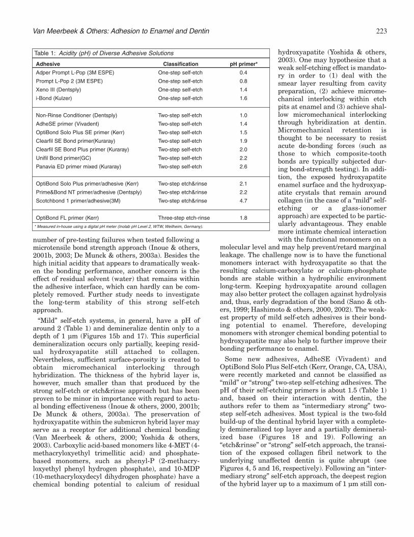
number of pre-testing failures when tested following amicrotensile bond strength approach (Inoue & others,2001b, 2003; De Munck & others, 2003a). Besides thehigh initial acidity that appears to dramatically weak-en the bonding performance, another concern is theeffect of residual solvent (water) that remains withinthe adhesive interface, which can hardly can be com-pletely removed. Further study needs to investigatethe long-term stability of this strong self-etchapproach.
“Mild” self-etch systems, in general, have a pH ofaround 2 (Table 1) and demineralize dentin only to adepth of 1 µm (Figures 15b and 17). This superficialdemineralization occurs only partially, keeping resid-ual hydroxyapatite still attached to collagen.Nevertheless, sufficient surface-porosity is created toobtain micromechanical interlocking throughhybridization. The thickness of the hybrid layer is,however, much smaller than that produced by thestrong self-etch or etch&rinse approach but has beenproven to be minor in importance with regard to actu-al bonding effectiveness (Inoue & others, 2000, 2001b;De Munck & others, 2003a). The preservation ofhydroxyapatite within the submicron hybrid layer mayserve as a receptor for additional chemical bonding(Van Meerbeek & others, 2000; Yoshida & others,2003). Carboxylic acid-based monomers like 4-MET (4-methacryloxyethyl trimellitic acid) and phosphate-based monomers, such as phenyl-P (2-methacry-loxyethyl phenyl hydrogen phosphate), and 10-MDP(10-methacryloxydecyl dihydrogen phosphate) have achemical bonding potential to calcium of residual
hydroxyapatite (Yoshida & others,2003). One may hypothesize that aweak self-etching effect is mandato-ry in order to (1) deal with thesmear layer resulting from cavitypreparation, (2) achieve microme-chanical interlocking within etchpits at enamel and (3) achieve shal-low micromechanical interlockingthrough hybridization at dentin.Micromechanical retention isthought to be necessary to resistacute de-bonding forces (such asthose to which composite-toothbonds are typically subjected dur-ing bond-strength testing). In addi-tion, the exposed hydroxyapatiteenamel surface and the hydroxyap-atite crystals that remain aroundcollagen (in the case of a “mild” self-etching or a glass-ionomerapproach) are expected to be partic-ularly advantageous. They enablemore intimate chemical interactionwith the functional monomers on a
molecular level and may help prevent/retard marginalleakage. The challenge now is to have the functionalmonomers interact with hydroxyapatite so that theresulting calcium-carboxylate or calcium-phosphatebonds are stable within a hydrophilic environmentlong-term. Keeping hydroxyapatite around collagenmay also better protect the collagen against hydrolysisand, thus, early degradation of the bond (Sano & oth-ers, 1999; Hashimoto & others, 2000, 2002). The weak-est property of mild self-etch adhesives is their bond-ing potential to enamel. Therefore, developingmonomers with stronger chemical bonding potential tohydroxyapatite may also help to further improve theirbonding performance to enamel.
Some new adhesives, AdheSE (Vivadent) andOptiBond Solo Plus Self-etch (Kerr, Orange, CA, USA),were recently marketed and cannot be classified as“mild” or “strong” two-step self-etching adhesives. ThepH of their self-etching primers is about 1.5 (Table 1)and, based on their interaction with dentin, theauthors refer to them as “intermediary strong” two-step self-etch adhesives. Most typical is the two-foldbuild-up of the dentinal hybrid layer with a complete-ly demineralized top layer and a partially demineral-ized base (Figures 18 and 19). Following an“etch&rinse” or “strong” self-etch approach, the transi-tion of the exposed collagen fibril network to theunderlying unaffected dentin is quite abrupt (seeFigures 4, 5 and 16, respectively). Following an “inter-mediary strong” self-etch approach, the deepest regionof the hybrid layer up to a maximum of 1 µm still con-
Adhesive Classification pH primer*
Adper Prompt L-Pop (3M ESPE) One-step self-etch 0.4
Prompt L-Pop 2 (3M ESPE) One-step self-etch 0.8
Xeno III (Dentsply) One-step self-etch 1.4
i-Bond (Kulzer) One-step self-etch 1.6
Non-Rinse Conditioner (Dentsply) Two-step self-etch 1.0
AdheSE primer (Vivadent) Two-step self-etch 1.4
OptiBond Solo Plus SE primer (Kerr) Two-step self-etch 1.5
Clearfil SE Bond primer(Kuraray) Two-step self-etch 1.9
Clearfil SE Bond Plus primer (Kuraray) Two-step self-etch 2.0
Unifil Bond primer(GC) Two-step self-etch 2.2
Panavia ED primer mixed (Kuraray) Two-step self-etch 2.6
OptiBond Solo Plus primer/adhesive (Kerr) Two-step etch&rinse 2.1
Prime&Bond NT primer/adhesive (Dentsply) Two-step etch&rinse 2.2
Scotchbond 1 primer/adhesive(3M) Two-step etch&rinse 4.7
OptiBond FL primer (Kerr) Three-step etch-rinse 1.8
Table 1: Acidity (pH) of Diverse Adhesive Solutions
* Measured in-house using a digital pH meter (Inolab pH Level 2, WTW, Weilheim, Germany).
224 Operative Dentistry
tains hydroxyapatite, by which the transition of thehybrid layer to the underlying unaffected dentin is moregradual (Figures 18 and 19). These adhesives are moreacidic than the “mild” self-etch adhesives, by which bet-ter micromechanical interlocking is achieved at enameland dentin. The residual hydroxyapatite at the hybridlayer base may still allow for chemical intermolecularinteraction, as was shown before for the “mild” self-etchadhesives. Based on the acidity (Table 1), the one-stepself-etch adhesives i-Bond (Kulzer) and Xeno III(Dentsply, Milford, DE, USA) must also categorized as“intermediary strong” self-etch adhesives. Their result-ant interfacial interaction is consequently expected to besimilar to that produced by the intermediary “strong”two-step self-etch adhesives documented above.
Unicem (3M ESPE, St Paul, MN, USA) was recentlylaunched as a possible first step towards self-adhesiveresin-based restorative materials. This luting materialis designed to be applied without any pre-treatment.TEM of the resultant interface showed a very superfi-cial interaction with dentin (Figure 20). When appliedto bur-cut dentin, a layer about 0.5-1 µm deepappeared less mineralized and most likely representedinfiltration of Unicem components with a partially dis-solved bur smear layer. This layer did not appear whenUnicem was applied to fractured dentin that was freeof cutting smear. Then, the interaction of Unicem withdentin could barely be morphologically detected. Theactual bonding mechanism of this self-adhesive cementshould be investigated in depth.
MEASURING BONDING EFFECTIVENESS:LABORATORY VERSUS CLINICAL TESTING
Laboratory Testing of Adhesives: Can TheyPredict Clinical Effectiveness?
Clinical trials are the ultimate test for dental restora-tions, but they cannot differentiate the true reason forfailure due to the simultaneous impact of diversestresses on restorations within the aggressive oral cav-ity. Lab testing can evaluate the effect of a single vari-able, while keeping all other variables constant. Basedon this type of research, clear recommendations can beformulated toward clinicians with regard to the appro-priate use and selection of dental materials. In gener-al, laboratory testing is easy, fast and relatively cheapto screen new materials/techniques. They are useful indetermining the “effectiveness” of adhesive materialswithin the specific test set-up. Ideally, the final objec-tive should always be predicting clinical behavior long-term, though direct translation to the clinical situationis often difficult or even impossible.
Bond Strength Testing
In the mouth, the interface between restoration andtooth is exposed to diverse forces that act simultane-
ously. Already during setting of composite, resinshrinkage puts stress on the bond, pulling it away fromthe cavity wall (Versluis, Tantbirojn & Douglas, 1998).During function, mechanical stress by chewing forces,thermal and chemical stress with changes in tempera-ture and pH will have an effect on the bond integrity aspart of bio-tribocorrosive effects. The rationale behindbond strength testing is that the higher the actualbonding capacity of an adhesive, the better it will with-stand such stresses and the longer the restoration willsurvive in vivo. Bond strength testing is relatively easyand fast and, in fact, besides a material tester does notrequire special equipment. It, therefore, remains themost popular methodology for measuring bondingeffectiveness in the laboratory. Van Noort & others(1989), however, emphasized that bond strength can-not be regarded as a material property. The dataobtained from bond strength tests largely depend onthe actual test set-ups that may differ between labora-tories for parameters such as specimen geometry, sizeof surface area, the type of composite and more. It is,therefore, not surprising that bond strength data sub-stantially vary among laboratories throughout theworld. The many variables involved make standardi-zation of test methodologies for bond-strength meas-urements hardly achievable.
Most commonly, bond strength is measured by sub-jecting composites bonded-to-enamel/dentin to tensileor shear stress. However, at bond strength values high-er than 20 MPa in a shear test, cohesive failures of thesubstrate will more likely occur (Schreiner & others,1998). Therefore, a new test needed to be developedthat differentiates between adhesives that producehigher bond strengths. A microtensile bond strength(µTBS) methodology was introduced by Sano and oth-ers in 1994(a). These authors showed that microtensilebond strength was inversely related to the bonded sur-face area (Sano & others, 1994a; Shono & others, 1999;Phrukkanon, Burow & Tyas, 1998a,b; Pashley & oth-ers, 1999) and that although much higher bondstrengths were measured, most failures still occurredat the interface between tooth substrate and adhesive.Other advantages of µTBS-testing are that regionalbond strengths and bonding effectiveness to clinicallyrelevant tooth substrates such as carious (Nakajima &others, 1995; Yoshiyama & others, 2000) and scleroticdentin (Tay & others, 2000; Kwong & others, 2002) canbe measured (Pashley & others, 1999). The major dis-advantage of µTBS-testing is the rather labor-inten-sive, technically demanding and relatively fragile sam-ple preparation technique. Special care should betaken to avoid/reduce the production of microfracturesat the interface during specimen preparation. Theymay weaken the bond and, thus, reduce the actualbond strength (Ferrari & Cardoso, 2002). Otherwise,one could argue that clinical restoration margins are
225Van Meerbeek & Others: Adhesion to Enamel and Dentin
subjected to similar stresses during finishing of com-posite restorations with diamonds. They also inducemicrofractures at the restoration-tooth transition. Inthis way, µTBS-sample preparation may actually bet-ter simulate clinical circumstances. Eventually, if allspecimens are prepared in the same manner, no addi-tional variable is introduced. In order to standardizesample preparation, at BIOMAT Leuven, the IowaMicroSpecimen Former (Armstrong, Keller & Boyer,2001; De Munck & others, 2003a,c) is used, which
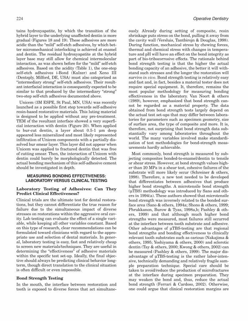
Figure 21. Chart presenting the micro-tensile bond strength (µTBS) toenamel of diverse commercial adhesives. The data were gathered fromdiverse laboratory studies carried out at BIOMAT Leuven strictly follow-ing the same experimental protocol. The color code refers to the differ-ent kinds of adhesives following the classification presented in Figure 1.For the two-step self-etch adhesives, the light-green colored bars repre-sent the data produced by “mild” self-etch adhesives, the intermediary-green colored bars those produced by “intermediary strong” self-etchadhesives, and the dark-green bars represent the µTBS-data producedby the “strong” self-etch adhesives. For the one-step self-etch adhesives,the light-yellow colored bars represent the data produced by “mild” self-etch adhesives, the intermediary-yellow colored bars those produced by“intermediary strong” self-etch adhesives, and the dark-yellow bars rep-resent the µTBS-data produced by the “strong” self-etch adhesives. Alldata are pooled per group of adhesives underneath the chart.
Figure 22. Statistical analysis of the pooled enamel µTBS-data demon-strate that three-step etch&rinse adhesives bond equally well to enam-el as two-step etch&rinse adhesives. Etch&rinse adhesives bond slight-ly, but statistically significantly better to enamel than two-step self-etchadhesives, that on their turn bond significantly much better than one-step self-etch adhesives. The actual p-values are mentioned in the tableinsert. All red-colored figures indicate statistical significant difference;black-colored figures indicate absence of statistical difference.
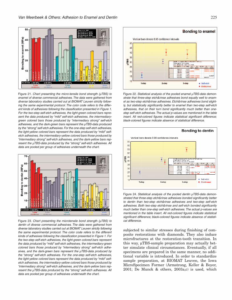
Figure 23. Chart presenting the microtensile bond strength (µTBS) todentin of diverse commercial adhesives. The data were gathered fromdiverse laboratory studies carried out at BIOMAT Leuven strictly followingthe same experimental protocol. The color code refers to the differentkinds of adhesives following the classification presented in Figure 1. Forthe two-step self-etch adhesives, the light-green colored bars representthe data produced by “mild” self-etch adhesives, the intermediary-greencolored bars those produced by “intermediary strong” self-etch adhe-sives, and the dark-green bars represent the µTBS-data produced bythe “strong” self-etch adhesives. For the one-step self-etch adhesives,the light-yellow colored bars represent the data produced by “mild” self-etch adhesives, the intermediary-yellow colored bars those produced by“intermediary strong” self-etch adhesives, and the dark-yellow bars rep-resent the µTBS-data produced by the “strong” self-etch adhesives. Alldata are pooled per group of adhesives underneath the chart.
Figure 24. Statistical analysis of the pooled dentin µTBS-data demon-strated that three-step etch&rinse adhesives bonded significantly betterto dentin than two-step etch&rinse adhesives and two-step self-etchadhesives. Both two-step etch&rinse and self-etch bonded significantlymuch better than one-step self-etch adhesives. The actual p-values arementioned in the table insert. All red-colored figures indicate statisticalsignificant difference; black-colored figures indicate absence of statisti-cal difference.
226 Operative Dentistry
enables the production of stick-type specimens that arecylindrically constricted at the interface to ensure thatthe maximum tensile stress is concentrated at theactual interface. Hence, this µTBS-testing protocolmust become the new standard for measure bondingeffectiveness in the laboratory.
A modification of this test is the “micro-shear” test,which makes it more difficult to standardize the loca-tion of the force (Shimada & others, 2000). Nevertheless,
the results obtained did notdiffer substantially from thosegathered following a µTBS-protocol (Phrukkanon & oth-ers, 1998a,b).
Another less common tech-nique is the push-out test(Frankenberger, Krämer &Petschelt, 1999, 2000b; Franken-berger & others, 2000a). Asmall resin composite cylinderin the middle of a dentin disc ispushed out, resulting in ashear stress at the interface.The main advantage of thistechnique is that failure isforced to occur along the adhe-sive interface (Drummond &others, 1996). However, thistest is more time-consumingand cannot be applied for eval-uating enamel bond strength.Also, push-out data are verycomparable to traditionalshear-bond strength testing(Drummond & others, 1996).
µTBS to Enamel—At Leuven, the µTBS of a largegroup of commercial and experimental adhesives tobur-cut enamel and dentin has been determined (Inoue& others, 2001a,b, 2003; De Munck & others, 2003a,c),always following the same experimental protocol,using one particular composite material (Z100, 3MESPE). When bonding to enamel, an etch&rinseapproach still results in the highest bonding effective-
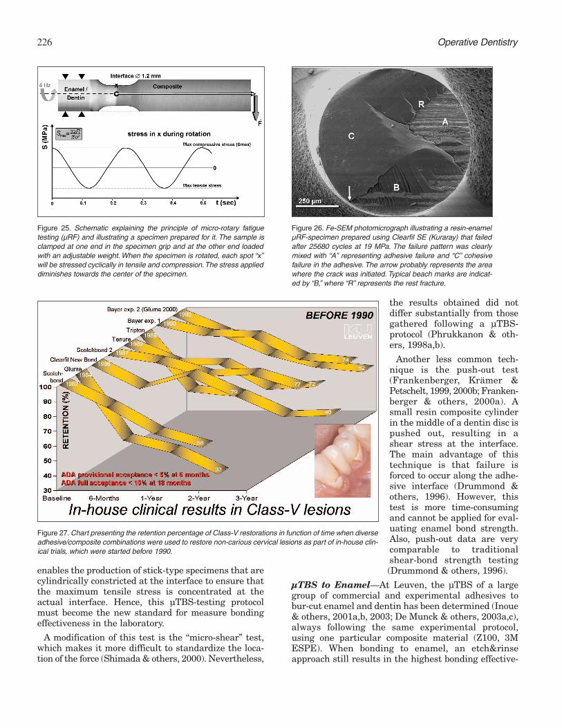
Figure 25. Schematic explaining the principle of micro-rotary fatiguetesting (µRF) and illustrating a specimen prepared for it. The sample isclamped at one end in the specimen grip and at the other end loadedwith an adjustable weight. When the specimen is rotated, each spot “x”will be stressed cyclically in tensile and compression.The stress applieddiminishes towards the center of the specimen.
Figure 26. Fe-SEM photomicrograph illustrating a resin-enamelµRF-specimen prepared using Clearfil SE (Kuraray) that failedafter 25680 cycles at 19 MPa. The failure pattern was clearlymixed with “A” representing adhesive failure and “C” cohesivefailure in the adhesive. The arrow probably represents the areawhere the crack was initiated. Typical beach marks are indicat-ed by “B,” where “R” represents the rest fracture.
Figure 27. Chart presenting the retention percentage of Class-V restorations in function of time when diverseadhesive/composite combinations were used to restore non-carious cervical lesions as part of in-house clin-ical trials, which were started before 1990.
ness irrespective of a two- or three-step procedure andthe product tested (Figure 21). When pooling theµTBSs of all etch&rinse adhesives tested (or of whichthe µTBS was repeatedly measured as part of differentstudies), a µTBS of 39 and 40 MPa was achieved,respectively, for the three-step and two-stepetch&rinse adhesives. As has been known fromBuonocore (1955), bonding to enamel only requires anacid-etch step followed by the application of a fluidresin without the need for an intermediary primerstep. The latter, on the other hand, does not negativelyinfluence bonding effectiveness and is even mandatorywhen a “wet-bonding” procedure is carried out.
A self-etch procedure, in general, has resulted in alower bonding effectiveness, though some adhesivesapproached the bonding effectiveness of etch&rinseadhesives (Figure 21). A pooled µTBS of about 30 MPawas obtained for two-step self-etch adhesives. TheµTBS of the “strong” two-step self-etch adhesiveNRC/Prime&Bond NT (Dentsply) was not significantlylower than that of the etch&rinse adhesives. AlthoughClearfil SE (Kuraray) and the experimental successorClearfil SE Plus (Kuraray; containing the anti-micro-bial monomer methacryloyloxydodecylpyridinium bro-mide or MDPB; Imazato & others, 1997) belong to thegroup of “mild” two-step self-etch adhesives, theirµTBS is not that much lower than etch&rinse adhe-sives. This may indicate that although they only inter-act superficially with enamel and, thus, their potentialfor micromechanical interlocking is much less than a
phosphoric-acid treatment, theadditional chemical bondingcapacity to hydroxyapatitemay have contributed to theactual favorable bonding effec-tiveness. Likewise, the bond-ing effectiveness of the “inter-mediary strong” two-step self-etch adhesive Optibond SoloPlus Self-etch (Kerr, Orange,CA, USA) approached that ofetch&rinse adhesives.
One-step self-etch adhesivesproduced significantly lowerµTBSs than etch&rinse andtwo-step self-etch adhesives(Figure 21). The pooled µTBSwas about 16 MPa for the one-step self-etch adhesives. The“strong” one-step adhesives,Prompt L-Pop (3M ESPE), andits successor Adper Prompt(3M ESPE), presented withµTBSs in the same range asthat recorded for the two-stepself-etch adhesives. This mostlikely can be attributed to their
higher acidity and, consequently, higher potential toachieve micromechanical interlocking at enamel.
The glass-ionomer adhesive Fuji Bond LC (GC) per-formed equally well as the two-step self-etch adhesives(Figure 21). However, during bond-strength testing,the glass-ionomer adhesive tended to fail more fre-quently in the glass-ionomer material itself than at theactual interface, where their actual bonding effective-ness to enamel was never measured and should atleast be higher than the cohesive strength of the glass-ionomer adhesive (Inoue & others, 2000, 2001a).
Statistical analysis of the pooled enamel µTBS data(Figure 22) showed that etch&rinse adhesives, irre-spective of a two- or three-step application procedure,bonded slightly but significantly stronger to enamelthan two-step self-etch adhesives and significantlymore strong than one-step self-etch adhesives.
µTBS to Dentin—At dentin, three-step etch&rinseadhesives still surpassed all other adhesives that usesimplified application procedures (Figure 23). No sig-nificant difference could be recorded between the bond-ing effectiveness to dentin of two-step etch&rinse andtwo-step self-etch adhesives. Again, the “mild” two-step self-etch adhesive Clearfil SE (Kuraray) and the“intermediary strong” two-step self-etch adhesiveOptibond Solo Plus Self-etch (Kerr) most closelyapproached the bonding effectiveness of the conventionalthree-step adhesives. The lowest µTBS was again
227Van Meerbeek & Others: Adhesion to Enamel and Dentin
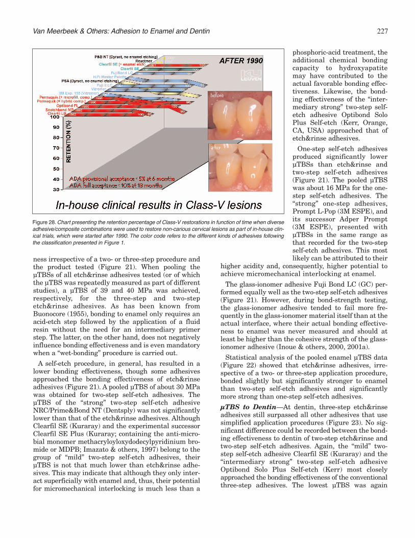
Figure 28. Chart presenting the retention percentage of Class-V restorations in function of time when diverseadhesive/composite combinations were used to restore non-carious cervical lesions as part of in-house clin-ical trials, which were started after 1990. The color code refers to the different kinds of adhesives followingthe classification presented in Figure 1.
228 Operative Dentistry
recorded for the one-step self-etch adhesives that per-formed similarly to the glass-ionomer adhesive FujiBond LC (GC).
Statistical analysis of the pooled dentin µTBS-data(Figure 24) showed that three-step etch&rinse adhe-sives bonded significantly more strongly to dentin thantwo-step etch&rinse and two-step self-etch adhesives.Both latter systems did not perform significantlydifferent from each other. Again, the significantly leastfavorable µTBS-results were recorded for one-step self-etch adhesives.
µTBS to Hydroxyapatite—As mentioned above, theactual bonding effectiveness of glass ionomers and“mild” self-etch adhesives may result from combinedmicromechanical and chemical interaction with thetooth substrate. It is, however, currently not known howmuch chemical interaction contributes to the actualbonding effectiveness. Therefore, the authors deter-mined the µTBS of a various group of adhesive materialsto synthetic hydroxyapatite that, besides not havingorganic collagen, was highly polished and, thus, devoidof mechanical interlocking sites (Van Meerbeek & oth-ers, 2003). Among the adhesives tested, all specimensprepared with three-step etch&rinse adhesive OptibondFL (Kerr) failed prior to µTBS-testing (pre-testing fail-ures), proving that any micromechanical retention wasexcluded. The two-step self-etch adhesive Clearfil SE(Kuraray) presented with a rather low µTBS, along witha high number of pre-testing failures, indicating thatthere is some chemical interaction that, however slightly(for about 7% as compared to its µTBS to dentin), con-tributed to the actual bond strength achieved at dentin.Clearly, much less pre-testing failure and a significantlyhigher µTBS were recorded for the resin-modified glass-ionomer adhesive Fuji Bond LC (GC) and the conven-tional glass-ionomer restorative material Fuji IX (GC).For Fuji Bond LC, the chemical interaction accountedfor about 40% of the actual bond strength achieved atdentin. Chemical bonding of glass ionomer to hydroxya-patite depended greatly on the use of a separatepolyalkenoic acid conditioner (Cavity Conditioner (GC);see also Figure 8). Without this pre-treatment, all spec-imens failed prior to testing. Equally effective as glass-ionomer materials with regard to chemical bondingpotential, the resin-based luting material Panavia F(Kuraray) presented with a µTBS that accounted forabout 67% of its actual bond strength to dentin. No pre-testing failures were recorded for Panavia F, indicatingits relatively strong chemical bonding potential.Although Panavia F was applied following a self-etchapproach using a 10-MDP-based primer solution, as inClearfil SE, its chemical bonding effectiveness is muchhigher than the two-step self-etch adhesive Clearfil SE.Further in-depth analysis of the actual differences incomposition and application procedures should helpexplain this difference in chemical bonding potential.
Finally, the self-adhesive luting material Unicem (3MESPE) presented with a relatively negligible chemicalbonding potential in the same range recorded for thetwo-step self-etch adhesive (Clearfil SE). Note that theratio of chemical bonding to eventual “total” bondingeffectiveness must be regarded as arbitrary, since differ-ences in substrate properties such as roughness, stiff-ness and so on between the hydroxyapatite and dentinspecimens were ignored.
Effect of Aging—Most current adhesives perform wellin bond-strength tests, at least when tested shortly afterapplication and under controlled in vitro conditions(Inoue & others, 2001b, 2003; De Munck & others,2003a,c). However, the oral cavity with temperaturechanges, chewing loads and chemical attacks by acidsand enzymes forms a severe challenge for tooth-compositebonds to survive for a long time. Clinically, marginaldeterioration of composite restorations remains prob-lematic and forms the major reason that dramaticallyshortens the lifetime of adhesive restorations (VanMeerbeek & others, 1998a). A factor known to degradetooth-composite bonds is exposure to water (Gwinnett &Yu, 1995; Sano & others, 1999, Armstrong & others, 2001).Among different forms of marginal leakage, nano-leakage, or the ingress of oral fluids through nanometer-sized channels along collagen fibrils within the hybridlayer, is considered detrimental to the bond integrity(Sano & others, 1995; Hashimoto & others, 2000, 2002).
In a recent paper (De Munck & others, 2003c), theauthors studied the long-term degradation of resin-dentin bonds using a µTBS-testing methodologythrough exposure to water for four years, either directlyor indirectly, when the resin-dentin interface was sur-rounded by resin bonded to enamel. The microtensilebond strength (µTBS) to dentin of two three-stepetch&rinse adhesives (Optibond Dual-Cure, Kerr;Scotchbond Multi-Purpose, 3M ESPE) was compared totwo two-step etch&rinse adhesives (Optibond Solo,Kerr; Scotchbond 1, 3M ESPE) after four years of storagein water. Direct exposure to water resulted in a signifi-cant decrease in the µTBS of the two-step but not of thethree-step etch&rinse adhesives. Indirect exposure towater did not significantly reduce the µTBS of any adhe-sive, indicating that resin bonded to enamel protectedthe resin-dentin bond against degradation. This meansthat, in the clinical situation, one can rely on durabledentin bonding using three- or two-step etch&rinseadhesives if all cavity margins are located in enamel.For cavities with margins ending in dentin, three-steptotal-etch adhesives are preferred.
Marginal Sealing Effectiveness
Clinically, early loss of restoration is no longer a clini-cal problem when reliable (mostly conventional three-step etch&rinse) adhesives are used, even long-term(Van Meerbeek & others, 1994, 1998a; van Dijken,

229Van Meerbeek & Others: Adhesion to Enamel and Dentin
2000a,b, 2001, 2002; De Munck & others, 2003b).However, marginal leakage and consequent marginaldiscoloration remains clinically the most frequent rea-son to replace/repair an adhesive restoration.Therefore, besides bond-strength testing, testing themarginal sealing effectiveness of adhesives is needed.
Marginal leakage has been defined as the clinicallyundetectable passage of bacteria, fluids, molecules orions between a cavity wall and the restorative mate-rial applied to it (Kidd, 1976). All resin-based restora-tive materials shrink, which introduces shrinkagestress, pulling the adhesive away from the cavity wall,which may eventually form a gap. Today’s adhesivesare incapable of completely sealing restoration mar-gins and, thus, preventing microleakage long-term(Pilo & Ben-Amar, 1999; Bouillaguet & others, 2000;Ceballos & others, 2001). Many methodologies havebeen introduced to assess microleakage and can be fur-ther subdivided in qualitative, semi-quantitative ortrue quantitative measurements of sealing effective-ness.
Qualitative Measurement of SealingEffectiveness—The use of organic dyes as tracers isone of the oldest, most common methods of detectingleakage in vitro. A number of dyes varying in particlesize and affinity to substrates have been used and areknown to significantly influence microleakage results(Alani & Toh, 1997). In general, this method involvesimmersion of a restored tooth into a dye solution afterhaving coated the unrestored tooth parts covered witha waterproof varnish until close to the restorationmargin. After a certain time interval, the specimensare washed and sectioned into two or more slices tovisually determine the extent of dye penetration alongthe restoration margin (Alani & Toh, 1997). The mainproblem is that this methodology basically is a quali-tative evaluation method. It can be made semi-quanti-tative by applying a non-parametric scale(Castelnuovo, Tjan & Liu, 1996).
Semi-Quantitative Measurement of SealingEffectiveness or Marginal Analysis—A number ofin vitro studies have tested the performance of adhe-sives by semi-quantitatively evaluating by using SEMthe marginal gap formation around restorationsplaced in extracted teeth (Roulet & others, 1989;Krejci, Kuster & Lutz, 1993; Roulet, 1994; Gladys &others, 1995; Blunck, Neumann & Roulet, 2000;Blunck & Roulet, 1999, 2002). This method assumesthat if the forces generated during shrinkage or ther-mo-mechanical strains exceed the bond strength toenamel/dentin, an observable gap will form at themargin of the restoration. Although the literature alsolacks clear evidence of any correlation of gap forma-tion in vitro with the interfacial failures observed invivo, it is reasonable to assume that this semi-quanti-
tative mar-ginal gap analysis is clinically relevant(Roulet, 1994), certainly, when measurements arerepeated after thermocycling (Krejci & others, 1993,Schuckar & Geurtsen, 1997).
Blunck and Roulet (2002) have semi-quantitativelyanalyzed the marginal adaptation of cervical restora-tions for a diverse group of adhesives, consistently fol-lowing the same experimental protocol. Basically,their results correlated well with the µTBS-datarecorded by the authors of this study at BIOMATLeuven. After one-year water storage and two ther-mocycling sessions, still on average, 93% of therestoration margin length was gap-free for the three-step etch&rinse adhesive Optibond FL (Kerr) and 91%for the “mild” two-step self-etch adhesive Clearfil SE(Kuraray) (Blunck & Roulet, 2002). Two-stepetch&rinse adhesives such as Excite (Vivadent),Optibond Solo Plus (Kerr) and Scotchbond 1 (3MESPE) revealed significantly lower percentages ofgap-free margin lengths of 80%, 82% and 63%, respec-tively. Less than half (48%) of the margin length wasgap-free for the “strong” one-step self-etch adhesivePrompt-L Pop (3M ESPE).
Quantitative Measurement of SealingEffectiveness or Flow Measurement—A quantita-tive method to assess microleakage is to measure theflow along the interface (Pagliarini & others, 1996) orfrom the pulp to a sealed dentin surface (Derkson,Pashley & Derkson, 1986; Del-Nero, Escribano & de laMacorra, 2000; Bouillaguet & others, 2000). The mar-ginal sealing effectiveness is quantified using a “Flodecdevice” (De Marco Engineering, Geneva, Switzerland).The adhesively-restored tooth is brought under pres-sure with water from inside the dental pulp. The per-meability of the tooth-restoration interface is thenquantitatively determined through accurate measure-ment of the displacement of an air bubble within awater-filled micro-pipet (∅=0.7 mm) using a computer-driven optical system (Flodec device). The main advan-tages of this method are that it is fully quantitative andthat the specimens can be longitudinally followed sinceit is a non-destructive method. However, one majorproblem using this technique is that leakage may alsooccur through the dental substrate itself and, thus,falsely increase the leakage values.
Nanoleakage—Sano and others (1994b, 1995)revealed that leakage can occur between the hybridlayer and intact dentin, even in the absence of a mar-ginal gap. This leakage was assessed using Ag-ions thatare extremely small (0.059 nm). It is hypothesized thatit represents permeation through demineralized sub-micron spaces that have not been filled with adhesiveresin (Sano & others, 1995). These voids are so smallthat bacteria may not be able to pass through, butthese spaces may be more susceptible to degrada-
230 Operative Dentistry
tion by water and bacterial side products such as acidsand enzymes (Paul & others, 1999). This phenomenoncan be quantitatively assessed by measuring the dyepenetration depth using, for instance, confocal laserscanning microscopy (Dörfer & others, 2000; Pioch &others, 2001, 2002) or TEM (Tay & others, 2002b).
Dynamic Fatigue Testing
Bonding effectiveness to tooth tissue is typically meas-ured statically, for example, by shear bond or microten-sile bond strength (µTBS) testing (see above). In theclinical situation, however, tooth-composite bonds areseldom imposed to such acute tensile/shear stresses.During its lifetime, a restoration is subjected to cyclicloading, each load is insufficient to provoke failure, butin the long-term, can possibly lead to marginal deteri-oration and loss of the restoration. Therefore, fatiguetesting of dental adhesives is expected to better predicttheir in vivo performance.
There is, however, no standard fatigue test for dentaladhesives. Possible methods are a cyclic shear test(Ruse, Shew & Feduik, 1995; Drummond & others,1996; Dewji & others, 1998), a cyclic tensile test(Aquilino, Diaz-Arnold & Piotrowski, 1991; Givan &others, 1995), a cyclic fracture toughness test (Destoop,2002) or a cyclic push-out test (Frankenberger & oth-ers, 1999). Another possibility is loading not only theinterface but the whole tooth until the tooth-restora-tion complex fails (Fissore, Nicholls & Yuodelis, 1991).
At BIOMAT Leuven, the authors have developed amicro-rotary fatigue device that dynamically teststooth-composite interfaces (De Munck & others, 2002).A macro-version was used prior to determine thefatigue resistance of soldered joints (Wiskott, Nicholls& Belser, 1994). In our test set-up, standard microten-sile bond strength (µTBS) bar-type samples preparedwith a rounded, constricted interface (Figure 25 and26) were clamped in a pin-chuck and connected to astepping motor with the free end loaded with a certainweight. By rotating the specimen, each spot at theouter surface of the interface underwent successivelycompressive and tensile loading following a sinusoidalfunction (Wiskott & others, 1994). Depending on thesurvival/failure of each sample after 105 cycles, the loadimposed to the next sample was increased/decreasedwith ± 5%. The results of the fatigue test were ana-lyzed using a logistic regression to determine the loadat which 50% of the samples failed and was called themedian micro-Rotary Fatigue Resistance (µRFR). In apilot study, the µRFR of the three-step etch&rinseadhesive Optibond FL (Kerr) and the two-step self-etchadhesive Clearfil SE (Kuraray) to enamel and dentinwas determined. The ranking of median µRFRs was inaccordance with the ranking of the respective µTBSsobtained for the two adhesives bonded to enamel anddentin. They were about three-fourths of the respective
µTBSs, except for Optibond FL bonded to dentin,which appeared to lose more of its static bond strengthwhen tested dynamically. From this preliminary study,it could be concluded that fatiguing of tooth-compositeinterfaces is feasible, with consistent results provided.Because of the cyclic loading and high number of cycles(105), the resulting data might also be more clinicallyrelevant, especially for assessing long-term bondingeffectiveness, which is still a major shortcoming of con-temporary adhesives.
Clinical Testing of Adhesives
New adhesives are continually being introduced to thedental profession, unfortunately, often without suffi-cient clinical validation (Van Meerbeek & others,1998a, 2001a). In the mouth, multiple and mutually-interactive clinical variables related to the quality oftooth substrate and its immediate oral environment co-determine the eventual effectiveness of adhesives (VanMeerbeek & others, 1994). Adhesives have mainlybeen clinically tested in non-prepared cervical abra-sions and erosions. Such “model” lesions are ideal testcavities, because they are located mainly in dentin andare widely available. They present no macro-mechani-cal undercuts, and they are usually found in anteriorteeth or premolars with good access and in patientswho have better than average oral hygiene. However,such clinical trials are limited in number and requireseveral years with regular recalls in order to achievesufficient clinical validation. Nevertheless, the moreexpensive and long-lasting clinical trials remain neces-sary to validate laboratory observations. Laboratorytesting on near ideal substrates and under optimal invitro conditions is valuable as a pre-clinical screeningtest of adhesive materials, at best, only a good predic-tion of clinical performance. Most Class-V clinical tri-als run for three years, although longer follow-up timesmay be desirable. However, after three years, mostadhesives are outdated and are replaced by a successorthat claims to be better.
At Leuven, the clinical effectiveness of adhesives hasbeen routinely investigated in controlled follow-upstudies using the same experimental protocol foralmost 20 years. The clinical effectiveness of modernadhesives has significantly improved, allowing adhe-sive restorations to be placed with a high predictablelevel of clinical success. Most modern adhesive systemsare superior to their predecessors, especially in termsof retention, making it no longer the main cause of pre-mature clinical failure. This must, in part, be attrib-uted to the introduction in the early 1990s of the “total-etch” (now referred to as “etch&rinse”) technique, bywhich phosphoric acid is also applied to dentin. Earlieradhesives often showed many failures within the firstsix months when applied strictly to dentin without anyselective phosphoric acid-etching of adjacent enamel(Figure 27). When following the same protocol in more
231Van Meerbeek & Others: Adhesion to Enamel and Dentin
recent clinical trials (etch&rinse systems applied selec-tively to dentin), almost any early debonding failureswere recorded (Figure 28). This must, to a great extent,be attributed to the enamel immediately adjacent todentin always being (unintentionally) etched and theguarantee of a durable bond to the enamel margin.Adequate bonding to enamel, alone, may also keepsuch restorations in place. Nevertheless, bonding todentin has improved substantially. However, in orderto be considered clinically effective, adhesive systemsshould not only keep the restoration in place for a sig-nificant period of time, but also, and what clinicallymay even be more important, completely seal therestoration margins against the ingress of oral fluidsand microorganisms. However, none of today’s systemsyet appears able to guarantee leakage-free margins fora significant amount of time, especially at the dentinsite (Van Meerbeek & others, 1998a; De Munck & oth-ers, 2003b).
At Leuven, the excellent clinical performance of thethree-step etch&rinse adhesive Optibond FL (Kerr),with a 100% retention rate at five years, is noteworthy(De Munck & others, 2003b). Likewise, 96% of therestorations were still in place at five years when thethree-step etch&rinse adhesive Permaquick(Ultradent, South Jordan, UT, USA) was used (DeMunck & others, 2003b). Besides the favorable clinicalperformance of etch&rinse adhesives, glass ionomerscommonly present with high retention results, evenup to three years of clinical service (Figure 28). In arecent double-blind, split-mouth, randomized con-trolled clinical trial, the clinical effectiveness of the“mild” two-step self-etch adhesive Clearfil SE(Kuraray) was evaluated following two experimentalprotocols (Peumans & others, 2003). Clearfil SE wasapplied either following the manufacturer’s instruc-tions or including prior selective acid etching of theenamel cavity margins with 40% phosphoric acid. Attwo years, no restoration losses were recorded foreither experimental group (Figure 28). Besides a high-er tendency toward small (but of clinically negligiblerelevance) marginal defects at the enamel side (whenenamel was not etched beforehand with phosphoricacid), the “mild” self-etch approach of Clearfil SE stillappears to be a clinically reliable, predictable and sim-plified adhesive technique.
In general, two-step etch&rinse adhesives performclinically less favorably than conventional three-stepadhesives (Sunnegardh & van Dijken, 2000; vanDijken, 2000a). For instance, still favorable seven-yearretention rates of 84% and 79%, respectively, wererecorded for the three-step etch&rinse adhesivesClearfil Liner Bond (Kuraray) and Optibond Dual-Cure(Kerr) (van Dijken, 2001). Two-step etch&rinse adhe-sives generally perform clinically less favorably inClass-V lesions. The results reported for this group
vary more among the different research centers, whichis probably indicative of their higher technique sensi-tivity. For instance, only 45% of the acetone-basedadhesive One-Step (BISCO, Schaumberg, IL, USA)were retained at five years (van Dijken, 2001), and only52% of Gluma 2000 (Kulzer) at five years (van Dijken,2000a). Also, 25% of the Scotchbond 1 restorations werealready lost at the three-year recall in a study by vanDijken (2001), while only 3% were lost at three years ina study by Ripps, Burgess and Rappold (2000). A lossrate of only 7% was recorded for Optibond Solo at threeyears (Swift & others, 2001). At three years, excellentand reasonably good clinical effectiveness was reportedfor Prime&Bond 2.1 (Dentsply), with a retention rate of100% at three years by Martin, Jedynakiewicz andFletcher (2002), and 89% at three years by Swift andothers (2001).
Regarding the clinical effectiveness of two-step self-etch adhesives, less data is available in the literature.Latta and others (2000) reported a still favorable 92%retention rate at three years for Clearfil Liner Bond 2and van Dijken (2002) reported a 91% retention rate attwo years.
Finally, regarding one-step self-etch adhesives,strongly varying results were recorded for PSA(applied along with Dyract, Dentsply). Only a 5% lossrate at five years was reported by Folwaczny and oth-ers (2001), whereas, even 41% of the restorationsplaced using PSA (Dentsply) de-bonded within a four-year observation period, as reported by Unlu, Belli andOzer (2001). A rather favorable retention rate of 84%at five years was reported by van Dijken (2000a).Several studies reported on the clinical performance ofPrompt L-Pop (3M ESPE). Rather favorable short-term retention rates of 100% at six months and 96% atone year, respectively, were recorded by Munoz andothers (2001) and by Boghosian (2002). However, rela-tively high loss rates of 21% at two years and 35% atone year were reported, respectively, by van Dijken(2002) and Brackett, Covey and St Germain (2002).
CONCLUSIONS
A great diversity in laboratory testing of adhesivesexists. Modern determination of bonding effectivenessin the laboratory should involve (1) microtensile bondstrength testing, (2) sealing effectiveness testing usingsemi-quantitative marginal analysis or fully quantita-tive margin permeability measurement and possibly (3)dynamic fatigue testing. There is a lack of standardiza-tion of testing methodologies. Nevertheless, good corre-lation exists between laboratory and clinical effective-ness, by which it can be concluded that laboratory test-ing CAN predict clinical effectiveness.
Diverse types of adhesives exist which can be classi-fied following their bonding mechanism and clinical
232 Operative Dentistry
application approach into etch&rinse, glass-ionomerand self-etch adhesives. Although there is a tendencytoward adhesives with simplified application proce-dures, simplification does not guarantee equal orimproved bonding effectiveness. Three-step etch&rinseadhesives still perform best in laboratory and clinicalresearch. Because of an additional chemical bondingpotential to hydroxyapatite, the mild self-etch approachmay be most promising in terms of durable bonding todental hard tissue using a simple, low, technique-sensi-tive application technique.
Acknowledgements
The Buonocore Memorial lecture is supported by a grant fromCaulk Dental Manufacturing Co to the Academy of OperativeDentistry.
(Presented 27 February 2003)
References
Alani AH & Toh CG (1997) Detection of microleakage around den-tal restorations: A review Operative Dentistry 22(4) 173-185.
Armstrong SR, Keller JC & Boyer DB (2001) The influence ofwater storage and C-factor on the dentin-resin compositemicrotensile bond strength and debond pathway utilizing afilled and unfilled adhesive resin Dental Materials 17(3) 268-276.
Aquilino SA, Diaz-Arnold AM & Piotrowski TJ (1991) Tensilefatigue limits of prosthodontic adhesives Journal of DentalResearch 70(3) 208-210.
Blunck U, Neumann O & Roulet J-F (2000) Marginal adaptationof four one-bottle-adhesives depending on the application tech-nique Journal of Dental Research 79(Special Issue) Abstract#41 p 149.
Blunck U & Roulet JF (1999) Marginal adaptation of compomerClass V restorations in vitro Journal of Adhesive Dentistry 1(2)143-151.
Blunck U & Roulet JF (2002) Effect of one-year water storage onthe effectiveness of dentin adhesives in Class V composite resinrestorations Journal of Dental Research 81(Special Issue A)Abstract #0946 p A-139.
Boghosian A (2002) Clinical evaluation of a self-etching adhesive:1 year results Journal of Dental Research 81(Special Issue A)Abstract #0192 p A-52.
Bouillaguet S, Duroux B, Ciucchi B & Sano H (2000) Ability ofadhesive systems to seal dentin surfaces: An in vitro studyJournal of Adhesive Dentistry 2(3) 201-208.
Bouschlicher MR, Reinhardt JW & Vargas MA (1997) Surfacetreatment techniques for resin composite repair AmericanJournal of Dentistry 10(6) 279-283.
Brackett WW, Covey DA & St Germain HA Jr (2002) One-yearclinical performance of a self-etching adhesive in Class V resincomposites cured by two methods Operative Dentistry 27(3) 218-222.
Buonocore DH (1955) A simple method of increasing the adhesionof acryl filling materials to enamel surfaces Journal of DentalResearch 34 849-853.
Castelnuovo J, Tjan AHL & Liu P (1996) Microleakage of multi-step and simplified-step bonding systems American Journal ofDentistry 9(6) 245-248.
Ceballos L, Osorio R, Toledano M & Marshall GW (2001)Microleakage of composite restorations after acid or Er:YAGlaser cavity treatments Dental Materials 17(4) 340-346.
Degrange H & Roulet JF (1997) Minimally invasive dentistry withbonding Chicago Quintessence Publishing.
Del-Nero MO, Escribano N & de la Macorra JC (2000) Analysis ofsealing vs tensile bond strength of eight adhesive restorativematerial systems Journal of Adhesive Dentistry 2(2) 117-127.
De Munck J, Van Meerbeek B, Inoue S, Vargas M, Yoshida Y,Armstrong S, Lambrechts P & Vanherle G (2003a) Micro-tensilebond strength of one- and two-step self-etch adhesives to bur-cutenamel and dentin American Journal of Dentistry (in press).
De Munck J, Van Meerbeek B, Peumans M & Lambrechts P(2003b) Five-year clinical effectiveness of two three-step total-etch adhesives and two composites in cervical lesions Journal ofDental Research 82(Special Issue) Abstract 0907 (in press).
De Munck J, Van Meerbeek B, Yoshida Y, Inoue S, Vargas M,Suzuki K, Lambrechts P & Vanherle G (2003c) Four-year waterdegradation of total-etch adhesives bonded to dentin Journal ofDental Research 82(2) 136-140.
De Munck J, Van Meerbeek B, Lambrechts P & Braem M (2002)Micro-rotary fatigue of tooth-biomaterial interfaces inProgramme and abstracts 2002, European Festival of OralScience, Cardiff, Wales, September 25-28, 2002 Abstract #406.
Denehy G, Bouschlicher M & Vargas M (1998) Intraoral repair ofcosmetic restorations Dental Clinics of North America 42(4)719-737.
Derkson GD, Pashley DH & Derkson ME (1986) Microleakagemeasurement of selected restorative materials: A new in vitromethod Journal of Prosthetic Dentistry 56(4) 435-440.
Destoop V (2002) The cracking resistance and adhesion to dentineof dental restorative materials: A fracture mechanics approachUniversité Catholique de Louvain Doctoral dissertation.
Dewji HR, Drummond JL, Fadavi S & Punwani I (1998) Bondstrength of BIS-GMA and glass ionomer pit and fissure sealantsusing cyclic fatigue European Journal of Oral Sciences 106(1)594-599.
Dörfer CE, Staehle HJ, Wurst MW, Duschner H & Pioch T (2000)The nanoleakage phenomenon: Influence of different dentinbonding agents, thermocycling and etching time EuropeanJournal of Oral Sciences 108(4) 346-351.
Drummond JL, Sakaguchi RL, Racean DC, Wozny J & SteinbergAD (1996) Testing mode and surface treatment effects on dentinbonding Journal of Biomedical Materials Research 32(4) 533-541.
Ferrari M & Cardoso PEC (2002) SEM evaluation of microtensilesample integrity before being tested Journal of Dental Research81(Special Issue A) Abstract #0951 p A-139.
Fissore B, Nicholls JI & Yuodelis RA (1991) Load fatigue of teethrestored by a dentin bonding agent and a posterior compositeresin Journal of Prosthetic Dentistry 65(1) 80-85.
Folwaczny M, Mehl A, Kunzelmann KH & Hickel R (2001)Clinical performance of a resin-modified glass-ionomer and acompomer in restoring non-carious cervical lesions. 5-yearresults American Journal of Dentistry 14(3) 153-156.
233Van Meerbeek & Others: Adhesion to Enamel and Dentin
Frankenberger R, Krämer N & Petschelt A (1999) Fatigue behav-iour of different dentin adhesives Clinical Oral Investigation3(1) 11-17.
Frankenberger R, Kramer N, Oberschachtsiek H & Petschelt A(2000a) Dentin bond strength and marginal adaption afterNaOCl pre-treatment Operative Dentistry 25(1) 40-45.
Frankenberger R, Kramer N & Petschelt A (2000b) Technique sen-sitivity of dentin bonding: Effect of application mistakes on bondstrength and marginal adaptation Operative Dentistry 25(4)324-330.
Fukuda R, Yoshida Y, Nakayama Y, Okazaki M, Inoue S, Sano H,Shintani H, Snauwaert J & Van Meerbeek B (2003) Bondingefficacy of polyalkenoic acids to hydroxyapatite, enamel anddentin Biomaterials (in press).
Givan DA, Fitchie JG, Anderson L & Zardiackas LD (1995)Tensile fatigue of a 4-META cement bonding three base metalalloys to enamel and comparison to other resin cements Journalof Prosthetic Dentistry 73(4) 377-385.
Gladys S, Van Meerbeek B, Inokoshi S, Willems G, Braem M,Lambrechts P & Vanherle G (1995) Clinical and semi-quantita-tive marginal analysis of four tooth-coloured inlay systems atthree years Journal of Dentistry 23(6) 329-338.
Gwinnett AJ & Yu S (1995) Effect of long-term water storage ondentin bonding American Journal of Dentistry 8(2) 109-111.
Hashimoto M, Ohno H, Kaga M, Endo K, Sano H & Oguchi H(2000) In vivo degradation of resin-dentin bonds in humans over1 to 3 years Journal of Dental Research 79(6) 1385-1391.
Hashimoto M, Ohno H, Sano H, Tay FR, Kaga M, Kudoi Y, OguchiH, Araki Y & Kuboto M (2002) Micromorphological changes inresin-dentin bonds after 1 year of water storage Journal ofBiomedical Materials Research 63(3) 306-311.
Imazato S, Kinomoto Y, Tarumi H, Torii M, Russell RR & McCabeJF (1997) Incorporation of antibacterial monomer MDPB intodentin primer Journal of Dental Research 76(3) 768-772.
Inoue S, Van Meerbeek B, Abe Y, Yoshida Y, Lambrechts P,Vanherle G & Sano H (2001a) Effect of remaining dentin thick-ness and the use of conditioner on micro-tensile bond strengthof a glass-ionomer adhesive Dental Materials 17(5) 445-455.
Inoue S, Van Meerbeek B, Vargas M, Lambrechts P & VanherleG (2000) Adhesion mechanism of self-etching adhesives inTagami J, Toledano M, Prati C (eds) Advanced AdhesiveDentistry Proceedings book of the Granada 3rd InternationalKuraray Symposium: December 3-4, 1999, Cirimido: GraficheErredue p 131-148.
Inoue S, Vargas MA, Abe Y, Yoshida Y, Lambrechts P, Vanherle G,Sano H & Van Meerbeek B (2001b) Micro-tensile bond strengthof eleven contemporary modern adhesives to dentin Journal ofAdhesive Dentistry 3(3) 237-245.
Inoue S, Vargas MA, Abe Y, Yoshida Y, Lambrechts P, Vanherle G,Sano H & Van Meerbeek B (2003) Micro-tensile bond strengthof eleven modern adhesives to enamel American Journal ofDentistry (in press).
Kidd EAM (1976) Microleakage: A review Journal of Dentistry4(5) 199-206.
Krejci I, Kuster M & Lutz F (1993) Influence of dentinal fluid andstress on marginal adaptation of resin composites Journal ofDental Research 72(2) 490-494.
Kwong SM, Cheung GS, Kei LH, Itthagarun A, Smales RJ, TayFR & Pashley DH (2002) Micro-tensile bond strengths to scle-
rotic dentin using a self-etching and a total-etching techniqueDental Materials 18(5) 359-369.
Latta MA, Barkmeier WW, Triolo PT & Cavel WT (2000) Three-year clinical evaluation of the Clearfil Liner Bond 2 systemJournal of Dental Research 79(Special Issue) Abstract #1030p 272.
Martin N, Jedynakiewicz NM & Fletcher JM (2002) A clinicalevaluation of a new compomer restorative at 3 years in therestoration of non-retentive cervical cavities Journal of DentalResearch 81(Special Issue A) Abstract #0195 p A-52.
Munoz CA, Dunn JR, Bernal G, Torres J & Wilson A (2001)Clinical evaluation of Prompt L-Pop at 6 months Journal ofDental Research 80(Special Issue) Abstract #237 p 65.
Nakajima M, Sano H, Burrow MF, Tagami J, Yoshiyama M,Ebisu S, Ciucchi B, Russell CM & Pashley DH (1995) Tensilebond strength and SEM evaluation of caries-affected dentinusing dentin adhesives Journal of Dental Research 74(10)1679-1688.
Pagliarini A, Rubini R, Rea M, Campese C & Grandini R (1996)Effectiveness of the current enamel-dentinal adhesives: A newmethodology for its evaluation Quintessence International27(4) 265-270.
Pashley DH & Tay FR (2001) Aggressiveness of contemporaryself-etching adhesives. Part II: Etching effects on ungroundenamel Dental Materials 17(5) 430-444.
Pashley DH, Carvalho RM, Sano H, Nakajima M, Yoshiyama M,Shono Y, Fernandes CA & Tay F (1999) The microtensile bondtest: A review Journal of Adhesive Dentistry 1(4) 299-309.
Paul SJ, Welter DA, Ghazi M & Pashley D (1999) Nanoleakageat the dentin adhesive interface vs microtensile bond strengthOperative Dentistry 24(3) 181-188.
Peumans M, Van Meerbeek B, De Munck J & Lambrechts P(2003) Two-year clinical effectiveness of a self-etch adhesive incervical lesions Journal of Dental Research 82(Special Issue)Abstract #0911 (in press).
Phrukkanon S, Burrow MF & Tyas MJ (1998a) Effect of cross-sectional surface area on bond strengths between resin anddentin Dental Materials 14(2) 120-128.
Phrukkanon S, Burrow MF & Tyas MJ (1998b) The influence ofcross-sectional shape and surface area on the microtensile bondtest Dental Materials 14(3) 212-221.
Pilo R & Ben-Amar A (1999) Comparison of microleakage forthree one-bottle and three multiple-step dentin bonding agentsJournal of Prosthetic Dentistry 82(2) 209-213.
Pioch T, Staehle HJ, Duschner H & García-Godoy F (2001)Nanoleakage at the composite-dentin interface: A reviewAmerican Journal of Dentistry 14(4) 252-258.
Pioch T, Staehle HJ, Wurst M, Duschner H & Dorfer C (2002) Thenanoleakage phenomenon: Influence of moist vs dry bondingJournal of Adhesive Dentistry 4(1) 23-30.
Ripps A, Burgess J & Rappold A (2000) Clinical evaluation of selfetching primer with compomer restorative material Journal ofDental Research 79(Special Issue) Abstract #1035 p 273.
Roeters JJ (2000) A simple method to protect patient and envi-ronment when using sandblasting for intraoral repair Journalof Adhesive Dentistry 2(3) 235-238.
Roulet J-F (1994) Marginal integrity: Clinical significanceJournal of Dentistry 22(Supplement 1) 9-12.
234 Operative Dentistry
Roulet J-F, Reich T, Blunk U & Noack M (1989) Quantitativemargin analysis in the scanning electron microscope ScanningMicroscopy 3(1) 147-158.
Ruse ND, Shew R & Feduik D (1995) In vitro fatigue testing of adental bonding system on enamel Journal of BiomedicalMaterials Research 29(3) 411-415.
Sano H, Shono T, Sonoda H, Takatsu T, Ciucchi B, Carvalho R &Pashley DH (1994a) Relationship between surface area foradhesion and tensile bond strength—Evaluation of a micro-ten-sile bond test Dental Materials 10(4) 236-240.
Sano H, Shono T, Takatsu T & Hosoda H (1994b) Microporousdentin zone beneath resin-impregnated layer OperativeDentistry 19(2) 59-64.
Sano H, Takatsu T, Ciucchi B, Horner JA, Matthews WG &Pashley DH (1995) Nanoleakage: Leakage within the hybridlayer Operative Dentistry 20(1) 18-25.
Sano H, Yoshikawa T, Pereira PN, Kanemura N, Morigami M,Tagami J & Pashley DH (1999) Long-term durability of dentinbonds made with a self-etching primer, in vivo Journal ofDental Research 78(4) 906-911.
Schreiner RF, Chappell RP, Glaros AG & Eick JD (1998)Microtensile testing of dental adhesives Dental Materials 14(3)194-201.
Schuckar M & Geurtsen W (1997) Proximo-cervical adaptation ofClass II-composite restorations after thermocycling: A quanti-tative and qualitative study Journal of Oral Rehabilitation24(10) 766-775.
Shimada Y, Antonucci JM, Schumacher GE, McDonough WG &Tagami J (2000) Effects of regional tooth structure and section-ing orientation on micro-shear bond strength in Tagami J,Toledano M & Prati C (eds) Advanced Adhesive DentistryProceedings book of the Granada 3rd International KuraraySymposium: December 3-4, 1999, Cirimido: Grafiche Erredue p91-103.
Shono Y, Terashita M, Shimada J, Kozono Y, Carvalho RM,Russell CM & Pashley DH (1999) Durability of resin-dentinbonds Journal of Adhesive Dentistry 1(3) 211-218.
Sunnegardh K & van Dijken JWV (2000) Three-year evaluationof 3 dentin bonding systems Journal of Dental Research79(Special Issue) Abstract #1032 p 272.
Swift EJ Jr, Perdigão J, Wilder AD Jr, Heymann HO, SturdevantJR & Bayne SC (2001) Clinical evaluation of two one-bottledentin adhesives at three years Journal of the American DentalAssociation 132(8) 1117-1123.
Tay FR, Smales RJ, Ngo H, Wei SH & Pashley DH (2001) Effectof different conditioning protocols on adhesion of a GIC todentin Journal of Adhesive Dentistry 3(2) 153-167.
Tay FR, Pashley DH, Suh BI, Carvalho RM & Itthagarun A(2002a) Single-step adhesives are permeable membranesJournal of Dentistry 30(7-8) 371-382.
Tay FR, Pashley DH & Yoshiyama M (2002b) Two modes ofnanoleakage expression in single-step adhesives Journal ofDental Research 81(7) 472-476.
Tay FR, Kwong SM, Itthagarun A, King NM, Yip HK, MouldingKM & Pashley DH (2000) Bonding of a self-etching primer tonon-carious cervical sclerotic dentin: Interfacial ultrastructureand microtensile bond strength evaluation Journal of AdhesiveDentistry 2(1) 9-28.
Tay FR, Gwinnett AJ, Pang KM & Wei SH (1996) Resin perme-ation into acid-conditioned, moist, and dry dentin: A paradigmusing water-free adhesive primers Journal of Dental Research75(4) 1034-1044.
Tyas MJ, Anusavice KJ & Frencken JE (2000) Minimal interven-tion dentistry—a review International Dental Journal 50(1) 1-12.
Unlu N, Belli S & Ozer F (2001) Clinical evaluation of a com-pomer resin in Class V abrasion lesions: Four year resultsJournal of Dental Research 81(Special Issue B) Abstract #27p B-236.
van Dijken JW (2000a) Clinical effectiveness of 12 adhesive sys-tems in Class-V non-retentive lesions. A 5-year evaluation inProgramme and abstracts of the 4th joint meeting of CED andNOF of IADR Warsaw, Poland, August 24-27, 2000 Abstract #42.
van Dijken JW (2000b) Clinical evaluation of three adhesive sys-tems in Class V non-carious lesions Dental Materials 16(4) 285-291.
van Dijken JW (2001) Data presented at symposium held duringthe CED-IADR meeting in Rome, Italy, September 2001.
van Dijken JWV (2002) Simplified adhesive systems in Class Vnon-carious cervical dentin lesions in Programme and abstractsof the European Festival of Oral Science, Cardiff, Wales,September 25-28, 2002 Abstract #8.
Van Meerbeek B, Conn L Jr, Duke ES, Robinson SJ, Eick JD &Guerrero D (1996) Correlative transmission electronmicroscopy examination of non-demineralized and deminer-alised resin-dentin interface formed by two dentin adhesive sys-tems Journal of Dental Research 75(3) 879-888.
Van Meerbeek B, De Munck J, Yoshida Y, Shirai K, Inoue S,Shintani H & Lambrechts P (2003) Chemical bonding potentialof adhesive materials to hydroxyapatite Journal of DentalResearch 82(Special Issue) (abstract in press).
Van Meerbeek B, Perdigão J, Lambrechts P & Vanherle G (1998a)The clinical performance of adhesives Journal of Dentistry26(1) 1-20.
Van Meerbeek B, Peumans M, Verschueren M, Gladys S, BraemM, Lambrechts P & Vanherle G (1994) Clinical status of tendentin adhesive systems Journal of Dental Research 73(11)1690-1702.
Van Meerbeek B, Vargas S, Inoue S, Yoshida Y, Peumans M,Lambrechts P & Vanherle G (2001a) Adhesives and cements topromote preservation dentistry Operative Dentistry(Supplement 6) 119-144.
Van Meerbeek B, Yoshida Y, Inoue S, Fukuda R, Okazaki M,Lambrechts P & Vanherle G (2001b) Interfacial characteriza-tion of resin-modified glass-ionomers to dentin Journal ofDental Research 80(Special Issue) Abstract #1701 p 739.
Van Meerbeek B, Yoshida Y, Inoue S, Vargas M, Abe Y, Fukuda R,Okazaki M, Lambrechts P & Vanherle G (2000) Bonding mech-anism and micro-tensile bond strength of a 4-MET-based self-etching adhesive Journal of Dental Research 79(SpecialIssue) Abstract #845 p 249.
Van Meerbeek B, Yoshida Y, Lambrechts P & Vanherle G (1998b)Mechanisms of bonding of a resin-modified glass-ionomer adhe-sive to dentin Journal of Dental Research 77(Special Issue)Abstract #2236 p 911.
Van Meerbeek B, Yoshida Y, Lambrechts P, Vanherle G, Duke ES,Eick JD & Robinson SJ (1998c) A TEM study of two water-based adhesive systems bonded to dry and wet dentin Journalof Dental Research 77(1) 50-59.
235Van Meerbeek & Others: Adhesion to Enamel and Dentin
Van Noort R, Noroozi S, Howard IC & Cardew G (1989) A critiqueof bond strength measurements Journal of Dentistry 17(2) 61-67.
Versluis A, Tantbirojn D & Douglas WH (1998) Do dental com-posites always shrink toward the light? Journal of DentalResearch 77(6) 1435-1445.
Wilson NHF, Setcos JC & Brunton P (2001) Replacement orrepair of dental restorations in Wilson NHF, Roulet JF, Fuzzi M(eds) Advances in Operative Dentistry. Challenges of the futureChicago Quintessence Publishing p 105-115.
Wiskott AHW, Nicholls JI & Belser UC (1994) Fatigue resistanceof soldered joints: A methodological study Dental Materials10(3) 215-220.
Yip HK, Tay FR, Ngo HC, Smales RJ & Pashley DH (2001)Bonding of contemporary glass ionomer cements to dentinDental Materials 17(5) 456-470.
Yoshida Y & Van Meerbeek B (2002) Unpublished observations.
Yoshida Y, Van Meerbeek B, Nakayama Y, Snauwaert J,Hellemans L, Lambrechts P, Vanherle G & Wakasa K (2000)Evidence of chemical bonding at biomaterial-hard tissue inter-faces Journal of Dental Research 79(2) 709-714.
Yoshida Y, Van Meerbeek B, Nakayama Y, Yoshioka M,Snauwaert J, Lambrechts P, Vanherle G, Wakasa K & OkazakiM (2001) Adhesion to and decalcification of hydroxyapatite bycarboxylic acids Journal of Dental Research 80(6) 1565-1569.
Yoshida Y, Van Meerbeek B, Snauwaert J, Hellemans L,Lambrechts P, Vanherle G, Wakasa K & Pashley DH (1999) Anovel approach to AFM characterization of adhesive tooth-bio-material interfaces Journal of Biomedical Materials Research47(1) 85-90.
Yoshida Y, Van Meerbeek B, Okazaki M, Shintani H & Suzuki K(2003) Comparative study on adhesive performance of func-tional monomers Journal of Dental Research 82(SpecialIssue) abstract (in press).
Yoshioka M, Yoshida Y, Inoue S, Lambrechts P, Vanherle G,Nomura Y, Okazaki M, Shintani H & Van Meerbeek B (2002)Adhesion/decalcification mechanisms of acid interactions withhuman hard tissues Journal of Biomedical Materials Research59(1) 56-62.
Yoshiyama M, Urayama A, Kimochi T & Matsuo T (2000)Practical application of sealed restoration to dentin caries—Resin adhesion to caries-affected dentin in Tagami J, ToledanoM, Prati C (eds) Advanced Adhesive Dentistry Proceedings bookof the Granada 3rd International Kuraray Symposium:December 3-4, 1999, Cirimido: Grafiche Erredue p 177-194.