buccal nifedipine in the management of preendoscopy hypertension
TRANSCRIPT
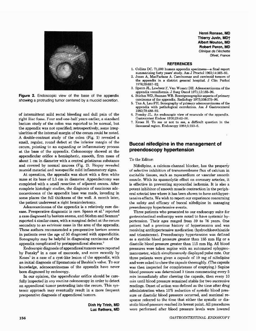
Figure 2. Endoscopic view of the base of the appendixshowing a protruding tumor centered by a mucoid secretion.
of intermittent mild rectal bleeding and dull pain of theright iliac fossa. Four and one-half years earlier, a standardbarium study of the colon was reported to be normal, butthe appendix was not opacified; retrospectively, some irregularities of the internal margin of the cecum could be noted.A double-contrast study of the colon (Fig. 1) revealed asmall, regular, round defect at the inferior margin of thececum, pointing to an expanding or inflammatory processat the base of the appendix. Colonoscopy showed at theappendicular orifice a hemispheric, smooth, firm mass ofabout 1 cm in diameter with a central gelatinous substanceand covered by normal mucosa (Fig. 2). Biopsy revealedmucoid material and nonspecific mild inflammatory signs.
At operation, the appendix was short with a firm whitemass at its base of 1.5 cm in diameter. Appendectomy wascompleted with a small resection of adjacent cecum. Aftercomplete histologic studies, the diagnosis of mucinous adenocarcinoma of the appendix base was made, invading insome places the full thickness of the wall. A month later,the patient underwent a right hemicolectomy.
Adenocarcinoma of the appendix is a relatively rare disease. Preoperative diagnostic is rare. Spann et a1.3 reporteda case diagnosed by barium enema, and Stiehm and Seaman4
reported 4 similar cases, with a marginal defect at the cecumsecondary to an extrinsic mass in the area of the appendix.These authors recommended a preoperative barium enemain patients over the age of 50 diagnosed with appendicitis.Sonography may be helpful in diagnosing carcinoma of theappendix complicated by periappendiceal abscess.5
Endoscopic diagnosis of appendiceal tumors were reportedby Ponsky6 in a case of mucocele, and more recently byKroes7 in a case of a cyst-like lesion of the appendix, withan initial diagnosis of lipomatosis of Bauhin's valve. To ourknowledge, adenocarcinomas of the appendix have neverbeen diagnosed by endoscopy.
In our opinion, the appendicular orifice should be carefully inspected in any routine colonoscopy in order to detectan appendiceal tumor protruding into the cecum. This systemic approach may eventually result in a more frequentpreoperative diagnosis of appendiceal tumors.
Dinh Hy Trinh, MDLuc Rethers, MD
156
Henri Ronsse, MDThierry Juvin, MDtAlbert Mouton, MDRobert Peron, MDClinique de I'Archette
Olivet, France
REFERENCES1. Collins DC. 71,000 human appendix specimens-a final report
summarizing forty years' study. Am J ProctoI1963;14:365-81.2. Jones A, MacFarlane A. Carcinomas and carcinoid tumors of
the appendix in a district general hospital. J Clin Pathol1976;29:687-92.
3. Spann JL, Lowbeer F, Van Womer DE. Adenocarcinoma oftheappendix vermiformis. J Surg OncoI1971;13:185-96.
4. Stiehm WD, Seaman WB. Roentgenographic aspects ofprimarycarcinoma of the appendix. Radiology 1973;108:275-96.
5. Tan A, Lau PH. Sonography of primary adenocarcinoma of theappendix with pathological correlation. Am J Gastroenterol1983;78:488-92.
6. Ponsky JL. An endoscopic view of mucocele of the appendix.Gastrointest Endosc 1976;23:42-50.
7. Kroes H. To see or not to see, a difficult question in theileocaecal region. Endoscopy 1984;5:193-6.
Buccal nifedipine in the management ofpreendoscopy hypertension
To the Editor:
Nifedipine, a calcium-channel blocker, has the propertyof selective inhibition of transmembrane flux of calcium inexcitable tissues, such as myocardium or vascular smoothmuscle. With its spasmolytic effect on coronary arteries, itis effective in preventing myocardial ischemia. It is also apotent inhibitor of smooth muscle contraction in the peripheral arterial tree where it has been shown to have antihypei-tensive effects. We wish to report our experience concerningthe safety and efficacy of buccal nifedipine in managingpreendoscopy hypertensive events.
Three patients who presented to our endoscopy suite forgastrointestinal endoscopy were noted to have systemic hypertension. Their ages ranged from 59 to 76 years. Onepatient had a previous history of hypertension and wasreceiving antihypertensive medication (hydrochlorothiazideand triamterene). Preendoscopy hypertension was definedas a systolic blood pressure greater than 185 mm Hg or adiastolic blood pressure greater than 115 mm Hg. All bloodpressures were taken supine with an automated sphygmomanometer, which simultaneously displayed pulse rate. Ourthree patients were given a capsule of 10 mg of nifedipineand were asked to chew the capsule thoroughly. (The capsulewas then inspected for completeness of emptying.) Supineblood pressure was determined 3 times commencing every 5min immediately after chewing the capsule, then every 10min until blood pressure remained stable for two successivereadings. Onset of action was defined as the time after drugadministration when 10% reduction of systolic blood pressure or diastolic blood pressure occurred, and maximal response referred to the time that either the systolic or diastolic blood pressure reached its lowest point. All procedureswere performed after blood pressure levels were lowered
GASTROINTESTINAL ENDOSCOPY

with light sedation using intravenous diazepam and meperidine.
The mean pretreament systolic and diastolic blood pressures were 188.3 ± 2.4 and 115.0 ± 4.1 mm Hg. Averageposttreatment systolic and diastolic blood pressures were146.7 ± 4.7 and 84.0 ± 2.8 mm Hg. The mean drop in bloodpressure was 41.6 mm Hg systolic and 31.0 mm Hg diastolic.The reduction in systolic and diastolic blood pressures wasnot associated with reflex tachycardia. Mean pretreatmentand posttreatment (maximal antihypertensive response)pulse rates per minutes were 80.0 ± 8.2 and 78.7 ± 1.9,respectively.
A prompt antihypertensive response was observed in allpatients. The onset of the response was within 5 min,whereas the time for maximal antihypertensive effect was15 to 25 min. We noted no hypotension, reflex tachycardia,or other reported adverse effects such as syncope, palpitation, or symptoms suggestive of cerebral or coronary ischemia. The one patient with a history of hypertension wasinstructed to resume his previous antihypertensive medication, and the other two patients were referred for hypertensive evaluation.
Buccal nifedipine seems to be especially suited for use inthe management of preendoscopy hypertension for the following reasons: (1) Blood pressure is lowered in direct relation to the magnitude of the pretreatment values. Profoundhypotension is rarely observed and invasive hemodynamicmonitoring is not mandatory. (2) Reflex cardiac stimulationprecipitating angina or myocardial infarction is rarely seenbecause of the concomitant coronary vasodilator action ofthe drug. (3) Nifedipine can easily be given buccally. Theonset of blood pressure reduction is usually prompt (5 to 15min), constant, and predictable (at least 20% average reduction). (4) The duration of action is 3 to 5 hours after a singledose, which allows the endoscopist time to perform theprocedure and observe recovery afterwards.
Precautions should be taken, particularly in the elderlyand in patients with hypovolemic chronic renal failure,cerebrovascular disease, and hypertensive encephalopathy,not to reduce the blood pressure too precipitously or belowthe lower limit of autoregulation for the brain, heart, kidneys, or other vital organs.
Chih-Hung Chen, MDE. A. Gelzayd, MD
L. C. Maas, MDSouthfield, Michigan
Garren-Edwards1M gastric bubble: deflationwith Nd:YAG laser
To the Editor:
The Garren-EdwardsT" gastric bubble is a medical device
recently approved by the Food and Drug Administrationthat is indicated for use as a therapy adjunct to a diet toreduce weight in morbid obesity patients.! The gastric bubble placement is an easily performed procedure and is described in detail by the manufacturer. On the other hand,
VOLUME 34, NO.2, 1988
the removal of the gastric bubble may be difficult, and fewdetailed recommendations are provided by the manufacturer.
Two main problems are present during removal: deflatingthe bubble and pulling it out of the stomach. Punctureneedles are frequently used to deflate the bubble, but this isdifficult to achieve because the bubble, which is made of anelastomeric plastic, is hard and the hole produced by theneedle are too small to evacuate the air effectively.
In order to solve the deflation problem, we have used anNd:YAG laser (Sharplan 2100, Laser Industries) at a powerof 80 Wand a pulse duration of 0.3 sec to make several holeson both ends of the bubble. The air is evacuated duringremoval of the bubble, which is easily performed with agrasping forceps. We have found this procedure easy toperform and effective in the removal of the gastric bubble.
Alexander Fich, MDEran Goldin, MD
Moshe Ligumsky, MDDepartment of Gastroenterology
Hebrew University-Hadassah Medical SchoolJerusalem, Israel
REFERENCE
1. Garren LR, Garren ML. The Garren gastric bubble: an aid totreatment of obesity. Gastrointest Endosc 1985;31:162.
Getting the air out of the balloon
To the Editor:
The Garren-EdwardsT" balloon has become widely used in
the treatment of obesity. Problems removing the balloonhave been occurring with increasing frequency. We havedeveloped a technique for removing the Garren-EdwardsT"
balloon that has worked without a failure and is simple andsafe.
The balloon is visualized using a conventional gastroscope. A Microvasive 25-gauge, 4-mm sclerotherapy needleis passed through the biopsy channel of the endoscope. TheGarren-Edwards'" balloon is punctured in a single spot withthe needle left in the balloon. A wall suction unit on itsmaximum setting is then attached to the sclerotherapyneedle. In a matter of minutes, the balloon is observed tocollapse. At the termination of this procedure, the ballooncontains no air. An Olympus-FG8L rat tooth forcep is thenused to grasp the balloon. It must be grasped on the reinforced edge in order to be removed without difficulty. Thismethod has been, in our experience, the most effective,simple, and safe way to remove Garren-EdwardsT
" balloons.
Martin S. Kleinman, MDEllen Coyne, RN
Delois SmithUniversity of Rochester
School of Medicine &DentistryStrong Memorial Hospital
Rochester, New York
157