bronchial mucosal inflammation in asthma modulation … gertrude marie.pdf · bronchial mucosal...
TRANSCRIPT
BRONCHIAL MUCOSAL INFLAMMATION IN ASTHMA
Modulation by glucocorticoids
ONTSTEKING IN DE BRONCHIALE MUCOSA BIJ ASTMA
Be'lnvloeding door glucocortico'lden
Cover: Electronmicroscopic photograph of an immunogold staining with antihuman IL-4 mAb of a secondary granule of an eosinophil from a patient with allergic asthma. Gold particles are seen in the crystalloid core of the secondary granule, indicating the presence of IL-4. Magnification x 30,000.
CIP-GEGEVENS KONINKLlJKE BIBLIOTHEEK, DEN HAAG
Moller, Gertrude Marie
Bronchial mucosal inflammation in asthma. Modulation by glucocorticoids/ Gertrude M. Moller; [ill.: T.M. van Os]. - Rotterdam: Department of Immunology, Erasmus University Rotterdam. - III. Thesis Rotterdam. - With ref. - With summary in Dutch. ISBN 90-73436-29-X NUGI743 Subject headings: asthma / glucocorticoids / airway inflammation
No part of this thesis may be reproduced or transmitted in any form by any means, electronic or mechanical, Including photocopying, recording or any information storage and retrieval system, without permission in writing from the publisher (G.M. Moffer, Department of Immunology, Erasmus University and University Hospital Dijkzigt, P.O. Box 1738, 3000 DR Rotterdam, The Netherlands).
BRONCHIAL MUCOSAL INFLAMMATION IN ASTHMA
Modulation by glucocorticoids
ONTSTEKING IN DE BRONCHIALE MUCOSA BIJ ASTMA
Be'invloeding door glucocortico'iden
PROEFSCHRIFT
ter verkrijging van de graad van doctor aan de Erasmus Universiteit Rotterdam
op gezag van de rector magnificus Prof. Dr. P.W.C. Akkermans M. A.
en volgens besluit van het College voor Promoties. De openbare verdediging zal plaatsvinden op
woensdag 10 april 1996 om 11.45 uur
door
Gertrude Marie Moller
geboren te Baltimore, V.S.
PROMOTIE-COMMISSIE:
Promotor:
Co-promotor:
Overige leden:
~.IMMUNOlOGIE I 0 I , E l {) A I! •
prof. dr. R. Benner
dr. H.C. Hoogsteden
prof. dr. C. Hilvering prof. dr. Th.H. van der Kwast prof. dr. D.S. Postma
Dit proefsehrift is tot stand gekomen binnen de afdelingen Immunologie en Longziekten van de Erasmus Universiteit Rotterdam en het Aeademiseh Ziekenhuis Dijkzigt Rotterdam.
Het onderzoek en het drukken van dit proefsehrift werd mede mogelijk gemaakt door finanei;;le steun van Glaxo Welleome B. V.
Het proefsehrift werd gedrukt door Haveka B.V. te Alblasserdam.
"The patient is the centre of the medical universe around which all our work revolves and towards which all our efforts tend" J.B. Murphy (1857-1916)
Aan mijn ouders Aan Paul

CONTENTS
Chapter 1
Chapter 2
2.1
2.2
Chapter 3
3.1
3.2
Chapter 4
4.1
BRONCHIAL MUCOSAL INFLAMMATION IN ASTHMA
Modulation by glucocorticoids
General introduction 9
1.1 Asthma
1.2 Immunopathogenesis of allergic asthma
1.3 Pathophysiology of the bronchial mucosa in asthma
1.4 Glucocorticoids
1.5 Aims of the studies
Effects of glucocorticoid therapy on airway inflammation
Effects of long-term treatment with beclomethasone on airway inflammation in the bronchial mucosa of atopic asthmatics Submitted
Influence of fluticasone propionate on airway inflammation in atopic asthmatics Submitted
Dendritic cells in asthma and modulation by glucocorticoids
Increased numbers of dendritic cells in the bronchial mucosa of atopic asthmatics: downregulation by inhaled corticosteroids Glin Exp Allergy 1996 (in press)
Fluticasone propionate therapy is associated with modulation of dendritic cells in the bronchial mucosa of atopic asthmatics Submitted
Cytokine expression in asthma
Immunolocalization of interleukin 4 in eosinophils in the bronchial mucosa of atopic asthmatics Am J Respir Cell Mol BioI 1996 fin press)
35
37
53
69
71
85
103
105
4.2
Chapter 5
5.1
Chapter 6
Summary
Samenvatting
Abbreviations
Dankwoord
Curriculum vitae
Publications
Ultrastructural immunogold localization of interleukin 5 to the crystalloid core compartment of eosinophil secondary granules in patients with atopic asthma J Histoehem Cytoehem 1996; 44:67-69 11 9
Eosinophils in the bronchial mucosa in relation to indices from methacholine log-dose response (MLDR) curves in atopic asthmatics 127
Eosinophils in the bronchial mucosa in relation to indices from methacholine log-dose response (MLDR) curves in atopic asthmatics Am J Respir Crit Care Med 1995 151; 4:A 133 (modified) 131
General discussion 137
149
155
161
167
173
177
CHAPTER 1
GENERAL INTRODUCTION
1.1 Asthma
1.2 Immunopathogenesis of allergic asthma
1.3 Pathophysiology of the bronchial mucosa in asthma
1.4 Glucocorticoids
1.5 Aims of the studies
1.6 References

General introduction
1.1 Asthma
In this thesis studies are presented concerning airway inflammation in the bronchial mucosa of allergic asthmatics and its modulation by glucocorticoids. Asthma is one of the most common chronic disorders in the Western world and affects about 10% of the population. Its prevalence, morbidity and mortality appear to be rising (1,2). Initially, the Greek term "U(HflU" recognized by Hippocrates (460-357 B.C.) was the name given to the disorder occurring in people with "difficult breathing". At present, "asthma" is still difficult to define. The current working definition of asthma recommended by the National Asthma Education Program Expert Panel Report (3) is as follows:
'asthma is a lung disease with the fol/owing characteristics: (1) airway obstruction that is reversible (but not completely so in some patients) either spontaneously or with treatment; (2) airway inflammation; and (3) increased airway responsiveness to a variety of stimuli'.
Asthma is defined as a syndrome characterized by "variable airway obstruction". Pathophysiological changes that contribute to the airway narrowing are increased bronchial smooth muscle contraction, enhanced mucus production, and enhanced vascular permeability with mucosal edema. Histologic evaluation of the airways of asthmatics shows a chronic inflammation in the airway wall (4). The airflow obstruction in asthmatics is traditionally considered to be reversible if the patient's forced expiratory volume in one second (FEV 1) increases by at least 15% after inhalation of Bragonists (5). Asthma is characterized by increased airway hyperresponsiveness, defined as a decreased threshold of airway narrowing in response to a variety of non·specific stimuli (6). These non-specific stimuli include tobacco smoke (7), fog, nitrogen dioxide (8), ozone (9), viral infections (10), and inhaled pharmacologic agents (histamine (11), methacholine (12)), as well as physical stimuli (exposure to cold air (13), exercise (14)). Exposure to specific stimuli such as allergens (house dust mite, pollen, animal dander) can lead to an increase in airway hyperresponsiveness (15), which often precedes asthma (16). Atopy, the genetic predisposition for directing an IgE response to common environmental allergens such as house dust mite and pollen, is another risk factor for the development of asthma (17) and may affect 40% of the population of western European countries (18). A recent study demonstrates that a trait for elevated level of serum total immunoglobulin E ((gE) is coinherited with a trait for bronchial hyperresponsiveness and that a gene governing bronchial hyperresponsiveness is located near a major locus on chromosome 5q that regulates IgE levels (19). IgE can bind with high
11
Chapter 1
affinity to specific receptors (Fc8RI) expressed on the surface of mast cells, basophils, monocytes and eosinophils, and with somewhat lower affinity to macrophages and eosinophils (Fc8RII, CD23). Cross-linking of receptor-bound IgE with specific allergen results in the release of inflammatory mediators, including histamine, prostaglandin D2 (PGD2) and leukotriene C4 (L TC4) which, through their direct effects on airway smooth muscle and microvasculature, are responsible for the allergen-induced bronchial narrowing and wheezing (20).
Ag
-'7$~t"'o '".~~ 6~'(-'c """" t_lj t .co Y
dendritic cell
t • Ag
:::;(1 ~L-4' IL-13 IL-6 IL-12 mast cell
+-IL-13 :t:~ • (jX:'
IL-3 IL-4 IL-5
GM-CSF eosinophil
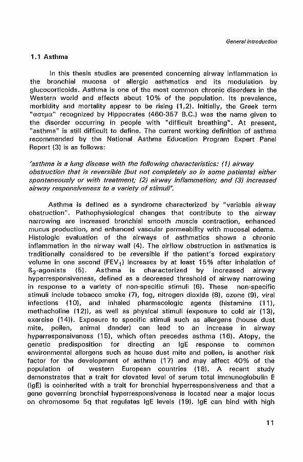
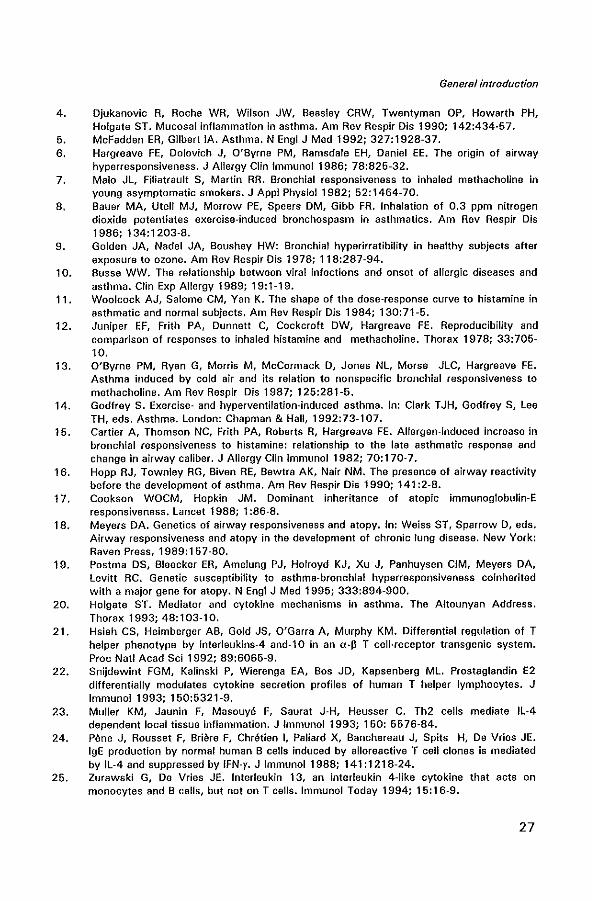
Figure 1. Schematic presentation of the allergic Immune response.
1.2 Immunopathogenesis of allergic asthma
IL-5
mediators of inflammation
The initial event in the allergic immune reponse is thought to be the presentation of antigen to T lymphocytes by antigen-presenting cells (APC). APC comprise monocytes, macrophages, B lymphocytes and in particular dendritic cells (DC). Recognition of the presented antigen by the T lymphocyte and the generation of costimulatory signals by the APC result in proliferation of T lymphocytes and production of inflammatory mediators by both APC and T lymphocytes (Figure 1). T lymphocytes will develop into CD4 + helper T lymphocytes, termed Th2 lymphocytes, which produce predominantly IL-4, IL-5, IL-6, IL-10 and IL-13. The development of Th2
12
General introduction
lymphocytes is influenced by a balance between different cytokines. IL-4, IL-10 and prostaglandin E2 (PGE2), produced by APC promote the generation of Th2 cells, while IFN-y and IL-12 strongly promote Thl lymphocyte development (21,22). IL-4 stimulates the development and maturation of naive T lymphocytes towards the Th2 lymphocyte phenotype (23), and supports mast cell growth. In addition, IL-4 and IL-13 are obligatory cytokines required for isotype switching of B lymphocytes to IgE production (24,25). Although T lymphocytes have been proposed as the main origin of Th2-type cytokines, recent studies also identified the mast cell as an important source of IL-4 (26). In addition, IL-5 together with IL-3 and granulocyte macrophage colony-stimulating factor (GM-CSF) are involved in growth, differentiation, recruitment and increasing the effector capacity of eosinophils (27,28). Once recruited into the airway, activated eosinophils secrete a variety of lipid mediators such as platelet activating factor (PAF) , L TC4, and four cationic proteins with cytotoxic properties. These include major basic protein (MBP) localized to the crystalloid core of the specific or secondary granule, eosinophil cationic protein (ECP), eosinophil-derived neurotoxin (EDN), and eosinophil peroxidase (EPa) (29,30). These products are highly toxic to the respiratory epithelium and cause epithelial damage (shedding) (31). This loss of epithelial cells may further contribute to bronchial hyperresponsiveness, possibly by a reduced production of epithelium-derived relaxing factors (32,33). Respiratory viral infections are also suspected to be important in the pathogenesis of asthma (34). Many respiratory viruses, particularly respiratory syncytial virus (RSV) and parainfluenza virus, directly damage airway epithelium (35) and increase airway hyperresponsiveness (36).
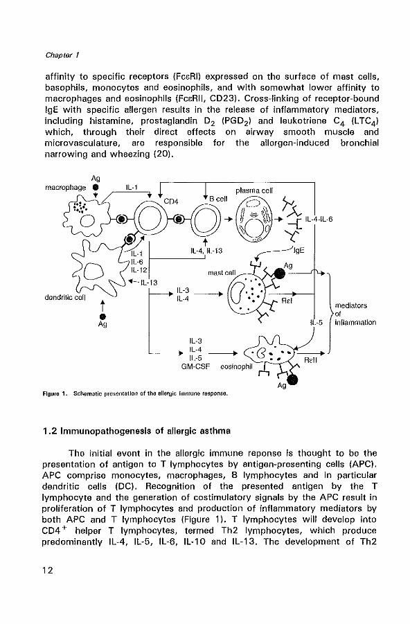
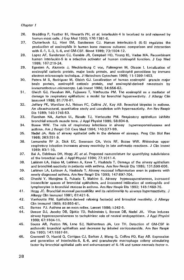
Figure 2. Schematic diagram of a bronchus.
1 epithelium and basement membrane
lamina propria
smooth muscle and cartilage
mucosa
1 submucosa
13
Chapter 1
1.3 Pathophysiology of the bronchial mucosa in asthma
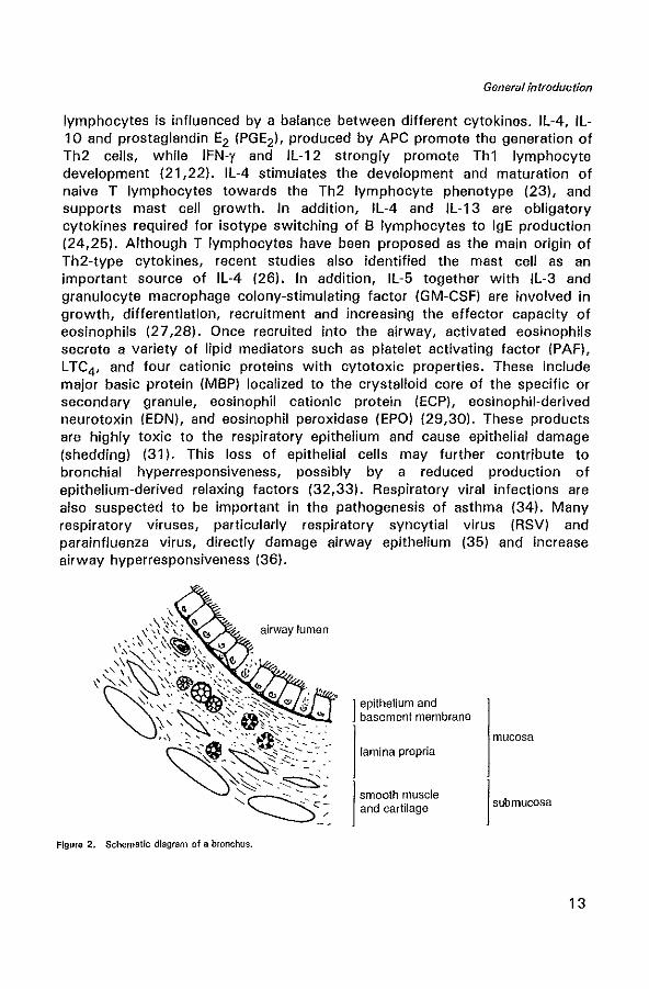
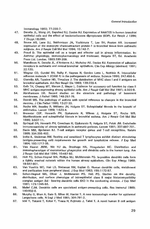
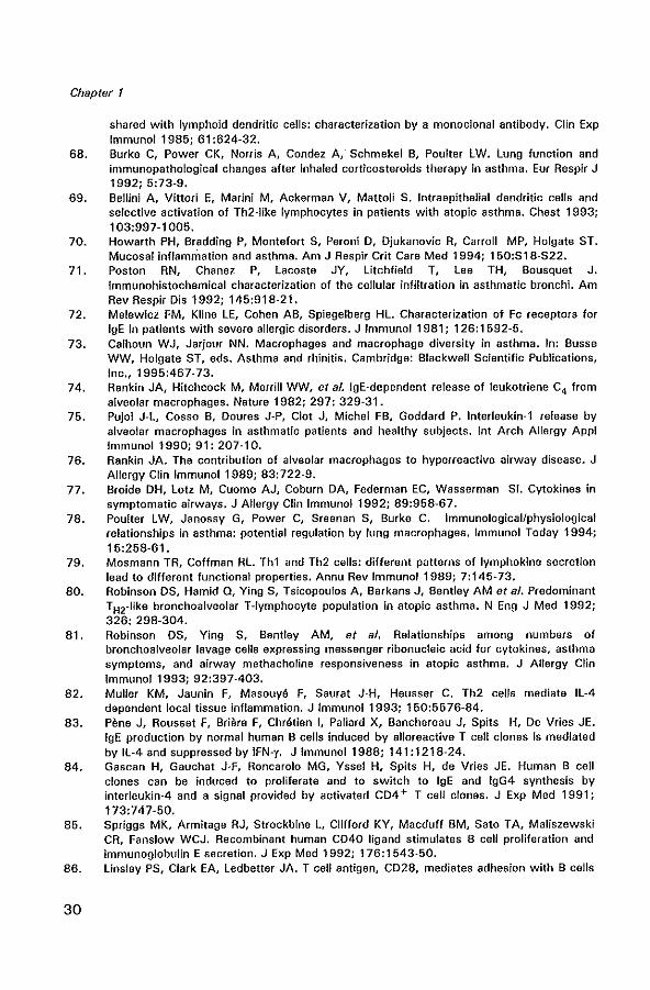
The bronchial mucosa consists of epithelium, basement membrane and lamina propria. The submucosa contains smooth muscle, glands and cartilage (Figure 2) (37). More specific knowledge has become available since the introduction of the fibreoptic bronchoscope into asthma research, providing an opportunity to take bronchial biopsies (Figures 3 and 4) and to obtain cells from the epithelial lining fluid by bronchoalveolar lavage (BAL) (5). From several biopsy studies, it became apparent that inflammatory changes are already present in mild or newly diagnosed asthmatics (38,39).
Figure 3. Schematic overview of the human lungs, Indicating that biopsies were taken from the carinae of the lingula or tlw right upper, middle or lower lobes (*).
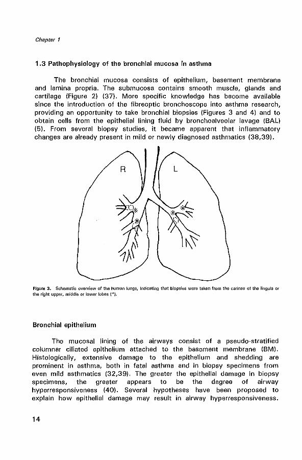
Bronchial epithelium
The mucosal lining of the airways consist of a pseudo-stratified columnar ciliated epithelium attached to the basement membrane (BM). Histologically, extensive damage to the epithelium and shedding are prominent in asthma, both in fatal asthma and in biopsy specimens from even mild asthmatics (32,39). The greater the epithelial damage in biopsy specimens, the greater appears to be the degree of airway hyperresponsiveness (40). Several hypotheses have been proposed to explain how epithelial damage may result in airway hyperresponsiveness.
14
General introduction
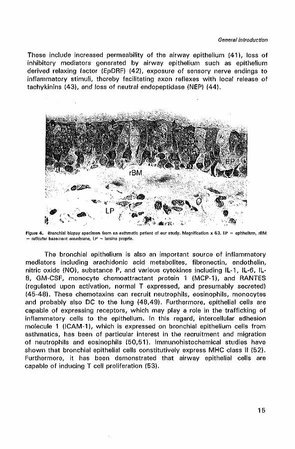
These include increased permeability of the airway epithelium (41), loss of inhibitory mediators generated by airway epithelium such as epithelium derived relaxing factor (EpDRF) (42). exposure of sensory nerve endings to inflammatory stimuli, thereby facilitating axon reflexes with local release of tachykinins (43), and loss of neutral endopeptidase (NEP) (44).
Figure 4. Bronchial biopsy specimen from an asthmatic patient of our study. Magnification x 63. EP "" epithelium, rSM = reticular basement membrane, lP =: lamina propria.
The bronchial epithelium is also an important source of inflammatory mediators including arachidonic acid metabolites, fibronectin, endothelin, nitric oxide (NO). substance P, and various cytokines including IL-1, IL-6, IL-8, GM-CSF, monocyte chemoattractant protein 1 (MCP-1), and RANTES (regulated upon activation, normal T expressed, and presumably secreted) (45-48). These chemotaxins can recruit neutrophils, eosinophils, monocytes and probably also DC to the lung (48,49). Furthermore, epithelial cells are capable of expressing receptors, which may playa role in the trafficking of inflammatory cells to the epithelium. In this regard, intercellular adhesion molecule 1 (ICAM-1), which is expressed on bronchial epithelium cells from asthmatics, has been of particular interest in the recruitment and migration of neutrophils and eosinophils (50,51). Immunohistochemical studies have shown that bronchial epithelial cells constitutively express MHC class)) (52). Furthermore, it has been demonstrated that airway epithelial cells are capable of inducing T cell proliferation (53).
15
Chapter 1
Reticular basement membrane
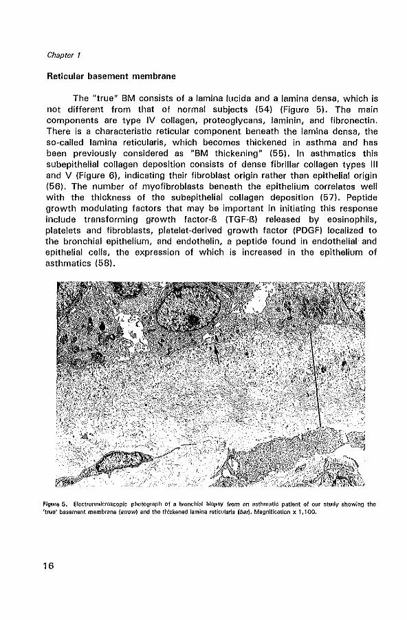
The "true" 8M consists of a lamina lucida and a lamina densa, which is not different from that of normal subjects (54) (Figure 5). The main components are type IV collagen, proteoglycans, laminin, and fibronectin. There is a characteristic reticular component beneath the lamina densa, the so-called lamina reticularis, which becomes thickened in asthma and has been previously considered as "8M thickening" (55). In asthmatics this subepithelial collagen deposition consists of dense fibrillar collagen types III and V (Figure 6), indicating their fibroblast origin rather than epithelial origin (56). The number of myofibroblasts beneath the epithelium correlates well with the thickness of the subepithelial collagen deposition (57). Peptide growth modulating factors that may be important in initiating this response include transforming growth factor-B (TGF-B) released by eosinophils, platelets and fibroblasts, platelet-derived growth factor (PDGF) localized to the bronchial epithelium, and endothelin, a peptide found in endothelial and epithelial cells, the expression of which is increased in the epithelium of asthmatics (58).
Figure 6. Electronmlcroscopic photograph of a bronchial biopsy from an asthmatlc patlent of our study showing the 'true' basement membrane (arrow) and the thickened lamina reticu!aris (bar). Magnification x 1,100.
16
General introduction
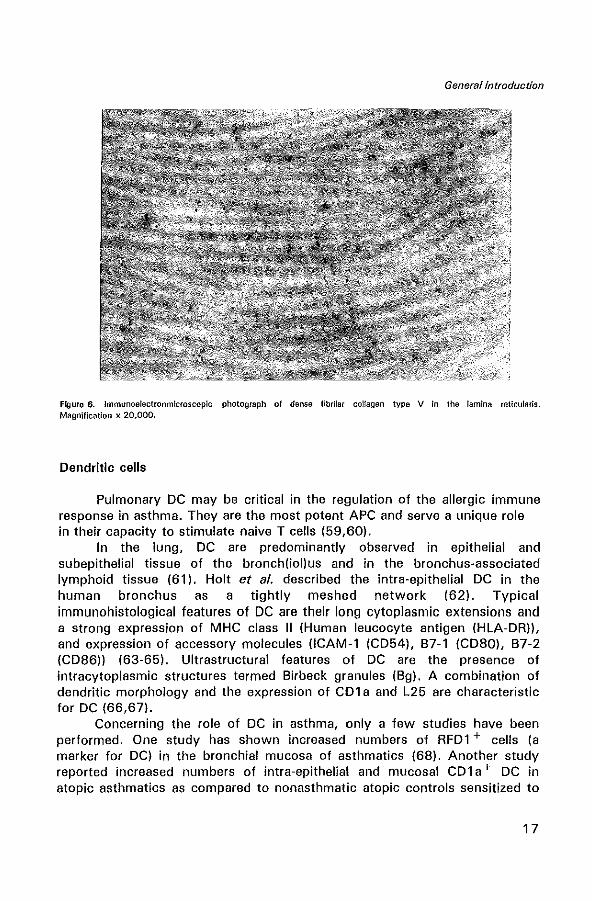
Figure 6. Immunoelectronmicroscopic photograph of dense fibrilar collagen type V in the lamina reticularis. Magnification x 20,000.
Dendritic cells
Pulmonary DC may be critical in the regulation of the allergic immune response in asthma. They are the most potent APC and serve a unique role in their capacity to stimulate naive T cells (59,60).
In the lung, DC are predominantly observed in epithelial and subepithelial tissue of the bronch(iol)us and in the bronchus-associated lymphoid tissue (61). Holt et al. described the intra-epithelial DC in the human bronchus as a tightly meshed network (62). Typical immunohistological features of DC are their long cytoplasmic extensions and a strong expression of MHC class II (Human leucocyte antigen (HLA-DR)), and expression of accessory molecules (lCAM-1 (C054), B7-1 (C080), B7-2 (C086)) (63-65). Ultrastructural features of DC are the presence of intracytoplasmic structures termed Birbeck granules (Bg). A combination of dendritic morphology and the expression of CD1 a and L25 are characteristic for DC (66,67).
Concerning the role of DC in asthma, only a few studies have been performed. One study has shown increased numbers of RFD1 + cells (a marker for DC) in the bronchial mucosa of asthmatics (68). Another study reported increased numbers of intra-epithelial and mucosal C01 a + DC in atopic asthmatics as compared to nonasthmatic atopic controls sensitized to
17
Chapter 1
the same allergen (69). However, the pathogenetic role of DC in allergic airway inflammation in asthma has not been completely established.
Macrophages and monocytes
Alveolar macrophages (AM) represent the largest cell population, accounting for 80% to 90% of the airway cells in BAL fluid in asthmatics and normal subjects (70). Increased numbers of macrophages were found in biopsies of atopic asthmatics (71). AM and monocytes express FCERII (CD23) (72). Via these receptors, AM can be activated by antigen and release a variety of mediators, including L TB4, LTC4, PAF and PGD2 (73,74). Moreover, macrophages and monocytes are also activated by antigen in an IgE-dependent manner to release IL-l B, tumor necrosis factor-a (TNF-al, GMCSF, and superoxide anion (°2-) (75-77).
In bronchial biopsies of asthmatics a reduced proportion of immunosuppressive macrophages was found, compared with normal lung tissue (78), supporting the hypothesis that T cell-mediated inflammation in asthma may partly result from a reduced capacity of a macrophage subpopulation to suppress T cell activity. Changes in subpopulations of AM, which favor proinflammatory functions, or lack downregulating capabilities, could be a mechanism by which airway inflammation in asthma is controlled (73). The contribution of AM to the pathogenesis of asthma has not yet been fully elucidated and requires additional study.
Lymphocytes
APC present antigenic peptides to T lymphocytes, and recognition of the peptide-MHC molecule complex by the T cell receptor is a crucial step in T cell stimulation, resulting in proliferation and cytokine production. Three distinct subsets, ThO, Th 1 and Th2, can be distinguished, based on their different cytokine profiles (79). Th 1 cells predominantly secrete IL-2 and IFNy, whereas Th2 cells produce IL-4, IL-5, IL-10 and IL-13, and ThO cells produce all of these cytokines. Asthma is generally considered to be a Th2-mediated disorder. Recent studies have shown elevated percentages of cells that express mRNA encoding IL-3, IL-4, IL-5, and GM-CSF in BAL fluid from mild atopic asthmatics, consistent with a Th2-type pattern of cytokine synthesis (80,81).
IL-4 stimulates the development and maturation of naive T cells to the Th2 lymphocyte phenotype (82), and supports mast cell growth. IL-4 and IL-13 play a critical role in isotype switching of B lymphocytes to IgE production (25,83). For the induction of IgE synthesis by IL-4, a physical T-B cell interaction is required (84). After recognition of the peptide-MHC class II complex on B cells by the T cell receptor, T cells become activated, express
18
General introduction
the CD40 ligand (CD40L, gp39), and stimulate B cell proliferation and IgE secretion (B5). A second required costimulatory signal is mediated by the T cell antigen CD28. The antigen B7 expressed on DC, activated B cells and monocytes, has been shown to be the ligand for CD28 (86). CTLA-4 can also act as a ligand for B7 and is expressed on activated T lymphocytes (87). Furthermore, IL-4 increases expression of vascular cellular adhesion molecule (VCAM-l) on endothelial cells, which may be involved in eosinophil adhesion in the pulmonary circulation (88). IL-5 promotes the differentiation (27), recruitment (89), activation and survival of eosinophils (28). In several studies of bronchial biopsies taken from asthmatics, the expression of cell surface activation molecules, such as the IL-2 receptor (CD25), MHC class II, and very late activation antigen 1 (VLA-l), is increased on T-Iymphocytes (90,91). Interestingly, the total numbers of both helper (CD4) and suppressor/cytotoxic (CD8) T lymphocytes in bronchial mucosa of asthmatics are comparable to those in controls (92); CD4 cells predominate over CD8 cells. The CD4 + subset can be further subdivided by the expression of the leucocyte common antigen (CD45) isoforms by stage of maturation; CD45RA is expressed on naive cells, while CD45RO is expressed on "memory" cells, which have been previously activated by exposure to specific antigen (93). Immunohistochemical studies have shown that the numbers of activated T lymphocytes can be correlated with the degree of asthma severity and with the numbers of total and activated eosinophils (90,93).
There are virtually no B lymphocytes within histologic sections of the normal airway mucosa. However, distinct B cell areas were detected in the bronchus-associated lymphoid tissue (BAL T) (64). In the bronchi of asthmatics a very few B lymphocytes are present (71).
Eosinophils
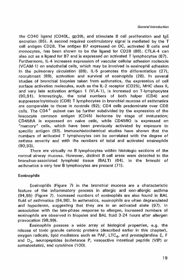
Eosinophils (Figure 7) in the bronchial mucosa are a characteristic feature of the inflammatory process in allergic and non·allergic asthma (94,95) (Figure 7). Increased numbers of eosinophils are also found in BAL fluid of asthmatics (94,96). In asthmatics, eosinophils are often degranulated and hypodense, suggesting that they are in an activated state (97). In association with the late-phase response to allergen, increased numbers of eosinophils are observed in biopsies and BAL fluid 3-24 hours after allergen provocation (98,99).
Eosinophils possess a wide array of biological properties, e.g. the release of toxic granule cationic proteins (described earlier in this chapter), oxygen radicals, lipid mediators such as PAF, LTC4, and prostaglandins E, F and D2, neuropeptides (substance P, vasoactive intestinal peptide (VIP) or somatostatin), and cytokines (100).
19
Chapter 1
Eosinophils are a newly recognised source of several cytokines including IL-la (101), IL-3 (102), IL-4 (103), IL-5 (104), IL-6 (105), IL-8 (106), GM-CSF (107), TNF-a (108), macrophage inflammatory protein-la (MIP-la) (109), transforming growth factor-a (TGF-a) (110) and TGF-B (111). Important for eosinophil survival, activation and recruitment are IL-3, IL-5, GM-CSF, TNF-a, and MIP-la, indicating an autocrine regulation which may prolong the eosinophilic inflammation in the airways.
Figure 7. Electronmicroscop!c photograph of an eosinophil in the bronchial epithelium of an asthmatic patient of our study. Magnification x 3,000.
Mast cells
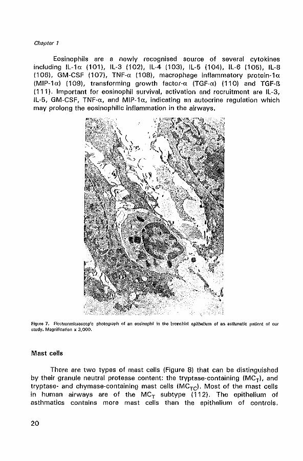
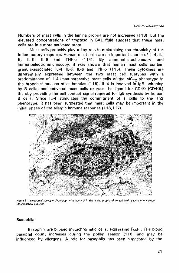
There are two types of mast cells (Figure 8) that can be distinguished by their granule neutral protease content: the tryptase-containing (MCT), and tryptase- and chymase-containing mast cells (MCTe). Most of the mast cells in human airways are of the MCT subtype (112). The epithelium of asthmatics contains more mast cells than the epithelium of controls.
20
General introduction
Numbers of mast cells in the lamina propria are not increased (113), but the elevated concentrations of tryptase in BAL fluid suggest that these mast cells are in a more activated state.
Mast cells probably playa key role in maintaining the chronicity of the inflammatory response. Human mast cells are an important source of IL-4, IL-5, IL-6, IL-8 and TNF-o: (114). By immunohistochemistry and immunoelectronmicroscopy, it was shown that human mast cells contain granule-associated IL-4, IL-5, IL-6 and TNF-o: (115). These cytokines are differentially expressed between the two mast cell subtypes with a predominance of IL-4 immunoreactive mast cells of the MCTe phenotype in the bronchial mucosa of asthmatics (115). IL-4 is involved in IgE switching by B cells, and activated mast cells express the ligand for CD40 (CD40L) thereby providing the cell contact signal required for IgE synthesis by human B cells. Since IL-4 stimulates the commitment of T cells to the Th2 phenotype, it has been suggested that mast cells may be important in the initial phase of the allergic immune response (116,117).
FIgure 8. Electronmicroscopic photograph of a mast cell in the lamina proplia of an asthmatic patient of our study. Magnification x 3,000.
Basophils
Basophils are bilobed metachromatic cells, expressing FCERI. The blood basophil count increases during the pollen seaSOn (118) and may be influenced by allergens. A role for basophils has been suggested by the
21
Chapter 1
finding that during the late-phase response to allergen, the numbers of basophils are increased in the SAL fluid (119). Clearly, the role of basophils in asthma requires further study.
Neutrophils
The role of neutrophils in asthma is not yet clear. The numbers of neutrophils seen in bronchial biopsies of asthmatics are extremely low (120). No differences in the numbers of neutrophils have been found in bronchial biopsies and lavages between asthmatics and healthy controls (5,71,98), suggesting that these cells do not play an important role in the mechanisms of the inflammatory process in asthma.
Adhesion molecules
Cellular adhesion mechanisms are necessary for the migration of inflammatory cells from the circulation into the surrounding tissue and the airway lumen. Several families of adhesion molecules have been described so far, but in asthma the main attention has been focused on ICAM-1 (CD54), a member of the immunoglobulin superfamily, ligand for the lymphocyte function-associated antigen (LFA-1 (CD11 a/CD18) integrin on human T cells) (121) and the major receptor for rhinovirus (122). Another important adhesion molecule is endothelial leucocyte adhesion molecule (ELAM-1), which belongs to the selectin family and whose complementary ligand is Sialyl-LeX (123). Vascular cellular adhesion molecule-1 (VCAM-1), a ligand for very late activation antigen (VLA-4), is expressed on eosinophils and is an important molecule in selective eosinophil recruitment (88). Expression of VCAM-1 appears to be upregulated by IL-4 on vascular endothelium (88). Immunohistochemical studies have demonstrated enhanced expression of ICAM-1, ELAM-1, and VCAM-1 in allergic asthma (124). In a monkey model of experimentally induced allergic asthma, antibodies to ICAM-1 were reported to reduce symptoms of hyperresponsiveness and to prevent eosinophil influx into the airways (51).
1.4 Glucocorticoids
In humans glucocorticoids, such as hydrocortisone (cortisol) and cortisone, are hormones naturally produced by the adrenal cortex. Synthetic glucocorticoids are also available, such as prednisolone and dexamethasone. Inhaled glucocorticoids are by far the most effective anti-inflammatory agents used in asthma. Inhaled glucocorticoids suppress inflammation in the airways of asthmatics, although their precise molecular mechanism of action
22
General introduction
is not yet clear (125). There have recently been important advances in understanding the molecular mechanisms of glucocorticoids (126).
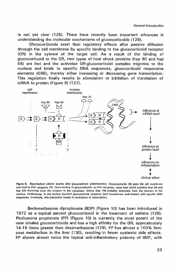
Glucocorticoids exert their regulatory effects after passive diffusion through the cell membrane by specific binding to the glucocorticoid receptor (GR) in the cytosol of the target cell. As a result of the binding of glucocorticoid to the GR, two types of heat shock proteins (hsp 90 and hsp 56) are lost and the activated GR-glucocorticoid complex migrates to the nucleus and binds to specific DNA sequences, glucocorticoid responsive elements (GRE), thereby either increasing or decreasing gene transcription. This regulation finally results in stimulation or inhibition of translation of mRNA to protein (Figure 9) (127).
cell membrane
hsp 90 hsp 56
''If_.Lf [D ~ [D :::t [Dill
t
W ?
nuclear membrane
hsp 70 00 I .... ··,',':·,,·::."·
/' " / """
-j~~,,~J r~; , \,"''''1 1' ',,,.,
?
inflUence at mRNA-level
1 inflUence at protein-level
inflUence on inflammation ,. ,. clinical effect
Figura 9. Hypothetical cellular events after glucocorticoid administration. Glucocortlcolds (S) PI)SS the cell me:mbr<loe and bind to their receptors (A). Upon binding of gfucocorticoids to their receptors, some heat shock proteins (hsp 56 and hsp 90) dissociate from the receptor in the cytoplasm. Others (hsp 70) prob<lbly dissoci<lte from the receptor In the nucleus. Furthermore, in the nucleus liganded glucocortIcoid receptors form homodimers and interact with specifiC DNA sequences. Eventuallv. this Interaction results in modulation of transcription.
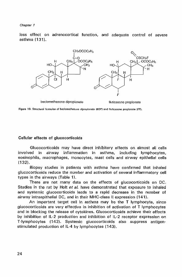
Beclomethasone dipropionate (BOP) (Figure 10) has been introduced in 1972 as a topical aerosol glucocorticoid in the treatment of asthma (128). Fluticasone propionate (FP) (Figure 10) is currently the most potent of the new inhaled glucocorticoids and has a high affinity for the GR, approximately 14-18 times greater than dexamethasone (129). FP has almost a 100% firstpass metabolism in the liver (130), resulting in fewer systemic side effects. FP shows almost twice the topical anti-inflammatory potency of BOP, with
23
Chapter 1
less effect on adrenocortical function, and adequate control of severe asthma (1 31 ).
o o
beclomethasone dipropionate
O~ CSCH2F
H CH3 .... OCOC2HS HO i .... CH3
CH3 H A---' , F
F
, H
fluticasone propionate
····H
Figure 10. Structural formulae of beclomethasone diproplonate (BOP) and fluticasone propionate IFP).
Cellular effects of glucocorticoids
Glucocorticoids may have direct inhibitory effects on almost all cells involved in airway inflammation in asthma, including lymphocytes, eosinophils, macrophages, monocytes, mast cells and airway epithelial cells (132).
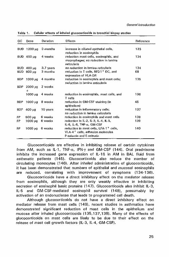
Biopsy studies in patients with asthma have confirmed that inhaled glucocorticoids reduce the number and activation of several inflammatory cell types in the airways (Table 1).
There are not many data on the effects of glucocorticoids on DC. Studies in the rat by Holt et al. have demonstrated that exposure to inhaled and systemic glucocorticoids leads to a rapid decrease in the number of airway intraepithelial DC, and in their MHC-class " expression (141).
An important target cell in asthma may be the T lymphocyte, since glucocorticoids are very effective in inhibition of activation of T lymphocytes and in blocking the release of cytokines. Glucocorticoids achieve their effects by inhibition of IL-2 production and inhibition of IL-2 receptor expression on T-Iymphocytes (142). Systemic glucocorticoids also suppress antigenstimulated production of IL-4 by lymphocytes (143).
24
General introduction
Table 1. Cellular effects of inhaled glucocorticoids in bronchial biopsy studies
GC Dose Duration Effects Reference
BUD 1200 ~g 3 months increase in ciliated epithelial cells, 133 reduction in eosinophils
BUD 400 ~g 4 weeks reduction mast celts, eosinophils, and 134 macro phages; no reduction in lamina reticularis
BUD 400~g 3.7 years no reduction in lamina reticularis 134 BUD 800 ~g 3 months reduction in T cells, RFD 1 + DC, and 68
expression of HLA-OR BOP 1 000 ~g 4 months reduction in eosinophifs and mast cells; 135
reduction in lamina reticularis BOP 2000 ~g 2 weeks
+ 1 000 ~g 4 weeks reduction in eosinophils, mast cells, and 136
T cells BOP 10001,g 8 weeks reduction in GM-CSF staining (in 45
epithelium) BOP 400 ~g 10 years reduction in inflammatory cells; 137
no reduction in lamina reticularis FP 500 rg 6 weeks reduction in eosinophils and mast cells 13B FP 1000 rg 6 weeks reduction in IL-2, IL-3, IL-4, IL-5, 139
IL-6. IL-B. TNF-a, GM-CSF FP 10001'9 6 weeks reduction in mast celis, LFA-1 + cells, 140
VLA-4 + cells, adhesion molecules P-selectin and E-selectin
Glucocorticoids are effective in inhibiting release of certain cytokines from AM, such as IL-1, TNF-a, IFN-y and GM-CSF (144). Oral prednisone inhibits the increased gene expression of IL-1 B in AM in SAL fluid from asthmatic patients (145). Glucocorticoids also reduce the number of circulating monocytes (146). After inhaled administration of glucocorticoids, it has been demonstrated that numbers of epithelial and mucosal eosinophils are reduced, correlating with improvement of symptoms (134-136).
Glucocorticoids have a direct inhibitory effect on the mediator release from eosinophils, although they are only weakly effective in inhibiting secretion of eosinophil basic proteins (147). Glucocorticoids also inhibit IL-3, IL-5 and GM-CSF-mediated eosinophil survival (148), presumably by activation of an endonuclease that leads to programmed cell death.
Although glucocorticoids do not have a direct inhibitory effect on mediator release from mast cells (149), recent studies in asthmatics have demonstrated significant reduction of mast cells in the epithelium and mucosa after inhaled glucocorticoids (135,137,138). Many of the effects of glucocorticoids on mast cells are likely to be due to their effect on the release of mast cell growth factors (IL-3, IL-4, GM-CSF).
25
Chapter 1
The bronchial epithelium is another important target for inhaled glucocorticoids (150). Glucocorticoids inhibit the increased expression of GM-CSF and RANTES in the epithelium of asthmatic patients (45,47,135).
1 .5 Aims of the studies
The aim of the studies presented in this thesis was to investigate the cellular aspects of chronic airway inflammation in the bronchial mucosa of allergic asthmatic patients by using (immuno)electronmicroscopy and immuno-histochemistry. Furthermore, the effects of glucocorticoids on the bronchial mucosal inflammation were studied, with a special emphasis on dendritic cells, eosinophils and cytokines, such as IL-4 and IL-5.
Chapter 2 describes the effects of long-term (2.5 years) and short-term (3 months) treatment with glucocorticoids, i.e. BDP and FP, respectively, on pulmonary function data, the number of several inflammatory cell types and the thickness of the lamina reticularis of the BM in the bronchial mucosa in allergic asthma.
. In chapter 3 the distribution of DC in the bronchial mucosa of asthmatics compared to controls is investigated. Furthermore, the influence of inhaled glucocorticoids on the presence of DC in the bronchial mucosa is investigated. The implications of these findings with regard to the role of pulmonary DC in the initiation and propagation of the allergic immune response are discussed.
Chapter 4 describes the localization of IL-4 immunoreactivity in the bronchial mucosa of allergic asthmatics and the ultrastructural immunogold localization of IL-5 in the crystalloid core compartment of the secondary granules of eosinophils.
In chapter 5 the relationship between eosinophils in the bronchial mucosa and indices from the methacholine log-dose response curves in allergic asthmatics is studied.
In chapter 6 the presented data are discussed in the context of the literature. Also a summary of the thesis is given.
1.6 REFERENCES
1. Lang DM, Polansky M. Patterns of asthma mortality in Philadelphia from 1969 to 1991. N Engl J Med 1994; 331:1542-6.
2. Burney PGJ, Chinn S, Rona RJ. Has the prevalence of asthma increased in children? Evidence from the national study of health and growth 1973~1986. Br Med J 1990; 300: 1306-1 O.
3. Sheffer AL. Guidelines for the diagnosis and management of asthma. National Heart, lung, and Blood Institute National Asthma Education Program Expert Panel Report. J Allergy elin Immunol 1991; 88:425-534.
26
General introduction
4. Djukanovic R, Roche WH, Wilson JW, Beasley CRW, Twentyman OP, Howarth PH, Holgate ST. Mucosal inflammation in asthma. Am Rev Hespir Dis 1990; 142:434-57.
5. McFadden ER, Gilbert IA. Asthma. N Engl J Med 1992; 327:1928-37. 6. Hargreave FE, Dolovich J, O'Byrne PM, Ramsdale EH, Daniel EE. The origin of airway
hyperresponsiveness. J Allergy Clin Immunol 1986; 78:825-32. 7. Malo JL, Filiatrault S, Martin RR. Bronchial responsiveness to inhaled methacholine in
young asymptomatic smokers. J Appl Physiol1982; 52:1464-70. 8. Bauer MAr UteH MJ, Morrow PE, Speers OM, Gibb FR. Inhalation of 0.3 ppm nitrogen
dioxide potentiates exercise-induced bronchospasm in asthmatics. Am Rev Hespir Dis 1986; 134: 1203-8.
9. Golden JA, Nadel JA, Boushey HW: Bronchial hyperirratibility in healthy subjects after exposure to ozone. Am Hev Respir Dis 1978; 118:287-94.
10. Busse WW. The relationship between viral infections and onset of allergic diseases and asthma. Clin Exp Allergy 1989; 19:1-19.
11. Woolcock AJ, Salome CM, Yan K. The shape of the dose-response curve to histamine in asthmatic and normal subjects. Am Rev Respir Dis 1984; 130:71-5.
12. Juniper EF, Frith PAl Dunnett C, Cockcroft OW, Hargreave FE. Reproducibility and comparison of responses to inhaled histamine and methacholine. Thorax 1978; 33:705-10.
13. O'Byrne PM, Ryan G, Morris M, McCormack 0, Jones NL, Morse JLC, Hargreave FE. Asthma induced by cold air and its relation to nonspecific bronchial responsiveness to methacholine. Am Rev Respir Dis 1987; 125:281-5.
14. Godfrey S. Exercise- and hyperventilation-induced asthma. In: Clark T JH, Godfrey S, Lee TH, eds. Asthma. London: Chapman & Hall, 1992:73-107.
15. Cartier A, Thomson NC, Frith PA, Roberts R, Hargreave FE. Allergen-induced increase in bronchial responsiveness to histamine: relationship to the late asthmatic response and change in airway caliber. J Allergy Clin Immunol 1982; 70: 170-7.
16. Hopp RJ, Townley RG, Biven RE, Bewtra AK, Nair NM. The presence of airway reactivity before the development of asthma. Am Rev Respir Dis 1990; 141 :2-8.
17. Cookson WOCM, Hopkin JM. Dominant inheritance of atopic immunogiobulin·E responsiveness. Lancet 1988; 1 :86-8.
18. Meyers OA. Genetics of airway responsiveness and atopy. In: Weiss ST, Sparrow 0, eds. Airway responsiveness and atopy in the development of chronic lung disease. New York: Raven Press, 1989:157-80.
19. Postma OS, Bleecker ER, Amelung PJ, Holroyd KJ, Xu J, Panhuysen CIM, Meyers OA, Levitt RC. Genetic susceptibility to asthma-bronchial hyperresponsiveness coinherited with a major gene for atopy. N Engl J Med 1995; 333:894·900.
20. Holgate ST. Mediator and cytokine mechanisms in asthma. The Altounyan Address. Thorax 1993; 48: 1 03-1 O.
21. Hsieh CSt Heimberger AS, Gold JS, O'Garra A, Murphy KM. Differential regulation of T helper phenotype by interleukins-4 and-lOin an a·p T cell·receptor transgenic system. Proc Natl Acad Sci 1992; 89:6065-9.
22. Snijdewint FGM, Kalinski p, Wierenga EA, Bas JD, Kapsenberg ML. Prostaglandin E2 differentially modulates cytokine secretion profiles of human T helper lymphocytes. J Immunol 1993; 150:5321-9.
23. Muller KM, Jaunin F, Masouye F, Saurat J-H, Heusser C. Th2 cells mediate IL·4 dependent local tissue inflammation. J Immunol 1993; 150: 5576-84.
24. Pene J, Rousset F, Briere F, Chretien I, Paliard X, Banchereau J, Spits H, De Vries JE. IgE production by normal human B cells induced by alloreactive T cell clones is mediated by IL-4 and suppressed by IFN-y. J Immunol 1988; 141: 1218-24.
25. Zurawski G, De Vries JE. Interleukin 13, an inter leu kin 4-like cytokine that acts on monocytes and S cells, but not on T cells.lmmunol Today 1994; 15:16-9.
27
Chapter 1
26. Bradding P, Feather IH, Howarth PH, et al. Interleukin-4 is localized to and released by human mast cells. J Exp Med 1992; 176:1381-6.
27. Clutterbuck EJ, Hirst EM, Sanderson CJ. Human interleukin-5 (lL-5) regulates the production of eosinophils in human bone marrow cultures: comparison and interaction with IL-1, IL-3, IL-6, and GM-CSF. Blood 1989; 73:1504-12.
28. Lopez AF, Sanderson CJ, Gamble JR, Campbell HO, Young IG, Vadas MA. Recombinant human interleukin-5 is a selective activator of human eosinophil function. J Exp Med 1988; 167:219-24.
29. Egesten A, Alumets J, Mecklenburg C von, Palmegren M, Olsson I. Localization of eosinophil cationic protein, major basic protein, and eosinophil peroxidase by immuno electron microscopic technique. J Histochem Cytochem 1986; 11: 1399-1403.
30. Peters M S, Rodriguez M, Gleich GJ. localization of human eosinophil granule major basic protein, eosinophil cationic protein, and eosinophil-derived neurotoxin by immunoelectron microscopy. Lab Invest 1986; 54:656-62.
31. Gleich GJ, Flavahan NA, Fujisawa T, Vanhoutte PM. The eosinophil as a mediator of damage to respiratory epithelium: a model for bronchial hyperreactivity. J Altergy Clin Immunol1988; 81:776-81.
32. Jeffery PK, Wardlaw AJ, Nelson FC, Collins JV, Kay AB. Bronchial biopsies in asthma. An ultrastructural, quantitative study and correlation with hyperreactivity. Am Rev Respir Dis 1989; 140:1745-53.
33. Flavahan NA, Aarhus LLI Rimele T J, Vanhoutte PM. Respiratory epithelium inhibits bronchial smooth muscle tone. J Appl Physiol 1985; 58:834-8.
34. Busse WW. The role of respiratory infections in airway hyperresponssiveness and asthma. Am J Respir Crit Care Med 1994; 140:S77-S9.
35. Nadel JA. Role of airway epithelial celts in the defense of airways. Prog Clin Bioi Res 1988; 263:331-9.
36. Lemanske RF Jrl Dick ECI Swenson CA, Vrtis RFt Busse WW. Rhinovirus upper respiratory infection increases airway reactivity in late asthmatic reactions. J Clin Invest 1989; 83:1-10.
37. Bai A, Eidelman DH, Hogg JC at al. Proposed nomenclature for quantifying subdivisions of the bronchial wall. J Appl Physiol1994; 77:1011-4.
38. Laitinen LA, Heino M, Laitinen A, Kava T, Haahtela T. Damage of the airway epithelium and bronchial reactivity in patients with asthma. Am Rev Respir Dis 1985; 131 :599-606.
39. Laitinen LA, Laitinen A, Haahtela T. Airway mucosal inflammation even in patients with newly diagnosed asthma. Am Rev Respir Dis 1993; 147:697·704.
40. Ohashi Y, Motojima S, Fukuda T, Makino S. Airway hyperresponsiveness, increased intracellular spaces of bronchial epithelium, and increased infiltration of eosinophils and lymphocytes in bronchial mucosa in asthma. Am Rev Respir Dis 1992; 145:1469-76.
41. Hogg JC. Bronchial mucosal permeability and its relationship to airways hyperreactivity. J Allergy Clin Immunol1981; 67:421-5.
42. Vanhoutte PM. Epithelium+derived relaxing factor(s) and bronchial reactivity. J Allergy Clin Immunol 1989; 83:855-61.
43. Barnes PJ. Asthma as an axon reflex. Lancet 1986; i:242-5. 44. Dusser OJ, Jacoby DB, Djokic TO, Rubinstein I, Borson DB, Nadel JA. Virus induces
airway hyperresponsiveness to tachykinins: role of neutral endopeptidase. J Appl Physiol 1989; 67:1504-11.
45. Sousa AR, Poston AN, Lane SJ, Nakhosteen JA, Lee TH. Detection of GM-CSF in asthmatic bronchial epithelium and decrease by inhaled corticosteroids. Am Rev Respir Dis 1993; 147:1557-61.
46. Cromwell 0 , Hamid a, Corrigan CJ, Barkan J, Meng a, Collins PO, Kay AB. Expression and generation of interleukin-8, IL-6, and granulocyte macrophage colony stimulating factor by bronchial epithelial cells and enhancement of IL-1 IS and tumor necrosis factor a.
28
General introduction
Immunology 1992; 77:330-7. 47. Devalia JL, Wang JH, Sapsford RJ, Davies RJ. Expression of RANTES in human bronchial
epithelial cells and the effect of beclomethasone dipropionate (BOP). Eur Respir J 1994; 7 (SuppI.18):98S.
48. Sousa AR, Lane SJ, Nakhosteen JA, Yoshimura T, Lee TH, Poston AR. Increased expression of the monocyte chemoattractant protein-l in bronchial tissue from asthmatic subjects. Am J Respir Cell Mol Bioi 1994; 10:142-7.
49. Proud D. The epithelial cell as a target and effector cell in airway inflammation. In: Asthma: physiology, immunopharmacology and treatment. Holgate ST, Ed. Academic Press Ltd. London. 1993:199-209.
50. ManoHtsas N, Devalia JL, d' Ardenne AJ, McAulay AE, Davies RJ. Expression of adhesion receptors in asthmatic and normal bronchial epithelium. elin Exp Allergy (abstractl 1991; 22:120.
51. Wegner CD, Gundel RH, Reilly P, Haynes N, Gordon Letts L, Rothlein R. Intercellular adhesion molecule 1 (lCAM-1) in the pathogenesis of asthma. Science 1990; 247:456-9.
52. Glanville AR, Tazelaar HD, Theodore J. The distribution of MHC class I and II antigens on bronchial epithelium. Am Rev Respir Dis 1989; 139:330-4.
53. Kalb TH, Chuang MT, Marom Z, Mayer L. Evidence for accessory cell function by class II MHC antigen-expressing airway epithelial cells. Am J Respir Cell Mol Bioi 1991; 4:320-9.
54. Abrahamson DR. Recent studies on the structure and pathology of basement membranes. J Patho11986; 149:257-78.
55. Dunnill MS. The pathology of asthma with special reference to changes in the bronchial mucosa. J Clin Pathol 1960; 13:27-33.
56. Roche WR, Beasley R, Williams JH, Holgate ST. Subepithelial fibrosis in the bronchi of asthmatics. Lancet 1989; 1 :520-4.
57. Brewster CEP, Howarth PH, Djukanovic R, Wilson J, Holgate ST, Roche WR. Myofibroblasts and subepithelial fibrosis in bronchial asthma. Am J Respir Cell Mol Bioi 1990; 3:507-11.
58. Springall DR, Howarth PH, Couniham H, Ojukanovic R, Holgate ST, Polak JM. Endothelin immunoreactivity of airway epithelium in asthmatic patients. Lancet 1991; 337:697-701.
59. Davis MM, Bjorkman BJ. T-cell antigen receptor genes and T-cell recognition. Nature 1988; 334:395-402.
60. Inaba K, Steinman RM. Resting and sensitized T lymphocytes exhibit distinct stimulating (antigen-presenting cell) requirements for growth and Iymphokine release. J Exp Med 1984; 160:1717-35.
61. Van Haarst JMW, Wit HJ de, Drexhage HA, Hoogsteden HC. Distribution and immunophenotype of mononuclear phagocytes and dendritic cells in the human lung. Am J Respir Cell Mol 8io11994; 10:487-92.
62. Holt PG, Schon-He grad MA, Phillips MJ, McMenamin PG. la-positive dendritic cells form a tightly meshed network within the human airway epithelium. Clin Exp Allergy 1989; 19:597-601.
63. Van Voorhis WC, Hair LS, Steinman RM, Kaplan G. Human dendritic cells, enrichment and purification from peripheral blood. J Exp Med 1982; 155:1172-87.
64. Schon-Hegrad MA, Oliver J, McMenamin PG, Holt PG. Studies on the density, distribution, and surface phenotype of intraepithelial class II major histocompatibility complex antigen (I a) - bearing dendritic cells (DC) in the conducting airways. J Exp Med 1991; 173:1345-56.
65. Melief CJM. Dendritic cells are specialized antigen-presenting cells. Res Immunol 1989; 140:902-6.
66. Murphy G, ahan A, Sato S, Mihm M, Harrist T. A new immunologic marker for epidermal Langerhans cells. N Engl J Med 1981; 304:791-2.
67. Ishii y, Takami T, Kokai y, Yuasa H, Fujimoto J, Takei T. A novel human a cell antigen
29
Chapter 1
shared with lymphoid dendritic cells: characterization by a monoclonal antibody. Clin Exp Immunol 1985; 61 :624-32.
68. Burke C, Power CK, Norris A, Condez A, Schmekel B, Poulter LW. Lung function and immunopathological changes after inhaled corticosteroids therapy in asthma. Eur Respir J 1992; 5:73-9.
69. Bellini A, Vittori E, Marini M, Ackerman V, Mattol! S. Intra epithelial dendritic celts and selective activation of Th2-like lymphocytes in patients with atopic asthma. Chest 1993; 103:997-1005.
70. Howarth PH, Bradding P, Montefort 5, Peroni 0, Ojukanovic R, Carroll M?, Holgate ST. Mucosal inflammation and asthma. Am J Respir Crit Care Med 1994; 150:518-522.
71. Poston RN, Chanez P, Lacoste JY, litchfield T, Lee TH, Bousquet J. Immunohistochemical characterization of the cellular infiltration in asthmatic bronchi. Am Rev Respir Dis 1992; 145:918-21.
72. Melewicz FM, Kline LE, Cohen AB, Spiegelberg Hl. Characterization of Fc receptors for IgE in patients with severe allergic disorders. J Immunol 1981; 126: 1592-5.
73. Calhoun WJ, Jarjour NN. Macrophages and macrophage diversity in asthma. In: Busse WW, Holgate ST, eds. Asthma and rhinitis. Cambridge: Blackwell Scientific Publications, Inc" 1995:467-73.
74. Rankin JA, Hitchcock M, Merrill WW, et al. IgE-dependent release of leukotriene C4 from alveolar macrophages. Nature 1982; 297: 329-31.
75. Pujol J·L, Cosso B, Doures J-P, Clot J, Michel FB, Goddard ? Interleukin-1 release by alveolar macro phages in asthmatic patients and healthy subjects. Int Arch Allergy Appl Immunol1990; 91: 207-10.
76. Rankin JA. The contribution of alveolar macrophages to hyperreactive airway disease. J Allergy Clin Immunol 1989; 83:722-9.
77. Broide OH, lotz M, Cuomo AJ, Coburn OA, Federman EC, Wasserman 51. Cytokines in symptomatic airways. J Allergy Clin Immunol 1992; 89:958-67.
78. Poulter LW, Janossy G, Power C, Sreenan S, Burke C. Immunological/physiological relationships in asthma: potential regulation by lung macrophages. Immunol Today 1994; 15:258-61.
79. Mosmann TR, Coffman Rl. Thl and Th2 cells: different patterns of Iymphokine secretion lead to different functional properties. Annu Rev Immunol 1989; 7: 145-73.
80. Robinson OS, Hamid 0, Ying S, Tsicopoulos A, Barkans J, Bentley AM et al. Predominant TH2-like bronchoalveolar T-Iymphocyte popUlation in atopic asthma. N Eng J Med 1992; 326: 298-304.
81. Robinson OS, Ying S, Bentley AM, et al. Relationships among numbers of bronchoalveolar lavage cells expressing messenger ribonucleic acid for cytokines, asthma symptoms, and airway methacholine responsiveness in atopic asthma. J Allergy Clin Immunol 1993; 92:397-403.
82. Muller KM, Jaunin F, Masouye F, Saurat J-H, Heusser C. Th2 cells mediate IL-4 dependent local tissue inflammation. J Immunol1993; 150:5576·84.
83. Pene J, Rousset F, Briere F, Chretien I, Paliard X, Banchereau J, Spits H, De Vries JE. IgE production by normal human B cells induced by alloreactive T cell clones is mediated by IL-4 and suppressed by IFN-y. J Immunol 1988; 141 :1218-24.
84. Gascan H, Gauchat J-F, Roncarolo MG, Yssel H, Spits H, de Vries JE. Human B cell clones can be induced to proliferate and to switch to IgE and IgG4 synthesis by interleukin-4 and a signal provided by activated CD4 + T cell clones. J Exp Med 1991; 173:747-50.
85. Spriggs MK, Armitage RJ, Strockbine L, Clifford KY, Macduff 8M, Sato TA, Maliszewski CR, Fanslow WCJ. Recombinant human CD40 ligand stimulates B cell proliferation and immunoglobulin E secretion. J Exp Med 1992; 176:1543~50.
86. Linsley PS, Clark EA, Ledbetter JA. T cell antigen, C028, mediates adhesion with B cells
30
General introduction
by interacting with the activation antigen, B7/BB-1. Proc Nat! Acad Sci 1990; 87:5031-35.
87. Linsley PS, Brady W, Grosmaire L, Aruffo A, Damla NK, ledbetter JA. CTlA-4 is a second receptor for the B cell activation antigen B7. J Exp Med 1991; 174:562-9.
88. Schleimer RP, Sterbisky SA, Kaiser J, Bickel CA, Klunk OA, Tomioka K, Newman W, Luscinskas FW, Gimbrone Jr MA, Mcintyre BW. Il-4 induces adherence of human eosinophils and basophHs but not neutrophils to endothelium. Association with expression of VCAM-1. J Immunol1992; 148:1086-92.
89. Wang JM, Rambaldi A, Biondi A, Chan ZG, Sanderson CJ, Mantovani A. Recombinant interleukin 5 is a selective eosinophil chemoattractant. Eur J Immunol1989; 19:701-5.
90. Bradley BL, Azzawi M, Jacobson M, Assoufi B, Collins JV, Irani A- MA at al. Eosinophils, T-Iymphocytes, mast cells, neutrophils and macro phages in bronchial biopsy specimens from atopic subjects without asthma and normal control subjects and relationship to bronchial hyperresponsiveness. J Allergy Clin Immunol1991: 88:661-74.
91. Hamid a, Barkans J, Robinson OS, Durham SR, Kay AB. Co-expression of CD25 and CD3 in atopic allergy and asthma. Immunology 1992; 75:659-63.
92. Akbar AN, Salmon M, Janossy G. The synergy between naive and memory T cells during actiVation. Immunol Today 1991; 12:184-8.
93. Azzawi M, Bradley B, Jeffery PK, et al. Identification of activated T lymphocytes and eosinophils in bronchial biopsies in stable atopic asthma. Am Rev Respir Dis 1990; 142:1410·13.
94. Bousquet T, Chanez P, Lacoste JY, Barneon G, Ghavanian N, Enander I, Venge P, Ahlstedt S, Lafontaine J, Godard P, Michel FB. Eosinophilic inflammation in asthma. N Eng J Med 1990; 323:1033·9.
95. Bentley AM, Menz G, Storz C, Robinson OS, Bradley B, Jeffery PK, Durham SR, Kay AB. Identification of T lymphocytes, macro phages, and activated eosinophils in the bronchial mucosa of intrinsic asthma: relationships to symptoms and bronchial responsiveness. Am Rev Respir Dis 1992; 145:500-05.
96. Wardlaw AJ, Dunnette S, Gleich GJ, Collins JV, Kay AB. Eosinophils and mast cells in bronchoalveolar lavage in mild asthma: relationship to bronchial hyperreactivity. Am Rev Respir Dis 1988; 137:62-70.
97 _ Fukuda T, Dunnette S, Reed CE, Ackerman SJ, Peters MS, Gleich GJ. Increased numbers of hypodense eosinophils in the blood of patients with bronchial asthma. J Allergy Clin Immunol1985; 132:981·5.
98. De Monchy JGR, Kauffman HF, Venge P, Koeter GH, Jansen HM, Sluiter HJ, De Vries K. Bronchoalveolar eosinophilia during allergen-induced late asthmatic reactions. Am Rev Raspir Dis 1985; 131 :373·6.
99. Aalbers R, Smith M, De Monchy JGR, Huitema 8, Kauffman HF, Koeter GH, Timens W. Activated eosinophils in the bronchial mucosa, before, 3h, and 24h after allergen challenge. Am Rev Respir Dis 1992; 145:A20.
100. Dahl R, Venge P, Fredens K_ Eosinophi/s. In: Asthma: Basic Mechanisms and Clinical Management. Barnes PJ, Rodger I, Thomson N, eds. london: Academic Press, 1992:111-24.
101. Del Pozo V, De Andres B, Martin E, Maruri N, Zubeldia JM, Palomino P, Lahoz C. Murine eosinophils and IL-1: all-1 mRNA detection by in situ hybridization. Production and release of IL-l from peritoneal eosinophils. J Immunol 1990; 144:3117-22.
102. Kita H, Ohnishi T, Okubo Y, Weiler D, Abrams JS, Gleich GJ. Granulocyte/macrophage colony-stimulating factor and inter leu kin 3 release from human peripheral blood eosinophils and neutrophils. J Exp Med 1991; 174:745-8.
103. Nonaka M, Nonaka R, Woolley K, Adelroth E, Miura K. Okhawara y, Glibetic M, Nanako K, O'Byrne P, Dolovich J, Jordana M. Distinct immunohistochemical localization of Il-4 in human inflamed airway tissues. J Immunol 1995; 155:3234-44.
31
Chapter 1
104. Desreumaux P, Janin A, Colombel JF, Prin L, Plumas J, Emilie D, Torpier G, Capron A, Capron M. Interleukin 5 messenger RNA expression by eosinophils in the intestinal mucosa of patients with coeliac disease. J Exp Med 1992; 175:293-6.
105. Hamid 0, Barkans J, Meng A, Ying S, Abrams JS, Kay AB, Moqbel R. Human eosinophils synthesize and secrete interleukin-6 in vitro. Blood 1992; 80: 1496-1501.
106. Braun RK, Franchini M, Erard F, Rihs S, De Vries IJ, Blaser K, Hansel TT, Walker C. Human peripheral blood eosinophils produce and release interleukin-B on stimulation with calcium ionophore. Eur J Immunol 1993; 23:956-60.
107. Moqbel R, Hamid 0, Ying S, Sarkans J, Hartnell A, Tsicopoulos A, Wardlaw AJ, Kay AB. Expression of mRNA and immunoreactivity for the granulocyte/macrophage colonystimulating factor in activated human eosinophils. J Exp Med 1991; 174:749-52.
108. Beil WJ, Weller PF, Tzizik DM, Galli SJ, Dvorak AM. Ultrastructural immunogold localization of tumor necrosis factor-a to the matrix compartment of eosinophil secondary granules in patients with idiopathic hypereosinophilic syndrome. J Histochem Cytochem 1993; 41:1611-5.
109. Costa JJ, Matossian M, Resnick MS, Beil WJ, Wong DTW, Gordon JR, Dvorak AM, Weller PF, Galli SJ. Human eosinophils can express the cytokines tumor necrosis factor-a and macrophage inflammatory protein-1 Ct. J Clin Invest 1993; 91 :2673-84.
110. Wong DTW, Weller PF, Galli SJ, Elovic A, Rand TH, Gallagher GT, Chiang GT, Chou MY, Motossian K, McBride J, Todd R. Human eosinophils express transforming growth factor(I.. J Exp Med 1990; 172:673-81.
111. Ohno I, Lea RG, Flanders KC, Clark DA, Banwatt D, Dolovich J, Denburg J, Harley CS, Gauldie J, Jordana M. Eosinophils in chronically inflamed human upper airway tissues express transforming growth tactor P1 gene (TGF P1). J Clin Invest 1992; 89:1662-8.
112. Schwartz LB, Bradford TR, Irani AA, Deblois G, Craig SS.The major enzymes of human mast cell secretory granules. Am Rev Respir Dis 1987; 135: 1186-9.
113. Pesci A, Foresci A, Bertorelli G, Chetta A, Oliveri D. Histochemical characteristics and degranulation of mast cells in epithelium and lamina propria of bronchial biopsies from asthmatic and normal subjects. Am Rev Respir Dis 1993; 147:684-9.
114. Bradding P, Roberts JA, Britten KM, Montetort S, Djukanovic R, Heusser C, Howarth PH, Holgate ST. Interleukin-4, -5, -6, and TNF-a in normal and asthmatic airways: evidence for the human mast cell as an important source of these cytokines. Am J Respir Cell Mol 81011994 ;10:471-80.
115. Bradding P, Okayama Y, Howarth PH, Church MK, Holgate ST. Heterogeneity of human mast cells based on cytokine content. J Immunol 1995i 155:297-301.
116. Gauchat JF, Henchoz S, Mazzei G, Aubry JP, Brunner T, Blasei H, Life P, Talabot D, Flores-Romo L, Thompson J, Kishi K, Butterfield J, Dahinden C, Bonnefoy JY. Induction of human IgE synthesis in B cells by mast cells and basophils. Nature 1993; 365:340-3.
117. Swain SL, Weinberg AD, English M, Huston G. ILA directs the development of Th2-like helper effectors. J Immunol 1990; 145:3796-806.
118. Chavance M, Herbeth 8, Kauffmann F. Seasonal patterns of circulating basophils. Int Arch Allergy Appllmmunol 1988; 86:462-4.
119. Guo C-S, Liu MC, Galli SJ, Kagey-Sobotka A, Lichtenstein LM. The histamine containing cells in the late phase response in the lung are basophils. J Allergy Clin Immunol (abstract) 1990; 85:172.
120. Henson PM, Barish LC. Neutrophil mediators in asthma. In: Asthma and rhinitis. Busse WW, Holgate ST, eds. Cambridge: Blackwell Scientific Publications, Inc., 1995: 367-82.
121. Dustin M, Rothlein R, Bhan AK, Dinarello CA, Springer TA. Induction by IL*l and IFN-y: tissue distribution, biochemistry and function of a natural adherence molecule (lCAM-l). J Immunol 1986; 137:245-54.
122. Greve JM, Davis G, Meyer AM, ef al. The major human rhinovirusreceptor is ICAM*l. Cell 1989;56:839-47.
32
General introduction
123. Philips Ml, Nudelman E, Gaeta FCA, et at: HAM·1 mediates cell adhesion by recognition of a carbohydrate ligand Sialyl·le2x. Science 1990; 250:1130·2.
124. Montefort S, Roche WR, Howarth PH, Djucanovic A, Gratziou C, Caroll M, Smith l, Britten KM, Haskard 0, Lee TH: Intercellular adhesion molecule·l (lCAM-1) and endothelial leucocyte adhesion molecule-l (HAM-l) expression in the bronchial mucosa of normal and asthmatic subjects. Eur Respir J 1992; 5:815-23.
125. Barnes PJ, Pedersen S. Efficacy and safety of inhaled corticosteroids in asthma. Am Rev Respir Dis 1993; 148:S1-26.
126. Miesfield AL. Molecular genetics of corticosteroid action. Am Rev Aespir Dis 1990; 141:S11-7.
127. Van Hal PTH, Hoogsteden HC. 1995. Anti-inflammatory mechanisms of glucocorticoids. In: Human monocytes and alveolar macro phages : modulation of phenotype and fUnction by cytokines and glucocorticoids in vitro and in asthma. Hal PTH van, thesis. Aotterdam. 59-90.
128. Clark T JH. Effect of beclomethasone dipropionate delivered by aerosol in patients with asthma. Lancet 1972;i: 1361-4.
129. Hogger P, Rohdewald P. Binding kinetics of f/uticasone propionate to the human glucocorticoid receptor. Steroids 1994; 59:597-602.
130. Harding SM.The human pharmacology of fluticasone propionate. Respir Med 1990; 84: ISuppl AI:25-9.
131. Barnes NC, Marone G, DiMaria GU, Visser S, Utama I, Payne SL. A comparison of f1uticasone propionate, 1 mg daily with beclomethasone dipropionate, 2 mg daily, in the treatment of severe asthma. Eur Respir J 1993; 6:877-84.
132. Schleimer RP. Effects of glucocorticosteroids on inflammatory cells relevant to their therapeutic applications in asthma. Am Rev Respir Dis 1990; 141 :S59-69.
133. Laitinen LA, Laitinen A, Haahtela T. A comparative study of the effects of an inhaled corticosteroid, budesonide, and a ~2"agonist, terbutaline, on airway inflammation in newly diagnosed asthma. J Allergy Clin Immunol 1992; 90:32-42.
134. Jeffery PK, Godfrey RW, Adelroth E, Nelson F, Rogers A, Johansson S-A. Effects of treatment on airway inflammation and thickening of basement membrane reticular collagen in asthma. A quantitative light and electron microscopic study. Am Rev Respir Dis 1992; 145:890-9.
135. Trigg CJ, Manolitsas NO, Wang J, Calderon MA, McAulay A, Jordan SE, Herdman MJ, Jhalli N, Duddle JM, Hamilton SA, Devalia JL, Davies RJ. Placebo-controlled immunopathologic study of four months of inhaled corticosteroids in asthma. Am J Respir Crit Care Med 1994: 150:17-22.
136. Djukanovic R, Wilson JW, Britten KM, Wifson SJ, Walls AF, Roche WR, Howarth PH, Holgate ST. Effect of' inhaled corticosteroid on airway inflammation in asthma. Am Rev Respir Dis 1992: 145:669-74.
137. Lundgren R, Soderberg M, Horstedt P, Stenling R. Morphological studies of bronchial mucosal biopsies from asthmatics before and after ten years of treatment with inhaled steroids. Eur Respir J 1988; 1 :883-9.
138. Del Donno M, Foresi A, Chetta A, Betorelli G, Pesci A. Casalini A, Testi R, Olivieri D. Effect of six week treatment with low dose of inhaled fluticasone propionate on airway inflammation in mild asthma. Eur Respir J (abstract) t 995; 8:S302.
139. Howarth PH, Feather I, Montefort S, Underwood J, Holgate ST. The influence of the inhaled corticosteroid, f1uticasone propionate on airway cytokine immunoreactivity in asthma. Am J Respir Crit Care Med (abstract) 1995; 151 :A40.
140. Montefort SI Feather I, Underwood J, Madden J, Porter C, Holgate ST, Howarth PH. The influence of inhaled fluticasone propionate on symptoms, pulmonary physiology and airway inflammation in asthma. Am J Respir Crit Care Med (abstract) 1995; 151 :A40.
141. Nelson OJ, McWilliam AS, Haining S, Holt PG. Modulation of airway intra epithelial
33
Chapter 1
dendritic cells following exposure to steroids. Am J Respir Crit Care Med 1995; 151:4 75~ 81.
142. Reed JC, Abidi AH, Alpers JD, Hoover RG, Robb RJ, Nowell PC. Effect of cyclosporin A and dexamethasone on interleukin 2 receptor gene expression. J Immunol 1986; 137:150A.
143. Wu CY, Fargeas C, Nakajima T, Delespesse G. Glucocorticoids suppress the production of interleukin 4 by human lymphocytes. Eur J Immunol1991; 21:2645~7.
144. Guyre PM, Munck A. Glucocorticoid actions on monocytes and macro phages. In: Schleimer RP, Claman HN, Oronsky AR, eds. Antiinflammatory steroid action. Basic and clinical aspects. New York: Academic Press, 1991 :199~225.
145. Barish L, Mascali JJ, Dishuck J, Beam WR, Martin RJ, Rosenwasser LJ. Detection of alveolar macrophage-derived IL~ 1 r! in asthma. Inhibition with corticosteroids. J Immunol 1992; 149:3078·82.
146. Thompson JI van FUrth R. The effect of glucocorticosteroids on the kinetics of mononuclear phagocytes. J Exp Med 1970; 131 :429-42.
147. Kita HI Abu-Ghazaleh RI Sanderson CJ, Gleich GJ. Effect of steroids on immunoglobu1in~ induced eosinophil degranulation. J Allergy Clin Immunol1991; 87:70~7.
148. Wallen NI Kita H, Weller D, Gleich GJ. Glucocorticoids inhibit cytokine-mediated eosinophil survival. J Immunol1991; 147:3490-5.
149. Cohan VL, Undem BJ, Fox CC, Adkinson NF, Lichtenstein LM, Schleimer RP. Dexamethasone does not inhibit the release of mediators from human lung mast cells residing in airway, intestine, or skin. Am Rev Respir Dis 1989: 140:951-4.
150. Barnes PJ, Greening AP, Crompton GK. Glucocorticoid resistance in asthma. Am J Respir Crit Care Med 1995; 152:S125A2.
34
CHAPTER 2
EFFECTS OF GLUCOCORTICOID THERAPY ON AIRWAY INFLAMMATION
2.1 Effects of long-term treatment with beclomethasone on airway inflammation in the bronchial mucosa of atopic asthmatics Submitted
2.2 Influence of fluticasone propionate on airway inflammation in atopic asthmatics Submitted

CHAPTER 2.1
EFFECTS OF LONG-TERM TREATMENT WITH BECLOMETHASONE ON AIRWAY INFLAMMATION IN THE BRONCHIAL MUCOSA OF ATOPIC
ASTHMATICS'
G.M. Moller1.2, S.E. Overbeekl, C.G. van Helden-Meeuwsen2, T. Godthelp2, E.P. Prens2, P.G. Mulder3, R. Aalbers4, D.S. Postma5,
H .C. Hoogsteden 1
Departments of Pulmonary Diseases', Immunology2, and Epidemiology and Biostatistics3, Erasmus University and University Hospital Dijkzigt, Rotterdam, Dept. of Pulmonary Diseases,
Martini Hospita~ and Dept. of Pulmonary Diseases5, University Hospital, Groningen, The Netherlands.
It Submitted for publication.

Effects of beclomethasone on airway inflammation
ABSTRACT
Glucocorticoids are known to be the most effective drugs in improving asthma symptoms and airway hyperresponsiveness, but their precise mechanism of action is not yet clear. We have investigated the effects of 2.5 years of inhaled beclomethasone dipropionate (BDP) on the interleukin (lL)-4 immunoreactivity, eosinophils, (activated) T lymphocytes, and mast cells and on the thickness of the lamina reticularis in sixteen mild to severe atopic asthmatics. These patients were randomly sampled from a double-blind, multi centre study in chronic nonspecific lung disease (CNSLD) (1). Biopsies were obtained by fiberoptic bronchoscopy after 2.5 years of treatment with terbutaline, 2000 I1g daily plus either inhaled BDP, 800 I1g daily (BA+CS), ipratropium bromide, 160 I1g daily (BA+AC) or placebo (BA+PL). Following treatment, we found a significant increase in FEV1 (p=0.01) and a significant decrease in hyperresponsiveness (PC20 histamine) (p = 0.01) in the glucocorticoid group (n = 5) compared to the bronchodilator (BA + AC/PL) group (n = 11). In the glucocorticoid group the numbers of IL-4 expressing cells in the lamina propria were marginally significantly lower (p =0.05), as were the numbers of total eosinophils (p = 0.02) compared to the bronchodilator group. The number of activated eosinophils and mast cells in the glucocorticoid group were generally lower, but this did not reach statistical significance. No difference in the number of T lymphocyte subsets and the thickness of the lamina reticularis were Seen between both groups. These data support the hypothesis that the beneficial effects of long-term inhaled BDP treatment in asthma may result from a decrease in IL-4 immunoreactivity and from an inhibition of local bronchial eosinophilic inflammation.
INTRODUCTION
Inhaled glucocorticoids are the most effective drugs for asthma currently available. Numerous studies have documented their long-term clinical efficacy in adults and children (1-5) though the precise mode of action of these anti-inflammatory drugs is yet unknown. Several studies have demonstrated increased numbers of inflammatory cells, increased levels of cytokines in bronchial biopsies, bronchoalveolar lavage (BAL) fluid and peripheral blood (PB) even in mild asthmatics (6-12). Thickening of the lamina reticularis of the basement membrane is often reported in asthmatic subjects and is mainly attributed to increased depositions of collagen III and V and fibronectin but not laminin (13,14).
39
Chapter 2.1
CD4 + T helper lymphocytes can be classified according to their cytokine production pattern into a T helper 1 (Th1) subset, which produce IL-2 and IFN-y, a T helper 2 (Th2) subset which produce IL-4, IL-5 and IL-10 and a naive Tho subset, producing all of these cytokines (15). The levels of mRNA for interleukin-4 (ILA) and interleukin·5 (IL-5) are increased in BAL and PB (9) of asthmatics and have been shown to correlate positively with disease severity (10,12). IL·4 is involved in the isotype switching of B·cells to IgG4 and IgE (16,17), and is a growth factor for B cells (18,19). Though the initial source of IL-4 required for the Th2 commitment is not fully clear, mast cells do secrete ILA after crosslinking of the high affinity receptor for IgE (20). IL-5 promotes the differentiation (21), vascular adhesion, recruitment and activation of eosinophils (22,23). In addition to T lymphocytes, IL-4 and IL-5 are also produced by mast cells and eosinophils (24,25). Thus, IL-4 and IL-5 are considered to play a key role in the pathogenesis of asthma (26,27). Glucocorticoids have been shown to suppress cytokine production by inhibiting transcription factors which regulate gene expression (28·31).
We hypothesize that the beneficial anti·inflammatory actions of glucocorticoids in asthma may partly result from a reduction of cytokine production in vivo, concomitant with an inhibition of local bronchial eosinophilic inflammation. Therefore we have examined the effects of longterm beclomethasone dipropionate (BDP) on FEV l' airway hyperresponsiveness (PC20 histamine), IL-4 immunoreactivity and the inflammatory cell infiltration in bronchial biopsies from 16 asthmatic patients randomly sampled from a previous double-blind, multi centre study in chronic non-specific lung disease (CNSLD) (1).
MATERIAL AND METHODS
Patients and non·asthmatic controls
Sixteen non-smoking atopic asthmatic patients (seven women, nine men, median age 43 yr, range 24 - 61 yr) were randomly sampled in two participating centres, Groningen and Rotterdam from the Dutch CNSLD study group (1,32).
The diagnosis of asthma was based on a history of attacks of breathlessness and wheezing without chronic cough or sputum production (i.e. for more than 3 months per year), according to the criteria of the American Thoracic Society (33). Atopy was defined as a positive skin prick test to house-dust mite or at least two of twelve common aeroallergens (mean wheal size> 0.7 times the histamine wheal size (32)). All patients showed airway hyperreactivity to histamine with a provocative concentration
40
Effects of beclomethasone on airway inflammation
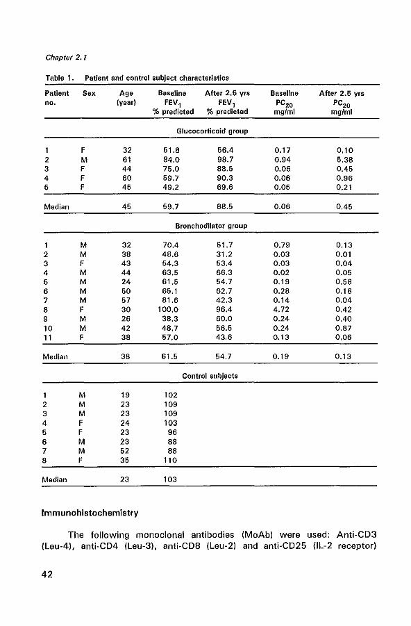
of histamine causing a 20% decrease in FEV 1 (PC20) of S; 8 mg/ml (32,34). The patients had a baseline reversibility ~ 9% of predicted. Patients were treated double-blind with an inhaled 1l2-agonist (terbutaline, two 250 ).lg puffs) plus either inhaled corticosteroid (BOP, two 100 ).lg puffs) (BA + CS) (n = 5), an anticholinergic bronchodilator (ipratropium bromide, two 20 fIg puffs) (BA+AC) (n=5) or placebo (BA+PL) (n=6). All medication was taken 4 times daily. As no significant differences were found between the BA + AC group and the BA + PL group with regard to FEV l' PC20 and the profiles of inflammatory cells the data were subsequently pooled for analysis in one bronchodilator group (n = 11). Fiberoptic bronchoscopy was performed at the end of the 2.5 year study in the same period (between August and December) in both centres, before breaking the code. A control group was composed of eight non-smoking non-asthmatic subjects (three women and five men with a median age of 23 years, range 19 to 52 years). All controls had a PC20 histamine of more than 8 mg/ml and a median FEV 1 of 103 (88-110) % of the predicted value. Patient and control characteristics are shown in Table 1. Further details of the study methods have been described previously (32). The study protocol was approved by the Medical Ethics Committee; all patients gave written informed consent.
Bronchoscopy
Fiberoptic bronchoscopy (Olympus model BF IT 10 Tokyo, Japan) was performed with atropine 0.5 mg intramuscularly as premedication. Terbutaline, 2 puffs of 250 ).lg per Nebuhaler, was given 30 min before the procedure. The nose, throat and vocal cords were anaesthetized with topical lidocaine spray. An Olympus alligator forceps model FB15C and the fenestrated forceps model FB 19C were used to take two biopsies from segmental or subsegmental divisions of the main bronchi.
Bronchial biopsies
Each biopsy sample for immunohistochemistry was immediately placed in ice-cooled isotonic saline and snap-frozen within 20 min in Tissue-Tek II OCT embedding medium (Miles, Naperville, Illinois, USA). Samples were stored at -80°C until use. One biopsy sample for electron microscopic analysis was placed immediately in glutaraldehyde for future studies. Frozen sections (6 flm) were cut on a Reichert-Jung 2800 Frigocut cryostat. From each biopsy two sections were placed on poly-L-Iysine-coated (Sigma Diagnostics, St. Louis, MO, USA) microscopic slides. Sections were air dried for 30 min and stored at -80°C until use.
41
Chapter 2.1
Table 1. Patient and control subject characteristics
Patient Sex Age Baseline After 2.6 yrs Baseline After 2.6 yrs no. (year) FEV, FEV, PC20 PC20
% predicted % predicted mgfml mg/ml
Glucocorticoid group
F 32 51.8 56.4 0.17 0.10 2 M 61 84.0 98.7 0.94 5.38 3 F 44 75.0 88.5 0.06 0.45 4 F 60 59.7 90.3 0.06 0.96 5 F 45 49.2 69.6 0.05 0.21
Median 45 59.7 88.5 0.06 0.45
Bronchodilator group
M 32 70.4 51.7 0.79 0.13 2 M 38 48.6 31.2 0.03 0.01 3 F 43 54.3 53.4 0.03 0.04 4 M 44 63.5 66.3 0.02 0.05 5 M 24 61.5 54.7 0.19 0.58 6 M 50 65.1 62.7 0.28 0.18 7 M 57 81.6 42.3 0.14 0.04 8 F 30 100.0 96.4 4.72 0.42 9 M 26 38.3 60.0 0.24 0.40 10 M 42 48.7 56.5 0.24 0.87 11 F 38 57.0 43.6 0.13 0.06
Median 38 61.5 54.7 0.19 0.13
Control subjects
M 19 102 2 M 23 109 3 M 23 109 4 F 24 103 5 F 23 96 6 M 23 88 7 M 52 88 8 F 35 110
Median 23 103
Immunohistochemistry
The following monoclonal antibodies (MoAb) were used: Anti-CD3 (Leu-4), anti-CD4 (Leu-3), anti-CDB (Leu-2) and anti-CD25 (lL-2 receptor)
42
Effects of beclomethasone on airway inflammation
from Becton Dickinson, San Jose, CA, USA; anti-CD45RO (UCHL-1) from Dakopatts, Glostrup, Denmark; anti-IL-4 from Genzyme, Cambridge, MA, USA; EG 1 recognizing eosinophil catonic protein (ECP) in resting and activated eosinophils, and EG2 recognizing the cleaved form of ECP in activated eosinophils from Pharmacia, Uppsala, Sweden; anti-tryptase recognizing mast cells from Chemicon Brunschwig Chemie, Temecula, CA, USA.
The MoAb staining was detected by the immuno-alkaline phosphatase anti-alkaline phosphatase (APAAP) method. The sections were fixed in acetone for 10 min at 20°C, rinsed in phosphate-buffered saline (PBS, pH 7.2) and placed in a half-automatic stainer (Shandon, Pittsburgh, PA, USA). In this stainer the slides were sequentially incubated with bovine serum albumin (BSA) 2% in PBS for 10 min, incubated with normal rabbit serum (Central Laboratory of the Netherlands Red Cross Blood Transfusion Service, Amsterdam, The Netherlands) for 10 min and incubated with the MoAb in the optimal dilution for 30 min at 20°C. The sections were subsequently rinsed in PBS for 5 min and incubated for 30 min with a rabbit anti-mouse (RaM) (1 :20) immunoglobulin antiserum, rinsed in PBS, incubated with APAAP (1 :40) (Dakopatts) for 30 min at 20°C, rinsed in PBS and TRIS buffer (pH 8.0), and incubated for 30 min with New Fuchsin substrate (Chroma, Stuttgart, Germany), which stained positive cells red. Finally, the sections were rinsed with distilled water, counterstained with Mayer's haematoxylin, and mounted in glycerin gelatin. Control staining was performed by substitution with PBS and incubation with an irrelevant MoAb of the same isotype and protein concentration.
For staining with anti-tryptase and anti-IL-4 a supersensitive immunodetection system (Biotin-Streptavidin Amplified Detection System AZOOOUM, Biogenex, San Ramon, CA, USA) was used. This protocol followed the APAAP protocol up to the incubation with normal rabbit serum for 10 min. The sections were then incubated for 1 h with the MoAb. The sections were rinsed with PBS for 5 min and successively linked with biotinylated rabbit anti-mouse (1 :50) for 30 min, rinsed with PBS for 5 min and labelled with streptavidine alkaline phosphatase (1 :30) for 30 min, rinsed in PBS for 5 min and TRIS buffer (pH 8.0) for 5 min, and incubated with New Fuchsin. The rest of the protocol was identical to the APAAP protocol.
Quantification of the bronchial biopsies
Biopsies were coded and two sections 120 11m apart were counted in a blinded fashion for each antibody and each biopsy at a magnification of 10x40 by one person (G.M.M.) and the mean value was calculated. With an eye piece graticule the numbers of positively stained cells were counted in a zone 100 11m deep in the bronchial mucosa along the length of the epithelial
43
Chapter 2.1
basement membrane (BM), which had to be covered with epithelium over at least 500 flm.
Cells were counted if they stained red and contained a nucleus. The cell counts were expressed as the number per unit length (1 mm) of basement membrane.
Quantification of the lamina reticularis
Biopsies were coded prior to analysis. Subepithelial reticular collagen thickness was measured by using a 1 Ox1 00 oil objective and a digital imageprocessing system (IBAS 2000 system, Kontron, Miinchen, Germany). The thickness of the lamina reticularis was quantitated by measuring only the BM covered by at least a basal epithelial cell layer and where the lamina propria under the BM was at least 100 f1m deep. The thickness of the BM was measured in five at random fields in two sections of each patient and control subject.
Statistical analysis
The numbers of positive cells for most of the MoAb used showed a positive skewed distribution and therefore were analyzed using nonparametric statistics. Median cell counts and thickness of the lamina reticularis from biopsies from asthmatics who received inhaled glucocorticoids were compared with those who received bronchodilators only using the Mann-Whitney U test. The thickness of the lamina reticularis of asthmatics was compared with the thickness of the control subjects using the Mann-Whitney U test. A value of p <0.05 was considered significant.
RESULTS
Pulmonary function data
Details of patient and pulmonary function data have been given before (1). In short: Two and a half years of inhaled BDP resulted in a significant improvement in FEV 1 of 14.7% of predicted as compared to the bronchodilator group (-3.6% of predicted, p=0.01) (Table 2). Log 2PC20 in the glucocorticoid group improved 2.5 dose steps, compared to an deterioration of -0.63 dose steps in the bronchodilator group (p = 0.01) (Table 2).
44
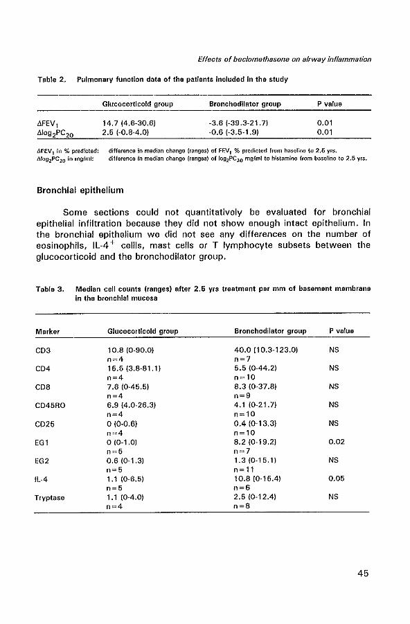
Effects of beclomethasone on airway inflammation
Table 2. Pulmonary function data of the patients included in the study
MEV 1 in % predicted: dfo92PC20 In mgfmf:
Glucocorticoid group
14.7 (4.6-30.6) 2.5 (-0.8-4.0)
Bronchodilator group
-3.6 (-39.3-21. 7) -0.6 (-3.5-1.9)
P value
0.01 0.01
difference In median change (ranges) of FEV1 % predicted from baseline to 2.5 yrs. difference in median change (ranges) of fog2PC20 mgfml to histamine from baseline to 2.5 yrs.
Bronchial epithelium
Some sections could not quantitatively be evaluated for bronchial epithelial infiltration because they did not show enough intact epithelium. In the bronchial epithelium we did not see any differences on the number of eosinophils, IL-4 + cellls, mast cells or T lymphocyte subsets between the glucocorticoid and the bronchodilator group.
Table 3. Median cell counts (ranges) after 2.5 yrs treatment per mm of basement membrane in the bronchial mucosa
Marker Glucocorticoid group Bronchodilator group P value
CD3 10.8 10-90.0) 40.0 110.3-123.0) NS 11=4 11=7
CD4 15.5 (3.8-81.11 5.5 (0-44.2) NS 11=4 n~10
CD8 7.6 (0-45.5) 8.3 (0-37.8) NS 11=4 n=9
CD45RO 6.9 14.0-26.3) 4.1 (0-21.7) NS n=4 n~ 10
CD25 0(0·0.6) 0.4 (0-13.3) NS 11=4 n= 10
EGl 0(0-1.0) 8.2 (0-19.2) 0.02 11=5 11=7
EG2 0.6 (0-1.3) 1.3 (0-15.1) NS n=5 n~ 11
Il-4 1.1 10-6.5) 10.8 (0-15.4) 0.05 n=5 11=6
Tryptase 1.1 (0-4.0) 2.5 (0-12.4) NS n=4 n=8
45
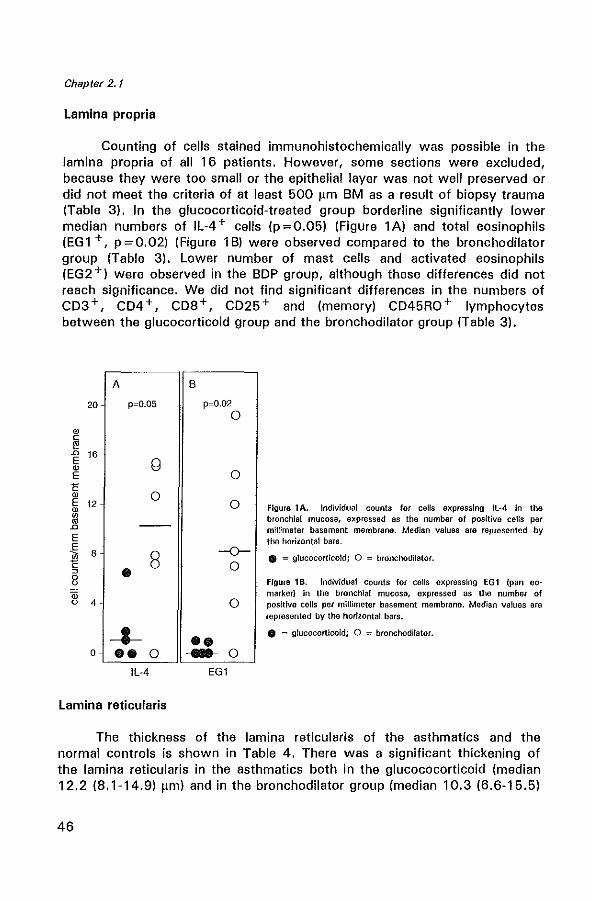
Chapter 2.1
Lamina propria
Counting of cells stained immunohistochemically was possible in the lamina propria of all 16 patients. However, some sections were excluded, because they were too small or the epithelial layer was not well preserved or did not meet the criteria of at least 500 11m 8M as a result of biopsy trauma (Table 3). In the glucocorticoid-treated group borderline significantly lower median numbers of IL-4 + cells (p; 0.05) (Figure 1 A) and total eosinophils (EG1 +, p;0.02) (Figure 1B) were observed compared to the bronchodilator group (Table 3). Lower number of mast cells and activated eosinophils (EG2+) were observed in the BOP group, although these differences did not reach significance. We did not find significant differences in the numbers of C03 +, C04 +, C08 +, C025 + and (memory) C045RO + lymphocytes between the glucocorticoid group and the bronchodilator group (Table 3).
20
'" ~ E 16
'" E E '" ~ 12
ill D
E .€ 8 ~ ~
8 ID 0 4 -
0
A
p=O.05
g
o
It 8
-t-ee 0
IL-4
Lamina reticularis
B
p=O.02 o
o
o
-0-o
o
•• .... 0
EGI
Figure lA. Individual counts for celts expressing IL-4 in the bronchial mucosa, expressed as the number of positive celts per millimeter basement membrane. Median values are represented by the horizontal bars .
• = glucocorticoid; 0 = bronchodilator.
Figure 1 B. Individual counts for cens expressing EG 1 (pan eomarker) In the bronchial mucosa, expressed as the number of positive celfs per millimeter basement membrane. Median values are represented by the horizontal bars.
• = glucocorticoId; 0 = bronchodilator •
The thickness of the lamina reticularis of the asthmatics and the normal controls is shown in Table 4. There was a significant thickening of the lamina reticularis in the asthmatics both in the glucococorticoid (median 12.2 (8.1-14.9) 11m) and in the bronchodilator group (median 10.3 (6.6-15.5)
46
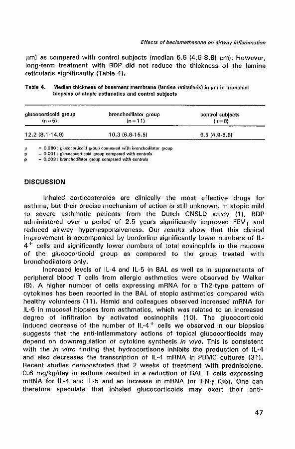
Effects of beclomethasone on airway inflammation
f\m) as compared with control subjects (median 6.5 (4.9-8.8) f\m). However, long-term treatment with BDP did not reduce the thickness of the lamina reticularis significantly (Table 4).
Table 4. Median thickness of basement membrane (lamina reticularis) In pm in bronchial biopsies of atopic asthmatics and control subjects
glucocorticoid group In~5)
12.218.1-14.9)
bronchodilator group In~ 11)
10.316.6-16.5)
p = 0.280: glucocorticoid group compared with bronchodilator group p = 0.001 : glucococorticoid group compared with controls p = 0.003 : bronchodilator group compared with controls
DISCUSSION
control subjects In~8)
6.5 14.9-8.8)
Inhaled corticosteroids are clinically the most effective drugs for asthma, but their precise mechanism of action is still unknown. In atopic mild to severe asthmatic patients from the Dutch CNSLD study (1 ),BDP administered over a period of 2.5 years significantly improved FEV 1 and reduced airway hyperresponsiveness. Our results show that this clinical improvement is accompanied by borderline significantly lower numbers of IL-4 + cells and significantly lower numbers of total eosinophils in the mucosa of the glucocorticoid group as compared to the group treated with bronchodilators only.
Increased levels of IL-4 and IL-5 in BAL as well as in supernatants of peripheral blood T cells from allergic asthmatics were observed by Walker (9). A higher number of cells expressing mRNA for a Th2-type pattern of cytokines has been reported in the BAL of atopic asthmatics compared with healthy volunteers (11). Hamid and colleagues observed increased mRNA for IL-5 in mucosal biopsies from asthmatics, which was related to an increased degree of infiltration by activated eosinophils (10). The glucocorticoid induced decrease of the number of IL-4 + cells we observed in our biopsies suggests that the anti-inflammatory actions of topical glucocorticoids may depend on downregulation of cytokine synthesis in vivo. This is consistent with the in vitro finding that hydrocortisone inhibits the production of IL-4 and also decreases the transcription of IL-4 mRNA in PBMC cultures (31). Recent studies demonstrated that 2 weeks of treatment with prednisolone, 0.6 mg/kg/day in asthma resulted in a reduction of BAL T cells expressing mRNA for IL-4 and IL-5 and an increase in mRNA for IFN-y (35). One can therefore speculate that inhaled glucocorticoids may exert their anti-
47
Chapter 2.1
inflammatory actions by suppressing IL-4 production in the bronchial mucosa. The importance of IL-4 as a mediator of allergic airway inflammation regulating antigen-induced eosinophil recruitment is supported by studies of Brusselle et a/. (36). Unfortunately, because this study was set up later as a part from the multicentre study (1), we do not have biopsies at the beginning of the study prior to treatment. While it is extremely likely that the reduced number of IL-4 + cells and eosinophils present in the glucocorticoid group is a direct effect of beclomethasone, it can not be excluded with this design of the study that there has not been an adverse effect in the group given bronchodilators alone without BDP.
Eosinophils are known to be effector cells in the pathophysiology of asthma (37,38). Bronchial biopsies from patients treated with long-term (3.7 yr) inhaled BDP showed reduced numbers of mast cells and eosinophils. These changes were not seen in subjects treated with terbutaline (14). An uncontrolled study by Djukanovic and colleagues on 6 weeks of treatment with inhaled BDP has shown a significant decrease in airway hyperresponsiveness accompanied by a reduction in eosinophils, mast cells and T lymphocytes in bronchial mucosal biopsies (39). Our data, showing a reduction in the number of total eosinophils in the bronchial mucosa after long-term treatment with BDP compared with the bronchodilator group are in agreement with these observations. The reduction in eosinophils numbers we found, could possibly be a result of an inhibition of chemoattractants for eosinophils, like IL-3, IL-5 and granulocyt-macrophage colony stimulating factor (GM-CSF), since oral glucocorticoids reduce the concentrations of circulating IL-3, IL-5 and GM-CSF in asthmatics (40). In culture, glucocorticoids decrease cytokine-mediated survival of eosinophils, presumably by stimulating programmed cell death (apoptosis) (41). This process may also explain the reduction in the number of eosinophils in the bronchial mucosa after glucocorticoid treatment.
Interestingly, we detected a significant reduction in total eosinophils, as identified by EG1, but not in EG2+ eosinophils in the glucocorticoid group. EG2 identifies the secreted form of ECP and hence only stains eosinophils that are undergoing activation and/or secretion. Thus, our results suggest that glucocorticoids have no effect on eosinophil degranulation. Recently, it has been reported that eosinophil degranulation is not an important direct target of glucocorticoid action in humans (42).
Topical glucocorticoids have been shown to reduce the numbers of mast cells in bronchial biopsies, in parallel with clinical improvement (43,44). In our study we also observed a reduction in the median numbers of mast cells in the bronchial mucosa of the glucocorticoid group, although it did not reach statistical significance in this limited number of patients. Finally, in agreement with Wilson and colleagues (44). treatment with BDP did not differ from bronchodilator therapy with regard to the total number of T
48
Effects of beclomethasone on airway inflammation
lymphocytes, or their CD4 +, CD8+ or CD45RO+ subsets in the bronchial mucosa.
We did not observe a difference in the thickening of the lamina reticularis between the glucocorticoid and the bronchodilator group as measured by staining with hematoxylin and eosin and the use of a digital image-processing system. These findings are in agreement with another study, which showed that neither short-term (four weeks) nor long-term (3.7 years) corticosteroid treatment reduced the thickening of the reticular basement membrane (14). These results indicate that even long-term treatment with beclomethasone does not reduce the deposition of collagen fibrils and suggest that this deposition may be associated with an irreversible subepithelial fibrosis and airflow limitation.
In conclusion, our data support the hypothesis that the beneficial effects of long-term inhaled BDP therapy may result at least in part from an inhibition of IL-4 production in the bronchial mucosa and in a downregulation of the local eosinophilic inflammation. Recent studies suggest that mast cells as well as eosinophils can store and release IL-4 (24,25). Our findings suggest that inhaled glucocorticoids are very important in suppressing airway inflammation in asthma possibly through their ability to downregulate cytokine production.
ACKNOWLEDGMENTS
We are grateful to Prof. Dr. R. Benner for his continuous support. We thank Mr. T.M. van Os for photographical assistance, Mrs. M. Smith for technical assistance and Mrs. P. Assems for secretarial assistance. This study was financially supported by Glaxo Wellcome B.V., Zeist, The Netherlands.
REFERENCES
1. Kerstjens HAM, Brand PlP, Hughes MD et al. A comparison of bronchodilator therapy with or without inhaled corticosteroid therapy for obstructive airways disease. N Engl J Med 1992; 327: 1413-9.
2. Haahtela T, Jarvinen M, Kava T et al. Comparison of a B2-agonist, terbutaline with an inhaled corticosteroid, budesonide, in newly detected asthma. N Eng J Med 1991; 325: 388-92.
3. Juniper EF, Kline PA, Vanzieleghem MA, Ramsdale EH, O'Byrne PM, Hargraeve FE. Effect of long-term treatment with an inhaled corticosteroid Ibudesonide) on airway hyper responsiveness and clinical asthma in nonsteroid-dependent asthmatics. Am Rev Respir Dis 1990; 142: 832-6.
4. Lundgren R, Soderberg M, Horstedt P, Stenking R. Morphological studies of bronchial mucosal biopsies from asthmatics before and after ten year of treatment with inhaled steroids. Eur Respir J 1988; 1: 881-9.
5_ Van Essen-Zandvliet EE, Hughes MD, Waalkens HJ et al. Effects of 22 months of
49
Chapter 2.1
treatment with inhaled corticosteroids andlor beta-2-agonists on lung function, airway responsiveness, and symptoms in children with asthma. Am Rev Respir Dis 1992; 146: 547-54.
6. Bradley Bl, Azzawi M, Jacobson M of al. Eosinophils, T-Iymphocytes, mast cells, neutrophils and macro phages in bronchial biopsy specimens from atopic subjects with asthma: comparison with biopsy specimens from atopic subjects without asthma and normal subjects and relationship to bronchial hyperresponsiveness. J Allergy elin Immunol1991; 88: 661-74.
7. Bentley AM, Menz G, Storz C et al. Identification of T-Iymphocytes, macro phages, and activated eosinophils in the bronchial mucosa in intrinsic asthma. Am Rev Respir Dis 1992; 146: 500-6.
8. Ojukanovic R, Wilson JW, Britten KM et al. Quantification of mast cells and eosinophils in the bronchial mucosa of symptomatic atopic asthmatics and healthy control subjects using immunohistochemistry. Am Rev Respir Dis 1990; 142: 863-71.
9. Walker C, Bode E, Boer l, Hansel TT, Blaser K, Virchow JC. Allergic and non allergic asthmatics have distinct patterns of T-cell activation and cytokine production in peripheral blood and bronchoalveolar lavage. Am Rev Respir Dis 1992; 146: 109-15.
10. Hamid 0, Azzawi M, Ying S et al. Expression of mRNA for interleukfn-5 in mucosal biopsies from asthma. J Clin Invest 1991; 87: 1541-6.
11. Robinson OS, Hamid 0, Ying S et al. Predominant T H2-like bronchoalveolar T-Iymphocyte population in atopic asthma. N Eng J Med 1992; 326: 298-304.
12. Robinson OS, Ying S, Bentley AM et al. Relationships among numbers of bronchoalveolar lavage cells expressing messenger ribonucleic acid for cytokines, asthma symptoms, and airway methacholine responsiveness in atopic asthma. J Allergy Clin Immunol 1993; 92: 397-403.
13. Roche WR, 8easly R, Williams JH, Holgate ST. Subepithelial fibrosis in the bronchi of asthmatics. lancet 1989; 1: 520-3.
14. Jeffery PK, Godfrey RW, Adelroth E, Nelson F, Rogers A, Johansson S-A. Effects of treatment on airway inflammation and thickening of basement membrane reticular collagen in asthma. A quantitative light and electron microscopic study. Am Rev Respir Dis 1992; 145: 890-9.
15. Mosmann TR, Coffman RL. Thl and Th2 cells: different patterns of Iymphokine secretion lead to different functional properties. Ann Rev Immunol1989; 7: 145-73.
16. Verce1li 0, Jabara HH, lanener RP, Geha RS. IL-4 inhibits the synthesis of IFN-gamma and induces the synthesis of IgE in hUman mixed lymphocyte cultures. J Immunol 1990; 144; 570-3.
17. Del Prete GF, Maggi E, Parronchi P. Il-4 is an essential co-factor for the IgE synthesis induced in vitro by human T-cell clones and their supernatants. J Immunol 1988; 140: 4193-7.
18. Howard M, Farrar J, Hilfiker MT et al. Identification of a T cell-derived B cell growth factor distinct from interleukin 2. J Exp Med 1982; 155: 914-23.
19. Isakson PC, Pure E, Vitetta ES, Krammer PH. T cell-derived B cell differentiation factor (s). J Exp Mod 1982; 155: 734-48.
20. Bradding P, Feather IH, Howarth PH et al. Interleukin 4 is localized to and released by human mast cells. J Exp Med 1992; 176: 1381-6.
21. Clutterbuck EJ, Hirst MA, Sanderson CJ. Human interleukin-5 (lL-5) regulates the production of eosinophils in human bone marrow cultures: comparison and interaction with IL-1, IL-3, IL-6, and GM-CSF. 8100d 1989; 73: 1504-12.
22. lopez AFt Sanderson CJ, Gamble JR, Campbell HO, Young IG, Vadas MA. Recombinant human interleukin-5 is a selective activator of human eosinophil function. J Exp Med 1988; 167: 219-24.
23. Walsh GM, Hartnell At Wardlaw AJ, Kurihara K, Sanderson CJ, Kay AB. Il-5 enhances
50
Effects of beclomethasone on airway inflammation
the in vitro adhesion of human eosinophils, but not neutrophils, in a leucocyte integrin (CD11/18}-dapandent mannar. Immunology 1990; 71: 258-65.
24. Bradding P, Roberts JA, Britten KM et al. Interleukin-4, -5, and -6 tumor necrosis factor -a in normal and asthmatic airways: evidence for the human mast cell as a source of these cytokines. Am J Respir Cell Mol Bioi 1994; 10: 471-80.
25. Nonaka, M., R. Nonaka, K. Woolley et al. Distinct immunohistochemical localization of IL-4 in human inflamed airway tissues. J Immunol 1995; 155: 3234A4.
26. Corrigan CJ, Hackzu A, Gemou-Engesaeth V et al. CD4-T lymphocyte activation in asthma is accompanied by increased serum concentrations of interleukin-5. Am Rev Resp!r Dis 1993; 147: 540-7.
27. Ohnishi T, Kita H, Weiler D. IL-5 is the predominant eosinophil-active cytokine in the antigen-induced pulmonary late-phase reaction. Am Rev Respir Dis 1993; 147: 901-7.
28. Barnes PJ, Pedersen S. Efficacy and safety of inhaled corticosteroids in asthma. Am Rev Respir Dis 1993; 148: 51-26.
29. Robinson OS, Hamid 0, Ying S et al. Prednisolone treatment in asthma is associated with modulation of bronchoalveolar lavage cell interleukin-4, interleukin-5, and interferon-y cytokine gene expression. Am Rev Respir Dis 1993; 148: 901-6.
30. Guyre PM, Girard MT, Morganelli PM, Manginiello PD. Glucocorticoid effects on the production and action of immune cytokines. J Steroid Biochem 1988; 30: 89-93.
31. Byron KA, Varigos G, Wootton A. Hydrocortisone inhibition of human interleukin-4. Immunology 1992; 77: 624-6.
32. Brand PLP, Kerstjens HAM, Postma OS et al. Long-term multi centre trial in chronic nonspecific lung disease: methodology and baseline assessment in adult patients. Eur RespirJ 1992; 5: 21-31.
33. ATS Task Group. Standards for the diagnosis and care of patients with chronic obstructive pulmonary disease (COPD) and asthma. Am Rev Respir Dis 1987; 136: 225-31.
34. Cockcroft OW, Kilian ON, Mellon JJA, Hargreave FE. Bronchial hyperreactivity to inhaled histamine: a method and clinical survey. Clin Allergy 1977; 7: 235-43.
35. Robinson D. Hamid Q, Ying S et al. Prednisolone treatment in asthma is associated with modulation of bronchoalveolar lavage cell interleukin-4, interleukin-5, and interferon-y cytokine gene expression. Am Rev Respir Dis 1993; 148: 401-6.
36. Brusselle GG, Kips Je, Tavernier JH et al. Attenuation of allergic airway inflammation in IL-4 daficient mica. Clin and Exp Allergy 1994; 24: 73-80.
37. Bousquet T, Chanez P, Lacoste JJ et al. Eosinophilic inflammation in asthma. N Eng J Mad 1990; 323: 1033-9.
38. Aalbers R, de Monchy JG, Kauffman HF et al. Dynamics of eosinophil infiltration in the bronchial mucosa before and after the late asthmatic reaction. Eur Respir J 1993; 6: 840-7.
39. Djukanovic R, Wilson JW, Britten KM et al. Effect of an inhaled corticosteroid on airway inflammation and symptoms in asthma. Am Rev Respir Dis 1992; 146: 669-74.
40. Corrigan CJ, Hamid Q, North J, Barkans J, Moqbel R, Durham S, Gemou-Engesaeth V, Kay AB. Peripheral blood CD4 but not COB T -lymphocytes in patients with exacerbation of asthma transcribe and translate messenger RNA encoding cytokines which prolong eosinophil survival in the context of a Th2-type pattern: effect of glucocorticoid therapy. Am J Raspir Cell Mol 8io11995; 12: 567-78.
41. Barnes PJ.lnhaled glucocorticoids for asthma. New Engl J Med 1995: 332: 868-75. 42. Kita H, Abu-Ghazaleh R, Sanderson CJ, Gleich GJ. Effect of steroids on immunoglobulin
induced eosinophil degranulation. J Allergy Clin Immunol1991; 87: 70-7. 43. Laitinen LA, Laitinen A, Haahtela T. A comparative study of the effects of an inhaled
corticosteroid, budesonide, and of a B2-agonist, terbutaline on airway inflammation in newly diagnosed asthma. J Allergy Clin Immunol 1992; 90: 32-42.
51
Chapter 2.1
44. Wilson JW, Djukanovic H, Howarth PH, Holgate ST. Inhaled beclomethasone dipropionate downregulates airway lymphocyte activation in atopic asthma. Am J Respir Crit Care Mad 1994; 149: 86-9.
52
CHAPTER 2.2
INFLUENCE OF FLUTICASONE PROPIONATE ON AIRWAY INFLAMMATION IN ATOPIC ASTHMATICS'
Gertrude M. Moller1,2, Shelley E. Overbeek 1, Cornelia G. van HeldenMeeuwsen2, Paul G. Mulder3, Henk C. Hoogsteden 1
Departments of Pulmonary Diseases', Immunolog0, Epidemiology and Biostatistics3, Erasmus University and University Hospital Dijkzigt, Rotterdam, The Netherlands
!If Submitted for publication.

Effects of f/uticasone on airway inflammation
ABSTRACT
Fluticasone propionate is a new, topically active inhaled glucocorticoid with a high affinity for the glucocorticoid receptor. We investigated the effects of 3 months inhaled fluticasone propionate therapy on bronchial mucosal inflammation and the thickness of the lamina reticularis in subjects with atopic asthma.
Bronchial biopsies were obtained from 24 asthmatics before and after 3 months of treatment with fluticasone propionate 1000 I1g daily, or matched placebo in a randomized, double-blind parallel group study. Sections (6 !Im) were stained for T lymphocyte subsets, eosinophils, mast cells and adhesion molecules.
When the treated (n ~ 11) and placebo (n ~ 13) groups were compared, an improvement in FEV 1 (p ~ 0.009) and in methacholine PC20 (an increase of 3.7 doubling doses, p ~ 0.001) were observed after treatment with fluticasone propionate. This was accompanied by a significant reduction in eosinophils (p~0.005) and a trend toward a decrease in mast cells (p ~ 0.087) and expression of intercellular adhesion molecule-1 (ICAM-I) (p ~ 0.091). In the treated group there was a significant correlation between the reduction in the number of total eosinophils and the improvement in airway hyperresponsiveness (r ~ 0.96, p ~ 0.011). No change in total helper or activated helper T cells was seen. The thickness of the bronchial lamina reticularis was not reduced after 3 months treatment with fluticasone propionate.
In conclusion, 3 months fluticasone propionate treatment bronchial mucosal infiltration of eosinophils and mast downregulates ICAM-1 expression.
INTRODUCTION
reduces the cells, and
Fluticasone propionate (FP) is a new, topically active, inhaled glucocorticoid with a high affinity for the human glucocorticoid receptor and a negligible oral bioavailability (12). Glucocorticoids are the most effective therapy in asthma and suppress the chronic airway inflammation largely by inhibiting transcription factors that regulate abnormal gene expression (1,19).
Bronchial biopsies obtained from asthmatics have demonstrated local infiltration by inflammatory cells in the bronchial mucosa even in mild asthmatics (2). In addition, thickening of the lamina reticularis of the basement membrane is often reported as a characteristic feature of the bronchial mucosa in asthma and is mainly composed of Type III and Type V collagen (11,20). Cell adhesion molecules playa key role in the recruitment of leucocytes from the circulation to the site of the inflammation (14).
55
Chapter 2.2
Vascular cell adhesion molecule-1 (VCAM-I) is important for eosinophil migration, because very late activation antigen-4 (VLA-4), a ligand for VCAM-I, is expressed on the surface of eosinophils (21). In primates with
. experimentally induced allergic asthma, antibodies to intercellular adhesion molecule-1 (lCAM-I, CD54) were reported to reduce symptoms of hyperresponsiveness and airway eosinophilia (27). E-selectin is also involved in eosinophil accumulation in the bronchial mucosa (7). To date, increased expression of these molecules has been demonstrated in bronchial biopsies of asthmatics (17). However, the influence of FP therapy on the expression of these adhesion molecules has not been elucidated.
At the time of design of this study, placebo-controlled studies on the effects of 3 months inhaled FP on inflammatory cells in the bronchial mucosa were not available. Treatment with another inhaled glucocorticoid, beclomethasone dipropionate (BOP), 1000 !Ig daily dose for 4 months in a placebo controlled study showed a significant reduction in mast cells, eosinophils and no change in T lymphocyte subsets (22). Six weeks BOP (daily dose 2000 I1g for 2 wks and 1000 I1g for 4 wks) did not alter the total numbers of T cells or the CD4 + and CD8 + subsets when analyzed in peripheral blood or bronchoalveolar lavage (BAL) fluid. However, activated T cells recovered by BAL were reduced (28). Intranasal FP treatment of rhinitics for 3 months did reduce Langerhans' cells, but did not reduce the number of T lymphocytes in nasal mucosa (9).
The aim of this study was to examine the effect of FP, 500 I1g twice a day, on the number of bronchial mucosal inflammatory cells, the expression of adhesion molecules and the thickness of the lamina reticularis after a period of 3 months in comparison with a placebo group receiving inhaled B2-agonists alone.
MATERIALS AND METHODS
Patients and non-asthmatic controls
Bronchial mucosal biopsy specimens were obtained from 24 nonsmoking atopic asthmatic subjects (six women and eighteen men with a median age of 26 years, range 21 to 56 years). Asthma was defined as a history of episodic wheezing and reversible airway obstruction characterized by an increase in forced expiratory volume in one second (FEV 1) of " 9 % after inhalation of 1000 I1g terbutaline. The asthmatic subjects had a mean FEV 1 of 84.0 ± 16.1 % of the predicted value; the mean of the 210gs of the provocative concentrations of inhaled methacholine required to reduce their FEV 1 by 20% (PC20) was 0.32 ± 2.94 mg/ml. Individual PC20 data were obtained by linear interpolation of the logs of adjacent concentration values
56
Effects of f/uticasone on airway inflammation
(8). Atopy was defined by one or more positive skin-prick tests to extracts of 16 common aeroallergens. All patients were receiving inhaled i32-agonists, and none had taken oral or inhaled glucocorticoids in the month prior to the study. After a run-in period of 2 weeks, during which time they took inhaled i32-agonists only, spirometry was performed and methacholine PC 20 was measured according to the standardized 2 min tidal breathing technique (8). followed by the first fiberoptic bronchoscopy. Patients were randomized in a double-blind fashion to receive 3 months FP, 500 fIg or matched placebo twice daily, in addition to their inhaled i32-agonists. After 3 months of therapy, patients reattended the hospital for repeat measurement of spirometry and bronchial responsiveness, followed by a second fiberoptic bronchoscopy one week later. All measurements of PC 20 and both fiberoptic bronchoscopies were performed at the same time of day. There were no significant differences at baseline between the FP and the placebo group with regard to FEV 1 and PC20•
A control group was composed of eight non-smoking non-asthmatic subjects (three women and five men with a median age of 23 years, range 19 to 52 years). All controls had a PC20 histamine of more than 8 mg/ml and a median FEV1 of 103 (88-110)% of the predicted value. Patient and control characteristics are shown in Table 1. The study was approved by the local Ethics Committee and all participants gave their written informed consent.
Bronchial biopsy and tissue processing
Bronchial biopsy specimens were taken from the carinae of the lingula or the right upper, middle or lower lobes via an Olympus BF IT 10 fiberoptic bronchoscope (Tokyo, Japan) using alligator forceps, Olympus FB 15C by the same operator (S.E.O.). For light microscopic examination, each biopsy specimen was immediately placed in isotonic saline and frozen within 20 min in Tissue-Tek II OCT embedding medium (Miles, Naperville, Il, USA). Samples were stored at -80°C until use. Frozen sections (6flm) were cut on a Reichert-Jung 2800 Frigocut cryostat. Two sections from each biopsy were placed on poly-l-Iysine-coated (Sigma, Diagnostics, St louis, MO, USA) slides, air dried and stored at -80°C until use.
Immunohistochemistry
The following monoclonal antibodies (MoAb) were used: anti-CD3 (leu 4) for identification of total T lymphocytes, anti-CD4 (leu 3) and anti-CD8 (leu 2) for identification of T helper and T suppressor/cytotoxic lymphocyte subpopulations, anti-CD25 (ll-2R) for identification of cells expressing the activation marker interleukin-2 receptor (ll-2R). all from Becton Dickinson,
57
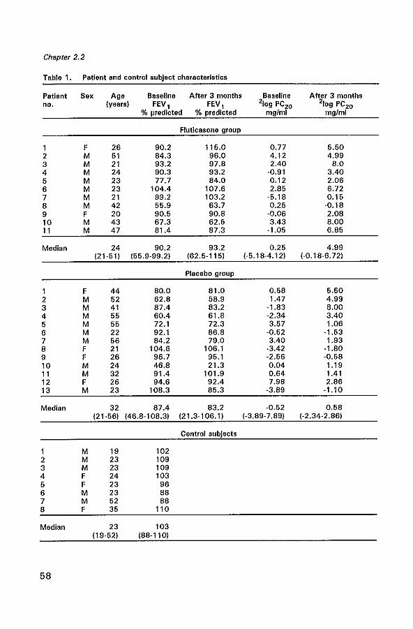
Chapter 2.2
Table 1. Patient and control subject characteristics
Patient Sex Age Baseline After 3 months Baseline After 3 months no. {years} FEV, FEV, 210g PC20 210g PC20
% predicted % predicted mgfml mglml
Fluticasone group
1 F 26 90.2 115.0 0.77 5.60 2 M 51 84.3 96.0 4.12 4.99 3 M 21 93.2 97.8 2.40 8.0 4 M 24 90.3 93.2 ·0.91 3.40 6 M 23 77.7 84.0 0.12 2.06 6 M 23 104.4 107.6 2.85 6.72 7 M 21 99.2 103.2 ·5.18 0.15 8 M 42 55.9 63.7 0.25 ·0.18 9 F 20 90.5 90.8 ·0.06 2.08 10 M 43 67.3 62.5 3.43 8.00 11 M 47 81.4 87.3 ·1.05 6.85
Median 24 90.2 93.2 0.25 4.99 121·51) 155.9·99.2) 162.6-115) 1-5.18-4.12) 1-0.18-6.72)
Placebo group
1 F 44 80.0 81.0 0.68 5.50 2 M 52 62.8 58.9 1.47 4.99 3 M 41 87.4 83.2 -1.83 8.00 4 M 55 60.4 61.8 -2.34 3.40 6 M 56 72.1 72.3 3.57 1.06 6 M 22 92.1 86.8 -0.52 -1.53 7 M 56 84.2 79.0 3.40 1.93 8 F 21 104.6 106.1 -3.42 -1.80 9 F 26 96.7 96.1 -2.56 -0.58 10 M 24 46.8 21.3 0.04 1.19 11 M 32 91.4 101.9 0.64 1.41 12 F 26 94.6 92.4 7.98 2.86 13 M 23 108.3 85.3 -3.89 -1.10
Median 32 87.4 83.2 -0.52 0.58 121-56) 146.8-108.3) 121.3-106.1) 1-3.89-7.89) 1-2.34-2.86)
Control subjects
1 M 19 102 2 M 23 109 3 M 23 109 4 F 24 103 5 F 23 96 6 M 23 88 7 M 52 88 8 F 35 110
Median 23 103 (19-52) 188-110)
58
Effects of fluticBsone on airway inflammation
San Jose, CA, USA; EG 1 recognizing eosinophil catonic protein (ECP) in resting and activated eosinophils, and EG2 recognizing the cleaved form of ECP in activated eosinophils from Pharmacia, Uppsala, Sweden; anti-tryptase recognizing mast cells from Chemicon Brunschwig Chemie, Temecula, CA, USA; anti-intercellular leucocyte adhesion molecule-1 (lCAM-1) (BBA3; British Bio-technology, Abingdon, Oxon, UK), anti-vascular cellular adhesion molecule-1 (VCAM-I) (BBA5; British Bio-technology) and anti-E-selectin (BBA 1; British Biotechnology) for identification of vessels expressing these adhesion molecules.
The MoAb staining was detected by the immuno-alkaline phosphatase anti-alkaline phosphatase (APAAP) method. The sections were fixed in acetone for 10 min at 20°C, rinsed in phosphate-buffered saline (PBS, pH 7.2) and placed in a half-automatic stainer (Shandon, Pittsburgh, PA, USA). In this stainer the slides were sequentially incubated with bovine serum albumin (BSA) 2% in PBS for 10 min, incubated with normal rabbit serum (ClB, Amsterdam, the Netherlands) for 10 min and incubated with the MoAb in the optimal dilution for 30 min at 20°C. The sections were subsequently rinsed in PBS for 5 min and incubated for 30 min with a rabbit anti-mouse (RaM) (1 :20) immunoglobulin antiserum, rinsed in PBS, incubated with APAAP (1 :40) (Dakopatts, Glostrup, Denmark) for 30 min at 20°C, rinsed in PBS and TRIS buffer (pH 8.0). and incubated for 30 min with New Fuchsin substrate (Chroma, Stuttgart, Germany) as a chromagen. The sections were counterstained with Mayers hematoxylin.
Control slides were treated similarly, but in one case excluding the primary antibody and in the other using an unrelated antibody of the same isotype and protein concentration.
Quantification
Biopsies were coded and two sections were counted in a blinded fashion for each antibody and each biopsy at a magnification of 10x40 by one person (G.M.M.) and the mean value was calculated. With an eye piece graticule the numbers of positively stained cells were counted in the epithelium and in a zone 100 !lm deep in the lamina propria along the length of the epithelial basement membrane (BM), which had to be covered with epithelium over at least 500 !lm.
Cells were counted if they stained red and contained a nucleus. The cell counts were expressed as the number per unit (1 mm) of basement membrane. The adhesion molecules were scored semi quantitatively on a 0-3 scale (0 = negative; 1 = weak; 2 = strong; 3 = very strong). There were no significant differences at baseline between the FP and the placebo group with regard to the profiles of the inflammatory cell types and the expression of the adhesion molecules.
59
Chapter 2.2
Quantification of the lamina reticularis
Biopsies were coded prior to analysis. Subepithelial reticular collagen thickness was measured by using a 10x100 oil objective and a digital imageprocessing system (IBAS 2000 system, Kontron, Munchen, Germany). The thickness of the BM was quantitated by measuring only the BM covered by at least a basal epithelial cell layer and where the lamina propria under the BM was at least 100 f.lm deep. The thickness of the BM was measured in five at random fields in two sections of each patient and control subject.
Statistical analysis
The median cell numbers showed a positive skewed distribution and therefore were analyzed using non-parametric statistics. Comparison of treatment effects between FP and placebo was performed by Mann-Whitney U test on percentual changes (after treatment minus before treatment divided by before treatment, multiplied by 100%). Correlation coefficients were obtained by Pearson's rank method.
Median thickness of the lamina reticularis in biopsies from asthmatics taken before treatment was compared with median thickness of biopsies from control subjects using the Mann-Whitney U test. A value of p <0.05 was considered significant.
RESULTS
Pulmonary function data
Three months of FP resulted in a significant improvement in FEV 1 of 6.06% of predicted value as compared with placebo group (-4.33% of predicted, p =0.009) (Table 2). 2LogPC20 in the FP group improved 3.71 dose steps, compared with 0.04 dose steps in the placebo group (p = 0.001) (Table 2). After FP treatment, the reduction in the number of total eosinophils correlated significantly with the improvement in aiway hyperresponsiveness (r = 0.96, p=0.011). In the placebo group no correlations were found.
60
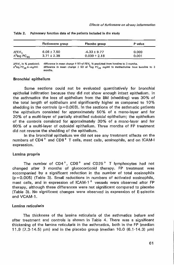
Effects of fluticasone on airway inflammation
Table 2. Pulmonary function data of the patients Included in the study
Fluticasone group
6.06 ± 7.50 3.71 ± 2.38
Placebo group
-4.33 ± 9.77 0.039 ± 2.18
P value
0.009 0.001
.6.FEV1 in % predicted: difference In mean change ± SO of FEV1 % predicled from baseline to 3 months •
.6.210g PC20 In mg/mf: difference in mean change ± SD of log PC20 mg/ml to methacholine f(Om baseline to 3 months.
Bronchial epithelium
Some sections could not be evaluated quantitatively for bronchial epithelial infiltration because they did not show enough intact epithelium. In the asthmatics the loss of epithelium from the 8M (shedding) was 30% of the total length of epithelium and significantly higher as compared to 10% shedding in the controls (p=0.003). In the sections of the asthmatic patients the epithelium consisted for approximately 50% of a mono-layer and for 20% of a mUlti-layer of partially stratified cuboidal epithelium; the epithelium of the controls consisted for approximately 30% of a mono-layer and for 60% of a mUlti-layer of cuboidal epithelium. Three months of FP treatment did not reverse the shedding of the epithelium.
In the bronchial epithelium we did not see any treatment effects on the numbers of CD4 + and CDS+ T cells, mast cells, eosinophils, and on ICAM-I expression.
Lamina propria
The number of CD4 +, CDS + and CD25 + T lymphocytes had not changed after 3 months of glucocorticoid therapy. FP treatment was accompanied by a significant reduction in the number of total eosinophils (p=0.005) (Table 3). Small reductions in numbers of activated eosinophils, mast cells, and in expression of ICAM-1 + vessels were observed after FP therapy, although these differences were not significant compared to placebo (Table 3). No significant changes were observed in expression of E-selectin and VCAM-1.
Lamina reticularis
The thickness of the lamina reticularis of the asthmatics before and after treatment and controls is shown in Table 4. There was a significant thickening of the lamina reticularis in the asthmatics, both in the FP (median 11.9 (7.3-14.5) f1m) and in the placebo group (median 10.0 (6.1-14.3) fjm)
61
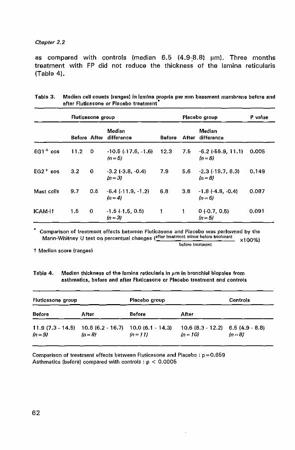
Chapter 2.2
as compared with controls (median 6.5 (4.9-.8.8) J.lm). Three months treatment with FP did not reduce the thickness of the lamina reticularis (Table 4).
Table 3. Median cell counts (ranges) in lamina propria per mm basement membrane before and after Flutlcasone or Placebo treatment·
Flutlcasone group Placebo group P value
Median Median Before After difference Before After difference
EG1+ eos 11.2 0 -10.5 (-17.6, -1.61 12.3 7.5 -6.2 (-55.9, 11.1) 0.005 In~5} In~8}
EG2 + eos 3.2 0 -3.2 (-3.8, -0.4) 7.9 5.6 -2.3 (-19.7, 8.3) 0.149 In~3} In~8}
Mast cells 9.7 0.5 -6.4 (-11.9, -1.2) 6.8 3.8 -1.8 (-4.8, -0.4) 0.087 In~4} In~6}
ICAM-It 1.5 0 -1.5 (-1.6, 0.5) 0(-0.7, 0.51 0.091 In~3} In~5}
Comparison of treatment effects between Fluticasone and Placebo was performed by the Mann-Whitney U test on percentual changes (after treatment minus before treatment xl00%)
before treatment t Median score (ranges)
Table 4. Median thickness of the lamina reticularis In pm in bronchial biopsies from asthmatics. before and after Fluticasone or Placebo treatment and controls
Fluticasone group
Before
11.9 (7.3 - 14.5) In~9}
After
10.6 (6.2 - 16.7) In~8}
Placebo group
Before
10.0 (6.1 - 14.3) In~ll}
After
10.6 (8.3 - 12.21 In ~ 10)
Comparison of treatment effects between Fluticasone and Placebo: p = 0.659 Asthmatics (before) compared with controls: p < 0.0005
62
Controls
6.5 (4.9 - 8.8) In~8}
Effects of fluticasone on airway inflammation
DISCUSSION
This double-blind placebo-controlled study has shown the antiinflammatory benefit of inhaled FP, 1000 I1g daily, in asthma. After 3 months of treatment there was a significant improvement in FEV 1 and airway hyperresponsiveness (PC20 methacholine) accompanied by a significant reduction in numbers of total eosinophils in bronchial mucosal biopsies in the FP group. The decrease in eosinophils after FP therapy was significantly correlated with improvement in hyperresponsiveness. There was a concomitant trend toward a reduction in mast cells and ICAM-I expression.
The effect on eosinophils and mast cells was in line with preliminary findings of others in short- and long-term studies with FP (6,15,10) and BOP (5,22). In a study with FP therapy, 500 I'g for six weeks significantly reduced eosinophil counts in both BAL and peripheral blood (15). Moreover, six weeks treatment with FP, 500 I1g daily dose was effective in reducing the numbers of eosinophils and mast cells in the lamina propria (6). In an uncontrolled study, after 6 weeks treatment with inhaled BOP there was a reduction in eosinophils, mast cells and T lymphocytes in bronchial mucosal biopsies (5). Glucocorticoids appear to inhibit the cytokine-mediated survival of eosinophils, presumably by stimulating programmed cell death (apoptosis) (25). This process may partly explain the reduction in the number of eosinophils in the bronchial mucosa after glucocorticoid treatment. IL-5 is an important selective eosinophil chemoattractant (16) and IL-3 and GM-CSF can induce chemotactic activity for eosinophils (26). Glucocorticoids reduce the concentrations of circulating IL-3, IL-5 and GM-CSF in asthmatics (3) and inhibit the increased expression of GM-CSF in the epithelium of asthmatics (24). This may be another important mechanism for the eosinopenic effect of glucocorticoids.
Interestingly, we detected a significant reduction in total eosinophils, as identified by EG1, but not in EG2 + eosinophils in the FP group. EG2 identifies the secreted form of ECP and hence only stains eosinophils that are undergoing activation and secretion. Thus, our results suggest that glucocorticoids are only weakly effective in inhibiting secretion of eosinophil basic proteins. This observation has been confirmed by others in the literature (13).
Modification of cytokines by glucocorticoids, which we did not investigate, has been reported (3,10,18). In a double-blind study of mild atopic asthmatics, prednisolone therapy was associated with clinical improvement and a reduction in the percentages of BAL fluid cells expressing mRNA encoding IL-4 and IL-5 (18). In biopsies from asthmatic patients receiving 6 weeks FP, 500 I1g twice daily, there was a significant reduction in immunostaining for IL-2, IL-3, IL-4, IL-5, IL-6, IL-8, TNF-a, and GM-CSF but not IFN-y (10). Oral glucocorticoid therapy of severe asthmatics is
63
Chapter 2.2
associated with a reduction in the percentages of peripheral blood CD4 + T lymphocytes expressing IL-3, IL-5, and GM-CSF mRNA and corresponding proteins, which are responsible for eosinophil survival (3).
There were no significant changes in the number of other inflammatory cells, including total T lymphocytes or their CD4 + or CD8 + subsets. These data are in agreement with findings of others (22,28). However, activated T cells recovered by SAL were reduced by steroids (28). FP treatment does not appear to affect the number of T cells or their subpopulations, although the function of T lymphocytes and their cytokine production profile may be influenced.
The downregulation of the numbers of eosinophils may be only one of the mechanisms whereby glucocorticoids modulate inflammation in asthma. The anti-inflammatory effects of FP may be related to the inhibition of the expression of adhesion molecules on endothelial cells. We did find a trend towards a reduction in the expression of ICAM-1 on vessels after FP treatment. It did not reach significance, probably because of the low number of biopsies. Recently, it was shown that dexamethasone was able to downregulate ICAM-1 expression in both a human monocytic and a bronchial epithelial cell line (23). Another study showed that dexamethasone inhibited the expression of adhesion molecules ELAM-1 and ICAM-1 by endothelial cells (4).
In this study, we found a significant thickening of the lamina reticularis of asthmatics compared to controls and we did not observe a reduction in thickening of the lamina reticularis by 3 months FP therapy. These findings are in agreement with earlier data on 2.5 years treatment with beclomethasone which did not show a reduction of the subepithelial collagen deposition (data not shown). Jeffery et al. showed that neither short-term (four weeks) nor long-term (3.7 years) glucocorticoid treatment reduced the thickening of the lamina reticularis (11). These results may implicate that the subepithelial collagen deposition in asthmatics may be associated with irreversible fibrosis and airflow limitation.
Lung function and airway hyperresponsiveness improved after 3 months FP therapy. The PC20 methacholine improved 3.7 doubling doses and the FEV1 6.1 % predicted in the FP group. Our observation that the reduction in eosinophils after FP treatment correlated significantly with the improvement in airway hyperresponsiveness provides strong evidence that there exists a relationship between airway responsiveness and eosinophils in the lamina propria.
Other investigators have also reported improvement in hyperresponsiveness. Donno et al. (6) and Li et al. (15) found improvement in PC20 methacholine after 6 weeks of FP (500 j.lg daily), whereas Montefort et al. (18) showed improvement in PC 20 histamine after 6 weeks of FP, 1000 j.lg daily. These placebo-controlled studies showed clearly that FP
64
Effects of fluticasone on airway inflammation
modifies airway inflammation in association with clinical benefit. In conclusion, this study shows that three months treatment with
1000 J.lg FP reduces the number of eosinophils and mast cells, and downregulates the expression of ICAM-I on vessels in the bronchial mucosa of asthmatic patients in parallel with improved FEV 1 and decreased hyperresponsiveness.
ACKNOWLEDGEMENTS
We thank Prof. Dr. R. Benner and Dr. E. P. Prens for continuous support, and Mrs. P. Assems for secretarial assistance.
REFERENCES
1. Barnes PJ, Pedersen S. Efficacy and safety of inhaled corticosteroids in asthma. Am Rev Respir Dis 1993: 148: 51-526.
2. Bradley Bl, Azzawi M, Jacobson M, Assoufi B, Collins JV, Irani A-MA, Schwartz lB, Durham SR, Jeffery PK, Kay AB. Eosinophils, T-Iymphocytes, mast cells, neutrophils and macrophages in bronchial biopsy specimens from atopic subjects with asthma: comparison with biopsy specimens from atopic subjects without asthma and normal subjects and relationship to bronchial hyperresponsiveness. J Allergy Clin Immunol 1991: 88: 661-74.
3. Corrigan CJ, Hamid a, North J, Barkans J, Moqbel R, Durham S, Gemou-Engesaeth V, Kay AB. Peripheral blood CD4 but not COB T-Iymphocytes in patients with exacerbation of asthma transcribe and translate messenger RNA encoding cytokines which prolong eosinophil survival in the context of a Th2-type pattern: effect of glucocorticoid therapy. Am J Respir Cell Mol Bioi 1995: 12: 567-78.
4. Cronstein BN, Kimmel SC, levin RI, Martiniuk F, Weissmann G. A mechanism for the antiinflammatory effects of corticosteroids: the glucocorticoid receptor regulates leucocyte adhesion to endothelial celfs and expression of endothelial-leucocyte adhesion molecule 1 and intercellular adhesion molecule 1. Proc Nat! Acad Sci USA 1992: 89: 9991-5.
5. Djukanovic A, Wilson JW, Britten KM, Wilson SJ, Walls AF, Roche WR, Howarth PH, Holgate ST. Effect of inhaled corticosteroid on airway inflammation in asthma. Am Rev Respir Dis 1992: 145: 669·74.
6. Donno MD, Foresi A, Chetta A, Bertorelli G, Pesci A, Casalini A, TesU R, Olivieri D. Effect of six week treatment with low dose of inhaled fluticasone propionate on airway inflammation in mild asthma. Eur Respir J 1995: 8: 8302.
7. Georas SN, Liu Me, Newman W, Beall lD, Stealey BA, Bochner BS. Altered adhesion molecule expression and endothelial cell activation accompany the recruitment of human granulocytes to the lung after segmental antigen challenge. Am J Respir Cell Mol Bioi 1992: 7: 261-9.
8. Hargreave FE, Sterk PJ, Ramsdale EH, Oolovich J, Zamel N. Inhalation challenge tests and airway responsiveness in man. Chest 1985: 87S: 202-6.
9. Holm A, Fokkens WJ, Godthelp T, Mulder PG, Vroom TM, Rijntjes E. The effect of three months nasal steroid therapy on nasal T cells and langerhans cells in patients suffering from allergic rhinitis. Allergy 1995: 50: 204-9.
10. Howarth PH, Feather I, Montefort S, Underwood J, Holgate ST. The influence of the
65
Chapter 2.2
inhaled corticosteroid, fluticasone propionate on airway cytokine immunoreactivity in asthma. Am J Respir Crit Care Med 1995: 151: A40.
11. Jeffery PK, Godfrey RW, Adelroth E, Nelson F, Rogers A. Johansson S-A. Effects of treatment on airway inflammation and thickening of basement membrane reticular collagen in asthma. A quantitative light and electron microscopic study. Am Rev Respir Dis 1992: 145: 890-9.
12. Johnson M. The anti·inflammatory profile of fluticasone propionate. Allergy 1995: 50: 11-4.
13. Kita H, Abu-Ghazaleh R, Sanderson CJ, Gleich GJ. Effect of steroids on immunoglobulininduced eosinophil degranulation. J Allergy Clin Immunol1991: 87: 70-7.
14. left AR, Hamann KJ, Wegner CD. Inflammation and cell-cell interactions in airway responsiveness. Am J Physiol 1991: 260: l189-l206.
15. li X, Wilson JW, Bao X. Fluticasone propionate suppress airway inflammation in asthma. Am J Raspir Crit Cara Mad 1995: 151: A386.
16. lopez AF, Sanderson CJ, Gamble JR, Campbell HO, Young IG. Vadas MA. Recombinant human interleukin-5 is a selective activator of human eosinophil function. J Exp Med 1988: 167: 219-24.
17. Montefort S, Roche WR, Howarth PH, Djucanovic R, Gratziou C, Carol! M, Smith l, Britten KM, Haskard 0, lee TH. Intercellular adhesion molecule-l (ICAM-H and endothelial leucocyte adhesion molecule-1 (ElAM-1) expression in the bronchial mucosa of normal and asthmatic subjects. Eur Respir J 1992: 5: 815-23.
18. Montefort S, Feather I, Underwood J, Madden J, Porter C, Holgate ST, Howarth PH. The influence of inhaled fluticasone propionate on symptoms, pulmonary physiology and airway inflammation in asthma. Am J Respir Crit Care Med 1995: 151: A40.
19. Robinson D, Hamid a, Ying S, Bentley A, Assoufi B, Durham S, Kay AB. Prednisolone treatment in asthma is associated with modulation of bronchoalveolar lavage cell interleukin-4, interleukin-5, and interferon-y cytokine gene expression. Am Rev Respir Dis 1993: 148: 401-6.
20. Roche WR, Beasley R, Williams JH, Holgate ST. Subepithelial fibrosis in the bronchi of asthmatics. lancet 1989: 1: 520-3.
21. Schleimer RP, Sterbinsky SA, Kaiser J, Bickel CA, Klunk OA, Tomioka K, Newman W, luscinskas FW, Gimbrone MA, Mcintyre BW, Bochner BS. Il-4 induces adherence of human eosinophils and basophils but not neutrophils to endothelium. Association with expression of VCAM-I, J Immunol 1992: 148: 1086-92.
22. Trigg CJ, Manolitsas ND, Wang J, Calderon MA, McAulay A, Jordan SEt Herdman MJ, Jhalli N, Duddfe JM, Hamilton SA, Devalia Jl, Davies RJ. Placebo·controlled immunopathologic study of four months of inhaled corticosteroids in asthma. Am J Respir Crit Care Med 1994: 150: 17-22.
23. van de Stolpe A, Caldenhove E, Raaijmakers JAM, van der Saag PT. Koenderman l. Glucocorticoid mediated repression of intercellular adhesion molecule-1 expression in human monocytic and bronchial epithelial cell lines. Am J Respir Cell Mol Bioi 1993: 8: 340-7.
24. Sousa AR, Poston RN, lane SJ, Narhosteen JA, lee TH. Detection of GM-CSF in asthmatic bronchial epithelium and decrease by inhaled corticosteroids. Am Rev Respir Dis 1993: 147: 1557-61.
25. Wallen N. Kita H, Weiller 0, Gleich GJ. Glucocorticoids inhibit cytokine mediated eosinophil survival. J Immunol1991: 147: 3490-5.
26. Warringa RAJ, Koenderman l, Kok PTM, Kreukniet J, Bruijnzeel PlB. Modulation and induction of eosinophil chemotaxis by granulocyte-macrophage colony-stimulating factor and interleukin·3. Blood 1991: 77: 2694-2700.
27. Wegner CD, Gundel AH, Reilly P, Haynes N, Gordon Letts l, Rothlein R. Intercellular adhesion molecule-l (tCAM-l) in the pathogenesis of asthma. Science 1990: 247: 456-
66
Effects of fluticasone on airway inflammation
9. 28. Wilson JW, Ojukanovic R, Howarth PH, Holgate ST. Inhaled beclomethasone
dipropionate down regulates airway lymphocyte activation in atopic asthma. Am J Respir Crit Care Med 1994: 149: 86·90.
67

CHAPTER 3
DENDRITIC CELLS IN ASTHMA AND MODULATION BY GLUCOCORTICOIDS
3.1 Increased numbers of dendritic cells in the bronchial mucosa of atopic asthmatics: downregulation by inhaled corticosteroids G/in Exp Allergy 1996 {in press}
3.2 Fluticasone propionate therapy is associated with modulation of dendritic cells in the bronchial mucosa of atopic asthmatics Submitted

CHAPTER 3.1
INCREASED NUMBERS OF DENDRITIC CELLS IN THE BRONCHIAL MUCOSA OF ATOPIC ASTHMATICS: DOWNREGULATION BY INHALED
CORTICOSTEROIDS'
Gertrude M. Miiller1•2, Shelley E. Overbeek 1, Cornelia G. van HeldenMeeuwsen2, Jan Maarten W. van Haarst2, Errol P. Prens2, Paul G. Mulder3,
Dirkje S. Postma4, Henk C. Hoogsteden 1
Departments of Pulmonary Diseases', Immunology2, and Epidemiology and Biostatistics3, Erasmus University and University Hospital Dijkzigt, Rotterdam, and Department of Pulmonary
Diseases4, University Hospital Groningen, The Netherlands
lie/in Exp Allergy 1996 (in press)

Dendritic cells in bronchial mucosa in asthma
SUMMARY
Background: Dendritic cells (DC) are the most potent antigenpresenting cells (A PC) and stimulators of T cells. Dendritic cells are also likely to be essential for the initiation of allergic immune responses in the lung. However, there are not many data on the presence of dendritic cells in the airways of patients with atopic asthma and on the effects of corticosteroid-treatment on such dendritic cells. Objective: We investigated the distribution of dendritic cells in the bronchial epithelium and mucosa of 16 non-smoking atopic asthmatic patients and 8 healthy control subjects using detailed immunohistochemistry (COl a, HLADR, L25 as markers for dendritic cells). Methods: Eleven asthmatics were treated for 2.5 years with bronchodilators only and five with bronchodilators and inhaled beclomethasone dipropionate (BOP), 800 !lg daily. The patients were randomly sampled from a doubleblind multicentre study. Results: There were higher numbers of CDla+ DC (p=0.003), L25+ DC (p = 0.002) and HLA-DR expression (p = 0.042) in the bronchial mucosa of asthmatic patients compared with healthy controls. After 2.5 years of treatment, we found a significant increase in flow expiratory volume in 1 second (FEV 1) (p = 0.009) and a significant decrease in hyperresponsiveness (PC20 histamine) (p = 0.013) in the corticosteroid group (n = 5) compared with the bronchodilator group (n = 11). This clinical improvement in the corticosteroid-treated group was accompanied by significantly lower numbers of CDla+ DC (p=0.008), and HLA-DR expression (p=0.028) in the bronchial mucosa than in the bronchodilator-treated group. Conclusion: Our data suggest that dendritic cells are involved in asthmatic inflammation and that corticosteroids may downregulate the number of dendritic cells.
INTRODUCTION
Atopic asthma is associated with a chronic T cell-mediated inflammatory process in the airways (1-3). Antigen-presenting cells (APC) play an essential role in asthma since T lymphocytes cannot respond to antigens without the help of APC (4). Dendritic cells (DC) are the most potent APC and in contrast to other APC unique in their capacity to stimulate naive T cells (5,6). Therefore, they may playa key role in the initiation of immune responses. In the lung, dendritic cells are predominantly observed in epithelial and subepithelial tissue of the bronch(iollus and in the bronchusassociated lymphoid tissue (7). Typical immunohistological features of DC are a strong major histocompatibility complex (MHC) class II(HLA-DR)
73
Chapter 3.1
expression (8-10), long cytoplasmic extensions, and the absence or a juxtanuclear spot of acid phosphatase. A combination of dendritic morphology and the expression of CD1 a is characteristic for DC (11,12). Increased numbers of CD1 a + DC and increased expression of MHC class II have been described in bronchial biopsies of asthmatic patients (13). Corticosteroids are the most effective therapy for asthma, but their precise mechanism of action is still unclear (14). Corticosteroids inhibit cytokine gene transcription and cytokine effects, thereby reducing chronic inflammation in asthma (14-17). To date, the effects of inhaled corticosteroids on DC in asthma are unknown. We hypothesized that the beneficial effects of corticosteroid therapy in atopic asthmatics may in part result from a downregulation of the numbers of DC, resulting in a decreased local T cell stimulation. Therefore we examined the distribution of the DC in the bronchial epithelium and mucosa of atopic asthmatics by immunohistochemistry. To this aim the effects of 2.5 years of double-blind treatment with bronchodilators alone or with bronchodilators plus beclomethasone dipropionate (8DP) were evaluated. The patients were randomly sampled from the Dutch Chronic Non-Specific Lung Disease (CNSLD)-study (18).
MATERIALS AND METHODS
Patients and Control subjects
Sixteen non-smoking atopic asthmatic patients (seven women, nine men, median age 43 years, range 24 - 61 years) were randomly sampled from the Dutch CNSLD study group (18,19) in two participating centres, Groningen and Rotterdam.
The diagnosis of asthma was based on a history of attacks of breathlessness and wheezing without chronic cough or sputum production (i.e. for more than 3 months per year), according to the criteria of the American Thoracic Society (20). Atopy was defined as a positive skin-prick test to house-dust mite or at least two of 12 common aeroallergens (mean weal size > 0.7 times the histamine weal size (21)). All patients showed airway hyperreactivity to histamine with a provocative concentration of histamine causing a 20% decrease in FEV1 (PC20) of,,; 8 mg/mL (19,21). Patients were treated in a double-blind fashion with an inhaled fl2-agonist (terbutaline, 250 fl9 two puffs) plus either inhaled corticosteroid (8DP, 100 fl9 two puffs) (BA+CS) (n;5), an anticholinergic bronchodilator (ipratropium bromide, 20 fl9 two puffs) (BA+AC) (n;5) or placebo (BA+PL) (n;6). All medication was taken four times daily. Because no significant differences were found between the BA + AC group and the BA + PL group with regard to
74
Dendritic cells in bronchial mucosa in asthma
FEV 1 and PC 20 the data were subsequently pooled for analysis in one bronchodilator group (n = 11). The fibreoptic bronchoscopy was performed at the end of the 2.5 years study in the same period (between August and December) in both centres, before breaking the code. Eight healthy nonsmoking subjects (three women, five men, median age 23 years, range 1 g -52 years) without medication were studied as controls. All controls had a PC20 of more than 8 mg/mL and a median FEV 1 of 103 (88-110) % predicted. Patient and control characteristics are shown in Table 1. Further details of the study methods have been described previously (19). The study protocol was approved by the Medical Ethics Committee; all patients and controls gave written informed consent.
Bronchoscopy
Fibreoptic bronchoscopy (Olympus model BF IT 10, Tokyo, Japan) was performed with atropine 0.5 mg intramuscularly as premedication. Terbutaline, two puffs of 250 !lg per Nebuhaler, was given 30 min before the procedure. The nose, throat and vocal cords were anaesthetized with topical lidocaine spray. An Olympus alligator forceps model FB15C and the fenestrated forceps model FB 19C were used to take two biopsies from segmental and subsegmental divisions of the main bronchi.
Bronchial biopsies
Each biopsy was immediately placed in isotonic saline and frozen within 20 min in Tissue-Tek II OCT embedding medium (Miles, Naperville, Illinois, USA). Samples were stored at -80°C until use. Frozen sections (6 !lm) were cut on a Reichert-Jung 2800 Frigocut cryostat. Two sections from each biopsy were placed on poly-L-Iysine-coated (Sigma Diagnostics, St Louis, MO, USA) microscopic slides. Sections were air dried for 30 min and stored at -80°C until use.
Immunohistochemistry
The following monoclonal antibodies (MoAb) were used: OKT6 (CD1 a) (American Type Culture Collection, Rockville, Maryland, USA), directed against the CD1 a antigen of DC (11); L25, a MoAb directed against B cells and DC (12) was kindly provided by Dr T. Takami (Gifu, Japan); HLA-DR (Central Laboratory of the Netherlands Red Cross Blood Transfusion Service
75
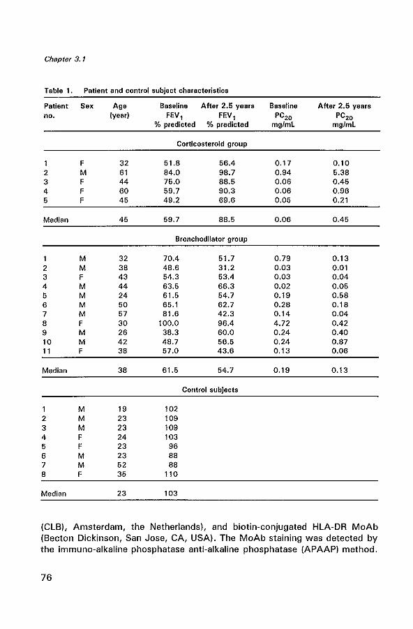
Chapter 3.1
Table 1. Patient and control subject characteristics
Patient Sex Age Baseline After 2.6 years Baseline After 2.6 years no. (year) FEV, FEV, PC20 PC20
% predicted % predicted mgfmL mg/mL
Corticosteroid group
F 32 51.8 56.4 0.17 0.10 2 M 61 84.0 98.7 0.94 5.38 3 F 44 75.0 88.5 0.06 0.45 4 F 60 59.7 90.3 0.06 0.96 5 F 45 49.2 69.6 0.05 0.21
Median 45 59.7 88.5 0.06 0.45
Bronchodilator group
M 32 70.4 51.7 0.79 0.13 2 M 38 48.6 31.2 0.03 0,01 3 F 43 54.3 63.4 0.03 0.04 4 M 44 63.5 66.3 0.02 0.05 5 M 24 61.5 54.7 0.19 0.58 6 M 50 66.1 62.7 0.28 0.18 7 M 57 81.6 42.3 0.14 0.04 8 F 30 100.0 96.4 4.72 0.42 9 M 26 38.3 60.0 0.24 0.40 10 M 42 48.7 56.5 0.24 0.87 11 F 38 57.0 43.6 0.13 0.06
Median 38 61.5 54.7 0.19 0.13
Control subjects
M 19 102 2 M 23 109 3 M 23 109 4 F 24 103 5 F 23 96 6 M 23 88 7 M 52 88 8 F 35 110
Median 23 103
(CLB), Amsterdam, the Netherlands), and biotin-conjugated HLA-DR MoAb (Becton Dickinson, San Jose, CA, USA). The MoAb staining was detected by the immuno-alkaline phosphatase anti-alkaline phosphatase (APAAP) method.
76
Dendritic cells in bronchial mucosa in asthma
The sections were fixed in acetone for 10 min at 20°C, rinsed in phosphate-buffered saline (PBS, pH 7.2) and placed in a half-automatic stainer (Shandon, Pittsburgh, PA, USA). In this stainer the slides were sequentially incubated with bovine serum albumin (BSA) 2 % in PBS for 10 min, incubated with normal rabbit serum (CLB, Amsterdam, the Netherlands) for 10 min and incubated with the MoAb in the optimal dilution for 30 min at 20°C. The optimal dilutions were determined by previous titration studies of the specific MoAb ( OKT6 10 f.lg/mL; L25 1 !lg/mL and HLA-DR 0.1 f.lg/mL). The sections were subsequently rinsed in PBS for 5 min and incubated for 30 min with a rabbit anti-mouse (RaM) (1 :20) immunoglobulin antiserum, rinsed in PBS, incubated with APAAP (1 :40) (Dakopatts, Glostrup, Denmark) for 30 min at 20°C, rinsed in PBS and TRIS buffer (pH B.O), and incubated for 30 min with New Fuchsin substrate (Chroma, Stuttgart. Germany). which stained positive cells red. Finally, the sections were rinsed with distilled water, counterstained with Mayer's haematoxylin, and mounted in glycerin gelatin. Control staining was performed by substitution with PBS and incubation with an irrelevant MoAb of the same isotype and concentration. To establish that L25 + DC were also expressing HLA-DR we performed a double-labelling with L25 and biotinylated HLA-DR. The procedure followed the APAAP protocol up to the incubation with APAAP for 30 min. After rinsing in PBS and blocking with normal mouse serum (1 :10) for 10 min, the slides were incubated for 45 min with the second MoAb biotinylated HLADR. Slides were rinsed in PBS and incubated with streptavidine - Jl galactosidase (Biogenex, San Ramon, CA, USA) for 45 min, and rinsed again with PBS. Slides were sequentially incubated with Jl galactoside (Serva, Heidelberg, Germany) which stained positive cells green, rinsed in TRIS buffer (pH B.O) and stained with New Fuchsin, which stained L25 + cells red. Finally, the sections were rinsed in distilled water and mounted in glycerin gelatin.
The immuno-double staining with L25 and HLA-DR was performed on three sections of three different patients, because of scarcity of biopsy material.
Quantification
Biopsies were coded and two sections 120 f.lm apart were counted in a blinded fashion for each antibody and each biopsy at a magnification of 10x40 by one person (G.M.M.) and the mean value was calculated. With an eye piece graticule the numbers of positively stained cells were counted in a zone 100 f.lm deep in the bronchial mucosa along the length of the epithelial basement membrane (BM), which had to be covered with epithelium over at least 500 f.lm. Cells were counted if they stained red and contained a nucleus. The cell counts were expressed as the number per unit (1 mm) of
77
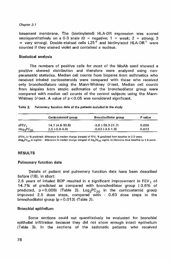
Chapter 3.1
basement membrane. The (biotinylated) HLA-DR expression was scored semiquantitatively on a 0-3 scale (0 = negative; 1 = weak; 2 = strong; 3 = very strong). Double-stained cells L25+ and biotinylated HLA-DR+ were counted if they stained violet and contained a nucleus.
Statistical analysis
The numbers of positive cells for most of the MoAb used showed a positive skewed distribution and therefore were analyzed using nonparametric statistics. Median cell counts from biopsies from asthmatics who received inhaled corticosteroids were compared with those who received only bronchodilators using the Mann-Whitney V-test. Median cell counts from biopsies from atopic asthmatics of the bronchodilator group were compared with median cell counts of the control subjects using the MannWhitney V-test. A value of p <0.05 was considered significant.
Table 2. Pulmonary function data of the patients included In the study
Corticosteroid group
14.7 (4.6-30.6) 2.5 (·0.8-4.0)
Bronchodilator group
-3.6 (·39.3·21.7) ·0.63 (·3.5·1.9)
P value
0.009 0.013
AFEV1 in % predicted: difference in median change (ranges) of FEV1 % predicted from baseline to 2.5 years . .6.!ogzPC20 In mg/ml: difference In median change (ranges) of logzPC2Cl mg/mL to histamine from baseline to 2.5 years.
RESULTS
Pulmonary function data
Detai)s of patient and pulmonary function data have been described before (18). In short: 2.5 years of inhaled BOP resulted in a significant improvement in FEV 1 of 14.7% of predicted as compared with bronchodilator group (-3.6% of predicted, p = 0.009) (Table 2). Log 2PC20 in the corticosteroid group improved 2.5 dose steps, compared with - 0.63 dose steps in the bronchodilator group (p = 0.013) (Table 2).
Bronchial epithelium
Some sections could not quantitatively be evaluated for bronchial epithelial infiltration because they did not show enough intact epithelium (Table 3). In the sections of the asthmatic patients who received
78
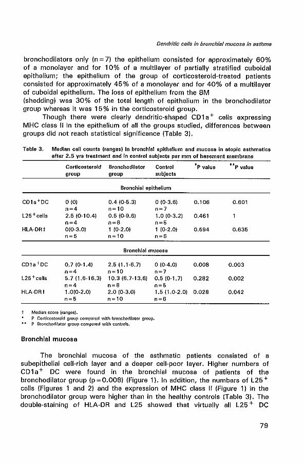
Dendritic cells in bronchial mucosa in asthma
bronchodilators only (n = 7) the epithelium consisted for approximately 60% of a monolayer and for 10% of a multilayer of partially stratified cuboidal epithelium; the epithelium of the group of corticosteroid-treated patients consisted for approximately 45 % of a monolayer and for 40% of a multilayer of cuboidal epithelium. The loss of epithelium from the 8M (shedding) was 30% of the total length of epithelium in the bronchodilator group whereas it was 15% in the corticosteroid group.
Though there were clearly dendritic-shaped CDl a + cells expressing MHC class" in the epithelium of all the groups studied, differences between groups did not reach statistical significance (Table 3).
Table 3. Median cell counts (ranges) In bronchial epithelium and mucosa in atopic asthmatics after 2.5 yrs treatment and in control subjects per mm of basement membrane
Corticosteroid Bronchodilator Control group group subjects
Bronchial epithelium
COla+OC 0(0) 0.4 (0-5.3) o (0-3.6) n=4 n~10 n=7
l25+cells 2.5 (0-10.4) 0.5 (0-9.6) 1.0 (0-3.2) n=4 n=8 n=5
HLA-DRt 0(0-3.0) 1 (0-2.0) 1 (0-2.0) n=5 n~10 n=6
Bronchial mucosa
COla+OC 0.7 (0-1.4) 2.5 (1.1-6.7) o (0-4.0) n=4 n~ 10 n=7
l25 +cells 5.7 (1.6-16.3) 10.3 (6.7-13.6) 0.5 (0-1. 7) n=4 n=8 n=5
HLA-DRt 1.0(0-2.0) 2.0 (0-3.0) 1.5 (1.0-2.0) n=5 n~ 10 n=6
Median score (ranges). P Corticosteroid group compared with bronchodilator group. P Bronchodilator group compared with controls.
Bronchial mucosa
'lip value
0.106 0.601
0.461
0.594 0.635
0.008 0.003
0.282 0.002
0.028 0.042
The bronchial mucosa of the asthmatic patients consisted of a subepithelial cell-rich layer and a deeper cell-poor layer. Higher numbers of CDla+ DC were found in the bronchial mucosa of patients of the bronchodilator group (p = 0.008) (Figure 1). In addition, the numbers of L25 + cells (Figures 1 and 2) and the expression of MHC class " (Figure 1) in the bronchodilator group were higher than in the healthy controls (Table 3). The double-staining of HLA-DR and L25 showed that virtually all L25 + DC
79
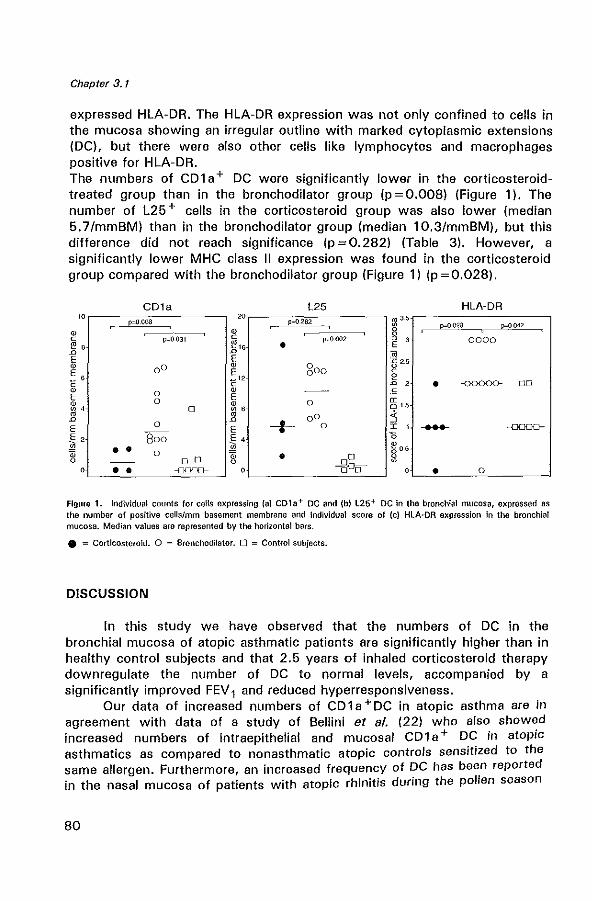
Chapter 3.1
expressed HLA-DR. The HLA-DR expression was not only confined to cells in the mucosa showing an irregular outline with marked cytoplasmic extensions (DC), but there were also other cells like lymphocytes and macrophages positive for HLA-DR. The numbers of CD1 a+ DC were significantly lower in the corticosteroidtreated group than in the bronchodilator group (p =0.008) (Figure 1). The number of L25 + cells in the corticosteroid group was also lower (median 5.7/mmBM) than in the bronchodilator group (median 10.3/mmBM), but this difference did not reach significance (p = O. 282) (Table 3). However, a significantly lower MHC class II expression was found in the corticosteroid group compared with the bronchodilator group (Figure 1) (p = 0.028).
CD1a L25 HLA-DR
" '" rn 3.5 ,",000 p=O.282 p=0,028 p=O ()42 ~ ~~ ~ 8 ,---------------,,-----.-------, C p=O.031 C p=OOO2 0 , 0000 ~ , .t 16 0 E n " E E .c 2.5 ~
0 0 @ goo u
~ , E c
-" e c c n , • -00000- 00 ~ 0
@ s E 0 E <r @ ~ 0 0 1.5 ~ , 0 ~ , • 00 :l n n
~ E 0 E a I 1 .... e-0
~ 2 800 ~ 4 '0
• • ~ 05
'" 0
'" • 00 u
u 0 0 0 ~
0 • • -==- 0 --000 • 0
Figure 1. Individual counts for cells expressing (aJ CD1e+ DC and (b) L26+ DC In the bronchIal mucosa, expressed as the number of positive cells/mm basement membrane and Individual score of (cl HLA·DR expression In the bronchial mucosa. Median values are represented by the horizontal bars .
• = CorticosteroId. 0 = Bronchodilator. 0 = Control subjects.
DISCUSSION
In this study we have observed that the numbers of DC in the bronchial mucosa of atopic asthmatic patients are significantly higher than in healthy control subjects and that 2.5 years of inhaled corticosteroid therapy downregulate the number of DC to normal levels, accompanied by a significantly improved FEV 1 and reduced hyperresponsiveness.
Our data of increased numbers of CD 1 a + DC in atopic asthma are in agreement with data of a study of Bellini et al. (22) who also showed increased numbers of intraepithelial and mucosal CD1 a + DC in atopic asthmatics as compared to nonasthmatic atopic controls sensitized to the same allergen. Furthermore, an increased frequency of DC has been reported in the nasal mucosa of patients with atopic rhinitis during the pollen season
80
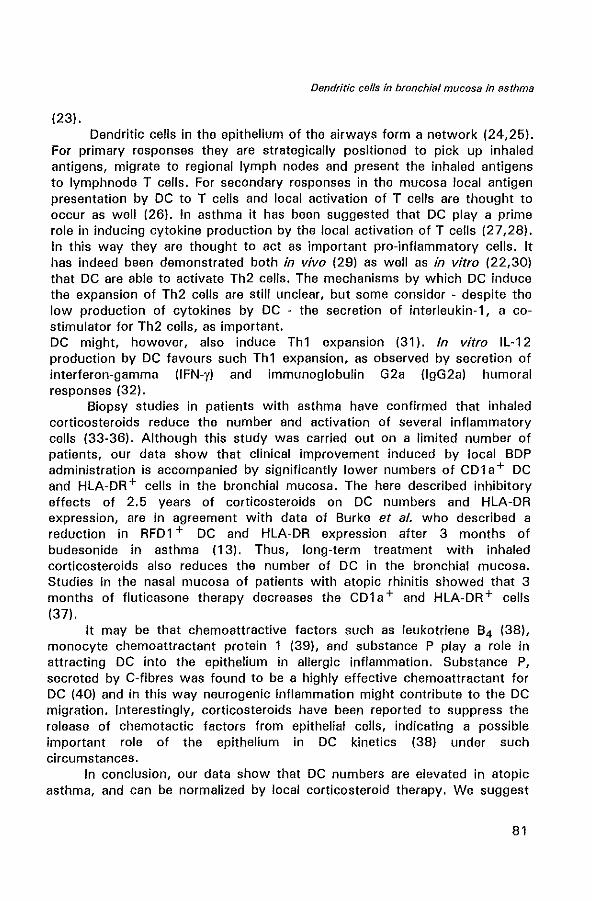
Dendritic cells in bronchial mucosa in asthma
(23). Dendritic cells in the epithelium of the airways form a network (24,25).
For primary responses they are strategically positioned to pick up inhaled antigens, migrate to regional lymph nodes and present the inhaled antigens to Iymphnode T cells. For secondary responses in the mucosa local antigen presentation by DC to T celis and local activation of T cells are thought to occur as well (26). In asthma it has been suggested that DC playa prime role in inducing cytokine production by the local activation of T cells (27,28). In this way they are thought to act as important pro-inflammatory cells. It has indeed been demonstrated both in vivo (29) as well as in vitro (22,30) that DC are able to activate Th2 cells. The mechanisms by which DC induce the expansion of Th2 cells are still unclear, but some consider - despite the low production of cytokines by DC - the secretion of interleukin-1, a costimulator for Th2 cells, as important. DC might, however, also induce Th1 expansion (31). In vitro IL-12 production by DC favours such Th1 expansion, as observed by secretion of interferon-gamma (lFN-y) and immunoglobulin G2a (lgG2a) humoral responses (32).
Biopsy studies in patients with asthma have confirmed that inhaled corticosteroids reduce the number and activation of several inflammatory cells (33-36). Although this study was carried out on a limited number of patients, our data show that clinical improvement induced by local BOP administration is accompanied by significantly lower numbers of CD1a+ DC and HLA-DR + cells in the bronchial mucosa. The here described inhibitory effects of 2.5 years of corticosteroids on DC numbers and HLA-DR expression, are in agreement with data of Burke et al. who described a reduction in RFD1 + DC and HLA-DR expression after 3 months of budesonide in asthma (13). Thus, long-term treatment with inhaled corticosteroids also reduces the number of DC in the bronchial mucosa. Studies in the nasal mucosa of patients with atopic rhinitis showed that 3 months of fluticasone therapy decreases the CD1a+ and HLA-DR+ cells (37).
It may be that chemoattractive factors such as leukotriene B4 (38), monocyte chemoattractant protein 1 (39), and substance P play a role in attracting DC into the epithelium in allergic inflammation. Substance P, secreted by C-fibres was found to be a highly effective chemoattractant for DC (40) and in this way neurogenic inflammation might contribute to the DC migration. Interestingly, corticosteroids have been reported to suppress the release of chemotactic factors from epithelial cells, indicating a possible important role of the epithelium in DC kinetics (38) under such circumstances.
In conclusion, our data show that DC numbers are elevated in atopic asthma, and can be normalized by local corticosteroid therapy. We suggest
81
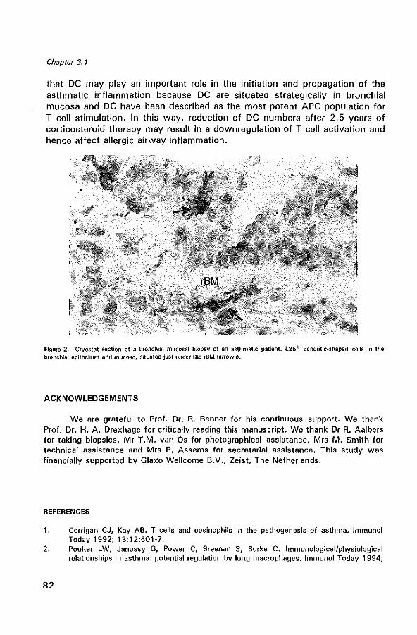
Chapter 3.1
that DC may play an important role in the initiation and propagation of the asthmatic inflammation because DC are situated strategically in bronchial mucosa and DC have been described as the most potent APC population for T cell stimulation. In this way, reduction of DC numbers after 2.5 years of corticosteroid therapy may result in a downregulation of T cell activation and hence affect allergic airway inflammation.
FigUre 2. Cryostat section of a bronchial mucosal biopsy of an asthmatic patient. l2S+ dendritic-shaped cens In the bronchial epithelium and mucosa, situated just under the r8M (arrowsl.
ACKNOWLEDGEMENTS
We are grateful to Prof. Dr. R. Benner for his continuous support. We thank Prof. Dr. H. A. Drexhage for critically reading this manuscript. We thank Dr R. Aalbers for taking biopsies, Mr T.M. van as for photographical assistance, Mrs M. Smith for technical assistance and Mrs P. Assems for secretarial assistance. This study was financially supported by Glaxo Wellcome B.V., Zeist, The Netherlands.
REFERENCES
1. Corrigan CJ, Kay AB. T cells and eosinophils in the pathogenesis of asthma. Immunol Today 1992: 13:12:501-7.
2. Poulter LW, Janossy G, Power C, Sreenan S, Burke C. Immunological/physiological relationships in asthma: potential regulation by lung macro phages. Immunol Today 1994;
82
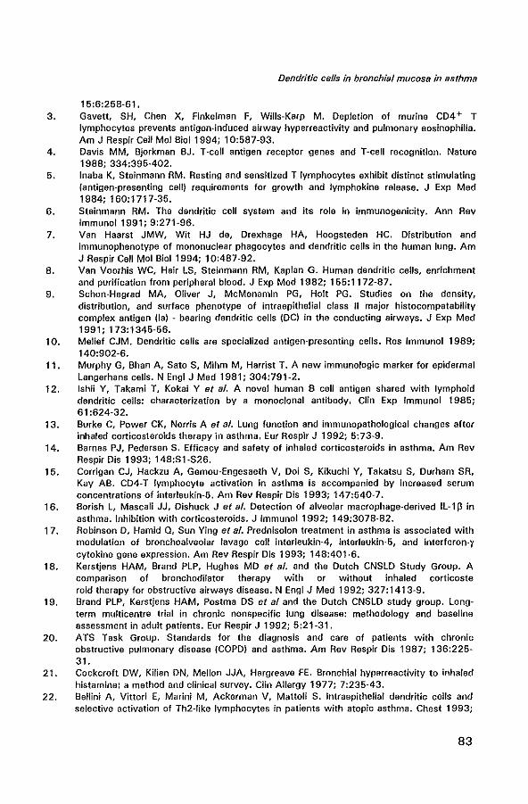
Dendritic cells in bronchial mucosa in asthma
15:6:258-61. 3. Gavett, SH, Chen X, Finkelman F, Wills-Karp M. Depletion of murine CD4 + T
lymphocytes prevents antigen-induced airway hyper reactivity and pulmonary eosinophilia. Am J Respir Cell Mol 8io11994; 10:587-93.
4. Davis MM, Bjorkman BJ. T-cell antigen receptor genes and T-cell recognition. Nature 1988; 334:395-402.
5. Inaba K, Steinmann RM. Resting and sensitized T lymphocytes exhibit distinct stimulating (antigen-presenting cell) requirements for growth and Iymphokine release. J Exp Med 1984; 160:1717-35.
6. Steinmann RM. The dendritic cell system and its role in immunogenicity. Ann Rev Immunol1991; 9:271-96.
7. Van Haarst JMW, Wit HJ de, Drexhage HA, Hoogsteden HC. Distribution and immunophenotype of mononuclear phagocytes and dendritic cells in the human lung. Am J Raspir Call Mol 8io11994; 10:487-92.
8. Van Voorhis WC, Hair lS, Steinmann RM, Kaplan G. HUman dendritic cells, enrichment and purification from peripheral blood. J Exp Med 1982; 155: 1172-87.
9. Schon-He grad MA, Oliver J, McMenamin PG, Holt PG. Studies on the density, distribution, and surface phenotype of intraepithelial class II major histocompatability complex antigen (la) - bearing dendritic cells (DC) in the conducting airways. J Exp Med 1991; 173:1345-56.
10. Meliet CJM. Dendritic cells are specialized antigen-presenting cells. Res Immunol 1989; 140:902-6.
11. Murphy G, Bhan A, Sato S, Mihm M, Harrist T. A new immunologic marker for epidermal Langarhans cells. N Engl J Mad 1981; 304:791-2.
12. Ishii V, Takami T, Kokai V et al. A novel human B cell antigen shared with lymphoid dendritic cells: characterization by a monoclonal antibody. Clin Exp Immunol 1985i 61 :624-32.
13. Burke C, Power CK, Norris A at al. lung function and immunopathological changes after inhaled corticosteroids therapy in asthma. Eur Respir J 1992~ 5: 73-9.
14. Barnes PJ, Pedersen S. Efficacy and safety of inhaled corticosteroids in asthma. Am Rev Raspir Dis 1993; 148:S1-S26.
15. Corrigan CJ, Hackzu A, Gemou-Engesaeth V, Doi S, Kikuchi V, Takatsu S, Durham SR, Kay AB. CD4-T lymphocyte activation in asthma is accompanied by increased serum concentrations of interleukin-5. Am Rev Respir Dis 1993; 147:540-7.
16. Borish L, Mascali JJ, Dishuck J et al. Detection of alveolar macrophage-derived IL-1P in asthma. Inhibition with corticosteroids. J Immunol1992; 149:3078-82.
17. Robinson 0, Hamid Q, Sun Ving et al. Prednisolon treatment in asthma is associated with modulation of bronchoalveolar lavage cell interleukin-4, interleukin-5, and interferon-y cytokine gene expression. Am Rev Respir Dis 1993; 148:401-6.
18. Kerstjens HAM, Brand PLP, Hughes MD et al. and the Dutch CNSLD Study Group. A comparison of bronchodilator therapy with or without inhaled corticoste raid therapy for obstructive airways disease. N Engl J Med 1992; 327: 1413-9.
19. Brand PlP, Kerstjens HAM, Postma DS et al and the Dutch CNSlD study group. Longterm multi centre trial in chronic nonspecific lung disease: methodology and baseline assessment in adult patients. Eur Respir J 1992; 5:21-31.
20. ATS Task Group. Standards for the diagnosis and care of patients with chronic obstructive pulmonary disease (COPD) and asthma. Am Rev Respir Dis 1987; 136:225-31.
21. Cockcroft DW, Kilian ON, Mellon JJA, Hargreave FE. Bronchial hyperreactivity to inhaled histamine: a method and clinical survey. Clin Allergy 1977; 7:235-43.
22. Bellini A, Vittori E, Marini M, Ackerman V, Mattoli S. Intra epithelial dendritic cells and selective activation of Th2-like lymphocytes in patients with atopic asthma. Chest 1993;
83
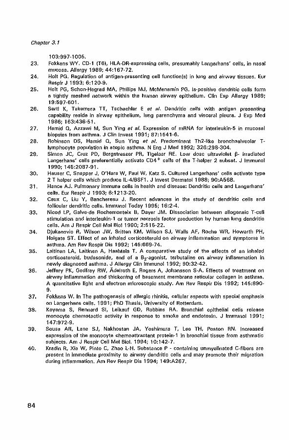
Chapter 3.1
103:997-1005. 23. Fokkens WY. CD~l (T6), HlA~DR-expressing cells, presumably langerhans' cells, in nasal
mucosa. Allergy 1989; 44:167-72. 24. Holt PG. Regulation of antigen-presenting cell function Is) in lung and airway tissues. Eur
Respir J 1993; 6: 120-9. 25. Holt PG, Schon-Hegrad MA, Phillips MJ, McMenamin PG. la-positive dendritic cells form
a tightly meshed network within the hUman airway epithelium. Clin Exp Allergy 1989; 19:597-601.
26. Sertl K, Takemura TT, Tschachler E ot al. Dendritic cells with antigen presenting capability reside in airway epithelium, lung parenchyma and visceral pleura. J Exp Med 1986; 163:436-51.
27. Hamid a, Azzawi M, Sun Ying et al. Expression of mRNA for interleukin-5 in mucosal biopsies from asthma. J Clin Invest 1991; 87:1541~6.
28. Robinson OS, Hamid a, Sun Ying et al. Predominant Th2~like bronchoalveolar Tlymphocyte population in atopic asthma. N Eng J Med 1992; 326:298-304.
29. Simon JC, Cruz PO, Bergstresser PR, Tigelaar RE. low dose ultraviolet p- irradiated langerhans' cells preferentially activate CD4 + cells of the T -helper 2 subset. J Immunol 1990; 145:2087·91.
30. Hauser C, Snapper J, O'Hara W, Paul W, Katz S. Cultured langerhans' cells activate type 2 T helper cells which produce Il-4/BSF1. J Invest Dermatol 1988; 90:A568.
31. Hance AJ. Pulmonary immune cells in health and disease: Dendritic cells and langerhans' cells. Eur Respir J 1993; 6:1213-20.
32. Caux C, Uu Y, Banchereau J. Recent advances in the study of dendritic cells and follicular dendritic cells. Immunol Today 1995; 16:2A.
33. Nicod lP, Galve-de Rochemonteix B, Dayer JM. Dissociation between allogeneic T-cell stimulation and interleukin~ 1 or tumor necrosis factor production by human lung dendritic cells. Am J Respir Cell Mol Bioi 1990; 2:615-22.
34. Djukanovic R, Wilson JW, Britten KM, Wilson SJ, Walls AF, Roche WR, Howarth PH, Holgate ST. Effect of an inhaled corticosteroid on airway inflammation and symptoms in asthma. Am Rev Respir Dis 1992; 145:669-74.
35. Laitinen lA, Laitinen A, Haahtela T. A comparative study of the effects of an inhaled corticosteroid, budesonide, and of a B2~agonist, terbutaline on airway inflammation in newly diagnosed asthma. J Allergy Clin Immunol 1-992; 90:32-42.
36. Jeffery PK, Godfrey RW, Adelroth E, Rogers A, Johansson S-A. Effects of treatment on airway inflammation and thickening of basement membrane reticular collagen in asthma. A quantitative light and electron microscopic study. Am Rev Respir Dis 1992; 145:890-9.
37. Fokkens W. In The pathogenesis of allergic rhinitis, cellular aspects with special emphasis on langerhans cells. 1991; PhD Thesis, University of Rotterdam.
38. Koyama S, Rennard SI, leikauf GO, Robbins RA. Bronchial epithelial cells release monocyte chemotactic activity in response to smoke and endotoxin. J Immunol 1991; 147:972·9.
39. Sousa AR, lane SJ, Nakhostan JA, Yoshimura T, lee TH, Poston RN. Increased expression of the monocyte chemoattractant protein-1 in bronchial tissue from asthmatic subjects. Am J Respir Cell Mol BioI. 1994; 10:142~7.
40. Kradin R, Xia W, Pinto C, Zhao l~H. Substance P ~ containing unmyelinated C-fibers are present in immediate proximity to airway dendritic cells and may promote their migration during inflammation. Am Rev Respir Dis 1994; 149:A267.
84
CHAPTER 3.2
FLUTICASONE PROPIONATE THERAPY IS ASSOCIATED WITH MODULATION OF DENDRITIC CELLS IN THE BRONCHIAL MUCOSA OF
ATOPIC ASTHMATICS'
Gertrude M. Moller 1,2, Shelley E. Overbeek 1, Cornelia G. van HeldenMeeuwsen2, Ton A.W. de Jong3 , Paul G. Mulder4, Hemmo A. Drexhage2,
Hank C. Hoogsteden 1
Departments of Pulmonary Diseases', Immunology, Pathologr, Epidemiology and Biostatistics4, Erasmus University and University Hospital Dijkzigt, Rotterdam, The Netherlands
• Submitted for pUblication

Modulation of dendritic cells by f/uticasone
ABSTRACT
Background: Dendritic cells (DC) are professional antigen-presenting cells (APC) and potent stimulators of resting T cells. Moreover DC express the high affinity receptor for IgE (FcBRI) and membrane bound IgE, instruments for IgE mediated antigen presentation. DC are therefore considered to play an important role in the initiation and propagation of allergic immune responses in the lung. Objective: We examined (1) the distribution of DC in the bronchial epithelium and lamina propria of 24 atopic asthmatics and 8 non-asthmatic controls using immunohistochemistry and (2) the effects of 3 months of an inhaled glucocorticoid (fluticasone propionate (FP)) on the numbers of DC. Methods: Bronchial biopsy specimens were taken from 8 control subjects and 24 subjects with atopic asthma before and after 3 months of treatment with FP, 1000 I1g daily, or matched placebo in a randomized double-blind parallel group study. Sections (6I1m) were stained for CDl a, L25, HLA-DR, IgE, and the high affinity receptor for IgE (FCBRI). Results: There were higher numbers of CDla+ DC (p= 0.025), L25+ DC (p = 0.008), IgE+ cells (p = 0.0003) and a higher HLA-DR expression (p = 0.057) in the lamina propria of asthmatics compared with non-asthmatic controls. When the treated (n = 11) and placebo (n = 13) groups were compared, there was a decrease in hyperresponsiveness (PC20 methacholine) (p = 0.001) after FP. This was accompanied by a decrease in bronchial mucosal CDl a + DC (p = 0.018), a reduction in HLA-DR expression (p=0.008), numbers of IgE+ cells (p=0.010), but not by a change in number of cells expressing FCBRI. Electron microscopic studies showed that electronlucent DC in the epithelium contained the characteristic intracytoplasmic Birbeck granules. Conclusion: This study shows that there are significantly more DC in the bronchial mucosa of asthmatics than in non-asthmatic controls and that fluticasone propionate therapy of asthmatics is associated with a reduction in the numbers of DC.
INTRODUCTION
Dendritic cells (DC) are professional antigen-presenting cells (APC) in the pulmonary tissue (1) and in contrast to other APC unique in their capacity to activate resting naive T cells (2-4). In the lung, DC are predominantly situated in epithelial and subepithelial tissue of the bronch(iol)us and in the bronchus-associated lymphoid tissue (5).
DC are characterized by their high constitutive expression of major histocompatibility complex (MHC) class" (HLA-DR) antigens (6-8), accessory
87
Chapter 3.2
molecules (intercellular adhesion molecule 1 (CD54), B7-1 (CD80), B7-2(CD86)), long cytoplasmic extensions, the absence or a juxtanuclear spot of acid phosphatase, and the presence of intracytoplasmic structures termed Birbeck granules (Bg). A combination of dendritic morphology and expression of CD1 a and L25 are characteristic for DC (9,10).
It is now known that a varying proportion of Langerhans' cells (LC), the representatives of the DC in the skin, carries surface immunoglobulin E (igE) (11,12). There is evidence that, this membrane IgE not only can capture antigen, but also that DC use the IgE for antigen presentation (13). It is also clear that the IgE-binding capacity of these cells is in fact due to the membrane bound expressing of a high affinity receptor for IgE (FCERI) (14,15). FCERI has been traditionally described on basophils and mast cells (16). More recently, the FCERI has also been found on monocytes of atopic dermatitis patients (17) and on eosinophils in parasitic infections (18).
Atopic asthma is associated with a chronic T cell-mediated inflammatory process in the airways (19,20). Since T lymphocytes cannot respond to antigens without the help of APC (21) and DC are superb APC, strategically positioned in the bronchial mucosa and carrying FCERI and IgE on their membrane, it is likely that pulmonary DC play a key role in the initiation and propagation of asthma. In an earlier study we found increased numbers of CD1 a+ DC and increased expression of MHC class II in bronchial mucosal biopsies from asthmatics (22).
Glucocorticoids are by far the most effective therapy for asthma, but their precise mechanism of action is still not certain (23), Glucocorticoids inhibit cytokine gene transcription and cytokine effects, thereby reducing chronic inflammation in asthma (23-26). Fluticasone propionate (FP) is a new topically-active glucocorticoid with a high affinity for the human glucocorticoid receptor and a negligible oral bioavailibility (27). To date, the effects of inhaled FP on DC in asthma are unknown. We hypothesized that one of the beneficial effects of glucocorticoid therapy of asthmatics might be associated with a reduction in the numbers of DC and other IgE-bearing cells, resulting in a decreased local T cell stimulation.
Our aim, therefore, was to evaluate the number of DC, the expression of MHC class II, the number of membrane bound IgE + cells, and of FCERI expression in the bronchial epithelium and the lamina propria in biopsy specimens from a group of asthmatics before and after 3 months FP therapy and from a group of non-asthmatic controls.
88
Modulation of dendritic cells by fluticasone
MATERIALS AND METHODS
Patients and non-asthmatic controls
Bronchial mucosal biopsy specimens were obtained from 24 nonsmoking atopic asthmatic subjects (six women and eighteen men with a median age of 26 years, range 21 to 56 years). Asthma was defined as a history of episodic wheezing and reversible airway obstruction characterized by an increase in forced expiratory volume in one second (FEV1) of ;" 9 % after inhalation of 1 000 I'g terbutaline. The asthmatic subjects had a mean FEV 1 of 84.0 ± 16.1 % of the predicted value; the mean of the 210gs of the provocative concentrations of inhaled methacholine required to reduce their FEV 1 by 20% (PC20) was 0.32 ± 2.94 mg/ml. Atopy was defined by one or more positive skin·prick tests to extracts of 16 common aeroallergens. All patients were receiving inhaled B-agonists, and none had taken oral or inhaled corticosteroids in the month prior to the study. After a run·in period of 2 weeks, during which time they took inhaled B·agonists only, spirometry was performed and methacholine PC 20 was measured according to the standardized 2 min tidal breathing technique (28), followed by the first fiberoptic bronchoscopy. Patients were randomized in a double-blind fashion to receive 3 months FP, 500 I'g or matched placebo twice daily, besides their inhaled B-agonists. After 3 months patients reattended the hospital for repeat measurement of spirometry and bronchial responsiveness, followed by a second fiberoptic bronchoscopy one week later. All measurements of PC20 and both fiberoptic bronchoscopies were performed at the same time of day. There were no significant differences at baseline between the FP and the placebo group with regard to FEV 1 and PC20.
A control group was composed of eight non-smoking non-asthmatic subjects (three women and five men with a median age of 23 years, range 19 to 52 years). All controls had a PC20 histamine of more than 8 mg/ml and a median FEV 1 of 103 (88-110) % of the predicted value. Patient and control characteristics are shown in Table 1. The study was approved by the local Ethics Committee and all participants gave their written informed consent.
Bronchial biopsy and tissue processing
Bronchial biopsy specimens were taken from the carinae of the lingula or the right upper, middle or lower lobes via an Olympus BF IT 10 fiberoptic bronchoscope (Tokyo, Japan) using alligator forceps, Olympus FB 15C by the same operator (S.E.O.). For light microscopic examination, each biopsy specimen was immediately placed in isotonic saline and frozen within 20 min in Tissue-Tek " OCT embedding medium (Miles, Naperville, Il, USA).
89
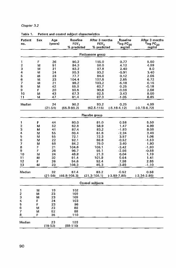
Chapter 3.2
Table 1. Patient and control subject characteristics
Patient Sex Age Baseline After 3 months Baseline After 3 months no. (years) FEV, FEV, 210g PC20 210g PC20
% predicted % predicted mglmi mg/ml
Fluticasone group
1 F 26 90.2 115.0 0.77 5.50 2 M 51 84.3 96.0 4.12 4.99 3 M 21 93.2 97.8 2.40 8.0 4 M 24 90.3 93.2 -0.91 3.40 5 M 23 77.7 84.0 0.12 2.06 6 M 23 104.4 107.6 2.85 6.72 7 M 21 99.2 103.2 -6.18 0.15 8 M 42 55.9 63.7 0.25 -0.18 9 F 20 90.5 90.8 -0.06 2.08 10 M 43 67.3 62.5 3.43 8.00 11 M 47 81.4 87.3 -1.05 6.85
Median 24 90.2 93.2 0.25 4.99 121-511 155.9-99.2) (62.5-115) (-5.18-4.12) (-0.18-6.72)
Placebo group
1 F 44 80.0 81.0 0.58 5.50 2 M 52 62.8 58.9 1.47 4.99 3 M 41 87.4 83.2 -1.83 8.00 4 M 55 60.4 61.8 -2.34 3.40 5 M 55 72.1 72.3 3.57 1.06 6 M 22 92.1 86.8 -0.52 -1.53 7 M 56 84.2 79.0 3.40 1.93 8 F 21 104.6 106.1 -3.42 -1.80 9 F 26 96.7 95.1 -2.56 -0.58 10 M 24 46.8 21.3 0.04 1.19 11 M 32 91.4 101.9 0.64 1.41 12 F 26 94.6 92.4 7.98 2.86 13 M 23 108.3 85.3 -3.89 -1.10
Median 32 87.4 83.2 -0.62 0.68 121-56) 146.8-108.3) 121.3-106.1) (-3.89-7.89) (-2.34-2.86)
Control subjects
1 M 19 102 2 M 23 109 3 M 23 109 4 F 24 103 5 F 23 96 6 M 23 88 7 M 52 88 8 F 35 110
Median 23 103 (19-52) (88-110)
90
Modulation of dendritic cells by fluticasone
Samples were stored at _80° C until use. Frozen sections (6I-lm) were cut on a Reichert-Jung 2800 Frigocut cryostat. Two sections from each biopsy were placed on poly-l-Iysine -coated (Sigma, Diagnostics, St louis, MO, USA) slides, air dried and stored at -80°C until use.
For electron microscopic evaluation, tissue was fixed in 1 % glutaraldehyde in 0.1 M phosphate buffer, pH 7.4, at 0-4°C for 24 hr, postfixed in 1 % Os04 in the same buffer, dehydrated, and embedded in lX 112 resin (Zeiss, Weesp, The Netherlands). Ultrathin (40-60 nm) sections were examined using a Zeiss electron microscope 902 operating at 80 kV.
Immunohistochemistry
The following mouse IgG 1 monoclonal antibodies (mAb) were applied at previously titrated optimal dilutions: OKT6 (10 flg/ml) (American Type Culture Collection, Rockville, MA, USA) directed against the CDl a antigen of DC9; l25 (0.1 I-lg/ml), directed against B cells and DC10, kindly provided by Dr. T. Takami, Gifu, Japan; anti-CD83 (2 I-lg/ml), an IgG3 mAb reacting with HB 15, expressed by DC subsets (29) from Dr. T. F. Tedder, Dana-Farber Cancer Institute, Boston, MA, USA; S100 (21.6 I-lg/ml), a rabbit anti cow polyclonal antibody from Dakopatts, Glostrup, Denmark, which stains lC; anti-HlA-DR (0.01 flg/ml) from Becton Dickinson, San Jose, CA, USA; IgG2b anti-lgE (10 I-lg/ml) from Central laboratory of the Netherlands Red Cross Blood Transfusion Service (ClB), Amsterdam, The Netherlands; and mAb 15-1 (anti-FcBRI), kindly provided by Dr. J.-P. Kinet (National Institutes of Health, Bethesda, MA, USA).
The mAb staining was detected by the immuno-alkaline phosphatase anti-alkaline phosphatase (APAAP) method. The sections were fixed in acetone for 10 min at 20°C, rinsed in phosphate-buffered saline (PBS, pH 7.2) and placed in a half-automatic stainer (Shandon, Pittsburgh, PA, USA). In this stainer the slides were sequentially incubated with bovine serum albumin (BSA) 2% in PBS for 10 min, incubated with normal rabbit serum (ClB) for 10 min and incubated with the mAb in the optimal dilution for 30 min at 20°C. The sections were subsequently rinsed in PBS for 5 min and incubated for 30 min with a rabbit anti-mouse (RaM) (1 :20) immunoglobulin antiserum, rinsed in PBS, incubated with APAAP (1 :40) (Dakopatts) for 30 min at 20°C, rinsed in PBS and TRIS buffer (pH 8.0), and incubated for 30 min with New Fuchsin substrate (Chroma, Stuttgart, Germany) as a chromogen. The sections were counterstained with Mayers hematoxylin. For staining with S100, the sections were incubated with normal goat serum (1:10) (ClB) for 10 min before incubation with the primary antibody, subsequently incubated for 30 min with biotinylated goat-anti-rabbit immunoglobulin antiserum (1 :2) (Biogenex, San Ramon, CA, USA), rinsed in
91
Chapter 3.2
PBS and incubated for 30 min with streptavidin-alkaline phoshatase (1 :50) (Biogenex). The sections were rinsed in PBS, incubated with APAAP (1 :40) and further processed as above.
Control slides were treated similarly, but in one case excluding the primary antibody and in the other using an unrelated antibody of the same isotype and protein concentration.
Quantification
Biopsies were coded and two sections were counted in a blinded fashion for each antibody and each biopsy at a magnification of 10x40 by one person (G.M.M.) and the mean value was calculated. With an eye piece graticule the numbers of positively stained cells were counted in the epithelium and in a zone 100 flm deep in the lamina propria along the length of the epithelial basement membrane (BM), which had to be covered with epithelium over at least 500 flm.
Cells were counted if they stained red and contained a nucleus. The cell counts were expressed as the number per unit (1 mm) of basement membrane. Since the HLA-DR expression was not only confined to inflammatory cells, HLA-DR expression was scored semiquantitatively on a 0-3 scale (0 ; negative; 1 ; weak; 2 ; strong; 3 ; very strong).
Statistical analysis
The median cell numbers showed a positively skewed distribution and therefore were analyzed using non-parametric statistics. There were no significant differences at baseline between the FP and the placebo group with regard to the profiles of the inflammatory cell types and the expression of HLA-DR. Comparison of treatment effects between Fluticasone and placebo was performed by Mann-Whitney U test on percentual changes (after treatment minus before treatment divided by before treatment, multiplied by 100%). Median cell counts from the biopsy specimens from the asthmatic subjects taken before treatment were compared with median cell counts of the control subjects using the Mann-Whitney U test. A value of p < 0.05 was considered significant. The staining with two DC-markers (S 1 00 and CD83) was performed on a few sections, because of scarcity of biopsy material; statistical analysis of these data was not attempted.
Figure 1. Electron microscopical ultrathin sectIon of the bronchial mucosa showing dendritic cells (DC), close to the basement membrane (BMlln the basal layers of the epithelium. Cilia te). x 1,100.
Figure 2. Dendritic cell in the bronchial epithelium, which contains a Birbeck granule (arrow) (see inset, x 50,000). x 3,000.
92
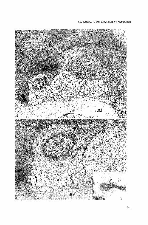
Modulation of dendritic cells by fluticasone
93
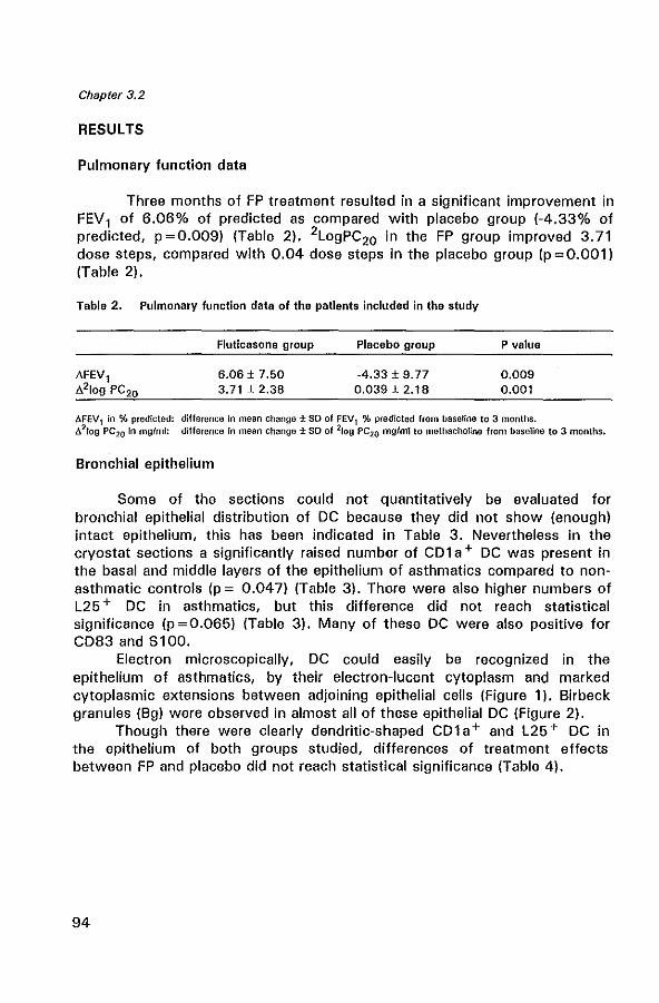
Chapter 3.2
RESULTS
Pulmonary function data
Three months of FP treatment resulted in a significant improvement in FEV 1 of 6.06% of predicted as compared with placebo group (-4.33% of predicted, p=0.009) (Table 2). 2LogPC20 in the FP group improved 3.71 dose steps, compared with 0.04 dose steps in the placebo group (p =0.001) (Table 2).
Table 2. Pulmonary function data of the patients included in the study
Fluticasone group
6.06 ± 7.50 3.71 ± 2.38
Placebo group
-4.33 ± 9.77 0.039 ± 2.18
P value
0.009 0.001
bFEV1 in % predicted: difference In mean change ± so of FEV1 % predicted from baseline to 3 months. I'/Iog PC20 In mgfm!: difference In mean change ± SD of 210g PC20 mg/ml to methacholine from baseline to 3 months.
Bronchial epithelium
Some of the sections could not quantitatively be evaluated for bronchial epithelial distribution of DC because they did not show (enough) intact epithelium, this has been indicated in Table 3. Nevertheless in the cryostat sections a significantly raised number of CD1 a + DC was present in the basal and middle layers of the epithelium of asthmatics compared to nonasthmatic controls (p= 0.047) (Table 3). There were also higher numbers of L25 + DC in asthmatics, but this difference did not reach statistical significance (p = 0.065) (Table 3). Many of these DC were also positive for CD83 and S 100.
Electron microscopically, DC could easily be recognized in the epithelium of asthmatics, by their electron-lucent cytoplasm and marked cytoplasmic extensions between adjoining epithelial cells (Figure 1). Birbeck granules (Bg) were observed in almost all of these epithelial DC (Figure 2).
Though there were clearly dendritic-shaped CD1a+ and L25+ DC in the epithelium of both groups studied, differences of treatment effects between FP and placebo did not reach statistical significance (Table 4).
94
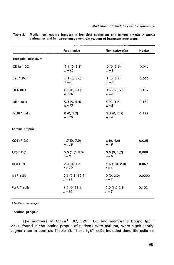
Modulation of dendritic cells by f/uticasone
Table 3. Median cell counts (ranges) in bronchial epithelium and lamina propria in atopic asthmatics and in non-asthmatic controls per mm of basement membrane
Asthmatics Non-asthmatics P value
Bronchial epithelium
CDla+ DC 1.7 (0, 9.1) 0(0,3.6) 0.047 n~ 19 n=8
L25+ DC 5.1 (0, 8.0) 1 (0,3.2) 0.065 n=6 n=5
HLA·DRt 0.9 (0, 2.0) 1.25 (0, 2.0) 0.107 n=20 n=6
IgE + cells 0.8 (0, 6.4) 0(0, 1.5) 0.159 n~ 17 n=6
FC6RI + cells 0(0, 1.2) 3.2 (0, 5.7) 0.132 n=20 n=5
lamina propria
CDla+ DC 1. 7 (0, 7.0) 0(0,4.0) 0.025 n= 19 n=8
L25+ DC 5.0 (1.7, 8.0) 0.5 (0, 1.7) 0.008 n=6 n=5
HLA·DRt 2.0 (0, 3.0) 1.5 (1.0, 2.0) 0.057 n=20 n=6
IgE+ cells 7.1(2.1,12.7) 0(0,2.0) 0.0003 n~ 17 n~6
Fo:RI + cells 3.2 (0, 11.7) 2.0 (1.2·2.8) 0.102 n~20 n=5
t Median score (ranges)
Lamina propria
The numbers of CDla+ DC, L25+ DC and membrane bound IgE+ cells, found in the lamina propria of patients with asthma, were significantly higher than in controls (Table 3). These IgE+ cells included dendritic cells as
95
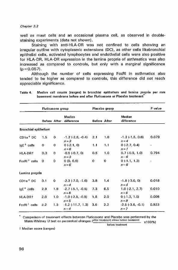
Chapter 3.2
well as mast cells and an occasional plasma cell, as observed in doublestaining experiments (data not shown).
Staining with anti-HLA-DR was not confined to cells showing an irregular outline with cytoplasmic extensions (DC), as other cells likebronchial epithelial cells, activated lymphocytes and endothelial cells were also positive for HLA-DR. HLA-DR expression in the lamina propria of asthmatics was also increased as compared to controls, but only with a marginal significance (p =0.057).
Although the number of cells expressing FcaRI in asthmatics also tended to be higher as compared to controls, this difference did not reach appreciable significance.
Table 4. Median cell counts (ranges) in bronchial epithelium and lamina propria per mm basement membrane before and after Fluticasone or Placebo treatment"
Fluticasone group Placebo group
Median Before After difference Before After
Bronchial epithelium
CDla+ DC 1.5
IgE+ cells 0
HLA-DRt 0.3
Fc£RI + cells 0
Lamina propria
CDla+DC 3.1
IgE+ cells 2,9
HLA-DRt 2.0
Fc£RI+ cells 2.2
o
o
o
o
-1.2 (-2.6, -0.4) 2.1 n=3 01-2.1,0) 1.1 n=6 -0.5 (-0.7, 0) 0.5 n=3 o to, 0.6) 0 n=6
o -2.3 (-7.0, -1.6) 3.8 n~4
1.9 -2.7 (-5.1, -0.5) 7.3 n=5
1.0 -1.0 (-2.5, -0.5) 1.8 n=5
1.3 -1.2 (-11.7, 1.3) 3.6 n=4
1.6
1.1
1.0
o
1.4
6.5
2.0
2.2
Median difference
-1.2 (-1.5, 0.6) n=4 o (-2.7, 0.4) n=1 0.7 (-0.5, 1.0) n=6 o {-1.1, 1.2) n=6
P value
0.079
0.794
-1.8 (-3.0, 0) 0.018 n~4
1.0 (-2.1, 2.7) 0.010 n=6 o (-1.3, 1.0) 0.008 n=8 -2.5 (-3.6, -0.1) 0.923 n=7
Comparison of treatment effects between Fluticasone and Placebo was performed by the Mann-Whitney U test on percentual changes (after treatment mInus before treatment xl00%)
before treatment
t Median score (ranges)
96
Modulation of dendritic cells by f/uticasone
The numbers of CD1a+ DC and IgE+ cells were significantly decreased in the glucocorticoid-treated group compared to the placebo group. No significant change in expression of FcaRI was seen after treatment (Table 4). There was a significant reduction in the score of MHC class" expression in the glucocorticoid group as compared to the placebo group.
DISCUSSION
In this double-blind placebo controlled study we firstly have shown that in the bronchial mucosa of asthmatics there are raised numbers of CD1a+ and L25+ DC, and of IgE+ cells. Secondly, we have shown that a decreased bronchial hyperresponsiveness after 3 months FP treatment was accompanied by a decrease in the numbers of CD1 a + DC, a reduction in MHC class " expression, and a reduction in the number of IgE + cells in the bronchial mucosa of subjects with atopic asthma.
The finding of Bg in the putative DC in the bronchial epithelium definitely shows them to be DC.
The DC network within the epithelium of the airways is strategically positioned to pick up inhaled antigens/allergens (30). For primary sensitization of naive T cells, DC are able to migrate to regional lymph nodes and present the inhaled antigens to lymph node T cells. For secondary responses in the mucosa, local antigen presentation by DC to T cells and local activation of T cells are thought to occur as well (31). The data showing increased numbers of CD1a+ and L25+ DC in atopic asthma are in agreement with our earlier findings (22) and with data of others, in which higher numbers of intraepithelial and mucosal CD1a+ DC in atopic asthmatics were described as compared to non-asthmatic atopic controls sensitized to the same allergen (32). In the nasal mucosa, DC and HLA-DR expression have been found to increase during symptomatic atopic rhinitis (33).
The increased number of cells bearing IgE in the lamina propria of asthmatics, of which the majority are DC and mast cells, likely contributes to an enhanced IgE-mediated antigen/allergen presentation. It has been proposed by Mudde et al (34) that IgE, bound to the FcaRI on the surface of APC or mast cells plays a role in antigen capturing before antigen processing and presentation. Although IgE is able to bind to mast cells, and to mediate antigen-induced degranulation, its role in maintaining chronic asthma is not yet fully understood. Increased expression of the high affinity receptor on monocytes of atopic dermatitis patients has been described (17). In our study the FcaRI expression in the lamina propria of asthmatics was upregulated as compared to controls, although not significant, probably due to the small number of control subjects.
Biopsy studies in patients with asthma have confirmed that
97
Chapter 3.2
glucocorticoids reduce the number and activation of several inflammatory cells (35-37). Although this study was carried out on a limited number of patients, our hypothesis that inhaled FP is able to downregulate the numbers of airway DC, was clearly supported by our results.
The described inhibitory effects of 3 months of FP on DC numbers and HLA-DR expression, are consistent with our previous findings indicating downregulation of DC and MHC-class II expression by 2.5 years beclomethasone therapy (22) and are in agreement with a study, which showed a reduction in RFD1 + DC and HLA-DR expression after three months of budesonide in asthma (37). In the present study, we found a considerable decrease in the number of CD1 a + DC in the lamina propria during FP therapy (median numbers from 3.1 to 0) and this decrease was larger than that reported in the study with 400 IIg budesonide treatment for three months (38). Similar studies in the rat by Holt et al. (39) have demonstrated that exposure to inhaled and systemic glucocorticoids leads to a rapid decrease in the number of airway intraepithelial DC, and in their MHC-class II expression. Studies in the nasal mucosa of patients with perennial allergic rhinitis showed a significant reduction in the number of CD1 a + cells and HLA-DR expression after 3 months of fluticasone therapy (40).
The precise mechanisms by which glucocorticoids downregulate DC, remain to be established. One possible mode of action is that glucocorticoids downmodulate the precursor recruitment from the circulation (39). For instance by suppressing the release of chemotaxins by bronchial epithelial cells, like monocyte chemoattractant protein 1 (41) and granulocyte macrophage colony stimulating factor (GM-CSF), which are involved in the recruitment and differentation of DC. The downregulation of DC by glucocorticoid therapy is probably associated with, and presumably dependent on, a decreased accessory function in vivo, since we showed previously that glucocorticoids are able to suppress the T cell stimulatory function of airway DC in vitro (42).
In conclusion, our data show that 3 months of topically-active glucocorticoid therapy downregulate DC and MHC class II expression in the bronchial mucosa of atopic asthmatics. Since DC play an important role in the initiation and propagation of the immune response, the decrease in DC numbers and their functional activity by glucocorticoids likely results in a downregulation of T cell activation.
ACKNOWLEDGEMENTS
We thank Prof. Dr. R. Benner and Dr. E. P. Prens for continuous support, and Mrs. P. Assems for secretarial assistance.
98
Modulation of dendritic cells by f/uticasone
REFERENCES
1. Holt PG, Schon-He grad MA, Oliver J. MHC class I[ antigen-bearing dendritic cells in pulmonary tissues of the rat. Regulation of antigen presentation activity by endogenous macrophage populations. J Exp Med 1988; 167:262-74.
2. Inaba K, Steinmann RM. Resting and sensitized T lymphocytes exhibit distinct stimulating (antigen-presenting cell) requirements for growth and Iymphokine release. J Exp Med 1984; 160:1717-35.
3. Steinman RM. The dendritic cell system and its role in immunogenicity. Ann Rev Immunol1991; 9:271-96.
4. Xia W, Pinto CE, Kradin RL. The antigen-presenting activities of la+ dendritic cells shift dynamically from lung to lymph node after an airway challenge with soluble antigen. J Exp Med 1995; 181 :1275·83.
6. Van Haarst JMW, Wit HJ de, Drexhage HA, Hoogsteden HC. Distribution and immunophenotype of mononuclear phagocytes and dendritic cells in the human lung. Am J Respir Cell Mol 8io11994; 10:487-92.
6. Van Voorhis WC, Hair LS, Steinman RM, Kaplan G. Human dendritic cells, enrichment and purification from peripheral blood. J Exp Med 1982; 166: 1172-87.
7. Schon-He grad MA, Oliver J, McMenamin PG, Holt PG. Studies on the density, distribution, and surface phenotype of intra epithelial class I[ major histocompatibility complex antigen (la) - bearing dendritic celfs (DC) in the conducting airways. J Exp Med 1991; 173: 1345-56.
8. Melief CJM. Dendritic cells are specialized antigen-presenting celfs. Res Immunol 1989; 140:902-6.
9. Murphy G, Bhan A, Sato S, Mihm M, Harrist T. A new immunologic marker for epidermal Langerhans cells. N Engl J Med 1981: 304:791-2.
10. Ishii Y, Takami T, Kokai Y, Yuasa H, Fujimoto J, Takei T. A novel human B cell antigen shared with lymphoid dendritic cells: characterization by a monoclonal antibody. Clin Exp Immunol 1985; 61 :624-32.
11. Sting I G, Bergstresser PRo Dendritic cells: a major story unfolds. Immunol Today 1996; 16:330-3.
12. Fokkens WJ, Vroom TM, Rijntjes E, Mulder PGH. CD-l (T6), HLA-DR-expressing cells, presumably Langerhans cells, in nasal mucosa. Allergy 1989; 44:167-72.
13. Mudde GC, van Reijsen, Boland GJ, de Gast GC, Bruijnzee! P, Bruijnzeel-Koomen CAFM. Allergen presentation by epidermal Langerhans' cells from patients with atopic dermatitis is mediated by IgE. Immunology 1990; 69:335-41.
14. Wang B, Rieger A, Kilgus 0, et al. Epidermal Langerhans cells from normal human skin bind monomeric IgE via FceRI. J Exp Med 1992; 175: 1353-8.
15. Bieber T, de la Salle H, Wollenberg A, et al. Human epidermal Langerhans cells express the high affinity receptor for immunoglobulin E (FceRI). J Exp Med 1992; 175: 1285-90.
16. Kinet J-P, and Metzger H. Genes, structures and actions of the high-affinity receptor for immunoglobulin E. In: Fe receptors and the action of antibodies. Metzger H, editor. American Society for Microbiology, Washington, DC: 1990:239-59.
17. Maurer 0, Fiebiger E, Reiniger S, et al. Expression of functional high affinity immunoglobulin E receptors (FceRI) on monocytes of atopic individuals. J Exp Med 1994; 179:745-50.
18. Gounni AS, Lamkhioued 8, Ochiai K, et al. High-affinity IgE receptor is involved in defence against parasites. Nature 1994; 367:183-6.
19. Corrigan CJ, Kay AB. T cells and eosinophils in the pathogenesis of asthma. Immunol Today 1992; 13:501-7.
20. Gavett, SH, Chen X, Finkelman F, Wills-Karp M. Depletion of murine CD4 + T lymphocytes prevents antigen-induced airway hyperreactivity and pulmonary
99
Chapter 3.2
eosinophilia. Am J Respir Cell Mol Bioi 1994; 10:587-93. 21. Davis MM, Bjorkman 8J. T-cell antigen receptor genes and T-cell recognition. Nature
1988; 334:395-402. 22. Moller GM, Overbeek SE, van Heiden-Meeuwsen CG et a/. Increased numbers of
dendritic cells in the bronchial mucosa of atopic asthmatic patients: downregulation by inhaled corticosteroids. Clin Exp Allergy 1996 (in press).
23. Barnes PJ, Pedersen S. Efficacy and safety of inhaled corticosteroids in asthma. Am Rev Respir Dis 1993; 148:51-526.
24. Corrigan CJ, Hamid Q, North J, et a/. Peripheral blood CD4 but not CD8 T -lymphocytes in patients with exacerbation of asthma transcribe and translate messenger RNA encoding cytokines which prolong eosinophil survival in the context of a Th2-type pattern: effect of glucocorticoid therapy. Am J Respir Cell Mol Bioi 1995; 12:567-78.
25. Borish L, Mascali JJ, Dishuck J, Beam WR, Martin RJ, Rosenwasser LJ. Detection of alveolar macrophage-derived IL-1~ in asthma. Inhibition with corticosteroids. J Immunol 1992; 149:3078-82.
26. Robinson D, Hamid Q, Sun Ying, ef a/. Prednisolon treatment in asthma is associated with modulation of bronchoalveolar lavage cell interleukin-4, interleukin-5, and interferony cytokine gene expression. Am Rev Respir Dis 1993; 148:401-6.
27. Johnson M. The anti-inflammatory profile of fluticasone propionate. Allergy 1995: 50 (suppl 23): 11-4.
28. Hargreave FE, Sterk PJ, Ramsdale EH, Dolovich J, Zamel N. Inhalation challenge tests and airway responsiveness in man. Chest 1985; 87S:202-6.
29. Zhou L-JI. Schwarting R, Smith HM, Tedder TF. A novel cell-surface molecule expressed by human interdigitating reticulum cells, Langerhans cells, and activated lymphocytes is a new member of the Ig superfamily. J Immunol 1992; 149:735-42.
30. Holt PG, Schon-Hegrad MA, Phillips MJ, McMenamin PG. la-positive dendritic cells form a tightly meshed network within the human airway epithelium. Clin Exp Allergy 1989; 19:597-601.
31. Sertl K, Takemura TT, Tschachler E, Ferrans VJ, Kaliner MA, Shevach EM. Dendritic cells with antigen-presenting capability reside in airway epithelium, lung parenchyma and visceral pleura. J Exp Med 1986; 163:436-51.
32. Bellini A, Vittori E, Marini M, Ackerman V, MattoH S. Intra epithelial dendritic cells and selective activation of Th2-like lymphocytes in patients with atopic asthma. Chest 1993; 103:997-1005.
33. Fokkens WJ, Vroom T, Rijntjes E, Mulder P. Fluctuation of the number of CDl a(T6)positive dendritic cells, presumably langerhans cells, in the nasal mucosa of patients with isolated grass-polfen allergy before. during, and after the grass-pollen season. J Allergy Clin Immunol 1989; 84:39-43.
34. Mudde GC, Bheekha R. Bruijnzeel-Koomen. IgE-mediated antigen presentation. Allergy 1995; 50:193-9.
35. Djukanovic R, Wilson JW, Britten KM, et al. Effect of an inhaled corticosteroid on airway inflammation and symptoms in asthma. Am Rev Respir Dis 1992; 145:669-74.
36. Laitinen LA, Laitinen A, Haahtela T. A comparative study of the effects of an inhaled corticosteroid, budesonide, and of a B2-agonist, terbutaline on airway inflammation in newly diagnosed asthma. J Allergy Clin Immunol 1992; 90:32-42.
37. Jeffery PK, Godfrey RW, Adelroth E, Rogers A, Johansson S-A. Effects of treatment on airway inflammation and thickening of basement membrane reticular collagen in asthma. A quantitative light and electron microscopic study. Am Rev Raspir Dis 1992: 145:890-9.
38. Burke C, Power CK, Norris A, Condez A, Schmekel B, Poulter lW. Lung function and immunopathological changes after inhaled corticosteroid therapy in asthma. Eu( Respir J 1992; 5:73-9.
100
Modulation of dendritic cells by fluticasone
39. Nelson DJ, McWilliam AS, Haining S, Holt PG. Modulation of airway intraepithelial dendritic cells following exposure to steroids. Am J Respir Crit Care Med 1995; 151 :475-81.
40. Holm A, Fokkens WJ, Godthelp T, Mulder PG, Vroom TM, Rijntjes E. The effect of three months nasal steroid therapy on nasal T cells and Langerhans cells in patients suffering from allergic rhinitis. Allergy 1995; 50:204-9.
41. Sousa AR, Lane SJ, Nakhostan JA, Yoshimura T, Lee TH, Poston RN. Increased expression of the monocyte chemoattractant protein-1 in bronchial tissue from asthmatic subjects. Am J Respir Cell Mol BioI. 1994; 10:142-7.
42. Van Haarst JMW, Verhoeven GT, de Wit HJ, Drexhage HA, Moller GM, Hoogsteden He. Glucocorticoids decrease allostimulatory function and 97 expression of human airway dendritic cells in vitro. In: Host defense, dendritic cells and the human lung. Van Haarst JMW, thesis, Rotterdam, 1995:55-67.
101

CHAPTER 4
CYTOKINE EXPRESSION IN ASTHMA
4.1 Immunolocalization of interleukin 4 in eosinophils in the bronchial mucosa of atopic asthmatics Am J Respir Cell Mol BioI 1996 (in press)
4.2 Ultrastructural immunogold localization of interleukin 5 to the crystalloid core compartment of eosinophil secondary granules in patients with atopic asthma J Histochem Cytochem 1996; 44:67-69

CHAPTER 4.1
IMMUNOLOCALIZATION OF INTERLEUKIN 4 IN EOSINOPHILS IN THE BRONCHIAL MUCOSA OF ATOPIC ASTHMATICS'
Gertrude M. Moller1,2, Tan A. W. de Jang3, Thea H. van der Kwast3, Shelley E. Overbeek', Annet F. Wierenga-Walf2, Thea Thepen4, Henk C.
Haagsteden'
Departments of Pulmonary Diseasesl~ Immunology2 and Patholog,j1, Erasmus University and University Hospital Dijkzigt, Rotterdam, and Dept. of Dermatology4, University Hospital Utrecht,
The Netherlands
* Am J Respir Cell Mol BioI 1996 (in press)

Human eosinophils store IL·4
ABSTRACT
Several studies have shown that human eosinophils can synthesize and release a number of cytokines. The aim of this study was to investigate whether eosinophils contain interleukin (IL)-4 protein. We examined the ultrastructural localization of IL-4 in eosinophils in bronchial mucosal biopsies of twenty-nine non-smoking atopic asthmatics and seven controls. No eosinophils were detected in the bronchial mucosa of controls. In the eosinophils (n = 42) of the asthmatics IL-4 was localized to the electrondense crystalloid core compartment of 85% of the secondary or specific eosinophil granules (n = 468). Other structures in the eosinophils were unlabeled. Control sections, incubated with an irrelevant primary antibody were negative. This study demonstrates that (pre-formed) IL-4 is stored in the secondary eosinophil granules. These results were extended by light microscopic immunodouble-staining for IL-4 protein and eosinophil cationic protein, which showed that a subpopulation of (activated) eosinophils express IL-4 immunoreactivity in bronchial mucosal biopsies of asthmatics as well as controls. These data indicate that eosinophils may be an important source of IL-4 in allergic inflammation.
INTRODUCTION
Asthma is characterized by chronic infiltration and activation of several types of inflammatory cells in the bronchial mucosa, particularly eosinophils and activated mast cells (1-3). Interleukin (lL)-4 and IL-5 are important immunoregulatory cytokines which appear to play a dominant role in the pathogenesis of asthma. IL-4 induces the isotype switching of B cells to IgE synthesis (4) and therefore contributes to the excessive IgE synthesis in atopic patients. Furthermore, IL-4 stimulates T cell proliferation, especially of the T helper 2 (Th2) phenotype (5) and increases the endothelial VCAM-I expression as well as the VLA-4 dependent selective accumulation of eosinophils in the lung (6). These cells are thought to playa critical role in epithelial cell damage and in mediating the airway inflammation. IL-5 promotes the differentiation (7), recruitment (8), activation and survival of the eosinophils (9).
Increased levels of IL-4 protein (10), and increased messenger RNA encoding IL-4 have been found in T cells recovered by bronchoalveolar lavage (BAL) of mild atopic asthmatics by in situ hybridization (11). IL-4 has been demonstrated in mast cells using immunoelectron microscopic analysis of mucosal samples taken from resected lung and nasal polyp tissues (12) and by immunohistochemistry studies with glycol methacrylate (GMA) embedded bronchial biopsies of asthmatics (13,14). Recently, IL-4 synthesis
107
Chapter 4.1
by eosinophils in nasal polyp tissues and bronchial biopsies of asthmatics has been described (15). Taken together, the cellular sources of IL-4 include T cells, mast cells, basophils and as it seems eosinophils. However, the subcellular immunolocalization of IL-4 protein in eosinophils in the bronchial mucosa of atopic asthmatics and of controls has not been described so far.
Therefore, in the present study we examined the (ultrastructural) localization of IL-4 by immunoelectron microscopy and immunodoublestaining in bronchial mucosal biopsies of atopic asthmatics and controls.
MATERIALS AND METHODS
Bronchial biopsies
Bronchial biopsies were obtained by fiberoptic bronchoscopy (Olympus model BF IT Tokyo, Japan) by the same operator (S.E.O.) from the segmental divisions of the main bronchi of twenty-nine patients (9 women, 20 men median age 30 yrs, range 18-55) with mild atopic asthma who were on bronchodilators only for at least 3 weeks prior to the study and seven nonatopic controls (3 women, 4 men median age 28 years, range 23-52). All were nonsmokers. The diagnosis of asthma was based on a history of attacks of breathlessness and wheezing without chronic cough or sputum production, according to the criteria of the American Thoracic Society (16). Atopy was defined as a positive skin-prick test to at least one of 16 common aeroallergens. The mean FEV, was 82.0 (46.8-108.3) % predicted and the geometric mean provocative concentration of inhaled histamine required to reduce their FEV 1 by 20% (PC20) was 0.87 mg/ml (0.03-4.72). The control subjects had a mean FEV, 99.3 (88-110) % predicted and a PC20 histamine of more than 8 mg/ml. Due to the low frequency of eosinophils in the biopsies processed for immunoelectron microscopy, a selection was made on semithin sections of the twenty-nine patients. Five biopsies each of five different patients, containing eosinophils were further processed for immunoelectron microscopy. In the biopsies of the controls processed for immunoelectron microscopy, no eosinophils were seen. Because of scarcity of biopsy material, light microscopic immunodouble staining was carried out on only twelve patients and three controls. The study protocol was approved by the local Medical Ethics Committee and all participants gave their written informed consent.
Fixation and embedding for immunoelectron microscopy
Biopsies were immediately placed into ice-cooled isotonic saline and within 20 min fixed in 1 % acroleine plus 0.4% glutaraldehyde (in 0.1 M
108
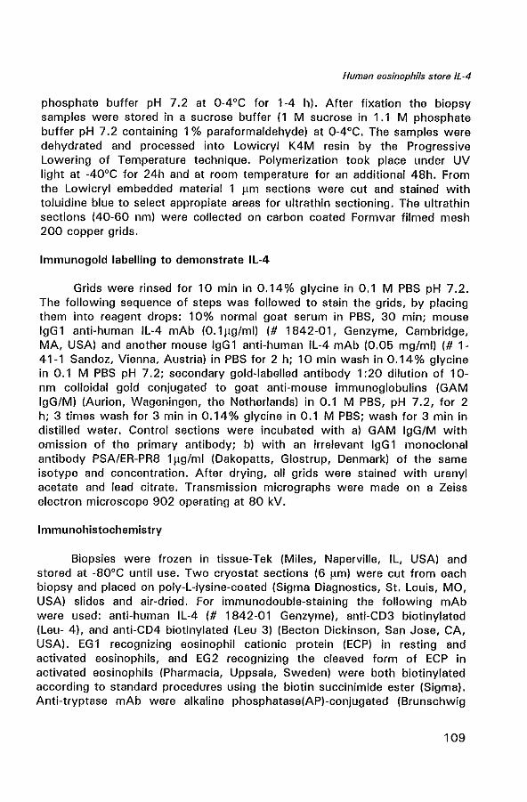
Human eosinophils store IL-4
phosphate buffer pH 7.2 at 0-4°C for 1-4 h). After fixation the biopsy samples were stored in a sucrose buffer (1 M sucrose in 1.1 M phosphate buffer pH 7.2 containing 1 % paraformaldehyde) at 0-4°C. The samples were dehydrated and processed into Lowicryl K4M resin by the Progressive Lowering of Temperature technique. Polymerization took place under UV light at -40°C for 24h and at room temperature for an additional 48h. From the Lowicryl embedded material 1 jlm sections were cut and stained with toluidine blue to select appropiate areas for ultrathin sectioning. The ultrathin sections (40-60 nm) were collected on carbon coated Formvar filmed mesh 200 copper grids.
Immunogold labelling to demonstrate IL-4
Grids were rinsed for 10 min in 0.14% glycine in 0.1 M PBS pH 7.2. The following sequence of steps was followed to stain the grids, by placing them into reagent drops: 10% normal goat serum in PBS, 30 min; mouse IgG1 anti-human IL-4 mAb (0.1 jlg/ml) (# 1842-01, Genzyme, Cambridge, MA, USA) and another mouse IgG 1 anti-human IL-4 mAb (0.05 mg/mi) (# 1-41-1 Sandoz, Vienna, Austria) in PBS for 2 h; 10 min wash in 0.14% glycine in 0.1 M PBS pH 7.2; secondary gold-labelled antibody 1 :20 dilution of 10-nm colloidal gold conjugated to goat anti-mouse immunoglobulins (GAM IgG/M) (Aurion, Wageningen, the Netherlands) in 0.1 M PBS, pH 7.2, for 2 h; 3 times wash for 3 min in 0.14% glycine in 0.1 M PBS; wash for 3 min in distilled water. Control sections were incubated with a) GAM IgG/M with omission of the primary antibody; b) with an irrelevant IgG 1 monoclonal antibody PSA/ER-PR8 1 jlg/ml (Dakopatts, Glostrup, Denmark) of the same isotype and concentration. After drying, all grids were stained with uranyl acetate and lead citrate. Transmission micrographs were made on a Zeiss electron microscope 902 operating at 80 kV.
Immunohistochemistry
Biopsies were frozen in tissue-Tek (Miles, Naperville, IL, USA) and stored at -80°C until use. Two cryostat sections (6 Ilm) were cut from each biopsy and placed on poly-L-Iysine-coated (Sigma Diagnostics, St. Louis, MO, USA) slides and air-dried. For immunodouble-staining the following mAb were used: anti-human IL-4 (# 1842-01 Genzyme), anti-CD3 biotinylated (Leu- 4), and anti-CD4 biotinylated (Leu 3) (Becton Dickinson, San Jose, CA, USA). EG 1 recognizing eosinophil cationic protein (ECP) in resting and activated eosinophils, and EG2 recognizing the cleaved form of ECP in activated eosinophils (Pharmacia, Uppsala, Sweden) were both biotinylated according to standard procedures using the biotin succinimide ester (Sigma). Anti-tryptase mAb were alkaline phosphatase(AP)-conjugated (Brunschwig
109
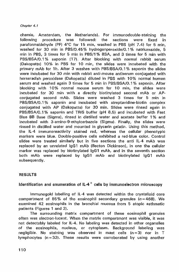
Chapter 4.1
chemie, Amsterdam, the Netherlands). For immunodouble·staining the following procedure was followed: the sections were fixed in paraformaldehyde (PF) 4°C for 15 min, washed in PBS (pH 7.4) for 5 min, washed for 30 min in PBS/0.45% hydrogenperoxide/O.l % natrium azide, 5 min in PBS, 3 times for 5 min in PBS/l % BSA, and 3 times for 5 min with PBS/BSA/O.l % saponin (17). After blocking with normal rabbit serum (Dakopatts) 10% in PBS for 10 min, the slides were incubated with the primary mAb for 1 h. After 3 washes with PBS/BSA/O.l % saponin the slides were incubated for 30 min with rabbit anti-mouse antiserum conjugated with horseradish peroxidase (Dakopatts) diluted in PBS with 10% normal human serum and washed again 3 times for 5 min in PBS/BSA/O.l % saponin. After blocking with 10% normal mouse serum for 10 min, the slides were incubated for 30 min with a directly biotinylated second mAb or APconjugated second mAb. Slides were washed 3 times for 5 min in PBS/BSA/O.l % saponin and incubated with streptavidine-biotin complex conjugated with AP (Dakopatts) for 30 min. Slides were rinsed again in PBS/BSA/O.l % saponin and IRIS buffer (pH 8.5) and incubated with Fast Blue BB Base (Sigma), rinsed in distilled water and acetate buffer 1 % and incubated with 3-amino-9-ethylcarbazole (Sigma). Finally, the slides were rinsed in distilled water and mounted in glycerin gelatin. Using this method, the IL-4 immunoreactivity stained red, whereas the cellular phenotypic markers were blue. Double-positive cells exhibited a red-blue color. Control slides were treated similarly but in five sections the anti IL-4 mAb was replaced by an unrelated IgGl mAb (Becton Dickinson), in one the cellular marker was replaced by biotinylated IgG 1 mAb, and in the seventh section both mAb were replaced by IgG 1 mAb and biotinylated IgG 1 mAb subsequently.
RESULTS
Identification and enumeration of IL·4 + cells by immunoelectron microscopy
Immunogold labelling of IL-4 was detected within the crystalloid core compartment of 85% of the eosinophil secondary granules (n=468). We examined 42 eosinophils in the bronchial mucosa from 5 atopic asthmatic patients (Figures 1 and 2).
The surrounding matrix compartment of these eosinophil granules often was electron-lucent. When the matrix compartment was visible, it was not detectably labeled for IL-4. No labeling was detected in other organelles of the eosinophils, nucleus, or cytoplasm. Background labeling was negligible. No staining was observed in mast cells (n = 3) nor in T lymphocytes (n = 32). These results were corroborated by using another
110
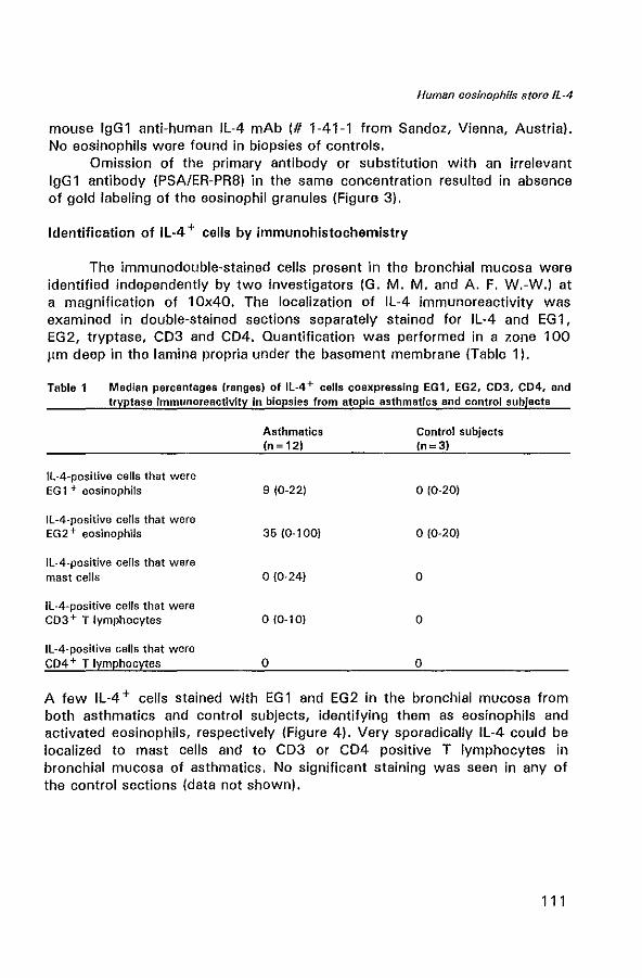
Human eosinophils store IL-4
mouse IgG1 anti-human IL-4 mAb (# 1-41-1 from Sandoz, Vienna, Austria). No eosinophils were found in biopsies of controls.
Omission of the primary antibody or substitution with an irrelevant IgG 1 antibody (PSA/ER-PR8) in the same concentration resulted in absence of gold labeling of the eosinophil granules (Figure 3).
Identification of IL-4 + cells by immunohistochemistry
The immunodouble-stained cells present in the bronchial mucosa were identified independently by two investigators (G. M. M. and A. F. W.-W.) at a magnification of 10x40. The localization of IL-4 immunoreactivity was examined in double-stained sections separately stained for IL-4 and EG 1, EG2, tryptase, CD3 and CD4. Quantification was performed in a zone 100 flm deep in the lamina propria under the basement membrane (Table 1).
Table 1 Median percentages (ranges) of Il-4+ cells coexpresslng EG1. EG2, CD3. CD4. and tryptase immunoreactivity in biopsies from atopic asthmatics and control SUbjects
Asthmatics Control subjects (n= 12) (n=3)
Il-4-positive cells that were EG 1 + eosinophils 9 (0-22) 0(0·20)
1l-4-positive cells that were EG2+ eosinophils 35 (0·'00) 0(0-20)
IL-4-positive cells that were mast cells 0(0-24) 0
IL-4-positive cells that were C03 + T lymphocytes 0(0·10) 0
Il-4-positive cells that were CD4 + T lymphocytes 0 0
A few IL-4 + cells stained with EG1 and EG2 in the bronchial mucosa from both asthmatics and control subjects, identifying them as eosinophils and activated eosinophils, respectively (Figure 4). Very sporadically IL-4 could be localized to mast cells and to CD3 or CD4 positive T lymphocytes in bronchial mucosa of asthmatics. No significant staining was seen in any of the control sections (data not shown).
111
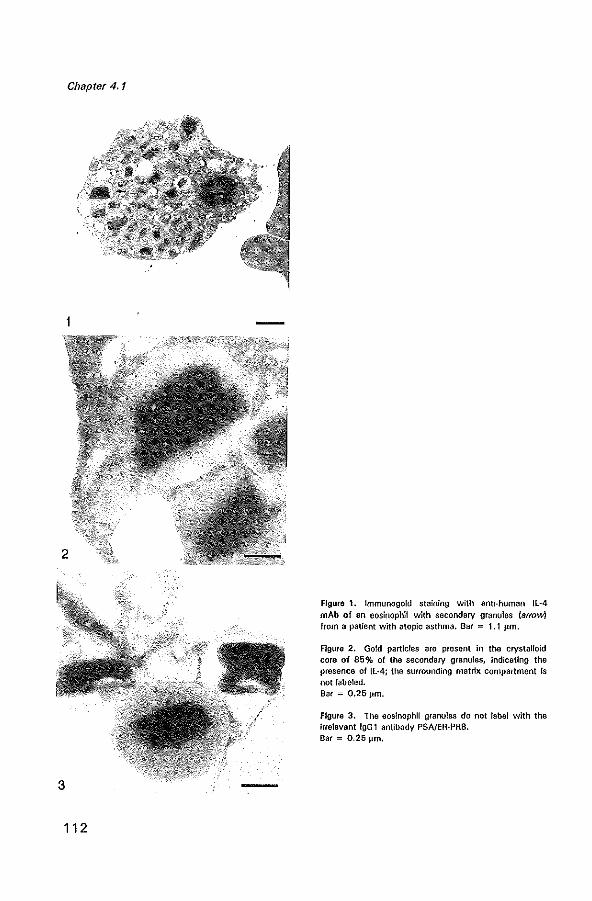
Chapter 4.1
112
Figura 1. Immunogold staining with anti-human Il-4 mAb of an eosinophil with secondary granules (arrow) from a patient with atopic asthma. Bar = 1.1 /-1m.
Figure 2. Gold partIcles are present in the crystalloid core of 85% of the secondary granules, indicating the presence of Il-4; the surrounding matrix compartment Is not lab-eled. Bar = 0.25 ~Im.
Figure 3. The eosinophil granules do not label with the irrelevant IgG 1 antibody PSA!ER·PRS. Bar = 0.25 /-1m.
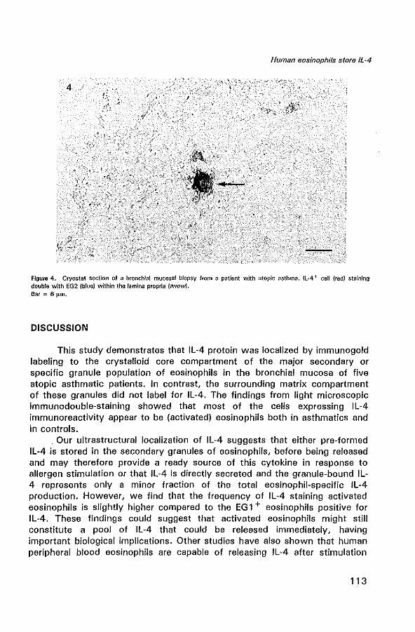
Human eosinophils store IL~4
Figure 4. Cryostat section of a bronchial mucosal biopsy from a patient with atopic asthma. IL-4 + cell (red! staining double with EG2 (blue) within the lamina propria (arrow)_ Bar = 6 ftm.
DISCUSSION
This study demonstrates that IL-4 protein was localized by immunogold labeling to the crystalloid core compartment of the major secondary or specific granule population of eosinophils in the bronchial mucosa of five atopic asthmatic patients. In contrast, the surrounding matrix compartment of these granules did not label for IL-4. The findings from light microscopic immunodouble-staining showed that most of the cells expressing IL-4 immunoreactivity appear to be (activated) eosinophils both in asthmatics and in controls.
Our ultrastructural localization of IL-4 suggests that either pre-formed IL-4 is stored in the secondary granules of eosinophils, before being released and may therefore provide a ready source of this cytokine in response to allergen stimulation or that IL-4 is directly secreted and the granule-bound IL-4 represents only a minor fraction of the total eosinophil-specific IL·4 production. However, we find that the frequency of IL-4 staining activated eosinophils is slightly higher compared to the EG 1 + eosinophils positive for IL·4. These findings could suggest that activated eosinophils might still constitute a pool of IL·4 that could be released immediately, having important biological implications. Other studies have also shown that human peripheral blood eosinophils are capable of releasing IL-4 after stimulation
113
Chapter 4.1
with slgA-immune complexes (151 or calcium ionophore (18(. IL-4 mRNA expression by a small percentage of activated eosinophils in BAL fluid and in biopsies from atopic asthmatics has been observed (19(. In peripheral blood eosinophils from asthmatics mRNA for IL-4 was detected by reverse transcriptase-PCR and IL-4 immunoreactivity was colocalized to blood eosinophils, as shown by cell fractionation and light microscopy (201. Obviously, IL-4 expression in eosinophils is not a chracteristic feature of pulmonary eosinophils. In combination these data on synthesis, storage and secretion of IL-4 by eosinophils, suggest a novel role for these cells in maintaining the IgE response and stimulating the Th2 commitment.
Ultrastructural immunohistochemical techniques with gold conjugated secondary antibodies have made it possible to identify a variety of enzymes and four different basic proteins with cytotoxic properties in the specific eosinophil granules (211. Major basic protein (MBPI was found to be localized to the crystalloid core compartment of the specific granules, while eosinophil cationic protein (ECPI, eosinophil derived-neurotoxin (EDNI, and eosinophil peroxidase (EPOI were localized to the matrix (22,231.
It is now well established that eosinophils are a source of several other cytokines in addition to IL-4, including IL-1a (241, IL-3 (251. IL-5 (261. IL-6 (271. IL-8 (281, GM-CSF (291. TNF-a (301. MIP-1a (311, TGF-a (321 and TGFB (331. However, the substructural localization of most of these cytokines remains to be determined.
Immunoelectron microscopic and immunohistochemical studies have demonstrated the presence of IL-4 in mast cells in bronchial and nasal mucosal biopsies from both patients with allergic asthma and rhinitis and controls (12,141. We could not confirm by immunogold labeling IL-4 immunoreactivity localized to mast cells granules. This could be due to the low number of mast cells examined in the ultrathin sections and to the variable distribution in the number of mast cells expressing cytokines (141. By immunodouble-staining we saw in only one patient a solitary mast cell positive for IL-4. We did not detect IL-4 immunoreactivity in CD3 or CD4 positive T lymphocytes. This does not exclude that these cells can produce and synthesize IL-4. It is likely, that once generated, T cells immediately secrete IL-4. In contrast to eosinophils and mast cells, they have little capacity for mediator storage.
In conclusion, our data indicate that eosinophils in the bronchial mucosa store IL-4 in the crystalloid core of secondary granules and may be an important source of IL-4 in chronic allergic airway inflammation.
ACKNOWLEDGMENTS
We are grateful to Prof. Dr. R. Benner and to Dr. Ir. H. F. J. Savelkoul for
114
Human eosinophils store /L-4
critically reading this manuscript. We thank Mrs. P. Assems for secretarial assistance. This study was financially supported by Glaxo Wellcome B.V., Zeist, The Netherlands.
REFERENCES
1. Bousquet, J., P. Chanez, J. Y. lacoste, G. Barneon, N. Ghavanian, I. Enander, P. Venge, S. Ahlstedt, J. Simony-Lafontaine, P. Godard, and F. B. Michel. Eosinophilic inflammation in asthma. N Engl J Med 1990; 323:1033-9.
2. Djukanovic, R., J. W. Wilson, K. M. Britten, S. J. Wilson, A. F. Walls, W. R. Roche, P. H. Howarth, and S. T. Holgate. Quantification of mast cells and eosinophils in the bronchial mucosa of symptomatic atopic asthmatics and healthy control subjects using immunohistochemistry. Am Rev Respir Dis 1990; 142:863-71.
3. Djukanovic, R., W. R. Roche, J. W. Wilson, C. R. W. Beasley, D. P. Twentyman, P. H. Howarth, and S. T. Holgate. Mucosal inflammation in asthma. Am Rev Resp!r Dis 1990; 142:434-57.
4. Del Prete, G. F., E. Maggi, P. Parrondi, I. Chretien, A. Tiri, D. Macchia, M. Ricci, J. Banchereau, J. E. de Vries, and S. Romagnani. Il-4 is an essential co-factor for the IgE synthesis induced in vitro by human T cell clones and their supernatants. J Immunol 1988; 140:4193-8.
5. Swain, S. l., A. D. Weinberg, M. English and G. Huston. Il·4 directs the development of TH2·like helper effectors. J Immunol1990; 145:3796-3806.
6. Schleimer, R. P., S. A. Sterbinsky, J. Kaiser, C. A. Sickel, D. A. Klunk, K. Tomioka, W. Newman, F. W. luscinskas, M. A. Gimbrone, B. W. Me Intyre, and B. S. Bochner. Il·4 induces adherence of human eosinophils and basophils but not neutrophils to endothelium. Association with expression of VCAM-1. J Immunol1992; 148:1086·92.
7. Clutterbuck, E. J., E. M. A. Hirst, and C. J. Sanderson. Human interleukin-5 (ll-5) regulates the production of eosinophils in human bone marrow cultures: comparison and interaction with IL-1, IL-3.IL-6, and GM-CSF. Blood 19B9; 73:1504-13.
8. Wang, J. M., A. Rambaldi, A. Biondi, Z. G. Chen, C. J. Sanderson, and A. Mantovani. Recombinant interleukin 5 is a selective eosinophil chemoattractant. Eur J Immunol 1989; 19:701-5.
9. lopez, A. F., C. J. Sanderson, J. R. Gamble, H. R. Campbell, I. G. Young, and M. A. Vadas. Recombinant human interleukin-5 is a selective activator of human eosinophil function. J Exp Med 1988; 167:219-24.
10. Walker, C., E. Bode, l. Boer, T. T. Hansel, K. Blaser, and J. C. Virchow. Allergic and non allergic asthmatics have distinct patterns of T-cell activation and cytokine production in peripheral blood and bronchoalveolar lavage. Am Rev Respir Dis 1992; 146:109-15.
11. Robinson, D. S., Q. Hamid, S. Ying, A. Tsicopoulos, J. Barkans, A. M. Bentley, C. Corrigan, S. R. Durham, and A. B. Kay. Predominant TH2-like bronchoalveolar Tlymphocyte population in atopic asthma. N Eng J Med 1992; 326:298-304.
12. Wilson, S. J., P. Bradding, C. Heusser, S. T. Holgate, P. H. Howarth. The subcellular localisation of interleukin 4 and interleukin 5 in the respiratory mucosa using immunoelectron microscopy. Am J Respir Crit Care Med (abstract) 1995; 151: A702.
13. Bradding, P., J. A. Roberts, K. M. Britten, S. Montefort, R. Djukanovic, R. Mueller, C. H. Heusser, P. H. Howarth, and S. T. Holgate. Interleukin-4. -5, and -6 and tumor necrosis factor-a. in normal and asthmatic airways: evidence for the human mast cell as a source of these cytokines. Am J Respir Cell Mol Bioi 1994; 10:471-80.
14. Bradding, P., Y. Okayama, P. H. Howarth, M. K. Church, S. T. Holgate. Heterogeneity of human mast cells based on cytokine content. J Immunol 1995; 155:297·307.
15. Nonaka, M., R. Nonaka, K. Woolley, E. Adelroth, K. Miura, Y. Okhawara, M. Glibetic, K.
115
Chapter 4.1
Nanako, P. O'Byrne, J. Dolovich, M. Jordana. Distinct immunohistochemical localization of IL-4 in human inflamed airway tissues. J Immunol 1995; 155:3234-44.
16. ATS Task Group. Standards for the diagnosis and care of patients with chronic obstructive pulmonary disease (COPD) and asthma. Am Rev Resp!r Dis 1987; 136:225-31.
17. Andersson, U., and J. Andersson. Immunolabeling of cytokine-producing cells in tissues and in suspension. In Cytokine producing cells. D. Fralelizie and D. Emilie, eds. Inserm. Paris. 1994; 1-16.
18. Bjerke, T., M. Gaustadnes, S. Nielsen, C. M. Rheimert, O. Schiotz, N. Rudiger, E. I. Christensen, R. Dahl, L. K. Poulsen. Human blood eosinophils express interleukin 4. J Allergy Clin Immunol 1995; 95:A801.
19. Ying, S., S. R. Durham, C. J. Corrigan, a. Hamid, and A. B. Kay. Phenotype of cells expressing mRNA for TH2-type Hnterleukin 4 and interleukin 5) and TH 1-type (interleukin 2 and interferon ..,.) cytokines in bronchoalveolar lavage and bronchial biopsies from atopic asthmatic and normal control subjects. Am J Respir Cell Mol Bioi 1995; 12:477-87.
20. Moqbel, R., S. Ying, J. Barkans, T. M. Newman, P. Kimmitt, M. Wakelin, C. J. Corrigan, S. R. Durham, A. B. Kay. Human eosinophils synthesise interleukin-4 Ul-4). The Immunologist (abstract) 1995; 3: A224.
21. Gleich, G. J., C. R. Adolphson. The eosinophil leukocyte: structure and function. Adv Immunol 1986; 39:177-253.
22. Egesten, A., J. Alumets, C. von Mecklenburg, M. Palmegren, I. Olsson. localization of eosinophil cationic protein, major basic protein, and eosinophil peroxidase by immuno electron microscopic technique. J Histochem Cytochem 1986; 11 :1399-403.
23. Peters, M. S., M. Rodriguez, G. J_ Gleich. localization of human eosinophil granule major basic protein, eosinophil cationic protein, and eosinophil-derived neurotoxin by immunoelectron microscopy. lab Invest 1986; 54:656-62.
24. Del Pozo, V., B. De Andres, E. Martin, N. Maruri, J. M. Zubeldia, P. Palomino, C. Lahoz. Murine eosinophils and fl-l: all-l mRNA detection by in situ hybridization. Production and release of IL-1 from peritoneal eosinophils. J Immunol 1990; 144:3117-22.
25. Kita, H., T. Ohnishi, Y. Okubo, D. Weiler, J. S. Abrams, G. J. Gleich. Granulocyte/macrophage colony-stimulating factor and interleukin 3 release from human peripheral blood eosinophils and neutrophils. J Exp Med 1991; 174:745-8.
26. Desreumaux, P., A. Janin, J. F. Colombe I, L. Prin, J. Plumas, D. Emilie, G. Torpier, A. Capron, M. Capron. Interleukin 5 messenger RNA expression by eosinophils in the intestinal mucosa of patients with coeliac disease. J Exp Med 1992; 175:293-6.
27. Hamid, a., J. Barkans, a. Meng, S. Ying, J. S. Abrams, A. B. Kay, R. Moqbel. Human eosinophils synthesize and secrete interleukin-6 in vitro. Blood 1992; 180: 1496-501.
28. Braun( R, K., M. Franchini, F. Erard, S. Rihs, I. J. De Vries, K. Blaser, T. T. Hansel, C. Walker. Human peripheral blood eosinophils produce and release interleukin-8 on stimulation with calcium ionophore. Eur J Immunol 1993; 23:956-60.
29. Moqbel, R., Q. Hamid, S. Ying, J. Barkans, A. Hartnell, A. Tsicopoulos, A. J. Wardlaw, A. B. Kay. Expression of mRNA and immunoreactivity for the granulocyte/macrophage colony-stimulating factor in activated human eosinophils. J Exp Med 1991; 174:749-52.
30. Beil, W. J., P. F. Weller, D. M. Tzizik, S. J. Galli, A. M. Dvorak. Ultrastructural immunogold localization of tumor necrosis factor-a to the matrix compartment of eosinophil secondary granules in patients with idiopathic hypereosinophilic syndrome. J Histochem Cytochem 1993; 41:1611-5.
31. Costa, J. J., K. Matossian, M. B. Resnick, W. J. Beil, D. T. W. Wong, J. R. Gordon, A. M. Dvorak, P. F. Weller, S. J. Galli. Human eosinophils can express the cytokines tumor necrosis factor-a and macrophage inflammatory protein-la. J Clin Invest 1993; 91:2673-84.
32. Wong, D. T. W., P. F. Weller, S. J. Galli, A. Elovic, T. H. Rand, G .. T. Gallagher, T.
116
Human eosinophils store IL-4
Chiang, M. Y. Chou, K. Motossian, J. McBride, R. Todd. Human eosinophils express transforming growth factor-a. J Exp Med 1990; 172:673-81.
33. Ohno, I., R. G. lea, K. C. Flanders, D. A. Clark, D. Banwatt, J. Dolovich, J. Denburg, C. B. Harley, J. Gauldie, M. Jordana. Eosinophils in chronically inflamed human upper airway tissues express transforming growth factor P1 gene (TGF PH. J Clin Invest 1992; 89:1662·8.
117

CHAPTER 4.2
ULTRASTRUCTURAL IMMUNOGOLD LOCALIZATION OF INTERLEUKIN 5 TO THE CRYSTALLOID CORE COMPARTMENT OF EOSINOPHIL SECONDARY
GRANULES IN PATIENTS WITH ATOPIC ASTHMA'
Gertrude M. Miiller1,2, Ton A. W. de Jong3, Shelley E. Overbeek1, Theo H. van der Kwast3, Dirkje S. Postma4, Henk C. Hoogsteden 1
Departments of Pulmonary Diseases!, Immunolog.r, and Path%g.j1, Erasmus University and University Hospital Dijkzigt, Rotterdam, the Netherlands, and Department of Pulmonary
Diseases4, University Hospital Groningen, The Netherlands
• J Histochem Cytochem 1996; 44:67-69

Localization of IL-5 in human eosinophil granules
SUMMARY
Bronchial biopsies from two patients with atopic asthma were analyzed by immunogold labeling to detect the ultrastructural location of interleukin 5 (IL-5). In eosinophils, IL-5 was localized to the electron-dense crystalloid core compartment of the secondary or specific eosinophil granules. Other structures in the eosinophils were unlabeled. No IL-5 was detected in mast cells. Control sections incubated with an irrelevant primary antibody were negative. This study demonstrates that pre-formed IL-5 is stored within the major population of secondary granules in human eosinophils.
INTRODUCTION
Interleukin-5 (lL-5) was demonstrated to be localized in eosinophils and, to a lesser extent, in mast cells in a recent immunohistochemical study using glycol methacrylate (GMA)-embedded bronchial biopsy specimens from asthmatic patients (1). The clinical importance of IL-5 is demonstrated by the fact that IL-5 is a growth and differentation factor (2), activator (3), and chemoattractant (4) for eosinophils and, as a consequence, is considered a pivotal cytokine in allergen-mediated eosinophilic responses. In the lower airways, increased IL-5 mRNA has been reported in the mucosa of mild asthmatics (5). In addition, increased concentration of IL-5 protein (6) and increased transcription of IL-5 has been found in T-cells recovered by bronchoalveolar lavage (BAL) from allergic asthmatics (7). In the present investigation we wanted to localize the IL-5 immunoreactivity by immunoelectron microscopy. We detected IL-5 exclusively in the major eosinophil granule population in mucosal biopsy specimens taken from two atopic asthmatic patients. Specifically, IL-5 was present in the electrondense crystalloid core compartment but not in the surrounding matrix of the secondary granules of eosinophils.
MATERIALS AND METHODS
Bronchial tissues
Bronchial biopsy specimens were obtained by fiberoptic bronchoscopy (Olympus model BF IT Tokyo, Japan) from the segmental divisions of the main bronchi of two non-smoking atopic asthmatic patients who were on bronchodilators only (terbutaline 500 ~'g four times a day). The diagnosis of asthma was based on a history of attacks of breathlessness and wheezing without chronic cough or sputum production, according to the criteria of the
121
Chapter 4.2
American Thoracic Society. The two patients were randomly sampled from the Dutch CNSLD study group (8). The study protocol was approved by the Medical Ethics Committee.
Fixation and embedding for immunoelectron microscopy
Biopsies were immediately placed in ice-cooled isotonic saline and within 20 min were fixed in 1 % acrolein plus 0.4% glutaraldehyde (in 0.1 M phosphate buffer, pH 7.2, at 0-4°C for 1-4 hrl. After fixation the biopsy samples were stored in a sucrose buffer (1 M sucrose in 1.1 M phosphate buffer, pH 7.2) with 1 % paraformaldehyde at 0-4°C. The samples were dehydrated and processed into Lowicryl K4M resin by the progressive lowering of temperature technique. Polymerization took place under UV light at -40°C for 24 hr and at room temperature (RT) for an additional 48 hr. From the Lowicryl-embedded material, 1-l'm sections were cut and stained with toluidine blue to select appropiate areas for ultra-thin sectioning. The ultra-thin sections (40-60 nm) were collected on carbon-coated Formvarfilmed mesh 200-mesh copper grids.
Immunogold labelling to demonstrate IL-5
Grids were rinsed for 10 min in 0.14% glycine in 0.1 M PBS, pH 7.2. The following sequence of steps was used to stain the grids, by placing them into reagent drops: (a) 10% normal goat serum (NGS) in PBS, 30 min; (b) 1: 1 00 dilution of primary mouse anti-human IL-5 monoclonal antibody (Mab7) (9) (Dr. L. A. McNamee, Glaxo, Greenford, Middlesex, UK) in PBS for 2 hr at RT; (c) wash in 0.14% glycine in 0.1 M PBS, pH 7.2; (d) secondary gold-labeled antibody [1 :20 dilution of 10-nm colloidal gold conjugated to goat anti-mouse immunoglobulins (GAM IgG/M) (Aurion, Wageningen, the Netherlands) in 0.1 M PBS, pH 7.2, for 2 hrJ; (e) wash in 0.14% glycine in 0.1 M PBS; (f) wash for 3 min in distilled water. Control sections were incubated (a) with GAM IgG/M with omission of the primary antibody and (b) with an irrelevant IgG 1 MAb PSA/ER-PR8 (Dakopatts, Glostrup, Denmark) of the same isotype and concentration. After drying, all grids were stained with uranyl acetate and lead citrate. Transmission micrographs were made on a Zeiss electron microscope 902 operating at 80 kV.
122
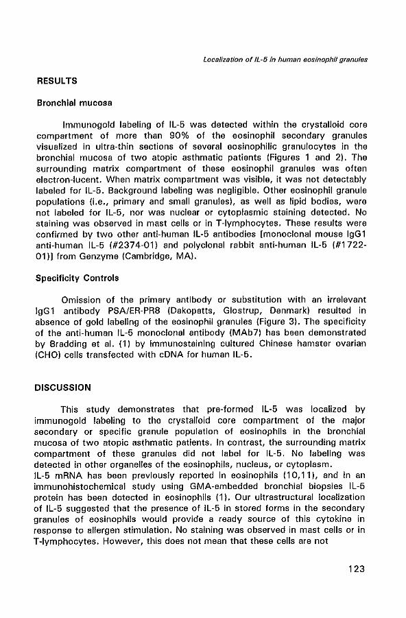
Localization of IL-5 in human eosinophil granules
RESULTS
Bronchial mucosa
Immunogold labeling of IL-5 was detected within the crystalloid core compartment of more than 90% of the eosinophil secondary granules visualized in ultra-thin sections of several eosinophilic granulocytes in the bronchial mucosa of two atopic asthmatic patients (Figures 1 and 2). The surrounding matrix compartment of these eosinophil granules was often electron-lucent. When matrix compartment was visible, it was not detect ably labeled for IL-5. Background labeling was negligible. Other eosinophil granule populations (i.e., primary and small granules). as well as lipid bodies, were not labeled for IL-5, nor was nuclear or cytoplasmic staining detected. No staining was observed in mast cells or in T-Iymphocytes. These results were confirmed by two other anti-human IL-5 antibodies [monoclonal mouse IgG 1 anti-human IL-5 (#2374-01) and polyclonal rabbit anti-human IL-5 (#1722-01)1 from Genzyme (Cambridge, MA).
Specificity Controls
Omission of the primary antibody or substitution with an irrelevant IgG 1 antibody PSA/ER-PR8 (Dakopatts, Glostrup, Denmark) resulted in absence of gold labeling of the eosinophil granules (Figure 3). The specificity of the anti-human IL-5 monoclonal antibody (MAb7) has been demonstrated by Bradding et al. (1) by immunostaining cultured Chinese hamster ovarian (CHO) cells transfected with cDNA for human IL-5.
DISCUSSION
This study demonstrates that pre-formed IL-5 was localized by immunogold labe)ing to the crystalloid core compartment of the major secondary or specific granule population of eosinophils in the bronchial mucosa of two atopic asthmatic patients. In contrast, the surrounding matrix compartment of these granules did not label for IL-5. No labeling was detected in other organelles of the eosinophils, nucleus, or cytoplasm. IL-5 mRNA has been previously reported in eosinophils (10,11), and in an immunohistochemical study using GMA-embedded bronchial biopsies IL-5 protein has been detected in eosinophils (1). Our ultrastructural localization of IL-5 suggested that the presence of IL-5 in stored forms in the secondary granules of eosinophils would provide a ready source of this cytokine in response to allergen stimulation. No staining was observed in mast cells or in T-Iymphocytes. However, this does not mean that these cells are not
123
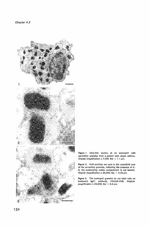
Chapter 4.2
2
124
Figure 1. Ultra-thin section of an eosinophil with secondary granules from a patient with atopic asthma. Original magnification x 7,000. Bar = 1.1 J.lm.
Figure 2. Gold particles are seen in the crystalloid core of the secondary granules, Indicating the presence of IL-5; the surrounding matrix compartment Is not labeled, Original magnification x 30,000. Bar = 0.25 ~Lm.
Figure 3. The eosinophil granules do not label with an irrelevant IgG 1 antibody, PSA/ER-PRB. Original magnification x 20,000. Bar = 0.4 ~lm,
producing and secreting IL-5. It is immediately secrete IL-5. In contrast little capacity for mediator storage.
Localization of IL-5 in human eosinophil granules
likely that once with eosinophils,
generated, T-cells lymphocytes have
Eosinophils have recently been identified as a pivotal source of several other cytokines in addition to IL-5, including IL-1a (12), IL-3 (13), IL-6 (14), IL-8 (15), GM-CSF (16), TNF-a (17), MIP-1a (18), TGF-a (19), and TGF-B (20). However, the substructural location of most of these cytokines remains to be determined.
Ultrastructural immunohistochemical techniques with gold conjugated secondary antibodies have made it possible to identify subcellular sites for a variety of eosinophil constituents (21). Four different basic proteins with cytotoxic properties and a number of enzymes are recognized in the specific eosinophil granules (22). The crystalline core compartment consists of major basic protein (MBP), and the surrounding matrix is composed of eosinophil cationic protein (ECP), eosinophil-derived neurotoxin (EDN) and eosinophil peroxidase (EPO).
Although the distribution of these eosinophil granule proteins has been known for some time, the ultrastructural localization of IL-5 in the crystalloid core of the secondary granules of eosinophils in bronchial mucosa of asthmatic patients has not previously been determined.
ACKNOWLEDGEMENTS
We thank Prof. Dr. R. Benner for continuous support and Ms. P. Assams for secretarial assistance.
REFERENCES
1. Bradding P, Roberts JA, Britten KM, Montefort S, Djukanovic R, Mueller R, Heusser CH, Howarth PH, Holgate ST. Interleukin-4, -5, and -6 tumor necrosis factor -ex in norm.al and asthmatic airways: evidence for the human mast cell as a source of these cytokines. Am J Respir Cell Mol Bioi 1994: 10:471-80.
2. Clutterbuck EJ, Hirst MA, Sanderson CJ. Human interleukln-5 UL-5) regulates the production of eosinophlls In human bone marrow cultures: comparison and interaction with IL-t. IL-3, IL-6. and GM-CSF. Blood 1989: 73:1504-13.
3. Lopez AF, Sanderson CJ, Gamble JR, Campbell HD, Young IG, Vadas MA. Recombinant human interleukin-5 is a selective activator of human eosinophil fUnction. J Exp Med 1988: 167:219-24.
4. Wang JM, Rambaldi A, Biondi A, Chen ZG, Sanderson CJ, Mantovanl A. Recombinant interleukin 5 is a selective eosinophil chemoattractant. Eur J Immunol 1989~ 19:701-5.
5. Hamid Q, Azzawi M, Sun Ying, Moqbel R, Wardlaw AJ, Corrigan CJ, Bradley B, Durham SR, Collins JV, Jeffery PK, Quint OJ, Kay AB. Expression of mRNA for interleukin-5 in mucosal biopsies from asthma. J Clin Invest 1991; 87:1541-6.
6. Walker C, Bode E, Boer L, Hansel TT, Blaser K, Virchow JC. Allergic and non allergic asthmatics have distinct patterns of T-cell activation and cytoklne production in
125

Chapter 4.2
peripheral blood and bronchoalveolar lavage. Am Rev Respir Dis 1992; 146:109-15. 7. Robinson OS, Hamid Q, Sun Ying, Tsicopoulos A, Barkans J, Bentley AM, Corrigan C,
Durham SR, Kay AB. Predominant T H2-like bronchoalveolar T-Iymphocyte population in atopic asthma. N Eng J Mad 1992; 326:298-304.
8. Kerstjens HAM, Brand PLP, Hughes MO, Robinson NJ, Postma OS, Sluiter HJ and the Dutch CNSLD Study Group. A comparison of bronchodilator therapy with or without inhaled corticosteroid therapy for obstructive airways disease. N Engl J Med 1992; 327:1413-9.
9. McNamee LA, Fattah 01, Baker T J, Bains T J, Hissey PH. Production, characterisation and use of monoclonal antibodies to human interlaukin 5 in an enzyme-linked immunosorbent assay. J Immunol Methods 1991; 141:81-8.
10. Broide DH, Paine MM, Firestein GS. Eosinophils express interleukin-5 and granulocyte macrophage colony-stimulating factor mRNA at sites of allergic inflammation in asthmatics. J Clin Invest 1992; 90:1414-24.
11. Desreumaux P, Janin A, Colombel JF, Prin L, Plumas J, Emilie 0, Torpier G, Capron A, Capron M. Interleukin 5 messenger RNA expression by eosinophils in the intestinal mucosa of patients with coeliac disease. J Exp Med 1992; 175:293-6.
12. Del Pozo V, De Andres B, Martin E, Maruri N, Zubeldia JM, Palomino P, Lahoz C. 1990. Murine eosinophils and IL-l: alL-l mRNA detection by in situ hybridization. Production and release of IL-l from peritoneal eosinophils. J Immunol 1990; 144:3117-22.
13. Kita H, Ohnishi T, Okubo Y, Weiler 0, Abrams JS, Gleich GJ. Granulocyte/macrophage colony-stimulating factor and interleukin 3 release from human peripheral blood eosinophils and neutrophils. J Exp Med 1991; 174:745-8.
14. Hamid a, Barkans J, Meng a, Sun Ying, Abrams JS, Kay Ab, Moqbel R. Human eosinophils synthesize and secrete interleukin-6 in vitro. Blood 1992; 80: 1496-501.
15. Braun RK, Franchini M,Erard F, Rihs S, De Vries S, Blaser K, Hansel TT, Walker C. Human peripheral blood eosinophils produce and release interleukin-8 on stimulation with calcium ionophore. Eur J Immunol 1993; 23:956-60.
16. Moqbel R, Hamid 0, Sun Ying, Barkans J, Hartnell A, Tsicopoulos A, Wardlaw AJ, Kay AB. Expression of mRNA and immunoreactivity for the granulocyte/macrophage colonystimulating factor in activated human eosinophils. J Exp Med 1991; 174:749-52.
17. Beil WJ, Weller PF, Tzizik OM, Galli SJ, Dvorak AM. Ultrastructural immunogold localization of tumor necrosis factor-a to the matrix compartment of eosinophil secondary granules in patients with idiopathic hypereosinophilic syndrome. J Histochem Cytocham 1993; 41:1611-5.
18. Costa JJ, Matossian K, Resnick MB, Beil WJ, Wong DTW, Gordon JR, Dvorak AM, Weller PF, Galli SJ. Human eosinophils can express the cytokines tumor necrosis factor-a and macrophage inflammatory protein-1a. J Clin Invest 1993; 91:2673-84.
19. Wong DTW, Weller PF, Galli SJ, Elovic A, Rand TH, Gallagher GT, Chiang T, Chou MY, Motossian K, McBride J, Todd R. Human eosinophils express transforming growth factora. J Exp Mad 1990; 172:673·81.
20. Ohno I, Lea RG, Flanders KC, Clark DA, Banwatt 0, Dolovich J, Denburg J, Harley CB, Gauldie J, Jordana M. Eosinophils in chronically inflamed human upper airway tissues express transforming growth factor Pl gene (TGF PlI, J Clin Invest 1992; 89:1662-8.
21. Dvorak AM, Ishizaka T, Weller PF, Ackerman SJ. Ultrastructural contributions to the understanding of the cell biology of human eosinophils: mechanisms of growth factorinduced development, secretion, and resolution of released constituents from the microenvironment. In Makino S, Fukuda T, eds. Eosinophils: biological and clinical aspects. Boca Raton, Fl: CRC Press, 1993: 13-32.
22. Gleich GJ, Adolphson CR. The eosinophil leucocyte: structure and function. Adv Immunol 1986; 39:177-253.
126

CHAPTER 5
EOSINOPHILS IN THE BRONCHIAL MUCOSA IN RELATION TO INDICES FROM METHACHOLINE LOG-DOSE RESPONSE (MLDR) CURVES IN ATOPIC
ASTHMATICS
5.1 Eosinophils in the bronchial mucosa in relation to indices from methacholine log-dose response (MLDR) curves in atopic asthmatics Am J Respir Crit Care Med 1995; 151 :4:A 133 (modified)

CHAPTER 5.1
EOSINOPHILS IN THE BRONCHIAL MUCOSA IN RELATION TO INDICES FROM METHACHOLINE LOG-DOSE RESPONSE (MLDR) CURVES IN ATOPIC
ASTHMATICS'
Gertrude M. Miiller1.2, Jan M. Bogaard1, Shelley E. Overbeek1, Cornelia G. van Helden-Meeuwsen 2, Henk C. Hoogsteden 1
Departments of Pulmonary Diseases', and Immunology2, Erasmus University Rotterdam, University Hospital Dijkzigt, The Netherlands
"Am J Respir Crit Care Med 1995; 151:4:A 133 (modified)

£osinophils and indices from methacholine log-dose curves
INTRODUCTION
Atopic asthma is characterized by local infiltration of several activated inflammatory cells in the bronchial mucosa, even in mild asthmatics (1). Particularly eosinophils, through their release of different basic proteins with cytotoxic properties (major basic protein (MBP), eosinophil cationic protein (ECP), eosinophil derived neurotoxin (EON) and eosinophil peroxidase (EPO)) have been associated with epithelial shedding (2,3). The numbers of eosinophils in the bronchial mucosa and in bronchoalveolar lavage (BAL) fluid has been correlated with the severity of asthma and the mechanisms which underlie airway hyperresponsiveness (4-7), one of the most prominent functional characteristics of asthma (8). Airway hyperresponsiveness is defined as a decreased threshold of airway narrowing in response to a variety of non-specific stimuli (9). At present the airway hyperresponsiveness is usually measured in log dose response curves as the provocative concentration histamine or methacholine causing a fall of 20% in the forced expiratory volume in one second (FEV 1) and is called the sensitivity (S; log2PC2o)' Previous studies (10,11) have focussed on the importance of defining airway hyperresponsiveness by a more detailed characterization of the entire histamine or methacholine log-dose response (MLDR) curve, i.e. the sensitivity (S), the steepest part of the slope of the curve (reactivity; R; .. FEV 1 (%)/ 1092 PC) and the maximal response value of airway narrowing (plateau; P, % FEV1). It was found that the log dose response curves from asthmatics, show a shift to the left, had a steeper slope and a higher maximal response value as compared to normals (12).
For the interpretation of the entire log- dose response curve a model fit to the data was necessary, because reaching of a plateau can be associated with a large drop in FEV l' and therefore extrapolation may be necessary. Moreover the model gives a smoothing of the individual data which often show marked random fluctuations. In our investigation indices were obtained as parameters of a Cumulative Gaussian Distribution (CGD) function fitted to the log-dose response curves (13).
Another pulmonary function variable, the specific (volumic) airway conductance (sGaw) can be derived from bodyplethysmography and can be considered as indicator for the airway lumen (14).
Airway inflammation produces hyperemia in the lamina propria (15). The process of vasodilation and plasma leakage may increase airway wall thickness, which may directly contribute to airway narrowing (15,16). Therefore, we tested the hypothesis that in mild to moderate asthma the indices from the entire MLDR curves and the baseline specific airway conductance (sGaw) are related to the presence and activation of eosinophils in the lamina propria.
131
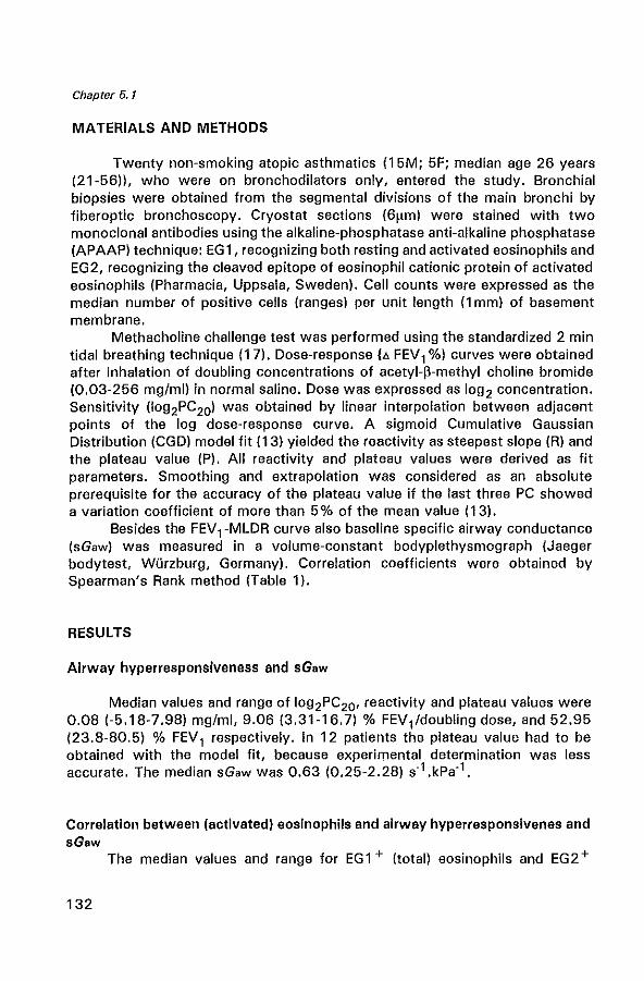
Chapter 5.1
MATERIALS AND METHODS
Twenty non-smoking atopic asthmatics (15M; 5F; median age 26 years (21-56)), who were on bronchodilators only, entered the study. Bronchial biopsies were obtained from the segmental divisions of the main bronchi by fiberoptic bronchoscopy. Cryostat sections (6I1m) were stained with two monoclonal antibodies using the alkaline-phosphatase anti-alkaline phosphatase (APAAP) technique: EG 1, recognizing both resting and activated eosinophils and EG2, recognizing the cleaved epitope of eosinophil cationic protein of activated eosinophils (Pharmacia, Uppsala, Sweden). Cell counts were expressed as the median number of positive cells (ranges) per unit length (1 mm) of basement membrane.
Methacholine challenge test was performed using the standardized 2 min tidal breathing technique (17). Dose-response (A FEV 1 %) curves were obtained after inhalation of doubling concentrations of acetyl-Jl-methyl choline bromide (0.03-256 mg/ml) in normal saline. Dose was expressed as 1092 concentration. Sensitivity (log2PC20) was obtained by linear interpolation between adjacent points of the log dose-response curve. A sigmoid Cumulative Gaussian Distribution (CGO) model fit (13) yielded the reactivity as steepest slope (R) and the plateau value (P). All reactivity and plateau values were derived as fit parameters. Smoothing and extrapolation was considered as an absolute prerequisite for the accuracy of the plateau value if the last three PC showed a variation coefficient of more than 5% of the mean value (13).
Besides the FEV l-MLOR curve also baseline specific airway conductance (sGaw) was measured in a volume-constant bodyplethysmograph (Jaeger body test, WOrzburg, Germany). Correlation coefficients were obtained by Spearman's Rank method (Table 1).
RESULTS
Airway hyperresponsiveness and sGaw
Median values and range of log2PC20' reactivity and plateau values were 0.08 (-5.18-7.98) mg/ml, 9.06 (3.31-16.7) % FEV1/doubiing dose, and 52.95 (23.8-80.5) % FEV1 respectively. In 12 patients the plateau value had to be obtained with the model fit, because experimental determination was less accurate. The median sGaw Was 0.63 (0.25-2.28) s-1.kPa-1.
Correlation between (activated) eosinophils and airway hyperresponsivenes and sGaw
The median values and range for EGl + (total) eosinophils and EG2 +
132
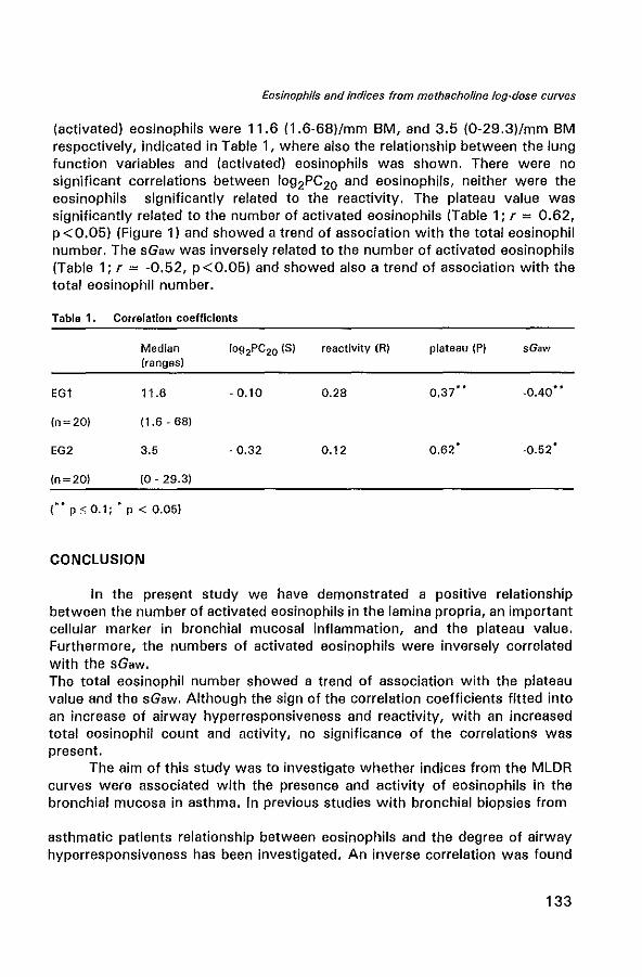
Eosinophils and indices from methacholine log-dose curves
(activated) eosinophils were 11.6 (1.6-68)/mm 8M, and 3.5 (0-29.3)/mm BM respectively, indicated in Table 1, where also the relationship between the lung function variables and (activated) eosinophils was shown. There were no significant correlations between 1092PC20 and eosinophils, neither were the eosinophils significantly related to the reactivity. The plateau value was significantly related to the number of activated eosinophils (Table 1; r = 0.62, p<0.05) (Figure 1) and showed a trend of association with the total eosinophil number. The sGaw was inversely related to the number of activated eosinophils (Table 1; r = -0.52, p <0.05) and showed also a trend of association with the total eosinophil number.
Table 1. Correlation coefficients
Median reactivity (R) plateau (P) sGaw (ranges)
EGl 11.6 - 0.10 0.28
(n~20) (1.6-68)
EG2 3.5 - 0.32 0.12 0.62' ·0.52'
(n~20) (0·29.3)
("' p s 0.1; • p < 0.05)
CONCLUSION
In the present study we have demonstrated a positive relationship between the number of activated eosinophils in the lamina propria, an important cellular marker in bronchial mucosal inflammation, and the plateau value. Furthermore, the numbers of activated eosinophils were inversely correlated with the sGaw. The total eosinophil number showed a trend of association with the plateau value and the sGaw. Although the sign of the correlation coefficients fitted into an increase of airway hyperresponsiveness and reactivity, with an increased total eosinophil count and activity, no significance of the correlations was present.
The aim of this study was to investigate whether indices from the MLDR curves were associated with the presence and activity of eosinophils in the bronchial mucosa in asthma. In previous studies with bronchial biopsies from
asthmatic patients relationship between eosinophils and the degree of airway hyperresponsiveness has been investigated. An inverse correlation was found
133
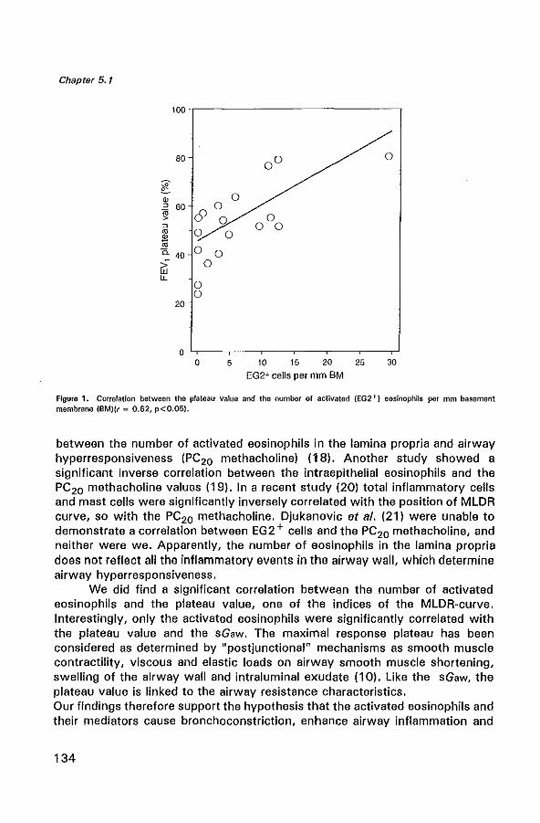
Chapter 5.1
100 -,---------------,
80
l w ~ 60 ~ ~
'" w 1il a. 40-;;: W LL
20
c9 0
0
o o
o
0
00 0
0 0
0 0 0 0
0
0
5 10 15 20 25 30
EG2+ cells per mm BM
Figure 1. Correlation between the plateau value and the number of activated (EG2 +) eosinophlls per mm basement membrane (BM)(r = 0.62, p<0.05).
between the number of activated eosinophils in the lamina propria and airway hyperresponsiveness (PC20 methacholine) (18). Another study showed a significant inverse correlation between the intraepithelial eosinophils and the PC20 methacholine values (19). In a recent study (20) total inflammatory cells and mast cells were significantly inversely correlated with the position of MLDR curve, so with the PC20 methacholine. Djukanovic et al. (21) were unable to demonstrate a correlation between EG2 + cells and the PC20 methacholine, and neither were we. Apparently, the number of eosinophils in the lamina propria does not reflect all the inflammatory events in the airway wall, which determine airway hyperresponsiveness.
We did find a significant correlation between the number of activated eosinophils and the plateau value, one of the indices of the MLDR-curve. Interestingly, only the activated eosinophils were significantly correlated with the plateau value and the sGaw. The maximal response plateau has been considered as determined by "post junctional" mechanisms as smooth muscle contractility, viscous and elastic loads on airway smooth muscle shortening, swelling of the airway wall and intraluminal exudate (10). Like the sGaw, the plateau value is linked to the airway resistance characteristics. Our findings therefore support the hypothesis that the activated eosinophils and their mediators cause bronchoconstriction, enhance airway inflammation and
134
Eosinophils and indices from methacholine log-dose curves
epithelial damage, and possibly cause an increase in airway hyperresponsiveness.
In conclusion, indices of MLDR curves .and also baseline sGaw are partly significantly related to the number of activated eosinophils in the bronchial mucosa. These results suggest a direct relationship between bronchial mucosal inflammation, characterized by eosinophil activity, and decrease in airway lumen. Our results stress the importance of modelling the entire MLDR curve and not only the use of 1092PC20 in follow up and characterization of airway hyperresponsiveness.
REFERENCES
1. Bradley BL, Azzawi M, Jacobson M, Assouffi a, Collins JV, Irani A-MA, Schwartz LB, Durham SR, Jeffery PK, Kay AB. Eosinophils, T-Iymphocytes, mast cells, neutrophils and macrophages in bronchial biopsy specimens from atopic subjects with asthma: comparison with biopsy specimens from atopic subjects without asthma and normal subjects and relationship to bronchial hyperresponsiveness. J Allergy Clin Immunol 1991: 88:661-74.
2. Gleich GJ, Adolphson CR. The eosinophil leucocyte: structure and function. Adv Immunol 1986; 39:177-253.
3. Bousquet J, Chanez P, Lacoste JY, Barm~on G, Ghavanian N, Enander I, Venge P, Ahlstedt S, Lafontaine J, Godard P, Michel FB. Eosinophific inflammation in asthma. N Engl J Med 1990; 323:1033-9.
4. Bentley AM, Menz G, Storz C, Robinson OS, Bradley B, Jeffery PK, Durham SR, Kay AB. Identification of T lymphocytes, macrophages and activated eosinophils in the bronchial mucosa of intrinsic asthma: relationship to symptoms and bronchial responsiveness. Am Rev Rsspir Dis 1992; 145:500-5.
5. Laitinen LA, Laitinen A, Heino M, Haahtela T. Eosinophilic airway inflammation during exacerbation of asthma and its treatment with inhaled corticosteroid. Case reports. Am Rev Rsspir Dis 1991; 144:83-7.
6. Wardlaw AJ, Dunnett S, Gleich GJ, Collins JV, Kay AB. Eosinophils and mast celfs in bronchoalveolar lavage in mild asthma: relationship to bronchial hyperreactivity. Am Rev Rsspir Dis 1988; 136:379-83.
7. Walker C, Kaegi MK, Braun p, Blaser K. Activated T ccells and eosinophilia in bronchoalveolar lavages from subjects with asthma correlated with disease severity. J Allergy Clin Immunol 1991; 88:935-42.
8. American Thoracic Society. Standards for the diagnosis and care of patients with CO PO and asthma. Am Rev Respir Dis 1987; 136:225-44.
9. Hargreave FE, Dolovich J, O'Byrne PM, Ramsdale EH, Daniel EE. The origin of airway hyperresponsiveness. J Allergy Clin Immunol1991; 88:425-534.
10. Sterk PJ, Bel EH. Bronchial hyperresponsiveness: The need for a distinction between hypersensitivity and excessive airway narrowing. Eur Respir J 1989; 2:267~74.
11. N. Eiser. Specificity and sensitivity of various parameters of dose-response curves. Eur Respir Rev 1991; 1: 41 4 7.
12. Woolcock AJ, Salome CM, Van K. The shape of the log dose-response curve to histamine in asthmatic and normal subjects. Am Rev Respir Dis 1984; 130:71 ~5.
13. Aerts JGJV, Bogaard JM, Overbeek SE, Verbraak AFM, Thio P. Extrapolation of methacholine log-dose response curves with a Cumulative Gaussian Distribution function. Eur Respir J 1994; 7:895-900.
14. Zarins LP, Clausen Tlo Bodyplethysmography. In: Pulmonary function testing, guidelines and controversies. Academic Press Inc. 1982; 141-53.
135
Chapter 5.1
15. Widdicombe JG. Anatomy and physiology of the airway circulation. Am Rev Resp!r Dis 1992; 146:S3-7.
16. Wagner EM, Mitzner W. Bronchial vascular engorgement and airway narrowing. Am J Respir Crit Care Med 1994; 149:A585.
17. Cockcroft DW, Killian DN, Mellon JJA, Hargreave FE. Bronchial reactivity to inhaled histamine: a method and clinical survey. Clin Allergy 1977; 7:235-43.
18. Bradley Bl, Azzawi M, Jacobson M, Assoufi B, Collins JV, Irani A-MA ot al. Eosinophils, Tlymphocytes, mast cells, neutrophils and macrophages in bronchial biopsy specimens from atopic subjects without asthma and normal control subjects and relationship to bronchial hyperresponsiveness. J Allergy Clin Immunol1991; 88:661-74.
19. Chetta A, Foresi A, Del Donno M, Bertorelli G, Pesci A, Olivieri D. Basement membrane thickness is related to epithelial cellular infiltrate, bronchial responsiveness and airway patency parameters in asthma. Am J Respir Crit Care Mad 1995; 151:A 132.
20. Foresi A, Chetta A, Del Donno M, Bertorelli G, Pesei A, Olivieri D. Dose-response curve to methacholine and chronic airway inflammation in asthma. Am J Respir Crit Care Med 1995; 151:A133.
21. Djukanovic A, Wilson JW, Britten KM, Wilson SJ, Walls AF, Roche WR, Howart PH, Holgate ST. Quantitation of mast cells and eosinophils in the bronchial mucosa of symptomatic atopic asthmatics and healthy control subjects using immunohistochemistry. Am Rev Respir Dis 1990; 142:863-71.
136

CHAPTER 6
GENERAL DISCUSSION

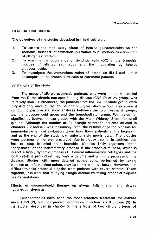
General discussion
GENERAL DISCUSSION
The objectives of the studies described in this thesis were:
1. To assess the modulatory effect of inhaled glucocorticoids on the bronchial mucosal inflammation in relation to pulmonary function data of allergic asthmatics.
2. To examine the occurrence of dendritic cells (DC) in the bronchial mucosa of allergic asthmatics and the modulation by inhaled glucocorticoids.
3. To investigate the immunolocalization of interleukin (ll)-4 and Il-5 in eosinophils in the bronchial mucosa of asthmatic patients.
limitations of the study
The group of allergic asthmatic patients, who were randomly sampled from the Dutch chronic non-specific lung disease (CNSLD) study group, was relatively small. Furthermore, the patients from the CNSLD study group were biopsied only once at the end of the 2.5 year study period. This made it difficult to perform statistical analyses between the two treatment groups, i.e. the glucocorticoid group and the bronchodilator group. We tested for significance between these groups with the Mann-Whitney U test for small groups. Although the number of 24 allergic asthmatic patients studied in chapters 2.2 and 3.2 was reasonably large, the number of paired biopsies for immunohistochemical evaluation taken from these patients at the beginning and at the end of the study was unfortunately much lower. The biopsies were too small or not well preserved, due to biopsy trauma. In addition, one has to keep in mind that bronchial biopsies likely represent static "snapshots" of the inflammatory process in the bronchial mucosa, which is in fact a highly dynamic process (1). Several inflammatory cell types and the local cytokine production may vary with time and with the progress of the disease. Studies with more detailed comparisons, performed by taking biopsies at different time points, may be required in the future. However, it is difficult to take bronchial biopsies from patients with severe asthma. Taken together, it is clear that studying allergic asthma by taking bronchial biopsies has its limitations.
Effects of glucocorticoid therapy on airway inflammation and airway hyperresponsiveness
Glucocorticoids have been the most effective treatment for asthma since 1 950 (2), but their precise mechanism of action is still unclear (3). In the studies described in chapter 2, the effects of two different types of
139
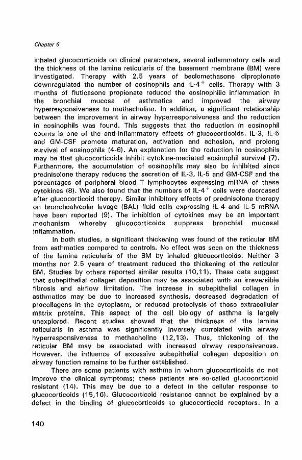
Chapter 6
inhaled glucocorticoids on clinical parameters, several inflammatory cells and the thickness of the lamina reticularis of the basement membrane (BM) were investigated. Therapy with 2.5 years of beclomethasone dipropionate downregulated the number of eosinophils and IL-4 + cells. Therapy with 3 months of fluticasone propionate reduced the eosinophilic inflammation in the bronchial mucosa of asthmatics and improved the airway hyperresponsiveness to methacholine. In addition, a significant relationship between the improvement in airway hyperresponsiveness and the reduction in eosinophils was found. This suggests that the reduction in eosinophil counts is one of the anti-inflammatory effects of glucocorticoids. IL-3, IL-5 and GM-CSF promote maturation, activation and adhesion, and prolong survival of eosinophils (4-6). An explanation for the reduction in eosinophils may be that glucocorticoids inhibit cytokine-mediated eosinophil survival (7). Furthermore, the accumulation of eosinophils may also be inhibited since prednisolone therapy reduces the secretion of IL-3, IL-5 and GM-CSF and the percentages of peripheral blood T lymphocytes expressing mRNA of these cytokines (8). We also found that the numbers of IL-4 + cells were decreased after glucocorticoid therapy. Similar inhibitory effects of prednisolone therapy on bronchoalveolar lavage (BAL) fluid cells expressing IL-4 and IL-5 mRNA have been reported (9). The inhibition of cytokines may be an important mechanism whereby glucocorticoids suppress bronchial mucosal inflammation.
In both studies, a significant thickening was found of the reticular BM from asthmatics compared to controls. No effect was seen on the thickness of the lamina reticularis of the BM by inhaled glucocorticoids. Neither 3 months nor 2.5 years of treatment reduced the thickening of the reticular BM. Studies by others reported similar results (10,11). These data suggest that subepithelial collagen deposition may be associated with an irreversible fibrosis and airflow limitation. The increase in subepithelial collagen in asthmatics may be due to increased synthesis, decreased degradation of procollagens in the cytoplasm, or reduced proteolysis of these extracellular matrix proteins. This aspect of the cell biology of asthma is largely unexplored. Recent studies showed that the thickness of the lamina reticularis in asthma was significantly inversely correlated with airway hyperresponsiveness to methacholine (12,13). Thus, thickening of the reticular BM may be associated with increased airway responsiveness. However, the influence of excessive subepithelial collagen deposition on airway function remains to be further established.
There are some patients with asthma in whom glucocorticoids do not improve the clinical symptoms; these patients are so-called glucocorticoid resistant (14). This may be due to a defect in the cellular response to glucocorticoids (15,16). Glucocorticoid resistance cannot be explained by a defect in the binding of glucocorticoids to glucocorticoid receptors. In a
140
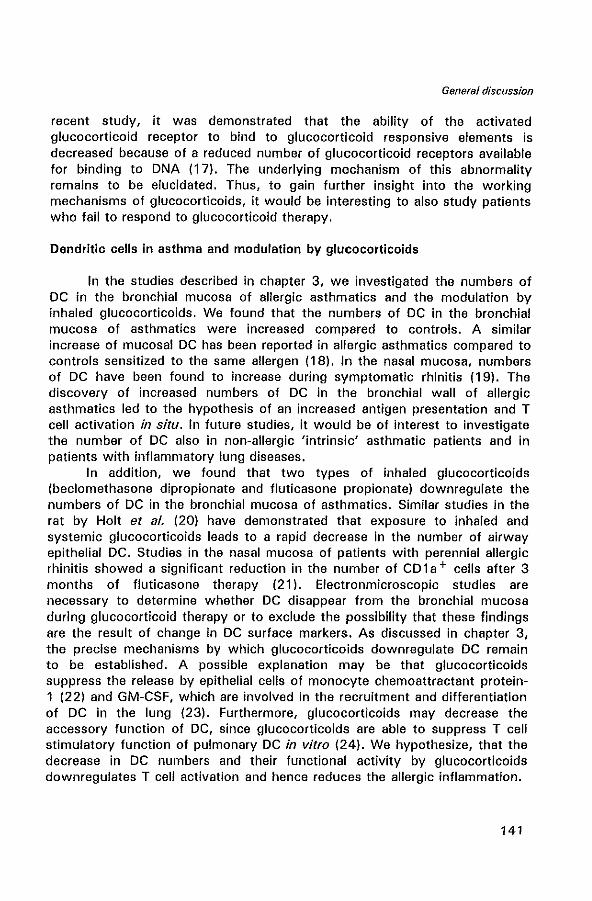
General discussion
recent study, it was demonstrated that the ability of the activated glucocorticoid receptor to bind to glucocorticoid responsive elements is decreased because of a reduced number of glucocorticoid receptors available for binding to DNA (17). The underlying mechanism of this abnormality remains to be elucidated. Thus, to gain further insight into the working mechanisms of glucocorticoids, it would be interesting to also study patients who fail to respond to glucocorticoid therapy.
Dendritic cells in asthma and modulation by glucocorticoids
In the studies described in chapter 3, we investigated the numbers of DC in the bronchial mucosa of allergic asthmatics and the modulation by inhaled glucocorticoids. We found that the numbers of DC in the bronchial mucosa of asthmatics were increased compared to controls. A similar increase of mucosal DC has been reported in allergic asthmatics compared to controls sensitized to the same allergen (18). In the nasal mucosa, numbers of DC have been found to increase during symptomatic rhinitis (19). The discovery of increased numbers of DC in the bronchial wall of allergic asthmatics led to the hypothesis of an increased antigen presentation and T cell activation in situ. In future studies, it would be of interest to investigate the number of DC also in non-allergic 'intrinsic' asthmatic patients and in patients with inflammatory lung diseases.
In addition, we found that two types of inhaled glucocorticoids (beclomethasone dipropionate and fluticasone propionate) downregulate the numbers of DC in the bronchial mucosa of asthmatics. Similar studies in the rat by Holt et al. (20) have demonstrated that exposure to inhaled and systemic glucocorticoids leads to a rapid decrease in the number of airway epithelial DC. Studies in the nasal mucosa of patients with perennial allergic rhinitis showed a significant reduction in the number of CDl a + cells after 3 months of fiuticasone therapy (21). Electronmicroscopic studies are necessary to determine whether DC disappear from the bronchial mucosa during glucocorticoid therapy or to exclude the possibility that these findings are the result of change in DC surface markers. As discussed in chapter 3, the precise mechanisms by which glucocorticoids downregulate DC remain to be established. A possible explanation may be that glucocorticoids suppress the release by epithelial cells of monocyte chemoattractant protein-1 (22) and GM-CSF, which are involved in the recruitment and differentiation of DC in the lung (23). Furthermore, glucocorticoids may decrease the accessory function of DC, since glucocorticoids are able to suppress T cell stimulatory function of pulmonary DC in vitro (24). We hypothesize, that the decrease in DC numbers and their functional activity by glucocorticoids downregulates T cell activation and hence reduces the allergic inflammation.
141
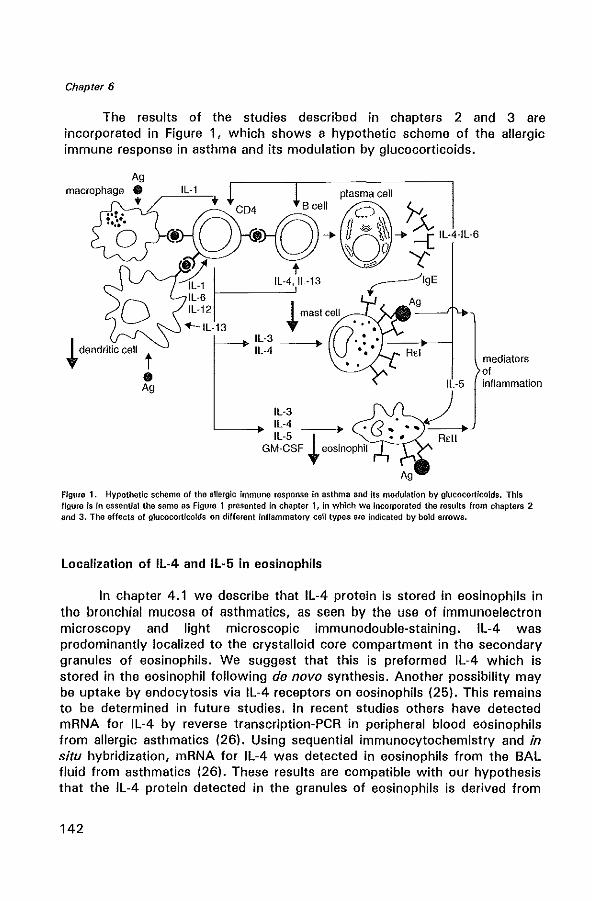
Chapter 6
The results of the studies described in chapters 2 and 3 are incorporated in Figure 1, which shows a hypothetic scheme of the allergic immune response in asthma and its modulation by glucocorticoids.
Ag
-i~~\;-{)5"'"f~~~"'''' :~~. t ," Y
~ dendrilic cell t
• Ag
=1(1 Hl-4,ll-13 ~19E Il-6 Ag IL-12 I mast cell --""'-_
+--ll-13 t U· ", Il-3 ~ .:, __ +-_-1 IL-4 •••
Il-5
mediators of inflammation
Figure 1. Hypothetic scheme of the allargic immune response In asthma and its modulation by glucocortlcolds. This figure is in essential the sarna as Figure 1 presented in chapter 1, in which we Incorporated the results from chapters 2 and 3. The effects of glucocorticolds on different Inflammatory cen types are indicated by bold arrows.
Localization of IL-4 and IL-5 in eosinophils
In chapter 4.1 we describe that IL-4 protein is stored in eosinophils in the bronchial mucosa of asthmatics, as seen by the use of immunoelectron microscopy and light microscopic immunodouble-staining. IL-4 was predominantly localized to the crystalloid core compartment in the secondary granules of eosinophils. We suggest that this is preformed IL-4 which is stored in the eosinophil following de novo synthesis, Another possibility may be uptake by endocytosis via IL-4 receptors on eosinophils (25). This remains to be determined in future studies, In recent studies others have detected mRNA for IL-4 by reverse transcription-PCR in peripheral blood eosinophils from allergic asthmatics (26). Using sequential immunocytochemistry and in situ hybridization, mRNA for IL-4 was detected in eosinophils from the BAL fluid from asthmatics (26). These results are compatible with our hypothesis that the IL-4 protein detected in the granules of eosinophils is derived from
142
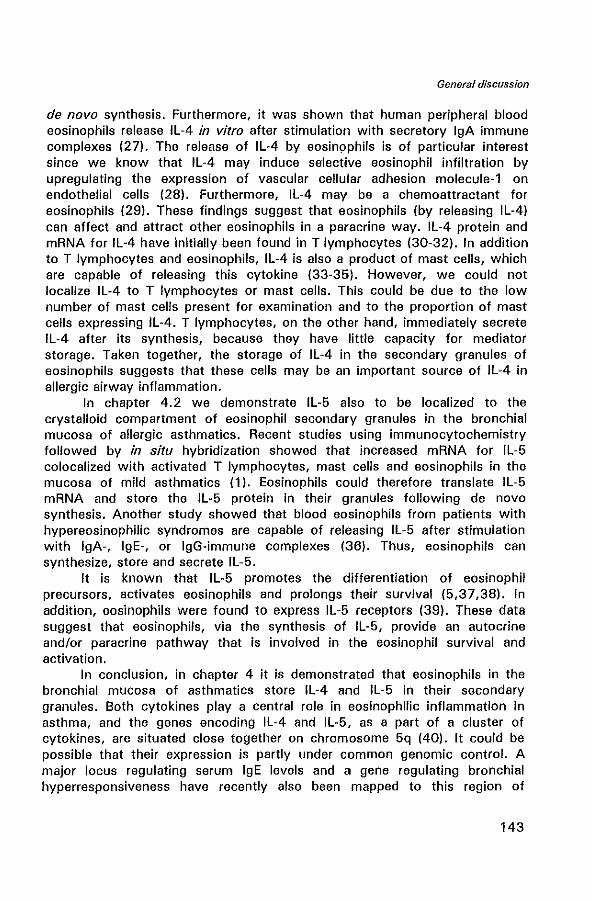
General discussion
de novo synthesis. Furthermore, it was shown that human peripheral blood eosinophils release IL-4 in vitro after stimulation with secretory IgA immune complexes (27). The release of IL-4 by eosinpphils is of particular interest since we know that IL-4 may induce selective eosinophil infiltration by upregulating the expression of vascular cellular adhesion molecule-1 on endothelial cells (28). Furthermore, IL-4 may be a chemoattractant for eosinophils (29). These findings suggest that eosinophils (by releasing IL-4) can affect and attract other eosinophils in a paracrine way. IL-4 protein and mRNA for IL-4 have initially been found in T lymphocytes (30-32). In addition to T lymphocytes and eosinophils, IL-4 is also a product of mast cells, which are capable of releasing this cytokine (33-35). However, we could not localize IL-4 to T lymphocytes or mast cells. This could be due to the low number of mast cells present for examination and to the proportion of mast cells expressing IL-4. T lymphocytes, on the other hand, immediately secrete IL-4 after its synthesis, because they have little capacity for mediator storage. Taken together, the storage of IL-4 in the secondary granules of eosinophils suggests that these cells may be an important source of IL-4 in allergic airway inflammation.
In chapter 4.2 we demonstrate IL-5 also to be localized to the crystalloid compartment of eosinophil secondary granules in the bronchial mucosa of allergic asthmatics. Recent studies using immunocytochemistry followed by in situ hybridization showed that increased mRNA for IL-5 colocalized with activated T lymphocytes, mast cells and eosinophils in the mucosa of mild asthmatics (1). Eosinophils could therefore translate IL-5 mRNA and store the IL-5 protein in their granules following de novo synthesis. Another study showed that blood eosinophils from patients with hypereosinophilic syndromes are capable of releasing IL-5 after stimulation with IgA-, IgE-, or IgG-immune complexes (36). Thus, eosinophils can synthesize, store and secrete IL-5.
It is known that IL-5 promotes the differentiation of eosinophil precursors, activates eosinophils and prolongs their survival (5,37,38). In addition, eosinophils were found to express IL-5 receptors (39). These data suggest that eosinophils, via the synthesis of IL-5, provide an autocrine and/or paracrine pathway that is involved in the eosinophil survival and activation.
In conclusion, in chapter 4 it is demonstrated that eosinophils in the bronchial mucosa of asthmatics store IL-4 and IL-5 in their secondary granules. Both cytokines playa central role in eosinophilic inflammation in asthma, and the genes encoding IL-4 and IL-5, as a part of a cluster of cytokines, are situated close together on chromosome 5q (40). It could be possible that their expression is partly under common genomic control. A major locus regulating serum IgE levels and a gene regulating bronchial hyperresponsiveness have recently also been mapped to this region of
143
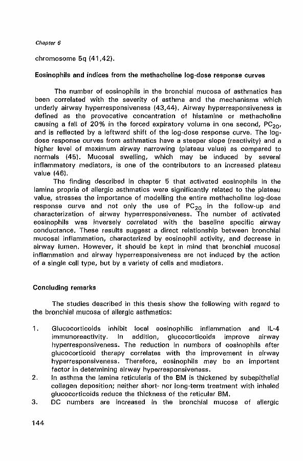
Chapter 6
chromosome 5q (41,42).
Eosinophils and indices from the methacholine log-dose response curves
The number of eosinophils in the bronchial mucosa of asthmatics has been correlated with the severity of asthma and the mechanisms which underly airway hyperresponsiveness (43,44). Airway hyperresponsiveness is defined as the provocative concentration of histamine or methacholine causing a fall of 20% in the forced expiratory volume in one second, PC20,
and is reflected by a leftward shift of the log-dose response curve. The logdose response curves from asthmatics have a steeper slope (reactivity) and a higher level of maximum airway narrowing (plateau value) as compared to normals (45). Mucosal swelling, which may be induced by several inflammatory mediators, is one of the contributors to an increased plateau value (46).
The finding described in chapter 5 that activated eosinophils in the lamina propria of allergic asthmatics were significantly related to the plateau value, stresses the importance of modelling the entire methacholine log-dose response curve and not only the use of PC 20 in the follow-up and characterization of airway hyperresponsiveness. The number of activated eosinophils was inversely correlated with the baseline specific airway conductance. These results suggest a direct relationship between bronchial mucosal inflammation, characterized by eosinophil activity, and decrease in airway lumen. However, it should be kept in mind that bronchial mucosal inflammation and airway hyperresponsiveness are not induced by the action of a single cell type, but by a variety of cells and mediators.
Concluding remarks
The studies described in this thesis show the following with regard to the bronchial muCOSa of allergic asthmatics:
1. Glucocorticoids inhibit local eosinophilic inflammation and IL-4 immunoreactivity. In addition, glucocorticoids improve airway hyperresponsiveness. The reduction in numbers of eosinophils after glucocorticoid therapy correlates with the improvement in airway hyperresponsiveness. Therefore, eosinophils may be an important factor in determining airway hyperresponsiveness.
2. In asthma the lamina reticularis of the 8M is thickened by subepithelial collagen deposition; neither short- nor long-term treatment with inhaled glucocorticoids reduce the thickness of the reticular 8M.
3. DC numbers are increased in the bronchial mucosa of allergic
144
General discussion
asthmatics compared to controls. Inhaled glucocorticoids downregulate the number of DC to normal levels. Since DC play an important role in the initiation and propagation of the allergic immune response, the decrease in DC numbers by glucocorticoids may downregulate T cell activation.
4. IL-4 and IL-5 are predominantly stored in the crystalloid core compartment of the secondary granules of eosinophils. Since eosinophils synthesize and release these cytokines after stimulation, these data suggest a novel role for these cells in the chronic mucosal inflammation in asthma.
REFERENCEs
1. Ying S, Durham SRi Corrigan CJ, Hamid 0, Kay AB. Phenotype of cells expressing mRNA for TH2-type Hnterleukin 4 and interleukin 5) and TH1-type Onterleukin 2 and interferon y) cytokines in bronchoalveolar lavage and bronchial biopsies from atopic asthmatic and normal control subjects. Am J Respir Cell Mol 8io11995; 12:477-87.
2. Carryer HM, Koelsche GA, Prickman LE, Maytum CK, Lake CF, Williams HL. Effects of cortisone on bronchial asthma and hay fever occurring in subjects sensitive to ragweed pollen. Proc Staff Meet, Mayo Clin 1950; 25:482-6.
3. Barnes PJ, Pedersen S. Efficacy and safety of inhaled corticosteroids in asthma. Am Rev Respir Dis 1993; 148:S 1-26.
4. Rothenberg ME, Owen WF, Silberstein OS, Woods J, Soberman RJ, Austen KF, Stevens RI. Human eosinophils have prolonged survival, enhanced functional properties and become hypo dense when exposed to hUman interleukin-3. J Clin Invest 1988; 81:1986-92.
5. Lopez AF, Williamson OJ, Gamble JR et al. Recombinant human granulocyte-macrophage colony stimulating factor stimulates in vitro mature human eosinophil and neutrophil function, surface receptor expression and survival. J Clin Invest 1986; 78:1220-8.
6. Lopez AF, Sanderson CJ, Gamble JR, Campbell HD, Young IG, Vadas MA. Recombinant human interleukin-5 is a selective activator of human eosinophil function. J Exp Med 1988; 167:219-24.
7. Wallen N, Kita H, Weiller 0, Gleich GJ. Glucocorticoids inhibit cytokine mediated eosinophil survival. J Immunol 1991: 147:3490-5.
8. Corrigan CJ, Hamid 0, North J, Barkans J, Moqbel R, Durham S, Gemou-Engesaeth V, Kay AB. Peripheral blood CD4 but not CDS T-Iymphocytes in patients with exacerbation of asthma transcribe and translate messenger RNA encoding cytokines which prolong eosinophil survival in the context of a Th2-type pattern: effect of glucocorticoid therapy. Am J Raspir Cerr Mol Bioi 1995: 12:567-78.
9. Robinson 0, Hamid Q, Ying S, Bentley A, Assoufi B, Durham S, Kay AB. Prednisolone treatment in asthma is associated with modulation of bronchoalveolar lavage cell interleukin-4, interleukin-5, and interferon-y cytokine gene expression. Am Rev Respir Dis 1993; 148:401-6.
10. Jeffery PK, Godfrey RW, Adelroth E, Nelson F, Rogers A, Johansson S-A. Effects of treatment on airway inflammation and thickening of basement membrane reticular collagen in asthma. A quantitative light and electron microscopic study. Am Rev Respir Dis 1992; 145:890-9.
11. Lundgren R, Soderberg M, Horstedt P, Stenling R. Morphological studies of bronchial mucosal biopsies from asthmatics before and after ten years of treatment with inhaled steroids. Eur Respir J 1988; 1 :883-9.
145
Chapter 6
12. Chetta A, Fores! A, Del Donna M, Bertorelli G, Pesci A, Olivieri D. Basement membrane thickness is related to epithelial cellular infiltrate, bronchial responsiveness and airway patency parameters in asthma. Am J Respir Crit Care Med 1995; 151:A132.
13. Sont JK, Willems lNA, Evertse CE, Neeskens P, Krieken van JHJM, Sterk PJ. Thickening of the airway subepithelial reticular layer is related to bronchial hyperresponsiveness to methacholine in subjects with atopic asthma. Am J Respir Crit Care Med 1995; 151 :A833.
14. Schwartz HJ, lowell FC, Melby JC. Steroid resistance in bronchial asthma. Ann Intern Mad 1968; 69:493·9.
15. Corrigan CJ, Brown PH, Barnes NC, Szefler SJ, Tsai J.J, Frew AJ, Kay AB. Glucocorticoid resistance in chronic asthma: glucocorticoid pharmacokinetics, glucocorticoid receptor characteristics, and inhibition of peripheral blood T cell proliferation by glucocorticoids in vitro. Am Rev Raspir Dis 1991; 144:1016-25.
16. Alvarez J, Surs W, Leung DYM, IkUl 0, Gelfland EW, Szefler SJ. Steroid-resistant asthma: immunologic and pharmacologic features. J Allergy Clin Immunol 1992; 89:714-21.
17. Adcock 1M, lane SJ, Brown CR, Peters MJ, Lee TH, Barnes PJ. Differences in binding of glucocorticoid receptor to DNA in steroid-resistant asthma. J Immunol 1995; 154:3500-6.
18. Bellini A, Vittori E, Marini M, Ackerman V, Mattoli S. Intraepithelial dendritic cells and selective activation of Th2-like lymphocytes in patients with atopic asthma. Chest 1993; 103:997-1005.
19. Fokkens WJ, Vroom T, Rijntjes E, Mulder P. Fluctuation of the number of COla (T6)positive dendritic cells, presumably langerhans cells, in the nasal mucosa of patients with isolated grass-pollen allergy before, during, and after the grass-pollen season. J Allergy Clin Immunol 1989; 84:39-43.
20. Nelson OJ, McWilliam AS, Halning 5, Holt PG. Modulation of airway intraepithelial dendritic cells following exposure to steroids. Am J Respir Crit Care Med 1995; 151:475-81.
21. Holm A, Fokkens WJ, Godthelp T, Mulder PG, Vroom TM, Rijntjes E. The effect of three months nasal steroid therapy on nasal T cells and langerhans cells in patients suffering from allergic rhinitis. Allergy 1995; 50:204-9.
22. Sousa AR, lane SJ, Nakhostan JA, Yoshimura T, Lee TH, Poston RN. Increased expression of the monocyte chemoattractant protein-1 in bronchial tissue from asthmatic subjects. Am J Resp!r Cell Mol Bioi 1994; 10:142·7.
23. Tazi A, Bouchonnet F, Grandsaigne M, Boumsell l, Hance AJ, Soler P. Evidence that granulocyte-macrophage colony stimulating factor regulates the distribution and differentiated state of dendritic cells Ilangerhans cells in human lung and lung cancers. J Clin Invest 1993; 91:566-76.
24. Van Haarst JMW, Verhoeven GT, de Wit HJ, Drexhage HA, Moller GM, Hoogsteden He. Glucocorticoids decrease allostimulatory function and B7 expression of human airway dendritic cells in vitro. In: Host defense, dendritic cells and the human lung. Van Haarst JMW, thesis, Rotterdam, 1995:55-67.
25. Dubois GR, Bruijnzeel-Koomen CAFM, Bruijnzeel PLB. Human eosinophils express interleukin-4 receptors. (abstract) J Allergy Clin Immunol 1995; 95:340.
26. Moqbel R, Ying S, Barkans J, Newman TM, Kimm!tt P, Wakelin M, Taborda-Barate l, Meng a, Corrigan CJ, Durham SR, Kay AB. Identification of messenger RNA for Il-4 in human eosinophils with granule localization and release of the translated product. J Immunol 1995; 155:4939-47.
27. Nonaka M, Nonaka R, Woolley K, Adelroth E, Miura K, Okhawara Y, Glibetic M, Nanako K, O'Byrne P, Dolovich J, Jordana M. Distinct immunohistochemical localization of IL-4 in human inflamed airway tissues. J Immunol 1995; 155:3234-44.
28. Schleimer RP, Sterbinsky SA, Kaiser J, Bickel CA, Klunk DA, Tomioka K, Newman W,
146
General discussion
luscinskas FW, Gimbrone MA, Mc Intyre aw, Bochner as. IL-4 induces adherence of human eosinophils and basophils but not neutrophils to endothelium. Association with expression of VCAM-1. J Immunol1992; 148:1086-92.
29. Dubois GR, Bruijnzeel-Koomen CAFM, Bruijnzeel PLB. IL-4 induces chemotaxis of blood eosinophils from atopic dermatitis patients, b4t nbt from normal individuals. J Invest Dermatol 1994; 102:843-6.
30. Del Prete GF, Maggi E, Parrondi P, Chretien I, Tiri A, Macchia 0, Ricci M, aanchereau J, de Vries JE, Romagnani S. IL-4 is an essential co-factor for the IgE synthesis induced in vitro by human T cell clones and their supernatants. J Immunol 1988; 140:4193-8.
31. Walker C, Bode E, Boer L, Hansel TT, Blaser K, Virchow JC. Allergic and non allergic asthmatics have distinct patterns of T-cell activation and cytokine production in peripheral blood and bronchoalveolar lavage. Am Rev Respir Dis 1992; 146:109-15.
32. Robinson OS, Hamid Q, Ying S, Tsicopoulos A, Barkans J, Bentley AM, Corrigan C, Durham SR, Kay AB. Predominant T H2-like bronchoalveolar T-Iymphocyte population in atopic asthma. N Eng J Med 1992; 326:298-304.
33. Bradding P, Feather IH, Howarth PH, Mueller R, Roberts JA, Britten K, Bews JPA, Hunt TC, Okayama Y, Heusser CH, Bullock GR, Church MK, Holgate ST. Interleukin-4 is localized to and released by human mast cells. J Exp Med 1992: 176: 1381-6.
34. Bradding P, Roberts JA, Britten KM, Montefort S, Ojukanovic R, Mueller R, Heusser CH, Howarth PH, Holgate ST. Interleukin-4, -5, and -6 and tumor necrosis factor-a in normal and asthmatic airways: evidence for the hUman mast cell as a source of these cytokines. Am J Respir Cell Mol Bioi 1994; 10:471-80.
35. Bradding P, Okayama V, Howarth PH, Church MK, Holgate ST. Heterogeneity of human mast cells based on cytokine content. J Immunol 1995; 155:297-307.
36. Dubucquoi S, Desreumaux P, Jan!n A, Klein 0, Goldman M, Tavernier J, Capron A, Capron M. Interleukin 5 synthesis by eosinophHs: association with granules and immunoglobulin-dependent secretion. J Exp Med 1994; 179:703-8.
37. Clutterbuck EJ, Hirst MA, Sanderson CJ. Human inter/eukin-5 (lL-5) regulates the production of eosinophils in human bone marrow cultures: comparison and interaction with IL-1, IL-3, IL·6, and GM-CSF. Blood 1989; 73:1504-12.
38. Yamaguchi Y, Hayashi Y, Suguma y, Miura Y, Kasahara T, Kitamura M, Torisu M, Mita S, Tominaga A, Takatsu K, Suda T. Highly purified murine interleukin-5 (lL-5) stimulates eosinophil function and prolongs in vitro survival. J Exp Med 1988; 167:1737-42.
39. Murata y, Takaki S, Migita M, Kikuchi Y, Tominaga A, Takatsu K. Molecular cloning and expression of the human interleukin 5 receptor. J Exp Med 1992; 175:341-51.
40. van Leeuwen BH, Martinson ME, Webb GC, Young IG. Molecular organization of the cytokine gene cluster, involving the human IL-3, IL-4, IL-5, and GM·CSF genes, on human chromosome 5. Blood 1989; 73:1142-8.
41. Marsh DG, Neely JO, Breazeale OR et al. Linkage analysis of IL-4 and other chromosome 5q31.1 markers and total serum immunoglobulin E concentrations. Science 1994; 264: 1152·6.
42. Postma OS, Bleecker ER, Amelung PJ, Holroyd KJ, Xu J, Panhuysen elM, Meyers OA, Levitt RC. Genetic susceptibility to asthma - bronchial hyperresponsiveness coinherited with a major gene for atopy. N Engl J Med 1995; 333:894-900.
43. Bentley AM, Menz G, Storz C, Robinson OS, Bradley 8, Jeffery PK, Durham SR, Kay AB. Identification of T lymphocytes, macrophages and activated eosinophils in the bronchial mucosa of intrinsic asthma: relationship to symptoms and bronchial responsiveness. Am Rev Respir Dis 1992; 145:500-5.
44. Laitinen LA, Laitinen A, Heino M. Haahtela T. Eosinophilic airway inflammation during exacerbation of asthma and its treatment with inhaled corticosteroid. Case reports. Am Rev Respir Dis 1991; 144:83-7.
45. Woolcock AJ, Salome CM, Yan K. The shape of the log dose-response curve to
147
Chapter 6
histamine in asthmatic and normal subjects. Am Rev Respir Dis 1984; 130:71·5. 46. Sterk PJ, Bel EH. Bronchial hyperresponsiveness: the need for a distinction between
hypersensitivity and excessive airway narrowing. Eur Respir J 1989; 2:267·74.
148

SUMMARY

Summary
The main subject of this thesis is the inflammatory process in the bronchial mucosa of allergic asthmatic patients. Bronchial biopsy studies were performed to gain more insight into the cellUlar aspects of the inflammation in the airways of asthmatic patients. Furthermore, the effects of inhaled glucocorticoids were studied on several inflammatory cell types in the bronchial mucosa, which may be useful in the treatment of asthmatic patients.
In chapter 1, asthma is defined as a chronic inflammatory disease of the airways with variable airway obstruction and airway hyperresponsiveness. Pathological changes that contribute to airway narrowing are increased bronchial smooth muscle contraction and enhanced mucus secretion in the airways. Asthma is characterized by increased airway hyperresponsiveness to a variety of non-specific stimuli, including tobacco smoke, fog, cold air and exercise. Atopy, the genetic predisposition for directing an IgE response to common allergens such as house dust mite and pollen, is a risk factor for the development of asthma. The clinical symptoms of asthma are recurrent episodes of cough, dyspnoea, wheezing and chest tightness.
Histological examination of the bronchial mucosa of asthmatics shows epithelial damage (shedding) which is associated with increased airway hyperresponsiveness. Also, the lamina reticularis of the basement membrane (BM) is thickened by a subepithelial collagen deposition, probably produced by myofibroblasts.
Allergic asthma is induced and propagated by the action of a variety of cells and mediators. In chapter 1 the role and function of bronchial epithelial cells, dendritic cells (DC), monocytes/macrophages, eosinophils and mast cells in the process of allergic asthma are described. The mechanisms of action of glucocorticoids and a summary of the most important bronchial biopsy studies of the anti-inflammatory actions of inhaled glucocorticoids (budesonide, beclomethasone dipropionate and fluticasone propionate) are described. Finally, the aims of the studies presented in this thesis are described.
Chapter 2.1 describes the effects of 2.5 years of treatment with beclomethasone dipropionate and bronchodilators on eosinophils, IL-4 immunoreactivity, T lymphocytes, mast cells and the thickness of the reticular BM in bronchial mucosal biopsies from allergic asthmatics from the Dutch Chronic Non-Specific Lung Disease (CNSLD) study group. The numbers of eosinophils and cells expressing IL-4 immunoreactivity were lower in the lamina propria of patients treated with glucocorticoids than in patients treated with bronchodilators only. In chapter 2.2 we show that 3 months of fluticasone propionate therapy is associated with a significant improvement in FEV 1 and PC20 methacholine (an increase of 3.7 doubling doses). This clinical improvement is associated with a reduction in eosinophils and mast
151
Summary
cells, and decreased expression of intercellular adhesion molecule-1 (ICAM-I) on blood vessels in the lamina propria. The reduction in eosinophils after fluticasone propionate treatment was significantly correlated with the improvement in airway hyperresponsiveness. In both studies in chapter 2 we found a significant thickening of the reticular BM of asthmatics compared to controls and the thickness of the lamina reticularis was not reduced after treatment with glucocorticoids.
In chapter 3.1 the distribution of DC in the bronchial mucosa of asthmatics from the CNSLD study group is investigated, as well as the effects of long-term beclomethasone dipropionate treatment on DC. Allergic asthma is associated with a chronic T cell-mediated inflammatory process in the airways. Since T lymphocytes cannot respond to antigens without the help of antigen-presenting cells (A PC), and DC are the most potent APC, it is likely that pulmonary DC playa key role in the initiation and propagation of asthma. Increased numbers of CD1 a + DC, L25 + DC and a higher expression of HLA-DR were observed in the bronchial mucosa of asthmatics compared to controls. With double-staining we showed that virtually all DC expressed HLA-DR, but there were also other HLA-DR + cells such as activated T lymphocytes and macrophages. After 2.5 years of treatment with beclomethasone dipropionate, the numbers of DC and the MHC class II expression were downregulated to normal levels. Chapter 3.2 describes the increased numbers of DC and IgE + cells in the lamina propria of asthmatics compared to controls, and the effects of 3 months of fluticasone propionate therapy of asthmatics on the numbers of DC, IgE+ cells, and cells positive for the high affinity receptor for IgE (FcERI) and on the expression of MHC class II. The IgE + cells, of which the majority are DC and mast cells, likely contribute to an enhanced IgE-mediated antigen/allergen presentation. Fluticasone propionate therapy resulted in a decrease in CD1 a + DC and IgE + cells, and in a reduction in the expression of MHC class II. This was accompanied by a decrease in bronchial hyperresponsiveness. No effects were seen on the number of FCERI + cells. The inhibitory effects of fluticasone propionate on DC are in agreement with the results of the study with beclomethasone dipropionate in chapter 3. 1. The precise mechanisms by which glucocorticoids reduce the number of DC remain to be established. We conclude that DC numbers, IgE + cells, and MHC class II expression are elevated in the bronchial mucosa of atopic asthmatics and that glucocorticoid therapy reduces these DC, IgE + cells and MHC class II expression. This may downregulate T cell activation and hence reduce allergic airway inflammation.
The immunolocalization of IL-4 in the bronchial mucosa of asthmatics is studied in chapter 4.1. Biopsy specimens (containing eosinophils) were processed for immunoelectron microscopy. Immunogold labelling of IL-4 was detected within the crystalloid core compartment of 85% of the eosinophil secondary granules (n ~ 468). No staining was detected in mast cells nor in T
152
Summary
lymphocytes. These results were extended by light microscopic immunodouble-staining for IL-4 protein and eosinophil cationic protein, which showed that a subpopulation of (activated) eosinophils expressed IL-4 immunoreactivity in bronchial mucosal biopsies of allergic asthmatics as well as controls. We conclude that eosinophils in the bronchial mucosa store IL-4 in the crystalloid core of secondary granules. These results in combination with data from the literature on synthesis and secretion of IL-4 by eosinophils, suggest a novel role for these cells in the mucosal inflammatory response.
In chapter 4.2 we describe the ultrastructural immunogold localization of IL-5 in the crystalloid core of the secondary granules of eosinophils in the bronchial mucosa of asthmatic patients. IL-5 is a growth and differentiation factor, activator and chemoattractant for eosinophils and considered a pivotal cytokine in allergen-mediated responses. Our ultrastructural localization of IL-5 suggests that IL-5 stored in the secondary granules of eosinophils would provide a ready source of this cytokine in response to allergen stimulation.
In chapter 5, the relationship between eosinophils and indices from the methacholine log-dose response curves in allergic asthmatics is studied. The number of EG2 + (activated) eosinophils in the lamina propria was significantly related to the maximum response value (the plateau value). Furthermore, the number of activated eosinophils was inversely correlated with the specific (volumic) airway conductance (sGaw), which can be considered as an indicator for the airway lumen. These results suggest a direct relationship between bronchial mucosal inflammation, characterized by eosinophil activity, and decrease in airway lumen.
153


---------SAMENVATTING---------

Samenvatting
In dit proefschrift staat het chronische ontstekingsproces in de bronchuswand van allergische astmapatienten centraal. Bij astmapatienten zijn perifere bronchusbiopten genomen om inzicht te krijgen in het onderliggende ontstekingsproces en om de effecten van therapie met inhalatie-glucocortico'iden te bestuderen. Het uiteindelijke doer van de in dit proefschrift beschreven studies was om bij astmapatienten het anti-inflammatoire effect van glucocortico'iden en hun invloed op verschillende celtypen en mediatoren in de luchtwegen te onderzoeken. Inzicht hierin is van groot belang voor de behandeling van astmapatienten.
In hoofdstuk 1 wordt astma gedefinieerd als een ziekte, die wordt gekenmerkt door een ehroniseh ontstekingsproces in de luehtwegen en een reversibele luehtwegvernauwing. Deze luehtwegvernauwing ontstaat door eontraetie van glad spierweefsel in de bronchus wand en door ophoping van slijm in de luchtwegen. Tevens is er een toename van prikkelbaarheid (hyperreaetiviteit) van de luehtwegen voor niet-allergisehe prikkels, lOals rook, mist, koude lueht en liehamelijke inspanning. Deze prikkels veroorzaken een luehtwegvernauwing met versehijnselen van hoesten, piepende adem haling en kortademigheid. Bij atopisehe astmapatienten bestaat een genetische predispositie tot sensibilisatie door allergenen lOals huisstof en pollen. Daardoor worden door zulke patienten veelal specifieke IgE antistoffen tegen deze allergenen gevormd.
Bij microscopisch onderzoek van de luchtwegen van astmapatienten wordt er schade aan het luchtwegepitheel gezien (shedding). die is geassocieerd met een toename in bronchiale hyperreaetiviteit. Eveneens wordt er een verdikte lamina reticularis van de basaalmembraan (BM) gezien, waarschijnlijk veroorzaakt door subepitheliale collageen deposities door myofibroblasten.
In hoofdstuk 1 worden tevens de rol en typische functie van het bronchusepitheel, dendritisehe cellen (DC), monocyten/maerofagen, Iymfocyten, eosinofiele granulocyten en mestcellen in allergisch astma beschreven. Uit de immunopathogenese van astma blijkt wei dat astma wordt ge'induceerd en in stand gehouden door een heel netwerk van verschillende cellen en mediatoren. Tot slot wordt ingegaan op de werkingsmechanismen van glueocortico'iden en worden de doelstellingen van de studie beschreven.
Hoofdstuk 2. 1 beschrijft de effeeten van 2,5 jaar behandeling met beclomethason dipropionaat en van luehtwegverwijders op eosinofiele granuloeyten, T Iymfoeyten, mestcellen, IL-4 + cellen en de dikte van de lamina reticularis in bronchusbiopten van astmapatienten, die deer uitmaken van het Stimuleringsprogramma GelOndheidonderzoek Chronische Aspecifieke Respiratoire Aandoeningen (SGO-CARA onderzoek). Het aantal eosinofiele granulocyten en IL-4 + cellen was significant lager in de mucosa van patienten behandeld met glucocortieo'iden, vergeleken met patienten behandeld
157
Samenvatting
met luchtwegverwijders. Er werd geen effect gezien op de dikte van de lamina reticularis.
Hoofdstuk 2.2 behandelt de effecten van 3 maanden therapie met het inhalatieglucocortico'ld fluticason propionaat op longfunctie parameters en het cellulaire ontstekingsproces in de lamina propria. Significante klinische verbeteringen in FEV 1 en in PC20 methacholine (3,7 verdubbelingsdoses) gingen gepaard met een afname in het aantal eosinofiele granulocyten en mestcellen, en verminderde expressie van het intercellulair adhesiemolecuul-1 (ICAM-I), dat aanwezig is op het endotheel in de lamina propria. De afname van het aantal eosinofielen onder invloed van fluticason propionaat was significant gecorreleerd met de verbetering in bronchiale hyperreactiviteit. De dikte van de lamina reticularis nam niet af onder invloed van glucocorticoiden. In beide studies werd er een significant dikkere reticulaire BM bij astmapatienten gevonden, vergeleken met gezonde controles.
In hoofdstuk 3 is het voorkomen van DC in de bronchiale mucosa van astmapatienten bestudeerd. DC zijn gespecialiseerde cellen van het immuunsysteem, die antigenen kunnen opnemen en tot peptiden kunnen afbreken om deze vervolgens te presenteren aan T-Iymfocyten. Indien T-Iymfocyten het peptide als lichaamsvreemd herkennen, worden ze geactiveerd. In astma spelen DC een essentiele rol in het op gang brengen van de allergische reactie.
In hoofdstuk 3.1 wordt beschreven dat het aantal CD1a+ DC en L25+ DC, en de expressie van HLA-DR in bronchusbiopten van astmapatienten uit het SGO-CARA onderzoek, die geen inhalatie-glucocortico'lden gebruikten, was verhoogd in vergelijking met gezonde controles. Met dubbelkleuringen werd aangetoond dat bijna aile DC MHC klasse II tot expressie brengen. Bij patienten die 2,5 jaar behandeld zijn met beclomethason dipropionaat werden aantallen DC gevonden, die vergelijkbaar waren met die van controles.
In hoofdstuk 3.2 wordt het effect van 3 maanden inhalatie-therapie met fluticason propionaat op DC, IgE + cellen, de expressie van MHC klasse II en op cellen positief voor de hoog affiene IgE receptor (FCERI) + onderzocht. De IgE + cellen, waarvan het merendeel DC en mestcellen zijn, drag en bij tot een verhoogde IgE gemedieerde antigeenpresentatie. Fluticason propionaat therapie resulteerde in een afname van het aantal DC en IgE + cellen en een reductie in MHC klasse II expressie. Er werd geen effect gevonden op het aantal FCERI + cellen. Uit deze resultaten kan worden geconcludeerd dat het aantal DC in astma is toegenomen en dat glucocortico'lden het aantal DC doen verminderen, wat mogelijk resulteert in een verminderde activatie van T-Iymfocyten. Het mechanisme van de afname van het aantal DC door glucocortico'lden is nog niet opgehelderd.
In hoofdstuk 4. 1 wordt de immunolocalisatie van IL-4 beschreven in de bronchiale mucosa van astmapatienten. Immunogoud labelling van IL-4 werd aangetroffen in het kristallo'lde internum in 85% van de secundaire of
158
Samenvatting
specifieke granula (n=468) van eosinofiele granulocyten. Mestcellen noch TIymfocyten waren positief voor IL-4. Deze· resultaten zijn uitgebreid met immunohistochemische dubbelkleuringen voor IL-4 en (geactiveerde) eosino!ielen. Een subpopulatie van (geactiveerde) eosinofielen was IL-4 positief, zowel bij astmapatienten als bij gezonde controles. Uit deze resultaten en gegevens uit de literatuur over synthese en uitscheiding van IL-4 door eosinofiele granulocyten kan worden geconcludeerd dat deze cellen een belangrijke rol spelen in allergisch astma.
In hoofdstuk 4.2 wordt de ultrastructurele immunolocalisatie van IL-5 in het kristallo'ide internum van de secundaire granula van eosinofiele granulacy ten beschreven. IL-5 is als groei- en differentiatiefactor en als chemoattractant voor eosinofiele granulocyten betrokken bij de allergische reactie. De aanwezigheid van IL-5 in opgeslagen vorm in de eosinofiele granulocyt maakt een snelle reactie na allergeen stimulatie mogelijk.
In hoofdstuk 5 wordt de relatie tussen indices van de methacholine log-dose respons curve van allergische astmapatienten en de aanwezigheid van eosinofiele granulocyten in de bronchiale mucosa onderzocht. Het aantal geactiveerde eosinofiele granulocyten in de lamina propria bleek gecorreleerd met de maximale respons (plateau waarde). Tevens was het aantal geactiveerde eosinofiele granulocyten negatief gecorreleerd met de specifieke luchtwegconductantie (sGaw), wat een maat is voor de luchtwegweerstand. Deze resultaten suggereren dat er een directe relatie be staat tussen de mate van ontsteking in de bronchiale mucosa en de luchtwegvernauwing.
De belangrijkste conciusies uit de in dit proefschrift beschreven studies zijn:
1. Inhalatie-glucocortico'iden verminderen het aantal eosinofiele granulocyten en IL-4 + cellen in het bronchiaal slijmvlies van allergische astmapatienten. De afname van het aantal eosinofiele granulocyten na glucocortico'id therapie is gecorreleerd met een vermindering in bronchiale hyperreactiviteit.
2. In allergisch astma is de lamina reticular is van de 8M verdikt door subepitheliale collageendeposities; kort- noch langdurende behandeling met inhalatie-glucocortico'iden doet de dikte van de reticulaire 8M afnemen.
3. Het aantal DC in het bronchiaal slijmvlies van astmapatienten is verhoogd vergeleken met gezonde controles. Inhalatie-glucocortico'iden reduceren het aantal DC tot normale waarden, wat zou kunnen resulteren in een verminderde T-Iymfocyt activatie.
4. IL-4 en IL-5 zijn voornamelijk gelocaliseerd in het kristallo'ide internum van de secundaire granula van eosinofiele granulocyten in het bronchiaal slijmvlies.
159

---------- ABBREVIATIONS----------

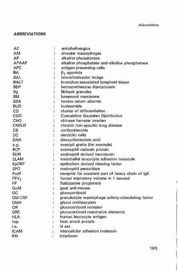
ABBREVIATIONS
AC AM AP APAAP APC BA BAL BALT BDP Bg BM BSA BUD CD CGD CHO CNSLD CS DC DNA e.g. ECP EDN ELAM EpDRF EPO FceR FEV1 FP GaM GC GM-CSF GMA GR GRE HLA hsp i.e. ICAM IFN
Abbreviations
anticholinergica alveolar macrophages alkaline phosphatase alkaline phosphatase anti-alkaline phosphatase antigen-presenting cells 112 agonists bronchoalveolar lavage bronchus-associated lymphoid tissue beclomethasone dipropionate Birbeck granules basement membrane bovine serum albumin budesonide cluster of differentiation Cumulative Gaussian Distribution chinese hamster ovarian chronic non-specific lung disease corticosteroids dendritic cells deoxyribonucleic acid exempli gratia (for example) eosinophil cationic protein eosinophil derived neurotoxin endothelial leucocyte adhesion molecule epithelium derived relaxing factor eosinophil peroxidase receptor for constant part of heavy chain of IgE forced expiratory volume in 1 second f)uticasone propionate goat anti-mouse glucocorticoid granulocyte macrophage colony-stimulating factor glycol methacrylate glucocorticoid receptor glucocorticoid responsive elements human leucocyte antigen heat shock protein id est intercellular adhesion molecule interferon
163
Abbreviations
Ig IL LC LFA LT mAb or MoAb MBP MCP MCr MCre MHC MIP MLDR mRNA n NEP NGS NO NS P PAF PB PBS PC20
PCR PG PGDF PL R RaM RANTES
rBM RNA RSV RT S sGaw TGF Th TNF
164
immunoglobulin interleukin Langerhans cells lymphocyte function-associated antigen leukotriene monoclonal antibodies major basic protein monocyte chemoattractant protein tryptase-containing mast cells tryptase- and chymase-containing mast cells major histocompatibility complex macrophage inflammatory protein methacholine log-dose response messenger RNA number in study or group neutral endopeptidase normal goat-serum nitric oxide not significant plateau value platelet activating factor peripheral blood phosphate-buffered saline provocative concentration (of a non-specific agent, e.g. histamine) which causes a 20% decrease in FEV1 polymerase chain reaction prostaglandin platelet-derived growth factor placebo reactivity rabbit anti-mouse regulated upon activation, normal T expressed, and presumably secreted reticular basement membrane ribonucleic acid Respiratory Syncytial Virus room temperature sensitivity specific airway conductance transforming growth factor T helper tumor necrosis factor
VCAM VIP VLA
vascular cellular adhesion molecule vasoactive intestinal peptide very late activation antigen
Abbreviations
165


DANKWOORD

Dankwoord
DANKWOORD
Aan de totstandkoming van dit proefschril! hebben velen een bijdrage geleverd. Ik ben iedere betrokkene daarvoor bijzonder dankbaar. Een aantal van hen wil ik hier met name noemen.
Prof. dr. R. Benner, beste Rob, dank voor de mogelijkheid die je mij hebt geboden om 4 jaar op jouw afdeling onderzoek te doen. Ook dank ik je voor de constructieve manier waarop jij inhoud hebt gegeven aan het zijn van mijn promotor. Van het gebruik van "de kopkamer" waar ik de laatste maanden in rust heb kunnen schrijven, heb ik genoten. Ik verheug mij op het continueren van klinisch wetenschappelijk onderzoek samen met de afdeling Immunologie.
Dr. H.C. Hoogsteden, beste Henk, dank voor je enthousiaste en zorgvuldige begeleiding van mijn onderzoek en voor de vrijheid die je mij binnen mijn onderzoek hebt gegeven. Je vertrouwen en het regelmatige overleg hebben mij gesteund en gestimuleerd. Ik hoop dat we deze onderzoekslijn in Rotterdam kunnen voortzetten.
Prof. dr. Th.H. van der Kwast, beste Theo, dank voor de hele prettige samenwerking met de afdeling Pathologie en voor het geven van je heldere kijk op mijn onderzoeksvragen, met name die op het gebied van de immunoelectronenmicroscopie. Hartelijk dank ook voor de snelle beoordeling van de manuscripten van mijn proefschril!.
Prof. dr. C. Hilvering, u wil ik bedanken voor het in mij gestelde vertrouwen, dat sprak uit het feit dat u mij destijds een opleidingsplaats in het vooruitzicht stelde. Verder dank ik u dat u zilting heel! willen nemen in de kleine commissie.
Prof. dr. D.S. Postma, beste Dirkje, voor het "SGO-onderzoek" heb ik in Groningen altijd op vee I steun en medewerking kunnen rekenen. De "indeling van de astmapatienten volgens Zwolle" is hier een legendarisch voorbeeld van. Hartelijk dank voor het kritisch doorlezen van mijn manuscripten.
Dr. E.P. Prens, beste Errol, met mijn longonderzoek kwam ik bij jou in de dermatologiegroep terecht en jouw manier van begeleiden als labhoofd paste goed bij mij. Ais ik je nodig had, kon ik altijd op je rekenen.
Prof. dr. H. Drexhage, beste Hemmo, dank voor de discussies en het becommentarieren van mijn manuscripten over dendritische cellen in de long.
Prof. dr. J.M. Bogaard, beste Jan, dank voor de prettige en deskundige begeleiding op het gebied van de Longfunctie.
Prof. dr. P.J. Sterk, dank ik dat hij zitting heel! willen nemen in mijn promotiecommissie.
Van de analisten wil ik Ton de Jong, Alex KleinJan, Corine van Helden-
169
Dankwoord
Meeuwsen, Annet Wierenga-Wolf en Mieke Smith noemen. Beste Ton, jij bent mijn rechterhand geweest voor de EM (Electronen
microscopie) op de afdeling Pathologie, waar ik altijd weer met een nieuwe opdracht terecht kon. Heel hartelijk dank hiervoor.
Beste Alex, jij hebt mij in het begin het meest geleerd van het vak 'Immunohistologie' .
Beste Corine en Annet, na 1,5 jaar werd jij, Corine 'mijn analist' en binnen een jaar had ik er twee, weliswaar in een 'duo-baan'. Jullie hebben je als een fantastisch koppel met veel enthousiasme en precisie over het snijden en kleuren van de longbiopten ontfermd; het werden in totaal meer dan 5000 coupes. Heel hartelijk dank.
Beste Mieke, dank voor het zorgvuldig invriezen van de SGO-biopten in Groningen. Ons overleg was altijd prettig.
Binnen onze eigen groep heb ik steeds collegialiteit ondervonden van Reno, Roger, Tom, Joost, en Eveline, en de lunches in het hok op Ee802 met Leo, Marie-Jose, Maarten Hazenberg, Maarten Hooijer, Johanneke, Annemieke en Ingrid waren een gezellige onderbreking van de dag. Ook de andere collega's dank ik voor de prettige sfeer op de afdeling.
Beste Tar, hartelijk dank voor het maken van mijn figuren, foto's en dia's, jij levert mooi vakwerkl
Het secretariaat, Geertje, Danielle, en Petra en onze administrateurs, Tom, Henk en Renate dank ik voor hun permanente hulpvaardigheid. Petra in het bijzonder, hartelijk dank voor al je secretariele ondersteuning, en het verzorgen van de lay-out van mijn proefschrift.
Een deel van het onderzoek werd verricht op de afdeling Longziekten van het Academisch Ziekenhuis Dijkzigt Rotterdam en op het Longfunctielaboratorium (o.l.v. Prof. dr. J.M. Bogaard), waarvan ik aile medewerkers en de participerende patienten wil bedanken.
Shelley Overbeek, beste Shelley, hartelijk dank voor het verzamelen van de patienten en het afnemen van de biopten. Veel succes met je eigen promotie.
Gert Verhoeven wens ik vee I succes met het verwerken van de biopten van de COPD-patienten en zijn promotie.
Paul Mulder wil ik hartelijk danken voor het samen met mij statistisch bewerken van aile gegevens.
Dr. Jan Raaijmakers, Katia van Geffen en Rolf Remorie van Glaxo Wellcome B.V. dank ik voor hun steun en interesse die ik ontving.
Dr PK Jeffery, dear Peter, it was a great pleasure and an inducement for my research to visit your Department of Lung Pathology, National Heart and Lung Institute, Royal Brompton Hospital.
170
Dankwoord
Oankzij lamilie en vrienden was deze onderzoeksperiode ook buiten het lab een hele lijne tijd.
Erik ten Berge wil ik niet onvernoamd laten omdat ik met hem destijds mee mocht naar het Bronkhorst Colloquium.
Jan Maarten, ik vind het heel leuk dat je nu mijn paraniml bent en straks weer mijn collega zult zijn.
Lieke en Joost, en de ouders van Paul, dank voor jullie interesse in mijn onderzoek.
Lieve pap en mam, jullie ben ik heel erg dankbaar voor alles wat jullie mij hebben meegegeven. Jullie mentale steun en warmte betekenen heel veel voor mij. Oit boekje is terecht aan jullie opgedragen.
En tenslotte, lielste Paul, het proelboek is al! Jouw nooit allatende steun en lielde zijn voorwaarden geweest voor de totstandkoming hiervan.
Rotterdam, lebruari 1 996
171


--------- CURRICULUM VITAE ---------

16 september 1 964 juni 1983 sept. 1983
1986-1987
mei-sept. 1988
okt. 1988 nov. 1988-mei 1989
mei 1991 juni 1991-jan. 1992
febr. 1992-maart 1996
1993-1994 april 1996
Curriculum vitae
CURRICULUM VITAE
Gertrude Marie (Trudeke) Moller
Geboren te Baltimore, V.S. Gymnasium-B, Stedelijk Gymnasium, Leiden Aanvang studie Geneeskunde, Rijksuniversiteit Leiden Student-assistentschap Macroscopische Anatomie Afstudeerproject "Vergelijking van oestrogeenreceptorwaarden in mammacarcinoom, bepaald met de immunohistochemie en de biochemie". Verricht op de afdeling Klinische Oncologie en het Laboratorium voor Pathologie, Academisch Ziekenhuis Leiden (o.Lv. Dr. H.J. Keizer en Prof. dr. Ph.M. Kluin) Doctoraalexamen Geneeskunde Co-assistentschap Radiotherapie: MGH Cancer Center, Massachusetts General Hospital, Harvard Medical School, Boston (o.Lv. Prof. dr. H.D. Suit) Artsexamen, Rijksuniversiteit Leiden Assistent geneeskundige niet in opleiding (AGNIO), afdeling Longziekten, Streekziekenhuis Midden Twente, Hengelo Assistent in opleiding (AIO), afdelingen Immunologie en Longziekten, Academisch Ziekenhuis Dijkzigt Rotterdam en Erasmus Universiteit Rotterdam (o.Lv. Prof. dr. R. Benner en dr. H.C. Hoogsteden). In opleiding tot Immunoloog (Stichting voor Opleidingen tot Medisch-Biologisch Wetenschappelijk Onderzoeker) Assistent practicum Klinische Immunologie Assistent geneeskundige in opleiding (AGIO), afdeling Interne Geneeskunde I, Academisch Ziekenhuis Dijkzigt Rotterdam (opleider Prof. dr. M.A.D.H. Schalekamp); vooropleiding "Inwendige Geneeskunde" in het kader van de opleiding tot "arts voor Longziekten en Tuberculose", Academisch Ziekenhuis Dijkzigt Rotterdam (opleider Prof. dr. C. Hilvering)
175


--------- PUBLICATIONS ---------

PUBLICATIONS
1. Moller GM, ten Berge EJFM, Stassen CM. Tracheocele: a rare cause of difficult endotracheal incubation and subsequent pneumomediastinum. Eur Respir J 1994; 7: 1376-7.
2. Hoogsteden HC, Moller GM. Medicamenteuze be.handeling van astma. Diagnose Informatie en Medische Statistiek 1995; 19: 12-7.
3. Moller GM, de Jong TAW, Overbeek SE, van der Kwast TH, Postma DS, Hoogsteden HC. Ultrastructural immunogold localization of interleukin 5 to the crystalloid core compartment of eosinophil secondary granules in patients with atopic asthma. J Histochem Cytochem 1996; 44:67-9.
4. Moller GM, Overbeek SE, van Heiden-Meeuwsen CG, van Haarst JMW, Prens EP, Mulder PG, Postma DS, Hoogsteden HC. Increased numbers of dendritic cells in the bronchial mucosa of atopic ashtmatics: down regulation by inhaled corticosteroids. Clin Exp Allergy 1996 (in press).
5. Moller GM, de Jong TAW, van der Kwast TH, Overbeek SE, WierengaWolf AF, Thepen T, Hoogsteden HC. Immunolocalization of interleukin-4 in eosinophils in the bronchial mucosa of atopic asthmatics. Am J Respir Cell Mol Bioi 1996 (in press).
6. Moller GM, Overbeek SE, van Heiden-Meeuwsen CG, de Jong TAW, Mulder PG, Drexhage HA, Hoogsteden HC. Fluticasone propionate therapy is associated with modulation of dendritic cells in the bronchial mucosa of atopic asthmatics. Submitted.
7. Moller GM, Overbeek SE, van Heiden Meeuwsen CG, Godthelp T, Prens EP, Mulder PG, Aalbers R, Postma DS, Hoogsteden HC. Effects of longterm treatment with beclomethasone on airway inflammation in the bronchial mucosa of atopic asthmatics. SUbmitted.
8. Moller GM, Overbeek SE, van Heiden-Meeuwsen CG, Mulder PG, Hoogsteden HC. Influence of fluticasone propionate on airway inflammation in atopic asthmatics. Submitted.
179
