brain contains twoformsofsynaptic vesicle protein2 twoformsofsynaptic vesicle protein2 sandram....
TRANSCRIPT
Proc. Natl. Acad. Sci. USAVol. 90, pp. 2150-2154, March 1993Neurobiology
Brain contains two forms of synaptic vesicle protein 2SANDRA M. BAJJALIEH, KAREN PETERSON, MICHAL LINIAL*, AND RICHARD H. SCHELLER
Howard Hughes Medical Institute and Department of Molecular and Cellular Physiology, Stanford University, Stanford, CA 94305-5426
Communicated by W. Maxwell Cowan, December 7, 1992
ABSTRACT Molecular cloning of a cDNA encoding syn-aptic vesicle protein 2 (SV2) revealed that it is homologous toa family of proton cotransporters from bacteria and fungi andto a related family of glucose transporters found in mammals.The similarity to proton cotransporters raised the possibilitythat SV2 might mediate the uptake of neurotransmitters intovesicles, an activity known to require a proton gradient. Todetermine whether SV2 is a member of a family of vesicularproteins, we used the SV2 clone to screen for similar cDNAs inrat brain. We characterized 42 clones, 25 of which encode SV2and 4 of which encode a protein, SV2B, that is 65% identicaland 78% similar to SV2. The protein encoded by the SV2BcDNA is recognized by the monoclonal antibody that defines theSV2 protein. When SV2B is expressed in COS cells, antibodylabeling is reticular in nature, suggesting that SV2B, like SV2(hence, SV2A), is segregated to intracellular membranes. Theexpression ofSV2B is limited to neural tissue. While both formsof SV2 are expressed in all brain regions, SV2B is expressed athighest levels in the cortex and hippocampus, whereas thehighest level of expression of SV2A is in subcortical regions.Therefore, the SV2 proteins, like other characterized synapticvesicle proteins, comprise a small gene family.
Efforts to understand the molecular events underlying neuralsecretion have focused on characterizing molecules unique tosynaptic structures. To date, several synaptic vesicle pro-teins have been characterized (1-3). One of these proteins,synaptic vesicle protein 2 (SV2), was originally identifiedwith a monoclonal antibody generated against cholinergicvesicles from the electric organ of the electric ray Discopygeommata (4). The epitope recognized by this antibody wasfound to be present on secretory vesicles of all neural andendocrine cells surveyed (5-7). Its presence across a widerange of species, from elasmobranchs to mammals, suggeststhat it plays a critical role in vesicle functioning.cDNAs encoding SV2 were cloned by screening a rat brain
library with DNA probes based on amino acid sequence (8)and by screening for immunoreactivity in CHO cells trans-fected with PC-12 cell cDNAs (9). Both methods identifiedthe same cDNA sequence. The predicted protein has 12putative membrane-spanning domains and shows significanthomology to a large family of transport proteins. Included inthis family are bacterial and fungal proteins that cotransportprotons with sugars, citrate, and drugs, and mammalianfacilitative glucose transporters (10, 11). The similarity to alarge class of transport proteins suggested that SV2 might bea transporter specific to synaptic vesicles. The transport ofneurotransmitters into vesicles is dependent on a protongradient across the vesicle membrane (12). Given that theactivity of bacterial transporters homologous to SV2 alsorequires a proton gradient, it seemed possible that SV2 mightbe a neurotransmitter transporter.The occurrence ofthe SV2 antigen in a wide range ofneural
and endocrine cells suggested either that the SV2 antigen
defines a family of proteins or that any transport activity ofSV2 is generic to all secretory vesicles. To address the firstpossibility, that the SV2 cDNA is one of a family of relatedgene products, we looked for related proteins by screening arat brain library with the region of SV2 most homologous tothe bacterial transporters. We report here a cDNA sequencethat encodes a protein recognized by the anti-SV2 antibody,which we denote SV2B.t
MATERIALS AND METHODSMaterials. Taq DNA polymerase was purchased from
Boehringer Mannheim, radiolabeled nucleotides were fromDuPont, and NucTrap push columns were from Stratagene.The rat brain cDNA library used was from Stratagene, andthe pCMV vector was from Invitrogen. Paraformaldehydewas purchased from EM Sciences. Sequenase (United StatesBiochemical) reagents were used for DNA sequencing. Rho-damine-conjugated goat anti-mouse secondary antibody waspurchased from Tago. RNA Stat-60 was purchased from TelTest "B" (Friendswood, TX). Enhanced chemiluminescenceantibody detection reagents were from Amersham. RNA sizestandards were from BRL. Nylon membrane was purchasedfrom Schleicher & Schuell.
Generation of a Probe and Library Screening. To generate aprobe recognizing the first six membrane domains of SV2, aPCR was done using 24-nucleotide primers coding for aminoacids 164-171 and 352-359. SV2 inSK- (4 fmol) was amplifiedin a reaction mixture containing 0.3 ,uM primers; 3.3 ,uM eachdTTP, dGTP, and dATP; reaction buffer (provided by themanufacturer); 0.5 unit of Taq DNA polymerase; and 500 ,uCi[32P]dCTP (3000 Ci/mmol; 1 Ci = 37 GBq; 3.3 ,uM). Primerswere annealed at 50°C for 1 min. Thirty amplification cycleswere done followed by a 10-min extension reaction. Theradiolabeled products were separated from other reactionconstituents by NucTrap push column purification.
This probe was used to screen a Lambda ZAP II rat braincDNA library by standard techniques (13). Filters were hy-bridized at 55°C in 1.2 M salt [6x standard saline citrate(SSC)]. Thirty-nine positive clones were isolated. To deter-mine which were reisolates ofSV2, sequencing reactions weredone using an oligonucleotide primer made to an internalregion of SV2. Clones identified as novel were sequenced bythe dideoxynucleotide chain-termination reaction.
Transient Expression of SV2B in COS Cells. The SV2BcDNA was subcloned into the pCMV expression vector.Plasmid DNA was purified by Triton lysis followed by adouble cesium chloride gradient procedure (13). COS cells,plated on two-chambered slides, were transfected with 10 ugofplasmidDNA using the DEAE-dextran method (19). Threedays after transfection, the cells were processed for anti-SV2immunoreactivity.
Immunohistochemistry. Cells were fixed in phosphate-buffered saline (PBS) containing 4% paraformaldehyde for 20
*Present address: The Hebrew University ofJerusalem, Departmentof Biological Sciences, Institute of Life Sciences, Jerusalem 91904,Israel.tThe sequence reported in this paper has been deposited in theGenBank data base (accession no. L10362).
2150
The publication costs of this article were defrayed in part by page chargepayment. This article must therefore be hereby marked "advertisement"in accordance with 18 U.S.C. §1734 solely to indicate this fact.
Proc. Natl. Acad. Sci. USA 90 (1993) 2151
min and rinsed three times in PBS with 0.1 M glycine. Cellswere permeabiized in PBS with 1% bovine serum albumin,2% normal goat serum, and 0.4% saponin (blocking buffer)and then incubated with the anti-SV2 antibody in blockingbuffer for 1 hr. Unbound antibody was removed with threePBS/glycine washes. The anti-SV2 antibody was labeledwith secondary rhodamine-conjugated goat anti-mouse anti-body.Western Blot Analyses. Transfected cells were homoge-
nized in 10 mM Hepes, pH 7.5/0.32 M sucrose using a metalDounce homogenizer chilled on ice. Unbroken cells andnuclei were sedimented at 250 x g for 5 min, after whichprotein content of the supernatant (homogenate) was deter-mined by the Bio-Rad protein assay with bovine serumalbumin as a standard. Twenty-five micrograms of eachhomogenate was loaded onto an 8-12.5% gradient gel. Alsoloaded was -0.5 ,g of synaptic vesicle protein purified fromrat brain (8). Gel-resolved proteins were transferred to ni-trocellulose and probed with affinity-purified anti-SV2 as-cites fluid at 1:1000. Antibody binding was detected with agoat anti-mouse secondary antibody coupled to horseradishperoxidase that was used with an enhanced chemilumines-cence kit.Northern Blot Analyses. Total RNA was obtained from
tissue frozen in liquid nitrogen immediately after sacrifice andstored at -80°C. Both the method of Chirgwin et al. (14) andof Chomczynski and Sacchi (15) were used to isolate RNA.RNA was resolved on 1% agarose formaldehyde gels andblotted to nylon. RNA was stained with 0.02% methyleneblue in 0.3 M NaOAc. Relative amounts of 18S and 28S RNAwere similar across lanes of the blots used. Prehybridizationbuffer contained 0.5 M Na2HPO4 (pH 7.2), 1 mM EDTA, and5% SDS. Probes were generated by PCR amplification of alarge region of each SV2 clone using [32P]dCTP as describedabove, except that nucleotide concentrations were lower(12-18 ,uM). Probes were incubated with filters overnight at65°C. Filters were washed in 0.2x SSC/0.1% SDS at 65°Cand then exposed to film. To reprobe, fiters were stripped byboiling in distilled water for 5 min.
RESULTSIsolation of a cDNA Encoding a Second Form of SV2. To
identify other forms of SV2, we screened a rat brain cDNAlibrary with DNA encoding the first six transmembranedomains. This region of SV2 contains the consensus se-quences that define the bacterial transporter family (10) withwhich SV2 shares homology. We reasoned that relatedproteins would contain these conserved sequences. Screen-ing -500,000 plaques at low stringency produced 25 reiso-lates of SV2. Sequencing reactions utilizing a primer from thecoding region revealed a single base difference in all 25reisolates when compared to the original clone. This differ-ence results in a change from phenylalanine to cysteine atamino acid 342. Sequencing in entirety a SV2 clone isolatedin this screen revealed no other differences.
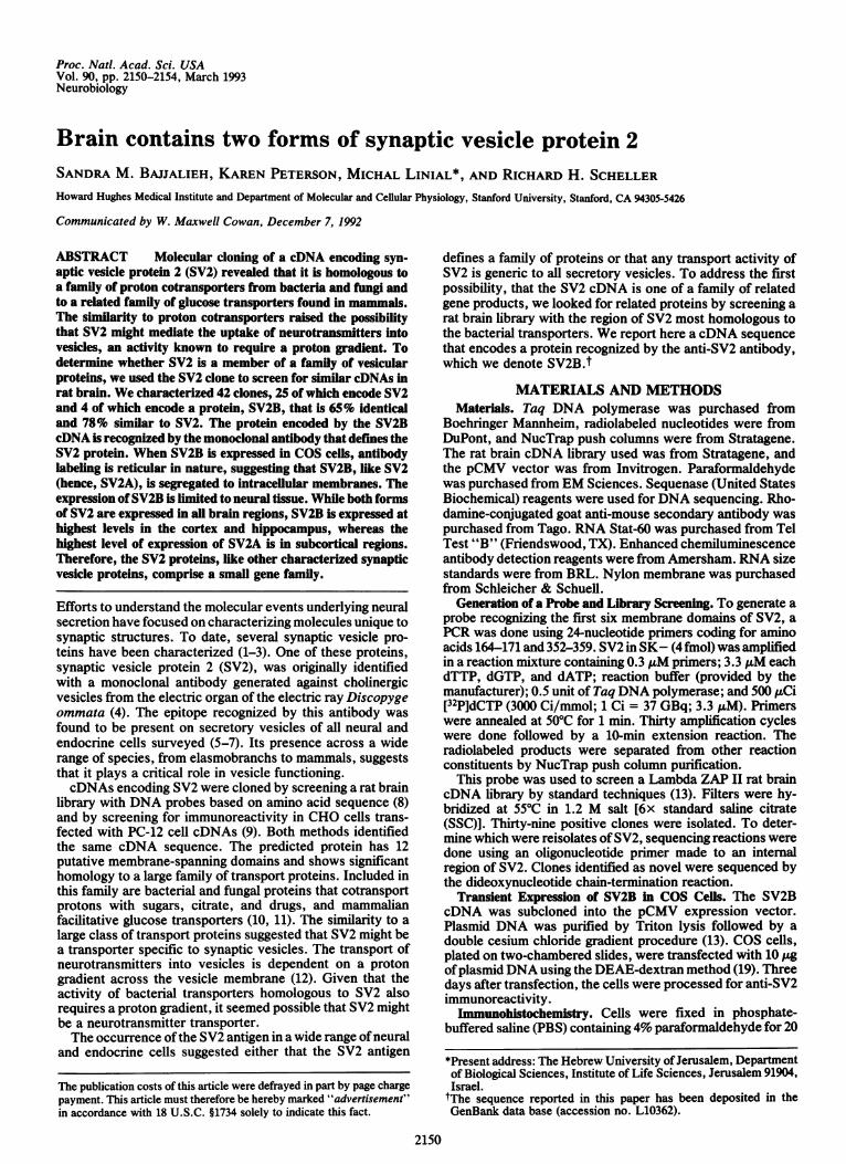
In addition to SV2, four other clones encoding a proteinsimilar to SV2 were isolated. The longest of the four waschosen for analysis and sequenced on both strands. Thenucleic and predicted amino acid sequences are shown in Fig.1. The 3660-nucleotide cDNA encodes a 682-amino acidprotein with a predicted molecular mass of 77.5 kDa. LikeSV2, this protein has an acidic N terminus with no apparentsignal sequence, 12 predicted membrane-spanning domains,and 3 consensus sites for N-glycosylation. BESTFIT (16)alignment withSV2 revealed 65% identity and 78% similaritytoSV2. Based on these similarities, we term this cDNA andthe protein it encodes SV2B and will refer to the originalSV2as SV2A.The region of SV2B showing the most divergence from
SV2A is the N-terminal cytoplasmic tail (Fig. 2). SV2B hasa shorter N terminus, lacking the first 39 amino acids of SV2A
and two other 9-amino acid segments in this region. The onlyother deletion in SV2B is a 2-amino acid stretch in the regionbetween membrane domains 7 and 8, the second mostdivergent region of SV2B. This region contains 38% noncon-servative substitutions. Other differences between the twoproteins are scattered throughout and include many conser-vative changes.SV2 Consensus Sequences. Identical and highly conserved
regions of protein families are often functionally important.Two shared features of the SV2 proteins deserve mention.The first is the conservation between SV2A and -B of thesugar transporter motifs. The consensus sequences used todefine this family of transporters (10) include an RXGRRsequence between the second and third membrane domains,a PESPR sequence at the end of the sixth membrane domain,and a diffused motif that spans the fourth and fifth membranedomains. In SV2B, the RXGRR sequence is KXGRK, con-servative changes which suggest that the presence of basicresidues in these positions is definitive of the consensussequence. Like SV2A, SV2B has two differences in thediffused motif at the same residues as in SV2A. In the sugartransporters the RXGRR and PESPR motifs are repeated inthe second halfofthe proteins, supporting the hypothesis thatthey arose via internal duplication. While the homologybetween SV2 and the transporters is less apparent in thesecond half of SV2, there are modified versions of these twoconsensus sequences in the second half ofboth forms of SV2.The region between the 7th and 8th membrane domains(analogous to the region between the 2nd and 3rd membranedomains) contains an RXGRXR sequence in both SV2 pro-teins. Likewise, after the 12th membrane domain, both SV2proteins have a PETK sequence, a variant of the PESPRsequence that is also found following the 12th membranedomain of the Escherichia coli arabinose and xylose trans-porters and the facilitative glucose transporter of mammalianhepatoma cells (10). Therefore, the SV2 proteins, like sugartransporters, shows some evidence of internal duplication.The large loop between the seventh and eighth membrane
domains distinguishes the SV2 proteins from the transporterswith which they share homology. Both SV2 proteins containthree conserved consensus sites for N-glycosylation and bothare rich in phenylalanine in this region. SV2A is 12% phe-nylalanine in this region; SV2B is 13% phenylalanine. Espe-cially interesting is a series of phenylalanines, often followedby charged residues, interrupted by nine amino acids. AFASTA (17) search of GenBank revealed similarity of thisregion to the mcbg protein of E. coli (18). mcbg is a 22-kDasoluble protein that appears to mediate antibiotic resistanceby working in conjunction with a transport protein in the cellmembrane. Like the region between the seventh and eighthmembrane domains of the SV2 proteins, mcbg is rich inphenylalanine (10% of the protein) and has a region ofphenylalanines interrupted by an identical number of resi-dues as in the SV2 proteins (Fig. 3). This similarity suggeststhat the SV2 proteins may be a compilation of two bacterialtransport systems and that the phenylalanine-rich loop me-
diates interaction of the transported substance with thedomains that span the membrane.SV2B Is Recognized by the Anti-SV2 Antibody. The epitope
recognized by the anti-SV2 monoclonal antibody is predictedto be in the N terminus of the protein (8). Since the majordifferences between the two forms ofSV2 are in this region,it seemed likely that the antibody might not recognize theSV2B protein. To determine whether the widespread immu-noreactivity found with the anti-SV2 antibody representsboth SV2A and SV2B, the immunoreactivity of the SV2Bprotein was assessed. SV2B, subcloned into the pCMVmammalian expression vector, was transiently expressed inCOS cells, an exocrine cell line that does not express SV2immunoreactivity. Cells transfected with SV2B cDNA were
Neurobiology: Bajalieh et al.

2152 Neurobiology: Bajjalieh et al. Proc. Natl. Acad. Sci. USA 90 (1993)
1 GCTACCCAGATCAACGGTGCTCTACCGCTGAATCGCTCGCTGCGGATTTTCCACCCAGGCCACAACCTTCAGTAGAAGGACAAACATCAC91 AGCCAGTGAGCTGCCAAGCAGTGCTCCGACCGACCTTGTCAAAGAGAGCAGCTGGTCCCAGTTTTAGTCAGCACCTCTCAGCATAAGTGT
181 CTGGGTCACAGAAGGAGAACAGGTCAACATTTCCAAAGACATTTAACACACCAGCACAAAAGGCTACTTAGGTTGTCTTTGTATGAATTT271 GGTTGACTAGAGAAGTAAATTAAAAAGGTGCTTTGATTGAAGAAGTCACATGGACATAATCACCATTTCAACAATATATTAGATGATGGT
M D D Y 4361 TATTGGAACAGCCCAAACATTTACCACAGAGCAAGAGATATAGCTTGAGTGGCTACTAGACTATTCCAGAATCAAGGAATGGATGACTAC
R Y R D N Y E G Y A P N D G Y Y R G N E Q N P E E D A Q S D 34451 AGGTATCGGGACAACTATGAGGGCTATGCCCCTAATGATGGCTACTACCGGGGCAATGAGCAGAACCCGGAAGAAGATGCACAGAGCGAT
V T E G H D E E D E I Y E G E Y Q G I P H P D D V K S K Q T 64541 GTTACAGAAGGCCACGATGAAGAGGATGAGATCTATGAGGGCGAGTACCAAGGCATCCCTCATCCAGATGATGTCAAGTCTAAGCAGACT
K M A P S R A D G L R G Q A D L M A E R M E D E E Q L A H 0 94631 AAGATGGCACCGTCCAGAGCAGATGGCCTTCGGGGCCAGGCAGACCTGATGGCTGAGAGAATGGAAGATGAGGAGCAGCTCGCTCACCAG
1Y E T I I D E C G H G R F Q W T L F F V L V L A L M A D G V 124
721 TACGAGACCATCATTGATGAGTGTGGCCATGGGCGCTTCCAGTGGACCCTCTTTTTCGTCTTGGTCTTGGCCTTGATGGCTGACGGAGTG
E V F V V S F A L P S A E K D M C L S S S K K G M L G L I V 154
811 GAAGTGTTTGTGGTGAGCTTTGCTCTGCCAAGTGCAGAGAAAGATATGTGTCTGTCAAGTTCCAAGAAAGGAATGCTCGGGCTGATTGTC2
Y L G M M A G A F I L G G L A D K L G R K K V L S M S L A I 184
901 TACCTAGGAATGATGGCAGGAGCCTTCATCCTGGGGGGCCTGGCTGATAAACTGGGAAGGAAGAAGGTCCTCAGCATGTCCTTGGCTATC3 4
N A S F A S L S S F V 0 G Y G A F L F C R L I S G I G I G G 214
991 AATGCTTCCTTTGCCTCCCTCTCCTCCTTCGTGCAGGGATATGGAGCTTTCCTCTTCTGCAGACTCATCTCAGGCATAGGTATTGGGGGC
S L P I V r A Y F S 3 F L S R E K R G E H L S W L G I F W M 2441081 TCCCTGCCAATTGTTTTTGCCTACTTTTCTGAGTTCTTATCACGGGAGAAACGCGGTGAGCATCTCAGCTGGCTGGGTATCTTCTGGATG
5T G G I Y A S A M A W S I I P H Y G W G F S M G T N Y H F H 274
1171 ACTGGGGGCATCTACGCATCTGCCATGGCCTGGAGCATCATTCCACACTATGGCTGGGGCTTCAGCATGGGAACCAATTATCACTTCCAC6
S W R V F V I V C A L P A T V S M V A L K F M P 3 S P R F L 3041261 AGCTGGAGAGTGTTTGTCATCGTCTGTGCTCTGCCTGCCACTGTGTCCATGGTGGCCCTGAAGTTCATGCCAGAAAGCCCCAGGTTCCTG
L E M G K H D E A W M I L K Q V H D T N M R A K G T P E K V 3341351 CTGGAGATGGGCAAGCATGATGAAGCCTGGATGATTCTCAAGCAAGTCCATGACACCAACATGAGAGCTAAGGGGACCCCTGAGAAGGTG
F T V S H I K T P K Q M D E F I E I Q S S T G T W Y 0 R W L 3641441 TTCACGGTTTCCCACATCAAAACTCCCAAGCAAATGGATGAATTCATTGAGATCCAGAGTTCAACAGGGACTTGGTACCAGCGCTGGTTG
V R F M T I F K Q V W D N A L Y C V M G P Y R M N T L IL..A 3941531 GTCAGGTTCATGACCATTTTCAAACAGGTGTGGGATAACGCCTTGTACTGTGTGATGGGACCCTACAGAATGAACACCCTGATTCTGGCT
7V V W F T M A L S Y Y G L T V W F P D M I R Y F Q D E E Y K 424
1621 GTGGTCTGGTTCACCATGGCCTTAAGTTACTATGGCCTGACAGTGTGGTTCCCCGACATGATCCGGTATTTCCAGGATGAAGAATATAAG0
S K M K V F F G E H V H G A T I N F T M E N Q I H Q H G K L 4541711 TCTAAAATGAAGGTGTTTTTTGGTGAGCACGTGCATGGCGCCACAATCAACTTCACCATGGAAAACCAGATCCACCAACATGGGAAGCTT
V N D K F I K M Y F K H V L F E D T F F D K C Y F E D V T S 4841801 GTGAACGATAAGTTCATAAAGATGTACTTTAAGCATGTCCTCTTTGAGGACACATTCTTTGACAAATGCTATTTTGAAGATGTGACATCC
0
T D T Y F K N C T I E S T T F Y N T D L Y K H K F I D C R F 5141891 ACAGATACTTATTTCAAGAACTGCACCATTGAATCGACTACCTTCTACAACACAGACCTCTACAAACACAAGTTCATTGACTGTCGGTTT
o
I N S T F L E Q K E G C H M D F E E D N D F L I Y L V S F L 5441981 ATCAATTCCACCTTTCTGGAGCAGAAGGAGGGCTGCCACATGGACTTTGAAGAGGACAATGATTTTCTGATTTACCTCGTCAGCTTCCTC
8G S L S V L P G N T I S A L L M D R I G R L K N I G G S M L 574
2071 GGCAGCCTGTCTGTCTTGCCTGGGAACATAATTTCTGCCCTGCTCATGGACAGAATCGGAAGACTTAAGATGATTGGTGGCTCCATGCTC9 10
I S A V C C F F L F F G N S E S A M I G W 0 C L F C G T S I 604
2161 ATCTCTGCAGTCTGCTGCTTCTTCCTGTTTTTTGGCAACAGCGAGTCTGCGATGATCGGCTGGCAATGCCTGTTCTGTGGGACCAGCATT
A A W N A L D V I T V E L Y P T N Q R A T A F G I L N G L C 634
2251 GCAGCCTGGAATGCTCTGGATGTGATCACAGTAGAGCTGTATCCCACCAACCAGAGGGCCACTGCCTTCGGCATCCTCAATGGACTGTGC11 12
K L G A I L G N T I F A S F V G I T K V V P I L L A A A S L 664
2341 AAACTTGGTGCCATCCTGGGAAACACTATCTTTGCTTCTTTTGTTGGGATCACCAAAGTGGTCCCCATCCTTCTGGCTGCTGCTTCTCTG
V G G G AV A L R L P E T R E Q V L M *
2431 GTTGGAGGTGGCTTGGTTGCCCTTCGACTGCCAGAGACTCGAGAGCAGGTCCTGATGTGAACAGCACCGTGGGGAGAGGAAGATGGGAAG FIG. 1. Sequence of SV2B. Nucleo-2521 ATCTTTTTTCAGGACACTTCAGCACAGCCACACCTCCTGCCTCTCACTGTCCACAGGACACCACGGACAGCACAGAAGGAGMAATGGACT . .p
. a e2611 GTGTGATCCCTAGTTTAGGACCCACTTCAGTTGTTAATACGTTTTTAACTCAGGTCATTGGCTAGGGGTGCCCAGAGTCAACCTTAGGAT tide and predicted amio acid sequence2701 CCCAGAGCTGTGCGCTTAGCTTCAGGTCTCTCGATCCAAGGCAGGGGGAGGACACTCCAGTAAATCCATATAGTAAGCAAGAAATTGCTT of SV2B. Predicted membrane-spanning2791 GCATTTCAGACTGACATTGAGGACAAGAAACACTTAATCTCCTCTATGTAGCTTCTGGGAGCCCAGTGACCTAGCAGATTGCCTCAGTTG domains are underlined and numbered.2881 GGAGTGGGATCAATACATAAATATCAGATTCACAGAACAGACTATATATTTGCATTTGAAAGTATCACCTATCATATTTTTCCTTTTGAA2971 AATTGGCACGGTAGTATTGAAGATACTCTGATATAGAAAAACCCAATATTGAAGCAAAATGTATATAGACTGACTTTTCATCTCAAGCGT N-glycosylation consensus sequences3061 CCTGAACTTTGCACAAAGAAGAGGTGACACGTCAAGAGTGATATTTATTATGGTTTTGCTCTACTCAGAGACACTTGAGGTTGTTATTA are denoted with a circle. Sugar trans-3151 AGTAAACTATGTTTGTATATAGCAGTTTTGAGGTTTTATTCCCACAAATGGTAGCTGCTAAGATTGCCAGCATTTTAGCCGAATTTGGGG3241 AGAACTCGCCATTCAAGGTCCCAGGAAACTGGTGCAAATGGATGATGTTTAAGCATGATGTCAGTGCAGCCATCTTGCCTGCTGTCAGGA porter consensus sequences are in bold-3331 GGATGTACTTGGCAAGCCACATGCTATTTCCTTTACCTGGGAATTTTTCTTCAACTTGGTAGGGCAGTTACTTTTCTCTCCATGCTGAT face type. Numbers on the left indicate3421 CGCTTGTGAAACTCAGGTCCATGGGTCGTTTGATGCTGTTTACCCAGGAAGAACAGGAAGAAAGGTTTATTTTTATTTTTATTTTTTAAT nucleotide number numbers on the nght3511 GGCTGCTTGCATTTTCCTTTTAAAGAAAAACAAAACAAAACAACACAGGTAACTGGCCAAGGATGGGAAAATGAAATCACAGTTCTTCAA inucetieanumber numbers3601 AGTGTGAGTCCTTACTGTTTGATTTCAAAGGAAAACACAATGCACTTTTATTACAGAT 3660 indicate ano acid number.
immunoreactive (Fig. 4B). The labeling of immunoreactive The immunoreactive peptide fragment used to obtain thecells was reticular, resembling the labeling of cells expressing amino acid sequence of SV2A was the N terminus of theSV2A. This suggests that SV2B, like SV2A, is segregated to protein, suggesting that the epitope recognized by the anti-intracellular membranes. Western blot analyses of cellular SV2 antibody is in this region of the protein (8). The findingprotein indicated that unmodified SV2B has a lower apparent that the antibody recognizes both forms of SV2 suggests thatmolecular mass than unmodified SV2A, consistent with its the epitope lies within the 19-amino acids (SV2B amino acidssmaller predicted size (Fig. 5). The immunoreactivity ofbrain 36-54) that comprise the longest series of identical residuessynaptic vesicles appears to be a sum of the SV2A and SV2B in the N termini of the SV2 proteins.forms. These results indicate that the anti-SV2 immunore- Northern Blot Analyses Reveal That SV2A and SV2B Areactivity found throughout the brain and endocrine tissues is Neural Specific and Differentially Expressed in the Brain. Toa product of both forms of SV2. compare the expression of both forms of SV2 across tissues

Proc. Natl. Acad. Sci. USA 90 (1993) 2153
FIG. 2. Predicted topology of SV2 proteins. Amino acids aredepicted as circles. Residues identical in both SV2 proteins are solidcircles, conservative changes are hatched circles, differences areopen circles, and residues present only in SV2A are stippled circles.The following amino acids were considered conservative replace-ments for each other D/E, R/K, M/L, M/I, S/T, V/I, V/M, L/T,and L/V. Arrow indicates the first amino acid of SV2B.
and brain regions total RNA from various tissues was sub-jected to Northern analyses. To be certain that the probes foreach form were specific, Southern analyses were done usingthe same hybridization conditions used for Northern analy-ses. The Southern blots indicated that the probes distin-guished between the two forms ofSV2 (Fig. 6A). 18S and 28SRNA, detected by methylene blue staining, were used tocompare the amount and integrity of the RNA (data notshown).Probes directed against SV2B recognized an -5-kb mes-
sage expressed in brain but not in nonneural tissues (Fig. 6B).The SV2B message is somewhat larger than the messagecoding for SV2A (-4 kb). It was surprising that neither SV2Anor -B was found in adrenal or pituitary (data not shown). Itmay be that SV2 is expressed at much lower levels in thesetissues than it is in brain or that there is an endocrine-specificform of SV2.A survey of different brain regions revealed that SV2A is
expressed across all regions with highest levels in the sub-cortex (basal ganglia, thalamus). SV2B is also expressed in allbrain regions, but its highest levels were found in the hippo-campus and cortex. Both forms were expressed at lowestlevels in the spinal cord (Fig. 6C). The exposure shown inFig. 6C was selected to best demonstrate differential expres-sion; however, longer exposures revealed that both forms arefound at significant levels in all regions.
DISCUSSIONThe monoclonal antibody that defines SV2 recognizes acomponent of all secretory vesicles in neural and endocrinecells, suggesting that SV2 has a generic role in vesiclefunctioning. cDNA cloning of SV2 revealed its homology to
SV2A FjXGLRLKSVS EDSLFEECY DVTSSNTFRNCTFINTNYNTDLFEYSV2B FIKMYFKHVLF1EDTFFDKCY DVTSTDT F1KNCTIEST7F1YNTDLYKH 1MCBG F ISLRLQKSIIFILSCRFRDC FKETDLRKSCrGSEFNNTE FRHSDLSHCD F
FIG. 3. Similarity of the lumenal domain of SV2 proteins to E.coli mcbg protein. Alignment of mcbg protein (amino acids 99-149)with the lumenal regions of SV2A (amino acids 517-567) and SV2B(amino acids 460-510).
FIG. 4. SV2B is recognized by anti-SV2 antibody. Expression ofSV2B protein in COS cells. COS cells were transfected with SV2BcDNA subcloned into the pCMV mammalian expression vector.Cells were also transfected with SV2A and with vector. Anti-SV2immunoreactivity was assessed 3 days after transfection. Cells weretransfected with SV2A (A), SV2B (B), orpCMV vector (C). Labelingof both SV2 proteins is reticular in nature, suggesting that both aresegregated to intracellular membranes. (Bar = 50 ,um.)
a large family oftransport proteins. Synaptic vesicles containseveral transport activities. They take up neurotransmitters,protons, chloride (to counteract the electrochemical gradientproduced by proton uptake), ATP, and calcium. They alsotransport compounds that regulate their internal osmolarity(12). The bacterial transporters homologous to SV2 require aproton gradient for activity, suggesting the possibility thatSV2 might transport neurotransmitters, an activity that alsorequires a proton gradient. The widespread occurrence of
> raC. cn u# >
97 -
66-
43-
29-
FIG. 5. Western blot analysis ofcells transfected with pCMV vector,SV2A, and SV2B. Homogenates oftransfected cells and synaptic vesicleproteins were analyzed for anti-SV2immunoreactivity as described. Num-bers on left are kDa.
Neurobiology: Bajjalieh et al.

2154 Neurobiology: Bajjalieh et al.
A
SV2A SV2B
AB AB
...,4.:_.&I,.
B
kb
4.4 -
2.4 -
1.4 -
SV2A SV2B
a~~~~~~~~~ Co~~~~~~~~~~~~~~~rLC
2 ..
,r.,e, .......... ..,~~~~~~~~~~~~~~~~~~~.,.... ..4,, <. ? :.e"'C> '"^' >"i:SS-'' *.I
C
kb4.4 -
2.4 -
1.4 -
SV2A SV2B
LIu
D0 ,c.0
Y
.......
0.24-
FIG. 6. Both SV2A and SV2B are neural specific. (A) Specificity of probes used in Northern analyses. Probes were tested for specificityin Southern analyses of SV2A and SV2B in KS-. Identical DNA blots were probed with the indicated cDNA. (B) Tissue distribution of SV2proteins. Total RNA (10 ,g) from the indicated tissues was resolved on a 1% agarose formaldehyde gel, blotted to nylon, and checked forconsistency of amount and integrity by methylene blue staining. Blot was probed with 32P-labeled SV2B cDNA and exposed to film. The sameblot was stripped and probed with 32P-labeled SV2A cDNA under identical conditions. (C) Differential expression of SV2A and SV2B in brain.Total RNA (10 ,ug) from the indicated brain regions was analyzed as in B.
anti-SV2 immunoreactivity suggested that SV2 might com-prise a family of proteins each transporting a different classof transmitter. Our search for other forms of SV2 in brainrevealed one other form. The second form of SV2, SV2B, is65% identical to the form ofSV2 first described (SV2A). Bothforms are recognized by the anti-SV2 antibody and areexpressed only in neural tissues. While the two forms areexpressed somewhat differently across brain regions, bothare present throughout, suggesting that their activities aresimilar if not identical.
Recently, two cDNAs encoding vesicular catecholaminetransporters have been cloned (20). Interestingly, while theypossess no sequence homology to SV2, they are homologousto another family of bacterial transporters, suggesting thatmore than one class of vesicular transporter is of bacterialdescent. Taken together, the finding that SV2 is not likely toconstitute a large family, that catecholamine transportersbelong to a different transporter family, and that SV2 occursin most or all neurons, the only possible transmitter sub-strates are amino acids, which are found in almost everyneuron and fall into two classes (excitatory and inhibitory).The second half of SV2 has been reported to show homol-
ogy to sodium-dependent neurotransmitter transporters thatare located on the plasma membrane (9). However, BESTFITcomparison of this region of SV2 to various regions of they-taminobutyric acid transporter (21) show a similarity ofminimal statistical significance (Z = 2 vs. Z = 7 for thesimilarity between SV2 and the bacterial transporters). Inaddition, the lineup that maximizes identities between SV2and the plasma membrane transporter pairs predicted mem-brane-spanning regions of the plasma membrane transporterswith regions of SV2 predicted to lie between membranedomains. Given this very weak homology, and the evidenceof internal duplication in SV2, we conclude that there is norelationship between SV2 and currently characterizedplasma membrane neurotransmitter transporters.The differences between the two forms of SV2 are found
primarily in the N-terminal region preceding the first mem-brane domain. The functional implications ofthese differencesare unknown. Despite these differences, however, both SV2Aand SV2B have a predominance of acidic residues in thisregion, often in series. Series of acidic residues are also foundin a calcium-binding protein of sarcoplasmic reticulum (22),suggesting that the SV2 proteins might bind calcium. How-ever, calcium overlay experiments found no evidence of
calcium binding by SV2A (S.M.B., unpublished observa-tions).Other synaptic vesicle-specific proteins also are present in
two or three forms. These isoforms are assumed to havesimilar or identical functions (2). The finding that braincontains two forms of SV2 suggests that, as assumed for theother synaptic vesicle proteins, both forms have a similaractivity not specific to a single class of neurons.We thank Dr. Jose Garcia-Arraras, Beverly Wendland, and Anne
Fleming for help with dissections and Dr. Garcia-Arraras for review-ing the manuscript.1. Trimble, W. S., Linial, M. & Scheller, R. H. (1991) Annu. Rev.
Neurosci. 14, 93-122.2. Sudof, T. C. & Jahn, R. (1991) Neuron 6, 665-677.3. Kelly, R. B. (1988) Neuron 1, 431-438.4. Buckley, K. & Kelly, R. B. (1985) J. Cell Biol. 100, 1284-1294.5. Floor, E. & Feist, B. (1989) J. Neurochem. 52, 1433-1437.6. Schmidle, T., Weiler, R., Desnos, C., Scherman, D., Fischer-
Colbrie, R., Floor, E. & Winkler, H. (1991) Biochim. Biophys. Acta1061, 251-261.
7. Lowe, A. W., Maddeddu, L. & Kelly, R. B. (1988) J. Cel Biol. 106,51-59.
8. Baijalieh, S. M., Peterson, K., Shinghal, R. & Schelier, R. H. (1992)Science 257, 1271-1273.
9. Feany, M. B., Lee, S., Edwards, R. B. & Buckley, K. M. (1992)Cell 70, 861-867.
10. Henderson, P. J. F. & Maiden, M. C. J. (1990) Phil. Trans. R. Soc.London Ser. B 326, 391-410.
11. Maiden, M. C. J., Davis, E. O., Baldwin, S. A., Moore, D. C. M.& Henderson, P. J. F. (1987) Nature (London) 325, 641-643.
12. Maycox, P. R., Hell, J. W., Jahn, R. (1990) Trends Neurosci. 13,83-87.
13. Sambrook, J., Fritsch, E. F. & Maniatis, T. (1989) MolecularCloning: A Laboratory Manual (Cold Spring Harbor Lab., ColdSpring Harbor, NY).
14. Chirgwin, J. M., Przybyla, A. E., MacDonald, R. J. & Rutter,W. J. (1979) Biochemistry 18, 5294-5299.
15. Chomczynski, P. & Sacchi, N. (1987) Anal. Biochem. 162, 156-159.16. Henderson, P. F. J., Haeberli, P. & Smithies, 0. (1984) Nucleic
Acids Res. 12, 387-395.17. Pearson, W. R. & Lipman, D. J. (1988) Proc. Natl. Acad. Sci. USA
85, 2444-2448.18. Garrido, M., Herrero, M., Kalter, R. & Moreno, F. (1988) EMBO
J. 7, 1853-1862.19. Cullen, B. R. (1987) Methods Enzymol. 152, 684-704.20. Liu, Y., Peter, D., Roghani, A., Schuldiner, S., Prive, G. G.,
Eisenberg, D., Brecha, N. & Edwards, R. H. (1992) Cell 70,539-551.
21. Guastella, J., Nelson, N., Nelson, H., Czyzyk, L., Keynan, S.,Miedel, M. C., Davidson, N., Lester, H. A. & Kanner, B. I. (1990)Science 249, 1303-1306.
22. Leberer, E., Charuk, J. H. M., Green, N. M. & MacLennan, D. H.(1989) Proc. Natl. Acad. Sci. USA 86, 6047-6051.
Proc. Natl. Acad. Sci. USA 90 (1993)