brachytherapy in oral cavity
TRANSCRIPT

Brachytherapy In Oral Cavity Carcinomas
Made by: Dr. Isha Jaiswal
Moderator: Dr. Rahat Hadi
Date: 25 Nov. 2014
Introduction
‘Brachytherapy’ means short range therapy.
First form of conformal radiation therapy.
High dose intensification with rapid dose fall off.
Organ & function sparing.
Interstitial Brachytherapy – refers to implanting radioactive sources directly into the target tissues
1898: Marie & Pierre Curie isolated Radium
1938: Manchester System by Paterson-Parker
1964: Bernard Pierquin et al. used Ir192 after-loading interstitial implant
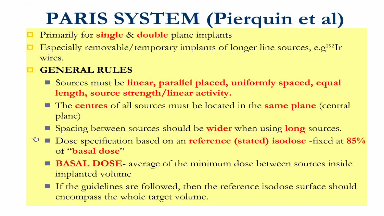
1978: Paris System – Pierquin and Dutreix
1980-90s: remote after-loading , computer planning & optimization
New possibilities in Interstitial Brachytherapy with advantages of
remote after-loading & computer optimization
History of Brachytherapy

CLINICAL ASPECTS OF INTERSTITIAL BRACHYTHERAPY

Intent of treatment
1. Radical : Brachytherapy alone as treatment
2. Boost: EBRT Brachytherapy to boost dose to the primary
3. As salvage therapy in recurrent cases who have been irradiated before or who are unfit for surgery

Brachytherapy in oral cavity carcinoma
Indications:
For T1 N0 and T2 N0 , tumour less than 30 mm in size, brachytherapy can be given as the sole
treatment for primary tumour.
For larger tumours or those with positive nodes, ideally combined surgery and post operative
radiation is preferable, if this is not feasible patients should have external beam radiation to the
primary and node with brachytherapy as a boost to the primary.
Contra-Indications:
Patient unfit for the procedure eg. comorbidities
Target volume not definable/ indistinct margins
T4 disease with bone involvement
Tumor access difficult

SELECTION CRITERIA
Easily accessible lesions
Early stage diseases (Ideal implant ≤ 5 cm)
Well localized tumor to organ of origin
No nodal or distant metastases
No local infections or inflammation
No comorbidities :DM / HTN
Proliferative/ ulcerative lesions preferred.
Favorable histology- mod. diff. i.e. SCC


General concepts based on ABS recommendation

The ABS recommends the use of brachytherapy as a component of the treatment of head-and-neck tumors.
Much of the implant process of HDR is similar to LDR
No definite evidence on use of concomitant chemotherapy; risk of increased mucosal toxicity compromising treatment – however, appears to be useful for the treatment of recurrences
regarding the sequencing of EBRT and brachytherapy, it may be advantageous to obtain shrinkage with EBRT before applying brachytherapy in advanced tumors
In case of brachytherapy boost, placement of radio-opaque markers before starting EBRT can help delineate the target volume, before any shrinkage occurs
The dose prescription volume and dose points should be clearly specified

Dental Preparation
Teeth with caries should be restored. Teeth with deep caries or poor periodontal support must be removed and complete healing obtained before starting RT.
A prosthesis (made of acrylic resin) including lead shielding (2mm thick) should be made for brachytherapy of the lips, tongue, and floor of mouth, to reduce dose to the mandible and prevent osteoradionecrosis.
The shielding is worn by the patient during whole duration of irradiation, to protect teeth, gum, and mandible as it reduces the transmitted dose by about 50%

CLINICAL:
Accurate assessment of :tumor dimensions, neck node, lesion type
via clinical examination & pre-treatment imaging: CT & MRI
Feasibility for Brachytherapy: Mouth opening, dental status, proximity of
bones to tumor
requirement of dental shields/spacers
Requirement of tracheostomy
Fitness for anaesthesia
Important Facts to be Noted in H&N Brachytherapy
Based on ABS recommendations

PHYSICAL:
Dose distribution even in a good implant is likely to be non-homogenous
(due to complex geometry)
Even minor displacement may produce significant hot/cold spots; increased
morbidity/recurrence
Peripheral fall off- may cause under-dosage of a site especially at borders
Interstitial edema may produce alteration in dose distribution calculated to
an extent of 10-15%.
Important Facts to be Noted in H&N Brachytherapy
Based on ABS recommendations

The isodose distribution should be computer optimized, whenever possible, to
conform to the CTV. The method of optimization should be noted.
The dwell times can be adjusted to minimize dose inhomogeneity.
It must be stressed that whereas optimization can make the isodose distribution more
homogenous, optimization vshould not be used as a substitute for good catheter
placement
PHYSICAL:

BIOLOGICAL:
– Total duration of EBRT + Brachytherapy should be kept as short as
possible (<8 weeks) to minimize tumor cell repopulation
– Interval between EBRT and Brachytherapy should be as short as
possible (<1–2 weeks) depending on degree of recovery from mucositis
– Interval between twice daily HDR fractions should be as long as
possible (minimum of 6 hours)
– Previous irradiation history for dose calculation
Important Facts to be Noted in H&N Brachytherapy

ABS Recommendations for recurrent head & neck cancer
Strongly emphasizes on using brachytherapy for recurrent tumors
The extent of disease should be carefully studied with CT, MRI, or PET scan as
necessary.
Complication risks are increased in patients with previous surgery, skin or mucosal
ulceration, deep soft tissue necrosis, bone exposure, or severe fibrosis.
Meticulous implant technique and adequate doses are necessary.
Generally larger margins are required for recurrent tumors, especially if additional
EBRT is not applied.
Because of the paucity of published data, the ABS cannot make specific
recommendations for the indications for HDR brachytherapy in recurrent head and neck
tumors.
However, in view of the normal tissue tolerance, it is advisable to keep the dose per
fraction relatively small.

N=220

Typical stages of a brachytherapy procedure.
3D PLANNING2D PLANNING

Prerequisites for brachytherapy treatment
Target Volume The aim should be to treat the gross tumour volume which is usually
palpable plus a margin of at least 1 cm all around it.
Pre-planning: measure the tumour carefully.
plan the exact number of radiation sources to be used with their length and separation.

Techniques Of Implantation
The two commonest techniques used for brachytherapy in the oral tongue are:
hypodermic needle technique
guide-gutter technique
Plastic loop technique
Brachytherapy sources should always be implanted in an operating room equipped for anesthesia, with adequate lighting and suction facilities and the means to deal with extensive bleeding
The brachytherapy technique should be based on a classic system for interstitial brachytherapy (like those designed in Paris, Manchester etc)
.

Hypodermic Needles
•Hollow, bevelled needles with outer diameter of
0.8mm and variable length (4 to 8 cm), open at both
ends.
• Cause little trauma - can be directly inserted in the
tissues
•The rigid steel and template system avoids
displacement of the sources due to the elasticity of
the soft tissues
•Can be used in lip tumours of ≤3cm in largest
diameter, not involving the lateral commissurae.

• Iridium hairpins with a fixed separation of 12
mm are used
• This limits width of volume which can be
treated to approximately 15 mm and the
technique can therefore only be used for
smaller tumours (≤30 mm in length).
• The guide gutter is first inserted and when
they are in position, the radioactive hairpins
can be cut to the desired length
• The pre-prepared suture is then tied over the
bridge of the hairpin to secure it within the
tongue
Guide-gutter technique:

Advantages:
• This allows a wider separation between the sources - can be used to
treat larger volumes.
• Remote after-loading that reduces the risk of exposure
• In case of local oedema inducing the risk of displacement of the
plastic tubes, one can wait for an acceptable local status before
loading the iridium wire.
• Self retaining assembly, no suturing required
Plastic-Tube Loop Technique:

Prognostic factors for brachytherapy in oral cavity

A:tumour size is one of the most important prognostic factors.
1:The largest retrospective analysis was done by Pernot of 448 patients with tongue carcinoma and showed the critical role of tumour volume on local control as well as on loco regional control and survival rate.( Pernot M, Malissard L, Hoffstetter S. The study of tumoral, radiobiological and general health factors that influence results and complications in a series
of 448 oral tongue carcinomas treated exclusively by irradiation. Int J Radiat Oncol Biol Phys 1994; 29: 673-9.)
2:In the Gustave Roussy experience for patients treated by brachytherapy alone, tumour size also plays a
role in local control: in 269 patients with mobile tongue carcinoma,
the local control rate was 93%,86%, 69% for T1, T2, T3 lesions respectively.(Gerbaulet A, Haie-Meder C, Marsiglia H, et al. Role of brachytherapy in the treatment of Head &Neck cancer. Selectron Brachy J,
1992; 3: 15-20.)
3:Mazeron studying the influence of other tumour characteristics, showed (in a series of 166 patients with
cancer of the mobile tongue treated by iridium implant alone that infiltrating tumours recurred in 22% of
cases, whereas only 9% of superficial ones did.
Mazeron JJ, Crook JM, Marinello G, et al. Prognostic factors of local outcome for T1, T2 carcinomas
of oral tongue treated by iridium 192 implantation. Radiother Oncol 1990; 19: 281-5.

Prognostic Factors For Local Control & Complications In Interstitial Brachytherapy Head And Neck
Dose to mandible Intersource spacing Treatment volume Safety margin Dose rate, total dose, dose per fraction Time delay

Factors Affecting Local Control: Indian data
N=28Factors influencing LC- dose rate source activity, interplaner distance, discontinuity in prescribed isodose

PHYSICAL ASPECTS OF INTERSTITIAL BRACHYTHERAPY

depends on several relevant Physical and Dosimetric characteristics
Ideal brachytherapy source must have following properties:
CHOICE OF RADIONUCLIDE
Pure γ emitter – less α/β emission
Medium γ energy – high enough to target tumor with homogenous dose & low enough to
avoid normal tissues & reduce shielding needs
High specific activity – small size & suitability for HDR
Stable ( not liquid/gaseous) daughter product
t ½ for permanent/temporary implant
Should be available in a form which does not powder or disperse if source is damaged or
dispensed

Brachytherapy sources:
Brachytherapy can be accomplished with either :
rigid cesium-137 needles
iridium-192 (192Ir) sources afterloaded into angiocaths.
The most common technique is afterloadingwith 192Ir .
Guide needles can be inserted either free-hand or with the aid of a custom template to help maintain optimal source spacing

Dose, dose rates & fractionation: NCCN 2014
HIGH DOSE RATE
a. Brachy Alone :
45 – 50 Gy @ 3-4Gy/# bid
b. External + Brachytherapy
Ext : 40-50 Gy in 4 1/2 - 5 1/2 wks.
Brachy : 21 Gy @ 3 Gy/#
LOW DOSE RATE(@0.4-0.5gy/hr)
a. Brachy Alone
60 - 70 Gy in 6 to 7 days.
b. External + Brachytherapy
Ext : 40-50 Gy in 4 1/2 - 5 1/2 wks.
Brachy : 20-35 Gy in 2-3 days

HDR Vs LDR :Indian evidence
N=84

LDR
Superior radiobiological role.
Minimum intersession variability in dose distribution
HDR
SHORT T/T TIME Geometry well
maintained Better patient
compliance / comfort Day care procedure
OPTIMIZATION
NO RADIATION HAZARDS
SMALL APPLICATOR Less tissue trauma
ADVANTAGES

RULES OF IMPLANTATION
Depending on the size of the lesion a single plane, double plane, or volume implant can be used to cover the tumor with a 1-cm margin.
For tumors <1 cm in thickness, single plane implants are adequate.
1-2.5 cm thickness double plane implant
> 2.5 cm thickness volume implant
Surface mold radiation can also be considered for small tumors <1 cm depth or superficial lesions of the lip, hard palate, lower gingiva, and floor of the mouth.
For lesions exceeding 2.5 cm, it is difficult to avoid significant cold spots in the implant volume.
It must be borne in mind is that as tumors get too close to the mandible or are large in volume, the risk of osteoradionecrosis increases

DOSIMETRY IN INTERSTITIAL IMPLANT:
Paterson-parker system Quimby system Paris system Computer system


TYPESOF IMPLANTS

For volume implants, AL should be at least 7.5% longer than TL for each uncrossed end.

The P-P tables are designed to give milligram hours / 1000 roentgens for various implant sizes both for planar and volume implants depending on the surface area of implant.
To convert the P-P tables from roentgens into rad in tissue one needs to account the following corrections.
P-P Tables


ICRU-58


Volumes:definitions
Gross Tumor Volume (GTV)
The palpable or visible/demonstrable extent and location of the malignant growth.
Clinical Volume (CTV)
The volume of the tissue that contains a gross tumor volume and/or sub clinical microscopic malignant disease which has to be eliminated.
Planning Target Volume (PTV)
The volume of tissue receiving the prescribed irradiation. For an interstitial brachytherapy, the PTV is, in general, identical to the CTV.
Treatment Volume (TV)
The volume of tissue, which is encompassed by an isodose surface that has been specified by the radiation oncologist. The dose value at this isodose surface is the minimum target dose

Central PlaneThe plane that is perpendicular to the main direction of the linear sources and passing through the estimated center of the implant.

Prescription Dose The prescribed dose is defined as the dose, which the physician intends to give, and enters in the
patient’s treatment chart.
Mean Central Dose
Calculated as average of doses to mean central dose points
Minimum Target Dose (MTD): The minimum dose at the periphery of the CTV = Minimum dose decided upon by the clinician as adequate to treat the CTV (minimum peripheral dose).
MTD ≅ 90% of the prescribed dose for interstitial therapy

Prescription Dose
Low Dose Volume
A low dose volume should be defined as a volume within the clinical target volume, encompassed by an isodose corresponding to 90% of the prescribed dose.
High Dose Volume
The volume of tissue that is encompassed that will receive more than 150% of the mean central dose be reported.

ICRU (58) recommendation for reporting interstitial brachytherapy
Description of the clinical, including GTV, CTV
Description of the techniques
Source specification
Treatment prescription
Mean Central Dose (MCD), Minimum Target Dose, Homogeneity Index
Volumes and their dimensions, including PTV, Treatment Volume,
High-dose regions, low-dose regions, reference volume, irradiated volume

CLINICAL APPLICATION OF INTERSTITIAL BRACHYTHERAPY

CLINICAL APPLICATIONS
Oral Cavity:
LIP:
Indications: T1-2N0 Lesions
T.V.: All visible & palpable tumour with 5-10 mm margin
Dose: 50-70Gy in 5-7 days LDR
Technique:
Rigid afterloading needles maintained in place by Template
Classical plastic tubes
Spacers to decrease dose to gingiva, teeth & other lip

1a&b: T1 squamous carcinoma, manifesting an ulcerous tumour affecting an external third of the inferior lip. 1c: Brach- therapy using rigid needles technique for LDR 192Ir sources 1d: tumour 2 months after brachytherapy with excellent cosmetic and functional results

CLINICAL APPLICATIONS…
Oral Tongue:
Indications: T1 N0, T2 N0 < 3cm lesion
T.V.: GTV + 5 mm margin
Dose: Alone:60-65 Gy LDR Boost 20-25 Gy after EBRT dose of 45-50 Gy
Techniques: Guide-gutter technique
Floor of Mouth:
Indications: T1-2N0 lesions, ≥ 5 mm away from mandible
Dose: Techniques same as for Tongue implants
Complication: Osteoradionecrosis:5-15%
AP X-ray


Advantage of Brachytherapy Delivers localized dose to the tumor
Rapid dose fall off outside the target volume allows excellent normal tissue sparing
Less intergal dose as compared to 3DCRT & IMRT
High biological efficacy
Decreased risk of tumor population
Elimination of set up errors as the source maintains a fixed relationship to target volume
High tolerance: Tolerable acute intense reaction
High control rate
Better cosmesis: May avoid disfigurement and mutilating surgery
Minimal radiation morbidity
Day care procedure
Organ preservation
Reirradiation for localized recurrence

Limitation of brachytherapy
Difficult for inaccessible regions
Limited for small tumors (T1-T2)
Nodal disease cannot be covered simultaneously
Invasive procedures, require GA
Greater conformation –small errors in placement of sources lead to extreme changes from the intended dose distribution
Quality of implant is operator dependent
Radioactive hazards (not now)

Decline in use of brachytherapy
Over the past decade or more improvements in reconstructive surgery techniques have diminished the practice frequency of brachytherapy in the treatment of oral cavity carcinoma.
In addition, a diminishing percentage of radiation oncologists remain highly skilled and experienced with the requisite implant techniques.
Finally, the steady advancement of highly conformal external beam techniques (IMRT, tomotherapy) has contributed to less frequent practice of brachytherapy in head and neck cancer overall.

Intraoral Cone enable boosting of radiation dose to sites within the oral cavity while avoiding
direct dose to the mandible
best suited for anterior oral cavity lesions in edentulous patients.
Treatment with intraoral cone involves either 100 to 250 kilovolt (peak) (kvp) x-rays or electron beams in the 6 to 12 MeV range
Lesions up to 3 cm are amenable to treatment with intraoral cone
requires careful daily positioning and verification by the physician. For this purpose the device is equipped with a periscope to visualize the lesion.
The cone abuts the mucosa and is centered directly over the lesion.
Intraoral cone treatment should take place prior to external beam radiation so that the lesion can be adequately visualized.
