book of abstracts twelfth international … book of...njit’s twelfth international undergraduate...
TRANSCRIPT
BOOK OF ABSTRACTS Twelfth International Undergraduate Summer Research Symposium
Thursday, August 1, 2019
Copyright © 2019 by New Jersey Institute of Technology (NJIT). All rights reserved.
New Jersey Institute of Technology University Heights Newark, NJ 07102-1982 Joel S. Bloom President
August 1, 2019
Welcome all – students, faculty, industry mentors, sponsors and friends of the university – to
NJIT’s Twelfth International Undergraduate Summer Research Symposium. It is exciting to see
so many ingenious inventions, and the bright, enterprising minds behind them, gathered in one
place. That some of you have joined our innovation hub from as far away as India is a testament
to the power of collaboration in the service of progress – not just in our own state or country, but
across the globe.
I want to especially thank the Provost’s office for making undergraduate research a high priority
on our campus, the students’ advisers for their ideas and precious time over the summer, and our
many sponsors for their generosity and commitment to helping forge the problem-solvers of
tomorrow - today.
And to the more than 130 of you exhibiting your work at the symposium, congratulations! By
thinking creatively, following through with diligence and tenacity – and even retooling when the
evidence requires it – you have embraced the rigors of professional science. You make us proud,
and we look forward to following your successes in the years to come.
Sincerely,
Joel S. Bloom
President
August 1, 2019
A message from the Provost:
Welcome to NJIT’s Twelfth International Undergraduate Summer Research Symposium. I would like to
congratulate all undergraduate summer research students, their faculty advisers, and program directors
for the impressive research work exhibited here. The symposium demonstrates excellent interdisciplinary
research and innovation by undergraduates who are honing their expertise as they prepare for leadership
roles in science and technology. As it is critically important for all of our students to develop such skills,
undergraduate research and innovation has been identified to be an integral part of NJIT’s 2020 Vision
Strategic Plan.
I thank all staff members, faculty advisers, and program directors for organizing this impressive
international symposium. Through the Undergraduate Research and Innovation (URI) initiative
established by Dr. Atam Dhawan, senior vice provost for research, this year’s summer research program
has been significantly expanded and includes more than 130 students from NJIT and partner institutions.
The online publication of the Book of Abstracts of NJIT’s Twelfth International Undergraduate Summer
Research Symposium is excellent, as it showcases the wonderful research work done by our students and
faculty, and will be archived through the URI website.
NJIT is committed to excellence in undergraduate education and research to provide our students
exceptional learning experiences that enable them to become leaders in our global society.
I look forward to meeting summer research teams at the symposium and learning more about their
exciting work.
Sincerely,
Fadi P. Deek Provost and Senior Executive Vice President
Fadi P. Deek Provost and Senior Executive Vice President
Undergraduate Research and Innovation
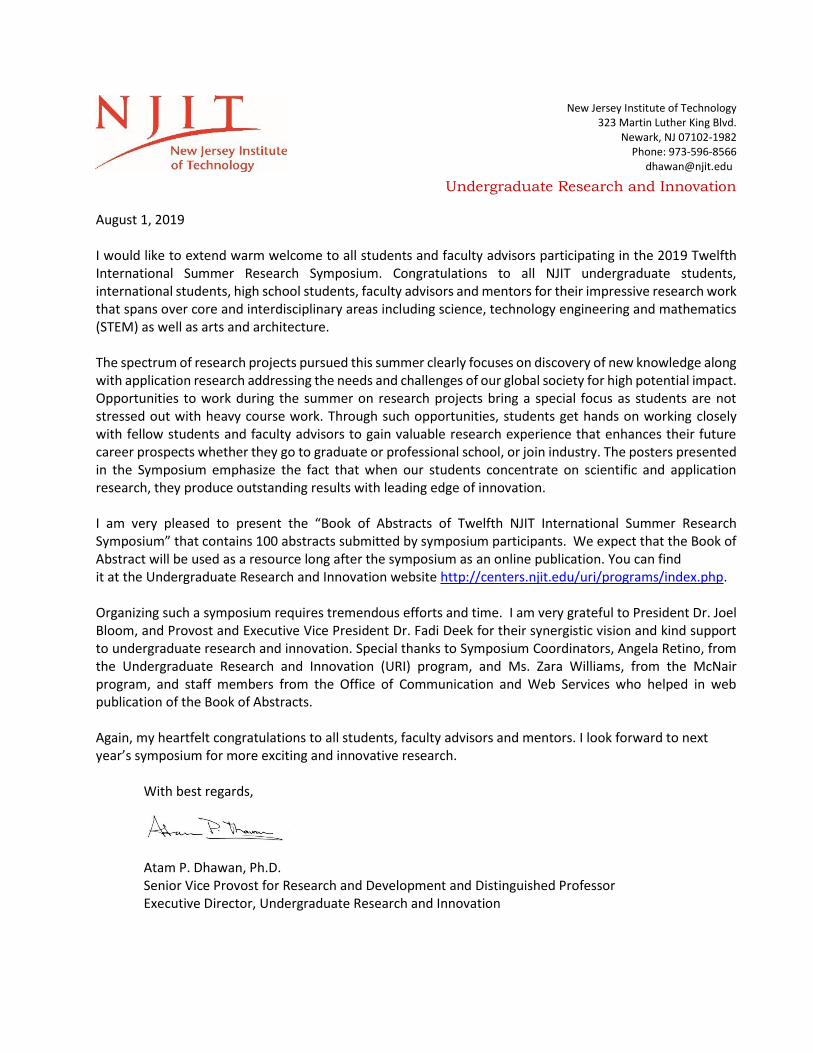
August 1, 2019 I would like to extend warm welcome to all students and faculty advisors participating in the 2019 Twelfth International Summer Research Symposium. Congratulations to all NJIT undergraduate students, international students, high school students, faculty advisors and mentors for their impressive research work that spans over core and interdisciplinary areas including science, technology engineering and mathematics (STEM) as well as arts and architecture. The spectrum of research projects pursued this summer clearly focuses on discovery of new knowledge along with application research addressing the needs and challenges of our global society for high potential impact. Opportunities to work during the summer on research projects bring a special focus as students are not stressed out with heavy course work. Through such opportunities, students get hands on working closely with fellow students and faculty advisors to gain valuable research experience that enhances their future career prospects whether they go to graduate or professional school, or join industry. The posters presented in the Symposium emphasize the fact that when our students concentrate on scientific and application research, they produce outstanding results with leading edge of innovation. I am very pleased to present the “Book of Abstracts of Twelfth NJIT International Summer Research Symposium” that contains 100 abstracts submitted by symposium participants. We expect that the Book of Abstract will be used as a resource long after the symposium as an online publication. You can find it at the Undergraduate Research and Innovation website http://centers.njit.edu/uri/programs/index.php. Organizing such a symposium requires tremendous efforts and time. I am very grateful to President Dr. Joel Bloom, and Provost and Executive Vice President Dr. Fadi Deek for their synergistic vision and kind support to undergraduate research and innovation. Special thanks to Symposium Coordinators, Angela Retino, from the Undergraduate Research and Innovation (URI) program, and Ms. Zara Williams, from the McNair program, and staff members from the Office of Communication and Web Services who helped in web publication of the Book of Abstracts. Again, my heartfelt congratulations to all students, faculty advisors and mentors. I look forward to next year’s symposium for more exciting and innovative research.
With best regards,
Atam P. Dhawan, Ph.D. Senior Vice Provost for Research and Development and Distinguished Professor Executive Director, Undergraduate Research and Innovation
New Jersey Institute of Technology 323 Martin Luther King Blvd.
Newark, NJ 07102-1982 Phone: 973-596-8566
New Jersey Institute of Technology
University Heights
Newark, NJ 07102-1982
973.596.5590
973.596.5201 fax
Ronald E. McNair Postbaccalaureate Achievement Program
August 1, 2019
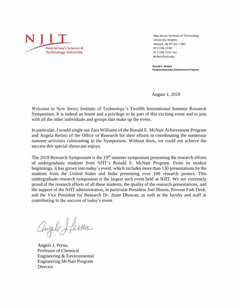
Welcome to New Jersey Institute of Technology’s Twelfth International Summer Research
Symposium. It is indeed an honor and a privilege to be part of this exciting event and to join
with all the other individuals and groups that make up the event.
In particular, I would single out Zara Williams of the Ronald E. McNair Achievement Program
and Angela Retino of the Office of Research for their efforts in coordinating the numerous
summer activities culminating in the Symposium. Without them, we could not achieve the
success this special showcase enjoys.
The 2019 Research Symposium is the 19th summer symposium presenting the research efforts
of undergraduate students from NJIT’s Ronald E. McNair Program. From its modest
beginnings, it has grown into today’s event, which includes more than 130 presentations by the
students from the United States and India presenting over 100 research posters. This
undergraduate research symposium is the largest such event held at NJIT. We are extremely
proud of the research efforts of all these students, the quality of the research presentations, and
the support of the NJIT administration, in particular President Joel Bloom, Provost Fadi Deek,
and the Vice President for Research Dr. Atam Dhawan, as well as the faculty and staff in
contributing to the success of today’s event.
Angelo J. Perna,
Professor of Chemical
Engineering & Environmental
Engineering McNair Program
Director
1
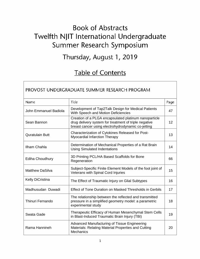
John Emmanuel Badiola Development of Tap2Talk Design for Medical Patients With Speech and Motion Deficiencies
47
Sean Bannon Creation of a PLGA encapsulated platinum nanoparticle drug delivery system for treatment of triple negative breast cancer using electrohydrodynamic co-jetting
12
Quratulain Butt Characterization of Cytokines Released for Post-Myocardial Infarction Therapy
13
Ilham Chahla Determination of Mechanical Properties of a Rat Brain Using Simulated Indentations
14
Ediha Choudhury 3D Printing PCL/HA Based Scaffolds for Bone Regeneration
66
Matthew DaSilva Subject-Specific Finite Element Models of the foot joint of Veterans with Spinal Cord Injuries
15
Kelly DiCristina The Effect of Traumatic Injury on Glial Subtypes 16
Madhusudan Duwadi Effect of Tone Duration on Masked Thresholds in Gerbils 17
Thinuri Fernando The relationship between the reflected and transmitted pressure in a simplified geometry model: a parametric experimental study
18
Swata Gade Therapeutic Efficacy of Human Mesenchymal Stem Cells in Blast-Induced Traumatic Brain Injury (TBI)
19
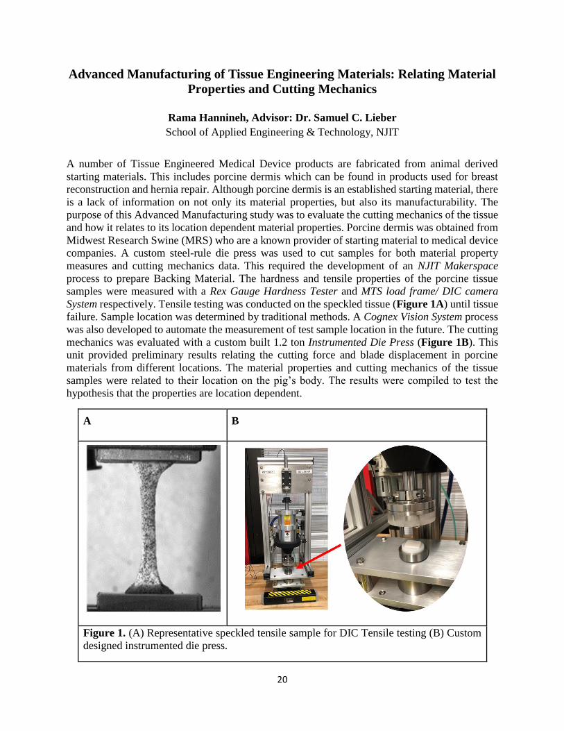
Rama Hannineh Advanced Manufacturing of Tissue Engineering Materials: Relating Material Properties and Cutting Mechanics
20

2
John Hawks High throughput assay for screening KaiC libraries 21
Omar Ilyas Rehabilitating Stroke Patients through Adaptive Digital Environments
22
AKM Islam Developing a Visualization Interface for Urban Data-driven Social Science Research
23
Supriya Iyer The relationship between the reflected and transmitted pressure in a simplified geometry model: a parametric experimental study
18
Rachel Lee Boron Kinetic Isotope Effect in Boronic Acid Oxidation 24
Nicole Loehle Engineering Nanoparticles for Brain Drug Delivery 25
Richard Marsh Sonochemical Degradation of Emerging Pollutants 26
Alvin Mathew Development of Tap2Talk Design for Medical Patients With Speech and Motion Deficiencies
47
Anna Mathew Novel Drug Delivery System using Anti-Angiogenic Peptides for Glioblastoma Multiforme
27
Brian McGrath Robotic Leg Prototype for Balance Stability Analysis and Control - PART III: The Nervous System
45
Michael Mobilio Encouraging the Use of Built-in Language Features for Learning Control Flow
28
Mahathi Mohan Gowda Investigating the role of a genetically-conserved spinal neuronal class, Dmrt3, in the functional control of locomotion in zebrafish.
29
Marcos Molina Integrated Solid-Fluid Interaction Potential for Modeling Gas Adsorption in Templated Mesoporous Carbons
30
Jorim Morainvil A Low-Cost Electro-Mechanical System to create 3D scans using 2D LIDARs
31
Zoraiz Naeem Theoretical Studies of Possible Topological Edge Modes in Novel Systems 32
James Nanchanatt Producing Well-Defined Fibrous Structures in Tissue Engineering Scaffolds Using an Adaptable Collector for Electrospinning
33
Randy Nutakor Accessing the extent and fate of legacy contaminant mixtures in sediments
34
Ishani Patel The Role of Neural Activity and Semaphorin Signaling in Neural Repair
35
Raghav Patel Understanding Unidentifiability in Dynamic Models from Ground Truth Data
36

3
Navya Pendyala Central Auditory Pathology in Blast Induced Tinnitus/Hearing Loss
37
Andre Pugliese Satellite Imagery of Insect Structures: Insights into Global
Ecological Declines 38
Sameer Rana Development of Tap2Talk Design for Medical Patients
With Speech and Motion Deficiencies 47
Lindsey Riggs Apolipoprotein E4 and Cholesterol Packaging in
Alzheimer’s Disease 39
Ian Rosenberg Virtual Design Theatre (VDT): Multi-User Iterative Production Design Tool
40
Ayushi Sangoi Assessing the Underlying Neural Mechanism of Vision
Therapy Through Phoria Adaptation 41
Sreya Sanyal Novel Approach Towards Cholesterol Management Using Hydrogel for PCSK9 Inhibition
42
Jinay Shah Computational Modeling of Two-Dimensional Nanomaterials for Water Desalination
43
Rahul Shah Evaluating the Effect of Skull and Brain Stiffness on Shock Wave Propagation in a Rodent Finite Element Model
44
Mahenoor Shaikh Robotic Leg Prototype for Balance Stability Analysis and
Control - PART I: The “Body” System 45
Divjyot Singh Numerical Models for Morphology and Optics of Soot Nanoparticles
46
Francis Stipa Development of Tap2Talk Design for Medical Patients With Speech and Motion Deficiencies
47
Donna Sunny Investigation of Particle Noise Produced by Tip Sonication
48
Neha Thati Molecular Mechanism of the Circadian clock in Cyanobacteria
49
Joseph Torsiello Computational Modeling of Friction between Two-Dimensional Materials
50
Nirali Trivedi In Vitro Modeling of Traumatic Brain Injury 51
Shruti Varshney Brain Function and Neuroplasticity with TBI 52
Abigail Varughese Neuromodulation of Sensory Encoding 53

4
Geetasravya Vegunta Measuring the dynamic properties of microglial cells after blast induced traumatic brain injury
54
Rashmi Venkatarama
Minocycline Loaded Albumin Nanoparticle (myn-ANP) Synthesis and Characterization: Potential Nanomedicine Approach to Traumatic Brain Injury by Targeting Microglial Cells Activation
55
Anuj Verma In Situ Ozone Nanobubble Technology for Water Disinfection and Pollutant Degradation
56
Michael Vitti Magnetic Spinner Model Provides a Material's Phonon Spectrum
57
Juliana Yang Fabrication of Microfluidic Cell Culture Systems for Bacteria and Eukaryotic Cells
58
Philip Zaleski Dynamics of cone-shaped meniscus on a substrate-supported drop in electric fields
59
Sara AbdelhamidEffect of Tank Bottom Shapes on Power Dissipation, Power Number and Njs in Stirred Vessels Under Different Baffling Configurations
61
Ehtesham AhmedRaspberry Pi as FSO Transceiver using UART Communication for Drone-Assisted Networking
62
Gabby AmparoExamination of water stress on the morphological evolution of Capsella bursa-pastoris
63
Iren AtallaAlignment analysis of Cardiomyocytes on Patterned vs Flat Scaffolds
64
David Bushiri Investigation of interlayer strength of 3D-printed Polymers 65
Ediha Choudhury3D Printing PCL/HA Based Scaffolds for Bone Regeneration
66
Felix De Dios Shear-Enhanced ESSENCE Biosensor for the Detection of a target DNA
67

5
Cruz DonatoDesign of a Cable Driven Exoskeleton for Hand Rehabilitation Post Stroke
68
Kevin Nino Database of Mechanism Animations 69
Jaime Siguenza Database of Mechanism Animations 70
Deva Craig Materials - Photon Interactions in Multilayers 72
Maria DeOliveira
Effects of UV exposure on the mechanical behavior of Cellulose Acetate
73
Nishaant Goswamy
The Effect of Autonomous Vehicles on Human Driving Behavior
74
Katherine Ji
An Exploratory Study into the Effects of Total Sleep Deprivation using fNIRS
75
Manisha Kannan
Apocynin loaded albumin nanoparticle formulation, characterization and evaluation in in vivo TBI model
76
Brian McGlew
Using Ozone Nanobubbles and Ultrasound to treat Sediment Contamination
77
George Mina
Fabrication of a Microscope Stage Compatible Incubator for Live Cell Imaging
78
Chandni Patel Changes in Synaptic Proteins as a Function of Time in Blast-Induced Traumatic Brain Injury
79
Xavier Reyes The Effect of Land Area on Ecological Niches: A study of ants and islands
80
Gregory Tanis
Collection and Analysis of Global Navigation Satellite System (GNSS) Positioning Data with SBAS and RTK for Autonomous Lawn Mowers
81

6
Zenit Winfield
Rodent to human scaling laws: The evaluation of biofidelic materials for the rat head model under shock wave loading
82
Shuhrah Chowdhury Mathematical Modeling of Circadian Rhythms, Tumor Growth, and Radiotherapy
104
Elizabeth Epstein Exploring the Viability of a PLSR-based Machine Learning Method for Predicting Circadian Phase in Cancer Patients
103
Mariia Goriachi Mathematical Modeling of Circadian Rhythms, Tumor Growth, and Radiotherapy
104
Luis Lara Mathematical Modeling of Circadian Rhythms, Tumor Growth, and Radiotherapy
104
Karen Reyes Exploring the Viability of a PLSR-based Machine Learning Method for Predicting Circadian Phase in Cancer Patients
103
Alexander Barrett Machine Learning Approaches for Optimizing Long-Short Portfolio
84
Peter Boyland Deep Learning with Application to Software Engineering 85
Andrew Chen Deep Learning with Application to Software Engineering 85
Peter Decker Eye Blinks Detection and Labeling 86
Mihail Kaburis Spatiotemporal Analysis of Racial Bias in NYPD Stop, Question, and Frisk Procedures
87

7
Samantha Kamath Spatiotemporal Analysis of Racial Bias in NYPD Stop, Question, and Frisk Procedures
87
Jasmine Medlock Spatiotemporal Analysis of Racial Bias in NYPD Stop, Question, and Frisk Procedures
87
Farukh Saidmuratov Deep Learning with Application to Software Engineering 85
Mohamad Sherif Machine Learning Approaches for Optimizing Long-Short Portfolio
84
Miriam Tan Eye Blinks Detection and Labeling 86
Ruohan Wu Machine Learning Approaches for Optimizing Long-Short Portfolio
84
John Desalvo Machine Learning Models for the Dynamics of Ferrofluids 89
Diego Ramos Design and Experimental Tests of an Energy Packet Switch Testbed for a Digital Microgrid
91
Lenin Ham Design and Experimental Tests of an Energy Packet Switch Testbed for a Digital Microgrid
91
Omar Aref III-Nitride Nanowire Deep Ultraviolet Light-Emitting Diodes for Precise Applications
93
Shweta Burgula Use of Rhenium Disulfide to Improve Sensitivity of the Lab on a Chip Device
94

8
Arijit Dutta
Visualization and Characterization of Etched-Based On-Chip Plasma Self-Separation
95
Ethan Espin Silver Nanowires as Infrared Transparent Electrodes
96
Kalid D-Luyando Flusa
Multi-Platform Optics and Photonics Educational Application
97
Seunghoon Kim Applications of Visible Light Communication Systems for Intelligent Consumer Messaging and Indoor Positioning
98
Eric Kraut Kalman Filter Implementation in Compression Optical Coherence Elastography Tissue Motion
99
Devynn Saunders Denoising fNIRS Data by Integrating Independent Component Analysis and Short Channel Separation Regression
100
Oladimeji Sobanjo CMOS Compatible RRAM Devices 101
Sahar Abulaimoun Collaborative Robotics for Cave and Tunnel Exploration 106
Erik Aleksanyan Collaborative Robotics for Cave and Tunnel Exploration 106
Adam Czyrsznic Collaborative Robotics for Cave and Tunnel Exploration 106
Karina Dsouza Collaborative Robotics for Cave and Tunnel Exploration 106
Jacqueline Farkas Collaborative Robotics for Cave and Tunnel Exploration 106
Matthew Frazier Collaborative Robotics for Cave and Tunnel Exploration 106
Kalib Guthrie Collaborative Robotics for Cave and Tunnel Exploration 106
Craig Iaboni Collaborative Robotics for Cave and Tunnel Exploration 106
Kyle LaPolice Collaborative Robotics for Cave and Tunnel Exploration 106
David Monroe Collaborative Robotics for Cave and Tunnel Exploration 106
Brittany Morales Collaborative Robotics for Cave and Tunnel Exploration 106
Aashka Patel Collaborative Robotics for Cave and Tunnel Exploration 106
Ben Ruoff Collaborative Robotics for Cave and Tunnel Exploration 106
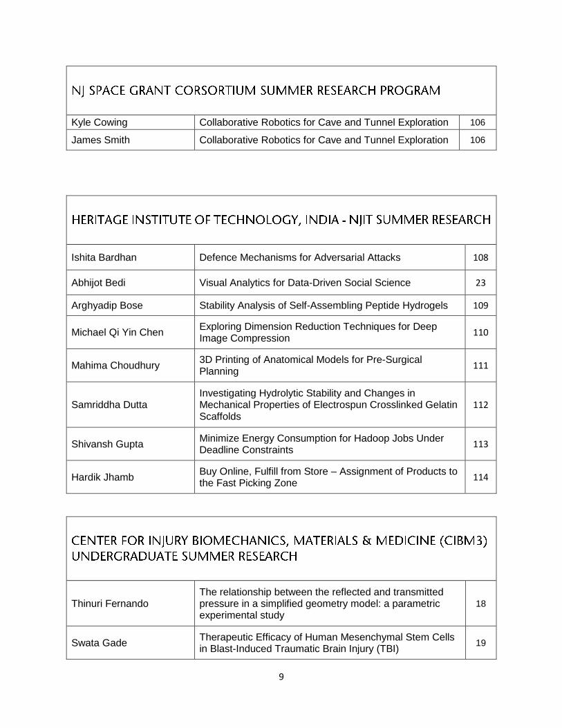
9
Kyle Cowing Collaborative Robotics for Cave and Tunnel Exploration 106
James Smith Collaborative Robotics for Cave and Tunnel Exploration 106
Ishita Bardhan Defence Mechanisms for Adversarial Attacks 108
Abhijot Bedi Visual Analytics for Data-Driven Social Science 23
Arghyadip Bose Stability Analysis of Self-Assembling Peptide Hydrogels 109
Michael Qi Yin Chen Exploring Dimension Reduction Techniques for Deep Image Compression
110
Mahima Choudhury 3D Printing of Anatomical Models for Pre-Surgical Planning
111
Samriddha Dutta Investigating Hydrolytic Stability and Changes in Mechanical Properties of Electrospun Crosslinked Gelatin Scaffolds
112
Shivansh Gupta Minimize Energy Consumption for Hadoop Jobs Under Deadline Constraints
113
Hardik Jhamb Buy Online, Fulfill from Store – Assignment of Products to the Fast Picking Zone
114
Thinuri Fernando The relationship between the reflected and transmitted pressure in a simplified geometry model: a parametric experimental study
18
Swata Gade Therapeutic Efficacy of Human Mesenchymal Stem Cells in Blast-Induced Traumatic Brain Injury (TBI)
19

10
Supriya Iyer The relationship between the reflected and transmitted pressure in a simplified geometry model: a parametric experimental study
18
Manisha Kannan Apocynin loaded albumin nanoparticle formulation, characterization and evaluation in in vivo TBI model
76
Chandni Patel Changes in Synaptic and Axonal Proteins as a Function of Time in Blast-Induced Traumatic Brain Injury
79
Navya Pendyala Central Auditory Pathology in Blast Induced Tinnitus/Hearing Loss
37
Rahul Shah
Evaluating the Effect of Skull and Brain Stiffness on Shock Wave Propagation in a Rodent Finite Element Model
44
Alekhya Thota
Blood Brain Barrier Permeability as Injury Criteria in Repeated Low-level Blast-Induced Traumatic Brain Injury (RbTBI)
116
Geetasravya Vegunta Measuring the dynamic properties of microglial cells after blast induced traumatic brain injury
54
Rashmi Venkatarama
Minocycline Loaded Albumin Nanoparticle (myn-ANP) Synthesis and Characterization: Potential Nanomedicine Approach to Traumatic Brain Injury by Targeting Microglial Cells Activation
55
Zenit Winfield Rodent to human scaling laws: The evaluation of biofidelic materials for the rat head model under shock wave loading
82
Manav Guzraty Nonlinear Dynamics of Tandem Flapping Wings 118
Tiffany Olivera Designing Amyloid-Inspired β-Sheet Fibrils from L- and D-
Handed Peptides 119

11
12
Creation of a PLGA encapsulated platinum nanoparticles drug delivery
system for treatment of triple negative breast cancer using
electrohydrodynamic co-jetting
Sean Bannon, Aida Lopez and Kathleen McEnnis
Department of Chemistry and Chemical Engineering
New Jersey Institute of Technology, Newark NJ 07102
The aim of this study is to develop a targeted drug delivery system for treatment of Triple Negative
Breast Cancer (TNBC). With no hormonal receptors to target, TNBC, accounting for almost 20%
of all breast cancers, only has invasive and painful forms of therapies for treatments, such as
chemotherapy, radiotherapy, and surgery. Chemotherapy drugs often make use of the anti-cancer
properties exhibited by platinum (such as cis-diamminedichloroplatinum(II), commonly known as
cisplatin). However, cisplatin is highly toxic and due to its systemic delivery kills healthy cells
indiscriminately from cancer. In this study, platinum nanoparticles (PtNps) are suggested as a less
toxic alternative to other platinum based chemotherapy drugs. A targeted drug delivery method is
also proposed as an alternative to its current systemic delivery in order to make treatments less
painful and more effective.
In this project, Electrohydrodynamic (EHD) co-jetting is used as the fabrication technique for drug
delivery devices. EHD co-jetting is a method where dissolved polymer in organic solvent is
pumped through a capillary needle with a high voltage applied to it in order to create solidified
polymer structures. This method of fabrication is desirable due to the high level of control of size
and shape of particles it allows for. Size and shape are two properties that are highly determinant
of how and where a drug is delivered in the body. The platinum nanoparticles (PtNPs) were
fabricated using a sonochemical technique. These PtNPs were then included in the jetting solution
to create a polymer encapsulated PtNP delivery device. The main goals of this study were to
determine how process parameters such as concentration of PtNP in solution, collection distance,
pump speed, and solution composition had on the size and shape of the particles. We also aimed
to create recipes of particles of approximately 50nm-200nm in size. Also in the scope of the project
was to create a new fully sealed acrylic box for EHD co-jetting particle fabrication to occur in.
The boxes used for earlier jets in our lab were not completely sealed as we were not jetting toxic
chemotherapy drugs. In order to jet the particles for this study, it was necessary to create a new
fully sealed box as an extra layer of protection.
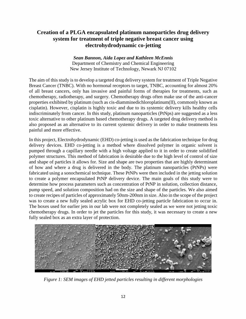
Figure 1: SEM images of EHD jetted particles resulting in different morphologies
13
Characterization of Cytokines Released for Post-Myocardial Infarction
Therapy
Quratulain Butt, Advisor: Dr. Eun Jung Lee, Ph.D.
Department of Biomedical Engineering
New Jersey Institute of Technology, Newark NJ 07102
Cardiovascular diseases, including myocardial infarction (MI), are one of the leading
causes of death and disability worldwide. The inflammatory response following MI is critical to
the cardiac remodeling process. In this signaling cascade, cytokines are released by numerous cell
types including macrophages and cardiomyocytes that contribute to both the pro-inflammatory and
anti-inflammatory response post-MI. In this project, a novel in vitro myocardial infarction model
is used to understand key cytokine interactions present between macrophages and cardiomyocytes
in the post-MI microenvironment. Tissue inhibitor metalloproteinase-1 (TIMP-1), an inhibitor of
matrix metalloproteinases, is a known anti-apoptotic and anti-fibrotic cytokine that is an interesting
target for cardiac regeneration therapy. Several studies have shown TIMP-1 promotes cellular
differentiation in numerous cell types. In our interest, overexpression of TIMP-1 in embryonic
stem cell transplanted hearts post-MI have shown to contain significantly more differentiated
cardiomyocytes compared to embryonic stem cell transplanted hearts [1]. Based on the results
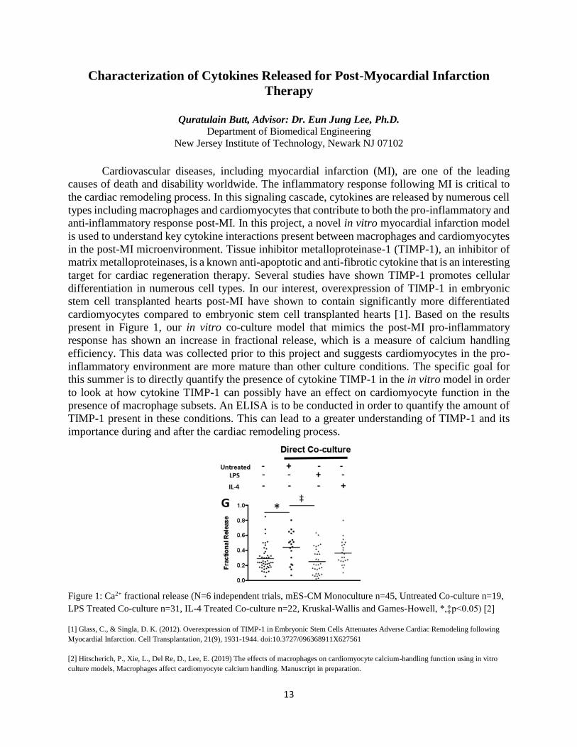
present in Figure 1, our in vitro co-culture model that mimics the post-MI pro-inflammatory
response has shown an increase in fractional release, which is a measure of calcium handling
efficiency. This data was collected prior to this project and suggests cardiomyocytes in the pro-
inflammatory environment are more mature than other culture conditions. The specific goal for
this summer is to directly quantify the presence of cytokine TIMP-1 in the in vitro model in order
to look at how cytokine TIMP-1 can possibly have an effect on cardiomyocyte function in the
presence of macrophage subsets. An ELISA is to be conducted in order to quantify the amount of
TIMP-1 present in these conditions. This can lead to a greater understanding of TIMP-1 and its
importance during and after the cardiac remodeling process.
Figure 1: Ca2+ fractional release (N=6 independent trials, mES-CM Monoculture n=45, Untreated Co-culture n=19,
LPS Treated Co-culture n=31, IL-4 Treated Co-culture n=22, Kruskal-Wallis and Games-Howell, *,‡p<0.05) [2]
[1] Glass, C., & Singla, D. K. (2012). Overexpression of TIMP-1 in Embryonic Stem Cells Attenuates Adverse Cardiac Remodeling following
Myocardial Infarction. Cell Transplantation, 21(9), 1931-1944. doi:10.3727/096368911X627561
[2] Hitscherich, P., Xie, L., Del Re, D., Lee, E. (2019) The effects of macrophages on cardiomyocyte calcium-handling function using in vitro
culture models, Macrophages affect cardiomyocyte calcium handling. Manuscript in preparation.
14
Determination of Mechanical Properties of a Rat Brain Using Simulated
Indentations
Ilham Chahla, Advisor: Xianlian Zhou
Department of Biomedical Engineering
New Jersey Institute of Technology, Newark NJ 07102
The key motivation is to understand mechanisms of Traumatic Brain Injury (TBI) with
accurate material models of living brain tissues and high fidelity physical simulations. TBI
simulations are challenging to conduct experimentally, causing researchers to veer toward the
development of computer models to study the biomechanics of TBI. We will correlate the material
properties of rat brain tissue material properties to indentation forces and displacements
measurable by a laser equipped probe through parametric computational simulations. The
proposed simulations will provide us the information needed to quantify the relation between
materials, boundary conditions, and measured force and displacement, resolving the difficulty of
traditional indentation methods with analytical deformation models on living tissues with irregular
geometry and inhomogeneity. We will extract useful data correlations from regression, curve
fitting, and implementation of machine learning models from simulation-generated data. The
discovered correlations will later be used in a laser indenter developed by Dr. Liu of ECE to predict
in vivo rat brain properties. This will ultimately enable us to study the
homogeneity/inhomogeneity, isotropy/anisotropy, and linearity/nonlinearity of brain properties
using both computation and physical measurements. These properties may be vital for developing
hypotheses about disease mechanisms and potential preventive measures against TBI.
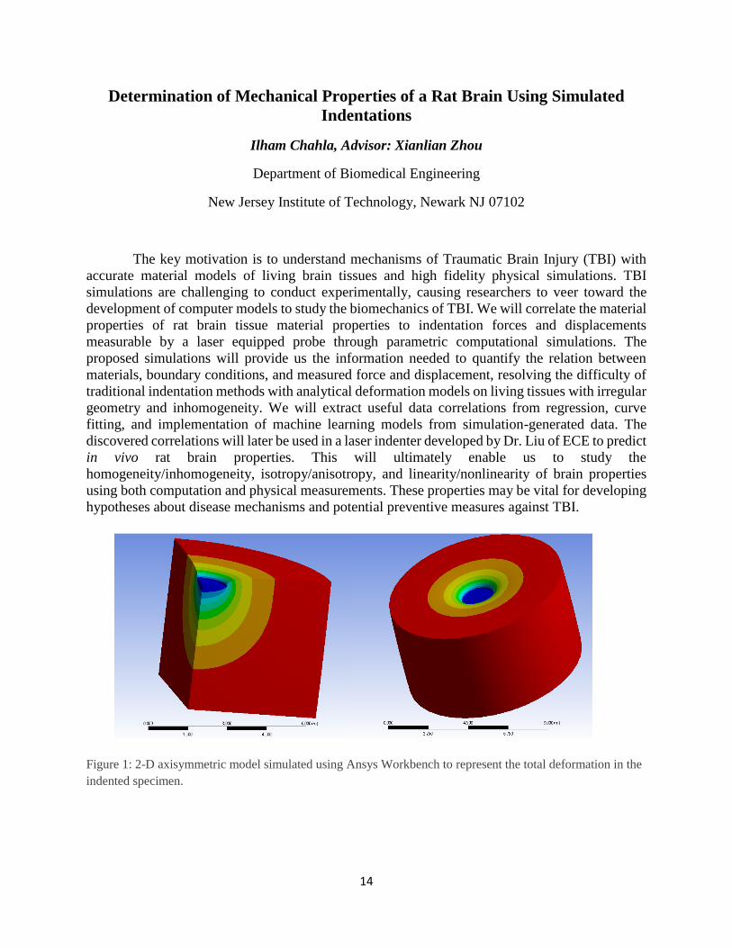
Figure 1: 2-D axisymmetric model simulated using Ansys Workbench to represent the total deformation in the
indented specimen.
15
Subject-Specific Finite Element Models of the Foot Joint of Veterans with
Spinal Cord Injury
Matthew DaSilva, Advisor: Dr. Saikat Pal Department of Biomedical Engineering
New Jersey Institute of Technology, Newark, NJ, 07102 USA
Abstract: The goal of this project is to develop and utilize subject-specific finite element models
to determine the risk of fractures of the calcaneus and other bones of the foot in Veterans with
chronic SCI. People with spinal cord injury (SCI) have increased forces applied on their feet with
upright standing and ambulatory maneuvers compared to their daily routine in a wheelchair. This
sedentary lifestyle has shown that a loss of bone below the level of lesion after SCI in combination
with weight-bearing activity may greatly increase the risk of fracture. The calcanei and, by
extrapolation, the other bones of the foot appear to be at an increased risk of sustaining a fragility
fracture while ambulating a powered exoskeleton. This becomes an interest as the VA’s Spinal
Cord Injury Services are prescribing exoskeletal devices for ambulation and require assistance
regarding issues of safety. Because of the emergence of exoskeletal devices, the field of SCI
Rehabilitation needs knowledge regarding the prevalence and extent of severe bone mineral
density (BMD) loss in the foot. BMD is a value used to understand bone health, with specific
regards to identifying the risk of fracture as seen in cases of severe loss.
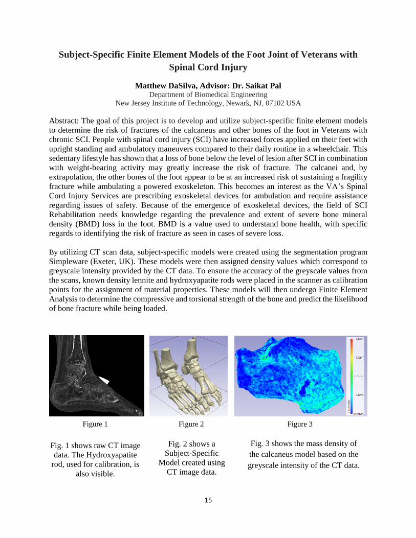
By utilizing CT scan data, subject-specific models were created using the segmentation program
Simpleware (Exeter, UK). These models were then assigned density values which correspond to
greyscale intensity provided by the CT data. To ensure the accuracy of the greyscale values from
the scans, known density lennite and hydroxyapatite rods were placed in the scanner as calibration
points for the assignment of material properties. These models will then undergo Finite Element
Analysis to determine the compressive and torsional strength of the bone and predict the likelihood
of bone fracture while being loaded.
Fig. 1 shows raw CT image
data. The Hydroxyapatite
rod, used for calibration, is
also visible.
Fig. 3 shows the mass density of
the calcaneus model based on the
greyscale intensity of the CT data.
Figure 1 Figure 2 Figure 3
Fig. 2 shows a
Subject-Specific
Model created using
CT image data.
16
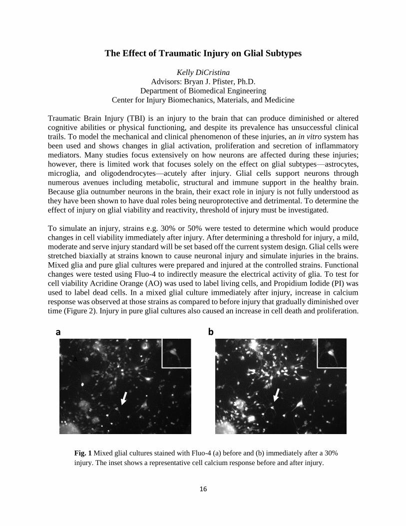
The Effect of Traumatic Injury on Glial Subtypes
Kelly DiCristina
Advisors: Bryan J. Pfister, Ph.D.
Department of Biomedical Engineering
Center for Injury Biomechanics, Materials, and Medicine
Traumatic Brain Injury (TBI) is an injury to the brain that can produce diminished or altered
cognitive abilities or physical functioning, and despite its prevalence has unsuccessful clinical
trails. To model the mechanical and clinical phenomenon of these injuries, an in vitro system has
been used and shows changes in glial activation, proliferation and secretion of inflammatory
mediators. Many studies focus extensively on how neurons are affected during these injuries;
however, there is limited work that focuses solely on the effect on glial subtypes—astrocytes,
microglia, and oligodendrocytes—acutely after injury. Glial cells support neurons through
numerous avenues including metabolic, structural and immune support in the healthy brain.
Because glia outnumber neurons in the brain, their exact role in injury is not fully understood as
they have been shown to have dual roles being neuroprotective and detrimental. To determine the
effect of injury on glial viability and reactivity, threshold of injury must be investigated.
To simulate an injury, strains e.g. 30% or 50% were tested to determine which would produce
changes in cell viability immediately after injury. After determining a threshold for injury, a mild,
moderate and serve injury standard will be set based off the current system design. Glial cells were
stretched biaxially at strains known to cause neuronal injury and simulate injuries in the brains.
Mixed glia and pure glial cultures were prepared and injured at the controlled strains. Functional
changes were tested using Fluo-4 to indirectly measure the electrical activity of glia. To test for
cell viability Acridine Orange (AO) was used to label living cells, and Propidium Iodide (PI) was
used to label dead cells. In a mixed glial culture immediately after injury, increase in calcium
response was observed at those strains as compared to before injury that gradually diminished over
time (Figure 2). Injury in pure glial cultures also caused an increase in cell death and proliferation.
Before Injury 30% Injury
Fig. 1 Mixed glial cultures stained with Fluo-4 (a) before and (b) immediately after a 30%
injury. The inset shows a representative cell calcium response before and after injury.
A
a b
17
Effect of Tone Duration on Masked Thresholds in Gerbils
Madhusudan Duwadi, Mentor: Nima Alamatsaz (PhD student) , Advisor: Antje Ihlefeld
Department of Biomedical Engineering
New Jersey Institute of Technology
For a long period, scientists and engineers have been working to improve hearing ability in
cochlear implant and hearing aid users. In quiet surroundings, hearing aid and cochlear implant
devices can restore speech intelligibility. However, in noisy places like a restaurant or an airport,
the majority of people with hearing loss still struggle to understand spoken words. Attempts at
improving these hearing devices alone seem to have been unsuccessful in the restoration of hearing
abilities. Several studies show that hearing loss changes not only the peripheral auditory apparatus
but also the central auditory system. Changes in physiology should affect the central processing of
auditory features in the brain. In particular, prior work shows that experience with hearing loss
adversely affects the ability to hear out targets in situations with background sound. This may be
due to widened neural filters or a reduced ability to combine information across neural filters or
both. This project measures how the duration of target sound affects the ability of a normal-hearing
animals to hear out the target when background sound is present. To establish the experimental
paradigm, Mongolian Gerbils, Meriones unguiculatus, were trained and
behavioral data were collected on normal hearing Gerbils.
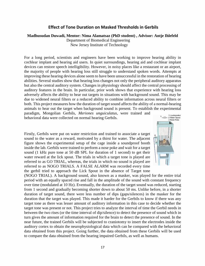
Firstly, Gerbils were put on water restriction and trained to associate a target
sound to the water as a reward, motivated by a thirst for water. The adjacent
figure shows the experimental setup of the cage inside a soundproof booth
inside the lab. Gerbils were trained to perform a nose poke and wait for a target
sound (1 kHz pure tone at 50 dB SPL for duration of 1 second) to get their
water reward at the lick spout. The trials in which a target tone is played are
referred to as GO TRIAL, whereas, the trials in which no sound is played are
referred to as NOGO TRIALS. A FALSE ALARM was recorded every time
the gerbil tried to approach the Lick Spout in the absence of Target tone
(NOGO TRIAL). A background sound, also known as a masker, was played for the entire trial
period with an equally spaced rise and fall in the amplitude of the sound with constant frequency
over time (modulated at 10 Hz). Eventually, the duration of the target sound was reduced, starting
from 1 second and gradually becoming shorter down to about 50 ms. Unlike before, in a shorter
duration of target sound, there were less number of dips (gaps/silences) in the masker for the
duration that the target was played. This made it harder for the Gerbils to know if there was any
target tone as there was lesser amount of auditory information in this case to decide whether the
target tone was present or not. This project tries to analyze the interval of time the Gerbil needs in
between the two rises (or the time interval of dip/silence) to detect the presence of sound which in
turn gives the amount of information required for the brain to detect the presence of sound. In the
near future, the trained Gerbils will be subjected to craniotomy to insert the electrodes inside the
auditory cortex to obtain the neurophysiological data which can be compared with the behavioral
data obtained from this project. Going further, the data obtained from these Gerbils will be used
to compare the data obtained from the hearing impaired Gerbils, as well as humans.
18
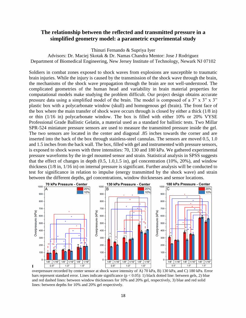
The relationship between the reflected and transmitted pressure in a
simplified geometry model: a parametric experimental study
Thinuri Fernando & Supriya Iyer
Advisors: Dr. Maciej Skotak & Dr. Namas Chandra Mentor: Jose J Rodriguez
Department of Biomedical Engineering, New Jersey Institute of Technology, Newark NJ 07102
Soldiers in combat zones exposed to shock waves from explosions are susceptible to traumatic
brain injuries. While the injury is caused by the transmission of the shock wave through the brain,
the mechanisms of the shock wave propagation through the brain are not well-understood. The
complicated geometries of the human head and variability in brain material properties for
computational models make studying the problem difficult. Our project design obtains accurate
pressure data using a simplified model of the brain. The model is composed of a 3” x 3” x 3”
plastic box with a polycarbonate window (skull) and homogenous gel (brain). The front face of
the box where the main transfer of shock wave occurs through is closed by either a thick (1/8 in)
or thin (1/16 in) polycarbonate window. The box is filled with either 10% or 20% VYSE
Professional Grade Ballistic Gelatin, a material used as a standard for ballistic tests. Two Millar
SPR-524 miniature pressure sensors are used to measure the transmitted pressure inside the gel.
The two sensors are located in the center and diagonal .85 inches towards the corner and are
inserted into the back of the box through stainless-steel cannulas. The sensors are moved 0.5, 1.0
and 1.5 inches from the back wall. The box, filled with gel and instrumented with pressure sensors,
is exposed to shock waves with three intensities: 70, 130 and 180 kPa. We gathered experimental
pressure waveforms by the in-gel mounted sensor and strain. Statistical analysis in SPSS suggests
that the effect of changes in depth (0.5, 1.0,1.5 in), gel concentration (10%, 20%), and window
thickness (1/8 in, 1/16 in) on internal pressure is significant. Further analysis will be conducted to
test for significance in relation to impulse (energy transmitted by the shock wave) and strain
between the different depths, gel concentrations, window thicknesses and sensor locations.
Fig. 1 The effect of window thickness, gel concentration, and sensor distance from the front window on peak
overpressure recorded by center sensor at shock wave intensity of A) 70 kPa, B) 130 kPa, and C) 180 kPa. Error
bars represent standard error. Lines indicate significance (p < 0.05): 1) black dotted line: between gels, 2) blue
and red dashed lines: between window thicknesses for 10% and 20% gel, respectively, 3) blue and red solid
lines: between depths for 10% and 20% gel respectively.
19
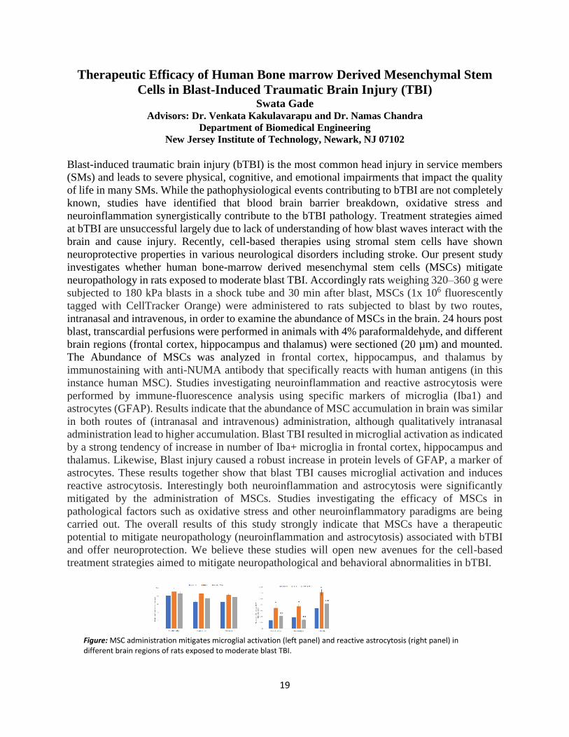
Therapeutic Efficacy of Human Bone marrow Derived Mesenchymal Stem
Cells in Blast-Induced Traumatic Brain Injury (TBI) Swata Gade
Advisors: Dr. Venkata Kakulavarapu and Dr. Namas Chandra
Department of Biomedical Engineering
New Jersey Institute of Technology, Newark, NJ 07102
Blast-induced traumatic brain injury (bTBI) is the most common head injury in service members
(SMs) and leads to severe physical, cognitive, and emotional impairments that impact the quality
of life in many SMs. While the pathophysiological events contributing to bTBI are not completely
known, studies have identified that blood brain barrier breakdown, oxidative stress and
neuroinflammation synergistically contribute to the bTBI pathology. Treatment strategies aimed
at bTBI are unsuccessful largely due to lack of understanding of how blast waves interact with the
brain and cause injury. Recently, cell-based therapies using stromal stem cells have shown
neuroprotective properties in various neurological disorders including stroke. Our present study
investigates whether human bone-marrow derived mesenchymal stem cells (MSCs) mitigate
neuropathology in rats exposed to moderate blast TBI. Accordingly rats weighing 320–360 g were
subjected to 180 kPa blasts in a shock tube and 30 min after blast, MSCs (1x 10⁶ fluorescently
tagged with CellTracker Orange) were administered to rats subjected to blast by two routes,
intranasal and intravenous, in order to examine the abundance of MSCs in the brain. 24 hours post
blast, transcardial perfusions were performed in animals with 4% paraformaldehyde, and different
brain regions (frontal cortex, hippocampus and thalamus) were sectioned (20 µm) and mounted.
The Abundance of MSCs was analyzed in frontal cortex, hippocampus, and thalamus by
immunostaining with anti-NUMA antibody that specifically reacts with human antigens (in this
instance human MSC). Studies investigating neuroinflammation and reactive astrocytosis were
performed by immune-fluorescence analysis using specific markers of microglia (Iba1) and
astrocytes (GFAP). Results indicate that the abundance of MSC accumulation in brain was similar
in both routes of (intranasal and intravenous) administration, although qualitatively intranasal
administration lead to higher accumulation. Blast TBI resulted in microglial activation as indicated
by a strong tendency of increase in number of Iba+ microglia in frontal cortex, hippocampus and
thalamus. Likewise, Blast injury caused a robust increase in protein levels of GFAP, a marker of
astrocytes. These results together show that blast TBI causes microglial activation and induces
reactive astrocytosis. Interestingly both neuroinflammation and astrocytosis were significantly
mitigated by the administration of MSCs. Studies investigating the efficacy of MSCs in
pathological factors such as oxidative stress and other neuroinflammatory paradigms are being
carried out. The overall results of this study strongly indicate that MSCs have a therapeutic
potential to mitigate neuropathology (neuroinflammation and astrocytosis) associated with bTBI
and offer neuroprotection. We believe these studies will open new avenues for the cell-based
treatment strategies aimed to mitigate neuropathological and behavioral abnormalities in bTBI.
Figure: MSC administration mitigates microglial activation (left panel) and reactive astrocytosis (right panel) in different brain regions of rats exposed to moderate blast TBI.
20
Advanced Manufacturing of Tissue Engineering Materials: Relating Material
Properties and Cutting Mechanics
Rama Hannineh, Advisor: Dr. Samuel C. Lieber
School of Applied Engineering & Technology, NJIT
A number of Tissue Engineered Medical Device products are fabricated from animal derived
starting materials. This includes porcine dermis which can be found in products used for breast
reconstruction and hernia repair. Although porcine dermis is an established starting material, there
is a lack of information on not only its material properties, but also its manufacturability. The
purpose of this Advanced Manufacturing study was to evaluate the cutting mechanics of the tissue
and how it relates to its location dependent material properties. Porcine dermis was obtained from
Midwest Research Swine (MRS) who are a known provider of starting material to medical device
companies. A custom steel-rule die press was used to cut samples for both material property
measures and cutting mechanics data. This required the development of an NJIT Makerspace
process to prepare Backing Material. The hardness and tensile properties of the porcine tissue
samples were measured with a Rex Gauge Hardness Tester and MTS load frame/ DIC camera
System respectively. Tensile testing was conducted on the speckled tissue (Figure 1A) until tissue
failure. Sample location was determined by traditional methods. A Cognex Vision System process
was also developed to automate the measurement of test sample location in the future. The cutting
mechanics was evaluated with a custom built 1.2 ton Instrumented Die Press (Figure 1B). This
unit provided preliminary results relating the cutting force and blade displacement in porcine
materials from different locations. The material properties and cutting mechanics of the tissue
samples were related to their location on the pig’s body. The results were compiled to test the
hypothesis that the properties are location dependent.
A B
Figure 1. (A) Representative speckled tensile sample for DIC Tensile testing (B) Custom
designed instrumented die press.
21
Directed evolution of KaiC, A circadian clock protein for Green Biofuel Production
John Hawks, Joydeep Chakraborty, Yong Ick Kim, Edgardo Farinas PhD*
Department of Chemistry and Environmental Science New Jersey Institute of Technology, Newark NJ 07102
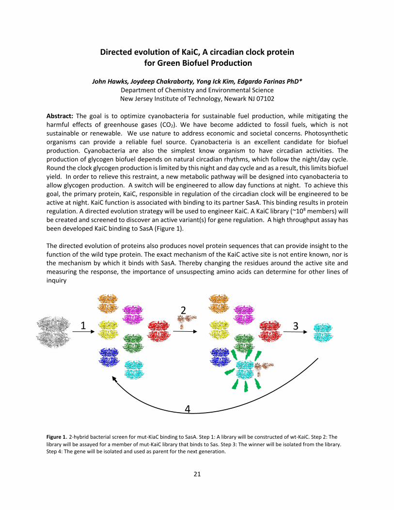
Abstract: The goal is to optimize cyanobacteria for sustainable fuel production, while mitigating the harmful effects of greenhouse gases (CO2). We have become addicted to fossil fuels, which is not sustainable or renewable. We use nature to address economic and societal concerns. Photosynthetic organisms can provide a reliable fuel source. Cyanobacteria is an excellent candidate for biofuel production. Cyanobacteria are also the simplest know organism to have circadian activities. The production of glycogen biofuel depends on natural circadian rhythms, which follow the night/day cycle. Round the clock glycogen production is limited by this night and day cycle and as a result, this limits biofuel yield. In order to relieve this restraint, a new metabolic pathway will be designed into cyanobacteria to allow glycogen production. A switch will be engineered to allow day functions at night. To achieve this goal, the primary protein, KaiC, responsible in regulation of the circadian clock will be engineered to be active at night. KaiC function is associated with binding to its partner SasA. This binding results in protein regulation. A directed evolution strategy will be used to engineer KaiC. A KaiC library (~108 members) will be created and screened to discover an active variant(s) for gene regulation. A high throughput assay has been developed KaiC binding to SasA (Figure 1). The directed evolution of proteins also produces novel protein sequences that can provide insight to the function of the wild type protein. The exact mechanism of the KaiC active site is not entire known, nor is the mechanism by which it binds with SasA. Thereby changing the residues around the active site and measuring the response, the importance of unsuspecting amino acids can determine for other lines of inquiry
Figure 1. 2-hybrid bacterial screen for mut-KiaC binding to SasA. Step 1: A library will be constructed of wt-KaiC. Step 2: The
library will be assayed for a member of mut-KaiC library that binds to Sas. Step 3: The winner will be isolated from the library. Step 4: The gene will be isolated and used as parent for the next generation.
1
2
3
4
22
Rehabilitating Stroke Patients through Adaptive Digital Environments
Omar A. Ilyas, Mentor/Advisor: Dr. Amy K. Hoover
Department of Informatics
New Jersey Institute of Technology, Newark, NJ 07102
Clinicians often instruct patients afflicted with strokes to perform in-home exercises such
as marching in place, balancing on one foot, and sidestepping. The clinician supervises these
exercises and makes adjustments based on the patients’ needs and individual performance.
However, because clinicians’ face-to-face time is limited, it can be difficult to keep a patient
performing said exercises consistently and correctly at home. Patients may become tired too
quickly or may develop a habit that reduces the effectiveness of the exercise. VSTEP is designed
to assist patients in performing their exercises and staying motivated.
The goal of VSTEP is to engage patients previously afflicted with strokes to perform their
fitness, balance, and mobility exercises optimally through the use of a video game! In-game
feedback and adjustment motivate patients to improve their exercise consistency, heart rate,
recovery and overall physical health. With a Kinect sensor and a heart rate monitor, VSTEP
simulates the patient’s movement and heart rate within the game, guides the patient to performing
correct exercises, and pushes the patient to achieve their ideal exercising heart rate. All of this,
while done with the assistance of a supervisor, can be done without the need for constant
readjustment from a clinician. Manual adjustments can be done as well, although the program is
intended for automatic motivation and adjustment.
To properly guide the patient in their exercises, the displays two targets whose positions
are affected by patient movement and patient heart rate. These targets indicate to the patient how
high and wide to raise their knees for the marching and balance exercise. If the patient is
successfully hitting these targets repeatedly, the targets raise slightly to encourage the patient to
push further. If, however, the patient is struggling to reach the new target positions over the course
of a few steps, the targets lower until the patient can reach them again. The targets also lower and
raise in response to the patient’s heart rate. If their heart rate is too high, and the targets will lower
slowly, and too low of a heart rate will raise them slowly. This is done to ensure that the patient is
within their ideal exercising heart rate ± 5 BPM according to either the Karvonan or Tanaka Heart
Rate Formula and performing the marching and/or balancing exercise correctly. While the patient
is exercising, all of their heart rate and performance data is being exported to a CSV file to be
viewable to clinicians for future records.

23
Visual Analytics for Data-Driven Social Science
Akm Islam, Abhijot Bedi, Riya Gupta, and Advisor: Dr. A Dasgupta
Department of Informatics New Jersey Institute of Technology, Newark, NJ 07102 USA
Abstract: Governments worldwide have started to implement open data initiatives and also launch
open data portals to enable the release of their large amounts of data in open and reusable formats.
The design of most open data sites follows a strategy, where opening more data, instead of opening
data better, has been the driving force.
We will develop a visual analytics tool for helping social scientists discover interesting data sets
that help them ask and answer new questions about cities and societies. Our proposed visual
analytics tool will transform the daily use of open urban data by social scientists and policy-makers
for formulating their research questions and decisions. We will address the challenge of scale and
complexity that analysts face while working with open data portals. These portals comprise too
many datasets and various categories that could be of interest, yet, there is little guidance on how
datasets could be potentially associated with answering specific analysis questions.
Interfaces like the NYC and Boston open data portals serve as collections with immense value for
advancing social science research or evaluating alternative urban policies.
However, to tap into the value of these collections, currently, analysts spend a lot of manual effort
for reconciling information from many datasets. This process is not only time-consuming and
cumbersome but could also be counter-productive with many explorations leading to analytical
dead ends. We will solve this problem by building novel visualization techniques that help social
scientists connect the dots and understand how many disparate data sets can be semantically
integrated for answering analysis questions.
24
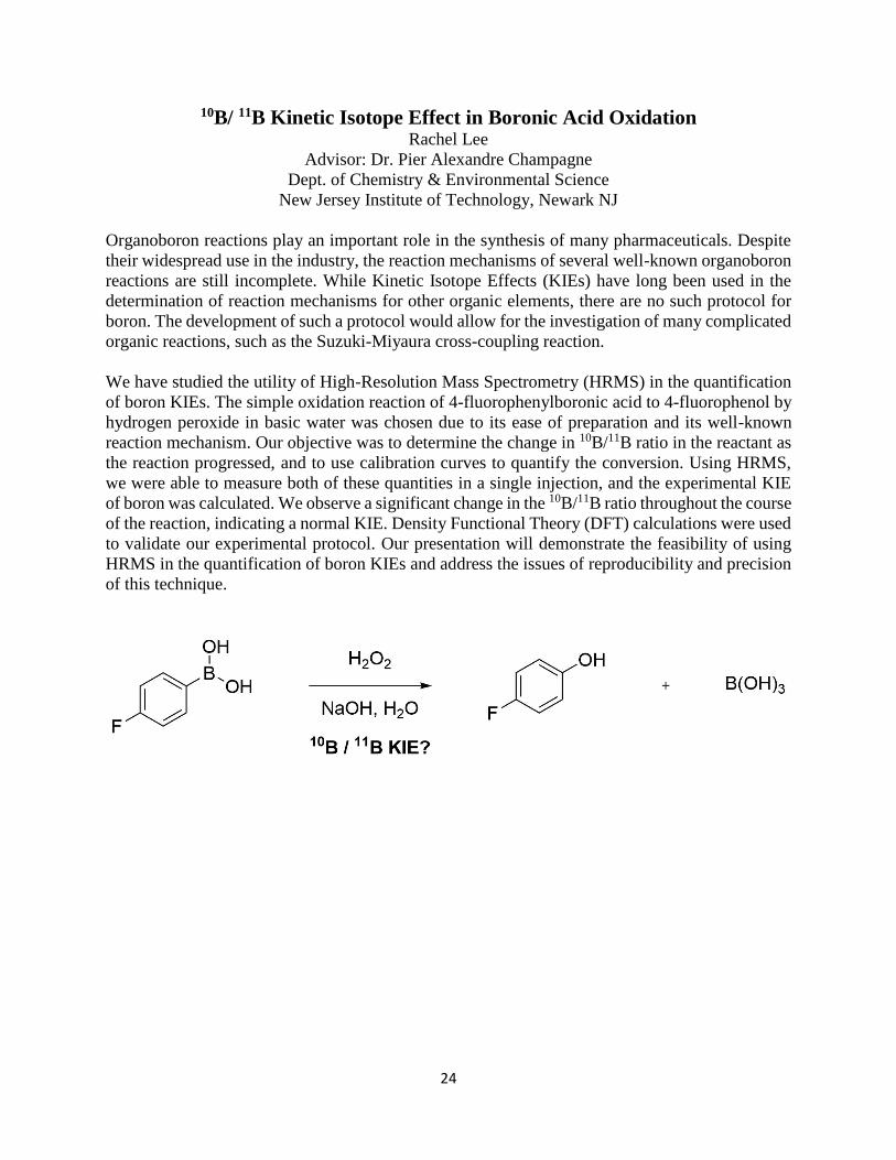
10B/ 11B Kinetic Isotope Effect in Boronic Acid Oxidation Rachel Lee
Advisor: Dr. Pier Alexandre Champagne
Dept. of Chemistry & Environmental Science
New Jersey Institute of Technology, Newark NJ
Organoboron reactions play an important role in the synthesis of many pharmaceuticals. Despite
their widespread use in the industry, the reaction mechanisms of several well-known organoboron
reactions are still incomplete. While Kinetic Isotope Effects (KIEs) have long been used in the
determination of reaction mechanisms for other organic elements, there are no such protocol for
boron. The development of such a protocol would allow for the investigation of many complicated
organic reactions, such as the Suzuki-Miyaura cross-coupling reaction.
We have studied the utility of High-Resolution Mass Spectrometry (HRMS) in the quantification
of boron KIEs. The simple oxidation reaction of 4-fluorophenylboronic acid to 4-fluorophenol by
hydrogen peroxide in basic water was chosen due to its ease of preparation and its well-known
reaction mechanism. Our objective was to determine the change in 10B/11B ratio in the reactant as
the reaction progressed, and to use calibration curves to quantify the conversion. Using HRMS,
we were able to measure both of these quantities in a single injection, and the experimental KIE
of boron was calculated. We observe a significant change in the 10B/11B ratio throughout the course
of the reaction, indicating a normal KIE. Density Functional Theory (DFT) calculations were used
to validate our experimental protocol. Our presentation will demonstrate the feasibility of using
HRMS in the quantification of boron KIEs and address the issues of reproducibility and precision
of this technique.
25
Engineering Nanoparticles for Drug Brain Delivery
Nicole Loehle
Advisor: Dr. Xiaoyang Xu, Mentor: William Ho, PhD Student
Otto H. York Department of Chemical and Materials Engineering
New Jersey Institute of Technology, Newark, NJ 07102
The Blood Brain Barrier (BBB) is the interface between the Central Nervous System (CNS) and
the blood. The BBB is extremely selective of what can pass through to the brain, only allowing small
molecules and specific proteins necessary for brain function. Unfortunately, this means life-saving
treatment of neurological diseases, including Parkinson’s, Alzheimer’s, stroke, and brain cancer, are
blocked from entering into the brain. Nanoparticles have the potential to overcome the BBB to deliver drugs
and therapeutics to the brain. This research is focused on creating PLGA-PEG nanoparticles and attaching
BBB- specific ligands to their surface to deliver therapeutics through the BBB into the brain.
To create our nanoparticles, we use Poly(lactic-co-glycolic acid) (PLGA) because it is a copolymer
known for its biodegradable and biocompatible nature, as well as its ability to easily functionalize ligands
through simple chemistry. We also use Polyethylene Glycol (PEG) because it is a polymer that allows for
‘stealth’ modification of the nanoparticle. This is important because nanoparticles can circulate in the blood
long enough to reach their targeted area and deliver their therapeutics. We plan to have our nanoparticles
undergo receptor-mediated transcytosis, where receptors must bind to their specific ligands in order for
cells to take up the therapeutic payload. If the receptor binds to the matching ligand, a vesicle, or cellular
sac used for transport, will encapsulate the ligand and carry it through the cell. We will conjugate a peptide
(ligand) that is recognized by BBB endothelial cells to the surface of the nanoparticle so our nanoparticle
can pass through the BBB endothelium using receptor-mediated transcytosis.
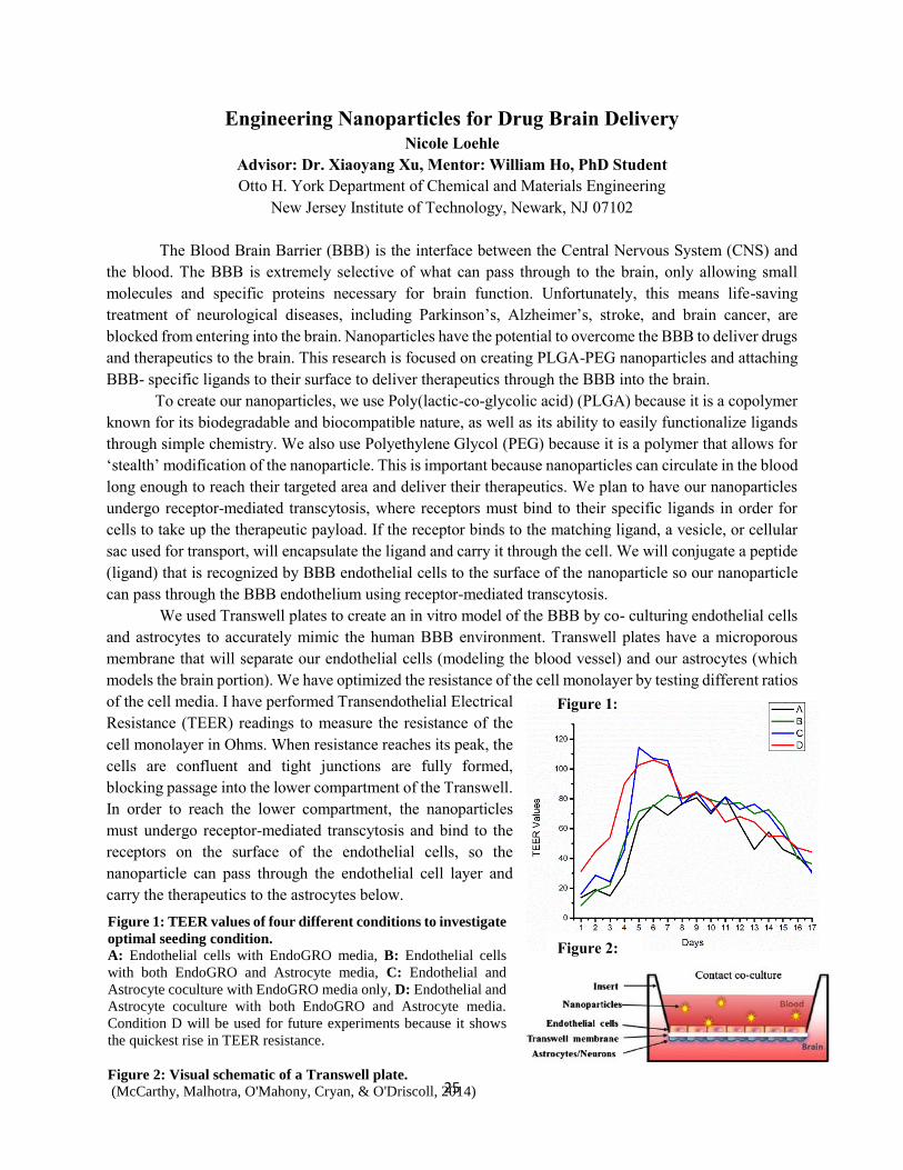
We used Transwell plates to create an in vitro model of the BBB by co- culturing endothelial cells
and astrocytes to accurately mimic the human BBB environment. Transwell plates have a microporous
membrane that will separate our endothelial cells (modeling the blood vessel) and our astrocytes (which
models the brain portion). We have optimized the resistance of the cell monolayer by testing different ratios
of the cell media. I have performed Transendothelial Electrical
Resistance (TEER) readings to measure the resistance of the
cell monolayer in Ohms. When resistance reaches its peak, the
cells are confluent and tight junctions are fully formed,
blocking passage into the lower compartment of the Transwell.
In order to reach the lower compartment, the nanoparticles
must undergo receptor-mediated transcytosis and bind to the
receptors on the surface of the endothelial cells, so the
nanoparticle can pass through the endothelial cell layer and
carry the therapeutics to the astrocytes below.
Figure 1:
Figure 2:
Figure 1: TEER values of four different conditions to investigate
optimal seeding condition. A: Endothelial cells with EndoGRO media, B: Endothelial cells
with both EndoGRO and Astrocyte media, C: Endothelial and
Astrocyte coculture with EndoGRO media only, D: Endothelial and
Astrocyte coculture with both EndoGRO and Astrocyte media.
Condition D will be used for future experiments because it shows
the quickest rise in TEER resistance.
Figure 2: Visual schematic of a Transwell plate. (McCarthy, Malhotra, O'Mahony, Cryan, & O'Driscoll, 2014)
26
Destruction of PFAS Using High-Frequency Power Ultrasound
Richard Marsh, Advisor: Dr. Jay Meegoda, Mentor: Jitendra Kewalramani, Ph.D. Student
Department of Civil and Environmental Engineering
New Jersey Institute of Technology, Newark, NJ 07102 USA
Abstract: Per- and Polyfluoroalkyl substances, known as PFAS, are a group of synthetic chemicals
containing an aliphatic carbon chain in which the hydrogen atoms have either been completely or partially
replaced by fluorine. Previously known as PFCs, these compounds have been manufactured since the 1940s
and were used in several consumer products including non-stick cookware, paints, carpets, floor polishes,
masking tape, and stain repellants. The unique properties of PFAS, including the strength of the C-F bond,
hydrophobicity, lipophobicity, and surface tension lowering capabilities, have contributed to its widespread
use in industrial and commercial areas. Due to these properties, most PFAS are extremely resistant to
degradation. The use of these compounds in numerous applications coupled with their resistance to
traditional environment remediation methods has led to the detection of a broad range of PFAS in the
environment and the human body.
This research uses ultrasound to induce the sonochemical degradation of two common PFAS,
perfluorooctanesulfonic acid (PFOS) and perfluorooctanoic acid (PFOA). The mechanisms causing
sonochemical reactions in liquids are the phenomenon of acoustic cavitation, which concentrates and
releases a tremendous amount of energy in localized areas, and pyrolysis, the splitting of molecules caused
by high temperatures. The concentrated energy in minute-scale cavitation bubbles produces various radicals
by the pyrolysis of water and dissolved gas molecules. Radicals are capable of initiating or promoting many
fast oxidation reactions. The sonochemical reactions caused by the hydroxyl radical will lead to the
destruction of PFAS within the reactor. For this study, the degradation of PFOS and PFOA was performed
using two different configurations to produce an ultrasonic field, a horn transducer operating at 20 kHz and
a plate transducer operating between 200 – 800 kHz. The 20 kHz transducer provides agitation, breaks up
clusters of molecules, and increases the number of bubbles in solution. The high-frequency ultrasound plate
produces the hydroxyl radicals, which oxidize and degrade organic compounds in suspension.
Variables such as sonication time, frequency, and PFAS concentration were examined to determine the
optimal conditions within the reactor to generate the most significant removal efficiency. Results were
analyzed using liquid chromatography/mass spectrometry to detect the presence of any organic pollutants
remaining. KI dosimetry and sonoluminescence were used to evaluate the presence of hydroxyl radicals
within the reactor.
27
Novel Drug Delivery System using Anti-Angiogenic Peptides for Glioblastoma
Multiforme
Anna Mathew, Advisor: Vivek Kumar, PhD
Department of Biomedical Engineering
New Jersey Institute of Technology, Newark, NJ 07102 USA
Glioblastoma, also known as glioblastoma multiforme (GBM), is an extremely aggressive
type of nervous system tumor that forms on the tissue of the brain. Nourished by a large and
complex network of blood vessels, the tumor cells reproduce quickly, making GBM one of the
deadliest cancers. Glioblastoma accounts for roughly half of all brain and central nervous system
cancers, with a one-year survival rate of less than forty percent and a five-year survival rate of less
than five percent. Current standard of care for patients with newly diagnosed glioblastoma includes
surgery, radiation therapy, and chemotherapy. One such treatment involves an in-situ drug delivery
system. Following surgery, carmustine wafers are implanted in the tumor cavity. The wafers then
slowly dissolve while releasing medication that will kill the remaining cancerous cells. These
wafers, however, lack the ability to homogeneously coat and conform to the tissue voids of the
resected tumor cavities, amongst other issues. To address this lack of proper fit, space-conforming
peptides modified with an anti-angiogenic mimic will be developed as an injectable hydrogel. As
indicated in previous literature, GBM is marked by aberrant vascular proliferation. These blood
vessels support the tumor through nutrient delivery and cancer cell migration. It has been
hypothesized that the peptides will be able to conform to the unique shape of the cavity and exhibit
anti-angiogenic properties to address this abundant vascularization, thus minimizing chances of
cancer recurrence.
Within this project, there are three main objectives to be accomplished. First, peptides of
choice will be synthesized and purified into hydrogels. The first is termed SL-Kr5, with its mimic
based on domain 5 of the extracellular protein Kringle. The second peptide, known as SL-LAM,
is based on the laminin-1 protein. Lastly, the final peptide, known as SL-HP is based on the
histidine-rich-glycoprotein. Once the gels are formed, their in vitro properties will be assessed
through a tube formation assay. The assay will be run on several different types of endothelial cells
(involved in blood vessel formation) to obtain a thorough understanding of the antiangiogenic
capabilities. The effects of the peptide on the cells will be understood by measuring the extent of
tube formation in terms of total length of tubules formed, as well as the number of segments,
branches, junctions, nodes, and segments of vessels. The assay will be run using rat brain
microvascular endothelial cells, rat retinal microvascular endothelial cells, and primary human
retinal microvascular endothelial cells. It is expected that the peptide will allow for a dose-
dependent destruction of the aforementioned capillary networks in each of the cell types, such that
at higher concentrations of the peptide there is a stronger anti-angiogenic response observed. This
will give us a clear picture of the anti-angiogenic effect of the peptides on these cell types, giving
a strong indicator of how they will perform in vivo in future experiments. The successful
completion of the objectives of this project will provide a clear and reliable understanding of the
potential of each peptide hydrogel as an effective drug delivery system for the treatment of GBM.

28
Encouraging the Use of Built-in Language Features for Learning Control
Flow
Student researcher:
Michael Mobilio (IT)
Faculty mentor:
Michael J. Lee, PhD (Informatics)
Abstract: This research attempts to develop a learning curriculum that encourages the use of
specific control flow patterns in the JavaScript and Python programming languages. This is a
technical approach that benefits the user by letting them construct, visualize, and follow specific
control flow patterns through the use of the built-in features from their choice of programming
language. This research aims to improve students' understanding and recognition of better
control flows.
Figure 1: Sample challenge inside the Gidget 4 interface
29
Investigating the role of a genetically-conserved spinal neuronal class,
Dmrt3, in the functional control of locomotion in zebrafish
Mahathi Mohan Gowda, Advisor: Kristen Severi
Department of Biological Sciences
New Jersey Institute of Technology, Newark, NJ 07102
Understanding the neural circuits that underlie locomotion is key in furthering knowledge
about the causes of motor behaviors and disorders. This project explores the role of spinal
interneurons, linked to the gene Dmrt3a, in larval zebrafish. The interneurons have been observed
in horses and mice, and these past studies suggest that they are linked to speed-shifting and
locomotor coordination1. Our hypothesis is that the activity of Dmrt3a neurons is necessary for
coordinating locomotion at different speeds. Although there is evidence for this hypothesis in other
models, the function of these neurons has never been tested in a moving animal which can be
manipulated experimentally. We will test the locomotive abilities of larval zebrafish lacking active
Dmrt3a interneurons, as well as the effects of stimulation of the interneurons while larvae are at
rest or swimming.
The aims of this experiment are accomplished through the use of a behavior rig and
FishTracker software2 that allows for the tracking of larval swimming behaviors like the following:
tail-angle, tail-beat frequency, bout duration, interbout duration, bout distance, and average
swimming speed versus grating speed. With these parameters it will be possible ascertain in a
statistically significant way if the Dmrt3a interneurons are involved in speed-shifting or locomotor
coordination. Furthermore, in order to accomplish the aforementioned, the experiment was split
into two portions.
In the first part of experimentation we will use an OMR grating (optomotor response grating
that facilitates swimming) at three different speeds and test it with two different groups of fish.
The first group of fish is the wild type larvae which will act as the control, and the second group
will be the experimental group or the Dmrt3a:Gal4;UAS:Botox-GFP transgenic line. The
transgenic line of larvae have their Dmrt3a interneurons silenced using botox3, which allows for
us to conclude that differences in locomotion between the experimental and control group come
about as a result of the lack of Dmrt3a interneurons. The second portion of experimentation uses
the same control group of larvae, while the experimental group will be the transgenic line of
Dmrt3a:Gal4;UAS:ChR2-YFP larvae. The transgenic larvae have channelrhodopsin2 on the
Dmrt3a interneurons, which activates the neurons when they have light shined on to them4 (this
experiment uses a laser and an optic fiber as the means of light). Furthermore, the larvae will also
be tested with an OMR grating, except this time the larvae will have their head embedded in
agarose (free swim is not possible when activation of neurons requires concentrated source of
light). This experiment will help to see if stimulation of the neurons leads to changes in locomotive
behavior of the larvae.
1. Andersson, L. S. et al. Mutations in DMRT3 affect locomotion in horses and spinal circuit function in mice. Nature
488, 642–6 (2012).
2. Marques, J. C., Lackner, S., Felix, R. & Orger, M. B. Structure of the Zebrafish Locomotor Repertoire Revealed with
Unsupervised Behavioral Clustering. Curr. Biol. (2018). doi:10.1016/j.cub.2017.12.002
3. Satou, C. et al. Transgenic tools to characterize neuronal properties of discrete populations of zebrafish neurons.
Development 140, 3927–3931 (2013).
4. Kateriya, S. et al. Channelrhodopsin-2, a directly light-gated cation-selective membrane channel. Proc. Natl. Acad. Sci.
100, 13940–13945 (2003).
30
Integrated Solid-Fluid Interaction Potential for Modeling Gas Adsorption in Templated Mesoporous Carbons
Marcos Molina, Mentor: Max Maximov, Advisor: Gennady Y. Gor Otto H. York Department of Chemical and Materials Engineering, New Jersey Institute of Technology,
University Heights, Newark, NJ 07102, USA
Due to their high surface area, nanoporous materials have numerous applications in chemical engineering, such as separation processes, catalysis, and energy storage. We focus here on three-dimensional ordered mesoporous (3DOm) carbons, which are specifically promising as frameworks for zeolite crystal synthesis and for solid natural gas storage.1 Because of the interconnection 3DOm carbons are also suitable for electrochemical applications as the narrow necks allow for efficient charge and discharge rate because of the improved diffusion of ions.2
Nitrogen adsorption is a standard technique used for the characterization of these materials with respect to surface area and pore size distribution. The relation between the experimental adsorption isotherms and the theoretical adsorption isotherm is commonly used to find the pore size distribution. The theoretical adsorption isotherm is often calculated using Monte Carlo molecular simulations using the fluid-solid interaction potential in a certain simplified geometry (e.g. sphere). However, the calculation of the potential in 3DOm carbons fails to account for the decrease in surface area due to the overlapping spherical pores.
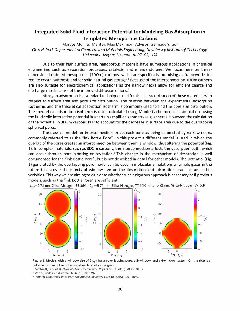
The classical model for interconnection treats each pore as being connected by narrow necks, commonly referred to as the “Ink Bottle Pore”. In this project a different model is used in which the overlap of the pores creates an interconnection between them, a window, thus altering the potential (Fig. 1). In complex materials, such as 3DOm carbons, the interconnection affects the desorption path, which can occur through pore blocking or cavitation.3 This change in the mechanism of desorption is well documented for the “Ink Bottle Pore”, but is not described in detail for other models. The potential (Fig. 1) generated by the overlapping pore model can be used in molecular simulations of simple gases in the future to discover the effects of window size on the desorption and adsorption branches and other variables. This way we are aiming to elucidate whether such a rigorous approach is necessary or if previous models, such as the “Ink Bottle Pore” are sufficient.
Figure 1. Models with a window size of 5 𝜎𝑓𝑓 for an overlapping pore, a 2-window, and a 4-window system. On the side is a
color bar showing the potential at each point in the graph. 1 Borchardt, Lars, et al. Physical Chemistry Chemical Physics 18.30 (2016): 20607-20614. 2 Macías, Carlos, et al. Carbon 63 (2013): 487-497. 3 Thommes, Matthias, et al. Pure and Applied Chemistry 87.9-10 (2015): 1051-1069.
31
A Low-Cost Electro-Mechanical System to create 3D scans using 2D LIDARs
for Pothole Detection
Jorim Morainvil, Advisor: Pramod Abichandani, Ph.D.
Department of Electrical and Computer Engineering Technology
New Jersey Institute of Technology, Newark, N.J. 07102
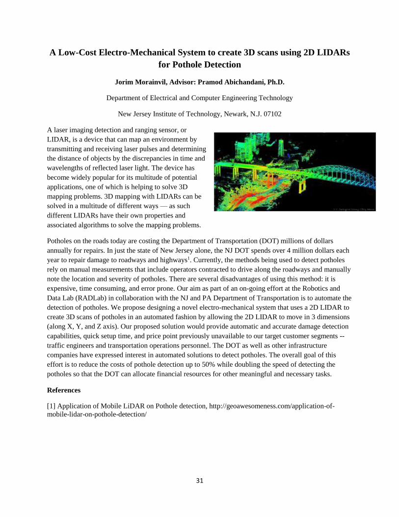
A laser imaging detection and ranging sensor, or
LIDAR, is a device that can map an environment by
transmitting and receiving laser pulses and determining
the distance of objects by the discrepancies in time and
wavelengths of reflected laser light. The device has
become widely popular for its multitude of potential
applications, one of which is helping to solve 3D
mapping problems. 3D mapping with LIDARs can be
solved in a multitude of different ways — as such
different LIDARs have their own properties and
associated algorithms to solve the mapping problems.
Potholes on the roads today are costing the Department of Transportation (DOT) millions of dollars
annually for repairs. In just the state of New Jersey alone, the NJ DOT spends over 4 million dollars each
year to repair damage to roadways and highways1. Currently, the methods being used to detect potholes
rely on manual measurements that include operators contracted to drive along the roadways and manually
note the location and severity of potholes. There are several disadvantages of using this method: it is
expensive, time consuming, and error prone. Our aim as part of an on-going effort at the Robotics and
Data Lab (RADLab) in collaboration with the NJ and PA Department of Transportation is to automate the
detection of potholes. We propose designing a novel electro-mechanical system that uses a 2D LIDAR to
create 3D scans of potholes in an automated fashion by allowing the 2D LIDAR to move in 3 dimensions
(along X, Y, and Z axis). Our proposed solution would provide automatic and accurate damage detection
capabilities, quick setup time, and price point previously unavailable to our target customer segments --
traffic engineers and transportation operations personnel. The DOT as well as other infrastructure
companies have expressed interest in automated solutions to detect potholes. The overall goal of this
effort is to reduce the costs of pothole detection up to 50% while doubling the speed of detecting the
potholes so that the DOT can allocate financial resources for other meaningful and necessary tasks.
References
[1] Application of Mobile LiDAR on Pothole detection, http://geoawesomeness.com/application-of-
mobile-lidar-on-pothole-detection/
32
Theoretical Studies of Possible Topological Edge Modes in Novel Systems
Zoraiz Naeem, Advisor: Dr. Ken Ahn
Department of Physics
New Jersey Institute of Technology, Newark, NJ 07102 USA
Abstract: This decade saw an immense development in the field of topology and has helped us
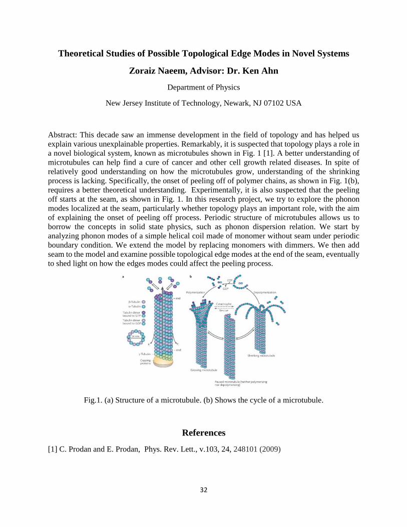
explain various unexplainable properties. Remarkably, it is suspected that topology plays a role in
a novel biological system, known as microtubules shown in Fig. 1 [1]. A better understanding of
microtubules can help find a cure of cancer and other cell growth related diseases. In spite of
relatively good understanding on how the microtubules grow, understanding of the shrinking
process is lacking. Specifically, the onset of peeling off of polymer chains, as shown in Fig. 1(b),
requires a better theoretical understanding. Experimentally, it is also suspected that the peeling
off starts at the seam, as shown in Fig. 1. In this research project, we try to explore the phonon
modes localized at the seam, particularly whether topology plays an important role, with the aim
of explaining the onset of peeling off process. Periodic structure of microtubules allows us to
borrow the concepts in solid state physics, such as phonon dispersion relation. We start by
analyzing phonon modes of a simple helical coil made of monomer without seam under periodic
boundary condition. We extend the model by replacing monomers with dimmers. We then add
seam to the model and examine possible topological edge modes at the end of the seam, eventually
to shed light on how the edges modes could affect the peeling process.
Fig.1. (a) Structure of a microtubule. (b) Shows the cycle of a microtubule.
References
[1] C. Prodan and E. Prodan, Phys. Rev. Lett., v.103, 24, 248101 (2009)
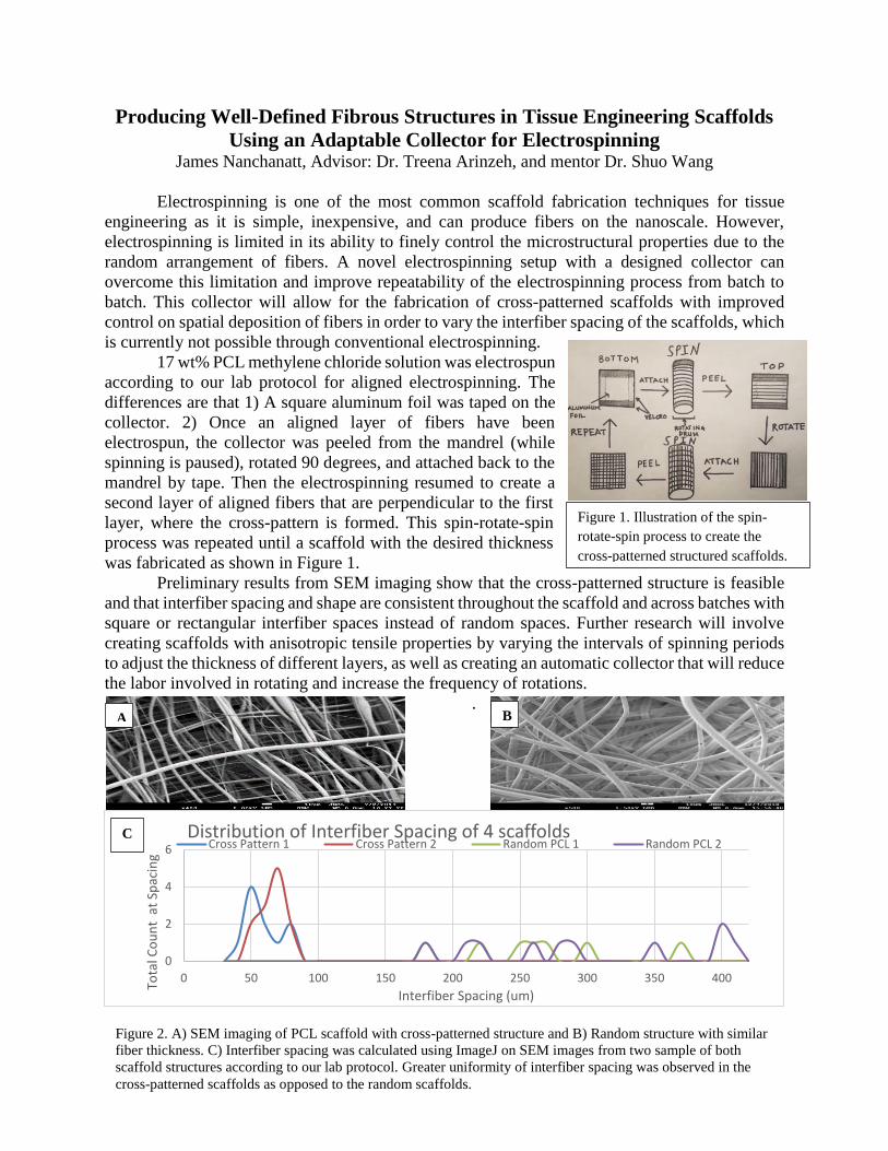
33
Producing Well-Defined Fibrous Structures in Tissue Engineering Scaffolds
Using an Adaptable Collector for Electrospinning James Nanchanatt, Advisor: Dr. Treena Arinzeh, and mentor Dr. Shuo Wang
Electrospinning is one of the most common scaffold fabrication techniques for tissue
engineering as it is simple, inexpensive, and can produce fibers on the nanoscale. However,
electrospinning is limited in its ability to finely control the microstructural properties due to the
random arrangement of fibers. A novel electrospinning setup with a designed collector can
overcome this limitation and improve repeatability of the electrospinning process from batch to
batch. This collector will allow for the fabrication of cross-patterned scaffolds with improved
control on spatial deposition of fibers in order to vary the interfiber spacing of the scaffolds, which
is currently not possible through conventional electrospinning.
17 wt% PCL methylene chloride solution was electrospun
according to our lab protocol for aligned electrospinning. The
differences are that 1) A square aluminum foil was taped on the
collector. 2) Once an aligned layer of fibers have been
electrospun, the collector was peeled from the mandrel (while
spinning is paused), rotated 90 degrees, and attached back to the
mandrel by tape. Then the electrospinning resumed to create a
second layer of aligned fibers that are perpendicular to the first
layer, where the cross-pattern is formed. This spin-rotate-spin
process was repeated until a scaffold with the desired thickness
was fabricated as shown in Figure 1.
Preliminary results from SEM imaging show that the cross-patterned structure is feasible
and that interfiber spacing and shape are consistent throughout the scaffold and across batches with
square or rectangular interfiber spaces instead of random spaces. Further research will involve
creating scaffolds with anisotropic tensile properties by varying the intervals of spinning periods
to adjust the thickness of different layers, as well as creating an automatic collector that will reduce
the labor involved in rotating and increase the frequency of rotations.
.
0
2
4
6
0 50 100 150 200 250 300 350 400
Tota
l Co
un
t a
t Sp
acin
g
Interfiber Spacing (um)
Distribution of Interfiber Spacing of 4 scaffoldsCross Pattern 1 Cross Pattern 2 Random PCL 1 Random PCL 2
Figure 1. Illustration of the spin-
rotate-spin process to create the
cross-patterned structured scaffolds.
Figure 2. A) SEM imaging of PCL scaffold with cross-patterned structure and B) Random structure with similar
fiber thickness. C) Interfiber spacing was calculated using ImageJ on SEM images from two sample of both
scaffold structures according to our lab protocol. Greater uniformity of interfiber spacing was observed in the
cross-patterned scaffolds as opposed to the random scaffolds.
A B
C
34
Effect of contaminant mixtures in the Biodegradation rates
Randy Nutakor, Advisor: Lucia Rodriguez-Freire, Mentors: Boran Wang PhD Student, Bo
Deng PhD student and Maedeh Soleimanifar PhD student
Department of Civil and Environmental Engineering
New Jersey Institute of Technology, Newark NJ 07102
Abstract: The goal of this research is to use extensive sampling methods to determine the
contaminant concentration together with laboratory experiments to assess contaminant mobility
from sediments and water. Sediments and water samples collected from different locations at the
Ringwood Mines/Landfill Superfund Site, NJ were analyzed in the lab to determine which samples
and locations has the highest amount of arsenic and benzene due to the possibility for enrichment
of specific microbial degraders. Wet sediments of between 5 g to 10 g were incubated under
varying conditions with 500mL to 100mL of synthetic water. The concentration of contaminants
was monitored with time with most emphasis on arsenic and benzene. Benzene biodegradation
experiments were carried out in two forms; under aerobic conditions using oxygen as an electron
acceptor and under anaerobic condition endogenous electron acceptors present in the sediments,
such as Fe(III), Mn(IV) and As(V).
Benzene concentration in water from locations with the highest concentration was measured using
purge-and-trap GC and sampled periodically during and after each experiment. Benzene
concentration in the sediments was also measured at the end of every experiment cycle.
Total arsenic concentration in was measured using the ICP-MS and periodically sampled during
each experiment while arsenic concentration in the sediments was also measured at the end of each
experiment cycle. The pH of the liquids was closely monitored during the experiment. Microbial
population was determined and the beginning and end of the experiments by DNA extraction using
DNA extraction soil kit and sequenced using Illumina sequencing. Sediments mineralogy was
evaluated using XRD and FESEM-EDS in the sediment before and after treatment. Speciation in
the sediments was also determined with XPS.
The results from these experiments are expected to inform local agencies and industries on the
distribution and fate of legacy contaminants mixtures in superfund sites and wetlands in New
Jersey and other states, as well as the attenuation potential of the mixtures in the contaminated site.
35
The Role of Neural Activity and Semaphorin Signaling in Neural Repair
Ishani Patel, Advisor: Dr. Gal Haspel
Department of Biological Sciences
New Jersey Institute of Technology
Our study focuses on understanding the mechanisms of neural repair at a cellular and molecular
level. Most mammalian neurons of the central nervous system (the brain and spinal cord) do not
have regenerative abilities. This deficit is the primary cause of the chronic effects of many
neurodegenerative diseases, as well as neurological injuries, such as spinal cord injuries. Spinal
cord injuries, caused by damage to the motor neurons of the spinal cord, often have permanent
debilitating effects, including partial or complete paralysis. There are more than 12,000 incidents
of SCI’s in the United States annually, yet, unfortunately, there are no reliable treatment options.
Through current rehabilitation treatments, patients can regain some function and sensation; often,
full function is not restored. A neurons’ ability to regenerate is dependent on several factors,
including cell signaling and activity, however, the precise effects of the pathways and mechanisms
are unknown. The limited treatment options along with the finite knowledge about neural
regeneration makes research on neural repair critical.
Our project aims to use an innovative technique to determine the contribution of semaphorin
signaling coupled with neural activity on dendritic branching patterns in mice cortical cells. The
semaphorin signaling pathway, composed of semaphorin, the signaling protein, and plexin, the
receptor protein, has a repelling effect on the direction of axonal growth during development.
However, in dendrites, semaphorin is known to promote increased growth and branching. Through
our study, we will determine whether dendritic growth is further affected by neural activity. We
will alter neural activity within the cell using optogenetics, a technique that controls cell activity
using light.
Two photosensitive opsins will be transgenically expressed within the cells: one that induces
activity and has a red fluorescent tag, pCAG-ChrimsonR-tdT, and one that inhibits activity and
has a cyan fluorescent tag, FCK-Arch-CFP. These opsins, when exposed to amber light (590 nm),
either open a protein channel or activate an iron pump to control cell activity. All the cells will be
transfected with a mixture of plasmids coding for the two optogenetic plasmids, and GFP that
makes the cells visible for imaging. The random expression of the plasmids will create four
experimental groups, which will be distinguishable by the different fluorescent tags on each
plasmid: activated and inactivated cells (Green, Red, and Cyan), activated cells (Red and Green),
inhibited cells (Cyan and Green), and a negative control (Green). During the five-day incubation
period for the cells, they will be exposed to 5 Ms pulses of 590 nm light at a frequency of 100 Hz,
to prevent overstimulation or habituation of the cells.
We will use Sholl analysis, a technique that uses counting within concentric circles around the cell
soma to quantify parameters such as dendritic branching and length, to compare the results of our
four experimental groups. We expect to see that activation will induce branching in the neurons,
and this effect will be additive to the effect of semaphorin, whereas inactivation will negate the
effects of the semaphorin.
36
Understanding Unidentifiability in Dynamic Models from Ground Truth Data
Raghav Patel, Advisor: Horacio G. Rotstein, PhD
Federated Department of Biological Sciences &
Institute for Brain and Neuroscience Research
New Jersey Institute of Technology, Newark, NJ 07102 USA
Abstract: A really useful tool to scientists would be a machine that can take in input data and output
useful equations that characterize that data and allow us to understand the dynamic mechanisms
that govern the generation of these data. Different techniques exist for fitting equations to data and
each one has different advantages and disadvantages. There are several difficulties associated with
these tools. These difficulties are both technical (algorithmic nature) and structural (degeneracy,
mathematical nature). Degeneracy refers to situations where multiple sets of parameter values can
produce the same observable output (oscillatory pattern in neurons), therefore making the inverse
problem (finding parameters given data) ill-posed. This is not a problem associated with
uncertainty in the knowledge of a unique parameter set from which the data was generated, but a
structural problem of mathematical models where multiple parameter sets are able to produce the
same result, and has been observed in neurobiology experiments [1]. In this project we have
developed ground truth models in order to calibrate and validate parameter estimation algorithms
in the presence of degeneracy. We used “faked” data, generated by models exhibiting degeneracy
in a number of attributes (e.g., oscillation frequency, oscillations amplitude, plateau/saturation
level) and the so-called genetic algorithms [2] to estimate the parameters used in generating these
data. Genetic algorithms are based on the theory of natural selection, where we make a random
initial population of parameter set values and keep “breeding” them until we get the most “fit”
parameters. The end result is a collection of parameter sets distributed in some fashion that
minimizes the error between the data and the simulated models according to some metric. In the
absence of degeneracy the distribution of estimated parameter sets is expected centered on the
“true values” with a distribution width that is smaller the lower the noise levels used to generate
the data (in the noiseless case, the distribution would be a delta function). In contrast, we show
that because of degeneracy in a given attribute or attributes the estimated parameters sets are
widely distributed with no apparent systematic rule except that of maintaining the attribute(s)
constant.
[1] Prinz A, Bucher D, Marder E. Similar network activity from disparate circuit parameters. Nature Neuroscience, 7:1345-1352, 2004.
[2] Deb K, Pratap A, Agarwal S, Meyarivan T. A fast and elitist multiobjective genetic algorithm: NSGA-II. IEEE Transactions on Evolutional Computation, 6:182-197.

37
Central Auditory Pathology of Blast Induced Tinnitus/Hearing Loss Navya Pendyala
Faculty Mentor: Dr. Venkata Kakulavarapu and Dr. Namas Chandra
Center for Injury Biomechanics, Materials, and Medicine NJIT
Blast-induced traumatic brain injury (bTBI) is one of the most prevalent forms of injury to
soldiers in battlefield due to the use of explosive devices that not only cause brain injury but also
create loud explosions that affect the auditory system. Partial hearing loss and/or tinnitus, a
phantom auditory perception, are the common auditory problems encountered by service members
discharged from active duty. While both peripheral and central auditory pathways are affected in
tinnitus/hearing loss, the mechanisms responsible for these events remains unknown.
Therefore, in this study I first attempted to distinguish partial hearing loss from tinnitus by
exploring peripheral auditory functions including gap detection and prepulse inhibition of acoustic
startle response and other behavior tests. In these tests, the startle response of the rats to four
different startle sounds were quantified to diagnose the rat for tinnitus or partial hearing loss. These
tests were done with rats subjected to one single 180kPa blast at the 1d, 7d, 15d, 28d, and 42d
timepoints. Preliminary data shows that only a few rats showed a significant difference in startle
response before and after blast injury, which was statistically insignificant. These studies are being
further continued using a large number of cohorts being exposed to mild, medium, high and
multiple low-level blast overpressures.
We next examined the central auditory system (CAS) abnormalities, particularly the
excitatory and inhibitory neurotransmitter receptor levels which are well known markers to
establish central auditory damage in the auditory cortex using the immunofluorescence method.
This data showed a significant decrease in the levels of NMDAR-1, a major receptor for the ligand
excitatory amino acid glutamate in auditory cortex, in rats exposed to high-level blast (180kPa) at
the 4h, 24h, 7d, 15d, and 30d timepoints (Figure 2). Interestingly, among these rats there was a
concomitant increase in the levels of the GABAA receptor for inhibitory amino acid GABA in the
auditory cortex (Figure 1). These results clearly indicate that an imbalance of excitatory and
inhibitory neurotransmission contributes to central auditory pathology in bTBI. The levels of these
receptors are being analyzed in animal models of low-level repeated blast at different time points.
Understanding the mechanisms of the CAS system abnormalities as related to blast injury will
significantly contribute to future therapeutic interventions to mitigate blast-induced tinnitus/partial
hearing loss.
Figure 1. This figure shows an increase in the GABAA
inhibitory receptor expression at five different
timepoints post high level single blast (180kPa).
Figure 2. This figure shows a decrease in the NMDAR1
excitatory receptor expression at five different
timepoints post high level single blast (180kPa).
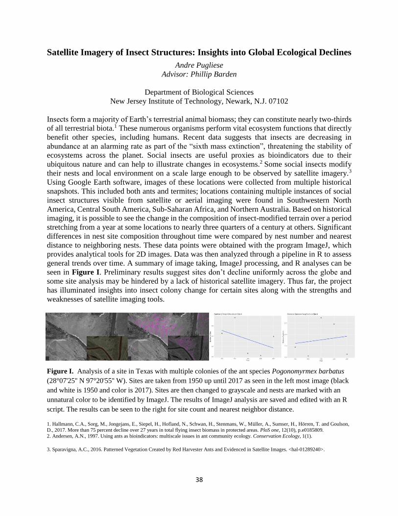
38
Satellite Imagery of Insect Structures: Insights into Global Ecological Declines
Andre Pugliese
Advisor: Phillip Barden
Department of Biological Sciences
New Jersey Institute of Technology, Newark, N.J. 07102
Insects form a majority of Earth’s terrestrial animal biomass; they can constitute nearly two-thirds
of all terrestrial biota.1 These numerous organisms perform vital ecosystem functions that directly
benefit other species, including humans. Recent data suggests that insects are decreasing in
abundance at an alarming rate as part of the “sixth mass extinction”, threatening the stability of
ecosystems across the planet. Social insects are useful proxies as bioindicators due to their
ubiquitous nature and can help to illustrate changes in ecosystems.2 Some social insects modify
their nests and local environment on a scale large enough to be observed by satellite imagery.3
Using Google Earth software, images of these locations were collected from multiple historical
snapshots. This included both ants and termites; locations containing multiple instances of social
insect structures visible from satellite or aerial imaging were found in Southwestern North
America, Central South America, Sub-Saharan Africa, and Northern Australia. Based on historical
imaging, it is possible to see the change in the composition of insect-modified terrain over a period
stretching from a year at some locations to nearly three quarters of a century at others. Significant
differences in nest site composition throughout time were compared by nest number and nearest
distance to neighboring nests. These data points were obtained with the program ImageJ, which
provides analytical tools for 2D images. Data was then analyzed through a pipeline in R to assess
general trends over time. A summary of image taking, ImageJ processing, and R analyses can be
seen in Figure I. Preliminary results suggest sites don’t decline uniformly across the globe and
some site analysis may be hindered by a lack of historical satellite imagery. Thus far, the project
has illuminated insights into insect colony change for certain sites along with the strengths and
weaknesses of satellite imaging tools.
-
Figure I. Analysis of a site in Texas with multiple colonies of the ant species Pogonomyrmex barbatus
(28°07'25'' N 97°20'55'' W). Sites are taken from 1950 up until 2017 as seen in the left most image (black
and white is 1950 and color is 2017). Sites are then changed to grayscale and nests are marked with an
unnatural color to be identified by ImageJ. The results of ImageJ analysis are saved and edited with an R
script. The results can be seen to the right for site count and nearest neighbor distance.
1. Hallmann, C.A., Sorg, M., Jongejans, E., Siepel, H., Hofland, N., Schwan, H., Stenmans, W., Muller, A., Sumser, H., Horren, T. and Goulson,
D., 2017. More than 75 percent decline over 27 years in total flying insect biomass in protected areas. PloS one, 12(10), p.e0185809.
2. Andersen, A.N., 1997. Using ants as bioindicators: multiscale issues in ant community ecology. Conservation Ecology, 1(1).
3. Sparavigna, A.C., 2016. Patterned Vegetation Created by Red Harvester Ants and Evidenced in Satellite Images. <hal-01289240>.
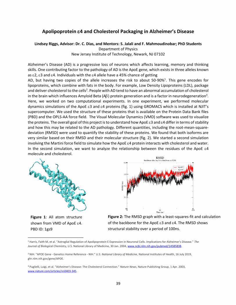
39
Apolipoprotein 4 and Cholesterol Packaging in Alzheimer’s Disease
Lindsey Riggs, Advisor: Dr. C. Dias, and Mentors: S. Jalali and F. Mahmoudinobar; PhD Students Department of Physics
New Jersey Institute of Technology, Newark, NJ 07102 Alzheimer’s Disease (AD) is a progressive loss of neurons which affects learning, memory and thinking skills. One contributing factor to the pathology of AD is the ApoE gene, which exists in three alleles known
as 2, 3 and 4. Individuals with the 4 allele have a 45% chance of getting AD, but having two copies of the allele increases the risk to about 50-90%1. This gene encodes for lipoproteins, which combine with fats in the body. For example, Low Density Lipoproteins (LDL), package and deliver cholesterol to the cells2. People with AD tend to have an abnormal accumulation of cholesterol
in the brain which influences Amyloid Beta (A) protein generation and is a factor in neurodegeneration3. Here, we worked on two computational experiments. In one experiment, we performed molecular
dynamics simulations of the ApoE 3 and 4 proteins (fig. 1) using GROMACS which is installed at NJIT’s supercomputer. We used the structure of these proteins that is available on the Protein Data Bank files (PBD) and the OPLS-AA force field. The Visual Molecular Dynamics (VMD) software was used to visualize
the proteins. The overall goal of this project is to understand how ApoE 3 and 4 differ in terms of stability and how this may be related to the AD pathology. Different quantities, including the root-mean-square-deviation (RMSD) were used to quantify the stability of these proteins. We found that both isoforms are very similar based on their RMSD and their molecular structure (fig. 2). We started a second simulation
involving the Martini force field to simulate how the ApoE 4 protein interacts with cholesterol and water.
In the second simulation, we want to analyze the relationship between the residues of the ApoE 4 molecule and cholesterol.
1 Harris, Faith M, et al. “Astroglial Regulation of Apolipoprotein E Expression in Neuronal Cells. Implications for Alzheimer's Disease.” The
Journal of Biological Chemistry, U.S. National Library of Medicine, 30 Jan. 2004, www.ncbi.nlm.nih.gov/pubmed/14585838.
2 NIH. “APOE Gene - Genetics Home Reference - NIH.” U.S. National Library of Medicine, National Institutes of Health, 16 July 2019,
ghr.nlm.nih.gov/gene/APOE.
3 Puglielli, Luigi, et al. “Alzheimer's Disease: The Cholesterol Connection.” Nature News, Nature Publishing Group, 1 Apr. 2003,
www.nature.com/articles/nn0403-345.
Figure 1: All atom structure
shown from VMD of ApoE 4.
PBD ID: 1gs9
Figure 2: The RMSD graph with a least-squares-fit and calculation
of the backbone for the ApoE 3 and 4. The RMSD shows
structural stability over a period of 100ns.
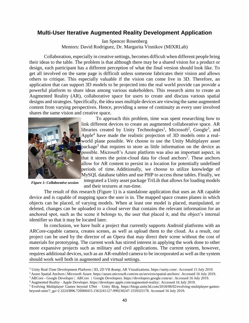
40
Multi-User Iterative Augmented Reality Development Application
Ian Spencer Rosenberg
Mentors: David Rodriguez, Dr. Margarita Vinnikov (MIXRLab)
Collaboration, especially in creative settings, becomes difficult when different people bring
their ideas to the table. The problem is that although there may be a shared vision for a product or
design, each participant has a different perception of what the final version should look like. To
get all involved on the same page is difficult unless someone fabricates their vision and allows
others to critique. This especially valuable if the vision can come live in 3D. Therefore, an
application that can support 3D models to be projected into the real world provide can provide a
powerful platform to share ideas among various stakeholders. This research aims to create an
Augmented Reality (AR), collaborative space for users to create and discuss various spatial
designs and strategies. Specifically, the idea uses multiple devices are viewing the same augmented
content from varying perspectives. Hence, providing a sense of continuity as every user involved
shares the same vision and creative space.
To approach this problem, time was spent researching how to
link different devices to create an augmented collaborative space. AR
libraries created by Unity Technologies1, Microsoft2, Google3, and
Apple4 have made the realistic projection of 3D models onto a real-
world plane possible. We choose to use the Unity Multiplayer asset
package5 that requires to store as little information on the device as
possible. Microsoft’s Azure platform was also an important aspect, in
that it stores the point-cloud data for cloud anchors2. These anchors
allow for AR content to persist in a location for potentially undefined
periods of time. Additionally, we choose to utilize knowledge of
MySQL database tables and use PHP to access those tables. Finally, we
integrated a Unity asset package TriLib that allows for loading models
and their textures at run-time.
The result of this research (Figure 1) is a standalone application that uses an AR capable
device and is capable of mapping space the user is in. The mapped space creates planes in which
objects can be placed, of varying models. When at least one model is placed, manipulated, or
deleted, changes can be uploaded to a cloud server that contains the relevant information for an
anchored spot, such as the scene it belongs to, the user that placed it, and the object’s internal
identifier so that it may be located later.
In conclusion, we have built a project that currently supports Android platforms with an
ARCore-capable camera, creates scenes, as well as upload them to the cloud. As a result, our
project can be used by the director of an Opera that may direct their scene without the cost of
materials for prototyping. The current work has stirred interest in applying the work done to other
more expansive projects such as military and civil applications. The current system, however,
requires additional devices, such as an AR-enabled camera to be incorporated as well as the system
should work well both in augmented and virtual settings.
1 Unity Real-Time Development Platform | 3D, 2D VR & AR Visualizations. https://unity.com/. Accessed 15 July 2019. 2 Azure Spatial Anchors | Microsoft Azure. https://azure.microsoft.com/en-us/services/spatial-anchors/. Accessed 16 July 2019. 3 ARCore - Google Developer | ARCore | Google Developers. https://developers.google.com/ar/. Accessed 16 July 2019. 4 Augmented Reality - Apple Developer. https://developer.apple.com/augmented-reality/. Accessed 16 July 2019. 5 Evolving Multiplayer Games beyond UNet – Unity Blog. https://blogs.unity3d.com/2018/08/02/evolving-multiplayer-games-
beyond-unet/?_ga=2.32243896.726900415.1563241127-999230247.1559325178. Accessed 16 July 2019.
Figure 1: Collaborative session
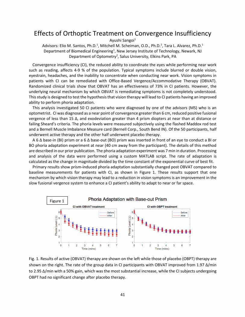
41
Effects of Orthoptic Treatment on Convergence Insufficiency Ayushi Sangoi1
Advisors: Elio M. Santos, Ph.D.1, Mitchell M. Scheiman, O.D., Ph.D.2, Tara L. Alvarez, Ph.D.1 Department of Biomedical Engineering1, New Jersey Institute of Technology, Newark, NJ
Department of Optometry2, Salus University, Elkins Park, PA
Convergence insufficiency (CI), the reduced ability to coordinate the eyes while performing near work such as reading, affects 4-9 % of the population. Typical symptoms include blurred or double vision, eyestrain, headaches, and the inability to concentrate when conducting near work. Vision symptoms in patients with CI can be remediated with Office-Based Vergence/Accommodative Therapy (OBVAT). Randomized clinical trials show that OBVAT has an effectiveness of 73% in CI patients. However, the underlying neural mechanism by which OBVAT is remediating symptoms is not completely understood. This study is designed to test the hypothesis that vision therapy will lead to CI patients having an improved ability to perform phoria adaptation.
This analysis investigated 50 CI patients who were diagnosed by one of the advisors (MS) who is an optometrist. CI was diagnosed as a near point of convergence greater than 6 cm, reduced positive fusional vergence of less than 15 Δ, and exodeviation greater than 4 prism diopters at near than at distance or failing Sheard’s criteria. The phoria levels were measured subjectively using the flashed Maddox rod test and a Bernell Muscle Imbalance Measure card (Bernell Corp., South Bend IN). Of the 50 participants, half underwent active therapy and the other half underwent placebo therapy.
A 6 Δ base-in (BI) prism or a 6 Δ base-out (BO) prism was inserted in front of an eye to conduct a BI or BO phoria adaptation experiment at near (40 cm away from the participant). The details of this method are described in our prior publication. The phoria adaptation experiment was 7 min in duration. Processing and analysis of the data were performed using a custom MATLAB script. The rate of adaptation is calculated as the change in magnitude divided by the time constant of the exponential curve of best fit.
Primary results show prism-induced phoria adaptation substantially changed post OBVAT compared to baseline measurements for patients with CI, as shown in Figure 1. These results support that one mechanism by which vision therapy may lead to a reduction in vision symptoms is an improvement in the slow fusional vergence system to enhance a CI patient’s ability to adapt to near or far space.
Fig. 1. Results of active (OBVAT) therapy are shown on the left while those of placebo (OBPT) therapy are
shown on the right. The rate of the group data in CI participants with OBVAT improved from 1.97 Δ/min
to 2.95 Δ/min with a 50% gain, which was the most substantial increase, while the CI subjects undergoing
OBPT had no significant change after placebo therapy.
Figure 1
42
Cholesterol Management Using Hydrogel for PCSK9 Inhibition
Sreya Sanyal
Advisor: Dr. Vivek Kumar Department of Biomedical Engineering
New Jersey Institute of Technology, Newark, N.J. 07102
Currently, most patients manage high LDL-c levels with statins, drugs that block the enzyme that produces cholesterol. However, statins are not universally effective, and may even result in adverse side effects, such as incident diabetes 1. This project seeks to investigate the efficacy of a new class of treatments, known as PCSK9 inhibitors. PCSK9 (proprotein convertase subtilisin/kexin type 9) is an enzyme that degrades cellular LDL receptors for LDL-c, as part of the body’s natural cholesterol metabolism. By binding to LDL receptors in conjunction with LDL-c, PCSK9 forces the LDL receptor to remain in the ‘active’ position so it is not recycled, and instead digested by lysosomes within liver cells. Pep2-8, a biological mimic identified by Zhang et al. 2, competitively binds to PCSK9, preventing it from degrading LDL receptors. The biological mimic is delivered via injected hydrogel—a 3 dimensional polymer that slowly releases Pep2-8 at precisely the targeted location. By inhibiting the activity of PCSK9, this therapy will allow the body to naturally lower high levels of LDL-c, and may be more effective and safe than current statin therapy.
The hydrogel EPCSK9 is initially a non-viscous liquid. When hydrogelation is induced, the peptide becomes viscous and exhibits shear-thinning. Substances with this property have cross-linking between polymers under normal conditions that results in a viscous gel, but lose this cross-linking when force is applied, resulting in a material that flows like a liquid. This enables the peptide to be easily loaded into and injected from the dosing syringe as liquid; once in the body, the gel regains its viscous properties and is slowly released as a Pep2-8 drug molecule.
The therapeutic effects of EPCK9 hydrogel were investigated using an in-vivo mouse model. Mice were fed high fat diets, and their blood was drawn daily over the course of 8 weeks. Levels of cholesterol will be determined from the drawn mouse serum using High Pressure Liquid Chromatography, a method separates the desired LDL-c molecules, and that will be paired with Mass Spectroscopy in order to accurately determine LDL-c levels throughout the in vivo study. Mouse livers were collected and analyzed for LDL receptor quantities using SDS-PAGE and Western Blot. SDS is a molecule used to coat proteins from the liver samples, and PAGE is an experimental procedure that allows the quantification of LDL receptors by running the coated proteins through a gel that separates them by mass. Western Blot was used to visualize the differing quantities of LDL receptors separated via SDS-PAGE by electrophoretically transferring the gel results to a membrane that was incubated with two different antibodies. The primary antibody binds to the LDL receptors, allowing quantification, and the secondary antibody binds to the primary, and fluoresces to allow visualization of the proteins of interest.
1. Spence, J. D., & Dresser, G. K. (2016). Overcoming Challenges With Statin Therapy. Journal of the American Heart Association, 5(1). doi:10.1161/jaha.115.002497
2. Lowe, J. (2013). Faculty of 1000 evaluation for Identification of a small peptide that inhibits PCSK9 binding to the low density lipoprotein receptor. F1000 - Post-publication Peer Review of the Biomedical Literature. doi:10.3410/f.718175417.793486831
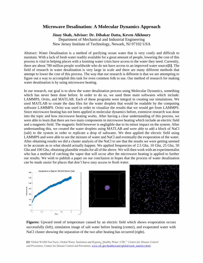
43
Microwave Desalination: A Molecular Dynamics Approach
Jinay Shah, Advisor: Dr. Dibakar Datta, Keven Alkhoury Department of Mechanical and Industrial Engineering
New Jersey Institute of Technology, Newark, NJ 07102 USA
Abstract: Water Desalination is a method of purifying ocean water that is very costly and difficult to
maintain. With a lack of fresh water readily available for a great amount of people, lowering the cost of this
process is vital in helping places with a looming water crisis have access to the water they need. Currently,
there are about 780 million people worldwide who do not have access to an improved water source[1]. The
field of research in water desalination is very large in scale and there are many different methods that
attempt to lower the cost of this process. The way that our research is different is that we are attempting to
figure out a way to accomplish this task for even common folk to use. Our method of research for making
water desalination is by using microwave heating.
In our research, our goal is to show the water desalination process using Molecular Dynamics, something
which has never been done before. In order to do so, we used three main softwares which include:
LAMMPS, Ovito, and MATLAB. Each of these programs were integral in creating our simulations. We
used MATLAB to create the data files for the water droplets that would be readable by the computing
software LAMMPS. Ovito was used in order to visualize the results that we would get from LAMMPS.
Since microwave heating has not been applied in molecular dynamics before, extensive research was done
into the topic and how microwave heating works. After having a clear understanding of this process, we
were able to learn that there are two main components in microwave heating which include an electric field
and a magnetic field. The magnetic field however is negligible due to its minor impact on the system. After
understanding this, we created the water droplets using MATLAB and were able to add a block of NaCl
(salt) to the system in order to replicate a drop of saltwater. We then applied the electric field using
LAMMPS and were able to see the mixture of water and NaCl and eventually the evaporation of the water.
After obtaining results we did a cluster analysis of the NaCl to see that the results we were getting seemed
to be accurate as to what should actually happen. We applied frequencies of 2.5 Ghz, 10 Ghz, 25 Ghz, 50
Ghz and 100 Ghz, obtaining plausible results for all of the above. We will then work with an experimentalist
who has a method of catching the vapor that will occur after the microwave heating is applied to further
our results. We wish to publish a paper on our conclusion in hopes that the process of water desalination
can be made easier for places that don’t have easy access to fresh water.
Figures: Upward trend of temperature caused by an electric field which shows evaporation occurs
successfully (left), simulation image of salt water before heating (center), and evaporated water with
NaCl cluster showing the separation of the two after heating has occurred (right).
[1] “Global WASH Fast Facts | Global Water, Sanitation and Hygiene | Healthy Water | CDC.” Centers for Disease Control
and Prevention, Centers for Disease Control and Prevention, www.cdc.gov/healthywater/global/wash_statistics.html.
44
Evaluating the Effect of Skull and Brain Stiffness on Shock Wave Propagation
in a Rodent Finite Element Model
Rahul Shah, Advisors: Molly Townsend, PhD and Namas Chandra, PhD
Center for Injury Biomechanics, Materials, and Medicine
Department of Biomedical Engineering
New Jersey Institute of Technology, Newark, NJ 07102
The injury mechanisms that characterize blast-induced traumatic brain injury (bTBI) have proven
to be difficult to investigate. Studies with humans are impractical and, therefore, animal models
are often used. Finite element (FE) modelling provides the necessary tools for identifying and
characterizing a rodent FE model, but requires accurate material models of the brain and skull.
These material models will be used in the simulation of bTBI exposures with varying
overpressures, the results of which will be compared to experimentally-measured intracranial
pressures (ICPs) to identify the best constitutive material models that can aid researchers in
selecting an appropriate constitutive model for the study of bTBI.
The rodent FE model used was developed from a micro computed tomography and magnetic
resonance imaging scan of a 10-week old male Sprague Dawley rat which was segmented into the
skin and soft tissues, skull, brain, and dura. The material properties for these biological tissues
were represented using constitutive material models found in literature. The brain constitutive
model was varied by parametrically varying the brain material properties, altering the elastic
modulus of the linear viscoelastic material between 1.314 kPa and 13.14 MPa. Additionally,
several viscoelastic models reported in literature were examined. Similarly, the linear elastic
constitutive material model of the skulls was parametrically varied, with the elastic moduli varying
between 100 MPa and 20 GPa with the Poisson’s ratio changing between 0.22 and 0.3. These
models were subjected to a mild primary blast wave of varying overpressures between the lower
end (70 kPa) and upper end (180 kPa) of blasts associated with mild bTBI. The influence of the
skull and brain material properties on the time varying pressure and strain profiles, including the
maximum ICP, the ICP profile, the pressure transmission pathway, and logarithmic strain values
within the rat head, was examined.
The baseline model, using the most common material models for the brain and skull, was shown
to accurately predict ICPs under a mild shock (70-180 kPa) by comparing simulated incident
pressure at the test location and simulated ICP with experimental measurements during a blast
exposure (n=4, p<0.01). Parametric variation of the brain material properties highlighted that the
ICP was largely insensitive to the viscoelastic brain constitutive model and highly dependent on
the elastic modulus. Varying the material properties of the skull increased maximum ICPs and
strain values in more compliant skulls. More compliant bone material models introduced
oscillations in the ICP profile, which are associated with increased flexure of the cranium. This is
evidenced by the increase in skull strain as pressure waves pass through.
The developed model demonstrates that a spike in ICP is exhibited closely following the incident
overpressure. This work identifies an appropriate constitutive model of a rat skull for use in FE
research through the cross-validation of simulation and experimental results. It further identifies
the effects of blast loading on rodents with varying skull and brain material properties, validated
with experimental ICP and strain measurements. Further work will seek to connect observed
mechanical loading pathways and patterns to microscale injury cascades.
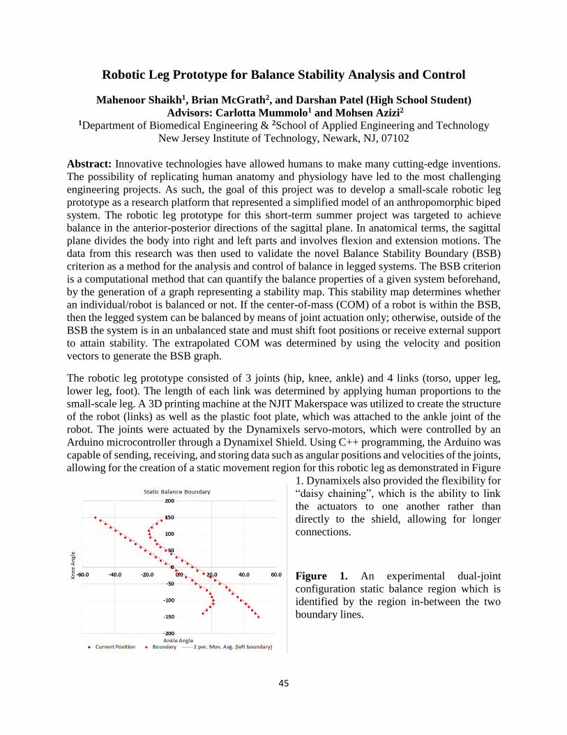
45
Robotic Leg Prototype for Balance Stability Analysis and Control
Mahenoor Shaikh1, Brian McGrath2, and Darshan Patel (High School Student)
Advisors: Carlotta Mummolo1 and Mohsen Azizi2 1Department of Biomedical Engineering & 2School of Applied Engineering and Technology
New Jersey Institute of Technology, Newark, NJ, 07102
Abstract: Innovative technologies have allowed humans to make many cutting-edge inventions.
The possibility of replicating human anatomy and physiology have led to the most challenging
engineering projects. As such, the goal of this project was to develop a small-scale robotic leg
prototype as a research platform that represented a simplified model of an anthropomorphic biped
system. The robotic leg prototype for this short-term summer project was targeted to achieve
balance in the anterior-posterior directions of the sagittal plane. In anatomical terms, the sagittal
plane divides the body into right and left parts and involves flexion and extension motions. The
data from this research was then used to validate the novel Balance Stability Boundary (BSB)
criterion as a method for the analysis and control of balance in legged systems. The BSB criterion
is a computational method that can quantify the balance properties of a given system beforehand,
by the generation of a graph representing a stability map. This stability map determines whether
an individual/robot is balanced or not. If the center-of-mass (COM) of a robot is within the BSB,
then the legged system can be balanced by means of joint actuation only; otherwise, outside of the
BSB the system is in an unbalanced state and must shift foot positions or receive external support
to attain stability. The extrapolated COM was determined by using the velocity and position
vectors to generate the BSB graph.
The robotic leg prototype consisted of 3 joints (hip, knee, ankle) and 4 links (torso, upper leg,
lower leg, foot). The length of each link was determined by applying human proportions to the
small-scale leg. A 3D printing machine at the NJIT Makerspace was utilized to create the structure
of the robot (links) as well as the plastic foot plate, which was attached to the ankle joint of the
robot. The joints were actuated by the Dynamixels servo-motors, which were controlled by an
Arduino microcontroller through a Dynamixel Shield. Using C++ programming, the Arduino was
capable of sending, receiving, and storing data such as angular positions and velocities of the joints,
allowing for the creation of a static movement region for this robotic leg as demonstrated in Figure
1. Dynamixels also provided the flexibility for
“daisy chaining”, which is the ability to link
the actuators to one another rather than
directly to the shield, allowing for longer
connections.
Figure 1. An experimental dual-joint
configuration static balance region which is
identified by the region in-between the two
boundary lines.
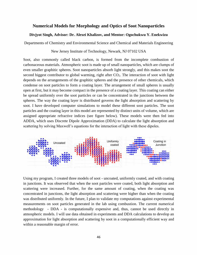
46
Numerical Models for Morphology and Optics of Soot Nanoparticles
Divjyot Singh, Advisor: Dr. Alexei Khalizov, and Mentor: Ogochukwu Y. Enekwizu
Departments of Chemistry and Environmental Science and Chemical and Materials Engineering
New Jersey Institute of Technology, Newark, NJ 07102 USA
Soot, also commonly called black carbon, is formed from the incomplete combustion of
carbonaceous materials. Atmospheric soot is made up of small nanoparticles, which are clumps of
even smaller graphitic spheres. Soot nanoparticles absorb light strongly, and this makes soot the
second biggest contributor to global warming, right after CO2. The interaction of soot with light
depends on the arrangements of the graphitic spheres and the presence of other chemicals, which
condense on soot particles to form a coating layer. The arrangement of small spheres is usually
open at first, but it may become compact in the presence of a coating layer. This coating can either
be spread uniformly over the soot particles or can be concentrated in the junctions between the
spheres. The way the coating layer is distributed governs the light absorption and scattering by
soot. I have developed computer simulations to model these different soot particles. The soot
particles and the coating layer in this model are represented by distinct units of volume, which are
assigned appropriate refractive indices (see figure below). These models were then fed into
ADDA, which uses Discrete Dipole Approximation (DDA) to calculate the light absorption and
scattering by solving Maxwell’s equations for the interaction of light with these dipoles.
Using my program, I created three models of soot - uncoated, uniformly coated, and with coating
in junctions. It was observed that when the soot particles were coated, both light absorption and
scattering were increased. Further, for the same amount of coating, when the coating was
concentrated in junctions, the light absorption and scattering were higher than when the coating
was distributed uniformly. In the future, I plan to validate my computations against experimental
measurements on soot particles generated in the lab using combustion. The current numerical
methodology - DDA - is computationally expensive and, thus, cannot be used directly in
atmospheric models. I will use data obtained in experiments and DDA calculations to develop an
approximation for light absorption and scattering by soot in a computationally efficient way and
within a reasonable margin of error.
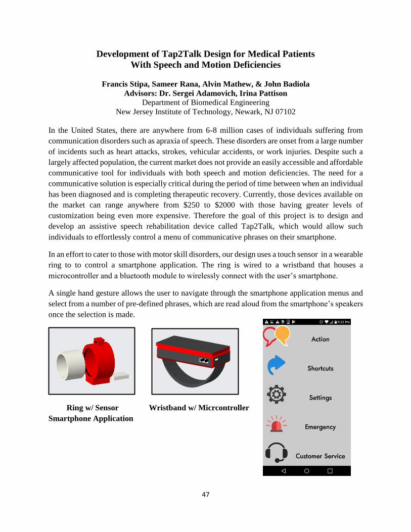
47
Development of Tap2Talk Design for Medical Patients
With Speech and Motion Deficiencies
Francis Stipa, Sameer Rana, Alvin Mathew, & John Badiola
Advisors: Dr. Sergei Adamovich, Irina Pattison
Department of Biomedical Engineering
New Jersey Institute of Technology, Newark, NJ 07102
In the United States, there are anywhere from 6-8 million cases of individuals suffering from
communication disorders such as apraxia of speech. These disorders are onset from a large number
of incidents such as heart attacks, strokes, vehicular accidents, or work injuries. Despite such a
largely affected population, the current market does not provide an easily accessible and affordable
communicative tool for individuals with both speech and motion deficiencies. The need for a
communicative solution is especially critical during the period of time between when an individual
has been diagnosed and is completing therapeutic recovery. Currently, those devices available on
the market can range anywhere from $250 to $2000 with those having greater levels of
customization being even more expensive. Therefore the goal of this project is to design and
develop an assistive speech rehabilitation device called Tap2Talk, which would allow such
individuals to effortlessly control a menu of communicative phrases on their smartphone.
In an effort to cater to those with motor skill disorders, our design uses a touch sensor in a wearable
ring to to control a smartphone application. The ring is wired to a wristband that houses a
microcontroller and a bluetooth module to wirelessly connect with the user’s smartphone.
A single hand gesture allows the user to navigate through the smartphone application menus and
select from a number of pre-defined phrases, which are read aloud from the smartphone’s speakers
once the selection is made.
Ring w/ Sensor Wristband w/ Micrcontroller
Smartphone Application
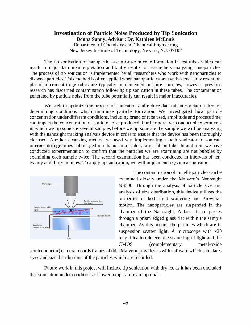
48
Investigation of Particle Noise Produced by Tip Sonication Donna Sunny, Advisor: Dr. Kathleen McEnnis
Department of Chemistry and Chemical Engineering
New Jersey Institute of Technology, Newark, N.J. 07102
The tip sonication of nanoparticles can cause micelle formation in test tubes which can
result in major data misinterpretation and faulty results for researchers analyzing nanoparticles.
The process of tip sonication is implemented by all researchers who work with nanoparticles to
disperse particles. This method is often applied when nanoparticles are synthesized. Low retention,
plastic microcentrifuge tubes are typically implemented to store particles, however, previous
research has discerned contamination following tip sonication in these tubes. The contamination
generated by particle noise from the tube potentially can result in major inaccuracies.
We seek to optimize the process of sonication and reduce data misinterpretation through
determining conditions which minimize particle formation. We investigated how particle
concentration under different conditions, including brand of tube used, amplitude and process time,
can impact the concentration of particle noise produced. Furthermore, we conducted experiments
in which we tip sonicate several samples before we tip sonicate the sample we will be analyzing
with the nanosight tracking analysis device in order to ensure that the device has been thoroughly
cleansed. Another cleansing method we used was implementing a bath sonicator to sonicate
microcentrifuge tubes submerged in ethanol in a sealed, large falcon tube. In addition, we have
conducted experimentation to confirm that the particles we are examining are not bubbles by
examining each sample twice. The second examination has been conducted in intervals of ten,
twenty and thirty minutes. To apply tip sonication, we will implement a Qsonica sonicator.
The contamination of micelle particles can be
examined closely under the Malvern’s Nanosight
NS300. Through the analysis of particle size and
analysis of size distribution, this device utilizes the
properties of both light scattering and Brownian
motion. The nanoparticles are suspended in the
chamber of the Nanosight. A laser beam passes
through a prism edged glass flat within the sample
chamber. As this occurs, the particles which are in
suspension scatter light. A microscope with x20
magnification detects the scattering of light and the
CMOS (complementary metal-oxide
semiconductor) camera records frames of this. Malvern provides us with software which calculates
sizes and size distributions of the particles which are recorded.
Future work in this project will include tip sonication with dry ice as it has been oncluded
that sonication under conditions of lower temperature are optimal.
49
Molecular Mechanism of the Circadian clock in Cyanobacteria
Neha Thati
Adviser: Dr. Yong-Ick Kim, Mentor: Manpreet Kaur
Department of Chemistry and Environmental Science
New Jersey Institute of Technology, Newark, NJ 07102
Abstract: The circadian clock is a 24-hour biological rhythm in the body which controls many
time-related activities, such as sleep and wakefulness. The simplest circadian clock is that of the
cyanobacterium; it is the only one that can be reconstituted in vitro, making it feasible to study.
The oscillator in cyanobacteria is composed of three proteins: KaiA, KaiB, and KaiC. Mixing these
proteins expressed from E. coli in a test tube can generate the circadian oscillation of KaiC
phosphorylation. The A-loop of KaiC is naturally buried in a network of hydrogen bonds. KaiA
activates KaiC phosphorylation by exposing the A-loop of KaiC. KaiB then sequesters KaiA by
binding to it, causing the A-loop conformation to return to its buried position and KaiC to
dephosphorylate. These processes of phosphorylation and dephosphorylation continue, generating
a 24-hour cycle. Previously published data have deduced that breaking the hydrogen bond network
can also expose the A-loop, causing KaiC to phosphorylate [1]. This experiment will determine if
KaiA truly needs to be present to promote KaiC phosphorylation. Confirming the role of this
protein can provide insight into the molecular mechanisms behind the circadian clock. One can
further understand how environmental time cues, such as temperature, the light-dark cycle, and
alterations of the biochemical system, can synchronize the 24-hour circadian cycle.
In this study, site-directed single mutations at the Glutamate-487 residue of the A-loop will be
performed to break the hydrogen bond network. E487S will modify this amino acid to Serine
and E487D to Aspartate. If a mutation that breaks the hydrogen bonds induces KaiC
phosphorylation, without the presence of KaiA, it can then be said that KaiA is not necessary to
initiate KaiC phosphorylation. Analysis of KaiC’s in vitro oscillation rhythm will confirm the
functional role of the protein KaiA. Understanding the molecular mechanism of the clock may
help uncover the links between biological clocks and medical problems related to the clock’s
disruption.
[1] Kim, Y.I., et al., (2008). The day/night switch in KaiC, a central oscillator component of the circadian clock of
cyanobacteria. Proc Natl Acad Sci U S A, 105(35): p. 12825-30.
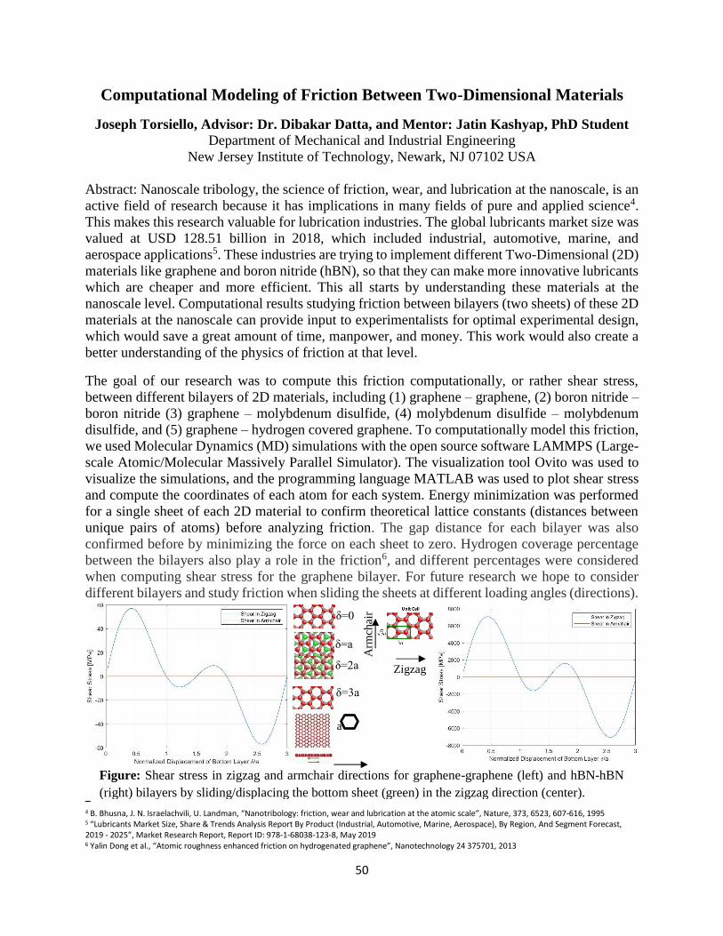
50
δ=0
δ=a
δ=2a
δ=3a
Zigzag
Arm
chair
a
Computational Modeling of Friction Between Two-Dimensional Materials
Joseph Torsiello, Advisor: Dr. Dibakar Datta, and Mentor: Jatin Kashyap, PhD Student Department of Mechanical and Industrial Engineering
New Jersey Institute of Technology, Newark, NJ 07102 USA
Abstract: Nanoscale tribology, the science of friction, wear, and lubrication at the nanoscale, is an
active field of research because it has implications in many fields of pure and applied science4.
This makes this research valuable for lubrication industries. The global lubricants market size was
valued at USD 128.51 billion in 2018, which included industrial, automotive, marine, and
aerospace applications5. These industries are trying to implement different Two-Dimensional (2D)
materials like graphene and boron nitride (hBN), so that they can make more innovative lubricants
which are cheaper and more efficient. This all starts by understanding these materials at the
nanoscale level. Computational results studying friction between bilayers (two sheets) of these 2D
materials at the nanoscale can provide input to experimentalists for optimal experimental design,
which would save a great amount of time, manpower, and money. This work would also create a
better understanding of the physics of friction at that level.
The goal of our research was to compute this friction computationally, or rather shear stress,
between different bilayers of 2D materials, including (1) graphene – graphene, (2) boron nitride –
boron nitride (3) graphene – molybdenum disulfide, (4) molybdenum disulfide – molybdenum
disulfide, and (5) graphene – hydrogen covered graphene. To computationally model this friction,
we used Molecular Dynamics (MD) simulations with the open source software LAMMPS (Large-
scale Atomic/Molecular Massively Parallel Simulator). The visualization tool Ovito was used to
visualize the simulations, and the programming language MATLAB was used to plot shear stress
and compute the coordinates of each atom for each system. Energy minimization was performed
for a single sheet of each 2D material to confirm theoretical lattice constants (distances between
unique pairs of atoms) before analyzing friction. The gap distance for each bilayer was also
confirmed before by minimizing the force on each sheet to zero. Hydrogen coverage percentage
between the bilayers also play a role in the friction6, and different percentages were considered
when computing shear stress for the graphene bilayer. For future research we hope to consider
different bilayers and study friction when sliding the sheets at different loading angles (directions).
4 B. Bhusna, J. N. Israelachvili, U. Landman, “Nanotribology: friction, wear and lubrication at the atomic scale”, Nature, 373, 6523, 607-616, 1995 5 “Lubricants Market Size, Share & Trends Analysis Report By Product (Industrial, Automotive, Marine, Aerospace), By Region, And Segment Forecast, 2019 - 2025”, Market Research Report, Report ID: 978-1-68038-123-8, May 2019 6 Yalin Dong et al., “Atomic roughness enhanced friction on hydrogenated graphene”, Nanotechnology 24 375701, 2013
Figure: Shear stress in zigzag and armchair directions for graphene-graphene (left) and hBN-hBN
(right) bilayers by sliding/displacing the bottom sheet (green) in the zigzag direction (center).
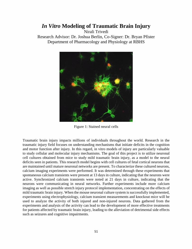
51
In Vitro Modeling of Traumatic Brain Injury Nirali Trivedi
Research Advisor: Dr. Joshua Berlin, Co-Signer: Dr. Bryan Pfister
Department of Pharmacology and Physiology at RBHS
Figure 1: Stained neural cells
Traumatic brain injury impacts millions of individuals throughout the world. Research in the
traumatic injury field focuses on understanding mechanisms that initiate deficits in the cognition
and motor function after injury. In this regard, in vitro models of injury are particularly valuable
to study cellular and molecular injury mechanisms. The goal of this project is to utilize neuronal
cell cultures obtained from mice to study mild traumatic brain injury, as a model to the neural
deficits seen in patients. This research model begins with cell cultures of fetal cortical neurons that
are maintained until mature neuronal networks are present. To characterize these cultured neurons,
calcium imaging experiments were performed. It was determined through these experiments that
spontaneous calcium transients were present at 13 days in culture, indicating that the neurons were
active. Synchronized calcium transients were noted at 21 days in culture, indicating that the
neurons were communicating in neural networks. Further experiments include more calcium
imaging as well as possible stretch injury protocol implementation, concentrating on the effects of
mild traumatic brain injury. When the mouse neuronal culture system is successfully implemented,
experiments using electrophysiology, calcium transient measurements and knockout mice will be
used to analyze the activity of both injured and non-injured neurons. Data gathered from the
experiments and analysis of the activity can lead to the development of more effective treatments
for patients affected by traumatic brain injury, leading to the alleviation of detrimental side effects
such as seizures and cognitive impairments.
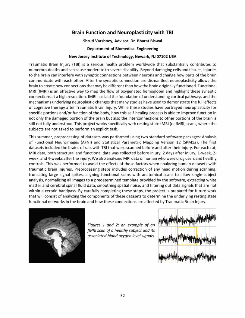
52
Brain Function and Neuroplasticity with TBI
Shruti Varshney, Advisor: Dr. Bharat Biswal
Department of Biomedical Engineering
New Jersey Institute of Technology, Newark, NJ 07102 USA
Traumatic Brain Injury (TBI) is a serious health problem worldwide that substantially contributes to numerous deaths and can cause moderate to severe disability. Beyond damaging cells and tissues, injuries to the brain can interfere with synaptic connections between neurons and change how parts of the brain communicate with each other. After the synaptic connection are dismantled, neuroplasticity allows the brain to create new connections that may be different than how the brain originally functioned. Functional MRI (fMRI) is an effective way to map the flow of oxygenated hemoglobin and highlight these synaptic connections at a high resolution. fMRI has laid the foundation of understanding cortical pathways and the mechanisms underlying neuroplastic changes that many studies have used to demonstrate the full effects of cognitive therapy after Traumatic Brain Injury. While these studies have portrayed neuroplasticity for specific portions and/or function of the body, how this self-healing process is able to improve function in not only the damaged portion of the brain but also the interconnections to other portions of the brain is still not fully understood. This project works specifically with resting state fMRI (rs-fMRI) scans, where the subjects are not asked to perform an explicit task.
This summer, preprocessing of datasets was performed using two standard software packages: Analysis of Functional Neuroimages (AFNI) and Statistical Parametric Mapping Version 12 (SPM12). The first datasets included the brains of rats with TBI that were scanned before and after their injury. For each rat, MRI data, both structural and functional data was collected before injury, 2 days after injury, 1-week, 2-week, and 4-weeks after the injury. We also analyzed MRI data of human who were drug users and healthy controls. This was performed to avoid the effects of those factors when analyzing human datasets with traumatic brain injuries. Preprocessing steps includes correction of any head motion during scanning, truncating large signal spikes, aligning functional scans with anatomical scans to allow single-subject analysis, normalizing all images to a predetermined template provided by the software, extracting white matter and cerebral spinal fluid data, smoothing spatial noise, and filtering out data signals that are not within a certain bandpass. By carefully completing these steps, the project is prepared for future work that will consist of analyzing the components of these datasets to determine the underlying resting state functional networks in the brain and how these connections are affected by Traumatic Brain Injury.
Figures 1 and 2: an example of an fMRI scan of a healthy subject and its associated blood oxygen level signals
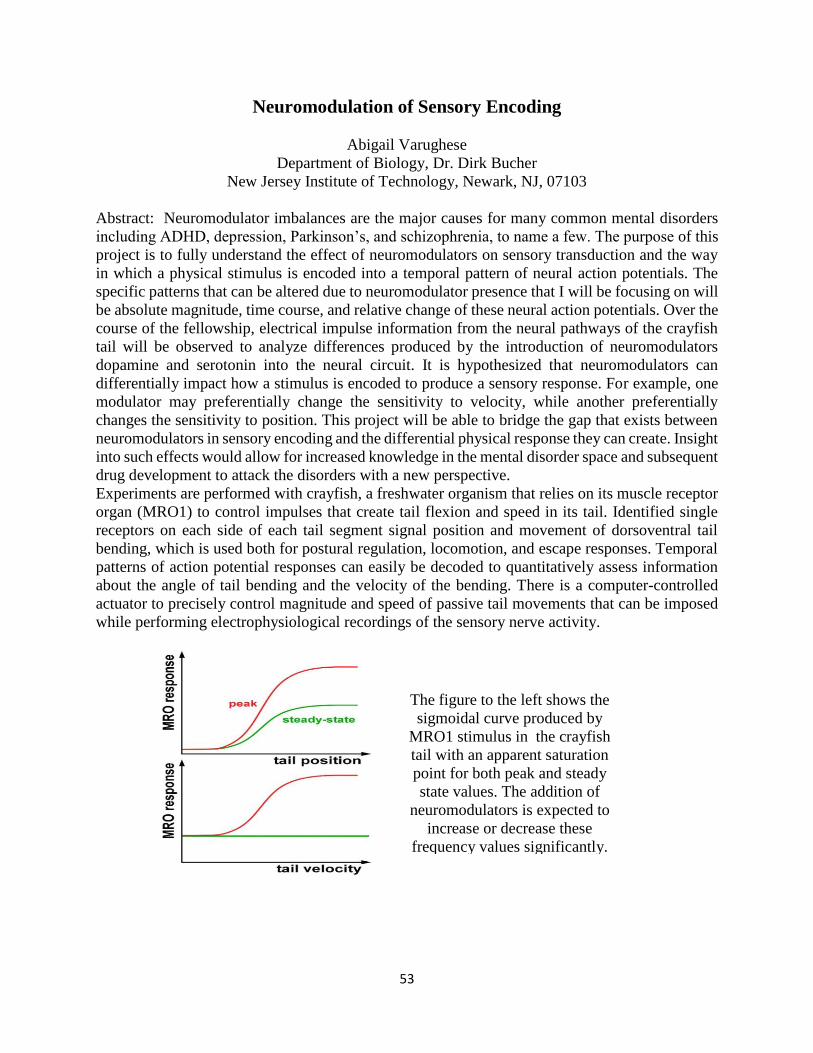
53
Neuromodulation of Sensory Encoding
Abigail Varughese
Department of Biology, Dr. Dirk Bucher
New Jersey Institute of Technology, Newark, NJ, 07103
Abstract: Neuromodulator imbalances are the major causes for many common mental disorders
including ADHD, depression, Parkinson’s, and schizophrenia, to name a few. The purpose of this
project is to fully understand the effect of neuromodulators on sensory transduction and the way
in which a physical stimulus is encoded into a temporal pattern of neural action potentials. The
specific patterns that can be altered due to neuromodulator presence that I will be focusing on will
be absolute magnitude, time course, and relative change of these neural action potentials. Over the
course of the fellowship, electrical impulse information from the neural pathways of the crayfish
tail will be observed to analyze differences produced by the introduction of neuromodulators
dopamine and serotonin into the neural circuit. It is hypothesized that neuromodulators can
differentially impact how a stimulus is encoded to produce a sensory response. For example, one
modulator may preferentially change the sensitivity to velocity, while another preferentially
changes the sensitivity to position. This project will be able to bridge the gap that exists between
neuromodulators in sensory encoding and the differential physical response they can create. Insight
into such effects would allow for increased knowledge in the mental disorder space and subsequent
drug development to attack the disorders with a new perspective.
Experiments are performed with crayfish, a freshwater organism that relies on its muscle receptor
organ (MRO1) to control impulses that create tail flexion and speed in its tail. Identified single
receptors on each side of each tail segment signal position and movement of dorsoventral tail
bending, which is used both for postural regulation, locomotion, and escape responses. Temporal
patterns of action potential responses can easily be decoded to quantitatively assess information
about the angle of tail bending and the velocity of the bending. There is a computer-controlled
actuator to precisely control magnitude and speed of passive tail movements that can be imposed
while performing electrophysiological recordings of the sensory nerve activity.
The figure to the left shows the
sigmoidal curve produced by
MRO1 stimulus in the crayfish
tail with an apparent saturation
point for both peak and steady
state values. The addition of
neuromodulators is expected to
increase or decrease these
frequency values significantly.
54
Blood Brain Barrier and Monocytes in Blast Traumatic Brain Injury
Geetasravya Vegunta
Faculty Mentors: Madhuvika Murugan and Namas Chandra
Department of Biomedical Engineering
Center for Injury Biomechanics, Materials, and Medicine
New Jersey Institute of Technology, Newark, NJ 07102
Blast traumatic brain injuries (blast TBI) are commonplace in the armed services. Exposure to
blast TBI can lead to chronic neuroinflammation, which then causes neurological and behavioral
deficits. Blast shockwaves can increase blood brain barrier (BBB) permeability, which allows
bloodborne particles and cells such as blood monocytes to enter the brain and release pro-
inflammatory neurochemicals, contributing to neural damage. In this study, we investigate whether
infiltration or blood monocytes is associated with increased BBB permeability after blast, and
whether depleting monocytes prevents behavioral changes observed after moderate blast TBI
exposure (180 kPa). To this end, we use double transgenic mice – CCR2 RFP/+:CX3CR1 GFP/+,
where infiltrating monocytes are tagged with RFP and resident immune cells (microglia) are
tagged with GFP. In order to investigate BBB integrity, mice were subjected to moderate blast and
the entry of intravenously administered Evans Blue dye in the blood, brain and liver were tested
at 0h (control), 4h, 1d, and 3d. To identify the contribution of infiltrated monocytes, clodronate
liposomes were administered to deplete total monocytes and behavioral assays including elevated
plus maze, open field test and novel object recognition test were performed at an acute (1d – 3d)
and chronic (28d – 30d) timepoint after blast TBI. DIL dye-containing liposomes were used as
vehicle control. In this study, we established a Evans Blue dosage for mice and a blood-serum
based validation method for efficient dye delivery. Spectrophotometric analysis of brain lysates
indicates that blast TBI resulted in a transient increase in BBB permeability at 4h but recovered to
control levels at 1d and 3d. Confocal imaging of brain tissue is underway and is expected to reveal
temporal and regional variations in BBB permeability changes within regions such as prefrontal
cortex, hippocampus and thalamus. The effect of clodronate-mediated monocyte depletion on
behavioral outcomes is inconclusive at present, warrants an increase in sample size, and is
currently under investigation. In summary, our study confirms that BBB permeability is altered
after blast TBI and may underlie the infiltration of monocytes and subsequent behavioral deficits.
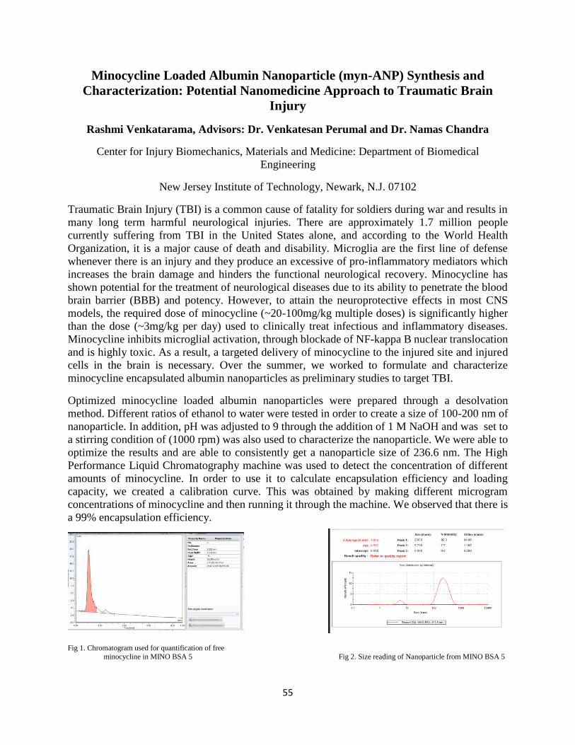
55
Minocycline Loaded Albumin Nanoparticle (myn-ANP) Synthesis and
Characterization: Potential Nanomedicine Approach to Traumatic Brain
Injury
Rashmi Venkatarama, Advisors: Dr. Venkatesan Perumal and Dr. Namas Chandra
Center for Injury Biomechanics, Materials and Medicine: Department of Biomedical
Engineering
New Jersey Institute of Technology, Newark, N.J. 07102
Traumatic Brain Injury (TBI) is a common cause of fatality for soldiers during war and results in
many long term harmful neurological injuries. There are approximately 1.7 million people
currently suffering from TBI in the United States alone, and according to the World Health
Organization, it is a major cause of death and disability. Microglia are the first line of defense
whenever there is an injury and they produce an excessive of pro-inflammatory mediators which
increases the brain damage and hinders the functional neurological recovery. Minocycline has
shown potential for the treatment of neurological diseases due to its ability to penetrate the blood
brain barrier (BBB) and potency. However, to attain the neuroprotective effects in most CNS
models, the required dose of minocycline (~20-100mg/kg multiple doses) is significantly higher
than the dose (~3mg/kg per day) used to clinically treat infectious and inflammatory diseases.
Minocycline inhibits microglial activation, through blockade of NF-kappa B nuclear translocation
and is highly toxic. As a result, a targeted delivery of minocycline to the injured site and injured
cells in the brain is necessary. Over the summer, we worked to formulate and characterize
minocycline encapsulated albumin nanoparticles as preliminary studies to target TBI.
Optimized minocycline loaded albumin nanoparticles were prepared through a desolvation
method. Different ratios of ethanol to water were tested in order to create a size of 100-200 nm of
nanoparticle. In addition, pH was adjusted to 9 through the addition of 1 M NaOH and was set to
a stirring condition of (1000 rpm) was also used to characterize the nanoparticle. We were able to
optimize the results and are able to consistently get a nanoparticle size of 236.6 nm. The High
Performance Liquid Chromatography machine was used to detect the concentration of different
amounts of minocycline. In order to use it to calculate encapsulation efficiency and loading
capacity, we created a calibration curve. This was obtained by making different microgram
concentrations of minocycline and then running it through the machine. We observed that there is
a 99% encapsulation efficiency.
Fig 1. Chromatogram used for quantification of free
minocycline in MINO BSA 5 Fig 2. Size reading of Nanoparticle from MINO BSA 5
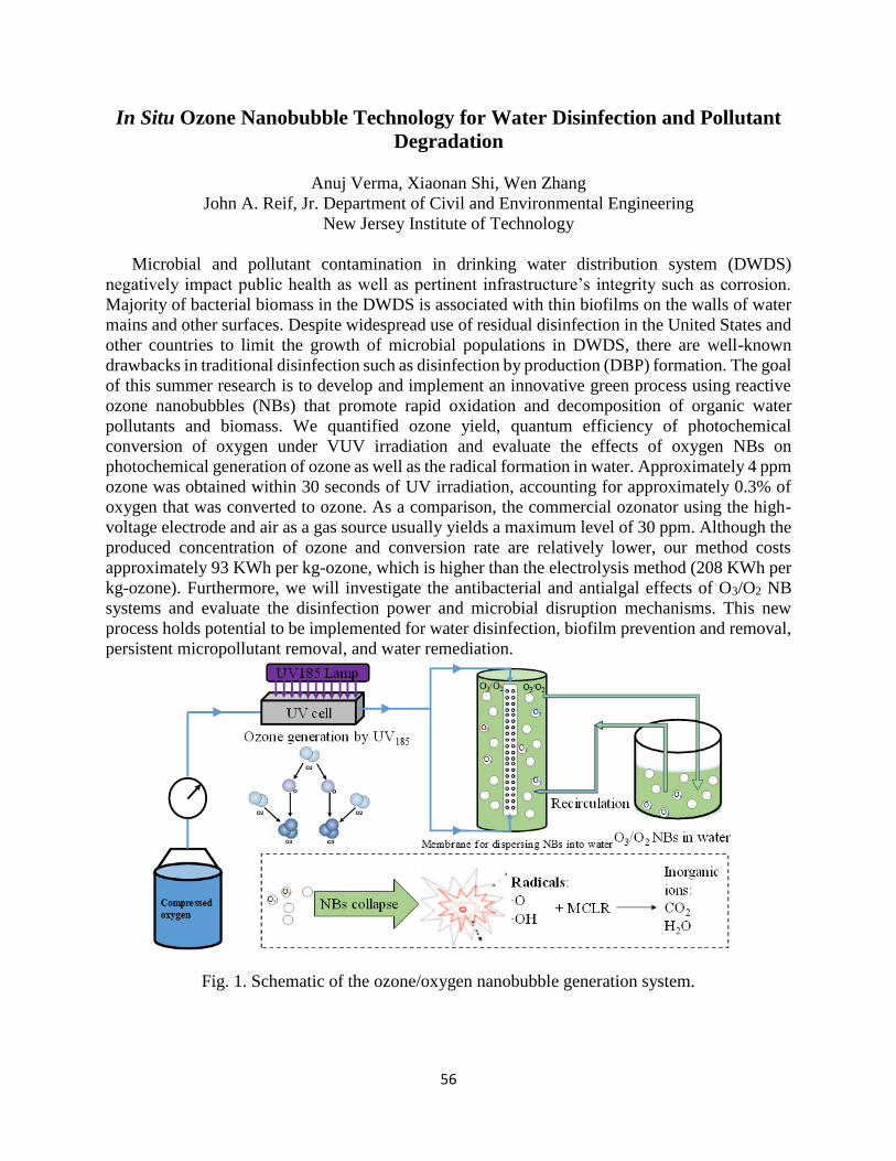
56
In Situ Ozone Nanobubble Technology for Water Disinfection and Pollutant
Degradation
Anuj Verma, Xiaonan Shi, Wen Zhang
John A. Reif, Jr. Department of Civil and Environmental Engineering
New Jersey Institute of Technology
Microbial and pollutant contamination in drinking water distribution system (DWDS)
negatively impact public health as well as pertinent infrastructure’s integrity such as corrosion.
Majority of bacterial biomass in the DWDS is associated with thin biofilms on the walls of water
mains and other surfaces. Despite widespread use of residual disinfection in the United States and
other countries to limit the growth of microbial populations in DWDS, there are well-known
drawbacks in traditional disinfection such as disinfection by production (DBP) formation. The goal
of this summer research is to develop and implement an innovative green process using reactive
ozone nanobubbles (NBs) that promote rapid oxidation and decomposition of organic water
pollutants and biomass. We quantified ozone yield, quantum efficiency of photochemical
conversion of oxygen under VUV irradiation and evaluate the effects of oxygen NBs on
photochemical generation of ozone as well as the radical formation in water. Approximately 4 ppm
ozone was obtained within 30 seconds of UV irradiation, accounting for approximately 0.3% of
oxygen that was converted to ozone. As a comparison, the commercial ozonator using the high-
voltage electrode and air as a gas source usually yields a maximum level of 30 ppm. Although the
produced concentration of ozone and conversion rate are relatively lower, our method costs
approximately 93 KWh per kg-ozone, which is higher than the electrolysis method (208 KWh per
kg-ozone). Furthermore, we will investigate the antibacterial and antialgal effects of O3/O2 NB
systems and evaluate the disinfection power and microbial disruption mechanisms. This new
process holds potential to be implemented for water disinfection, biofilm prevention and removal,
persistent micropollutant removal, and water remediation.
Fig. 1. Schematic of the ozone/oxygen nanobubble generation system.
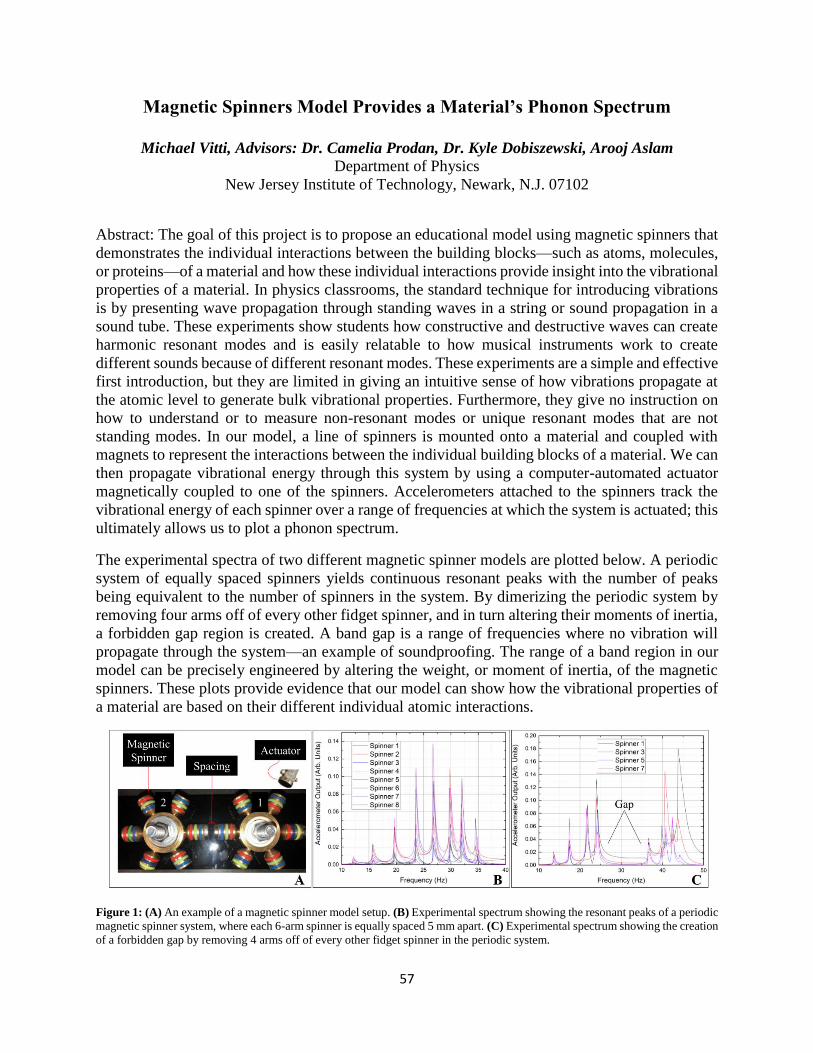
57
Magnetic Spinners Model Provides a Material’s Phonon Spectrum
Michael Vitti, Advisors: Dr. Camelia Prodan, Dr. Kyle Dobiszewski, Arooj Aslam
Department of Physics
New Jersey Institute of Technology, Newark, N.J. 07102
Abstract: The goal of this project is to propose an educational model using magnetic spinners that
demonstrates the individual interactions between the building blocks—such as atoms, molecules,
or proteins—of a material and how these individual interactions provide insight into the vibrational
properties of a material. In physics classrooms, the standard technique for introducing vibrations
is by presenting wave propagation through standing waves in a string or sound propagation in a
sound tube. These experiments show students how constructive and destructive waves can create
harmonic resonant modes and is easily relatable to how musical instruments work to create
different sounds because of different resonant modes. These experiments are a simple and effective
first introduction, but they are limited in giving an intuitive sense of how vibrations propagate at
the atomic level to generate bulk vibrational properties. Furthermore, they give no instruction on
how to understand or to measure non-resonant modes or unique resonant modes that are not
standing modes. In our model, a line of spinners is mounted onto a material and coupled with
magnets to represent the interactions between the individual building blocks of a material. We can
then propagate vibrational energy through this system by using a computer-automated actuator
magnetically coupled to one of the spinners. Accelerometers attached to the spinners track the
vibrational energy of each spinner over a range of frequencies at which the system is actuated; this
ultimately allows us to plot a phonon spectrum.
The experimental spectra of two different magnetic spinner models are plotted below. A periodic
system of equally spaced spinners yields continuous resonant peaks with the number of peaks
being equivalent to the number of spinners in the system. By dimerizing the periodic system by
removing four arms off of every other fidget spinner, and in turn altering their moments of inertia,
a forbidden gap region is created. A band gap is a range of frequencies where no vibration will
propagate through the system—an example of soundproofing. The range of a band region in our
model can be precisely engineered by altering the weight, or moment of inertia, of the magnetic
spinners. These plots provide evidence that our model can show how the vibrational properties of
a material are based on their different individual atomic interactions.
Figure 1: (A) An example of a magnetic spinner model setup. (B) Experimental spectrum showing the resonant peaks of a periodic
magnetic spinner system, where each 6-arm spinner is equally spaced 5 mm apart. (C) Experimental spectrum showing the creation
of a forbidden gap by removing 4 arms off of every other fidget spinner in the periodic system.
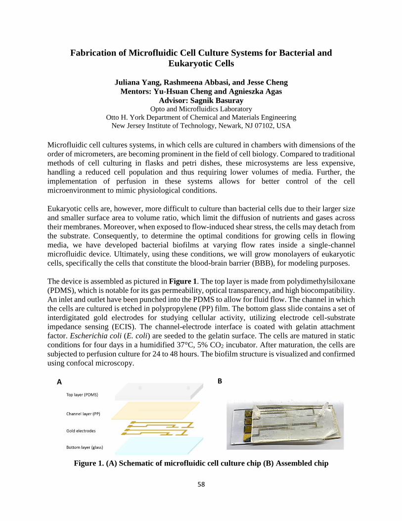
58
Fabrication of Microfluidic Cell Culture Systems for Bacterial and
Eukaryotic Cells
Juliana Yang, Rashmeena Abbasi, and Jesse Cheng
Mentors: Yu-Hsuan Cheng and Agnieszka Agas
Advisor: Sagnik Basuray Opto and Microfluidics Laboratory
Otto H. York Department of Chemical and Materials Engineering
New Jersey Institute of Technology, Newark, NJ 07102, USA
Microfluidic cell cultures systems, in which cells are cultured in chambers with dimensions of the
order of micrometers, are becoming prominent in the field of cell biology. Compared to traditional
methods of cell culturing in flasks and petri dishes, these microsystems are less expensive,
handling a reduced cell population and thus requiring lower volumes of media. Further, the
implementation of perfusion in these systems allows for better control of the cell
microenvironment to mimic physiological conditions.
Eukaryotic cells are, however, more difficult to culture than bacterial cells due to their larger size
and smaller surface area to volume ratio, which limit the diffusion of nutrients and gases across
their membranes. Moreover, when exposed to flow-induced shear stress, the cells may detach from
the substrate. Consequently, to determine the optimal conditions for growing cells in flowing
media, we have developed bacterial biofilms at varying flow rates inside a single-channel
microfluidic device. Ultimately, using these conditions, we will grow monolayers of eukaryotic
cells, specifically the cells that constitute the blood-brain barrier (BBB), for modeling purposes.
The device is assembled as pictured in Figure 1. The top layer is made from polydimethylsiloxane
(PDMS), which is notable for its gas permeability, optical transparency, and high biocompatibility.
An inlet and outlet have been punched into the PDMS to allow for fluid flow. The channel in which
the cells are cultured is etched in polypropylene (PP) film. The bottom glass slide contains a set of
interdigitated gold electrodes for studying cellular activity, utilizing electrode cell-substrate
impedance sensing (ECIS). The channel-electrode interface is coated with gelatin attachment
factor. Escherichia coli (E. coli) are seeded to the gelatin surface. The cells are matured in static
conditions for four days in a humidified 37°C, 5% CO2 incubator. After maturation, the cells are
subjected to perfusion culture for 24 to 48 hours. The biofilm structure is visualized and confirmed
using confocal microscopy.
Figure 1. (A) Schematic of microfluidic cell culture chip (B) Assembled chip
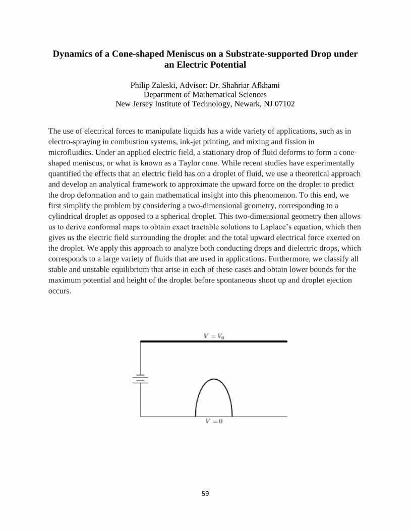
59
Dynamics of a Cone-shaped Meniscus on a Substrate-supported Drop under
an Electric Potential
Philip Zaleski, Advisor: Dr. Shahriar Afkhami
Department of Mathematical Sciences
New Jersey Institute of Technology, Newark, NJ 07102
The use of electrical forces to manipulate liquids has a wide variety of applications, such as in
electro-spraying in combustion systems, ink-jet printing, and mixing and fission in
microfluidics. Under an applied electric field, a stationary drop of fluid deforms to form a cone-
shaped meniscus, or what is known as a Taylor cone. While recent studies have experimentally
quantified the effects that an electric field has on a droplet of fluid, we use a theoretical approach
and develop an analytical framework to approximate the upward force on the droplet to predict
the drop deformation and to gain mathematical insight into this phenomenon. To this end, we
first simplify the problem by considering a two-dimensional geometry, corresponding to a
cylindrical droplet as opposed to a spherical droplet. This two-dimensional geometry then allows
us to derive conformal maps to obtain exact tractable solutions to Laplace’s equation, which then
gives us the electric field surrounding the droplet and the total upward electrical force exerted on
the droplet. We apply this approach to analyze both conducting drops and dielectric drops, which
corresponds to a large variety of fluids that are used in applications. Furthermore, we classify all
stable and unstable equilibrium that arise in each of these cases and obtain lower bounds for the
maximum potential and height of the droplet before spontaneous shoot up and droplet ejection
occurs.

60
61
Effect of Tank Bottom Shapes on Power Dissipation and 𝑵𝒋𝒔 in Stirred Vessels
Under Different Baffling Configurations
Sara A Abdelhamid, Advisor: Dr. Piero Armenante
Department of Chemical and Materials Engineering
New Jersey Institute of Technology, Newark, NJ 07102
Mechanically stirred mixing vessels are widely used in the chemical, biochemical, food, and other related
industries to homogenize the liquid content of the vessel and any phase dispersed in it, such as finely divided
solid particles. The power, P, dissipated by the impeller in these systems and the minimum agitation speed,
𝑁𝑗𝑠, to just suspend solid particles in the liquid are two of the most critical parameters for scaling up mixing
processes from laboratory scale to industrial scale. Both P (and its non-dimensional value 𝑃𝑜) and 𝑁𝑗𝑠
depend on several factors including type of impeller, its diameter, vessel geometry, and the dynamic
conditions at which the mixing is operated (agitation speed). Although most correlations available in the
literature for Po and 𝑁𝑗𝑠 were obtained at the laboratory scale using flat-bottom vessel, vessels with
hemispherical or torispherical bottoms are actually used in industrial applications. The effect of the shape
of the vessel bottom on 𝑃𝑜 and 𝑁𝑗𝑠 has not been investigated so far and is the focus of this work.
In agitated vessels, the degree of solid suspension is classified into three levels: on-bottom motion, complete
off-bottom suspension, and uniform suspension. Enough agitation is important to completely suspend the
solids off the vessel bottom. Below this needed agitation, the total surface area of the solid suspended within
the liquid is not completely or efficiently utilized. Therefore, it is important to be able to determine the
impeller agitation speed denoted by 𝑁𝑗𝑠, at which the just suspended state is achieved. Although 𝑁𝑗𝑠 is
critical to many industrial mixing processes, little information is available in the literature on the effect of
the tank bottom on 𝑁𝑗𝑠. Therefore, the main objective of this part of the work was to determine 𝑁𝑗𝑠 and the
power dissipation at these measured 𝑁𝑗𝑠 values for different tank bottom shapes experimentally. By using
the newly developed method in the Mixing Lab at NJIT, the area covered by the unsuspended solids was
measured at increasing agitation speeds, starting at low speeds below 𝑁𝑗𝑠 and increased until all particles
became suspended; thus, 𝑁𝑗𝑠 could be determined.
62
Raspberry Pi as FSO Transceiver using UART Communication for Drone-
Assisted Networking
Ehtesham Ahmed Advisor: Dr. Nirwan Ansari, Mentors: S. Zhang, W. Liu (Ph.D. Candidates)
Department of Electrical and Computer Engineering
New Jersey Institute of Technology, Newark, NJ 07102
Recent advancements in drone technology have created the unique opportunity to deploy cellular
networks on a platform of portable drones. Drone-mounted base-stations (DBSs) can
autonomously move in space and can fly at a relatively higher altitude than Macro Base Stations
(MBSs). It can improve the coverage and capacity of the networks to serve immense user demands
due to the potential Line-of-Sight (LoS) connection between the user and the DBSs. Nowadays, a
large number of institutions and research facilities are conducting research on Unmanned aerial
vehicle (UAV)-assisted networking to improve its reliability and flexibility. In traditional UAV-
assisted networks, access node and DBS employ FSO Transceiver or RF Transceiver to establish
the backhaul link (the link between DBS and MBS). However, in research labs, Universal Software
Radio Peripheral (USRP), which is costly as well as heavy, is commonly used as an RF transceiver
to establish the backhaul link. The aim of this research is to establish the backhaul link using the
Raspberry-Pi as it is inexpensive, light-weighted and capable of transmitting data at relatively high
speed. Raspberry-Pi has a built-in Universal Asynchronous Receiver/Transmitter (UART) which
is a serial communication protocol that transfers data bit by bit. Raspberry-Pi UART transmitter
uses a laser to convert the data into the optical signal and the UART receiver uses the laser detector
to convert the optical signal into data. Future research will concentrate on using Serial Peripheral
Interface (SPI) to achieve a higher data rate.
63
Examination of water stress on the morphological evolution of Capsella bursa-pastoris
Gabby Amparo, Advisor: Dr. Daniel Bunker, Mentor: Rebecca Panko
Department of Biology
New Jersey Institute of Technology, Newark, NJ 07102
As urbanization increases at a significant rate around the world, it is important for humans to
understand how this rapid growth can affect the environments they develop. Previous research
shows that the evolution of plants and animals can be affected by urbanization occurring in their
ecosystems. Such effects can vary from change in mating calls to physiological change. The effects
of urbanization on morphology of the common herb Capsella bursa-pastoris also known as
“shepherd's purse” will be examined. Capsella bursa-pastoris is often found in areas that
experience high abiotic stress. This plant has short generation times, self-pollinates, and is an
abundant species. That being said, Capsella bursa-pastoris is an ideal model species for this study.
The main abiotic stressor examined was water stress, specifically flood and drought conditions. In
urban environments with a majority of concrete streets, it is common for the soil to have low
porosity due to being surrounded by concrete. That being said, flooding becomes a recurring issue
in such an environment. Plants in urban areas may also experience extended periods of drought
due to low water availability. These factors are important to understand in order to better
understand our relationship. Germinated seeds of different populations were grown under artificial
conditions (for 10 weeks) and experienced flood and drought treatments. After these treatments,
various morphological traits such as leaf shape (or leaf surface area), stomatal density, and
pubescence were observed. These traits will determine changes in the morphology of shepherd's
purse. We are looking to prove that urban plants have evolved to better adapt themselves for their
constantly changing environment. It is expected for urban plants to display a lobed shape with
smaller surface area to aid in water retention. Urban plants display dense (enlarged) stomata on
their leaves to decrease the amount of water vapor that exits the leaves through transpiration. The
urban plants show more trichome on their leaves to aid them in the capturing of water or water
vapor.
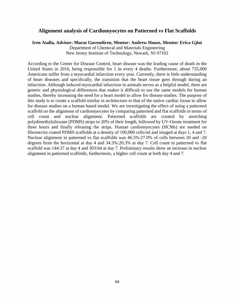
64
Alignment analysis of Cardiomyocytes on Patterned vs Flat Scaffolds
Iren Atalla, Adviser: Murat Guvendiren, Mentor: Andrew House, Mentee: Erica Gjini
Department of Chemical and Materials Engineering
New Jersey Institute of Technology, Newark, NJ 07102
According to the Center for Disease Control, heart disease was the leading cause of death in the
United States in 2016, being responsible for 1 in every 4 deaths. Furthermore, about 735,000
Americans suffer from a myocardial infarction every year. Currently, there is little understanding
of heart diseases and specifically, the transition that the heart tissue goes through during an
infarction. Although induced myocardial infarction in animals serves as a helpful model, there are
genetic and physiological differences that makes it difficult to use the same models for human
studies, thereby increasing the need for a heart model to allow for disease studies. The purpose of
this study is to create a scaffold similar in architecture to that of the native cardiac tissue to allow
for disease studies on a human based model. We are investigating the effect of using a patterned
scaffold on the alignment of cardiomyocytes by comparing patterned and flat scaffolds in terms of
cell count and nuclear alignment. Patterned scaffolds are created by stretching
polydimethylsiloxane (PDMS) strips to 20% of their length, followed by UV-Ozone treatment for
three hours and finally releasing the strips. Human cardiomyocytes (HCMs) are seeded on
fibronectin coated PDMS scaffolds at a density of 100,000 cells/ml and imaged at days 1, 4 and 7.
Nuclear alignment in patterned vs flat scaffolds was 46.5%:27.0% of cells between 20 and -20
degrees from the horizontal at day 4 and 34.3%:20.3% at day 7. Cell count in patterned vs flat
scaffold was 144:37 at day 4 and 303:64 at day 7. Preliminary results show an increase in nuclear
alignment in patterned scaffolds, furthermore, a higher cell count at both day 4 and 7.
65
Investigation of interlayer strength of 3D-printed Polymers
David Bushiri, Advisor: Dr. Murat Guvendiren, Mentor: Chya- Yan Liaw
Department of Biomedical Engineering
New Jersey Institute of Technology, Newark, NJ 07102
FDM (Fused deposition modeling) 3D printing is a manufacturing process which enables the
creation of complex and customized objects in a layer-by-layer fashion. Due to its versatility, FDM
technology has been increasingly adopted by many industries. Although FDM-based products are
widely used, the parts usually possess anisotropic and weaker mechanical properties compared to
the parts made by conventional manufacturing methods such as injection molding. Therefore,
understanding and improving the mechanical properties of FDM-printed products are crucial for
broadening the applications of 3D printing technology. Previous studies conducted on 3D printing
have focused on mechanical strength as a function of variety of processing parameters. However,
only a few studies have been conducted on the effects of processing parameters on interlayer
bonding strength. Interlayer bonding strength is important because it can determine how well the
design can withstand tension, compression, and torsion forces before breaking. In this study, we
will use DOE (design of experiments) for analysis and performance screening tests, to identify
important factors that influence the interlayer strength of ABS-printed parts. The approach of this
study is to focus on four important process parameters such as layer height, layer time, temperature,
and speed on. The layers of the built-up materials were stacked in the Z-direction during the
printing process. Mechanical tests including tensile test (ASTM D638 type V) and three-point
bending test (ISO 178) will be performed to study the effect of the above-mentioned processing
parameters on interlayer strength and to understand the feasibility of the use of these two tests in
assessing interlayer strength of the 3d printed objects.
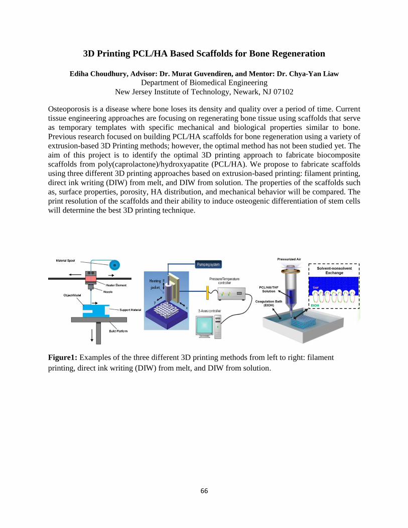
66
3D Printing PCL/HA Based Scaffolds for Bone Regeneration
Ediha Choudhury, Advisor: Dr. Murat Guvendiren, and Mentor: Dr. Chya-Yan Liaw
Department of Biomedical Engineering New Jersey Institute of Technology, Newark, NJ 07102
Osteoporosis is a disease where bone loses its density and quality over a period of time. Current
tissue engineering approaches are focusing on regenerating bone tissue using scaffolds that serve
as temporary templates with specific mechanical and biological properties similar to bone.
Previous research focused on building PCL/HA scaffolds for bone regeneration using a variety of
extrusion-based 3D Printing methods; however, the optimal method has not been studied yet. The
aim of this project is to identify the optimal 3D printing approach to fabricate biocomposite
scaffolds from poly(caprolactone)/hydroxyapatite (PCL/HA). We propose to fabricate scaffolds
using three different 3D printing approaches based on extrusion-based printing: filament printing,
direct ink writing (DIW) from melt, and DIW from solution. The properties of the scaffolds such
as, surface properties, porosity, HA distribution, and mechanical behavior will be compared. The
print resolution of the scaffolds and their ability to induce osteogenic differentiation of stem cells
will determine the best 3D printing technique.
Figure1: Examples of the three different 3D printing methods from left to right: filament
printing, direct ink writing (DIW) from melt, and DIW from solution.
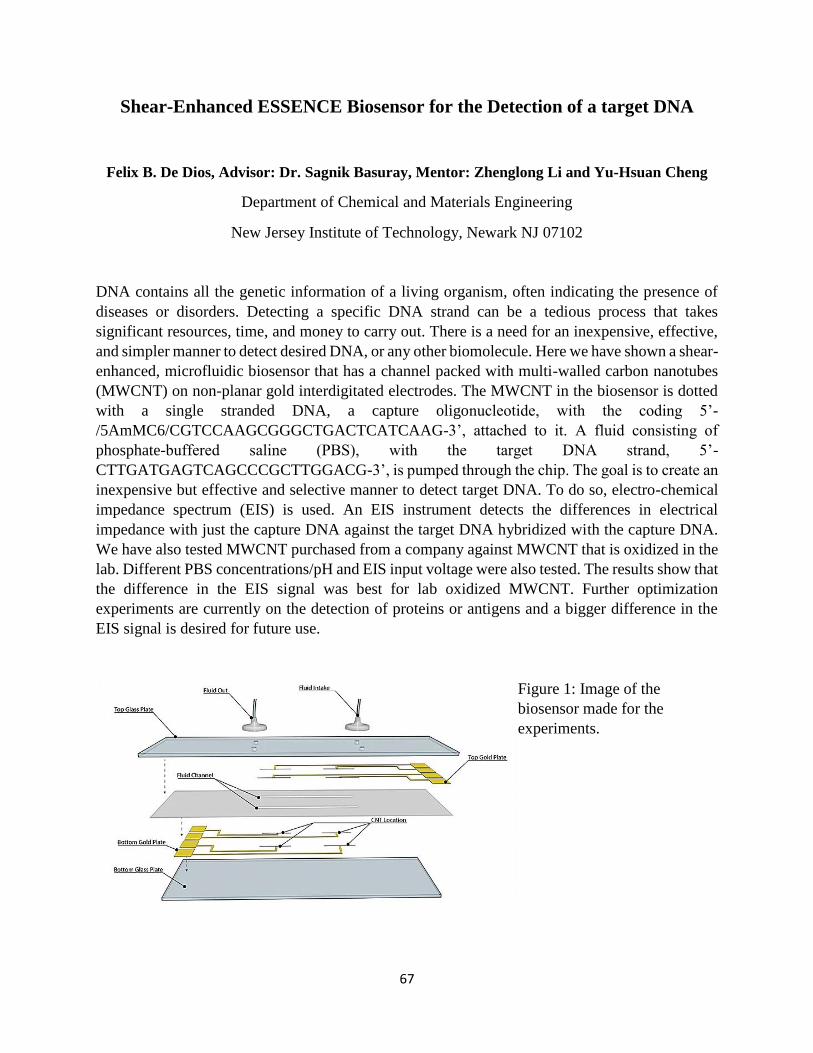
67
Shear-Enhanced ESSENCE Biosensor for the Detection of a target DNA
Felix B. De Dios, Advisor: Dr. Sagnik Basuray, Mentor: Zhenglong Li and Yu-Hsuan Cheng
Department of Chemical and Materials Engineering
New Jersey Institute of Technology, Newark NJ 07102
DNA contains all the genetic information of a living organism, often indicating the presence of
diseases or disorders. Detecting a specific DNA strand can be a tedious process that takes
significant resources, time, and money to carry out. There is a need for an inexpensive, effective,
and simpler manner to detect desired DNA, or any other biomolecule. Here we have shown a shear-
enhanced, microfluidic biosensor that has a channel packed with multi-walled carbon nanotubes
(MWCNT) on non-planar gold interdigitated electrodes. The MWCNT in the biosensor is dotted
with a single stranded DNA, a capture oligonucleotide, with the coding 5’-
/5AmMC6/CGTCCAAGCGGGCTGACTCATCAAG-3’, attached to it. A fluid consisting of
phosphate-buffered saline (PBS), with the target DNA strand, 5’-
CTTGATGAGTCAGCCCGCTTGGACG-3’, is pumped through the chip. The goal is to create an
inexpensive but effective and selective manner to detect target DNA. To do so, electro-chemical
impedance spectrum (EIS) is used. An EIS instrument detects the differences in electrical
impedance with just the capture DNA against the target DNA hybridized with the capture DNA.
We have also tested MWCNT purchased from a company against MWCNT that is oxidized in the
lab. Different PBS concentrations/pH and EIS input voltage were also tested. The results show that
the difference in the EIS signal was best for lab oxidized MWCNT. Further optimization
experiments are currently on the detection of proteins or antigens and a bigger difference in the
EIS signal is desired for future use.
Figure 1: Image of the
biosensor made for the
experiments.
68
Design of a Cable Driven Exoskeleton for Hand Rehabilitation Post Stroke
Cruz Donato, Advisors: Sergei Adamovich and Mentor: Ashley Mont
Department of Biomedical Engineering
New Jersey Institute of Technology, Newark, N.J. 07102
Stroke is one of the leading causes of disability in the United States, specifically due to
hemiparesis, the paralyzation of one side of the body. Post-stroke rehabilitation methods often use
repetitive tasks replicating activities of daily living to help stroke patients regain motor function.
However, most inpatient therapy focuses on treating lower extremity movement rather than upper
extremity movement, and as a result stroke survivors often struggle with distal upper extremity
movement. Robotic devices have been created to help in the rehabilitation process. As effective as
these devices have been, the designs could be improved to decrease weight, amount of material,
and possibly, cost. These devices could also work with software at home that would administer
tasks to the patients such as games in order to provide more positive reinforcement and more
individualized therapy. In this research, the design of a cable-driven, hand exoskeleton for stroke
rehabilitation is described. Individual parts of the exoskeleton were modeled on Creo Parametric
5.0, and measured to fit on to a healthy individual in the laboratory. After the parts were modeled
on Creo, they were 3D printed in Acrylonitrile butadiene styrene (ABS) to prototype, and, later
on, printed in NinjaFlex, a more flexible material that would be more appropriate for fit and
comfortability. Actuonix linear actuators were used to extend the index finger and thumb attached
to the actuators by a cable. The linear actuators were programmed using Arduino and a motor
driver to retract for one second and extend for one second. This provided cyclic stretching of the
fingers, replicating an effective stretching paradigm done in a previous study. Fishing line was
used as the cables connecting the linear actuators and corresponding digits. In the future, the
exoskeleton is expected to administer cyclic stretching to the digits, as well as be incorporated in
a virtual reality system developed previously in the lab.
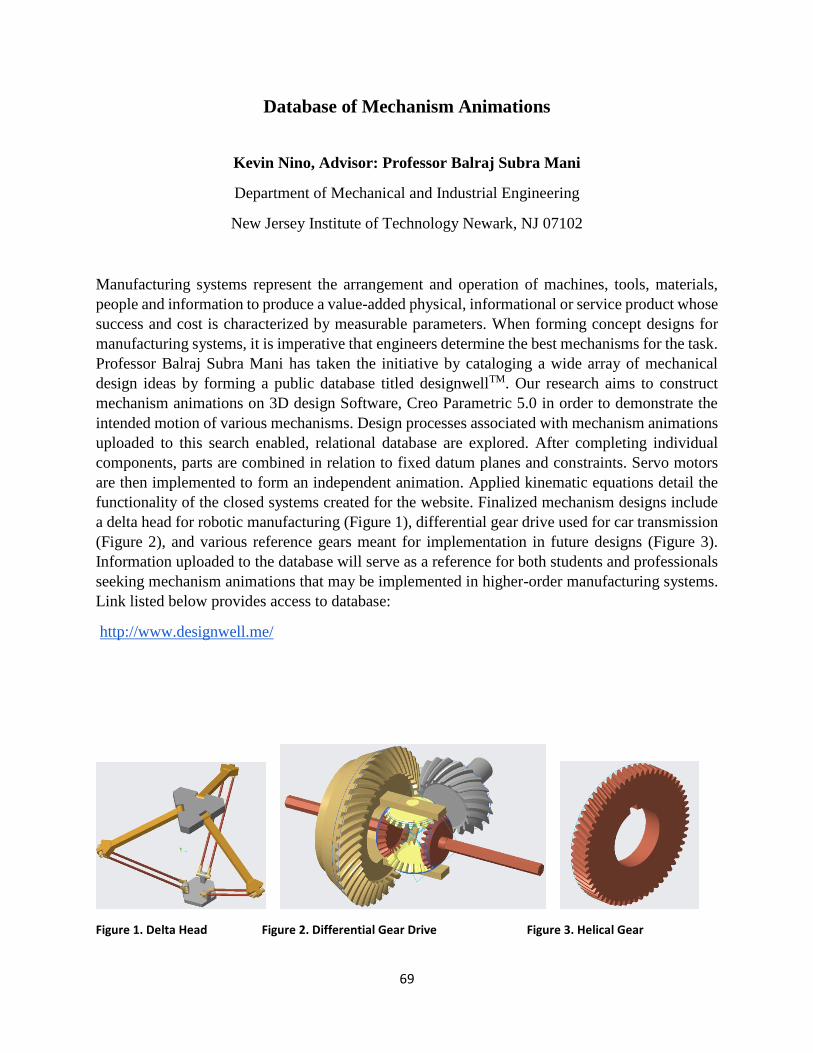
69
Database of Mechanism Animations
Kevin Nino, Advisor: Professor Balraj Subra Mani
Department of Mechanical and Industrial Engineering
New Jersey Institute of Technology Newark, NJ 07102
Manufacturing systems represent the arrangement and operation of machines, tools, materials,
people and information to produce a value-added physical, informational or service product whose
success and cost is characterized by measurable parameters. When forming concept designs for
manufacturing systems, it is imperative that engineers determine the best mechanisms for the task.
Professor Balraj Subra Mani has taken the initiative by cataloging a wide array of mechanical
design ideas by forming a public database titled designwellTM. Our research aims to construct
mechanism animations on 3D design Software, Creo Parametric 5.0 in order to demonstrate the
intended motion of various mechanisms. Design processes associated with mechanism animations
uploaded to this search enabled, relational database are explored. After completing individual
components, parts are combined in relation to fixed datum planes and constraints. Servo motors
are then implemented to form an independent animation. Applied kinematic equations detail the
functionality of the closed systems created for the website. Finalized mechanism designs include
a delta head for robotic manufacturing (Figure 1), differential gear drive used for car transmission
(Figure 2), and various reference gears meant for implementation in future designs (Figure 3).
Information uploaded to the database will serve as a reference for both students and professionals
seeking mechanism animations that may be implemented in higher-order manufacturing systems.
Link listed below provides access to database:
http://www.designwell.me/
Figure 1. Delta Head Figure 2. Differential Gear Drive Figure 3. Helical Gear
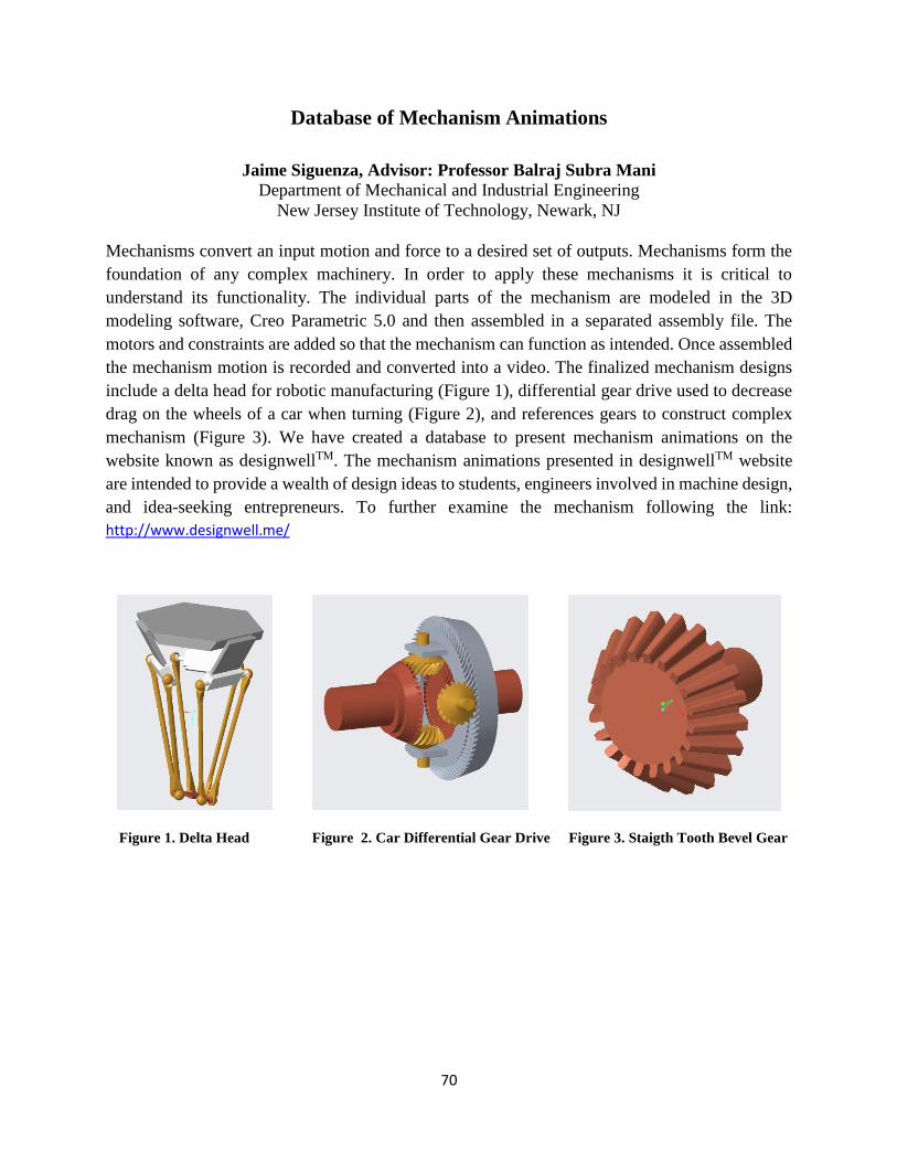
70
Database of Mechanism Animations
Jaime Siguenza, Advisor: Professor Balraj Subra Mani
Department of Mechanical and Industrial Engineering
New Jersey Institute of Technology, Newark, NJ
Mechanisms convert an input motion and force to a desired set of outputs. Mechanisms form the
foundation of any complex machinery. In order to apply these mechanisms it is critical to
understand its functionality. The individual parts of the mechanism are modeled in the 3D
modeling software, Creo Parametric 5.0 and then assembled in a separated assembly file. The
motors and constraints are added so that the mechanism can function as intended. Once assembled
the mechanism motion is recorded and converted into a video. The finalized mechanism designs
include a delta head for robotic manufacturing (Figure 1), differential gear drive used to decrease
drag on the wheels of a car when turning (Figure 2), and references gears to construct complex
mechanism (Figure 3). We have created a database to present mechanism animations on the
website known as designwellTM. The mechanism animations presented in designwellTM website
are intended to provide a wealth of design ideas to students, engineers involved in machine design,
and idea-seeking entrepreneurs. To further examine the mechanism following the link:
http://www.designwell.me/
Figure 1. Delta Head Figure 2. Car Differential Gear Drive Figure 3. Staigth Tooth Bevel Gear

71
72
Materials - Photon Interactions in Multilayers
Deva Craig, Jonathan Martinez, Jessy Nemati, Serlly Vega and Nuggehalli M. Ravindra
Department of Physics
New Jersey Institute of Technology, Newark, NJ 07102, USA
Abstract: This study investigates the optical properties such as transmittance, reflectance, and
absorptance/emittance of multilayer films of various thicknesses, in the wavelength range of 0.1-
10µm. By mathematically modelling the multilayers, using the transfer matrix method, we
investigate the optical properties of various layered films. The types of multilayers investigated
include Si/Ge, buried oxides such as SiO2 and SiN4 in semiconductors and antireflection coatings
for applications in solar cells. Surface roughness is considered in some of these case studies. The
simulated optical properties are compared with similar studies from the literature. Using these
simulations, we attempt to predict the properties of an optimized configuration of multilayered
films for these applications.
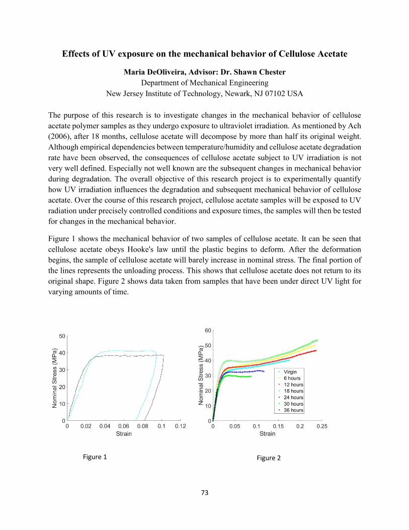
73
Effects of UV exposure on the mechanical behavior of Cellulose Acetate
Maria DeOliveira, Advisor: Dr. Shawn Chester
Department of Mechanical Engineering
New Jersey Institute of Technology, Newark, NJ 07102 USA
The purpose of this research is to investigate changes in the mechanical behavior of cellulose
acetate polymer samples as they undergo exposure to ultraviolet irradiation. As mentioned by Ach
(2006), after 18 months, cellulose acetate will decompose by more than half its original weight.
Although empirical dependencies between temperature/humidity and cellulose acetate degradation
rate have been observed, the consequences of cellulose acetate subject to UV irradiation is not
very well defined. Especially not well known are the subsequent changes in mechanical behavior
during degradation. The overall objective of this research project is to experimentally quantify
how UV irradiation influences the degradation and subsequent mechanical behavior of cellulose
acetate. Over the course of this research project, cellulose acetate samples will be exposed to UV
radiation under precisely controlled conditions and exposure times, the samples will then be tested
for changes in the mechanical behavior.
Figure 1 shows the mechanical behavior of two samples of cellulose acetate. It can be seen that
cellulose acetate obeys Hooke's law until the plastic begins to deform. After the deformation
begins, the sample of cellulose acetate will barely increase in nominal stress. The final portion of
the lines represents the unloading process. This shows that cellulose acetate does not return to its
original shape. Figure 2 shows data taken from samples that have been under direct UV light for
varying amounts of time.
Figure 1 Figure 2
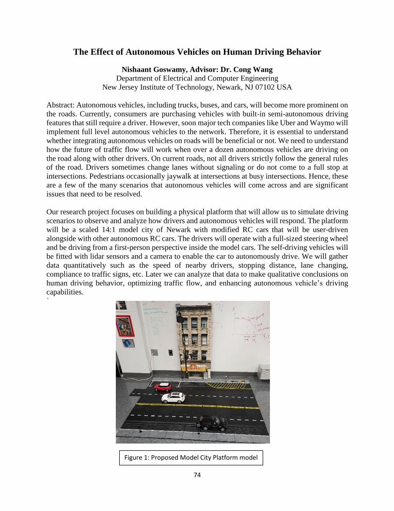
74
The Effect of Autonomous Vehicles on Human Driving Behavior
Nishaant Goswamy, Advisor: Dr. Cong Wang
Department of Electrical and Computer Engineering New Jersey Institute of Technology, Newark, NJ 07102 USA
Abstract: Autonomous vehicles, including trucks, buses, and cars, will become more prominent on
the roads. Currently, consumers are purchasing vehicles with built-in semi-autonomous driving
features that still require a driver. However, soon major tech companies like Uber and Waymo will
implement full level autonomous vehicles to the network. Therefore, it is essential to understand
whether integrating autonomous vehicles on roads will be beneficial or not. We need to understand
how the future of traffic flow will work when over a dozen autonomous vehicles are driving on
the road along with other drivers. On current roads, not all drivers strictly follow the general rules
of the road. Drivers sometimes change lanes without signaling or do not come to a full stop at
intersections. Pedestrians occasionally jaywalk at intersections at busy intersections. Hence, these
are a few of the many scenarios that autonomous vehicles will come across and are significant
issues that need to be resolved.
Our research project focuses on building a physical platform that will allow us to simulate driving
scenarios to observe and analyze how drivers and autonomous vehicles will respond. The platform
will be a scaled 14:1 model city of Newark with modified RC cars that will be user-driven
alongside with other autonomous RC cars. The drivers will operate with a full-sized steering wheel
and be driving from a first-person perspective inside the model cars. The self-driving vehicles will
be fitted with lidar sensors and a camera to enable the car to autonomously drive. We will gather
data quantitatively such as the speed of nearby drivers, stopping distance, lane changing,
compliance to traffic signs, etc. Later we can analyze that data to make qualitative conclusions on
human driving behavior, optimizing traffic flow, and enhancing autonomous vehicle’s driving
capabilities.
`
Figure 1: Proposed Model City Platform model
75
An Exploratory Study into the Effects of Total Sleep Deprivation using fNIRS
Katherine Ji; Advisor: Dr. Bharat Biswal
Department of Biomedical Engineering
New Jersey Institute of Technology, Newark, N.J. 07102
College students who work nights on end finishing projects or studying for tests with little
to no sleep go entirely unquestioned. Applauded, even. Since 1985, in the United States, the
number of adults sleeping less than six hours a night has increased by 31%. Previous studies using
functional magnetic resonance imaging, or fMRI, have indicated that healthy patients who
experience one to two nights of total sleep deprivation exhibit a lower accuracy rate and longer
reaction time in neurobehavioral tasks compared to results during restful wakefulness.
Additionally, fMRI has confirmed that areas of the brain in task-activated studies exhibit different
amounts of activation, especially in the prefrontal cortex and parietal cortex.
Our research project seeks to use functional near-infrared spectroscopy, or fNIRS, in order
to quantify how cognitive function and brain activity is affected by sleep deprivation. To our
knowledge, fNIRS has never been used in this capacity. Although fMRI is recognized as the gold
standard of brain imaging, We seek to use fNIRS, which has been shown to offer comparable
robustness. fNIRS detects changes in oxygenated and deoxygenated hemoglobin concentrations
based on the absorbance of infrared light. In the present study, we utilized different
neurobehavioral tasks that demonstrate brain activity covering attention, memory, working
memory, decision making, and interference resolution, which are particular brain functions that
have shown to be most affected by sleep.
Thus far, we have scanned 13 participants for both normal sleep and total sleep deprivation.
Initial results have demonstrated differences in hemodynamic function correlated with whether or
not the participant slept. In the coming weeks, more scans will be taken and data must be further
processed before a full analysis of results is conducted.
The purpose of this study is to add to existing literature surrounding the effects of total
sleep deprivation. Additionally, unlike fMRI, since fNIRS is a portable neuroimaging technique,
validating our scans against previous fMRI studies opens up the ability to study more physically
demanding tasks. For example, future studies may involve studying the effects of sleep deprivation
on motor function while driving, walking, playing sports, etc. Elucidating the cognitive
consequences of sleep deprivation is extremely important in a world observing a continued decline
in sleep duration, and will aid our understanding of the function of sleep in mental health and
cognitive impairment.
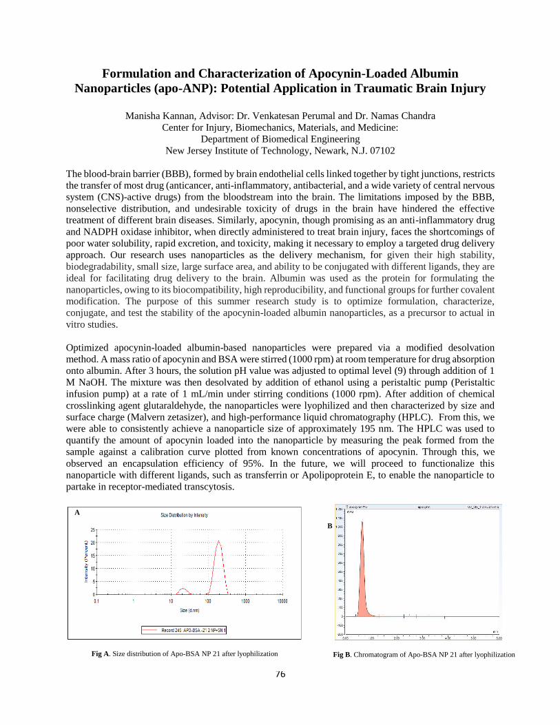
76
Formulation and Characterization of Apocynin-Loaded Albumin
Nanoparticles (apo-ANP): Potential Application in Traumatic Brain Injury
Manisha Kannan, Advisor: Dr. Venkatesan Perumal and Dr. Namas Chandra
Center for Injury, Biomechanics, Materials, and Medicine:
Department of Biomedical Engineering
New Jersey Institute of Technology, Newark, N.J. 07102
The blood-brain barrier (BBB), formed by brain endothelial cells linked together by tight junctions, restricts
the transfer of most drug (anticancer, anti-inflammatory, antibacterial, and a wide variety of central nervous
system (CNS)-active drugs) from the bloodstream into the brain. The limitations imposed by the BBB,
nonselective distribution, and undesirable toxicity of drugs in the brain have hindered the effective
treatment of different brain diseases. Similarly, apocynin, though promising as an anti-inflammatory drug
and NADPH oxidase inhibitor, when directly administered to treat brain injury, faces the shortcomings of
poor water solubility, rapid excretion, and toxicity, making it necessary to employ a targeted drug delivery
approach. Our research uses nanoparticles as the delivery mechanism, for given their high stability,
biodegradability, small size, large surface area, and ability to be conjugated with different ligands, they are
ideal for facilitating drug delivery to the brain. Albumin was used as the protein for formulating the
nanoparticles, owing to its biocompatibility, high reproducibility, and functional groups for further covalent
modification. The purpose of this summer research study is to optimize formulation, characterize,
conjugate, and test the stability of the apocynin-loaded albumin nanoparticles, as a precursor to actual in
vitro studies.
Optimized apocynin-loaded albumin-based nanoparticles were prepared via a modified desolvation
method. A mass ratio of apocynin and BSA were stirred (1000 rpm) at room temperature for drug absorption
onto albumin. After 3 hours, the solution pH value was adjusted to optimal level (9) through addition of 1
M NaOH. The mixture was then desolvated by addition of ethanol using a peristaltic pump (Peristaltic
infusion pump) at a rate of 1 mL/min under stirring conditions (1000 rpm). After addition of chemical
crosslinking agent glutaraldehyde, the nanoparticles were lyophilized and then characterized by size and
surface charge (Malvern zetasizer), and high-performance liquid chromatography (HPLC). From this, we
were able to consistently achieve a nanoparticle size of approximately 195 nm. The HPLC was used to
quantify the amount of apocynin loaded into the nanoparticle by measuring the peak formed from the
sample against a calibration curve plotted from known concentrations of apocynin. Through this, we
observed an encapsulation efficiency of 95%. In the future, we will proceed to functionalize this
nanoparticle with different ligands, such as transferrin or Apolipoprotein E, to enable the nanoparticle to
partake in receptor-mediated transcytosis.
Fig A. Size distribution of Apo-BSA NP 21 after lyophilization Fig B. Chromatogram of Apo-BSA NP 21 after lyophilization
A
B
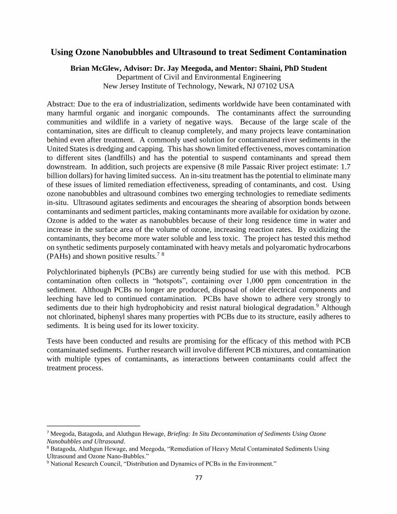
77
Using Ozone Nanobubbles and Ultrasound to treat Sediment Contamination
Brian McGlew, Advisor: Dr. Jay Meegoda, and Mentor: Shaini, PhD Student Department of Civil and Environmental Engineering
New Jersey Institute of Technology, Newark, NJ 07102 USA
Abstract: Due to the era of industrialization, sediments worldwide have been contaminated with
many harmful organic and inorganic compounds. The contaminants affect the surrounding
communities and wildlife in a variety of negative ways. Because of the large scale of the
contamination, sites are difficult to cleanup completely, and many projects leave contamination
behind even after treatment. A commonly used solution for contaminated river sediments in the
United States is dredging and capping. This has shown limited effectiveness, moves contamination
to different sites (landfills) and has the potential to suspend contaminants and spread them
downstream. In addition, such projects are expensive (8 mile Passaic River project estimate: 1.7
billion dollars) for having limited success. An in-situ treatment has the potential to eliminate many
of these issues of limited remediation effectiveness, spreading of contaminants, and cost. Using
ozone nanobubbles and ultrasound combines two emerging technologies to remediate sediments
in-situ. Ultrasound agitates sediments and encourages the shearing of absorption bonds between
contaminants and sediment particles, making contaminants more available for oxidation by ozone.
Ozone is added to the water as nanobubbles because of their long residence time in water and
increase in the surface area of the volume of ozone, increasing reaction rates. By oxidizing the
contaminants, they become more water soluble and less toxic. The project has tested this method
on synthetic sediments purposely contaminated with heavy metals and polyaromatic hydrocarbons
(PAHs) and shown positive results.7 8
Polychlorinated biphenyls (PCBs) are currently being studied for use with this method. PCB
contamination often collects in “hotspots”, containing over 1,000 ppm concentration in the
sediment. Although PCBs no longer are produced, disposal of older electrical components and
leeching have led to continued contamination. PCBs have shown to adhere very strongly to
sediments due to their high hydrophobicity and resist natural biological degradation.9 Although
not chlorinated, biphenyl shares many properties with PCBs due to its structure, easily adheres to
sediments. It is being used for its lower toxicity.
Tests have been conducted and results are promising for the efficacy of this method with PCB
contaminated sediments. Further research will involve different PCB mixtures, and contamination
with multiple types of contaminants, as interactions between contaminants could affect the
treatment process.
7 Meegoda, Batagoda, and Aluthgun Hewage, Briefing: In Situ Decontamination of Sediments Using Ozone
Nanobubbles and Ultrasound. 8 Batagoda, Aluthgun Hewage, and Meegoda, “Remediation of Heavy Metal Contaminated Sediments Using
Ultrasound and Ozone Nano-Bubbles.” 9 National Research Council, “Distribution and Dynamics of PCBs in the Environment.”
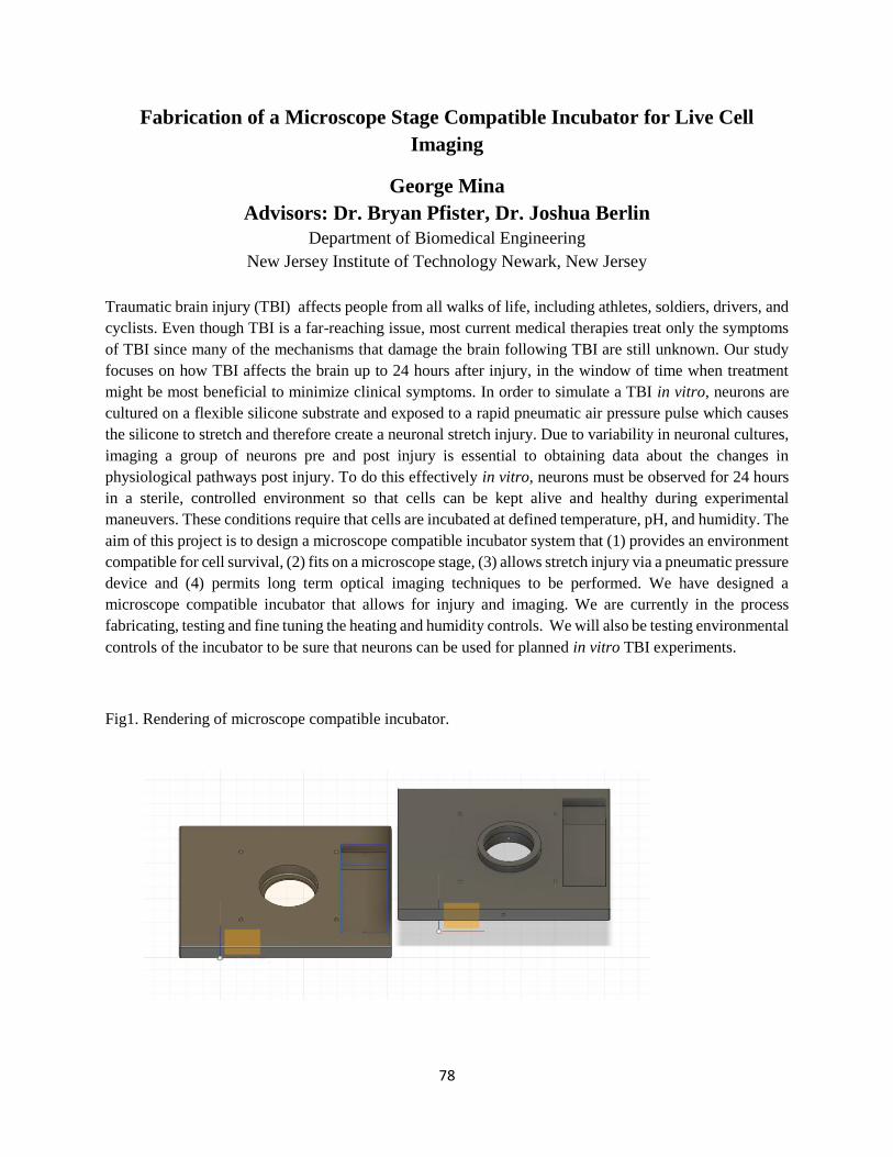
78
Fabrication of a Microscope Stage Compatible Incubator for Live Cell
Imaging
George Mina
Advisors: Dr. Bryan Pfister, Dr. Joshua Berlin
Department of Biomedical Engineering
New Jersey Institute of Technology Newark, New Jersey
Traumatic brain injury (TBI) affects people from all walks of life, including athletes, soldiers, drivers, and
cyclists. Even though TBI is a far-reaching issue, most current medical therapies treat only the symptoms
of TBI since many of the mechanisms that damage the brain following TBI are still unknown. Our study
focuses on how TBI affects the brain up to 24 hours after injury, in the window of time when treatment
might be most beneficial to minimize clinical symptoms. In order to simulate a TBI in vitro, neurons are
cultured on a flexible silicone substrate and exposed to a rapid pneumatic air pressure pulse which causes
the silicone to stretch and therefore create a neuronal stretch injury. Due to variability in neuronal cultures,
imaging a group of neurons pre and post injury is essential to obtaining data about the changes in
physiological pathways post injury. To do this effectively in vitro, neurons must be observed for 24 hours
in a sterile, controlled environment so that cells can be kept alive and healthy during experimental
maneuvers. These conditions require that cells are incubated at defined temperature, pH, and humidity. The
aim of this project is to design a microscope compatible incubator system that (1) provides an environment
compatible for cell survival, (2) fits on a microscope stage, (3) allows stretch injury via a pneumatic pressure
device and (4) permits long term optical imaging techniques to be performed. We have designed a
microscope compatible incubator that allows for injury and imaging. We are currently in the process
fabricating, testing and fine tuning the heating and humidity controls. We will also be testing environmental
controls of the incubator to be sure that neurons can be used for planned in vitro TBI experiments.
Fig1. Rendering of microscope compatible incubator.
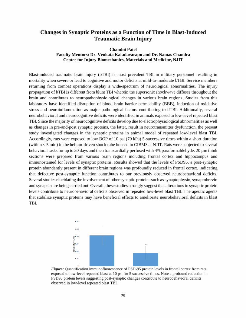
79
Changes in Synaptic Proteins as a Function of Time in Blast-Induced
Traumatic Brain Injury
Chandni Patel
Faculty Mentors: Dr. Venkata Kakulavarapu and Dr. Namas Chandra
Center for Injury Biomechanics, Materials and Medicine, NJIT
Blast-induced traumatic brain injury (bTBI) is most prevalent TBI in military personnel resulting in
mortality when severe or lead to cognitive and motor deficits at mild-to-moderate bTBI. Service members
returning from combat operations display a wide-spectrum of neurological abnormalities. The injury
propagation of bTBI is different from blunt TBI wherein the supersonic shockwave diffuses throughout the
brain and contributes to neuropathophysiological changes in various brain regions. Studies from this
laboratory have identified disruption of blood brain barrier permeability (BBB), induction of oxidative
stress and neuroinflammation as major pathological factors contributing to bTBI. Additionally, several
neurobehavioral and neurocognitive deficits were identified in animals exposed to low-level repeated blast
TBI. Since the majority of neurocognitive deficits develop due to electrophysiological abnormalities as well
as changes in pre-and-post synaptic proteins, the latter, result in neurotransmitter dysfunction, the present
study investigated changes in the synaptic proteins in animal model of repeated low-level blast TBI.
Accordingly, rats were exposed to low BOP of 10 psi (70 kPa) 5-successive times within a short duration
(within < 5 min) in the helium-driven shock tube housed in CIBM3 at NJIT. Rats were subjected to several
behavioral tasks for up to 30 days and then transcardially perfused with 4% paraformaldehyde. 20 µm think
sections were prepared from various brain regions including frontal cortex and hippocampus and
immunostained for levels of synaptic proteins. Results showed that the levels of PSD95, a post-synaptic
protein abundantly present in different brain regions was profoundly reduced in frontal cortex, indicating
that defective post-synaptic function contributes to our previously observed neurobehavioral deficits.
Several studies elucidating the involvement of other synaptic proteins such as synaptophysin, synaptobrevin
and synapsin are being carried out. Overall, these studies strongly suggest that alterations in synaptic protein
levels contribute to neurobehavioral deficits observed in repeated low-level blast TBI. Therapeutic agents
that stabilize synaptic proteins may have beneficial effects to ameliorate neurobehavioral deficits in blast
TBI.
Figure: Quantification immunofluorescence of PSD-95 protein levels in frontal cortex from rats
exposed to low-level repeated blast at 10 psi for 5 successive times. Note a profound reduction in
PSD95 protein levels suggesting post-synaptic changes contribute to neurobehavioral deficits
observed in low-level repeated blast TBI.
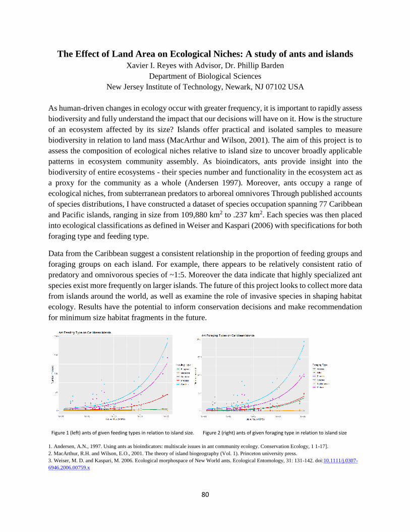
80
The Effect of Land Area on Ecological Niches: A study of ants and islands
Xavier I. Reyes with Advisor, Dr. Phillip Barden
Department of Biological Sciences
New Jersey Institute of Technology, Newark, NJ 07102 USA
As human-driven changes in ecology occur with greater frequency, it is important to rapidly assess
biodiversity and fully understand the impact that our decisions will have on it. How is the structure
of an ecosystem affected by its size? Islands offer practical and isolated samples to measure
biodiversity in relation to land mass (MacArthur and Wilson, 2001). The aim of this project is to
assess the composition of ecological niches relative to island size to uncover broadly applicable
patterns in ecosystem community assembly. As bioindicators, ants provide insight into the
biodiversity of entire ecosystems - their species number and functionality in the ecosystem act as
a proxy for the community as a whole (Andersen 1997). Moreover, ants occupy a range of
ecological niches, from subterranean predators to arboreal omnivores Through published accounts
of species distributions, I have constructed a dataset of species occupation spanning 77 Caribbean
and Pacific islands, ranging in size from 109,880 km2 to .237 km2. Each species was then placed
into ecological classifications as defined in Weiser and Kaspari (2006) with specifications for both
foraging type and feeding type.
Data from the Caribbean suggest a consistent relationship in the proportion of feeding groups and
foraging groups on each island. For example, there appears to be relatively consistent ratio of
predatory and omnivorous species of ~1:5. Moreover the data indicate that highly specialized ant
species exist more frequently on larger islands. The future of this project looks to collect more data
from islands around the world, as well as examine the role of invasive species in shaping habitat
ecology. Results have the potential to inform conservation decisions and make recommendation
for minimum size habitat fragments in the future.
1. Andersen, A.N., 1997. Using ants as bioindicators: multiscale issues in ant community ecology. Conservation Ecology, 1 1-17].
2. MacArthur, R.H. and Wilson, E.O., 2001. The theory of island biogeography (Vol. 1). Princeton university press.
3. Weiser, M. D. and Kaspari, M. 2006. Ecological morphospace of New World ants. Ecological Entomology, 31: 131-142. doi:10.1111/j.0307-
6946.2006.00759.x
Figure 1 (left) ants of given feeding types in relation to island size. Figure 2 (right) ants of given foraging type in relation to island size
81
Collection and Analysis of Global Navigation Satellite System (GNSS)
Positioning Data with SBAS and RTK for Autonomous Lawn Mowers
Gregory Tanis; Advisor: Dr. Cesar Bandera
Martin Tuchman School of Management
New Jersey Institute of Technology, Newark, NJ 07102 USA
Have you ever watched the seemingly aimless wanderings of a robotic vacuum cleaner and
thought to yourself “there must be a better solution?” They bounce around the carpet covered
pinball machine of your house, bumping into walls and furniture, retracing their path, and then off
to a remote corner of the kitchen where they become momentarily trapped. In Europe, there is a
growing market for small robotic lawn mowers that use similar technology, bouncing around the
perimeter of the property in a similarly aimless manner. These robotic lawn mowers have failed to
significantly impact the North American market because they cannot effectively mow large areas
and do not leave the stripes which Americans view as essential to a well-manicured lawn. The root
problem is that existing robotic vacuums and lawn mowers lack precise positioning.
The current Global Navigation Satellite System (GNSS) work well for autonomous
navigation in ships and planes. However, they lack the precision and accuracy crucial to
applications such as autonomous lawn mowers. GNSS is imprecise because of signal disruptions
as the signal travels from the satellites, through the atmosphere, and to the receiver, as well as
drifts in timing and position. This can result in an imprecision of several meters, unacceptable for
autonomous mowers. Another factor that can affect GNSS accuracy is obstacles such as trees and
buildings. The reflections off these obstacles can cause inaccuracy as it changes the distance which
the signal travels. In order to compensate for these inaccuracies, a base station is needed.
A base station is a receiver that remains stationary. The base station can track the drift of
the satellite signal over time and provide correction factors to the rover, the mobile receiver that is
being tracked. The three most common correction methods are Satellite Based Augmentation
System (SBAS), Real Time Kinematics (RTK), and cloud-based correction systems. SBAS uses a
handful of ground-based stations to send correction data to the satellites themselves. This is
convenient as any receiver can take advantage of this correction data as it is integrated into the
signal from the satellite. However, they are limited to accuracies in the meter range as the network
of base stations is relatively small. RTK and cloud-based correction systems, comparatively, offer
positioning resolution on the centimeter scale. RTK requires the user to have a second receiver
which uses a radio system to communicate with the rover. Cloud-based systems offer over-the-
internet correction data provided by the third-party providers receiver network.
In this research project, SBAS and RTK setups are used to collect location data in various
patterns. Tests are conducted in open areas with a clear sky view as well as in areas with obstacles
such as trees and houses. This provide real world data for the environments that autonomous
mowers are likely to encounter. The coordinates of each point are recorded and converted to
distances from a “zero point.” This data is then analyzed to find the accuracy precision, and other
characteristics of the positioning data. In the experiments performed to date, the SBAS positioning
data was found to be precise to about one meter (97 cm). The RTK research has not yet been
concluded but is expected to significantly reduce the range of error.
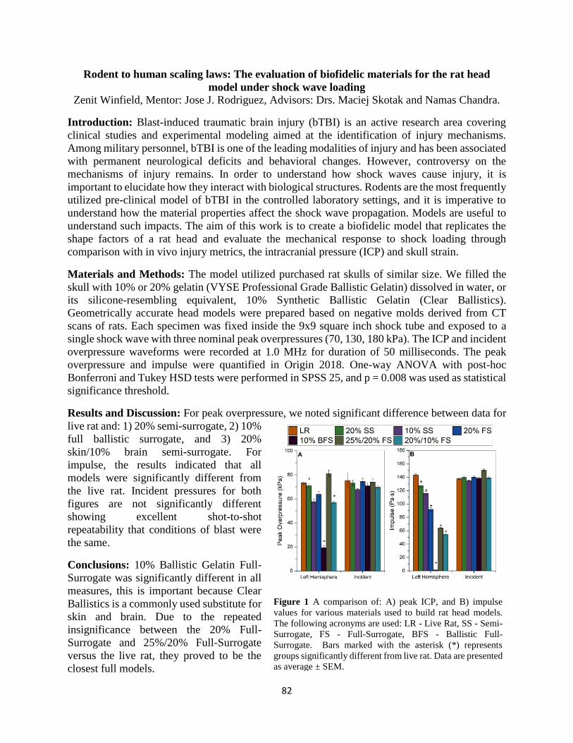
82
Rodent to human scaling laws: The evaluation of biofidelic materials for the rat head
model under shock wave loading
Zenit Winfield, Mentor: Jose J. Rodriguez, Advisors: Drs. Maciej Skotak and Namas Chandra.
Introduction: Blast-induced traumatic brain injury (bTBI) is an active research area covering
clinical studies and experimental modeling aimed at the identification of injury mechanisms.
Among military personnel, bTBI is one of the leading modalities of injury and has been associated
with permanent neurological deficits and behavioral changes. However, controversy on the
mechanisms of injury remains. In order to understand how shock waves cause injury, it is
important to elucidate how they interact with biological structures. Rodents are the most frequently
utilized pre-clinical model of bTBI in the controlled laboratory settings, and it is imperative to
understand how the material properties affect the shock wave propagation. Models are useful to
understand such impacts. The aim of this work is to create a biofidelic model that replicates the
shape factors of a rat head and evaluate the mechanical response to shock loading through
comparison with in vivo injury metrics, the intracranial pressure (ICP) and skull strain.
Materials and Methods: The model utilized purchased rat skulls of similar size. We filled the
skull with 10% or 20% gelatin (VYSE Professional Grade Ballistic Gelatin) dissolved in water, or
its silicone-resembling equivalent, 10% Synthetic Ballistic Gelatin (Clear Ballistics).
Geometrically accurate head models were prepared based on negative molds derived from CT
scans of rats. Each specimen was fixed inside the 9x9 square inch shock tube and exposed to a
single shock wave with three nominal peak overpressures (70, 130, 180 kPa). The ICP and incident
overpressure waveforms were recorded at 1.0 MHz for duration of 50 milliseconds. The peak
overpressure and impulse were quantified in Origin 2018. One-way ANOVA with post-hoc
Bonferroni and Tukey HSD tests were performed in SPSS 25, and p = 0.008 was used as statistical
significance threshold.
Results and Discussion: For peak overpressure, we noted significant difference between data for
live rat and: 1) 20% semi-surrogate, 2) 10%
full ballistic surrogate, and 3) 20%
skin/10% brain semi-surrogate. For
impulse, the results indicated that all
models were significantly different from
the live rat. Incident pressures for both
figures are not significantly different
showing excellent shot-to-shot
repeatability that conditions of blast were
the same.
Conclusions: 10% Ballistic Gelatin Full-
Surrogate was significantly different in all
measures, this is important because Clear
Ballistics is a commonly used substitute for
skin and brain. Due to the repeated
insignificance between the 20% Full-
Surrogate and 25%/20% Full-Surrogate
versus the live rat, they proved to be the
closest full models.
Figure 1 A comparison of: A) peak ICP, and B) impulse
values for various materials used to build rat head models.
The following acronyms are used: LR - Live Rat, SS - Semi-
Surrogate, FS - Full-Surrogate, BFS - Ballistic Full-
Surrogate. Bars marked with the asterisk (*) represents
groups significantly different from live rat. Data are presented
as average ± SEM.

83
84
Machine Learning Approaches for Long-Short Portfolio Optimization
Alexander Barrett, Mohamad Sherif, Ruohan Wu
Advisor: Dr. Zhi Wei
Mentor: Cheng Zhong
It is commonly believed that financial markets are predictable to some extent as observed in many
empirical studies. Many machine learning methods have been applied to predict stock returns,
dated decades back. However, the literature on long-short portfolio optimization using machine
learning approaches remain limited. Long-short portfolios aim to enhance return of investment in
specific market segments by exploiting profitable opportunities through taking a long position on
undervalued stocks while selling short overpriced stocks. To create an optimized portfolio, the
most overvalued and undervalued stocks must be identified through prediction. We create a novel
method of predicting stock market trends and thereby balancing long-short portfolio.
The stock data from the Center for Research in Security Prices (CRSP) is used for the purpose of
this study. Specifically, the data for individual stock returns and prices, S&P 500 index return,
industry categories, number of shares outstanding, sharecode, exchange code, and trading volume
spanning over 15 years (2004-18). The data is divided into three parts along time for model
training, validation, and testing. Using this dataset, linear regression, Random Forest, Support
Vector Regression (SVR), Gradient Boosting, and neural network models are implemented for
stock price prediction. For a given month in our data set, we rank the predicted return of each
stock, and comprise our long-short portfolio of the top and bottom 10% of these predicted stocks.
We then aggregate these returns over time to compute the cumulative return of a given model, and
use this as the metric of comparison between models. We find that most of our models positive
return over a five year period. In particular, our Gradient Boosting model yielded a return that was
approximately 26% greater than the return of the S&P 500 index over a five year span. This work
contributes to vastly growing interest in building more efficient tools to predict the stock market.
Future work in this project would involve the usage of deep learning methods and alternative
machine learning models for increased predictive accuracy.
85
A Detailed Look Into Existing Software Auto-Fix Techniques
Peter Boyland1, Andrew Chen1, Farukh Saidmuratov2
Advisor: Dr. David Shaohua Wang3, Mentor: Yi Li3
Department of Informatics
1Carnegie Mellon University, Pittsburgh, PA 15213 | 2Rensselaer Polytechnic Institute, Troy, NY 12180
| 3New Jersey Institute of Technology, Newark, NJ 07102
Within the workflow of software development, a major portion of time is dedicated to fixing
mistakes in programs. A popular adage that reflects this reality states, “If debugging is the process
of removing software bugs, then programming must be the process of putting them in.” However,
with recent advances in machine learning algorithms, Automated Program Repair (APR) methods
hold promise to automate the tedious task of correcting faulty programs. Recently, APR tools have
been implemented in two distinct fashions: rule-based, where fix ingredients are found in a search
space with the assistance of machine learning, and learning-based, where code patches are rather
predicted from deep learning models.
While a variety of these powerful and original APR tools have been explored, a baseline study of
the differences between the types of bug fixes and the run time of these procedures has not. In this
study, we present an overlook of the landscape of APR tools in order to provide insights of the
current state of automatic program repair research. We run a number of state-of-the-art APR tools
such as LSRepair and AVATAR on benchmark datasets containing buggy programs, and report
on the quantity and type of bugs these techniques can fix in a set time limit. This study considers
the following run times: 30 minutes, 1 hour, 2 hours, 3 hours, 4 hours, and 5 hours. By running
these techniques in parallel and averaging the correct bug fixes produced, we are able to discover
the effectiveness of each procedure given the limited run time.
With this analysis, future research can contextualize the effectiveness of novel approaches in the
backdrop of cutting edge past work. In addition, ensemble models could be built using a
combination of select systems reviewed in this study to produce a greater number of correct
patches.
86
Auto-Human Eye Blink Detection Dataset Labeling
Peter Decker1 and Miriam Tan2
Advisor: Dr. Guiling Wang3 | Mentor: Junyi Ye3 (PhD Student)
1 County College of Mars, Randolph, NJ 07869
2 Grove City College, Grove City, PA 16127
3 Department of Computer Science New Jersey Institute of Technology, Newark, NJ 07102
The initial labeling of new datasets used for training models is expensive and time consuming.
Manually generation of labels requires hours of repetitive, human labor while algorithmic methods
produce cheaper but inaccurate results [1]. Since these labels are used as the ground truth for
evaluating predictive methods, accuracy is imperative. Therefore we combine algorithmic labeling
with manual labeling to achieve 100% accuracy with fewer hours of human labor. We base our
algorithm on the Eye Aspect Ratio (EAR) method [2]. The algorithm generates an EAR threshold,
produces initial blink labels, and marks suspicious points to be manually investigated. Intersect-
Over-Union Evaluation determines the labels’ accuracy, precision, and recall. Automatically
generated EAR thresholds outperform any single hardcoded threshold and approach the accuracy
of EAR thresholds manually selected for each dataset. Furthermore, a frame count threshold and
EAR graphs pinpoint the sections requiring human inspection.
[1] AltexSoft, "How to Organize Data Labeling for Machine Learning: Approaches and Tools," 2019.
[Online]. Available: https://www.kdnuggets.com/2018/05/data-labeling-machine-learning.html.
[2] T. Soukupova and J. Cech, "Real-Time Eye Blink Detection using Facial Landmarks," Computer Vision
Winter Workshop, 2016.
87
Spatiotemporal Analysis of Racial Bias in NYPD Stop, Question, and Frisk
Procedures
Mihail Kaburis1, Samantha Kamath2, Jasmine Medlock3
Advisor: Dr. Xinyue Ye4 | Mentor: Jun Yuan (PhD Student)4
Department of Informatics 1University of South Florida, Tampa, FL 33620 | 2University of Miami, Coral Gables, FL 33146
3University of Maryland, Baltimore County, Baltimore MD 21250 | 4New Jersey Institute of Technology, Newark, NJ 07102
The treatment of racial minorities by law enforcement personnel during routine stops has become
a heavily debated topic in the United States. Determining the levels of bias in these stops could
have tremendous implications on police-civilian relations. Our research examines racial bias
within the NYPD’s Stop, Question, and Frisk (SQF) procedures, which have disproportionately
stopped black and hispanic individuals over their white counterparts. We determine racial bias via
the threshold test, a Bayesian statistical model that calculates a race and precinct-specific search
threshold — the point at which a police officer decides to search an individual for contraband.
Lower search thresholds for certain races imply racial bias against these races. However, previous
threshold test models have not accounted for spatiotemporal factors, including the fluctuation of
racial bias within police precincts over time and the amount of crime in the neighborhoods where
stops occur. To better understand racially-biased policing, the NYPD’s SQF Datasets and
aggregated Historical NYC Crime Data from 2003 to 2018 will be used to determine how racial
bias in SQF procedures varies according to these factors.
The datasets were first cleaned and filtered using R. The threshold model was applied to infer race
and precinct-specific search thresholds, which were plotted against crime numbers. Minority
search thresholds were compared to white search thresholds to determine the amount of racial bias
within each precinct over time. The model-predicted search and hit rates (the search success rate)
were compared to the actual values to determine the robustness of the model. Python scripts
employing geocoding APIs were utilized to determine the coordinates of stops. This information
allowed us to visualize racial bias within the NYPD’s stops across space and time with QGIS, a
geographic information system software.
While there was no significant correlation between the search thresholds and neighborhood crime
numbers, white search thresholds were consistently higher than black and Hispanic search
thresholds, implying consistent racial bias against these racial minorities. The model was generally
accurate at predicting the search rate of all races but slightly increased in error when predicting the
hit rate. This information, along with a robust visualization platform for racial bias, provides
valuable tools for policymakers, police, and residents to make informed decisions about policing
practices in the places that they call home.

88
89
Machine Learning Models for the Dynamics of Ferrofluids
John DeSalvo, Alex Janiak, Advisor: Dr. Shahriar Afkhami
Department of Mathematical Sciences
New Jersey Institute of Technology, Newark, NJ 07102 USA
The magnetic properties of ferrofluids allow for the potential of delivering drugs more precisely
towards specific locations in the human body. The probability of successfully targeting a location
is modeled via simulation of the motion these particles in a blood vessel where a magnetic field is
applied. Several machine learning techniques are used to improve this success rate by exploring
relationships with respect to various physical parameters. A significant dependence on the initial
position of the particle is indicated.

90
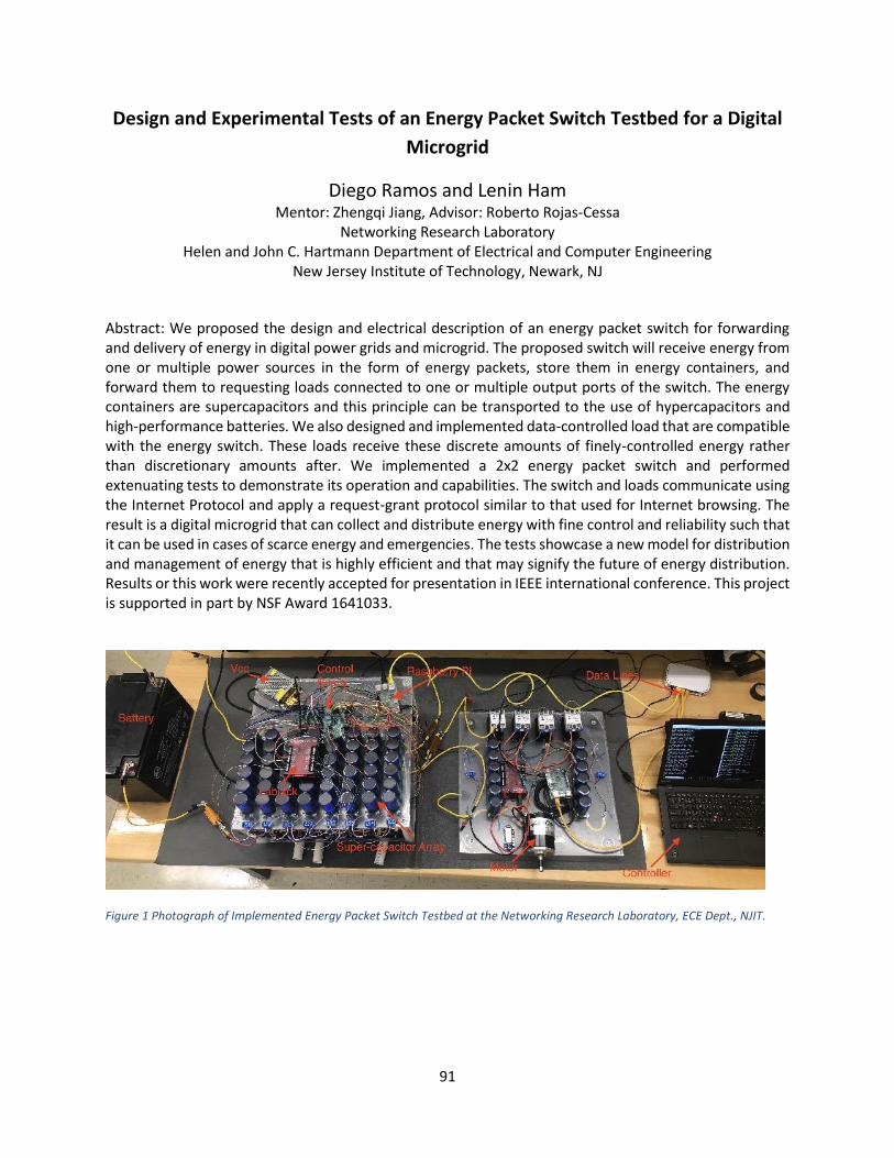
91
Design and Experimental Tests of an Energy Packet Switch Testbed for a Digital
Microgrid
Diego Ramos and Lenin Ham Mentor: Zhengqi Jiang, Advisor: Roberto Rojas-Cessa
Networking Research Laboratory Helen and John C. Hartmann Department of Electrical and Computer Engineering
New Jersey Institute of Technology, Newark, NJ
Abstract: We proposed the design and electrical description of an energy packet switch for forwarding and delivery of energy in digital power grids and microgrid. The proposed switch will receive energy from one or multiple power sources in the form of energy packets, store them in energy containers, and forward them to requesting loads connected to one or multiple output ports of the switch. The energy containers are supercapacitors and this principle can be transported to the use of hypercapacitors and high-performance batteries. We also designed and implemented data-controlled load that are compatible with the energy switch. These loads receive these discrete amounts of finely-controlled energy rather than discretionary amounts after. We implemented a 2x2 energy packet switch and performed extenuating tests to demonstrate its operation and capabilities. The switch and loads communicate using the Internet Protocol and apply a request-grant protocol similar to that used for Internet browsing. The result is a digital microgrid that can collect and distribute energy with fine control and reliability such that it can be used in cases of scarce energy and emergencies. The tests showcase a new model for distribution and management of energy that is highly efficient and that may signify the future of energy distribution. Results or this work were recently accepted for presentation in IEEE international conference. This project is supported in part by NSF Award 1641033.
Figure 1 Photograph of Implemented Energy Packet Switch Testbed at the Networking Research Laboratory, ECE Dept., NJIT.

92
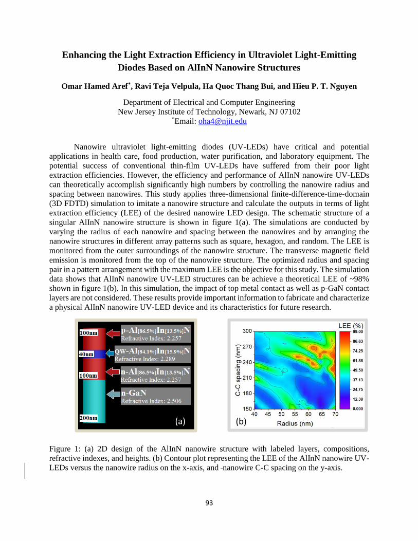
93
Enhancing the Light Extraction Efficiency in Ultraviolet Light-Emitting
Diodes Based on AlInN Nanowire Structures
Omar Hamed Aref*, Ravi Teja Velpula, Ha Quoc Thang Bui, and Hieu P. T. Nguyen
Department of Electrical and Computer Engineering
New Jersey Institute of Technology, Newark, NJ 07102 *Email: [email protected]
Nanowire ultraviolet light-emitting diodes (UV-LEDs) have critical and potential
applications in health care, food production, water purification, and laboratory equipment. The
potential success of conventional thin-film UV-LEDs have suffered from their poor light
extraction efficiencies. However, the efficiency and performance of AlInN nanowire UV-LEDs
can theoretically accomplish significantly high numbers by controlling the nanowire radius and
spacing between nanowires. This study applies three-dimensional finite-difference-time-domain
(3D FDTD) simulation to imitate a nanowire structure and calculate the outputs in terms of light
extraction efficiency (LEE) of the desired nanowire LED design. The schematic structure of a
singular AlInN nanowire structure is shown in figure 1(a). The simulations are conducted by
varying the radius of each nanowire and spacing between the nanowires and by arranging the
nanowire structures in different array patterns such as square, hexagon, and random. The LEE is
monitored from the outer surroundings of the nanowire structure. The transverse magnetic field
emission is monitored from the top of the nanowire structure. The optimized radius and spacing
pair in a pattern arrangement with the maximum LEE is the objective for this study. The simulation
data shows that AlInN nanowire UV-LED structures can be achieve a theoretical LEE of ~98%
shown in figure 1(b). In this simulation, the impact of top metal contact as well as p-GaN contact
layers are not considered. These results provide important information to fabricate and characterize
a physical AlInN nanowire UV-LED device and its characteristics for future research.
Figure 1: (a) 2D design of the AlInN nanowire structure with labeled layers, compositions,
refractive indexes, and heights. (b) Contour plot representing the LEE of the AlInN nanowire UV-
LEDs versus the nanowire radius on the x-axis, and nanowire C-C spacing on the y-axis.
(a) (b)
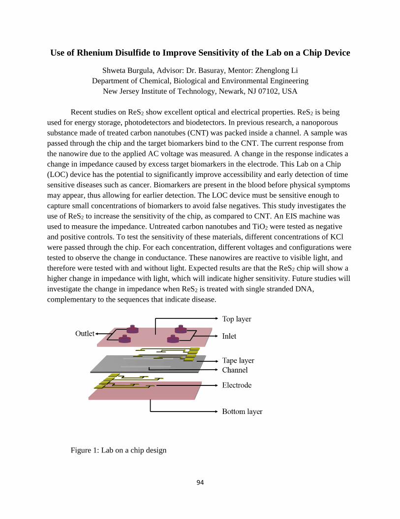
94
Use of Rhenium Disulfide to Improve Sensitivity of the Lab on a Chip Device
Shweta Burgula, Advisor: Dr. Basuray, Mentor: Zhenglong Li
Department of Chemical, Biological and Environmental Engineering
New Jersey Institute of Technology, Newark, NJ 07102, USA
Recent studies on ReS2 show excellent optical and electrical properties. ReS2 is being
used for energy storage, photodetectors and biodetectors. In previous research, a nanoporous
substance made of treated carbon nanotubes (CNT) was packed inside a channel. A sample was
passed through the chip and the target biomarkers bind to the CNT. The current response from
the nanowire due to the applied AC voltage was measured. A change in the response indicates a
change in impedance caused by excess target biomarkers in the electrode. This Lab on a Chip
(LOC) device has the potential to significantly improve accessibility and early detection of time
sensitive diseases such as cancer. Biomarkers are present in the blood before physical symptoms
may appear, thus allowing for earlier detection. The LOC device must be sensitive enough to
capture small concentrations of biomarkers to avoid false negatives. This study investigates the
use of ReS2 to increase the sensitivity of the chip, as compared to CNT. An EIS machine was
used to measure the impedance. Untreated carbon nanotubes and TiO2 were tested as negative
and positive controls. To test the sensitivity of these materials, different concentrations of KCl
were passed through the chip. For each concentration, different voltages and configurations were
tested to observe the change in conductance. These nanowires are reactive to visible light, and
therefore were tested with and without light. Expected results are that the ReS2 chip will show a
higher change in impedance with light, which will indicate higher sensitivity. Future studies will
investigate the change in impedance when ReS2 is treated with single stranded DNA,
complementary to the sequences that indicate disease.
Figure 1: Lab on a chip design
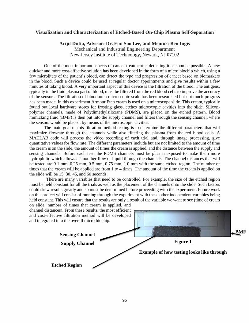
95
Visualization and Characterization of Etched-Based On-Chip Plasma Self-Separation
Arijit Dutta, Advisor: Dr. Eon Soo Lee, and Mentor: Ben Ingis
Mechanical and Industrial Engineering Department
New Jersey Institute of Technology, Newark, NJ 07102
One of the most important aspects of cancer treatment is detecting it as soon as possible. A new
quicker and more cost-effective solution has been developed in the form of a micro biochip which, using a
few microliters of the patient’s blood, can detect the type and progression of cancer based on biomarkers
in the blood. Such a device could be used at regular doctor appointments and give results within a few
minutes of taking blood. A very important aspect of this device is the filtration of the blood. The antigens,
typically in the fluid plasma part of blood, must be filtered from the red blood cells to improve the accuracy
of the sensors. The filtration of blood on a microscopic scale has been researched but not much progress
has been made. In this experiment Armour Etch cream is used on a microscope slide. This cream, typically
found out local hardware stores for frosting glass, etches microscopic cavities into the slide. Silicon-
polymer channels, made of Polydimethylsiloxane (PDMS), are placed on the etched pattern. Blood
mimicking fluid (BMF) is then put into the supply channel and filters through the sensing channel, where
the sensors would be placed, by means of the microscopic cavities.
The main goal of this filtration method testing is to determine the different parameters that will
maximize flowrate through the channels while also filtering the plasma from the red blood cells. A
MATLAB code will process the video recording of each trial and, through image processing, give
quantitative values for flow rate. The different parameters include but are not limited to the amount of time
the cream is on the slide, the amount of times the cream is applied, and the distance between the supply and
sensing channels. Before each test, the PDMS channels must be plasma exposed to make them more
hydrophilic which allows a smoother flow of liquid through the channels. The channel distances that will
be tested are 0.1 mm, 0.25 mm, 0.5 mm, 0.75 mm, 1.0 mm with the same etched region. The number of
times that the cream will be applied are from 1 to 4 times. The amount of the time the cream is applied on
the slide will be 15, 30, 45, and 60 seconds.
There are many variables that need to be controlled. For example, the size of the etched region
must be held constant for all the trials as well as the placement of the channels onto the slide. Such factors
could skew results greatly and so must be determined before proceeding with the experiment. Future work
on this project will consist of running through the experiment with these other independent variables being
held constant. This will ensure that the results are only a result of the variable we want to see (time of cream
on slide, number of times that cream is applied, and
channel distances). From these results, the most efficient
and cost-effective filtration method will be developed
and integrated into the overall micro biochip.
Figure 1
Example of how testing looks like through
microscope.
Sensing Channel
Supply Channel
Etched Region
BMF
96
Silver Nanowires as Infrared Transparent Electrodes
Ethan Espin
Advisor: Dr. Dong Ko, Mentor: Shihab Hafiz, Ph.D. Student
Department of Electrical and Computer Engineering
New Jersey Institute of Technology, Newark, NJ 07102
Abstract: Infrared detecting technology requires transparent and conductive electrodes which are
not present in current times. Infrared Cameras, for example, are used in the commercial and
military sector to identify targets, thieves, and criminals in low light and dark settings. The
materials used to fabricate the Infrared Transparent Contacts (ITC), although reliable, is proven to
be expensive and less effective compared to potential alternatives. Such materials consist mainly
of very thin metal contacts and carbon nanotubes. Silver nanowires (Ag NWs) are shown to be an
excellent infrared detecting electrode through the material’s high mechanical flexibility, high
transparency, and low sheet resistance. The objectives of this research are to understand the (1)
infrared transparency and (2) electrical conductivity characteristics of silver nanowire-derived
films as a function of processing conditions with the purpose of producing an optimized infrared
transparent conducting film. Former research determined that using diameters of Ag NWs in the
range of 100nm-120nm and lengths of 20 um or longer exhibits properties that are optimal for
infrared detecting applications. For this reason, two diameter sizes, 115nm and 120nm, are used
in the fabrication and experimental phase to determine the ideal Ag NW dimensions. Diluted
samples of the initial as-received solution were prepared to test a variety of silver nanowire
samples. The silver nanowire samples were fabricated by spin-coated the various solutions onto
sapphire substrates for 60 seconds at an RPM of 2000. The samples were then annealed at various
temperature and times to analyze the diverse resistivities. Resistivity was tested with a Hall
Measurement System which contained a four-probe resistivity testing chamber for the samples.
Optical transmittance of the silver nanowires film deposited on the sapphire substrate will be
characterized using FTIR (Fourier Transform Infrared) spectroscopy.
97
Multi-Platform Optics and Photonics Educational App
Kalid D. Luyando Flusa, Advisor: Dr. J Carpinelli, and Dr. A Khreishah
Department of Electrical and Computer Engineering
New Jersey Institute of Technology, Newark, NJ 07102 USA
The traditional method of teaching geometric optics relies on textbooks and outdated websites for
a visual demonstration of the content of geometric optics courses. Geometric optics define light as
a ray, which makes it useful for approximating the paths of light under certain circumstances. The
definition of light as a ray makes geometric optics heavily reliant on visualization. The Multi-
Platform Optics and Photonics Educational App is designed to enhance the learning environment
for students studying geometric optics by using a visual and interactive approach. The application
was developed using the game engine Unity and its’ physics library. Based on data collected
through the previous version of the application, changes in the Graphical User Interface design
and mechanics were determined for the feedback collection system and the quiz system. The
FeedbackPanel Graphical User Interface was enlarged from 490 of width and 923.5 of height to
the full extent in width and height of the devise's screen to accommodate more feedback fields and
to prevent the accidental clicking buttons outside of the feedback panel. The feedback form was
also modified to allow users to provide anonymous feedback. Text input fields were substituted
with dropdown lists to maintain consistency of data within the same attribute. A quiz feature to
evaluate the users was added to the scenes of the Concave Lens module, Convex Lens module,
and the Mirrors module. The new quiz evaluates the users employing multiple choice questions,
true or false questions, and mathematical exercise questions. The results of the quiz are shown to
the user and sent to the Firebase database of the project anonymously.

98
Applications of Visible Light Communication Systems for Intelligent
Consumer Messaging and Indoor Positioning
Seunghoon Kim, Edwin Hou
Department of Electrical and Computer Engineering
New Jersey Institute of Technology, Newark, NJ 07102
Visible light communication (VLC) has been a candidate for a future wireless
communication system since visible light has a much broader bandwidth that more users can
access compared to the bandwidth of radio waves. For the purpose of indoor use such as retail
stores, Wi-Fi may have electromagnetic interference as the number of users increases. Also, radio
waves can penetrate metals but are attenuated as they go through. Using visible light for wireless
communication has advantages in that it is electromagnetic interference free and has a bigger
frequency bandwidth. This research is aiming to provide a low-cost and simple scheme of indoor
messaging and localization using RGB light emitting diodes (LEDs) and light sensors where the
LEDs work as signal transmitters and the sensors work as signal receivers. We hope to make a
wireless communication system for consumers to be able to localize themselves as well as to get
a real time product information when they use the system. The reason for using RGB LEDs is that
combinations of the light colors can generate much more different codes than that of single white
light LEDs. In this research, characteristics of RGB LEDs and photosensors are investigated. Data
of light intensity is measured by the photodiode, and a method for localization is proposed based
on the data of the received light intensity. Also, by using this data, we suppose that a simple
messaging scheme can be constructed.
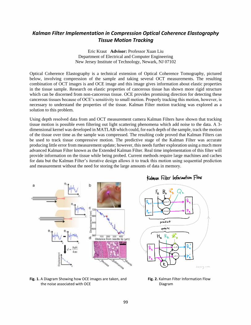
99
Kalman Filter Implementation in Compression Optical Coherence Elastography Tissue Motion Tracking
Eric Kraut Advisor: Professor Xuan Liu
Department of Electrical and Computer Engineering
New Jersey Institute of Technology, Newark, NJ 07102
Optical Coherence Elastography is a technical extension of Optical Coherence Tomography, pictured
below, involving compression of the sample and taking several OCT measurements. The resulting
combination of OCT images is and OCE image and this image gives information about elastic properties
in the tissue sample. Research on elastic properties of cancerous tissue has shown more rigid structure
which can be discerned from non-cancerous tissue. OCE provides promising direction for detecting these
cancerous tissues because of OCE’s sensitivity to small motion. Properly tracking this motion, however, is
necessary to understand the properties of the tissue. Kalman Filter motion tracking was explored as a
solution to this problem.
Using depth resolved data from and OCT measurement camera Kalman Filters have shown that tracking
tissue motion is possible even filtering out light scattering phenomena which add noise to the data. A 3-
dimensional kernel was developed in MATLAB which could, for each depth of the sample, track the motion
of the tissue over time as the sample was compressed. The resulting code proved that Kalman Filters can
be used to track tissue compressive motion. The predictive stage of the Kalman Filter was accurate
producing little error from measurement update; however, this needs further exploration using a much more
advanced Kalman Filter known as the Extended Kalman Filter. Real time implementation of this filter will
provide information on the tissue while being probed. Current methods require large machines and caches
for data but the Kalman Filter’s iterative design allows it to track this motion using sequential prediction
and measurement without the need for storing the large amounts of data in memory.
Fig. 1. A Diagram Showing how OCE images are taken, and Fig. 2. Kalman Filter Information Flow the noise associated with OCE Diagram
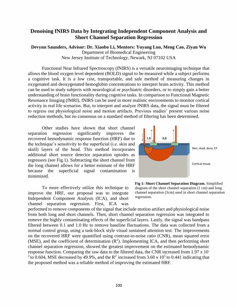
100
Denoising fNIRS Data by Integrating Independent Component Analysis and
Short Channel Separation Regression
Devynn Saunders, Advisor: Dr. Xiaobo Li, Mentors: Yuyang Luo, Meng Cao, Ziyan Wu Department of Biomedical Engineering
New Jersey Institute of Technology, Newark, NJ 07102 USA
Functional Near Infrared Spectroscopy (fNIRS) is a versatile neuroimaging technique that
allows the blood oxygen level dependent (BOLD) signal to be measured while a subject performs
a cognitive task. It is a low cost, transportable, and safe method of measuring changes in
oxygenated and deoxygenated hemoglobin concentrations to interpret brain activity. This method
can be used to study subjects with neurological or psychiatric disorders, or to simply gain a better
understanding of brain functionality during cognitive tasks. In comparison to Functional Magnetic
Resonance Imaging (fMRI), fNIRS can be used in more realistic environments to monitor cortical
activity in real life scenarios. But, to interpret and analyze fNIRS data, the signal must be filtered
to regress out physiological noise and motion artifacts. Previous studies’ present various noise
reduction methods, but no consensus on a standard method of filtering has been determined.
Other studies have shown that short channel
separation regression significantly improves the
recovered hemodynamic response function (HRF) due to
the technique’s sensitivity to the superficial (i.e. skin and
skull) layers of the head. This method incorporates
additional short source detector separation optodes as
regressors (see Fig.1). Subtracting the short channel from
the long channel allows for a better estimate of the HRF
because the superficial signal contamination is
minimized.
To more effectively utilize this technique to
improve the HRF, our proposal was to integrate
Independent Component Analysis (ICA), and short
channel separation regression. First, ICA was
performed to remove components of the signal that include motion artifact and physiological noise
from both long and short channels. Then, short channel separation regression was integrated to
remove the highly contaminating effects of the superficial layers. Lastly, the signal was bandpass
filtered between 0.1 and 1.0 Hz to remove baseline fluctuations. The data was collected from a
normal control group, using a task-block style visual sustained attention test. The improvements
on the recovered HRF were quantified using contrast-to-noise ratio (CNR), mean squared error
(MSE), and the coefficient of determination (R2). Implementing ICA, and then performing short
channel separation regression, showed the greatest improvement on the estimated hemodynamic
response function. Comparing the raw data to the filtered data, the CNR increased from 1.97 x 10-
2 to 0.604, MSE decreased by 49.9%, and the R2 increased from 3.60 x 10⁵ to 0.441 indicating that
the proposed method was a reliable method of improving the estimated HRF.
Fig 1: Short Channel Separation Diagram. Simplified
diagram of the short channel separation (1 cm) and long
channel separation (3cm) used in short channel separation
regression.
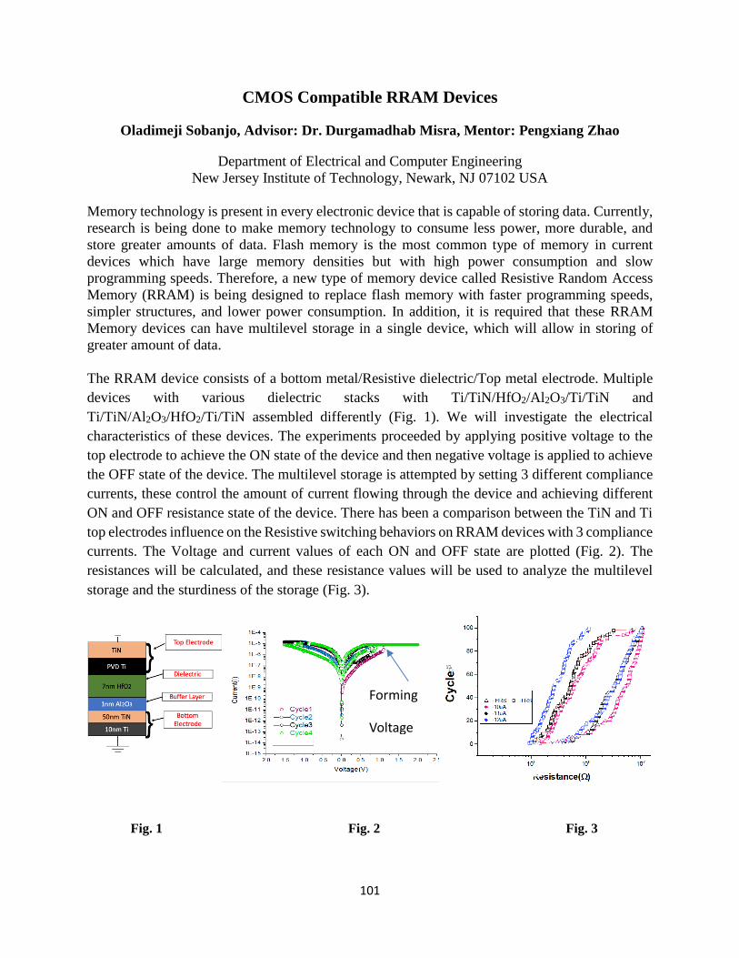
101
CMOS Compatible RRAM Devices
Oladimeji Sobanjo, Advisor: Dr. Durgamadhab Misra, Mentor: Pengxiang Zhao
Department of Electrical and Computer Engineering New Jersey Institute of Technology, Newark, NJ 07102 USA
Memory technology is present in every electronic device that is capable of storing data. Currently,
research is being done to make memory technology to consume less power, more durable, and
store greater amounts of data. Flash memory is the most common type of memory in current
devices which have large memory densities but with high power consumption and slow
programming speeds. Therefore, a new type of memory device called Resistive Random Access
Memory (RRAM) is being designed to replace flash memory with faster programming speeds,
simpler structures, and lower power consumption. In addition, it is required that these RRAM
Memory devices can have multilevel storage in a single device, which will allow in storing of
greater amount of data.
The RRAM device consists of a bottom metal/Resistive dielectric/Top metal electrode. Multiple
devices with various dielectric stacks with Ti/TiN/HfO2/Al2O3/Ti/TiN and
Ti/TiN/Al2O3/HfO2/Ti/TiN assembled differently (Fig. 1). We will investigate the electrical
characteristics of these devices. The experiments proceeded by applying positive voltage to the
top electrode to achieve the ON state of the device and then negative voltage is applied to achieve
the OFF state of the device. The multilevel storage is attempted by setting 3 different compliance
currents, these control the amount of current flowing through the device and achieving different
ON and OFF resistance state of the device. There has been a comparison between the TiN and Ti
top electrodes influence on the Resistive switching behaviors on RRAM devices with 3 compliance
currents. The Voltage and current values of each ON and OFF state are plotted (Fig. 2). The
resistances will be calculated, and these resistance values will be used to analyze the multilevel
storage and the sturdiness of the storage (Fig. 3).
Fig. 1 Fig. 2 Fig. 3
Forming
Voltage

102
103
Exploring the Viability of a PLSR-based Machine Learning Method for
Predicting Circadian Phase in Cancer Patients
Elizabeth Epstein, Karen Reyes, Evan Fagan, Fritz Huggins
Mentor: Jonathan Trinidad
Advisor: Casey Diekman
Department of Mathematical Sciences
New Jersey Institute of Technology, Newark NJ 07102
The circadian clock is an internal biological timekeeping system that regulates daily patterns in
physiology and behavior, such as an individual’s sleep-wake cycle. The molecular mechanism
underlying circadian rhythm generation is an intracellular clock composed of transcriptional and
translational feedback loops, which leads to 24-hour rhythms in gene expression. The tendency for
gene expression to peak at specific times of the day has implications in cancer therapy, as the effect
of such treatment is tied to the manner in which genes are expressed at the time of administration.
However, because there is significant variation in the phase of circadian rhythms across
individuals, the practical application of circadian-based chronotherapy requires an efficient and
cost-effective means through which a person’s unique circadian phase can be measured. The
currently accepted system used to assess an individual’s circadian rhythm is a tedious and
expensive process that necessitates gathering several blood samples from an individual over a
relatively short period of time. While effective, this system is not practical in a clinical setting.
This project aims to explore a recently developed method for assessing an individual’s circadian
phase more efficiently. While a number of methods of varying levels of accuracy have been
developed, most require a minimum of two samples to be taken from an individual. The Partial
Least Squares Regression (PLSR) method developed by Laing et al. (2017) stands out in the fact
that it requires only a single blood draw. The PLSR method uses a machine learning model to
extrapolate circadian time based on an assay of about 26,000 genes. The model in its current form
has only been validated for use in blood samples from healthy individuals and has never been
tested on cancer patients.
We are gaining familiarity with Laing et al.’s method by first applying it to a new dataset from
adipose tissue that was not part of the training set for the machine learning algorithm. Next, we
will apply the method to publicly available gene expression datasets from tumor samples to assess
how well the algorithm can predict circadian phase in cancerous tissues.
Laing, Emma E, et al. “Blood Transcriptome Based Biomarkers for Human Circadian Phase.” ELife, vol. 6, 2017,
doi:10.7554/elife.20214.
104
Mathematical Modeling of Circadian Rhythms, Tumor Growth, and
Radiotherapy
Mariia Goriachi, Luis Lara, Shuhrah Chowdhury
Mentors: Jonathan Trinidad and Emel Khan
Advisor: Casey Diekman
Department of Mathematical Sciences
New Jersey Institute of Technology, Newark, NJ 07102
The goal of this project is to study how to effectively utilize circadian rhythms in aiding
drug delivery against tumor growth. Circadian rhythms are 24-hour cycles in physiological
processes, such as cell growth and sleep/wake. In a number of studies, researchers have shown that
disruption of the circadian clock can increase or decrease the cell population growth rate. The
model presented in El Cheikh et al.10 is the first step in developing a multiscale model for the
interaction between the circadian clock and the cell cycle. Another paper, written by Checkley et
al.11, discussed a mathematical model for cancer cell population growth and simulated its reaction
to a combination of therapies.
In this project, we aim to integrate these two models and analyze the best time of day to
administer therapies to cancer patients. We hypothesize that by taking into account the phase of
circadian rhythms in healthy and cancer cells, appropriately timed therapies will be more effective
in killing cancer cells while being less toxic to healthy cells. We will implement our model in
MATLAB and conduct simulations to explore the complex interactions between the cell cycle, the
circadian clock, and tumor growth. Combined circadian rhythm/cell population and drug PK-PD
models can be a helpful tool for chronotherapy of cancer patients. Thus, this project will help
provide a foundation to develop more personalized and effective treatments specific to each
patient’s circadian phase and tumor properties.
10 El Cheikh, R., Bernard, S., & Khatib, N. E. (2014). Modeling circadian clock–cell cycle interaction effects on cell population
growth rates. Journal of Theoretical Biology, 363, 318-331. doi:10.1016/j.jtbi.2014.08.008
11 Checkley, S., Maccallum, L., Yates, J., Jasper, P., Luo, H., Tolsma, J., & Bendtsen, C. (2016). Bridging the gap between in
vitro and in vivo: Dose and schedule predictions for the ATR inhibitor AZD6738. Scientific Reports, 6(1). doi:10.1038/srep16545

105
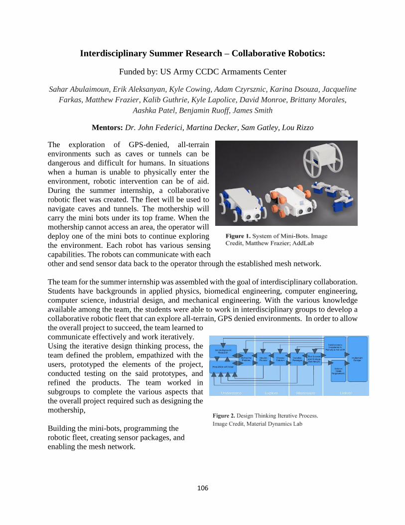
106
Interdisciplinary Summer Research – Collaborative Robotics:
Funded by: US Army CCDC Armaments Center
Sahar Abulaimoun, Erik Aleksanyan, Kyle Cowing, Adam Czyrsznic, Karina Dsouza, Jacqueline
Farkas, Matthew Frazier, Kalib Guthrie, Kyle Lapolice, David Monroe, Brittany Morales,
Aashka Patel, Benjamin Ruoff, James Smith
Mentors: Dr. John Federici, Martina Decker, Sam Gatley, Lou Rizzo
The exploration of GPS-denied, all-terrain
environments such as caves or tunnels can be
dangerous and difficult for humans. In situations
when a human is unable to physically enter the
environment, robotic intervention can be of aid.
During the summer internship, a collaborative
robotic fleet was created. The fleet will be used to
navigate caves and tunnels. The mothership will
carry the mini bots under its top frame. When the
mothership cannot access an area, the operator will
deploy one of the mini bots to continue exploring
the environment. Each robot has various sensing
capabilities. The robots can communicate with each
other and send sensor data back to the operator through the established mesh network.
The team for the summer internship was assembled with the goal of interdisciplinary collaboration.
Students have backgrounds in applied physics, biomedical engineering, computer engineering,
computer science, industrial design, and mechanical engineering. With the various knowledge
available among the team, the students were able to work in interdisciplinary groups to develop a
collaborative robotic fleet that can explore all-terrain, GPS denied environments. In order to allow
the overall project to succeed, the team learned to
communicate effectively and work iteratively.
Using the iterative design thinking process, the
team defined the problem, empathized with the
users, prototyped the elements of the project,
conducted testing on the said prototypes, and
refined the products. The team worked in
subgroups to complete the various aspects that
the overall project required such as designing the
mothership,
Building the mini-bots, programming the
robotic fleet, creating sensor packages, and
enabling the mesh network.

107
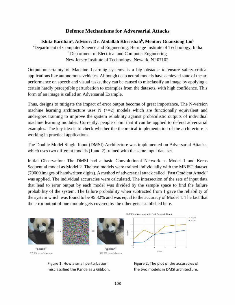
108
Defence Mechanisms for Adversarial Attacks
Ishita Bardhana, Advisor: Dr. Abdallah Khreishahb, Mentor: Guanxiong Liub
aDepartment of Computer Science and Engineering, Heritage Institute of Technology, India bDepartment of Electrical and Computer Engineering
New Jersey Institute of Technology, Newark, NJ 07102.
Output uncertainty of Machine Learning systems is a big obstacle to ensure safety-critical
applications like autonomous vehicles. Although deep neural models have achieved state of the art
performance on speech and visual tasks, they can be caused to misclassify an image by applying a
certain hardly perceptible perturbation to examples from the datasets, with high confidence. This
form of an image is called an Adversarial Example.
Thus, designs to mitigate the impact of error output become of great importance. The N-version
machine learning architecture uses N (>=2) models which are functionally equivalent and
undergoes training to improve the system reliability against probabilistic outputs of individual
machine learning modules. Currently, people claim that it can be applied to defend adversarial
examples. The key idea is to check whether the theoretical implementation of the architecture is
working in practical applications.
The Double Model Single Input (DMSI) Architecture was implemented on Adversarial Attacks,
which uses two different models (1 and 2) trained with the same input data set.
Initial Observation: The DMSI had a basic Convolutional Network as Model 1 and Keras
Sequential model as Model 2. The two models were trained individually with the MNIST dataset
(70000 images of handwritten digits). A method of adversarial attack called “Fast Gradient Attack”
was applied. The individual accuracies were calculated. The intersection of the sets of input data
that lead to error output by each model was divided by the sample space to find the failure
probability of the system. The failure probability when subtracted from 1 gave the reliability of
the system which was found to be 95.32% and was equal to the accuracy of Model 1. The fact that
the error output of one module gets covered by the other gets established here.
Figure 1: How a small perturbation
misclassified the Panda as a Gibbon.
Figure 2: The plot of the accuracies of
the two models in DMSI architecture.
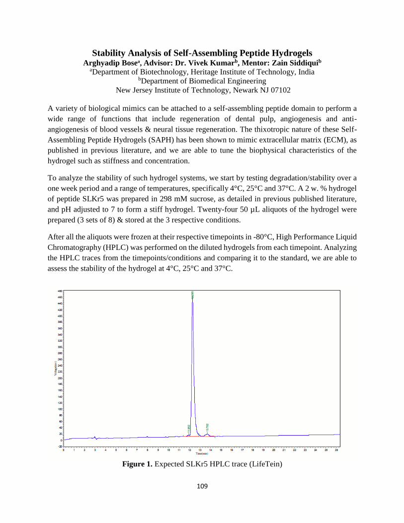
109
Stability Analysis of Self-Assembling Peptide Hydrogels
Arghyadip Bosea, Advisor: Dr. Vivek Kumarb, Mentor: Zain Siddiquib
aDepartment of Biotechnology, Heritage Institute of Technology, India bDepartment of Biomedical Engineering
New Jersey Institute of Technology, Newark NJ 07102
A variety of biological mimics can be attached to a self-assembling peptide domain to perform a
wide range of functions that include regeneration of dental pulp, angiogenesis and anti-
angiogenesis of blood vessels & neural tissue regeneration. The thixotropic nature of these Self-
Assembling Peptide Hydrogels (SAPH) has been shown to mimic extracellular matrix (ECM), as
published in previous literature, and we are able to tune the biophysical characteristics of the
hydrogel such as stiffness and concentration.
To analyze the stability of such hydrogel systems, we start by testing degradation/stability over a
one week period and a range of temperatures, specifically 4°C, 25°C and 37°C. A 2 w. % hydrogel
of peptide SLKr5 was prepared in 298 mM sucrose, as detailed in previous published literature,
and pH adjusted to 7 to form a stiff hydrogel. Twenty-four 50 µL aliquots of the hydrogel were
prepared (3 sets of 8) & stored at the 3 respective conditions.
After all the aliquots were frozen at their respective timepoints in -80°C, High Performance Liquid
Chromatography (HPLC) was performed on the diluted hydrogels from each timepoint. Analyzing
the HPLC traces from the timepoints/conditions and comparing it to the standard, we are able to
assess the stability of the hydrogel at 4°C, 25°C and 37°C.
Figure 1. Expected SLKr5 HPLC trace (LifeTein)
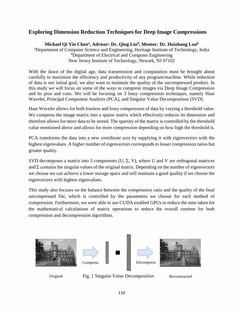
110
Original U Σ
V
Decompress
Reconstructed
Compress
Exploring Dimension Reduction Techniques for Deep Image Compressions
Michael Qi Yin Chena, Advisor: Dr. Qing Liub, Mentor: Dr. Huizhang Luob
aDepartment of Computer Science and Engineering, Heritage Institute of Technology, India
bDepartment of Electrical and Computer Engineering
New Jersey Institute of Technology, Newark, NJ 07102
With the dawn of the digital age, data transmission and computation must be brought about
carefully to maximize the efficiency and productivity of any program/machine. While reduction
of data is our initial goal, we also want to maintain the quality of the uncompressed product. In
this study we will focus on some of the ways to compress images via Deep Image Compression
and its pros and cons. We will be focusing on 3 lossy compression techniques, namely Haar
Wavelet, Principal Component Analysis (PCA), and Singular Value Decomposition (SVD).
Haar Wavelet allows for both lossless and lossy compression of data by varying a threshold value.
We compress the image matrix into a sparse matrix which effectively reduces its dimension and
therefore allows for more data to be stored. The sparsity of the matrix is controlled by the threshold
value mentioned above and allows for more compression depending on how high the threshold is.
PCA transforms the data into a new coordinate axis by supplying it with eigenvectors with the
highest eigenvalues. A higher number of eigenvectors corresponds to lesser compression ratios but
greater quality.
SVD decomposes a matrix into 3 components (U, Σ, V), where U and V are orthogonal matrices
and Σ contains the singular values of the original matrix. Depending on the number of eigenvectors
we choose we can achieve a lower storage space and still maintain a good quality if we choose the
eigenvectors with highest eigenvalues.
This study also focuses on the balance between the compression ratio and the quality of the final
uncompressed file, which is controlled by the parameters we choose for each method of
compression. Furthermore, we were able to use CUDA enabled GPUs to reduce the time taken for
the mathematical calculations of matrix operations to reduce the overall runtime for both
compression and decompression algorithms.
Fig. 1 Singular Value Decomposition
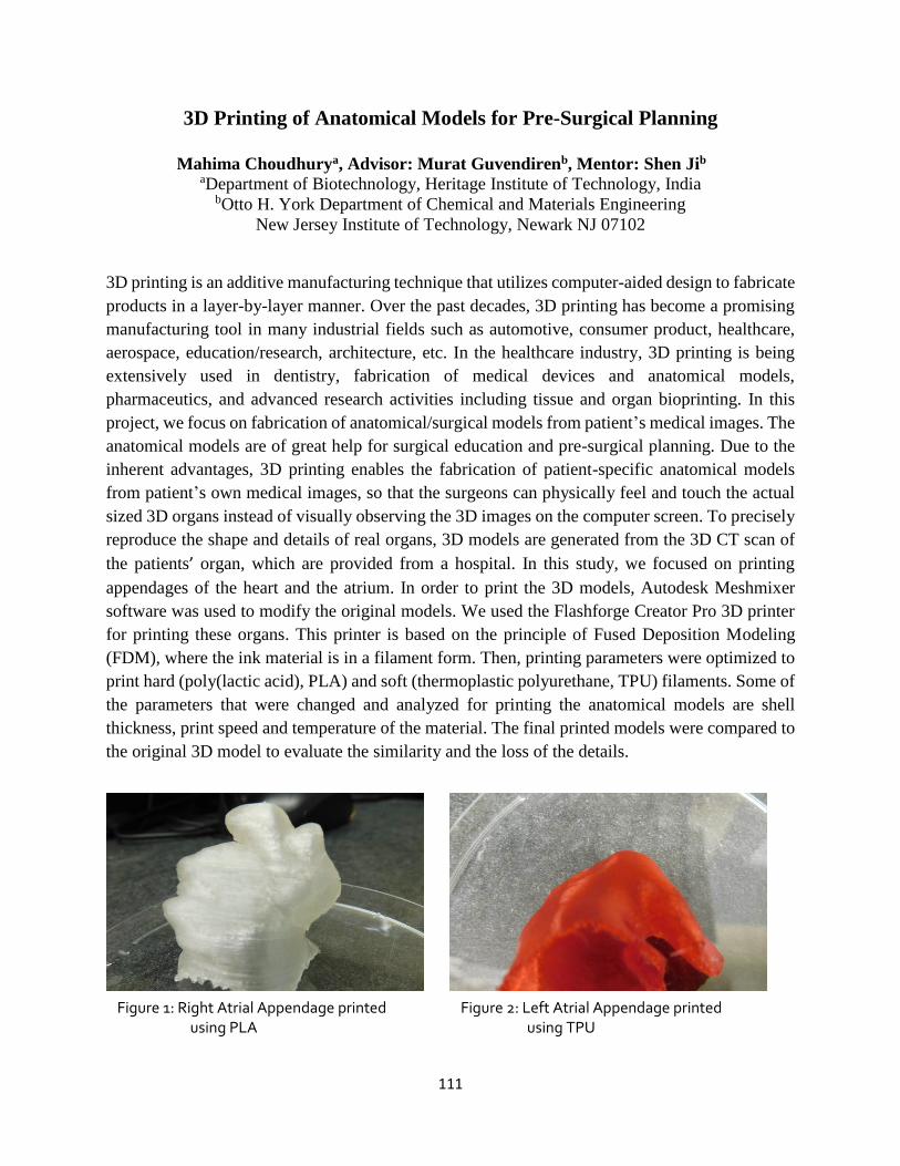
111
3D Printing of Anatomical Models for Pre-Surgical Planning
Mahima Choudhurya, Advisor: Murat Guvendirenb, Mentor: Shen Jib
aDepartment of Biotechnology, Heritage Institute of Technology, India bOtto H. York Department of Chemical and Materials Engineering
New Jersey Institute of Technology, Newark NJ 07102
3D printing is an additive manufacturing technique that utilizes computer-aided design to fabricate
products in a layer-by-layer manner. Over the past decades, 3D printing has become a promising
manufacturing tool in many industrial fields such as automotive, consumer product, healthcare,
aerospace, education/research, architecture, etc. In the healthcare industry, 3D printing is being
extensively used in dentistry, fabrication of medical devices and anatomical models,
pharmaceutics, and advanced research activities including tissue and organ bioprinting. In this
project, we focus on fabrication of anatomical/surgical models from patient’s medical images. The
anatomical models are of great help for surgical education and pre-surgical planning. Due to the
inherent advantages, 3D printing enables the fabrication of patient-specific anatomical models
from patient’s own medical images, so that the surgeons can physically feel and touch the actual
sized 3D organs instead of visually observing the 3D images on the computer screen. To precisely
reproduce the shape and details of real organs, 3D models are generated from the 3D CT scan of
the patients’ organ, which are provided from a hospital. In this study, we focused on printing
appendages of the heart and the atrium. In order to print the 3D models, Autodesk Meshmixer
software was used to modify the original models. We used the Flashforge Creator Pro 3D printer
for printing these organs. This printer is based on the principle of Fused Deposition Modeling
(FDM), where the ink material is in a filament form. Then, printing parameters were optimized to
print hard (poly(lactic acid), PLA) and soft (thermoplastic polyurethane, TPU) filaments. Some of
the parameters that were changed and analyzed for printing the anatomical models are shell
thickness, print speed and temperature of the material. The final printed models were compared to
the original 3D model to evaluate the similarity and the loss of the details.
Figure 1: Right Atrial Appendage printed using PLA
Figure 2: Left Atrial Appendage printed using TPU
112
INVESTIGATING HYDROLYTIC STABILITY AND CHANGES IN
MECHANICAL PROPERTIES OF ELECROSPUN CROSSLINKED
GELATIN SCAFFOLDS
Samriddha Dutta a, Advisor: Dr. Treena Livingston Arinzeh b, Mentor: Richard Vincent b
a Department of Biotechnology, Heritage Institute of Technology, India b Department of Biomedical Engineering,
New Jersey Institute of Technology, Newark, NJ 07102
Osteoarthritis (OA) is a degenerative joint disease that results in the loss of articular cartilage and
normal joint function. It is the most common chronic condition of the joints. Articular cartilage is
an avascular tissue with limited capacity to regenerate or heal after injury or degenerative disease.
While current therapies can alleviate symptomatic pain, surgical interventions do not restore
normal hyaline cartilage. Instead fibrocartilage forms which is mechanically inferior to native
hyaline cartilage. Therefore, a growing need exists for an effective treatment to repair articular
cartilage. An approach to promoting articular cartilage repair would be the use of tissue
engineering constructs called scaffolds that closely mimic the native cartilage microenvironment.
Electrospinning is a widely used process for fabricating these scaffolds to repair or regenerate
tissues. In this study, we have electrospun gelatin, a commonly used biological material that
promotes cell adhesion. However, it does not have suitable hydrolytic stability for long term
biological applications unless chemically crosslinked. Hence, chemical crosslinking was
performed using N-(3-dimethylaminopropyl)-N’-ethyl carbodiimide (EDC) with N-
hydroxysulfosuccinimide (NHS). In this study, the effect of crosslinking time and concentration
on hydrolytic stability and mechanical properties was assessed via changes in scaffold degradation,
tensile mechanical properties and rheological properties.

113
Minimize Energy Consumption of Hadoop Jobs Under Deadline Constraints
Shivansh Gupta, Advisor: Dr. Chase Wu, and Mentor: Qianwen Ye, Ph.D. Student
B.R.C.M. College of Engineering and Technology
Department of Computer Science
New Jersey Institute of Technology
Newark, NJ 07102, USA
Abstract:
Big data applications require massive computing, networking, and storage resources and are
typically executed in Hadoop systems deployed on large clusters for various data processing and
analysis purposes. These applications have become a main consumer of energy in today’s data
centers, and energy efficiency is critical to the sustainability of big data systems. The goal of this
research is to minimize the dynamic energy consumed by a batch of deadline-constrained Hadoop
jobs on a Hadoop cluster while meeting the deadline of each Hadoop job. Extensive experiments
are performed on a Hadoop cluster to measure the energy consumption and execution time of
different Hadoop jobs. In these experiments, we consider a homogeneous Hadoop cluster and a
workflow model consisting of n Hadoop jobs, each of which contains a set of map and reduce
tasks. We measure the energy consumption and execution time for each map task and reduce task.
Such performance measurements are then used in the design of a job mapping scheme in Hadoop
so that all jobs are completed within their respective deadlines and the energy consumption is
minimized. The proposed method, although focused on Hadoop systems, could be expanded and
applied to other big data systems.
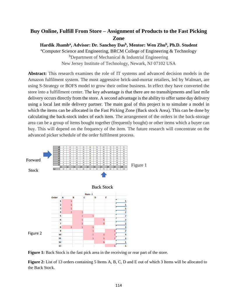
114
Buy Online, Fulfill From Store – Assignment of Products to the Fast Picking
Zone
Hardik Jhamba, Advisor: Dr. Sanchoy Dasb, Mentor: Wen Zhub, Ph.D. Student aComputer Science and Engineering, BRCM College of Engineering & Technology
bDepartment of Mechanical & Industrial Engineering
New Jersey Institute of Technology, Newark, NJ 07102 USA
Abstract: This research examines the role of IT systems and advanced decision models in the
Amazon fulfilment system. The most aggressive brick-and-mortar retailers, led by Walmart, are
using S-Strategy or BOFS model to grow their online business. In effect they have converted the
store into a fulfillment center. The key advantage is that there are no transshipments and last mile
delivery occurs directly from the store. A second advantage is the ability to offer same day delivery
using a local last mile delivery partner. The main goal of this project is to simulate a model in
which the items can be allocated in the Fast Picking Zone (Back stock Area). This can be done by
calculating the back-stock index of each item. The arrangement of the orders in the back-storage
area can be a group of items bought together (frequently bought) or other items which a buyer can
buy. This will depend on the frequency of the item. The future research will concentrate on the
advanced picker schedule of the order fulfilment process.
Figure 1
Figure 2
Figure 1: Back Stock is the fast pick area in the receiving or rear part of the store.
Figure 2: List of 13 orders containing 5 Items A, B, C, D and E out of which 3 Items will be allocated to
the Back Stock.
Forward
Stock
Back Stock

115
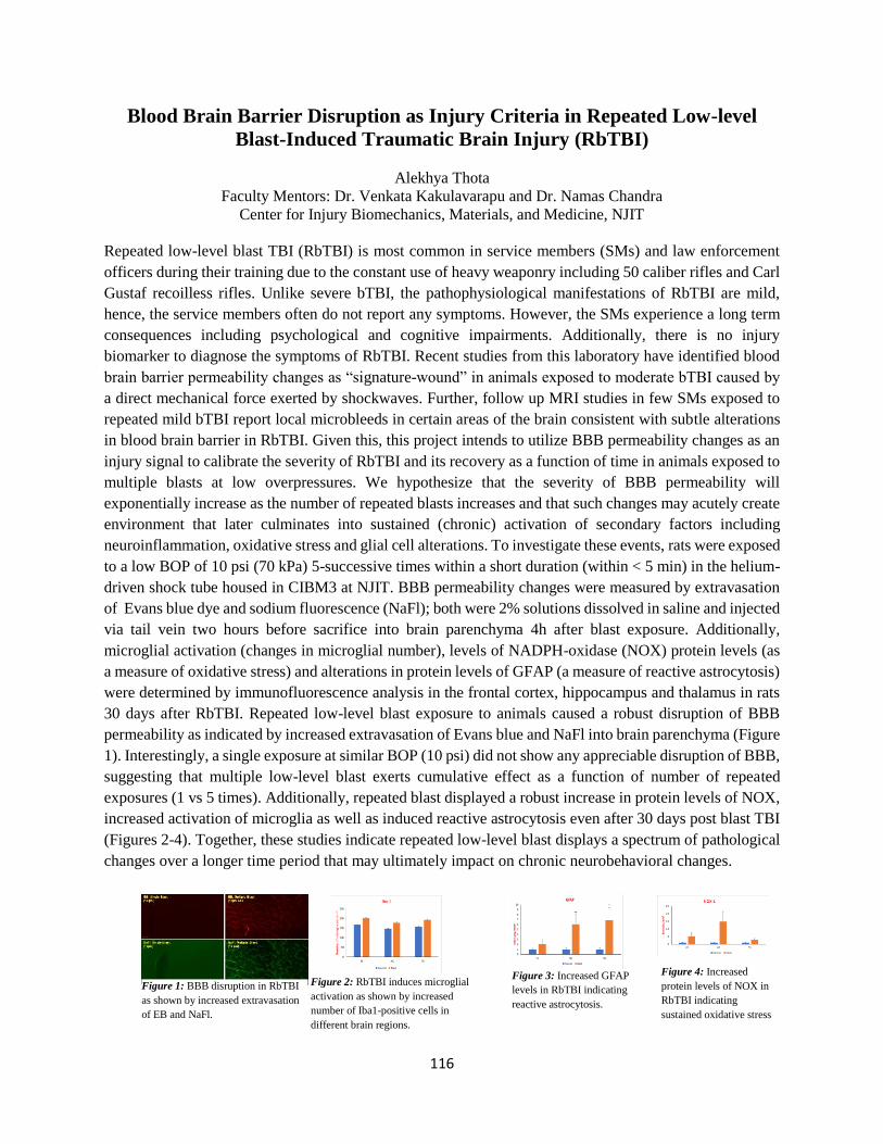
116
Blood Brain Barrier Disruption as Injury Criteria in Repeated Low-level
Blast-Induced Traumatic Brain Injury (RbTBI)
Alekhya Thota
Faculty Mentors: Dr. Venkata Kakulavarapu and Dr. Namas Chandra
Center for Injury Biomechanics, Materials, and Medicine, NJIT
Repeated low-level blast TBI (RbTBI) is most common in service members (SMs) and law enforcement
officers during their training due to the constant use of heavy weaponry including 50 caliber rifles and Carl
Gustaf recoilless rifles. Unlike severe bTBI, the pathophysiological manifestations of RbTBI are mild,
hence, the service members often do not report any symptoms. However, the SMs experience a long term
consequences including psychological and cognitive impairments. Additionally, there is no injury
biomarker to diagnose the symptoms of RbTBI. Recent studies from this laboratory have identified blood
brain barrier permeability changes as “signature-wound” in animals exposed to moderate bTBI caused by
a direct mechanical force exerted by shockwaves. Further, follow up MRI studies in few SMs exposed to
repeated mild bTBI report local microbleeds in certain areas of the brain consistent with subtle alterations
in blood brain barrier in RbTBI. Given this, this project intends to utilize BBB permeability changes as an
injury signal to calibrate the severity of RbTBI and its recovery as a function of time in animals exposed to
multiple blasts at low overpressures. We hypothesize that the severity of BBB permeability will
exponentially increase as the number of repeated blasts increases and that such changes may acutely create
environment that later culminates into sustained (chronic) activation of secondary factors including
neuroinflammation, oxidative stress and glial cell alterations. To investigate these events, rats were exposed
to a low BOP of 10 psi (70 kPa) 5-successive times within a short duration (within < 5 min) in the helium-
driven shock tube housed in CIBM3 at NJIT. BBB permeability changes were measured by extravasation
of Evans blue dye and sodium fluorescence (NaFl); both were 2% solutions dissolved in saline and injected
via tail vein two hours before sacrifice into brain parenchyma 4h after blast exposure. Additionally,
microglial activation (changes in microglial number), levels of NADPH-oxidase (NOX) protein levels (as
a measure of oxidative stress) and alterations in protein levels of GFAP (a measure of reactive astrocytosis)
were determined by immunofluorescence analysis in the frontal cortex, hippocampus and thalamus in rats
30 days after RbTBI. Repeated low-level blast exposure to animals caused a robust disruption of BBB
permeability as indicated by increased extravasation of Evans blue and NaFl into brain parenchyma (Figure
1). Interestingly, a single exposure at similar BOP (10 psi) did not show any appreciable disruption of BBB,
suggesting that multiple low-level blast exerts cumulative effect as a function of number of repeated
exposures (1 vs 5 times). Additionally, repeated blast displayed a robust increase in protein levels of NOX,
increased activation of microglia as well as induced reactive astrocytosis even after 30 days post blast TBI
(Figures 2-4). Together, these studies indicate repeated low-level blast displays a spectrum of pathological
changes over a longer time period that may ultimately impact on chronic neurobehavioral changes.
Figure 1: BBB disruption in RbTBI
as shown by increased extravasation
of EB and NaFl.
Figure 2: RbTBI induces microglial
activation as shown by increased
number of Iba1-positive cells in
different brain regions.
Figure 3: Increased GFAP
levels in RbTBI indicating
reactive astrocytosis.
Figure 4: Increased
protein levels of NOX in
RbTBI indicating
sustained oxidative stress

117
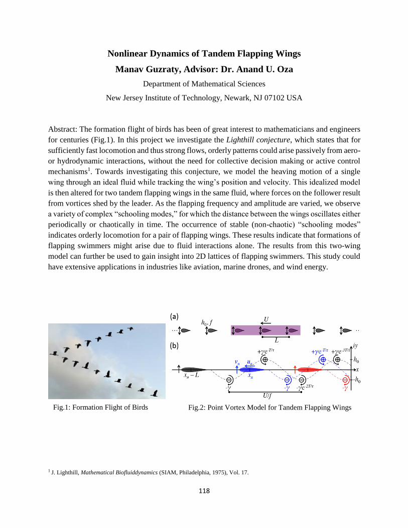
118
Nonlinear Dynamics of Tandem Flapping Wings
Manav Guzraty, Advisor: Dr. Anand U. Oza
Department of Mathematical Sciences
New Jersey Institute of Technology, Newark, NJ 07102 USA
Abstract: The formation flight of birds has been of great interest to mathematicians and engineers
for centuries (Fig.1). In this project we investigate the Lighthill conjecture, which states that for
sufficiently fast locomotion and thus strong flows, orderly patterns could arise passively from aero-
or hydrodynamic interactions, without the need for collective decision making or active control
mechanisms1. Towards investigating this conjecture, we model the heaving motion of a single
wing through an ideal fluid while tracking the wing’s position and velocity. This idealized model
is then altered for two tandem flapping wings in the same fluid, where forces on the follower result
from vortices shed by the leader. As the flapping frequency and amplitude are varied, we observe
a variety of complex “schooling modes,” for which the distance between the wings oscillates either
periodically or chaotically in time. The occurrence of stable (non-chaotic) “schooling modes”
indicates orderly locomotion for a pair of flapping wings. These results indicate that formations of
flapping swimmers might arise due to fluid interactions alone. The results from this two-wing
model can further be used to gain insight into 2D lattices of flapping swimmers. This study could
have extensive applications in industries like aviation, marine drones, and wind energy.
1 J. Lighthill, Mathematical Biofluiddynamics (SIAM, Philadelphia, 1975), Vol. 17.
Fig.2: Point Vortex Model for Tandem Flapping Wings Fig.1: Formation Flight of Birds
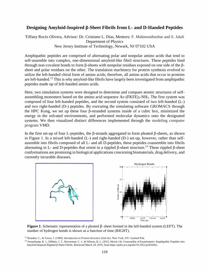
119
Designing Amyloid-Inspired β-Sheet Fibrils from L- and D-Handed Peptides
Tiffany Rocio Olivera, Advisor: Dr. Cristiano L. Dias, Mentors: F. Mahmoudinobar and S. Jalali
Department of Physics
New Jersey Institute of Technology, Newark, NJ 07102 USA
Amphipathic peptides are comprised of alternating polar and nonpolar amino acids that tend to
self-assemble into complex, one-dimensional amyloid-like fibril structures. These peptides bind
through non-covalent bonds to form β-sheets with nonpolar residues exposed on one side of the β-
sheet and polar residues on the other. The translation machinery for protein synthesis evolved to
utilize the left-handed chiral form of amino acids; therefore, all amino acids that occur in proteins
are left-handed.12 This is why amyloid-like fibrils have largely been investigated from amphipathic
peptides made up of left-handed amino acids.
Here, two simulation systems were designed to determine and compare atomic structures of self-
assembling monomers based on the amino acid sequence Ac-(FKFE)2-NH2. The first system was
composed of four left-handed peptides, and the second system consisted of two left-handed (L-)
and two right-handed (D-) peptides. By executing the simulating software GROMACS through
the HPC Kong, we set up these four β-stranded systems inside of a cubic box, minimized the
energy in the solvated environments, and performed molecular dynamics onto the designated
systems. We then visualized distinct differences implemented through the modeling computer
program VMD.
In the first set-up of four L-peptides, the β-strands aggregated to form pleated β-sheets, as shown
in Figure 1. In a mixed left-handed (L-) and right-handed (D-) set-up, however, rather than self-
assemble into fibrils composed of all L- and all D-peptides, these peptides coassemble into fibrils
alternating in L- and D-peptides that orient in a rippled β-sheet structure.13 These rippled β-sheet
conformations are promising in biological applications concerning biomaterials, drug delivery, and
currently incurable diseases.
12 Branden, C., & Tooze, J. (1999). Introduction to Protein Structure (2nd ed.). New York, NY: Garland Pub. 13 Swanekamp, R. J., DiMaio, J. T., Bowerman, C. J., & Nilsson, B. L. (2012, March 14). Coassembly of Enantiomeric Amphipathic Peptides into
Amyloid-Inspired Rippled β-Sheet Fibrils. Retrieved March 18, 2019, from https://pubs.acs.org/doi/10.1021/ja301642c.
Figure 1. Schematic representation of a pleated β–sheet formed in the left-handed system (LEFT). The
number of hydrogen bonds is shown as a function of time (RIGHT).