bones and joints chapter 6. skeleton bones joints connective tissue
TRANSCRIPT
BO N E S A N D J O I N T S
CHAPTER 6
SKELETON
• Bones• Joints• Connective tissue
FUNCTIONS OF BONES
Several functions• To serve as a firm framework for the body• To protect delicate structures such as the brain
and spinal cord• To work as levers to produce movement• To store calcium salts• To produce blood cells
BONE STRUCTURE
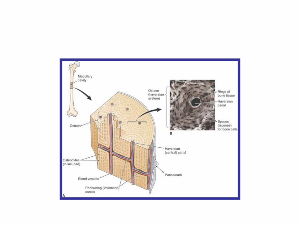
Types of bone (osseous) tissue• Compact bone• Haversion systems (osteons)
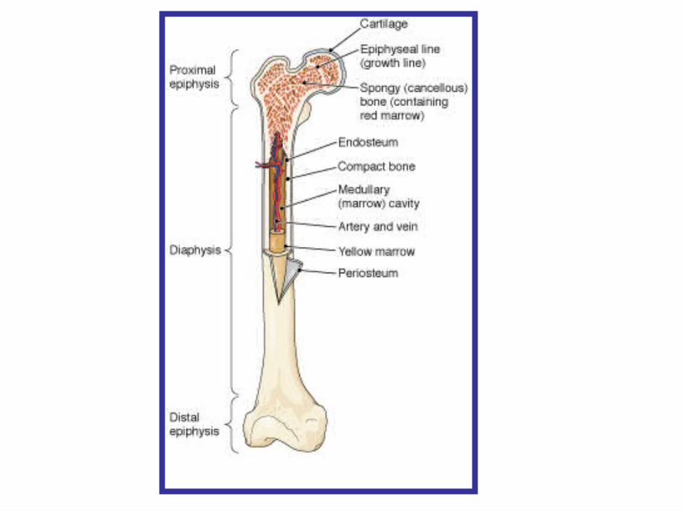
• Spongy (cancellous) bone• Bone marrow• Red marrow• Yellow marrow
• Bone membranes• Periosteum• Endosteum
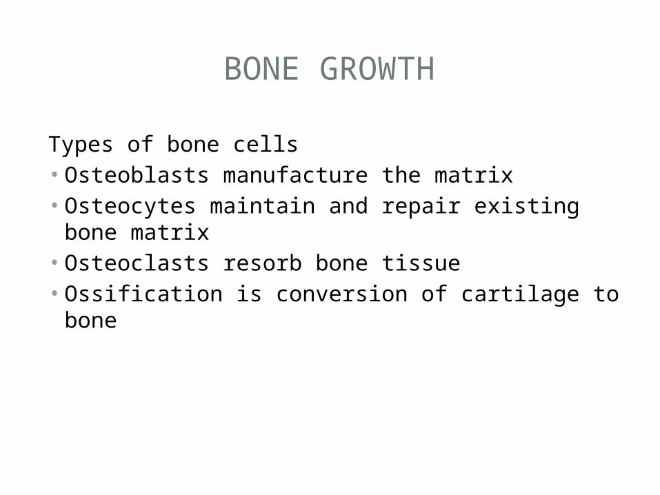
BONE GROWTH
Types of bone cells• Osteoblasts manufacture the matrix• Osteocytes maintain and repair existing bone
matrix• Osteoclasts resorb bone tissue• Ossification is conversion of cartilage to bone
FORMATION OF LONG BONE
• Cartilage begins to turn into bone• Epiphyseal plates develop across bone ends • Bones continue to lengthen • Bones stop lengthening • Bone resorption and formation continues
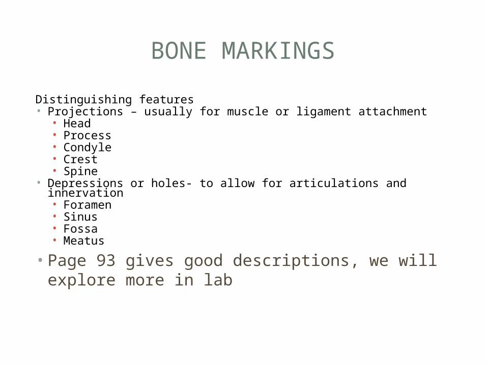
BONE MARKINGS
Distinguishing features• Projections – usually for muscle or ligament attachment• Head• Process• Condyle• Crest• Spine
• Depressions or holes- to allow for articulations and innervation • Foramen• Sinus• Fossa• Meatus
• Page 93 gives good descriptions, we will explore more in lab
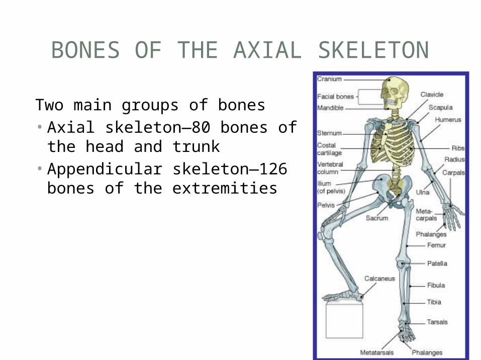
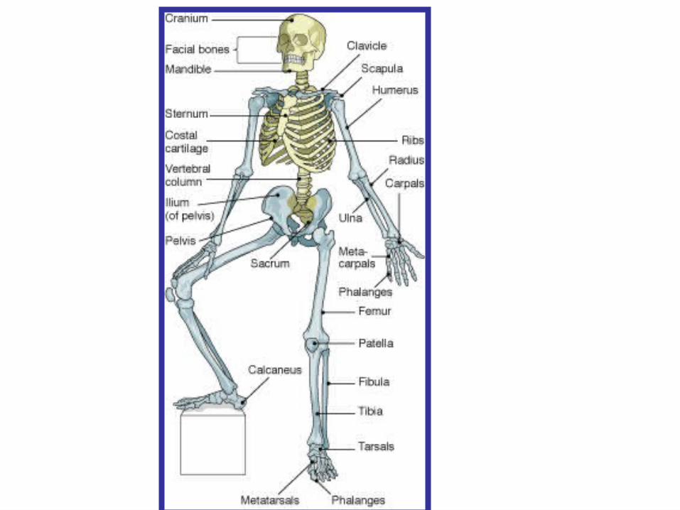
BONES OF THE AXIAL SKELETON
Two main groups of bones• Axial skeleton—80 bones of
the head and trunk• Appendicular skeleton—126
bones of the extremities
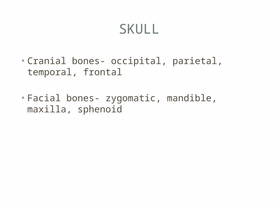
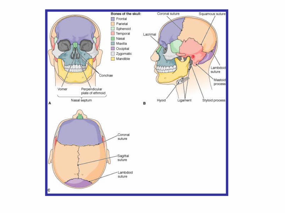
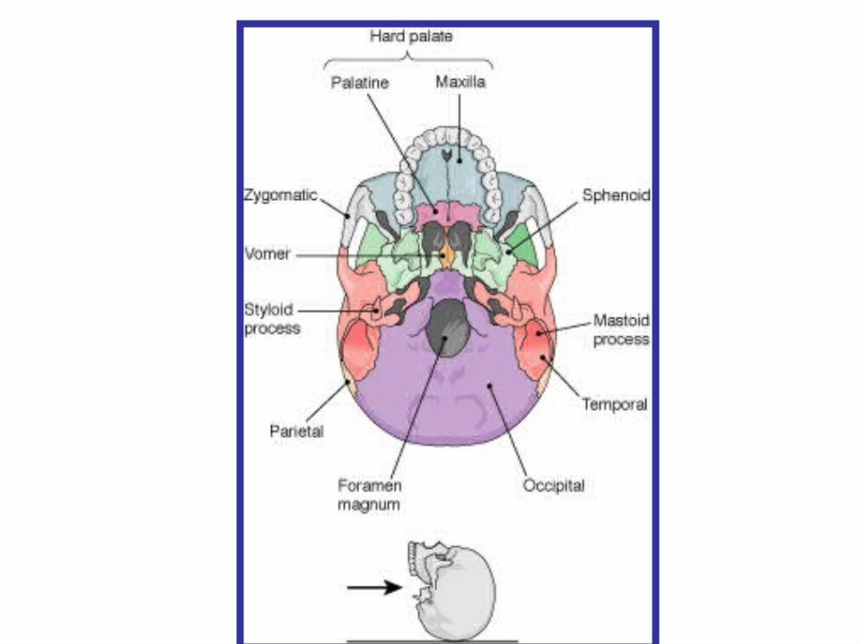
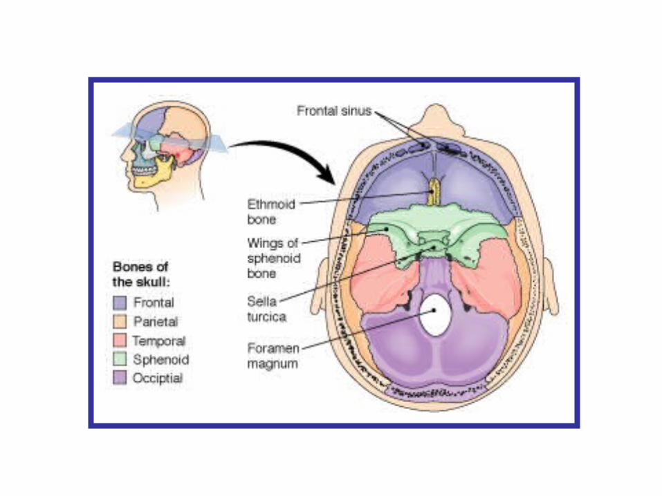
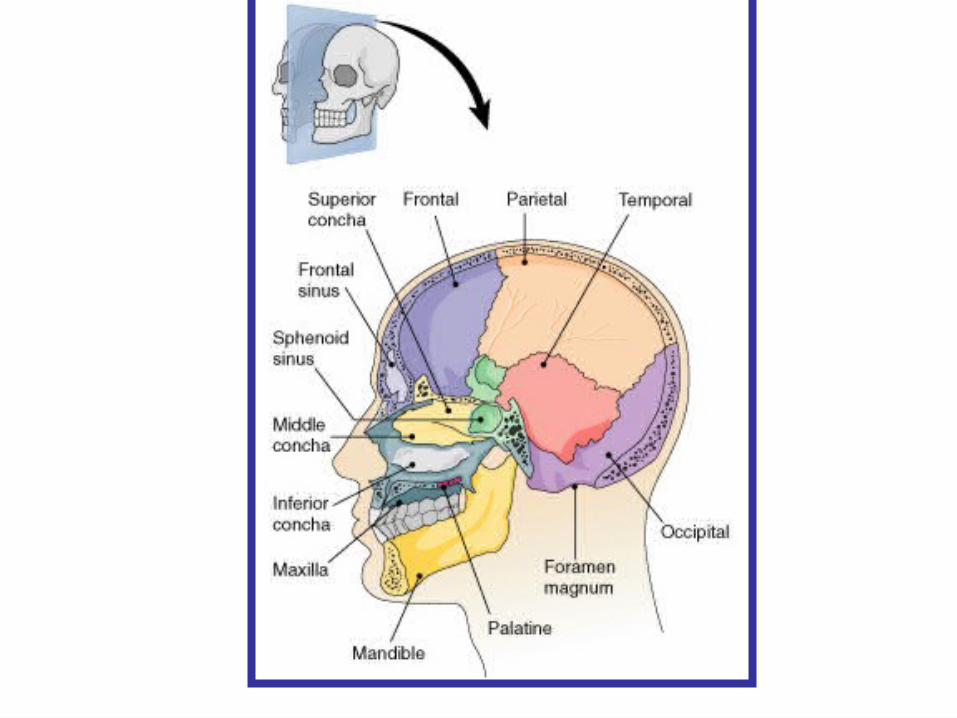
SKULL
• Cranial bones- occipital, parietal, temporal, frontal
• Facial bones- zygomatic, mandible, maxilla, sphenoid
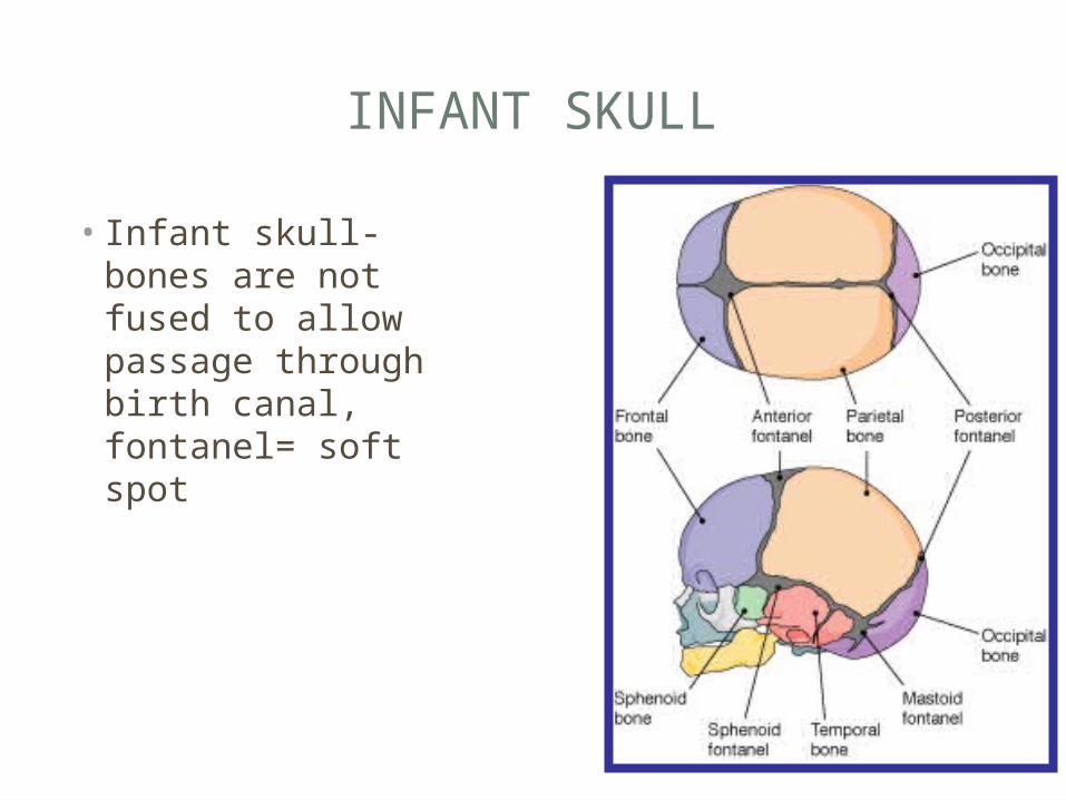
INFANT SKULL
• Infant skull- bones are not fused to allow passage through birth canal, fontanel= soft spot
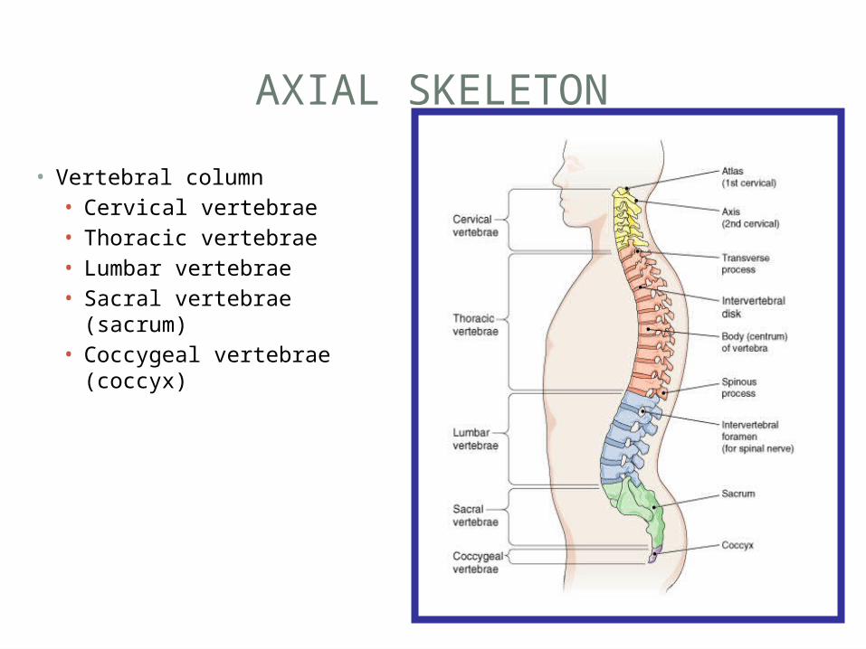
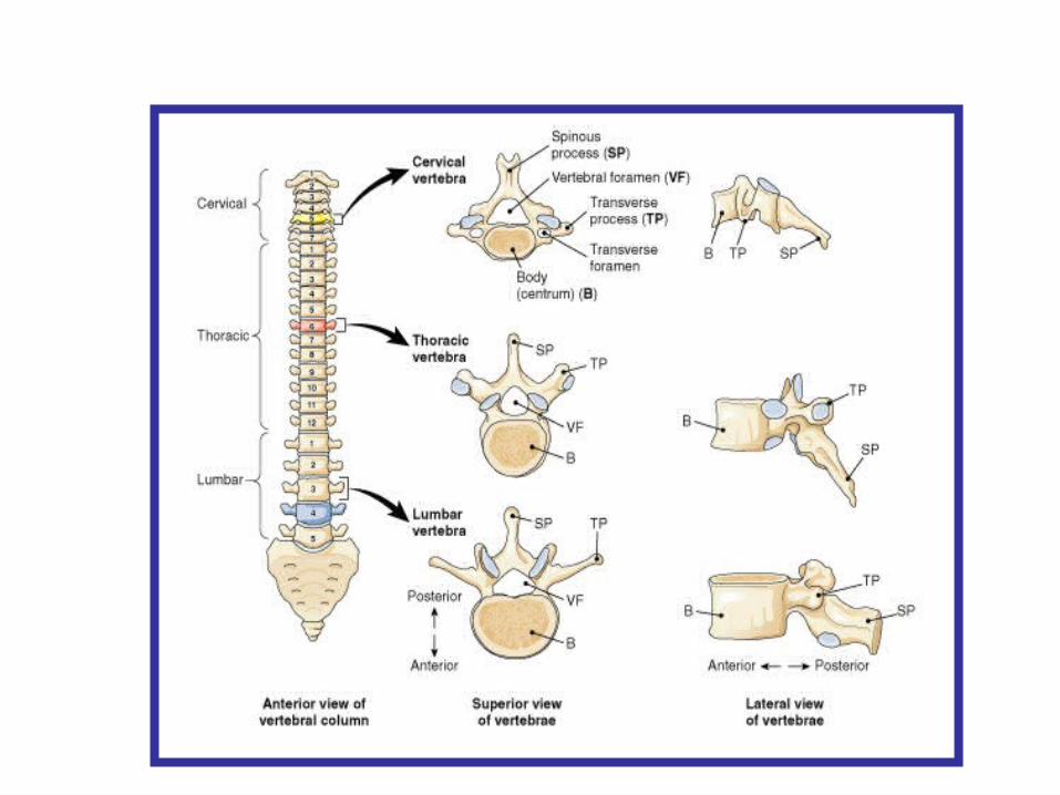
AXIAL SKELETON
• Vertebral column• Cervical vertebrae• Thoracic vertebrae• Lumbar vertebrae• Sacral vertebrae (sacrum)• Coccygeal vertebrae
(coccyx)
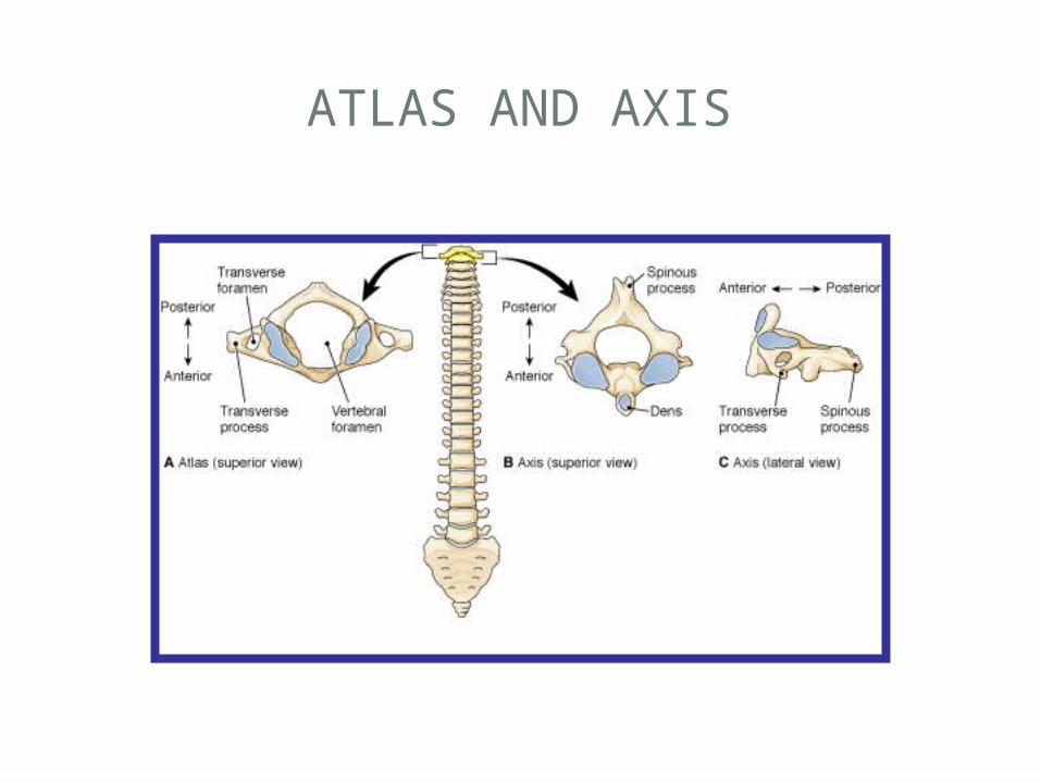
ATLAS AND AXIS
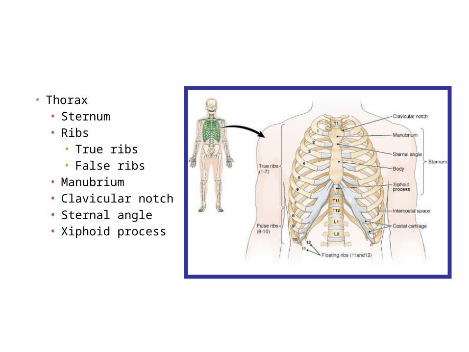
• Thorax• Sternum• Ribs• True ribs• False ribs
• Manubrium• Clavicular notch• Sternal angle• Xiphoid process
APPENDICULAR SKELETON
Two divisions • Upper• Lower
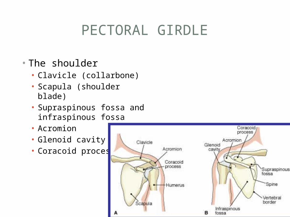
PECTORAL GIRDLE
• The shoulder • Clavicle (collarbone)• Scapula (shoulder blade)• Supraspinous fossa and
infraspinous fossa• Acromion• Glenoid cavity• Coracoid process
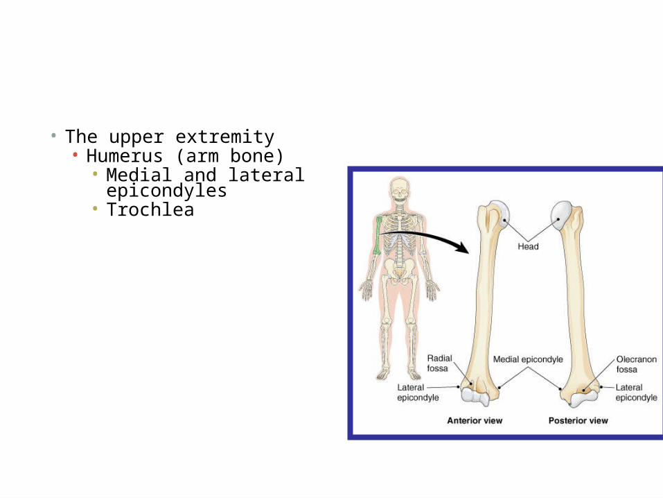
• The upper extremity• Humerus (arm bone)• Medial and lateral
epicondyles• Trochlea
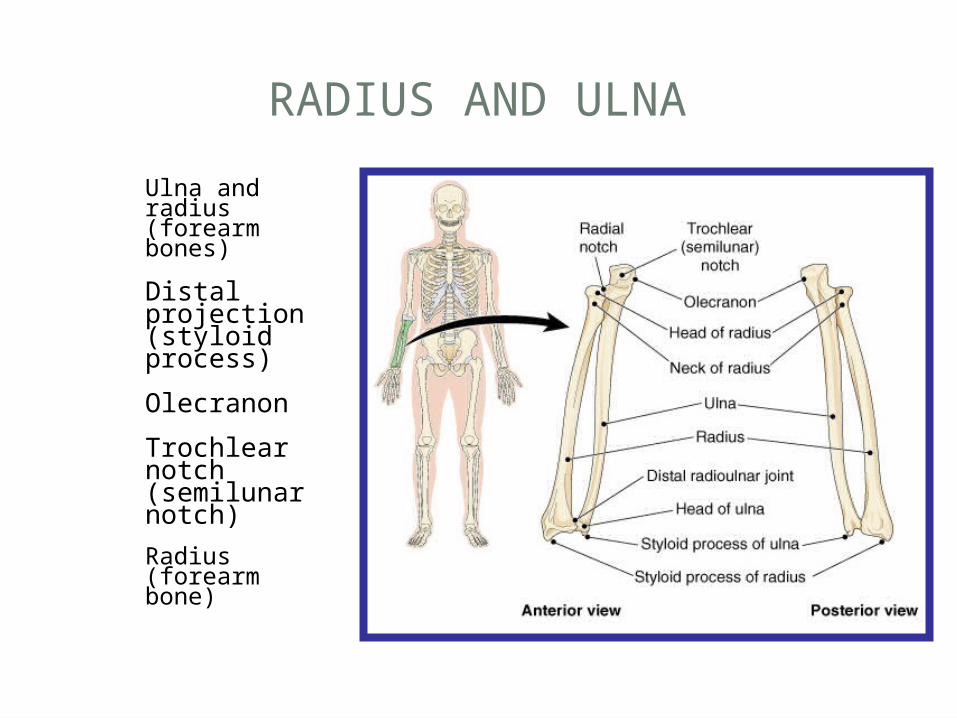
RADIUS AND ULNA
Ulna and radius (forearm bones)
Distal projection (styloid process)
Olecranon
Trochlear notch (semilunar notch)
Radius (forearm bone)
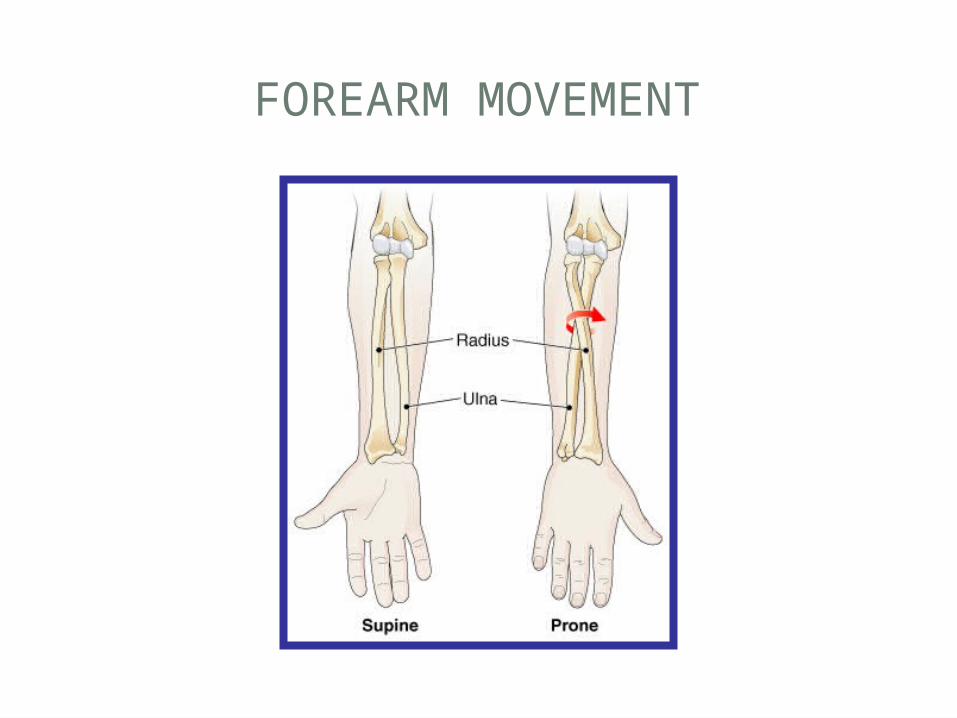
FOREARM MOVEMENT
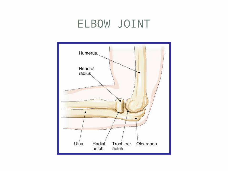
ELBOW JOINT
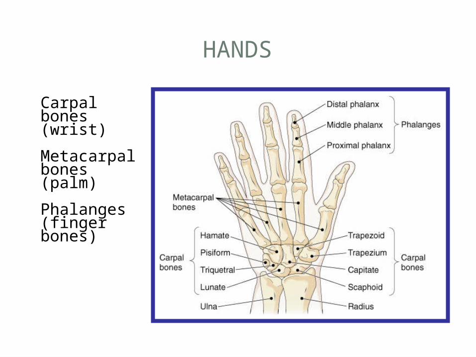
HANDS
Carpal bones (wrist)
Metacarpal bones (palm)
Phalanges (finger bones)
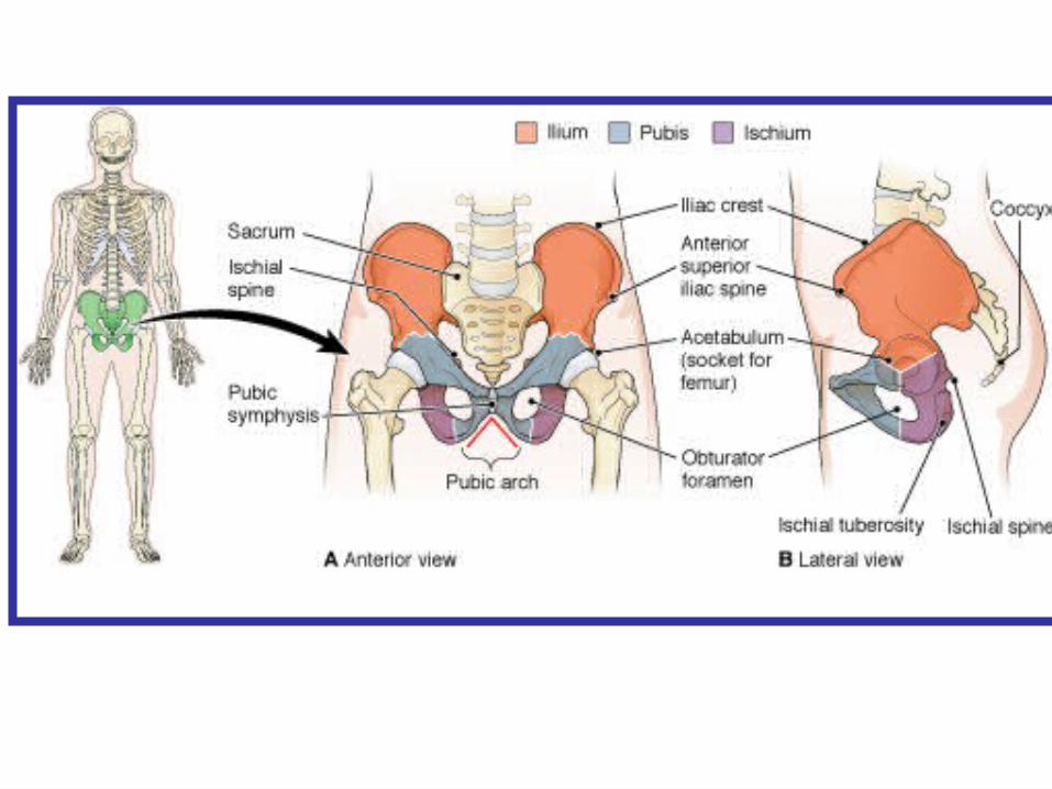
PELVIC GIRDLE
• The pelvic bones (ossa coxae)• Ilium• Iliac crest• Anterior superior iliac spine
• Ischium• Ischial spine• Ischial tuberosity
• Pubis• Pubic symphysis
• Features of pelvis• Acetabulum• Obturator foramen
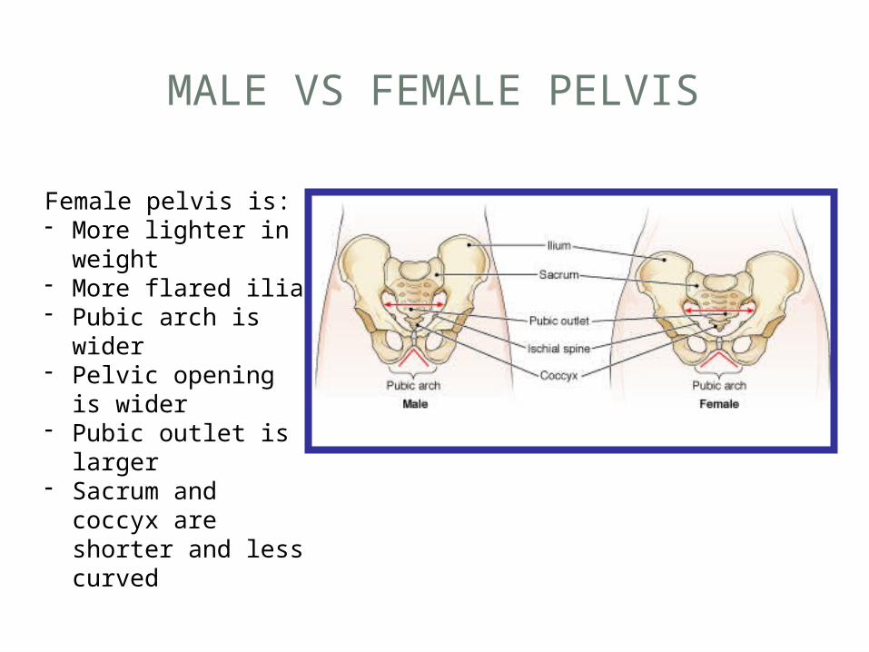
MALE VS FEMALE PELVIS
Female pelvis is:- More lighter in
weight- More flared ilia- Pubic arch is wider- Pelvic opening is
wider- Pubic outlet is
larger- Sacrum and
coccyx are shorter and less curved
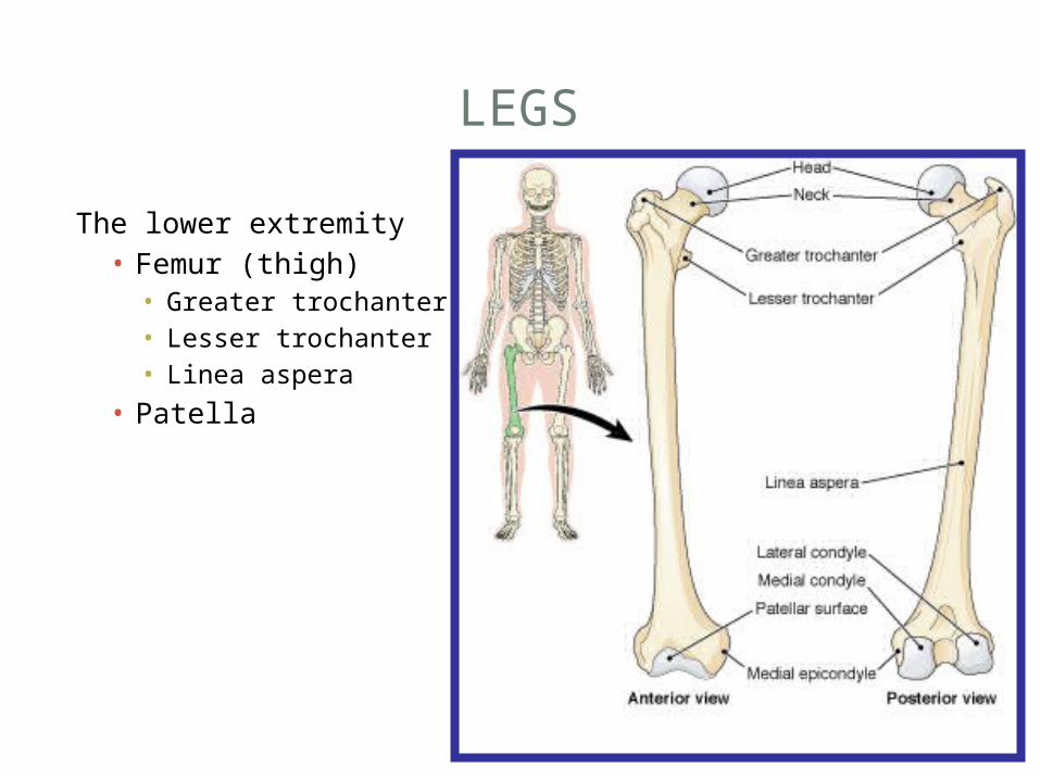
LEGS
The lower extremity• Femur (thigh)• Greater trochanter• Lesser trochanter• Linea aspera
• Patella
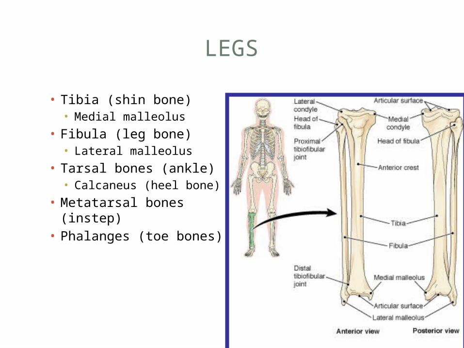
LEGS
• Tibia (shin bone)• Medial malleolus
• Fibula (leg bone)• Lateral malleolus
• Tarsal bones (ankle)• Calcaneus (heel bone)
• Metatarsal bones (instep)
• Phalanges (toe bones)
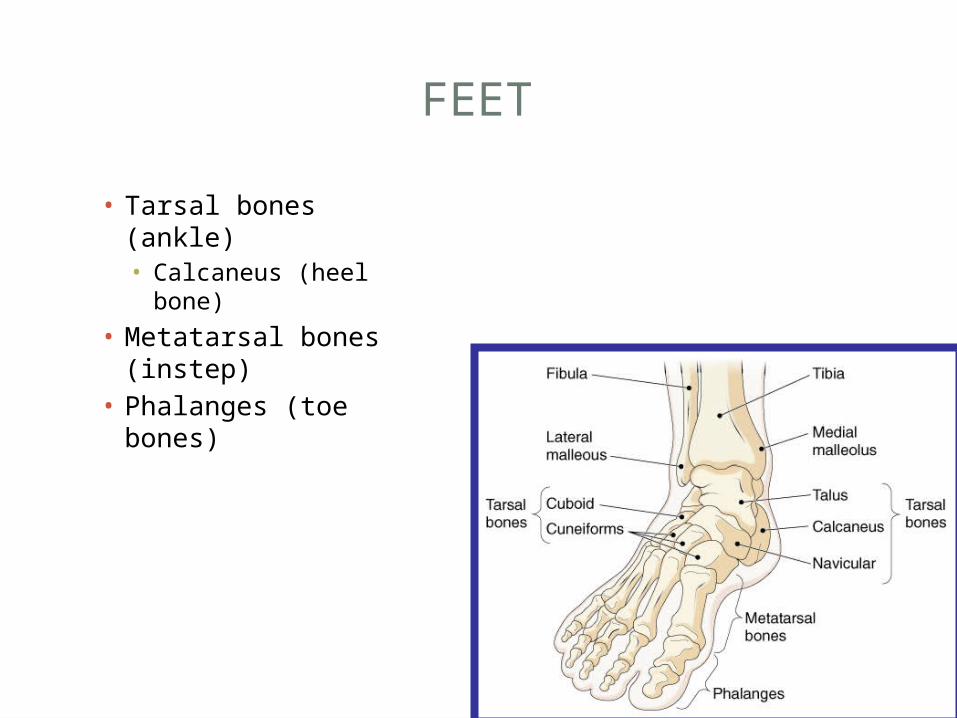
FEET
• Tarsal bones (ankle)• Calcaneus (heel
bone)
• Metatarsal bones (instep)
• Phalanges (toe bones)
THE AGING SKELETON
Bones undergo significant changes• Loss of calcium salts• Decrease in protein• Reduction in collagen• Loss of height• Decrease in chest diameter
JOINTS
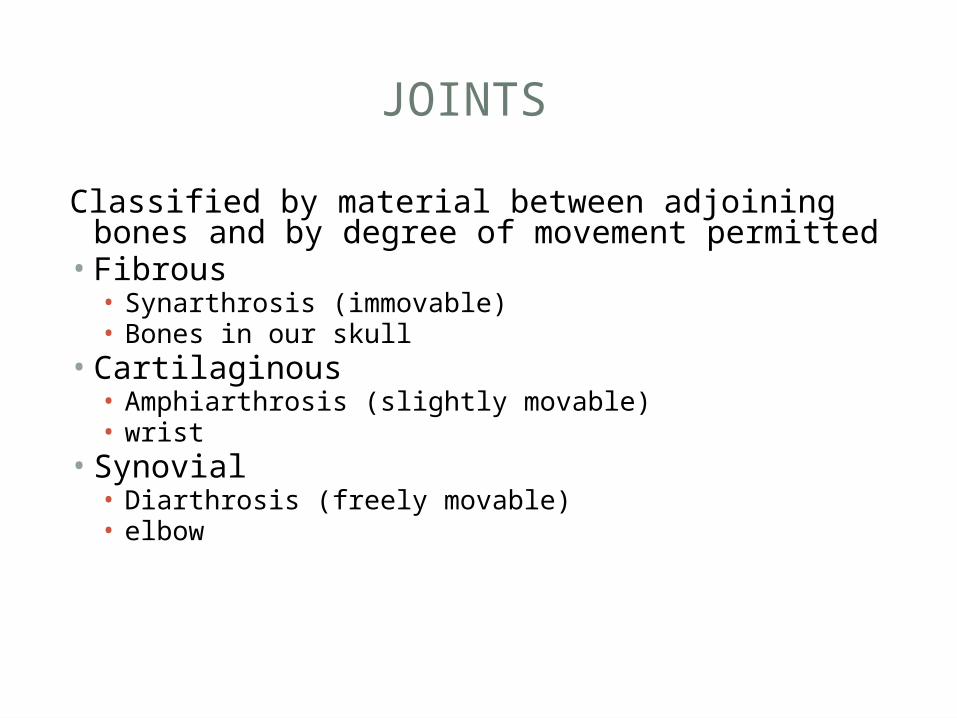
Classified by material between adjoining bones and by degree of movement permitted• Fibrous• Synarthrosis (immovable)• Bones in our skull
• Cartilaginous• Amphiarthrosis (slightly movable)• wrist
• Synovial• Diarthrosis (freely movable)• elbow
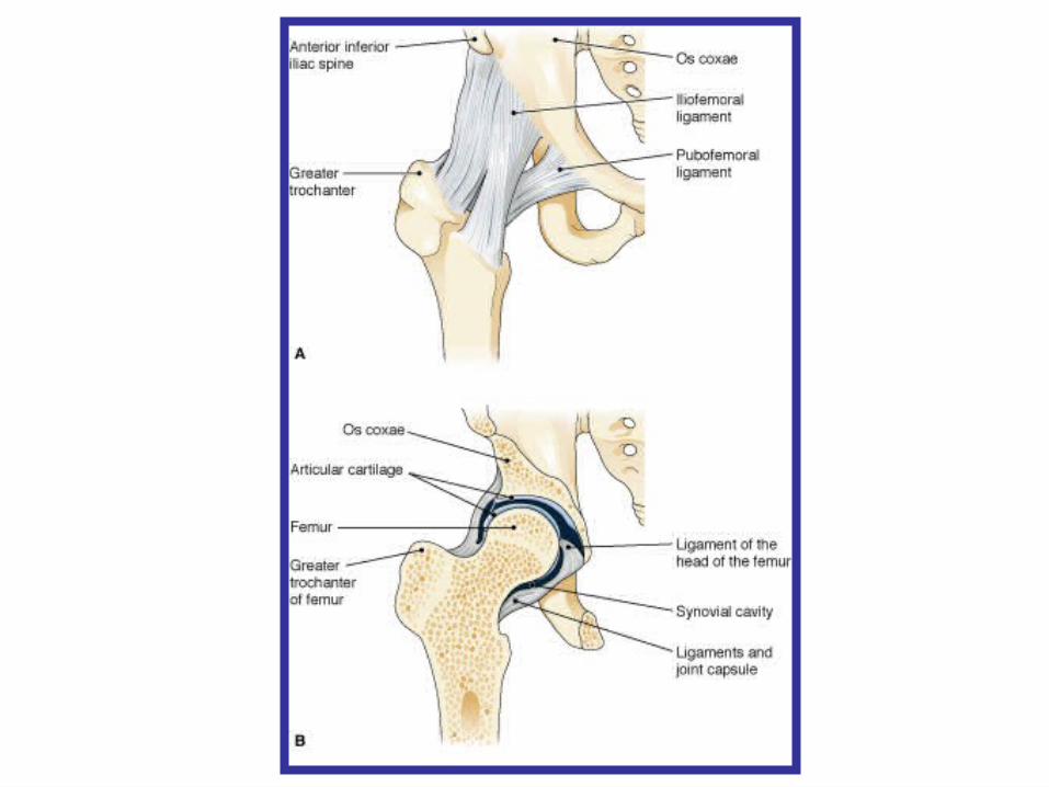
SYNOVIAL JOINTS
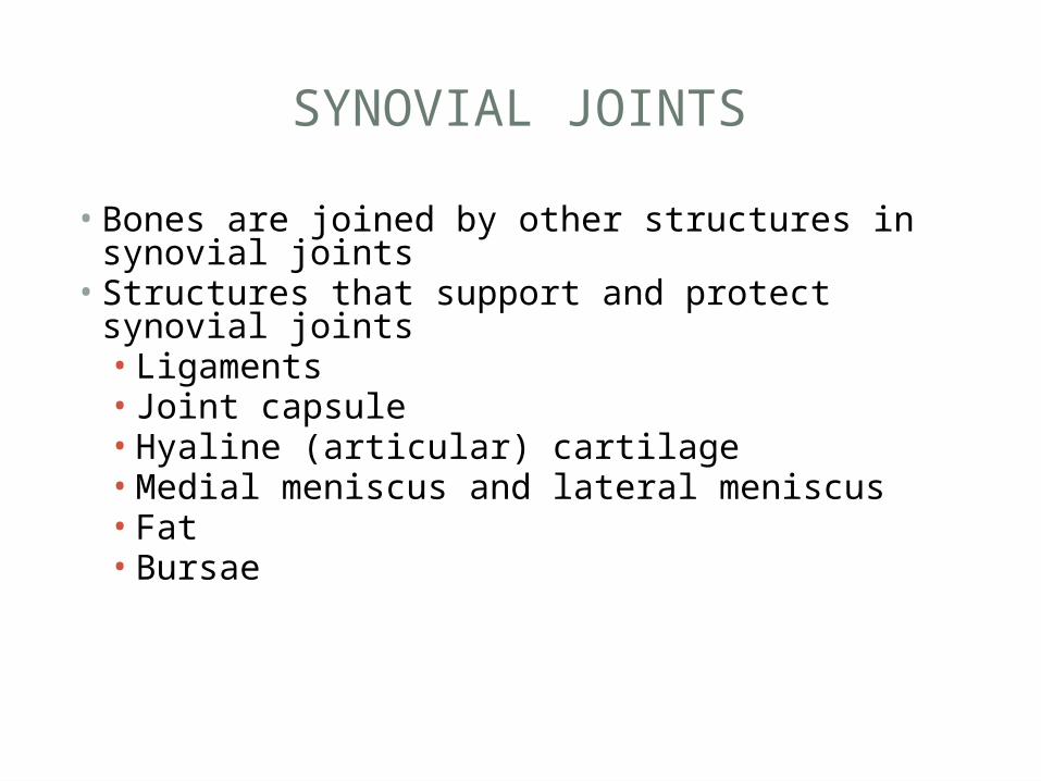
• Bones are joined by other structures in synovial joints• Structures that support and protect synovial joints• Ligaments• Joint capsule• Hyaline (articular) cartilage• Medial meniscus and lateral meniscus• Fat• Bursae
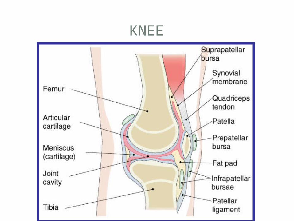
KNEE
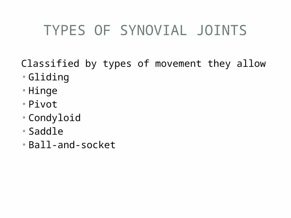
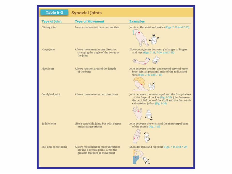
TYPES OF SYNOVIAL JOINTS
Classified by types of movement they allow• Gliding • Hinge• Pivot• Condyloid• Saddle• Ball-and-socket
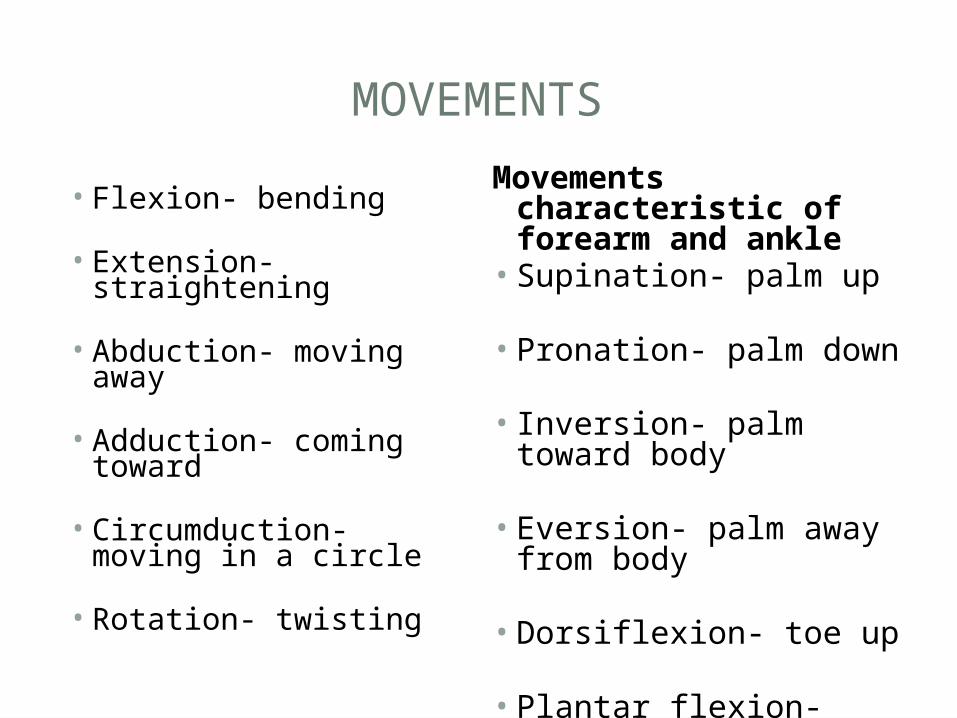
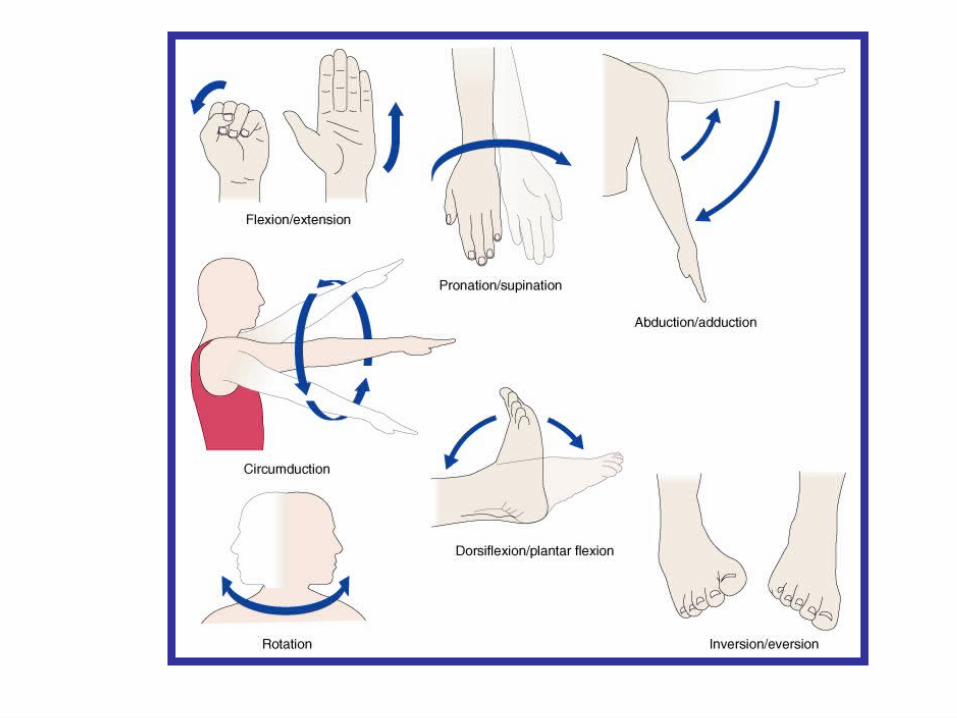
MOVEMENTS
• Flexion- bending
• Extension- straightening
• Abduction- moving away
• Adduction- coming toward
• Circumduction- moving in a circle
• Rotation- twisting
Movements characteristic of forearm and ankle• Supination- palm up
• Pronation- palm down
• Inversion- palm toward body
• Eversion- palm away from body
• Dorsiflexion- toe up
• Plantar flexion- pointed toe