blood stain typing by electrophoresis
DESCRIPTION
Blood Stain TypingTRANSCRIPT
C310/09(6)
Blood Stain Typing by Electrophoresis
An educational service of:
PO Box 752Beaumont, TX USA 77704-0752

28 1
Handbook for Rapid Blood Stain Typing Using Electrophoresis for the Crime Laboratory
Prepared by:
R.C. Briner, Ph.D.
C. R. Longwell, M.S.
SEMO Regional Crime LaboratorySoutheast Missouri State University
Cape Girardeau, MO 63701
Section 4 Final Notes
Titan®IIIandtheSuperZApplicatorSystemarefast,easyandpracticalforforensicinvestigationofmanydifferentproteinsandenzymes.Theexam-plesinthisbookletwillgivetheinvestigativelaboratorianspecifictechniquesforsomeassaysand theequipmentandreagentsareeasilymodified forotheruses.
PleasecontacttheauthorsatSEMORegionalCrimeLaboratoryorHelenaLaboratoriesifyouneedassistanceorhaveinformationtoshare.
For more information, call Helena Laboratories.Toll free 800-231-5663.

2 27
AcknowledgementsThefollowingstudentshaveprovidedvaluableassistanceinthedevelopmentofthesemethods:Retha(Mat-thews)Edens1,AliceAbbot2,RandallWebster3,Yvonne(Moll)Matthews4andRalphWillis5.Weespeciallyap-preciateMs.DonnaPruneau6forreviewingandupdatingthismanual(February,1989).TheseproceduresareinuseincourseFS552(BloodandBodyFluids),anelectiveintheScienceDivisionatSoutheastMissouriStateUniversity,CapeGirardeau,Missouri.TheauthorswouldliketothankHelenaLaboratories,Beaumont,Texas,forsupportofthisproject.PortionsofthisworkweresupportedbytheResearchCouncil,SoutheastMissouriStateUniversity,CapeGirardeau,Missouri.WewouldalsoliketothankMs.PattyButler7fortypingtheoriginaldraftofthishandbook.
1.No.11WestAdams,Cahokia,IL. 2.St.John’sMercyHospitalClinicalLaboratory,St.Louis,MO. 3.HannibalPoliceDepartment,Hannibal,MO. 4.P.O.Box945,CollegeStation,TX. 5.CrimeLaboratory,MissouriSouthernCollege,Joplin,MO. 6.SEMORegionalCrimeLaboratory,SoutheastMissouriStateUniversity,CapeGirardeau,MO. 7.Secretary,CollegeofScienceandTechnology,SoutheastMissouriStateUniversity,CapeGirardeau,MO.
Peptidase A (Pep A)(For Negroids)
1. TankBuffer:SameasforGLO-IandEsD
2. GelBuffer:SameasforGLO-IandEsD
3. Starch/AgarGel:SameasforGLO-IandEsD
4. SamplePreparationandApplication:Soakdriedbloodstainsforabout15minutesindistilledwaterandapplytogelinthesamemannerasGLO-IandEsD.Runaknowntype2-1asastandard.
5. Electrophoresis:250voltsfor45minutes.Amperageatthestartabout10mA,attheendabout6mA.
6. ReactionBuffer(SameasPGMreactionbuffer) 3.64gmTris,pH8.0 Dissolvein500mLofdistilledwater.AdjusttopH8.0withconcentratedHCl.
7. ReactionMixture 40mgL-valyl-L-leucine 20mgL-aminoacidoxidase(crudesnakevenom) 10mgMTT(lightsensitive) 4.0mgPMS(lightsensitive) 10mLreactionbuffer
8. Visualization:Overlay0.2gmagarose(SigmaTypeII)dissolvedin10mLofreactionbuffer.
Heattheagaroseuntildissolved.Allowtheagarosetocoolto55°Cbeforeaddingthereactionmixture.This isquicklypouredover thegel. Incubate thegel for1hourat37°C.
Thephenotypesappearasbluebandsonalightyellowbackground.
Figure 13 – Peptidase A (PepA)
Anode(+)
Applicationpoint
Cathode(–) Samplenumber 1 2 3 4 5 6 7 8
Electrophoretic patterns of three types of Pep A: Type 1-1: Samples 1, 2, 3, 7, 8 and 9 Type 2-1: Samples 4 and 6
Sample 5 is a suspected 1 + 2-1 mix from a vaginal/seminal sample

26 3
Table of Contents
Section 1 General Information I. Introduction 4
II. FactorsInfluencingResults 5
III. SampleExtractionandStorage 5
IV. Materials 5
V. ElectrophoresisTechniques 6
VI. ResultsandDiscussion 9
VII. References 9
Section 2 Typing of Dried Blood Stains With Cellulose Acetate Part I: Isoenzymes Phosphoglucomutase(PGM) 10
Erythrocyte/Vaginal/SeminalAcidPhosphatase (EAP/VAP/SAP) 12
AdenylateKinase(AK) 14
AdenosineDeaminase(ADA) 16
CarbonicAnhydraseII(CA-II)andHemoglobin 18
LactateDehydrogenase(LDH) 21
Glucose-6-PhosphateDehydrogenase(G-6-PD) 22
Part II: Proteins GroupSpecificComponent(Gc) 23
Haptoglobin 23
Section 3 Typing Dried Blood Stains Using Agar Starch Gels
Isoenzymes EsteraseD(EsD)andGlyoxylase1(GLO1) 24
PeptidaseA(PepA) 27
Section 4 Final Notes
Figure 11 – Glyoxylase-I (GLO-I)
Anode(+)
Applicationpoint
Cathode(–) Samplenumber 1 2 3 4 5 6 7 8
Electrophoretic patterns of three types of GLO-1: Type 1-1: Samples 5 Type 2-1: Samples 1, 3, 4, 6 and 7 Type 2-2: Samples 2 and 8
Figure 12 – Esterase D (EsD)
Anode(+)
Applicationpoint
Cathode(–) Samplenumber 1 2 3 4 5 6 7 8
Electrophoretic patterns of two types of EsD: Type 1-1: Samples 1, 3, 4, 5 and 6 Type 2-2: Samples 2, 7 and 8

4 25
Section 1 General Information I. Introduction
Inthepastdecade,progressindevelopingmethodologiesfortypingbloodfactorsusingisoenzymestudieshasmadebloodstainevidenceoneof themost important investiga-tive toolsavailable to forensicscientists.Originally,allprocedures for isoenzyme typinginvolved electrophoresis on starch gels.At the present time, four major electrophoreticsupportmediaorutilized:starchgel,agarose,polyacrylamidegelandcelluloseacetate.Thispresentationisaresultoftheresearchperformedinourlaboratoriesdirectedtowardadaptationofisoenzymetypingofbloodfactorsbyelectrophoresisonsupportedcelluloseacetatemembranes(CAM).
Traditionally,theonlymethodforidentifyinghumanbloodsamplesinthecrimelaboratorywastypingbytheABOsystem.UsingtheABOsystem,themostrarebloodtypeencoun-teredisAB,whichisfoundinonly3%ofthepopulation.WhilethepercentageofpersonshavingbloodtypeABisverylow,almost50%ofthehumanpopulationhastypeO.Itisap-parentfromthesestatisticsthatasystemformoredefiniteidentificationofbloodsystemsismandatoryifbloodtypingistobeusedinacrimelaboratory.
Intheearly1970s,researchwasinitiatedtoimprovethesystemfordeterminingthetypeofhumanbloodfoundondriedbloodstains.InadditiontotheABOsystem,othersystemswereutilized.TheerythrocyteMNandRh(primarilyRhD)antigensystemswereused.Pro-teinstudiesinvolvedhemoglobinandhaptoglobin.Severalenzymesystemswereutilized;themostusefulofthesebeingphosphoglucomutase(PGM),adenylatekinase(AK),eryth-rocyteacidphosphatase(EAP),esteraseD(EsD),andadenosinedeaminase(ADA).InitialworkwasperformedbyBryanJ.CullifordoftheMetropolitanPoliceForensicLaboratoryofLondon,England.1Determiningbloodtypesusingacombinationofthesystemscited,greatlydiminishestheoccurrenceofaparticulartypeinthepopulation.Althoughelectro-phoresiswasthemethodofchoiceindeterminingtheproteinspresentinabloodstain,thetimeinvolvedbecameaprimeconcernsincestarchgel(theonlymediausedatthattime)electrophoresisproceduresoftenrequired15to30hoursforcompletion.
Inthemid1970s,workersattheUniversityofPittsburghproposedamethodformultipleenzymeseparationonasingleelectrophoresisplate.ThisworkwascompletedunderthedirectionofB.Wraxall.2Bycoveringaparticularportionofthestarchgelanddevelopingeachportionspecificallyforthedesiredenzyme,itwaspossibletoidentifyseveralenzymesystemsatthesametime.AdditionalworkbyWraxalldevelopedamultiplesystemsusingastarch/agarosegel.
Since that time, the research of Dr. B. Grunbaum and P.L. Zajac using the BeckmanMicro-zone®SystemandadaptingthestarchgelprocedurestoSartorious®celluloseace-tatemembraneshasdrasticallyreducedthetimerequired.TheTitan®IIICelluloseAcetateElectrophoresisSystemfromHelenaLaboratoriesisnowbeingusedsuccessfullybytheSoutheastMissouriRegionalCrimeLaboratory.Methods for identifyinggeneticmarkersareperformedonsupportedcelluloseacetateelectrophoresisplates.Theresultsaredis-cernableandreadilyreproducible.Studiesonalltheenzymesystemscanbecompletedinlessthaneighthours.
TheEsDbandsappearaswhitefluorescentbandsonadarkbackground.UseanEsD2-1asacontrol.AftervisualizingEsD,markthepositionofthenumber2bandoftheEsDpattern.ThegeliscoveredwithGLOIreactionmixturebeginningwiththispositionandincubatedagain.
b. GLOI:20mgreducedGlutathione 50µL40%Methylglyoxal 7.5mLGLOIreactionbuffer
SoakmixtureintoWhatmanpapercuttofitthesizeofthegelfromthesecondbandoftheEsDpatterntotheedgeoftheplate(approximately6.5cmx10cm).Thefilterpaperisplacedonthegelbeginningatthecathodeside,gentlyrollingdowntherestofthepaper,beingsurenoairbubblesremainunderthepaper.Incubate30to40minutesat37°C.
Heat0.2gmsagarose(SigmaTypeII)in20mLdistilledwateruntildissolved.Allowtheagarosetocoolto60°Candthenadd2dropsofiodinesolution.(Iodinesolutioncontains1.65gmsofKI,2.94gmsofI2dissolvedin30mLsofwater.)Mixtheaga-rose/iodinemixture.Thismixturewillbealightorangecolor.Aftertheelectrophoresisgelhasbeenincubatedasabove,theagarose/iodinemixtureisquicklypouredontothegelstartingatonecornerofthegelallowingittoflowovertheentiregelsurface.Thephenotypeappearsasbluebandsonalightyellowbackground.Theheterozy-gote(2-1)hasthreebluebandspresentandshouldbeusedasacontrol.Asecondagarose/iodinemixturemaybeappliedtothegelifthebluebandsappearweakinthefirstgel.Thisisdoneafterremovingthefirstagarose/iodinegel.

24 5
II. Factors Influencing Results Severalvariablesinfluencetheresultsobtainedwithanelectrophoreticprocedure.These
includesamplepreparationandapplication,celluloseacetateplatepreparation,pHandionicstrengthofthebuffers,voltage,amperage,andcolordevelopmenttechniques.Thecelluloseacetateplatesmustbesoakedintheindividualsystemplatebufferpriortosam-pleapplication.Thesoakingbufferisusuallyadilutionofthetankbuffer.Thecorrectdilu-tionofplatebufferisessentialforgoodresolutionandforpreventionofhighamperageandexcessheat.Theplatemustbesoakedgraduallyfromthebottomuptoensuresaturationwithoutairbubbles.
Airbubblesinthecelluloseacetateimpedethemovementoftheelectriccurrentthroughtheplate.Oncetrappedintheplate,airbubblescannotberemovedandtheplatemustbediscarded.
ThepHofthetank,plateandreactionbuffersisextremelyimportant.AllenzymeshaveanoptimalpHforactivity.Denaturationoftheenzymesmayoccurifreagentswiththein-correctpHareused.Improperreactiontemperatureshavethesameeffect.Correctionicstrengthofthebuffersisalsoveryimportant.
Propervoltageandamperageareextremelyimportanttogoodelectrophoreticresolution.Voltageshouldremainconstant,allowingtheamperagetostartlow(about2milliamperesperplate)inordertoavoidoverheatingthesystem.Thetemperatureoftheelectrophore-sisplaterisesastheamperagerises.Ifthetemperatureexceedstheoptimalrange,theenzymewilldenature.
The amount of time required for electrophoresis varies with the enzyme system understudy.Noneofourproceduresrequiremorethanonehour.
Thetechniqueusedfordevelopingthecelluloseacetateplateaftercompletionofelectro-phoresisiscritical.Thetwobasictypesofdevelopmentarecolorimetricandfluorometric.Afterdevelopment,resultsmustberecordedandfiled.AllplatesarephotographedwithblackandwhitePolaroidfilm.
III. Sample Extraction and Storage Topreserveenzymaticactivity,anymaterialscontainingdriedbloodstainsshouldbefro-
zen.Atthetimeoftesting,cuta0.5cmx0.5cmpieceofstainedmaterialfromtheoriginalsampleandplaceinawellwith2to3dropsofsolvent.Distilledwater isroutinelyusedas thesolvent;however,Cleland’s reagentmustbeused forphosphoglucomutaseanderythrocyte acid phosphatase assays. Samples extracted with purified water may besoakedovernightinahumiditychamberat4°C.SamplesextractedinCleland’sreagent(dithioerythritol;availablefromSigma)arestablefor3to5hoursandshouldnotbestoredovernight.Theprocedure frommakingCleland’s reagent is included in thePGMproce-dure.
Section 3 Typing Dried Blood Stains Using Agar Starch Gels
Isoenzymes Esterase D (EsD) and Glyoxylase I (GLO I) Thisprocedureshouldbeperformedwithintwodaysofreceivingsample.Maximumsam-
pleageis10days.After10daysthesamplemaybetoodeterioratedtoproduceareadableresult.
1. TankBuffer:HEPES,pH7.5 5.95gmHEPES0.05M(N-2hydroxyethylpiperazine-N-2ethanesulfonicacid) Dissolvein400mLdistilledwater.AdjusttopH7.5with40%NaOH.Q.S.tofinalvol-
umeof500mLwithdistilledwater.
2. GelBuffer:1:14dilutionoftankbuffer,pH7.4. Dilute10mLoftankbufferwith140mLdistilledwater.
3. Starch/AgarGel 1%Agarose,SigmaTypeV(lowEEO) 2%Hydrolyzedstarch(ConnaughtLabs,Willowdale,Ontario,Canada). AvailablefromFisher,Cat.No.S-676. Dissolve0.3gmagaroseand0.6gmstarchin30mLgelbuffer.Afterdissolving,de-gas
thesolutionpriortopouringinto8cmx10cmx2mmdeepplasticplates.Gelmustsitovernightbeforeuse.
4. SamplePreparationandApplication a. Soakdriedbloodstains for15minutes in0.1NClelend’s reagent,pH8.0.Runa
knowntype2-1asastandard(GLOandEsD). b. Eightwells(1mmx4mm,3mmapart)arecutinthegel2cmfromthecathodicend
withametaltemplate.Apply5µLofbloodineachwellusingamicropipet.
5. Electrophoresis:250voltsfor30minutes. Itisveryimportanttokeeptheelectrophoresischamberverycoolduringtherun.The
tankbuffermustbekeptcolduntilreadyforuse.Tokeepthechamberatalowtem-perature,anotherlidormetalpanisfilledwithiceandplacedontopoftheclosedelec-trophoresistank.Plateisrunfacedownonspongewicks.Amperageatstart8to9mA;attheend,about10mA.
6. ReactionBuffer a. EsD: 0.41 gm sodium acetate, anhydrous (0.05M). Dissolve in 100 mL distilled
water.AdjusttopH6.5with1%aceticacid. b. GLOI:2.42gmNaH2PO4,anhydrous(0.2M) 1.31gmNa2HPO2,anhydrous(0.072M) Dissolvein100mLdistilledwater,adjusttopH6.2withphosphoricacid.
7. ReactionMixtureandVisualization a. EsD:4mgMU-Acetate(4-methylumbelliferylacetate) 1.0mLacetone(spectralgrade) 5mLreactionbuffer DissolveMU-acetateinabout1.0mLofacetoneandimmediatelyadd10.0mLofthe
reactionbuffer.SoakintoWhatman3MMpaper4cmx10cm.Laythispaperoveraportionofthegelfromtheorigintowardanode.Leaveatroomtemperatureforabout5minutes.RemovethepaperafterincubationatroomtemperatureandreadEsDbyusingUVlight.

6 23
IV. Materials
TitanIIICelluloseAcetateSystemavailablefromHelenaLaboratories. Hardware Cat. No. TITAN®plusPowerSupply(110V) 1504 ZipZone®Chamber 1283 SuperZApplicatorKit 4088 DevelopmentWeight 5014 TitanPhotoVuBoxCamera 5048 Bufferizer 5093 IODIncubator,OvenDryer(110V) 5116 Micro-Hood 8009 Consumables Titan®III-LipoCelluloseAcetate(60x76mm) 3900 Electra®HRBuffer 5085 Supre-HemeBuffer 5802 Electra®B1Buffer 5016 LDVisIsoenzymeReagent 5909 HemolysateReagent 5125 Blotters 5034 ZipZone®ChamberWicks 5081 DevelopmentSlides 5008 TitanPlasticEnvelope 5052 TitanIdentificationLabels 5006 HelenaMarker 5000
V. Electrophoresis Techniques
A. Preparation of Titan III-Lipo Plates
1. ProperlycodetherequirednumberofTitanIII-Lipoplatesbymarkingontheglossy,hardsidewithamagicmarker.Itissuggestedthatthemarkbeplacedinonecorneroftheplatesothatitisalwaysalignedwithsample#1.
2. Soaktheplatesfor20minutes in the individualsystemplatebuffer.Thesoakingbufferisusuallyadilutionofthetank buffer.The plates must be soaked gradually fromthebottomup toensuresaturationwithout trappingairbubblesinthecelluloseacetate.
The plates may be soaked in the Bufferizer (availablefrom Helena Laboratories, Cat. No. 5093) or they maybesoaked ina laboratorybeakerusing theprincipleoftheBufferizer.Placetheplatesinthebeakerinaverticalpositionandallowtheplatebuffertoslowlyflowdowntheside of the beaker from a separatory funnel or directlyfrom the buffer container.Alternately, fill a beaker withbufferandslowlyandsteadilyimmersetheplates(verti-calposition)intothebuffer.
Part II. Proteins Group Specific Component (Gc)
1. TankBuffer:Tris/Glycine,pH8.4 21.8gmGlycine 4.5gmTris Dissolvein900mLwater.AdjusttopH8.4withNaOH.Q.S.toafinalvolume
of1000mLwithdistilledwater.
2. PlateBuffer:Sameastankbuffer.
3. SamplePreparationandApplication:Extractbloodstainswith3dropsof6Murea(3.6gm:10mLH2O).Placeallofextractinasmallplasticcentrifugetube.Fillthetubehalfwaywithchloroform.Vortextomix.Centrifugethemixtureinamicrocentrifugeathighspeed.Thechloroformwashesdownthedebrisinthestain.
ExtractionofstainswithHemolysateReagent(HelenaCat.No.5125)willelim-inateagingbandswhichappearabovetheGcbands.ThiswillallowBCtypingtobeperformedonoldstains.*
4. Electrophoresis:500voltsfor10minutes;Initialamperage=2to4mA/plate.
5. Visualization(Immunofixation) a. Afterelectrophoresis,theplatesarecoveredwithhumanGcantiserumdi-
luted1:4withphysiologicalsaline(2mLserum+6mLsaline).Allowplatetosoakfor20minutes.
Thedilutedantiserumcanbereusedandisstableabout1monthafterdilu-tion,dependingonthenumberoftimesused.Storeintherefrigerator.
b. Removeexcessserumfromplateandsoakovernightinphysiologicalsa-line(0.85%).
c. Stainfor5minutesin0.2%PonceauS(v/vindistilledwater). d. Todestain,placeplatesinasolutionof5%aceticacid,agitatefor15min-
utes.Pouroutthissolutionandaddfreshaceticacid,agitateforanother15minutesuntil thebackgroundbecomeswhite.Platesmaybewashedovernight.Theplatesare thendried; thebandsaredarkpinkonawhitebackground.
TwosuggestedsourcesforGcantiseraare: Atlantic Antibodies In Vitro Research Sources, Inc. 10NonesuchRoad P.O.Box110 P.O.Box1032 Benson,MD21018 Scarborough,ME04074 (301)877-7110 (207)883-4154 Figures9and10onpage22illustratetheelectrophoreticpatternsofGc.
Haptoglobin (Hp) The stain extract prepared for the Gc procedure may be used for haptoglobin
determinationonpolyacrylamidegradientgel.However,beforeusingtheextract,preparea1:1mixturewith40%sucrose.
*PersonalCommunication:Willard(Bud)Stuver,Supervisor,Serology/BiologySection,Metro-DadePoliceCrimeLaboratory,Miami,FL33125.

22 7
B. Preparation of the Electrophoresis Tank (Zip Zone Chamber)
1. Prior to using the tank, fill the inner chamber halfway withwaterandkeepfrozenuntilthetankisused.
2. Whenpreparingforelectrophoresis,pour50mLoftankbufferintothecathodechamberanda1:1dilution(25mLtankbuf-fer/25mLH2O)intheanodechamber.(Exceptwhenstatedotherwise;checkspecificprocedures.)
3. Wet two Zip Zone® Chamber Wicks in the tank buffer anddrape one over each support bridge. Be sure they makecontactwiththebufferandthattherearenoairbubblesun-derthewicks.Donotallowthewickstotouchtheiceintheinnerchambers.Coverthetankuntilused.
C. Sample Application
1. Fillthewellsofthesamplewellplatewithbloodstainextractusingasmallcapillarypipette.
2. Primetheapplicatorbydepressingthetipsintothesamplewells 3 or 4 times.Apply this loading to a piece of blotterpaper.Primingtheapplicatormakesthesecondloadingmuchmoreuniform.Donotloadtheapplicatoragainatthispoint,butproceedquicklytothenextstep.
3. Remove the wettedTitan III plate from the buffer and blot
oncefirmlybetweentwoblotters.Placetheplateinthealign-ingbase, celluloseacetatesideup,aligning the topof theplate with the black scribe line marked “cathodic applica-tion”.Theidentificationmarkshouldbealignedwithsample#1.Mostproceduresrequireacathodicapplicationwiththeexceptionofphosphoglucomutase(PGM)whichrequiresanextremecathodicapplication.
HELENA
1 2 3 4 5 6 7 8
HELENA
1 2 3 4 5 6 7 8
Buffer
Ice
Buffer
CATHODE APPLICATION
CENTER APPLICATION
HELENA LABORATORIES
Placementofplateforextremecathodicapplication
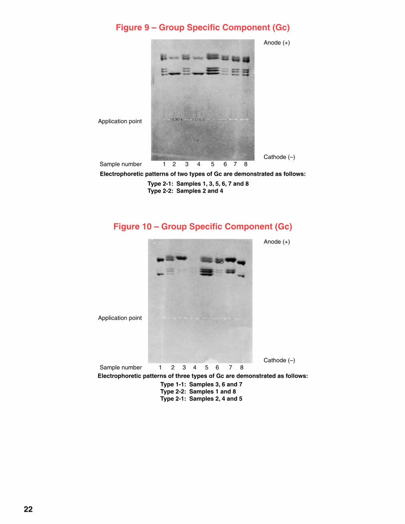
Figure 9 – Group Specific Component (Gc)
Anode(+)
Applicationpoint
Cathode(–) Samplenumber 1 2 3 4 5 6 7 8
Electrophoretic patterns of two types of Gc are demonstrated as follows:
Type 2-1: Samples 1, 3, 5, 6, 7 and 8 Type 2-2: Samples 2 and 4
Figure 10 – Group Specific Component (Gc)
Anode(+)
Applicationpoint
Cathode(–) Samplenumber 1 2 3 4 5 6 7 8
Electrophoretic patterns of three types of Gc are demonstrated as follows: Type 1-1: Samples 3, 6 and 7 Type 2-2: Samples 1 and 8 Type 2-1: Samples 2, 4 and 5

4. Apply thesampleto theplatebydepressingtheapplicatortipsintothesamplewells3or4timesand promptly transferring the applicator to thealigning base. Press the button down and holdfor5seconds.Makeasecondor thirdsuperim-posed application by repeating this step. Threeapplicationsof .25µLeach issufficient formostisoenzymestudies.
D. Electrophoresis of the Sample Plate
1. Quickly place the plate, cellulose acetate sidedown,intheelectrophoresistank.Placeaweight(plasticrod,glassslide,etc.)ontheplate(s)toin-surecontactwith thewicks.Cover thechambersecurely.
2. Electrophorese according to the instructions oftheappropriateprocedure.
E. Development
1. Foreachsampleplateused,areactionplatemustalsobesoakedaccordingtothesoakingmethoddescribedinStepA.Thereactionplateisusuallysoakedinthereactionbuffer.Seeindividualproce-dures,Section2.
2. Remove each plate from the reaction soaking buffer 5 minutes before completion of electrophoresis and blot gently. Place theplate, celluloseacetatesideup,ontoaglassdevelopmentslideandplaceonalevelcountertoporalevelingtable.
3. Pourapproximately2.0to5.0mLofreactionmix-tureontotheplate.Tilttheslideuntilthereactionmixturecovers theplate.Covereachplatewithasmallbox(ifthereactionmixtureislightsensi-tive)andallowtostandforfiveminutes.
4. Aftercompletionofelectrophoresis, remove thesample plate from the electrophoresis tank andblotlightlytoremoveexcessbuffer.
5. Carefullylayerthesampleplate,celluloseacetatesidedown,ontothereactionplate.Placeablotteroverthesandwichedplates.Removeexcesssub-stratefromthesandwichbydrawingtheedgeofthealigningbaseacrosstheplateseveraltimes.Blotawayexcesssubstrate.
6. PlacetheplatesbetweenmoistenedblottersandplaceonthetopofapreheatedMicro-HoodorinaHelenaIODorotherlaboratoryincubatorat37°C.Placeapreheateddevelopmentweightontopoftheblotters.
8 21
TITANPOWER SUPPLY
VOLTAGE CONTROL
II
II
II
II
IIII
II
II
II
II
+ _
Glucose-6-Phosphate Dehydrogenase (G-6-PD)
1. TankBuffer:Supre-HemeBuffer(HelenaCat.No.5802),pH8.4.Tris-EDTABoratebuffer.Diluteto1000mLwithdistilledwater.
2. PlateBuffer:Sameastankbuffer.
3. Preheatincubatoranddevelopmentweightto37°C.
4. SamplePreparationandApplication:Onecathodicapplicationofdistilledwaterextractofstain.
5. Electrophoresis:350voltsfor20minutes.
6. ReactionBuffer:Undilutedtankbuffer.
7. G-6-PDReactionMixture a.G-6-PDReagent,HelenaCat.No.5620or b.10.0mgGlucose-6-Phosphate 6.0mgNAD(NicotinamideAdenineDinucleotide) 7.0mgMTT(lightsensitive) 7.0mgPMS(lightsensititve) Reconstitutereagentwith3to5mLofundilutedtankbuffer.Thismakessufficient
reagentfor3to4reactionplates.
8. Incubation:20minutesat37°C.
9. Visualization:Separateplatesandsoakin7.5%trichloroaceticacid(TCA)for2to3minutes.Rinsein5%aceticacidfor5minutes.Blot,dryplatesandreadundertransmittedlight.

20 9
7. Aftertheappropriateincubationperiod,preparetheplatesforevalu-ationasdescribedinindividualprocedures(Section2).Afterdevel-opment,allplatesmustbedriedbeforereadingeitherbyairdryingorwithalightstreamofwarmforcedair.Bothplates,thesampleplateandreactionplate(negative),canberead.Inmostinstances,driedplatesmaybestoredindefinitelyforfuturereferenceifplacedinaTitanPlasticEnvelopeorsmallplasticstoragebag.
VI. Results and Discussion Atthepresenttime,thefollowingproceduresarebeingperformedrou-
tinely inSEMORegionalCrimeLaboratoryusing thecelluloseacetatemembrane(CAM)system:
Phosphoglucomutase(PGM) Erythrocyte/Vaginal/SeminalAcidPhosphatase(EAP/VAP/SAP) AdenylateKinase(AK) GroupSpecificComponent(Gc) Other procedures which are currently performed on CAM are: Glucose-6-PhosphateDehydrogenase(G-6-PD) LactateAcidDehydrogenase(LDH) CarbonicAnhydraseII(CA-II)5
Isoenzymetypingofbloodusingcelluloseacetatemembranesasasup-portmediumforelectrophoreticseparationhasproventobearelativelyin-expensiveandrapidmethodforidentifyingbloodstainsaccordingtopoly-morphicproteinsandenzymes.Usingtheseprocedures,determinationscanbeperformedusingmicrolitervolumesofspecimen(stainextract).
EsteraseD(EsD),Glyoxylase-I(GLO-I)andPeptidaseA(PepA)determi-nationsareperformedwithstarch-agarosegelduetosuperiorseparationandvisualization.Haptoglobindeterminationsareperformedutilizingagradientpolyacrylamidesystem(overnight).Ifthehaptoglobinprocedureis runat300volts, itcanbecompleted in fourhours. It ispossible foratrainedanalysttorunallsystems, includingspecies,ABOtypingandhaptoglobinineighthours.
VII. References 1. Culliford,B.J.,TheExaminationandTypingofBloodstainsintheCrime
Laboratory,LawEnforcementAssistanceAdministration,Washington,D.C.,20402,Stock#2700-0083.
2. Wraxall, Bryan G.D., Ed., Bloodstain Analysis System Procedures Manual,SerologicalResearchInstitute,Emeryville,California,1977.
3. Grunbaum,B.W.,Selvin,S.,Pace,N.,Black,D.M.,JofForensicSci,23,577,1979.
4. Grunbaum, B.W., Ed., Handbook for Individualization of Forensic Blood and Bloodstains,UniversityofCalifornia,Berkeley,California,1980.
5. Noppinger, K.E., Morrison, R.D., J of Forensic Sci, 26:1, 176-180,1981.
Lactate Dehydrogenase (LDH) 1. TankBuffer:ElectraHRBuffer(HelenaCat.No.5805),pH8.8. Trisbarbital-sodiumbarbitalbuffer.Diluteto1750mLwithdistilledwater.
2. PlateBuffer:Sameastankbuffer.
3. Preheatincubatoranddevelopmentweightto37°C.
4. SamplePreparationandApplication:Applytwocathodicapplicationsofdistilledwaterextractofstain.Seminalstainsmayhavetobeseriallydiluteduntilareadableresultisachieved.
5. Electrophoresis:180voltsfor30minutes;Initialamperage=8mAeach.
6. ReactionPlates:SoakplatesinPGMreactionbufferabout15minutesbeforecompletionofelectrophoresis.
7. LDHReactionMixture 5mLPGMreactionbuffer 25mgCalciumLactate 2mgNAD(NicotinamideAdenineDinucleotide) 2.5mgMTT(lightsensitive) 2.0mgPMS(lightsensititve) OruseLDVISIsoenzymeReagent,Cat.No.5909,HelenaLaboratories
8. Incubation:15minutesat37°Cwithdevelopmentweightontop.
9. Visualization:LDHpatternappearsasbluebands.Viewundertransmittedlight.
Figure 8 – Lactic Acid Dehydrogenase (LDH) Anode(+)
Applicationpoint
Cathode(–) Samplenumber 1 2 3 4 5 6 7 8
LDH electrophoretic patterns from various sources are illustrated as follows: Liver: Samples 1 and 2 Heart: Samples 3 and 4
Mixture of menstrual blood and seminal fluid: Samples 5, 6, 7, and 8

10 19
Section 2 Typing of Dried Blood Stains With Cellulose Acetate
Part 1: Isoenzymes Phosphoglucomutase (PGM)
1. TankBuffer:Tris/maleicacidbuffer0.075M;pH7.4 6.05gmTris:Tris(hydroxymethyl)aminomethane 5.81gmMaleicacid 1.46gmEDTA(FreeAcid):EthylenediaminetetraaceticAcid 3.47gmMgCl2•6H2O Dissolvein900mLofdistilledwaterandadjusttopH7.4with40%NaOH.
Q.S.tofinalvolumeof1000mLwithdistilledwater. CathodeBuffer:50mLoftankbuffer AnodeBuffer:15mLtankbuffer+35mLdistilledH2O
2. PlateBuffer:30mLtankbuffer+270mLdistilledH2O
3. Preheatincubatoranddevelopmentweightto37°C.
4. SamplePreparationandApplication Cleland’sReagent(0.01N) 154gm/dLDithiothreitol+100mLH2O.AdjustpHto8.0with6MNaOH. Soakbloodstainsfor15minutesinfreshCleland’sreagent.Postmortemblood
mayhavetobeseriallydilutedwithCleland’sreagentuntilareadableresultcanbeachieved.Runknowntype2-1standardoneachplate.Makeoneortwoapplications,extremelycathodic.
NOTE:TheextractmadeinCleland’sreagentisstablefor3to5hours.
5. Electrophoresis:250voltsfor30minutes;Initialamperage=2mA/plate.
6. ReactionBuffer:pH8.0 3.64gmTris Dissolvein500mLdistilledwater.AdjusttopH8.0withconcentratedHCl.
7. ReactionPlates:Soakplatesinreactionbufferapproximately15minutespriortocompletionofelectrophoresis.
8. PGMReactionMixture a)PreparedPGMmixturein3.0mLofreactionbufferor b)30.0mgGlucose-1-Phosphate* 0.3mgGlucose-1,6-Diphosphate* 20.0unitsGlucose-6-PhosphateDehydrogenase 2.5mgPMS:PhenazineMethosulfate(Lightsensitive) 2.5mgMTT:ThiazoylBlue(Lightsensitive) 2.0mgNADP:NicotinamideAdenineDinucleotidePhosphate 5.0mLreactionbuffer Stirmixtureuntildissolved.Pouronreactionplatesabout5minutes
beforecompletionofelectrophoresis. *AvailabletogetherfromSigmaChemicalCompany;-D-Glucose1-PhosphateDisodium
Salt,cat.no.G-1259
9. Incubation:15minutesat37°C,withweightontoporuntilbluebandsappear.
10.Visualization:Separateplatesandsoak5minutesin5%aceticacid.Blot, dryandreadusingtransmittedlight.
Figure 7 – Carbonic Anhydrase (CA-II)
Anode(+)
Applicationpoint
Cathode(–) Samplenumber 1 2 3 4 5 6 7 8
Electrophoretic patterns for two types of CA-II are demonstrated as follows: Type 1-1: Samples 1, 2, 4, 5 and 6 Type 2-1: Samples 3, 7, and 8

18 11
Figure 1 – Phosphoglucomutase (PGM) Anode(+)
Applicationpoint
Cathode(–) Samplenumber 1 2 3 4 5 6 7 8
Electrophoretic patterns for two types of PGM are demonstrated as follows: Type 1-1: Samples 2, 3, 4, 6, 7 and 8 Type 2-2: Samples 1 and 5
Figure 2 – Phosphoglucomutase (PGM) Anode(+)
Applicationpoint
Cathode(–) Samplenumber 1 2 3 4 5 6 7 8
Electrophoretic patterns for three types of PGM are demonstrated as follows: Type 1-1: Samples 1, 6, 7 and 8 Type 2-2: Samples 3 and 4 Type 2-1: Samples 2 and 5
Carbonic Anhydrase II (CA-II) and Hemoglobin (For Negroids)
1. TankBuffer:TAPS,0.075M,pH8.4 9.12gmTAPS(Tris(Hydroxymethyl)MethylaminopropaneSulfonicAcid) Dissolvein500mLdistilledwater.AdjusttopH8.4with40%NaOH(sodiumhydroxide).
MonitorpHclosely.
2. PlateBuffer:Sameastankbuffer.
3. Preheatincubatoranddevelopmentweightto37°C.
4. SamplePreparationandApplication:Prepareadistilledwaterextractofthestain.Runaknowntype2-1asstandard.Make5-6centerapplications.
5. Electrophoresis:Constantamperageof9.0mAperplatefor45minutes.Voltageisapproximately400.
Itisveryimportanttokeeptheelectrophoresischamberverycoolduringtherun.Thetankbuffermustbekeptcolduntilreadyforuse.Tokeepthechamberatalowtemperature,anotherlidormetalpanisfilledwithiceandplacedontopoftheclosedelectrophoresistank.
NOTE:Thehemoglobinbandsareeasilyseenasdistinctredbandsontheplatewithin10-15minutesafterstartingelectrophoresis.HemoglobinvariantscanbedeterminedwhiletheplateisstillinthetankandaremoredistinctbeforestainingforCA-II.
6. ReactionBuffer 3.60gmNa2HPO4,dibasic 0.05gmMgCl2•6H2O Dissolvein250mLdistilledwater.AdjusttopH7.5withHCl(1.0N).
7. ReactionMixture a.8.0mgFluoresceindiacetate 1.0mLAcetone 20mLreactionbuffer b.0.4gmagarose,SigmaTypeII 20mLdistilledwater c.Heatagaroseuntilitisdissolved.Coolto60°C.Dissolvefluoresceindiacetatein
1.0mLacetoneandimmediatelyadd20mLofreactionbuffer.Addthismixturetothecooledagarose.Pourtheagarose/fluoresceindiacetatemixtureintoflatPetridishes;makes3to4plates.Storeinrefrigerator.
d.Placethecelluloseacetateplateontotheagarose/fluoresceindiacetategel,celluloseacetatesidedown.
8. Incubation:20to30minutesat37°Corlongerifnecessary.
9. Visualization:CA-IIbandsarebrightyellowfluorescentbandsonadarkbackground.

12 17
Erythrocyte/Vaginal/Seminal Acid Phosphatase (EAP/VAP/SAP)
1. TankBuffer:CitratePhosphatepH5.5 4.42gmSodiumCitrate 3.36gmDibasicSodiumPhosphate(Na2HPO4•H2O)(2.98gmanhydrous) 0.372gmEDTA(FreeAcid) Dissolvein900mLofdistilledwaterandadjusttopH5.5withphosphoricacid.
Q.S.tofinalvolumeof1000mLwithdistilledwater. CathodeBuffer:50mLoftankbuffer AnodeBuffer:25mLtankbuffer+25mLdistilledH2O
2. PlateBuffer:60mLtankbuffer+232mLdistilledH2O
3. Preheatincubatoranddevelopmentweightto37°C.
4. SamplePreparationandApplication Cleland’sReagent(0.01N) 154gm/dLDithiothreitol+100mLH2O.AdjustpHto8.0with6MNaOH. Soakbloodstainsfor15minutesinfreshCleland’sReagent.Runknowntype
BAasstandard.Makethreecathodicapplications.
NOTE:TheextractmadeinCleland’sreagentisstablefor3to5hours.
5. Electrophoresis:200voltsfor60minutes;Initialamperage=2mA/plate.
6. ReactionBuffer 5.25gmCitricAcid(anhydrous) 2.0gmSodiumHydroxide Dissolvein500mLofdistilledwater.AdjusttopH5.0withsodiumhydroxide
orcitricaciddependingonpH.
7. ReactionPlates:Soakplatesinreactionbufferapproximately15minutespriortocompletionofelectrophoresis.
8. EAPReactionMixture 2.0mg4-methylumbelliferylphosphate 5.0mLreactionbuffer 0.75mLglycerol(finalsolutionis15%glycerolvol/vol) Pouronreactionplatesabout5minutesbeforecompletionofelectrophoresis.
9. Incubation:15minutesat37°C,withdevelopmentweightontop.
10. Visualization(Fluorescence):Separateanddryplates.Mistlightlywith0.1NNaOH.ReadunderU.V.lightbox.Storeplatesinplasticbags.Bandscanberegeneratedbymistingwith0.1NNaOH.
Figure 6 – Adenosine Deaminase (ADA)
Anode(+)
Applicationpoint
Cathode(–) Samplenumber 1 2 3 4 5 6 7 8
Electrophoretic patterns for two types of ADA are demonstrated as follows: Type 1-1: Samples 1, 2, 3,4, 5 and 8 Type 2-1: Samples 6 and 7

16 13
Figure 3 – Erythrocyte Acid Phosphatase (EAP)
Anode(+)
Applicationpoint
Cathode(–) Samplenumber 1 2 3 4 5 6 7 8
Electrophoretic patterns for six types of EAP are demonstrated as follows: Type BB: Samples 1 and 3 Type BA: Samples 2 and 8 Type DB: Samples 4 Type CB: Samples 5 Type CA: Samples 6 Type AA: Samples 7
Figure 4 – Vaginal/Seminal Acid Phosphatase
Anode(+)
Applicationpoint
Cathode(–) Samplenumber 1 2 3 4
Electrophoretic patterns for two types of vaginal/seminal fluid acid phosphatase are demonstrated as follows:
Type 1-1: Samples 1 and 3, seminal stain Type 2-2: Samples 2 and 4, vaginal stain
Adenosine Deaminase (ADA)
1.TankBuffer:CitratePhosphatepH5.5 4.42gmSodiumCitrate 3.36gmDibasicSodiumPhosphate(Na2HPO4•H2O)(2.98gmanhydrous) 0.372gmEDTA(FreeAcid) Dissolvein900mLofdistilledwaterandadjusttopH5.5withphosphoricacid.
Q.S.tofinalvolumeof1000mLwithdistilledwater. CathodeBuffer:50mLoftankbuffer AnodeBuffer:25mLtankbuffer+25mLdistilledH2O
2. PlateBuffer:60mLtankbuffer+232mLdistilledH2O
3. Preheatincubatoranddevelopmentweightto37°C.
4. SamplePreparationandApplication Cleland’sReagent(0.01N) 154gm/dLDithiothreitol+100mLH2O.AdjustpHto8.0with6MNaOH. Soakbloodstainsfor15minutesinfreshCleland’sReagent.RunaknownType2-1
asastandard.Makefourorfivecathodicapplications. NOTE:TheextractmadeinCleland’sreagentisstablefor3to5hours.
5. Electrophoresis:200voltsfor60minutes;Initialamperage=2mA/plate
6. ReactionBuffer:pH7.9 9.0gmTris 8.92gmMgCl2•6H2O Dissolvein500mLdistilledwater.AdjusttopH7.9with6MHCl.
7. ReactionMixture 10.0mgAdenosine 0.1units(4µL)XanthineOxidase 0.5units(0.05µL)NucleosidePhosphorylase 5.0mgPMS(lightsensitive) 5.0mgMTT(lightsensitive) 3mLReactionbuffer
NOTE:AddMTTandPMSjustbeforepouringonreactionplateandworkinthedarkbecausebothchemicalsarelightsensitive.Filterreactionmixtureontoreactionplate.
8. Incubation:15minutesat37°Coruntilbluebandsappear.
9. Visualization:Separateplatesandsoak5minutesin5%aceticacid.Blotwell,dryandreadresultsundertransmittedlight.
NOTE:Ifupperbandisdarker,thereisapossibilityofoxidation.UsemercaptoethanolratherthanCleland’sreagenttoextractthedriedstain.

14 15
Adenylate Kinase (AK)
1. TankBuffer:CitratePhosphatepH5.5 4.42gmSodiumCitrate 3.36gmDibasicSodiumPhosphate(Na2HPO4•H2O)(2.98gmanhydrous) 0.372gmEDTA(FreeAcid) Dissolvein900mLofdistilledwaterandadjusttopH5.5withphosphoricacid.
Q.S.tofinalvolumeof1000mLwithdistilledwater. CathodeBuffer:50mLoftankbuffer AnodeBuffer:25mLtankbuffer+25mLdistilledH2O
2. PlateBuffer:60mLtankbuffer+232mLdistilledH2O
3. Preheatincubatoranddevelopmentweightto37°C.
4. SamplePreparationandApplication:Twotothreecathodicapplicationsofdistilledwaterextractofstain.Ifpossible,runaknown2-1AKonthetestplatetohelpclarifyreadings.
5. Electrophoresis:200voltsfor60minutes;Initialamperage=2mA/plate. ADAandAKmayberuninsametank,utilizingseparateplatesforeachsystem.
6. ReactionBuffer:pH7.9 9.0gmTris 8.92gmMgCl2•6H2O Dissolvein500mLdistilledwater.AdjusttopH7.9with6MHCl.
7. ReactionMixture 150.0mgGlucose 10.0mgADP(Adenosine-5’diphosphate) 10.0mgNADP 5.0mgPMS(lightsensitive) 5.0mgMTT(lightsensitive) 14IUHexokinase* 6IUGlucose-6-PhosphateDehydrogenase* 5mLReactionbuffer NOTE:AddMTTandPMSjustbeforepouringonreactionplateandworkinthedark
becausebothchemicalsarelightsensitive.Filterreactionmixtureontothereactionplate. *AvailabletogetherfromSigmaChemicalCompany;Hexokinase-Glucose-6-PhosphateDehydrogenase,
cat.no.H-8629.
8. Incubation:5minutesat37°Coruntilbluebandsappear.
9. Visualization:Separateplatesandsoakfor5minutesin5%aceticacid.Blotwell,dryplatesandreadbluebandsundertransmittedlight.
Figure 5 – Adenylate Kinase (AK)
Anode(+)
Applicationpoint
Cathode(–) Samplenumber 1 2 3 4 5 6 7 8
Electrophoretic patterns for two types of AK are demonstrated as follows: Type 1-1: Samples 1, 2, 3, 4, 5, 7 and 8 Type 2-1: Sample 6

16 13
Figure 3 – Erythrocyte Acid Phosphatase (EAP)
Anode(+)
Applicationpoint
Cathode(–) Samplenumber 1 2 3 4 5 6 7 8
Electrophoretic patterns for six types of EAP are demonstrated as follows: Type BB: Samples 1 and 3 Type BA: Samples 2 and 8 Type DB: Samples 4 Type CB: Samples 5 Type CA: Samples 6 Type AA: Samples 7
Figure 4 – Vaginal/Seminal Acid Phosphatase
Anode(+)
Applicationpoint
Cathode(–) Samplenumber 1 2 3 4
Electrophoretic patterns for two types of vaginal/seminal fluid acid phosphatase are demonstrated as follows:
Type 1-1: Samples 1 and 3, seminal stain Type 2-2: Samples 2 and 4, vaginal stain
Adenosine Deaminase (ADA)
1.TankBuffer:CitratePhosphatepH5.5 4.42gmSodiumCitrate 3.36gmDibasicSodiumPhosphate(Na2HPO4•H2O)(2.98gmanhydrous) 0.372gmEDTA(FreeAcid) Dissolvein900mLofdistilledwaterandadjusttopH5.5withphosphoricacid.
Q.S.tofinalvolumeof1000mLwithdistilledwater. CathodeBuffer:50mLoftankbuffer AnodeBuffer:25mLtankbuffer+25mLdistilledH2O
2. PlateBuffer:60mLtankbuffer+232mLdistilledH2O
3. Preheatincubatoranddevelopmentweightto37°C.
4. SamplePreparationandApplication Cleland’sReagent(0.01N) 154gm/dLDithiothreitol+100mLH2O.AdjustpHto8.0with6MNaOH. Soakbloodstainsfor15minutesinfreshCleland’sReagent.RunaknownType2-1
asastandard.Makefourorfivecathodicapplications. NOTE:TheextractmadeinCleland’sreagentisstablefor3to5hours.
5. Electrophoresis:200voltsfor60minutes;Initialamperage=2mA/plate
6. ReactionBuffer:pH7.9 9.0gmTris 8.92gmMgCl2•6H2O Dissolvein500mLdistilledwater.AdjusttopH7.9with6MHCl.
7. ReactionMixture 10.0mgAdenosine 0.1units(4µL)XanthineOxidase 0.5units(0.05µL)NucleosidePhosphorylase 5.0mgPMS(lightsensitive) 5.0mgMTT(lightsensitive) 3mLReactionbuffer
NOTE:AddMTTandPMSjustbeforepouringonreactionplateandworkinthedarkbecausebothchemicalsarelightsensitive.Filterreactionmixtureontoreactionplate.
8. Incubation:15minutesat37°Coruntilbluebandsappear.
9. Visualization:Separateplatesandsoak5minutesin5%aceticacid.Blotwell,dryandreadresultsundertransmittedlight.
NOTE:Ifupperbandisdarker,thereisapossibilityofoxidation.UsemercaptoethanolratherthanCleland’sreagenttoextractthedriedstain.

12 17
Erythrocyte/Vaginal/Seminal Acid Phosphatase (EAP/VAP/SAP)
1. TankBuffer:CitratePhosphatepH5.5 4.42gmSodiumCitrate 3.36gmDibasicSodiumPhosphate(Na2HPO4•H2O)(2.98gmanhydrous) 0.372gmEDTA(FreeAcid) Dissolvein900mLofdistilledwaterandadjusttopH5.5withphosphoricacid.
Q.S.tofinalvolumeof1000mLwithdistilledwater. CathodeBuffer:50mLoftankbuffer AnodeBuffer:25mLtankbuffer+25mLdistilledH2O
2. PlateBuffer:60mLtankbuffer+232mLdistilledH2O
3. Preheatincubatoranddevelopmentweightto37°C.
4. SamplePreparationandApplication Cleland’sReagent(0.01N) 154gm/dLDithiothreitol+100mLH2O.AdjustpHto8.0with6MNaOH. Soakbloodstainsfor15minutesinfreshCleland’sReagent.Runknowntype
BAasstandard.Makethreecathodicapplications.
NOTE:TheextractmadeinCleland’sreagentisstablefor3to5hours.
5. Electrophoresis:200voltsfor60minutes;Initialamperage=2mA/plate.
6. ReactionBuffer 5.25gmCitricAcid(anhydrous) 2.0gmSodiumHydroxide Dissolvein500mLofdistilledwater.AdjusttopH5.0withsodiumhydroxide
orcitricaciddependingonpH.
7. ReactionPlates:Soakplatesinreactionbufferapproximately15minutespriortocompletionofelectrophoresis.
8. EAPReactionMixture 2.0mg4-methylumbelliferylphosphate 5.0mLreactionbuffer 0.75mLglycerol(finalsolutionis15%glycerolvol/vol) Pouronreactionplatesabout5minutesbeforecompletionofelectrophoresis.
9. Incubation:15minutesat37°C,withdevelopmentweightontop.
10. Visualization(Fluorescence):Separateanddryplates.Mistlightlywith0.1NNaOH.ReadunderU.V.lightbox.Storeplatesinplasticbags.Bandscanberegeneratedbymistingwith0.1NNaOH.
Figure 6 – Adenosine Deaminase (ADA)
Anode(+)
Applicationpoint
Cathode(–) Samplenumber 1 2 3 4 5 6 7 8
Electrophoretic patterns for two types of ADA are demonstrated as follows: Type 1-1: Samples 1, 2, 3,4, 5 and 8 Type 2-1: Samples 6 and 7

18 11
Figure 1 – Phosphoglucomutase (PGM) Anode(+)
Applicationpoint
Cathode(–) Samplenumber 1 2 3 4 5 6 7 8
Electrophoretic patterns for two types of PGM are demonstrated as follows: Type 1-1: Samples 2, 3, 4, 6, 7 and 8 Type 2-2: Samples 1 and 5
Figure 2 – Phosphoglucomutase (PGM) Anode(+)
Applicationpoint
Cathode(–) Samplenumber 1 2 3 4 5 6 7 8
Electrophoretic patterns for three types of PGM are demonstrated as follows: Type 1-1: Samples 1, 6, 7 and 8 Type 2-2: Samples 3 and 4 Type 2-1: Samples 2 and 5
Carbonic Anhydrase II (CA-II) and Hemoglobin (For Negroids)
1. TankBuffer:TAPS,0.075M,pH8.4 9.12gmTAPS(Tris(Hydroxymethyl)MethylaminopropaneSulfonicAcid) Dissolvein500mLdistilledwater.AdjusttopH8.4with40%NaOH(sodiumhydroxide).
MonitorpHclosely.
2. PlateBuffer:Sameastankbuffer.
3. Preheatincubatoranddevelopmentweightto37°C.
4. SamplePreparationandApplication:Prepareadistilledwaterextractofthestain.Runaknowntype2-1asstandard.Make5-6centerapplications.
5. Electrophoresis:Constantamperageof9.0mAperplatefor45minutes.Voltageisapproximately400.
Itisveryimportanttokeeptheelectrophoresischamberverycoolduringtherun.Thetankbuffermustbekeptcolduntilreadyforuse.Tokeepthechamberatalowtemperature,anotherlidormetalpanisfilledwithiceandplacedontopoftheclosedelectrophoresistank.
NOTE:Thehemoglobinbandsareeasilyseenasdistinctredbandsontheplatewithin10-15minutesafterstartingelectrophoresis.HemoglobinvariantscanbedeterminedwhiletheplateisstillinthetankandaremoredistinctbeforestainingforCA-II.
6. ReactionBuffer 3.60gmNa2HPO4,dibasic 0.05gmMgCl2•6H2O Dissolvein250mLdistilledwater.AdjusttopH7.5withHCl(1.0N).
7. ReactionMixture a.8.0mgFluoresceindiacetate 1.0mLAcetone 20mLreactionbuffer b.0.4gmagarose,SigmaTypeII 20mLdistilledwater c.Heatagaroseuntilitisdissolved.Coolto60°C.Dissolvefluoresceindiacetatein
1.0mLacetoneandimmediatelyadd20mLofreactionbuffer.Addthismixturetothecooledagarose.Pourtheagarose/fluoresceindiacetatemixtureintoflatPetridishes;makes3to4plates.Storeinrefrigerator.
d.Placethecelluloseacetateplateontotheagarose/fluoresceindiacetategel,celluloseacetatesidedown.
8. Incubation:20to30minutesat37°Corlongerifnecessary.
9. Visualization:CA-IIbandsarebrightyellowfluorescentbandsonadarkbackground.

10 19
Section 2 Typing of Dried Blood Stains With Cellulose Acetate
Part 1: Isoenzymes Phosphoglucomutase (PGM)
1. TankBuffer:Tris/maleicacidbuffer0.075M;pH7.4 6.05gmTris:Tris(hydroxymethyl)aminomethane 5.81gmMaleicacid 1.46gmEDTA(FreeAcid):EthylenediaminetetraaceticAcid 3.47gmMgCl2•6H2O Dissolvein900mLofdistilledwaterandadjusttopH7.4with40%NaOH.
Q.S.tofinalvolumeof1000mLwithdistilledwater. CathodeBuffer:50mLoftankbuffer AnodeBuffer:15mLtankbuffer+35mLdistilledH2O
2. PlateBuffer:30mLtankbuffer+270mLdistilledH2O
3. Preheatincubatoranddevelopmentweightto37°C.
4. SamplePreparationandApplication Cleland’sReagent(0.01N) 154gm/dLDithiothreitol+100mLH2O.AdjustpHto8.0with6MNaOH. Soakbloodstainsfor15minutesinfreshCleland’sreagent.Postmortemblood
mayhavetobeseriallydilutedwithCleland’sreagentuntilareadableresultcanbeachieved.Runknowntype2-1standardoneachplate.Makeoneortwoapplications,extremelycathodic.
NOTE:TheextractmadeinCleland’sreagentisstablefor3to5hours.
5. Electrophoresis:250voltsfor30minutes;Initialamperage=2mA/plate.
6. ReactionBuffer:pH8.0 3.64gmTris Dissolvein500mLdistilledwater.AdjusttopH8.0withconcentratedHCl.
7. ReactionPlates:Soakplatesinreactionbufferapproximately15minutespriortocompletionofelectrophoresis.
8. PGMReactionMixture a)PreparedPGMmixturein3.0mLofreactionbufferor b)30.0mgGlucose-1-Phosphate* 0.3mgGlucose-1,6-Diphosphate* 20.0unitsGlucose-6-PhosphateDehydrogenase 2.5mgPMS:PhenazineMethosulfate(Lightsensitive) 2.5mgMTT:ThiazoylBlue(Lightsensitive) 2.0mgNADP:NicotinamideAdenineDinucleotidePhosphate 5.0mLreactionbuffer Stirmixtureuntildissolved.Pouronreactionplatesabout5minutes
beforecompletionofelectrophoresis. *AvailabletogetherfromSigmaChemicalCompany;-D-Glucose1-PhosphateDisodium
Salt,cat.no.G-1259
9. Incubation:15minutesat37°C,withweightontoporuntilbluebandsappear.
10.Visualization:Separateplatesandsoak5minutesin5%aceticacid.Blot, dryandreadusingtransmittedlight.
Figure 7 – Carbonic Anhydrase (CA-II)
Anode(+)
Applicationpoint
Cathode(–) Samplenumber 1 2 3 4 5 6 7 8
Electrophoretic patterns for two types of CA-II are demonstrated as follows: Type 1-1: Samples 1, 2, 4, 5 and 6 Type 2-1: Samples 3, 7, and 8

20 9
7. Aftertheappropriateincubationperiod,preparetheplatesforevalu-ationasdescribedinindividualprocedures(Section2).Afterdevel-opment,allplatesmustbedriedbeforereadingeitherbyairdryingorwithalightstreamofwarmforcedair.Bothplates,thesampleplateandreactionplate(negative),canberead.Inmostinstances,driedplatesmaybestoredindefinitelyforfuturereferenceifplacedinaTitanPlasticEnvelopeorsmallplasticstoragebag.
VI. Results and Discussion Atthepresenttime,thefollowingproceduresarebeingperformedrou-
tinely inSEMORegionalCrimeLaboratoryusing thecelluloseacetatemembrane(CAM)system:
Phosphoglucomutase(PGM) Erythrocyte/Vaginal/SeminalAcidPhosphatase(EAP/VAP/SAP) AdenylateKinase(AK) GroupSpecificComponent(Gc) Other procedures which are currently performed on CAM are: Glucose-6-PhosphateDehydrogenase(G-6-PD) LactateAcidDehydrogenase(LDH) CarbonicAnhydraseII(CA-II)5
Isoenzymetypingofbloodusingcelluloseacetatemembranesasasup-portmediumforelectrophoreticseparationhasproventobearelativelyin-expensiveandrapidmethodforidentifyingbloodstainsaccordingtopoly-morphicproteinsandenzymes.Usingtheseprocedures,determinationscanbeperformedusingmicrolitervolumesofspecimen(stainextract).
EsteraseD(EsD),Glyoxylase-I(GLO-I)andPeptidaseA(PepA)determi-nationsareperformedwithstarch-agarosegelduetosuperiorseparationandvisualization.Haptoglobindeterminationsareperformedutilizingagradientpolyacrylamidesystem(overnight).Ifthehaptoglobinprocedureis runat300volts, itcanbecompleted in fourhours. It ispossible foratrainedanalysttorunallsystems, includingspecies,ABOtypingandhaptoglobinineighthours.
VII. References 1. Culliford,B.J.,TheExaminationandTypingofBloodstainsintheCrime
Laboratory,LawEnforcementAssistanceAdministration,Washington,D.C.,20402,Stock#2700-0083.
2. Wraxall, Bryan G.D., Ed., Bloodstain Analysis System Procedures Manual,SerologicalResearchInstitute,Emeryville,California,1977.
3. Grunbaum,B.W.,Selvin,S.,Pace,N.,Black,D.M.,JofForensicSci,23,577,1979.
4. Grunbaum, B.W., Ed., Handbook for Individualization of Forensic Blood and Bloodstains,UniversityofCalifornia,Berkeley,California,1980.
5. Noppinger, K.E., Morrison, R.D., J of Forensic Sci, 26:1, 176-180,1981.
Lactate Dehydrogenase (LDH) 1. TankBuffer:ElectraHRBuffer(HelenaCat.No.5805),pH8.8. Trisbarbital-sodiumbarbitalbuffer.Diluteto1750mLwithdistilledwater.
2. PlateBuffer:Sameastankbuffer.
3. Preheatincubatoranddevelopmentweightto37°C.
4. SamplePreparationandApplication:Applytwocathodicapplicationsofdistilledwaterextractofstain.Seminalstainsmayhavetobeseriallydiluteduntilareadableresultisachieved.
5. Electrophoresis:180voltsfor30minutes;Initialamperage=8mAeach.
6. ReactionPlates:SoakplatesinPGMreactionbufferabout15minutesbeforecompletionofelectrophoresis.
7. LDHReactionMixture 5mLPGMreactionbuffer 25mgCalciumLactate 2mgNAD(NicotinamideAdenineDinucleotide) 2.5mgMTT(lightsensitive) 2.0mgPMS(lightsensititve) OruseLDVISIsoenzymeReagent,Cat.No.5909,HelenaLaboratories
8. Incubation:15minutesat37°Cwithdevelopmentweightontop.
9. Visualization:LDHpatternappearsasbluebands.Viewundertransmittedlight.
Figure 8 – Lactic Acid Dehydrogenase (LDH) Anode(+)
Applicationpoint
Cathode(–) Samplenumber 1 2 3 4 5 6 7 8
LDH electrophoretic patterns from various sources are illustrated as follows: Liver: Samples 1 and 2 Heart: Samples 3 and 4
Mixture of menstrual blood and seminal fluid: Samples 5, 6, 7, and 8

4. Apply thesampleto theplatebydepressingtheapplicatortipsintothesamplewells3or4timesand promptly transferring the applicator to thealigning base. Press the button down and holdfor5seconds.Makeasecondor thirdsuperim-posed application by repeating this step. Threeapplicationsof .25µLeach issufficient formostisoenzymestudies.
D. Electrophoresis of the Sample Plate
1. Quickly place the plate, cellulose acetate sidedown,intheelectrophoresistank.Placeaweight(plasticrod,glassslide,etc.)ontheplate(s)toin-surecontactwith thewicks.Cover thechambersecurely.
2. Electrophorese according to the instructions oftheappropriateprocedure.
E. Development
1. Foreachsampleplateused,areactionplatemustalsobesoakedaccordingtothesoakingmethoddescribedinStepA.Thereactionplateisusuallysoakedinthereactionbuffer.Seeindividualproce-dures,Section2.
2. Remove each plate from the reaction soaking buffer 5 minutes before completion of electrophoresis and blot gently. Place theplate, celluloseacetatesideup,ontoaglassdevelopmentslideandplaceonalevelcountertoporalevelingtable.
3. Pourapproximately2.0to5.0mLofreactionmix-tureontotheplate.Tilttheslideuntilthereactionmixturecovers theplate.Covereachplatewithasmallbox(ifthereactionmixtureislightsensi-tive)andallowtostandforfiveminutes.
4. Aftercompletionofelectrophoresis, remove thesample plate from the electrophoresis tank andblotlightlytoremoveexcessbuffer.
5. Carefullylayerthesampleplate,celluloseacetatesidedown,ontothereactionplate.Placeablotteroverthesandwichedplates.Removeexcesssub-stratefromthesandwichbydrawingtheedgeofthealigningbaseacrosstheplateseveraltimes.Blotawayexcesssubstrate.
6. PlacetheplatesbetweenmoistenedblottersandplaceonthetopofapreheatedMicro-HoodorinaHelenaIODorotherlaboratoryincubatorat37°C.Placeapreheateddevelopmentweightontopoftheblotters.
8 21
TITANPOWER SUPPLY
VOLTAGE CONTROL
II
II
II
II
IIII
II
II
II
II
+ _
Glucose-6-Phosphate Dehydrogenase (G-6-PD)
1. TankBuffer:Supre-HemeBuffer(HelenaCat.No.5802),pH8.4.Tris-EDTABoratebuffer.Diluteto1000mLwithdistilledwater.
2. PlateBuffer:Sameastankbuffer.
3. Preheatincubatoranddevelopmentweightto37°C.
4. SamplePreparationandApplication:Onecathodicapplicationofdistilledwaterextractofstain.
5. Electrophoresis:350voltsfor20minutes.
6. ReactionBuffer:Undilutedtankbuffer.
7. G-6-PDReactionMixture a.G-6-PDReagent,HelenaCat.No.5620or b.10.0mgGlucose-6-Phosphate 6.0mgNAD(NicotinamideAdenineDinucleotide) 7.0mgMTT(lightsensitive) 7.0mgPMS(lightsensititve) Reconstitutereagentwith3to5mLofundilutedtankbuffer.Thismakessufficient
reagentfor3to4reactionplates.
8. Incubation:20minutesat37°C.
9. Visualization:Separateplatesandsoakin7.5%trichloroaceticacid(TCA)for2to3minutes.Rinsein5%aceticacidfor5minutes.Blot,dryplatesandreadundertransmittedlight.
22 7
B. Preparation of the Electrophoresis Tank (Zip Zone Chamber)
1. Prior to using the tank, fill the inner chamber halfway withwaterandkeepfrozenuntilthetankisused.
2. Whenpreparingforelectrophoresis,pour50mLoftankbufferintothecathodechamberanda1:1dilution(25mLtankbuf-fer/25mLH2O)intheanodechamber.(Exceptwhenstatedotherwise;checkspecificprocedures.)
3. Wet two Zip Zone® Chamber Wicks in the tank buffer anddrape one over each support bridge. Be sure they makecontactwiththebufferandthattherearenoairbubblesun-derthewicks.Donotallowthewickstotouchtheiceintheinnerchambers.Coverthetankuntilused.
C. Sample Application
1. Fillthewellsofthesamplewellplatewithbloodstainextractusingasmallcapillarypipette.
2. Primetheapplicatorbydepressingthetipsintothesamplewells 3 or 4 times.Apply this loading to a piece of blotterpaper.Primingtheapplicatormakesthesecondloadingmuchmoreuniform.Donotloadtheapplicatoragainatthispoint,butproceedquicklytothenextstep.
3. Remove the wettedTitan III plate from the buffer and blot
oncefirmlybetweentwoblotters.Placetheplateinthealign-ingbase, celluloseacetatesideup,aligning the topof theplate with the black scribe line marked “cathodic applica-tion”.Theidentificationmarkshouldbealignedwithsample#1.Mostproceduresrequireacathodicapplicationwiththeexceptionofphosphoglucomutase(PGM)whichrequiresanextremecathodicapplication.
HELENA
1 2 3 4 5 6 7 8
HELENA
1 2 3 4 5 6 7 8
Buffer
Ice
Buffer
CATHODE APPLICATION
CENTER APPLICATION
HELENA LABORATORIES
Placementofplateforextremecathodicapplication
Figure 9 – Group Specific Component (Gc)
Anode(+)
Applicationpoint
Cathode(–) Samplenumber 1 2 3 4 5 6 7 8
Electrophoretic patterns of two types of Gc are demonstrated as follows:
Type 2-1: Samples 1, 3, 5, 6, 7 and 8 Type 2-2: Samples 2 and 4
Figure 10 – Group Specific Component (Gc)
Anode(+)
Applicationpoint
Cathode(–) Samplenumber 1 2 3 4 5 6 7 8
Electrophoretic patterns of three types of Gc are demonstrated as follows: Type 1-1: Samples 3, 6 and 7 Type 2-2: Samples 1 and 8 Type 2-1: Samples 2, 4 and 5

6 23
IV. Materials
TitanIIICelluloseAcetateSystemavailablefromHelenaLaboratories. Hardware Cat. No. TITAN®plusPowerSupply(110V) 1504 ZipZone®Chamber 1283 SuperZApplicatorKit 4088 DevelopmentWeight 5014 TitanPhotoVuBoxCamera 5048 Bufferizer 5093 IODIncubator,OvenDryer(110V) 5116 Micro-Hood 8009 Consumables Titan®III-LipoCelluloseAcetate(60x76mm) 3900 Electra®HRBuffer 5085 Supre-HemeBuffer 5802 Electra®B1Buffer 5016 LDVisIsoenzymeReagent 5909 HemolysateReagent 5125 Blotters 5034 ZipZone®ChamberWicks 5081 DevelopmentSlides 5008 TitanPlasticEnvelope 5052 TitanIdentificationLabels 5006 HelenaMarker 5000
V. Electrophoresis Techniques
A. Preparation of Titan III-Lipo Plates
1. ProperlycodetherequirednumberofTitanIII-Lipoplatesbymarkingontheglossy,hardsidewithamagicmarker.Itissuggestedthatthemarkbeplacedinonecorneroftheplatesothatitisalwaysalignedwithsample#1.
2. Soaktheplatesfor20minutes in the individualsystemplatebuffer.Thesoakingbufferisusuallyadilutionofthetank buffer.The plates must be soaked gradually fromthebottomup toensuresaturationwithout trappingairbubblesinthecelluloseacetate.
The plates may be soaked in the Bufferizer (availablefrom Helena Laboratories, Cat. No. 5093) or they maybesoaked ina laboratorybeakerusing theprincipleoftheBufferizer.Placetheplatesinthebeakerinaverticalpositionandallowtheplatebuffertoslowlyflowdowntheside of the beaker from a separatory funnel or directlyfrom the buffer container.Alternately, fill a beaker withbufferandslowlyandsteadilyimmersetheplates(verti-calposition)intothebuffer.
Part II. Proteins Group Specific Component (Gc)
1. TankBuffer:Tris/Glycine,pH8.4 21.8gmGlycine 4.5gmTris Dissolvein900mLwater.AdjusttopH8.4withNaOH.Q.S.toafinalvolume
of1000mLwithdistilledwater.
2. PlateBuffer:Sameastankbuffer.
3. SamplePreparationandApplication:Extractbloodstainswith3dropsof6Murea(3.6gm:10mLH2O).Placeallofextractinasmallplasticcentrifugetube.Fillthetubehalfwaywithchloroform.Vortextomix.Centrifugethemixtureinamicrocentrifugeathighspeed.Thechloroformwashesdownthedebrisinthestain.
ExtractionofstainswithHemolysateReagent(HelenaCat.No.5125)willelim-inateagingbandswhichappearabovetheGcbands.ThiswillallowBCtypingtobeperformedonoldstains.*
4. Electrophoresis:500voltsfor10minutes;Initialamperage=2to4mA/plate.
5. Visualization(Immunofixation) a. Afterelectrophoresis,theplatesarecoveredwithhumanGcantiserumdi-
luted1:4withphysiologicalsaline(2mLserum+6mLsaline).Allowplatetosoakfor20minutes.
Thedilutedantiserumcanbereusedandisstableabout1monthafterdilu-tion,dependingonthenumberoftimesused.Storeintherefrigerator.
b. Removeexcessserumfromplateandsoakovernightinphysiologicalsa-line(0.85%).
c. Stainfor5minutesin0.2%PonceauS(v/vindistilledwater). d. Todestain,placeplatesinasolutionof5%aceticacid,agitatefor15min-
utes.Pouroutthissolutionandaddfreshaceticacid,agitateforanother15minutesuntil thebackgroundbecomeswhite.Platesmaybewashedovernight.Theplatesare thendried; thebandsaredarkpinkonawhitebackground.
TwosuggestedsourcesforGcantiseraare: Atlantic Antibodies In Vitro Research Sources, Inc. 10NonesuchRoad P.O.Box110 P.O.Box1032 Benson,MD21018 Scarborough,ME04074 (301)877-7110 (207)883-4154 Figures9and10onpage22illustratetheelectrophoreticpatternsofGc.
Haptoglobin (Hp) The stain extract prepared for the Gc procedure may be used for haptoglobin
determinationonpolyacrylamidegradientgel.However,beforeusingtheextract,preparea1:1mixturewith40%sucrose.
*PersonalCommunication:Willard(Bud)Stuver,Supervisor,Serology/BiologySection,Metro-DadePoliceCrimeLaboratory,Miami,FL33125.
24 5
II. Factors Influencing Results Severalvariablesinfluencetheresultsobtainedwithanelectrophoreticprocedure.These
includesamplepreparationandapplication,celluloseacetateplatepreparation,pHandionicstrengthofthebuffers,voltage,amperage,andcolordevelopmenttechniques.Thecelluloseacetateplatesmustbesoakedintheindividualsystemplatebufferpriortosam-pleapplication.Thesoakingbufferisusuallyadilutionofthetankbuffer.Thecorrectdilu-tionofplatebufferisessentialforgoodresolutionandforpreventionofhighamperageandexcessheat.Theplatemustbesoakedgraduallyfromthebottomuptoensuresaturationwithoutairbubbles.
Airbubblesinthecelluloseacetateimpedethemovementoftheelectriccurrentthroughtheplate.Oncetrappedintheplate,airbubblescannotberemovedandtheplatemustbediscarded.
ThepHofthetank,plateandreactionbuffersisextremelyimportant.AllenzymeshaveanoptimalpHforactivity.Denaturationoftheenzymesmayoccurifreagentswiththein-correctpHareused.Improperreactiontemperatureshavethesameeffect.Correctionicstrengthofthebuffersisalsoveryimportant.
Propervoltageandamperageareextremelyimportanttogoodelectrophoreticresolution.Voltageshouldremainconstant,allowingtheamperagetostartlow(about2milliamperesperplate)inordertoavoidoverheatingthesystem.Thetemperatureoftheelectrophore-sisplaterisesastheamperagerises.Ifthetemperatureexceedstheoptimalrange,theenzymewilldenature.
The amount of time required for electrophoresis varies with the enzyme system understudy.Noneofourproceduresrequiremorethanonehour.
Thetechniqueusedfordevelopingthecelluloseacetateplateaftercompletionofelectro-phoresisiscritical.Thetwobasictypesofdevelopmentarecolorimetricandfluorometric.Afterdevelopment,resultsmustberecordedandfiled.AllplatesarephotographedwithblackandwhitePolaroidfilm.
III. Sample Extraction and Storage Topreserveenzymaticactivity,anymaterialscontainingdriedbloodstainsshouldbefro-
zen.Atthetimeoftesting,cuta0.5cmx0.5cmpieceofstainedmaterialfromtheoriginalsampleandplaceinawellwith2to3dropsofsolvent.Distilledwater isroutinelyusedas thesolvent;however,Cleland’s reagentmustbeused forphosphoglucomutaseanderythrocyte acid phosphatase assays. Samples extracted with purified water may besoakedovernightinahumiditychamberat4°C.SamplesextractedinCleland’sreagent(dithioerythritol;availablefromSigma)arestablefor3to5hoursandshouldnotbestoredovernight.Theprocedure frommakingCleland’s reagent is included in thePGMproce-dure.
Section 3 Typing Dried Blood Stains Using Agar Starch Gels
Isoenzymes Esterase D (EsD) and Glyoxylase I (GLO I) Thisprocedureshouldbeperformedwithintwodaysofreceivingsample.Maximumsam-
pleageis10days.After10daysthesamplemaybetoodeterioratedtoproduceareadableresult.
1. TankBuffer:HEPES,pH7.5 5.95gmHEPES0.05M(N-2hydroxyethylpiperazine-N-2ethanesulfonicacid) Dissolvein400mLdistilledwater.AdjusttopH7.5with40%NaOH.Q.S.tofinalvol-
umeof500mLwithdistilledwater.
2. GelBuffer:1:14dilutionoftankbuffer,pH7.4. Dilute10mLoftankbufferwith140mLdistilledwater.
3. Starch/AgarGel 1%Agarose,SigmaTypeV(lowEEO) 2%Hydrolyzedstarch(ConnaughtLabs,Willowdale,Ontario,Canada). AvailablefromFisher,Cat.No.S-676. Dissolve0.3gmagaroseand0.6gmstarchin30mLgelbuffer.Afterdissolving,de-gas
thesolutionpriortopouringinto8cmx10cmx2mmdeepplasticplates.Gelmustsitovernightbeforeuse.
4. SamplePreparationandApplication a. Soakdriedbloodstains for15minutes in0.1NClelend’s reagent,pH8.0.Runa
knowntype2-1asastandard(GLOandEsD). b. Eightwells(1mmx4mm,3mmapart)arecutinthegel2cmfromthecathodicend
withametaltemplate.Apply5µLofbloodineachwellusingamicropipet.
5. Electrophoresis:250voltsfor30minutes. Itisveryimportanttokeeptheelectrophoresischamberverycoolduringtherun.The
tankbuffermustbekeptcolduntilreadyforuse.Tokeepthechamberatalowtem-perature,anotherlidormetalpanisfilledwithiceandplacedontopoftheclosedelec-trophoresistank.Plateisrunfacedownonspongewicks.Amperageatstart8to9mA;attheend,about10mA.
6. ReactionBuffer a. EsD: 0.41 gm sodium acetate, anhydrous (0.05M). Dissolve in 100 mL distilled
water.AdjusttopH6.5with1%aceticacid. b. GLOI:2.42gmNaH2PO4,anhydrous(0.2M) 1.31gmNa2HPO2,anhydrous(0.072M) Dissolvein100mLdistilledwater,adjusttopH6.2withphosphoricacid.
7. ReactionMixtureandVisualization a. EsD:4mgMU-Acetate(4-methylumbelliferylacetate) 1.0mLacetone(spectralgrade) 5mLreactionbuffer DissolveMU-acetateinabout1.0mLofacetoneandimmediatelyadd10.0mLofthe
reactionbuffer.SoakintoWhatman3MMpaper4cmx10cm.Laythispaperoveraportionofthegelfromtheorigintowardanode.Leaveatroomtemperatureforabout5minutes.RemovethepaperafterincubationatroomtemperatureandreadEsDbyusingUVlight.

4 25
Section 1 General Information I. Introduction
Inthepastdecade,progressindevelopingmethodologiesfortypingbloodfactorsusingisoenzymestudieshasmadebloodstainevidenceoneof themost important investiga-tive toolsavailable to forensicscientists.Originally,allprocedures for isoenzyme typinginvolved electrophoresis on starch gels.At the present time, four major electrophoreticsupportmediaorutilized:starchgel,agarose,polyacrylamidegelandcelluloseacetate.Thispresentationisaresultoftheresearchperformedinourlaboratoriesdirectedtowardadaptationofisoenzymetypingofbloodfactorsbyelectrophoresisonsupportedcelluloseacetatemembranes(CAM).
Traditionally,theonlymethodforidentifyinghumanbloodsamplesinthecrimelaboratorywastypingbytheABOsystem.UsingtheABOsystem,themostrarebloodtypeencoun-teredisAB,whichisfoundinonly3%ofthepopulation.WhilethepercentageofpersonshavingbloodtypeABisverylow,almost50%ofthehumanpopulationhastypeO.Itisap-parentfromthesestatisticsthatasystemformoredefiniteidentificationofbloodsystemsismandatoryifbloodtypingistobeusedinacrimelaboratory.
Intheearly1970s,researchwasinitiatedtoimprovethesystemfordeterminingthetypeofhumanbloodfoundondriedbloodstains.InadditiontotheABOsystem,othersystemswereutilized.TheerythrocyteMNandRh(primarilyRhD)antigensystemswereused.Pro-teinstudiesinvolvedhemoglobinandhaptoglobin.Severalenzymesystemswereutilized;themostusefulofthesebeingphosphoglucomutase(PGM),adenylatekinase(AK),eryth-rocyteacidphosphatase(EAP),esteraseD(EsD),andadenosinedeaminase(ADA).InitialworkwasperformedbyBryanJ.CullifordoftheMetropolitanPoliceForensicLaboratoryofLondon,England.1Determiningbloodtypesusingacombinationofthesystemscited,greatlydiminishestheoccurrenceofaparticulartypeinthepopulation.Althoughelectro-phoresiswasthemethodofchoiceindeterminingtheproteinspresentinabloodstain,thetimeinvolvedbecameaprimeconcernsincestarchgel(theonlymediausedatthattime)electrophoresisproceduresoftenrequired15to30hoursforcompletion.
Inthemid1970s,workersattheUniversityofPittsburghproposedamethodformultipleenzymeseparationonasingleelectrophoresisplate.ThisworkwascompletedunderthedirectionofB.Wraxall.2Bycoveringaparticularportionofthestarchgelanddevelopingeachportionspecificallyforthedesiredenzyme,itwaspossibletoidentifyseveralenzymesystemsatthesametime.AdditionalworkbyWraxalldevelopedamultiplesystemsusingastarch/agarosegel.
Since that time, the research of Dr. B. Grunbaum and P.L. Zajac using the BeckmanMicro-zone®SystemandadaptingthestarchgelprocedurestoSartorious®celluloseace-tatemembraneshasdrasticallyreducedthetimerequired.TheTitan®IIICelluloseAcetateElectrophoresisSystemfromHelenaLaboratoriesisnowbeingusedsuccessfullybytheSoutheastMissouriRegionalCrimeLaboratory.Methods for identifyinggeneticmarkersareperformedonsupportedcelluloseacetateelectrophoresisplates.Theresultsaredis-cernableandreadilyreproducible.Studiesonalltheenzymesystemscanbecompletedinlessthaneighthours.
TheEsDbandsappearaswhitefluorescentbandsonadarkbackground.UseanEsD2-1asacontrol.AftervisualizingEsD,markthepositionofthenumber2bandoftheEsDpattern.ThegeliscoveredwithGLOIreactionmixturebeginningwiththispositionandincubatedagain.
b. GLOI:20mgreducedGlutathione 50µL40%Methylglyoxal 7.5mLGLOIreactionbuffer
SoakmixtureintoWhatmanpapercuttofitthesizeofthegelfromthesecondbandoftheEsDpatterntotheedgeoftheplate(approximately6.5cmx10cm).Thefilterpaperisplacedonthegelbeginningatthecathodeside,gentlyrollingdowntherestofthepaper,beingsurenoairbubblesremainunderthepaper.Incubate30to40minutesat37°C.
Heat0.2gmsagarose(SigmaTypeII)in20mLdistilledwateruntildissolved.Allowtheagarosetocoolto60°Candthenadd2dropsofiodinesolution.(Iodinesolutioncontains1.65gmsofKI,2.94gmsofI2dissolvedin30mLsofwater.)Mixtheaga-rose/iodinemixture.Thismixturewillbealightorangecolor.Aftertheelectrophoresisgelhasbeenincubatedasabove,theagarose/iodinemixtureisquicklypouredontothegelstartingatonecornerofthegelallowingittoflowovertheentiregelsurface.Thephenotypeappearsasbluebandsonalightyellowbackground.Theheterozy-gote(2-1)hasthreebluebandspresentandshouldbeusedasacontrol.Asecondagarose/iodinemixturemaybeappliedtothegelifthebluebandsappearweakinthefirstgel.Thisisdoneafterremovingthefirstagarose/iodinegel.

26 3
Table of Contents
Section 1 General Information I. Introduction 4
II. FactorsInfluencingResults 5
III. SampleExtractionandStorage 5
IV. Materials 5
V. ElectrophoresisTechniques 6
VI. ResultsandDiscussion 9
VII. References 9
Section 2 Typing of Dried Blood Stains With Cellulose Acetate Part I: Isoenzymes Phosphoglucomutase(PGM) 10
Erythrocyte/Vaginal/SeminalAcidPhosphatase (EAP/VAP/SAP) 12
AdenylateKinase(AK) 14
AdenosineDeaminase(ADA) 16
CarbonicAnhydraseII(CA-II)andHemoglobin 18
LactateDehydrogenase(LDH) 21
Glucose-6-PhosphateDehydrogenase(G-6-PD) 22
Part II: Proteins GroupSpecificComponent(Gc) 23
Haptoglobin 23
Section 3 Typing Dried Blood Stains Using Agar Starch Gels
Isoenzymes EsteraseD(EsD)andGlyoxylase1(GLO1) 24
PeptidaseA(PepA) 27
Section 4 Final Notes
Figure 11 – Glyoxylase-I (GLO-I)
Anode(+)
Applicationpoint
Cathode(–) Samplenumber 1 2 3 4 5 6 7 8
Electrophoretic patterns of three types of GLO-1: Type 1-1: Samples 5 Type 2-1: Samples 1, 3, 4, 6 and 7 Type 2-2: Samples 2 and 8
Figure 12 – Esterase D (EsD)
Anode(+)
Applicationpoint
Cathode(–) Samplenumber 1 2 3 4 5 6 7 8
Electrophoretic patterns of two types of EsD: Type 1-1: Samples 1, 3, 4, 5 and 6 Type 2-2: Samples 2, 7 and 8

2 27
AcknowledgementsThefollowingstudentshaveprovidedvaluableassistanceinthedevelopmentofthesemethods:Retha(Mat-thews)Edens1,AliceAbbot2,RandallWebster3,Yvonne(Moll)Matthews4andRalphWillis5.Weespeciallyap-preciateMs.DonnaPruneau6forreviewingandupdatingthismanual(February,1989).TheseproceduresareinuseincourseFS552(BloodandBodyFluids),anelectiveintheScienceDivisionatSoutheastMissouriStateUniversity,CapeGirardeau,Missouri.TheauthorswouldliketothankHelenaLaboratories,Beaumont,Texas,forsupportofthisproject.PortionsofthisworkweresupportedbytheResearchCouncil,SoutheastMissouriStateUniversity,CapeGirardeau,Missouri.WewouldalsoliketothankMs.PattyButler7fortypingtheoriginaldraftofthishandbook.
1.No.11WestAdams,Cahokia,IL. 2.St.John’sMercyHospitalClinicalLaboratory,St.Louis,MO. 3.HannibalPoliceDepartment,Hannibal,MO. 4.P.O.Box945,CollegeStation,TX. 5.CrimeLaboratory,MissouriSouthernCollege,Joplin,MO. 6.SEMORegionalCrimeLaboratory,SoutheastMissouriStateUniversity,CapeGirardeau,MO. 7.Secretary,CollegeofScienceandTechnology,SoutheastMissouriStateUniversity,CapeGirardeau,MO.
Peptidase A (Pep A)(For Negroids)
1. TankBuffer:SameasforGLO-IandEsD
2. GelBuffer:SameasforGLO-IandEsD
3. Starch/AgarGel:SameasforGLO-IandEsD
4. SamplePreparationandApplication:Soakdriedbloodstainsforabout15minutesindistilledwaterandapplytogelinthesamemannerasGLO-IandEsD.Runaknowntype2-1asastandard.
5. Electrophoresis:250voltsfor45minutes.Amperageatthestartabout10mA,attheendabout6mA.
6. ReactionBuffer(SameasPGMreactionbuffer) 3.64gmTris,pH8.0 Dissolvein500mLofdistilledwater.AdjusttopH8.0withconcentratedHCl.
7. ReactionMixture 40mgL-valyl-L-leucine 20mgL-aminoacidoxidase(crudesnakevenom) 10mgMTT(lightsensitive) 4.0mgPMS(lightsensitive) 10mLreactionbuffer
8. Visualization:Overlay0.2gmagarose(SigmaTypeII)dissolvedin10mLofreactionbuffer.
Heattheagaroseuntildissolved.Allowtheagarosetocoolto55°Cbeforeaddingthereactionmixture.This isquicklypouredover thegel. Incubate thegel for1hourat37°C.
Thephenotypesappearasbluebandsonalightyellowbackground.
Figure 13 – Peptidase A (PepA)
Anode(+)
Applicationpoint
Cathode(–) Samplenumber 1 2 3 4 5 6 7 8
Electrophoretic patterns of three types of Pep A: Type 1-1: Samples 1, 2, 3, 7, 8 and 9 Type 2-1: Samples 4 and 6
Sample 5 is a suspected 1 + 2-1 mix from a vaginal/seminal sample

28 1
Handbook for Rapid Blood Stain Typing Using Electrophoresis for the Crime Laboratory
Prepared by:
R.C. Briner, Ph.D.
C. R. Longwell, M.S.
SEMO Regional Crime LaboratorySoutheast Missouri State University
Cape Girardeau, MO 63701
Section 4 Final Notes
Titan®IIIandtheSuperZApplicatorSystemarefast,easyandpracticalforforensicinvestigationofmanydifferentproteinsandenzymes.Theexam-plesinthisbookletwillgivetheinvestigativelaboratorianspecifictechniquesforsomeassaysand theequipmentandreagentsareeasilymodified forotheruses.
PleasecontacttheauthorsatSEMORegionalCrimeLaboratoryorHelenaLaboratoriesifyouneedassistanceorhaveinformationtoshare.
For more information, call Helena Laboratories.Toll free 800-231-5663.

C310/09(6)
Blood Stain Typing by Electrophoresis
An educational service of:
PO Box 752Beaumont, TX USA 77704-0752