birdshot retinochoroidopathy - mreh200.org.uk pioneer 0920 phuc lehoang.pdf · birdshot...
TRANSCRIPT

Birdshot Retinochoroidopathy
P. LEHOANG, MD, PhD
Universite Pierre et Marie Curie
Pitie-Salpetriere Hospital
Paris, France

Birdshot Meeting, September 28, 2013

Clinical Diagnosis
• Well defined criteria
– White, painless eyes
– Minimal anterior segment inflammation
– Floaters, photophobia, disturbed color vision
– Vitritis without snowballs or snowbanks
– Elongated cream-colored or depigmented
spots scattered throughout the post-equatorial
fundus
– Retinal vascular leakage leading to CME

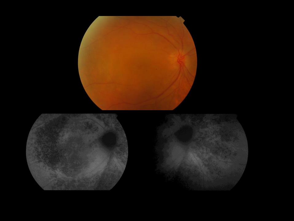
Typical BRC lesions : scattered cream-colored spots
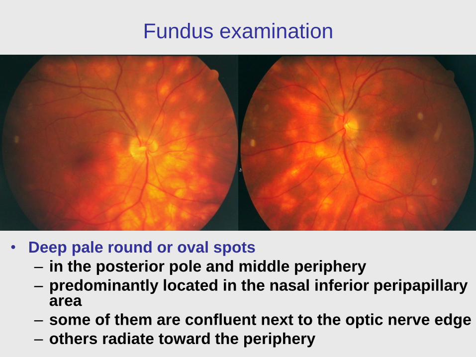
Fundus examination
• Deep pale round or oval spots
– in the posterior pole and middle periphery
– predominantly located in the nasal inferior peripapillary area
– some of them are confluent next to the optic nerve edge
– others radiate toward the periphery

Fluorescein angiography
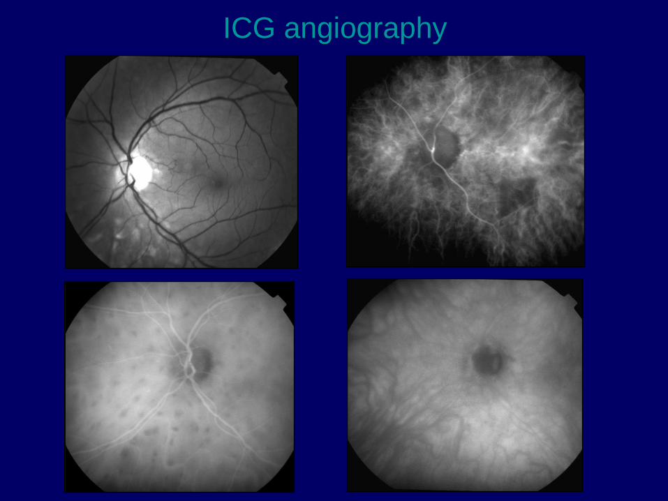
ICG angiography

Fundus : numerous deep pale spots
FA : late phase : macular cystoid leakage
ICGA : numerous hypofluorescent spots ; some of them are next to
the optic nerve head edge giving the well known shape in Mickey
mouse’s ears appearance
FA
FA
ICGA
ICGA

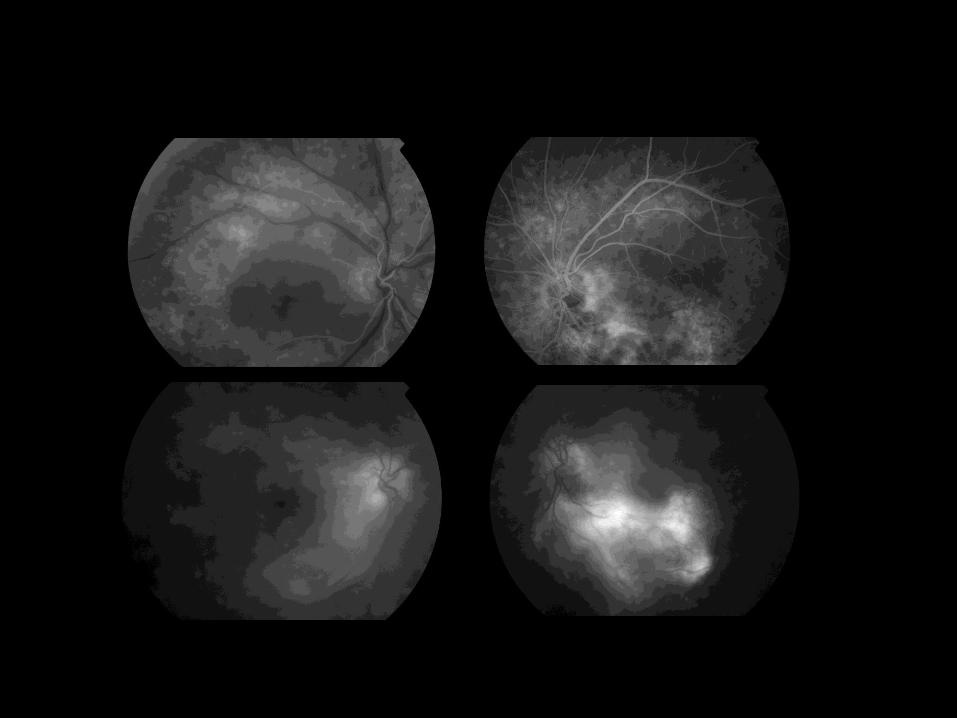
Fluorescein Angiography
Red free photograph : well defined spots
FA : hyperfluorescence of the optic disc, spots, CME
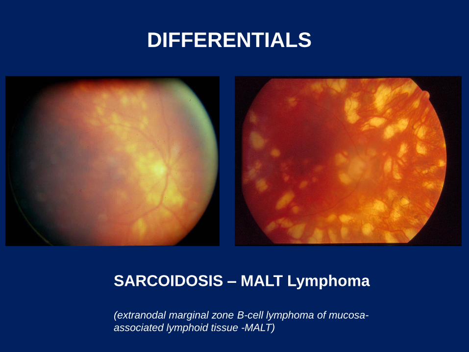
SARCOIDOSIS – MALT Lymphoma
(extranodal marginal zone B-cell lymphoma of mucosa-
associated lymphoid tissue -MALT)
DIFFERENTIALS
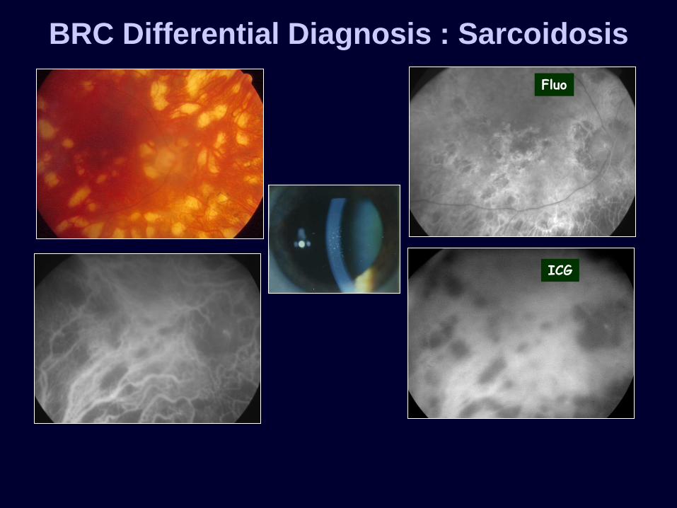
ICG
Fluo
BRC Differential Diagnosis : Sarcoidosis
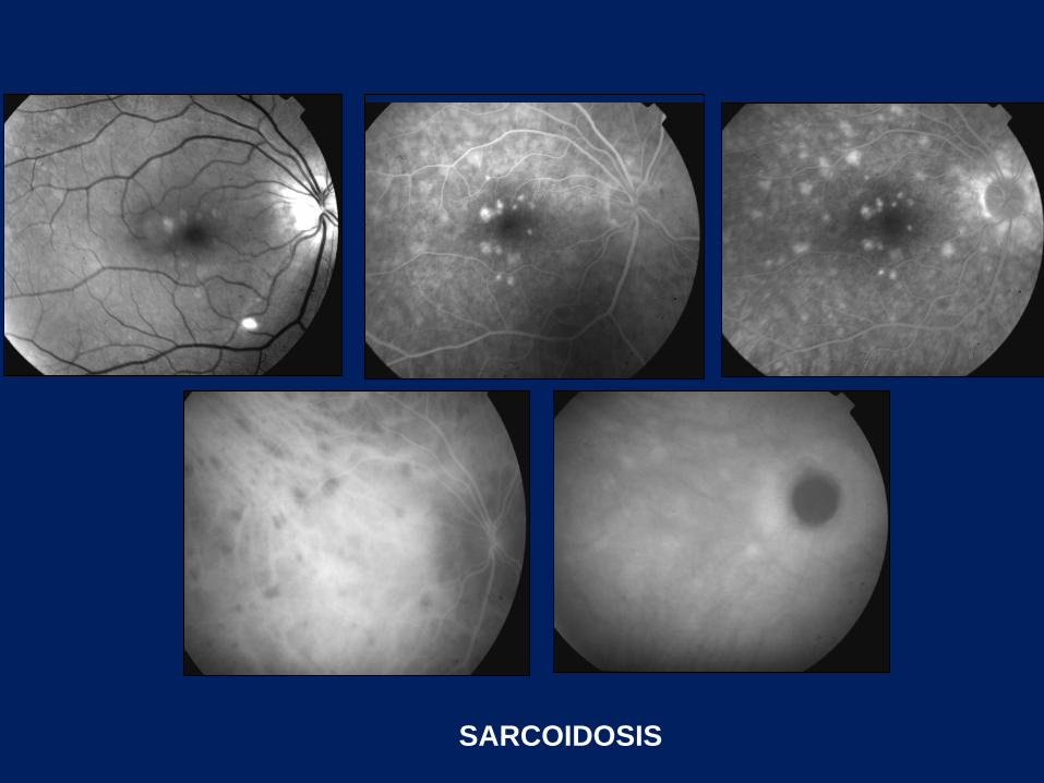
SARCOIDOSIS

large B-cell lymphoma.
Immunocytochemical staining was positive for CD19 and CD20
E. Miserocchi et al.; Ocul Immunol Inflam. 2012
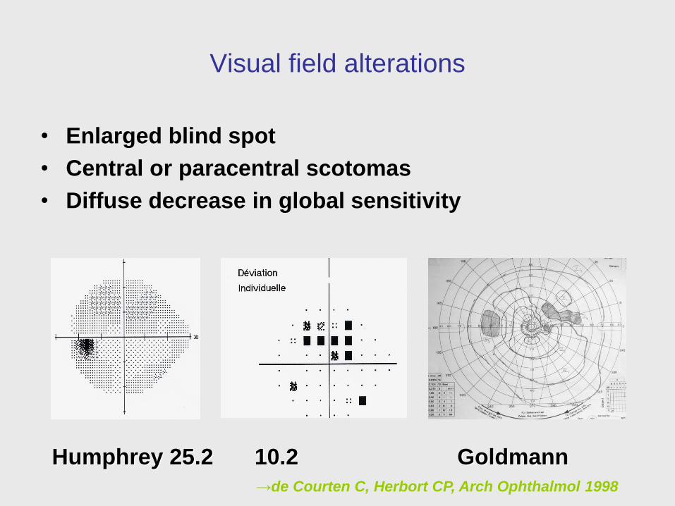
Visual field alterations
• Enlarged blind spot
• Central or paracentral scotomas
• Diffuse decrease in global sensitivity
Humphrey 25.2 10.2 Goldmann
→de Courten C, Herbort CP, Arch Ophthalmol 1998
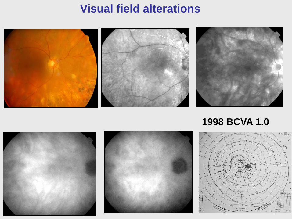
Visual field alterations
1998 BCVA 1.0

2003 BCVA 1.0
Visual field alterations
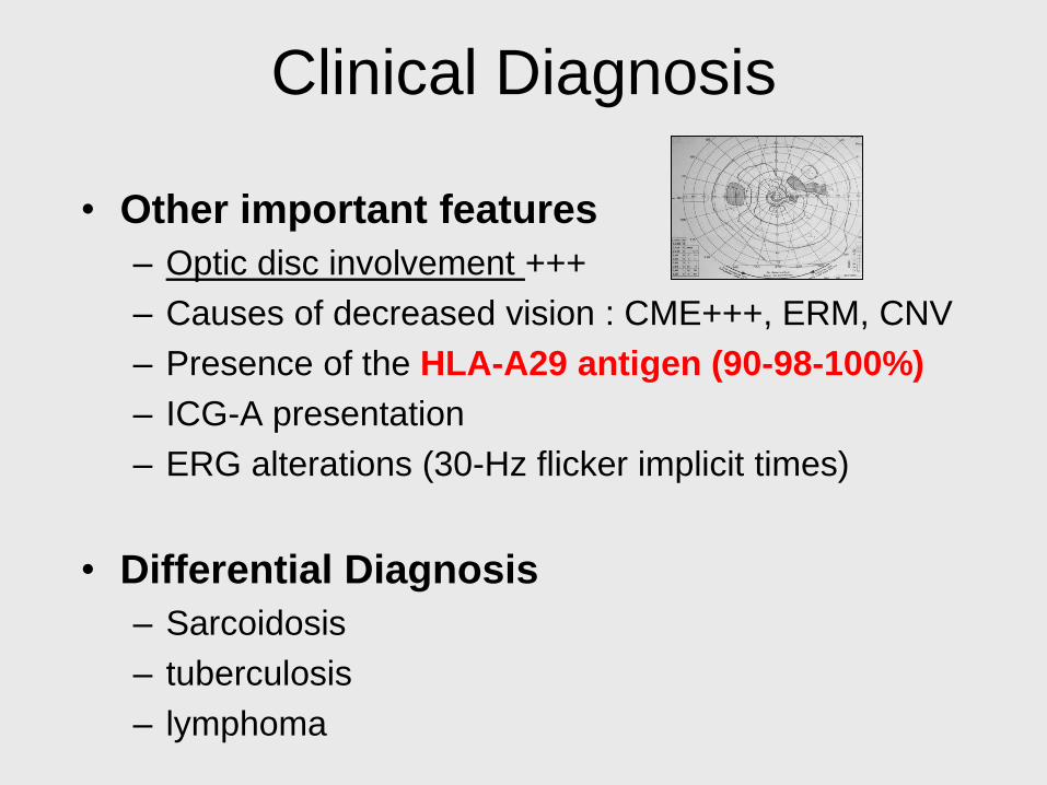
Clinical Diagnosis
• Other important features
– Optic disc involvement +++
– Causes of decreased vision : CME+++, ERM, CNV
– Presence of the HLA-A29 antigen (90-98-100%)
– ICG-A presentation
– ERG alterations (30-Hz flicker implicit times)
• Differential Diagnosis
– Sarcoidosis
– tuberculosis
– lymphoma
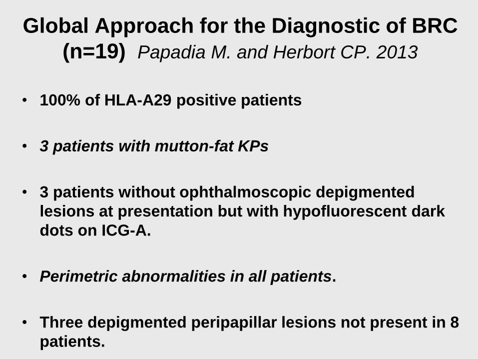
Global Approach for the Diagnostic of BRC
(n=19) Papadia M. and Herbort CP. 2013
• 100% of HLA-A29 positive patients
• 3 patients with mutton-fat KPs
• 3 patients without ophthalmoscopic depigmented
lesions at presentation but with hypofluorescent dark
dots on ICG-A.
• Perimetric abnormalities in all patients.
• Three depigmented peripapillar lesions not present in 8
patients.
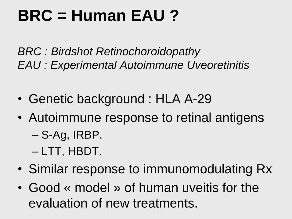
BRC = Human EAU ?
BRC : Birdshot Retinochoroidopathy
EAU : Experimental Autoimmune Uveoretinitis
• Genetic background : HLA A-29
• Autoimmune response to retinal antigens
– S-Ag, IRBP.
– LTT, HBDT.
• Similar response to immunomodulating Rx
• Good « model » of human uveitis for the
evaluation of new treatments.

Well delineated hypofluorecent choroidal spots
Indocyanine Green Angiography

Pathology and Imaging correlations
• Birdshot retinochoroidopathy
• Histological lesions account for the imaging observations.
• Focal lymphocytic infiltrates :
• NO real, genuine granulomas, (pathologically speaking)
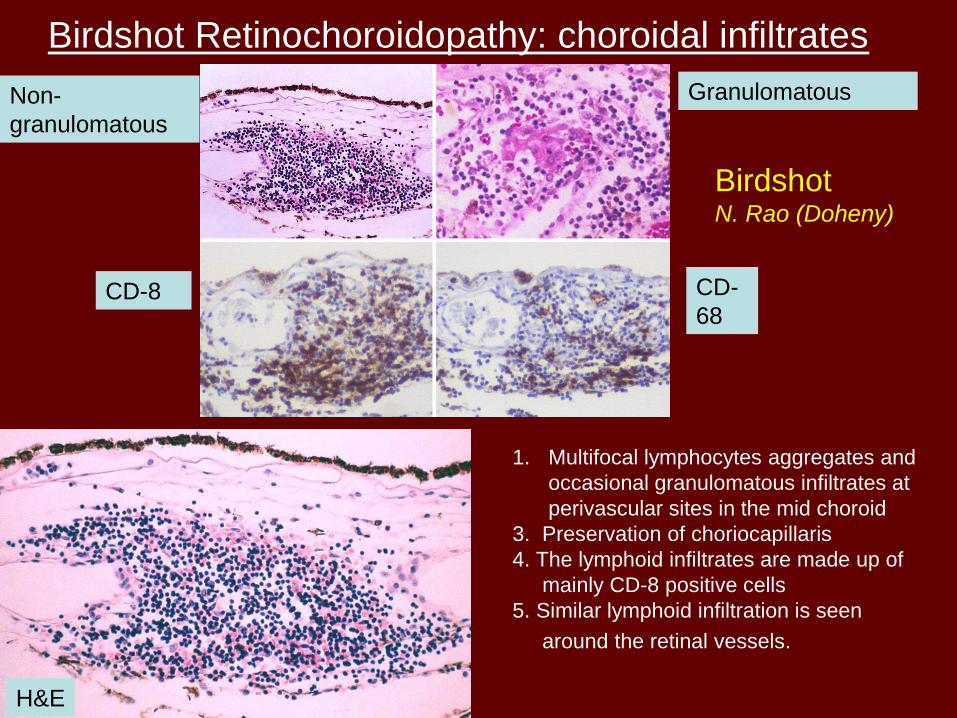
Birdshot Retinochoroidopathy: choroidal infiltrates
1. Multifocal lymphocytes aggregates and
occasional granulomatous infiltrates at
perivascular sites in the mid choroid
3. Preservation of choriocapillaris
4. The lymphoid infiltrates are made up of
mainly CD-8 positive cells
5. Similar lymphoid infiltration is seen
around the retinal vessels.
Non-
granulomatous
Granulomatous
CD-8 CD-
68
H&E
Birdshot N. Rao (Doheny)

Birdshot N. Rao (Doheny)
Birdshot: Retinal vasculitis
CD-8
CD-4
The retinal vascular infiltration
Non-granulomatous
lymphoid infiltration
of the retinal vessels, mostly perivascular
distribution
The infiltrate is made up of mainly CD-8
cells
Few CD-68 positive histiocytes and CD-4
positive T- cells are present

Retinal Vasculitis
Primate Model
LeHoang et al: Ophthalmic Res 2008; 40:181

primate
mice
Behcet
Birdshot

PA Gaudio et al., 2002

Histopathological sections, haematoxylin and eosin staining
• (A) Low power photomicrograph showing three foci of lymphocytic infiltrates (arrows) in the choroid (´5 magnification).
• (B) Higher power photomicrograph of focal lymphocytic infiltrate in the choroid. The choriocapillaris (top) is not involved (´50 magnification).
• (C) Choroidal lymphocytic focus abutting choroidal vessels (´50 magnification).
• (D) Lymphocytes surround a retinal vessel (focal retinal vasculitis) (´50 magnification).
• (E) Focal lymphocytic infiltrate (arrow) in prelaminar optic nerve (´25 magnification).
Birdshot
(Gaudio PA et al.
BJO 2002)

JS Pulido et al. (2012)

JS Pulido et al. (2012)
Absence of granulomatous lesions

Major Issue = Treatment
• Should we treat ?
• How aggressively should we treat ?
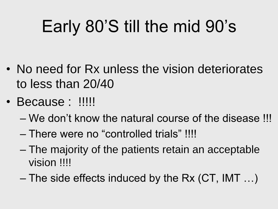
Early 80’S till the mid 90’s
• No need for Rx unless the vision deteriorates
to less than 20/40
• Because : !!!!!
– We don’t know the natural course of the disease !!!
– There were no “controlled trials” !!!!
– The majority of the patients retain an acceptable
vision !!!!
– The side effects induced by the Rx (CT, IMT …)

Should we treat ?
• Until recently: indications for Rx limited to very severe cases with VA below 20/40 (driver’s license)
• Since 2000, several longitudinal studies show the poor visual prognosis of BRC
• The same authors who denied the need for any Rx in the past, are now favoring an aggressive Rx in the early active phase of the disease.

Why should we treat ?
• Severity of the disease
• Poor visual outcome
• Chronic macular oedema+++
• Progressive loss of VA during follow-up
• The number of eyes with VA less than 20/200 increased :
– From 8% at onset
– To 30% after 5 years
– To 39% after ten years of follow-up (Rothova et al. Ophthalmology, 2004 )
• An aggressive therapeutic approach might be justified (CME+++)
(Thorne et al. Am J Ophthalmol, 2005)
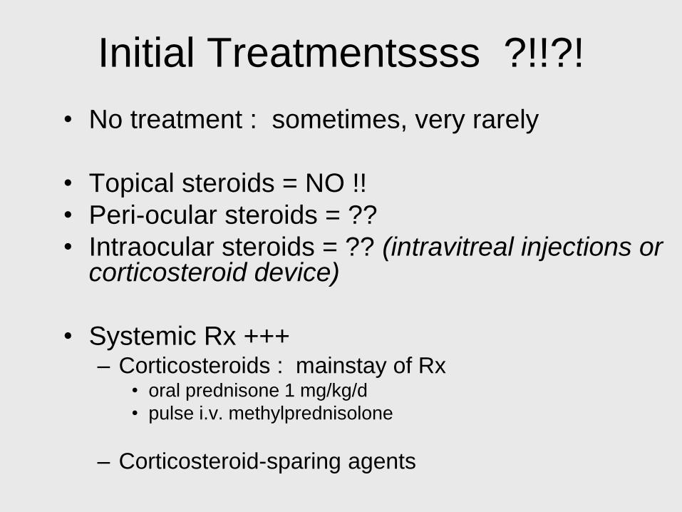
Initial Treatmentssss ?!!?!
• No treatment : sometimes, very rarely
• Topical steroids = NO !!
• Peri-ocular steroids = ??
• Intraocular steroids = ?? (intravitreal injections or corticosteroid device)
• Systemic Rx +++ – Corticosteroids : mainstay of Rx
• oral prednisone 1 mg/kg/d
• pulse i.v. methylprednisolone
– Corticosteroid-sparing agents
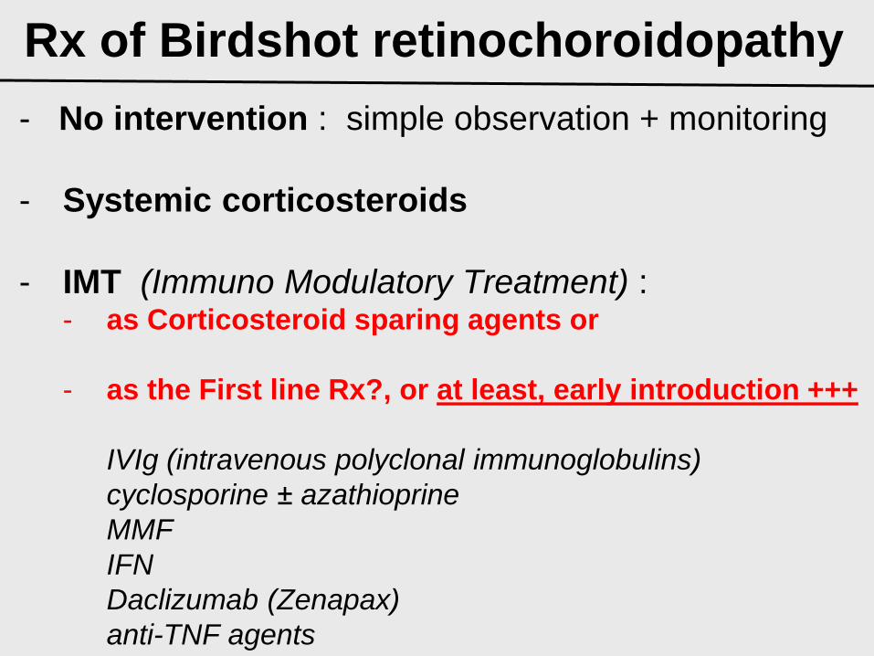
Rx of Birdshot retinochoroidopathy
- No intervention : simple observation + monitoring
- Systemic corticosteroids
- IMT (Immuno Modulatory Treatment) : - as Corticosteroid sparing agents or
- as the First line Rx?, or at least, early introduction +++
IVIg (intravenous polyclonal immunoglobulins)
cyclosporine ± azathioprine
MMF
IFN
Daclizumab (Zenapax)
anti-TNF agents
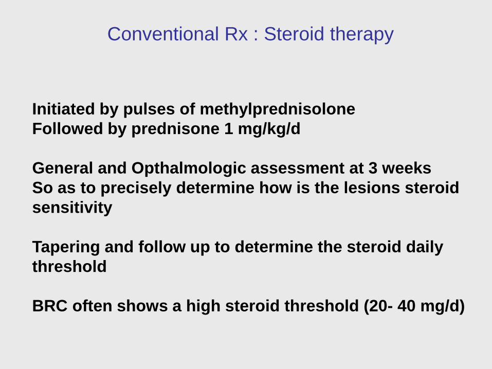
Conventional Rx : Steroid therapy
Initiated by pulses of methylprednisolone
Followed by prednisone 1 mg/kg/d
General and Opthalmologic assessment at 3 weeks
So as to precisely determine how is the lesions steroid
sensitivity
Tapering and follow up to determine the steroid daily
threshold
BRC often shows a high steroid threshold (20- 40 mg/d)

Fluo ICG
Fluo
Fluo
ICG ICG
ICG ICG
Retinal and choroidal signs of inflammation resolved
Before treatment
on steroid treatment

Corticosteroid-Sparing Agents (high dose corticosteroids dependence
> 20 mg/d oral prednisone)
• IvIg: polyclonal Ig (expensive,
unconvenient but effective in mild cases)
• Azathioprine
• Cyclosporine
• Mycophenolate Mofetil (MMF)
• Combination of Cyclosporine and MMF

Immunomodulation
• Polyclonal Immunoglobulins
from human origin (20 donnors)
induce saturation of idiotypic net
• Pilot study
Polyclonal immunoglobulins only
18 patients VA ↑ 53% ↓18% with a mean follow up of 39 m
Reduction of fluorescein leakage (50%)
• Retrospective study
Combined therapy (Steroid + PIG) 37 patients
Significant steroid threshold decrease → Ocular Immunol Inflamm 2000

CME recurrence under Prednisone (15 mg/d)
Control of inflammation under prednisone 5 mg/d and
polyclonal immunoglobulins 0.3g/kg/month

Cyclosporine = Not a New Rx
• 1983 = the first published BRC patient
treated with Cyclosporine (R.B. Nussenblatt)
• 1988 = published positive results in a series
of 19 BRC cyclosporine treated patients
(P. LeHoang)
• 1994 = effectiveness of low-dose
cyclosporine (A.T. Vitale)

Patients and Methods
• Retrospective case study (severe forms of BRC )
• January 1986-january 2000
• Inclusion criteria :
– Diagnosis based on : typical clinical presentation (Ryan and Maumenee, 1980)
– HLA-A29+
– Treated with CsA : dependence or resistance to high dose Ct, resistance to IvIg
• Cyclosporine A :
– Started at 3-5 mg/kg/d
– Slowly tapered
• Efficacy of CsA was evaluated : – VA, severity of macular edema (FA +/- OCT)
• Safety of CsA was evaluated : – Renal function and blood pressure

Results 1 • 168 BRC managed (1986 - 2000)
• 49 patients (98 eyes) required CsA (29.16%)
• Average age at onset : 48.5 years (29-68 y)
• Caucasian (100%)
• F/M : 1.2
• Mean duration of disease before CsA : 4.5 y (1-14 y)
• 28 patients required high level of Ct (57.1%) > 25 mg/d
• Resistance to Ct : 21 patients (42.9%)
• Control of ocular inflammation was achieved in most of cases
• Major renal toxicity
– observed in 2 patients
– required a rapid dosage tapering of cyclosporine
– Azathioprine was added

Visual acuity
• Improved by 2 lines or more : 42 eyes (42.6%)
• Stabilized : 37 eyes (38%)
• Decreased by 2 lines or more : 19 eyes (19.4%)
Aft
er
8 y
ears
of
Cs
A t
hera
py
Before CsA therapy
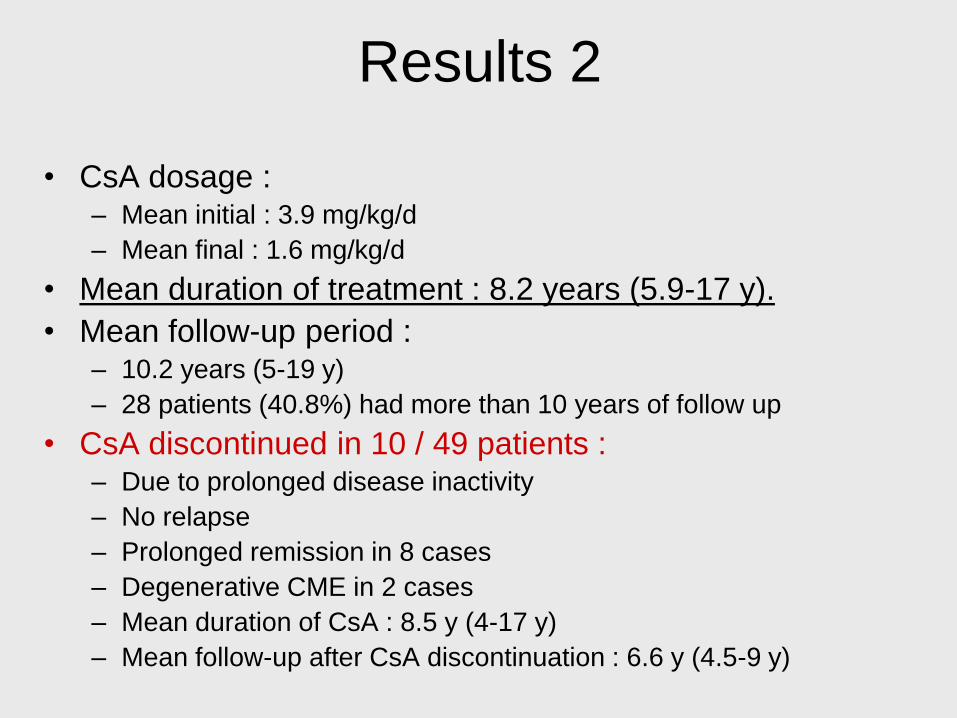
Results 2
• CsA dosage : – Mean initial : 3.9 mg/kg/d
– Mean final : 1.6 mg/kg/d
• Mean duration of treatment : 8.2 years (5.9-17 y).
• Mean follow-up period : – 10.2 years (5-19 y)
– 28 patients (40.8%) had more than 10 years of follow up
• CsA discontinued in 10 / 49 patients : – Due to prolonged disease inactivity
– No relapse
– Prolonged remission in 8 cases
– Degenerative CME in 2 cases
– Mean duration of CsA : 8.5 y (4-17 y)
– Mean follow-up after CsA discontinuation : 6.6 y (4.5-9 y)

Macular edema
Initial Final
NCME 21 (21.4%)
12 (12.2%)
CME 70 (71.4%)
19 (19.4%)
Macular oedema decreased significantly P<0.001
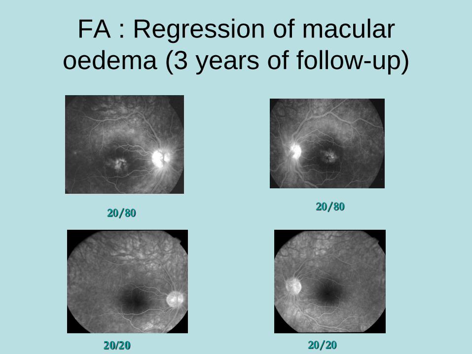
20/20 20/20
20/80 20/80
FA : Regression of macular
oedema (3 years of follow-up)
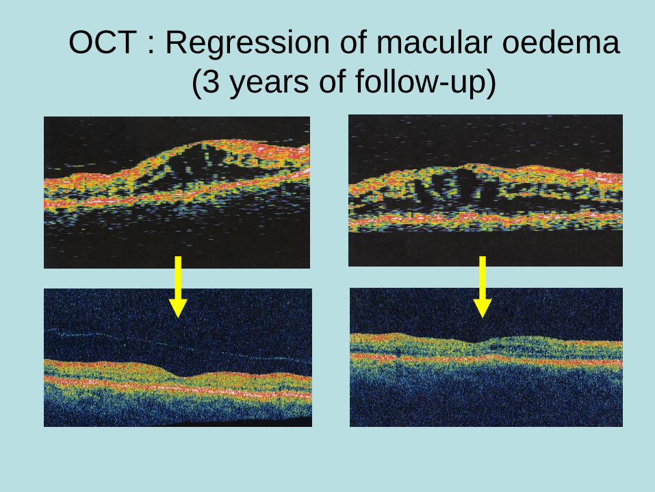
OCT : Regression of macular oedema
(3 years of follow-up)

Avril 2003
labbez
Good response to Rx

0.9
Timpagnon
ap Rx
CT+CSA
JUIN2001
before Rx
after Rx

Major side effects
• Long-term CsA Rx (5 mg/kg/d)
– High Prevalence of hypertension :
• High blood pressure : 78% patients after 12 months (p<0.0001)
– Significant renal impairment
• Significant decrease in creatinine clearance observed in all cases : mean 17.6% (12.4-24.5%) (p<0.0001)

Discussion 1
• Initial characteristics of our study-population : – Similar to those reported in previous series
– Caucasian (100%)
– Affects middle aged people
– Equilibrated sex-ratio (→Priem and Oosterhuis, Br J Ophthalmol. 1988)
• Efficacy of CsA reported in previous series : – (→ Nussenblatt et al. Am J Ophthalmol. 1983)
– (→LeHoang et al. Transplant Proc. 1988)
– (→Vitale et al. Ophthalmology, 1994)
• Previous studies : – Initial, mid-term and long-term efficacy of CsA
– Small cohort
• First study to evaluate a renal follow up period after CsA discontinuation : – Prolonged remission (8 / 49 cases; 16%)

Discussion 2
• Nephrotoxicity and hypertension : – Serious side-effects
– Well established in a single-center prospective cohort study
– Occur during the first 3 months of therapy
– Become maximal after 12 months
– Remain stable for the next few years
• Nephrotoxicity : – Dose dependent
– Highest doses (>3 mg/kg/d) induced more severe alterations than the lowest doses
– Tapering of CsA doses may improve renal function (only in the absence of interstitial fibrosis)
• Hypertension : – Independent of CsA dosage
(→Isnard Bagnis et al. J Am Soc Nephrol. 2002)

Discussion 3
• 28 patients
• Mean follow-up : 81 months
• 93% were treated with CsA
• 68 % were treated with a combination of azathioprine and CsA
• At the end of the follow-up :
– 85% of patients had either the same or improved VA
• Encouraging results
• By using this strategy, the preservation of visual function may be obtained ( Kiss et al. Ophthalmology, 2005)
• 11 patients (Aza, MTX, CyA, MMF, IvIg : 5 cases)
• Median follow-up : 6 years
• Reduction or stabilization of inflammation in all cases
(Becker et al. Ocular Immunol Inflam, 2005)
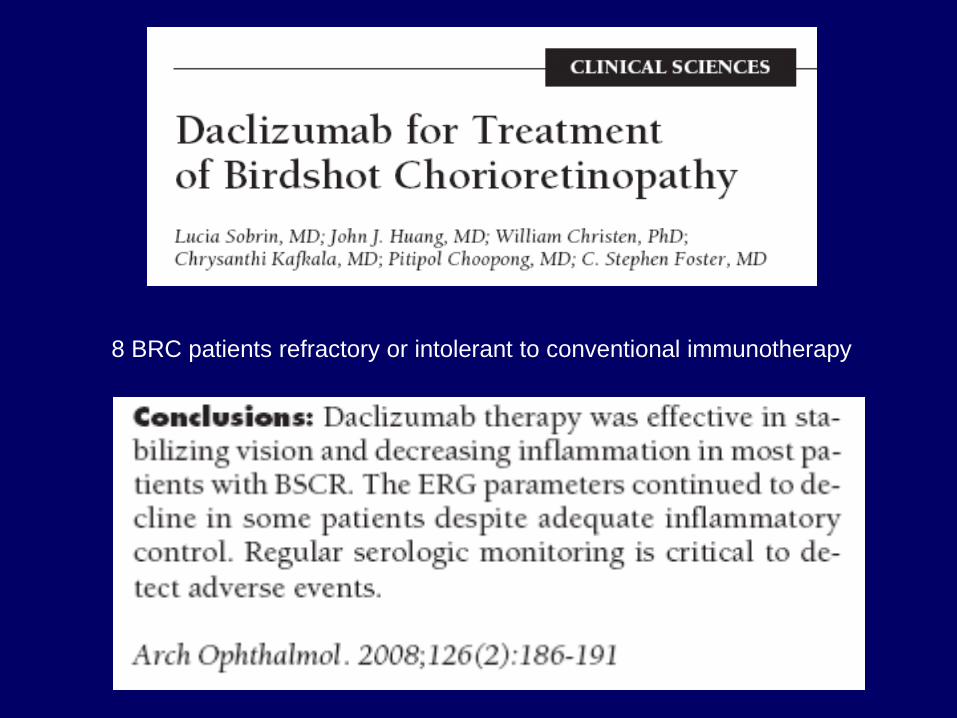
8 BRC patients refractory or intolerant to conventional immunotherapy
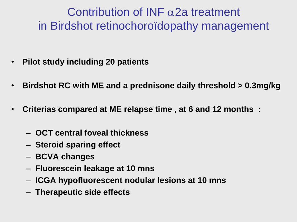
Contribution of INF a2a treatment
in Birdshot retinochoroïdopathy management
• Pilot study including 20 patients
• Birdshot RC with ME and a prednisone daily threshold > 0.3mg/kg
• Criterias compared at ME relapse time , at 6 and 12 months :
– OCT central foveal thickness
– Steroid sparing effect
– BCVA changes
– Fluorescein leakage at 10 mns
– ICGA hypofluorescent nodular lesions at 10 mns
– Therapeutic side effects
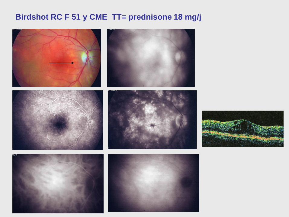
Birdshot RC F 51 y CME TT= prednisone 18 mg/j

INF a2a 3Mx3/w
Pred 8 mg/jd

Birdshot at presentation
- 2/3 VA < 20/50
- 1/3 VA < 20/200 (30 mths)
(Thorne JE et al. AJO 2005)
FIGURE 2 Spectral-domain optical
coherence tomography
(SD-OCT). SD-OCT at baseline showed
a disruption of the
hypereflective line representing the
junction between the inner
segments and the outer segments
(IS/OS junction), a distortion
of the normal lamination of the inner
retina, as well as a loss of
the normal hyporeflective space
corresponding to the photoreceptor
outer segments (PROS) in both the right
eye (A) and
the left eye (B). After 4 months, SD-OCT
demonstrated restored
continuity of the IS/OS junction,
restoration of the lamination
of the inner retina, as well as the
presence of the normal PROS
hyporeflective space in the fovea in the
right eye (C) and the
left eye (D). Red arrows depict the
IS/OS junction, while yellow
arrows depict the PROS hyporeflective
space. All scans are
from the same cross section through the
center of the fovea.

Contribution of INF a2a treatment
in Birdshot retinochoroïdopathy management
OCT Daily prednisone FA ICGA nodular
CFT Threshold leakage lesions
Initial time 327±70 34±18 34/34 (100%) 25/32 (78%)
At 6 months 223±62 15±13 11/34 (32%) 8/26 (30%)
At 12 months 211±49 11±9 5/24 (20%) 5/20 (25%)
P 0.001 0.001 0.01 0.01

Background Observations
- Progressive disease
- Sustained chronic inflammation + numerous
exacerbations -> further visual deterioration
- 2/3 VA < 20/50 ; 1/3 VA < 20/200 (30 mths)
- 20% are legally blind after 10 yrs
- Early agressive and continuous Rx for
preventing visual loss +++

Time of Therapeutic intervention =
too late
Choroidal disease responding well to
therapy while retinal disease is more
resistant,possibly explaining the slow
deterioration of functional parameters
despite therapy (CH Herbort).

Poor Prognosis
• Slow decline in VA despite Rx : persistent insidious inflammatory activity -> VA is not a good indicator for the monitoring of the Rx.
• Causes of visual alteration – Optic disc involvement
– CME+++, ERM, CNV
– Retinal atrophy
• Differential Diagnosis – Sarcoidosis
– Tuberculosis
– Lymphoma, MALT etc …
GAP

Corticosteroids used to be the mainstay of Rx
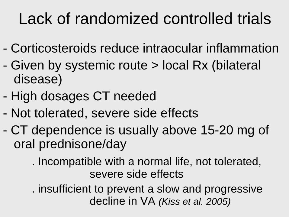
Lack of randomized controlled trials
• Peri-ocular and systemic corticosteroids are
ineffective :
– No sufficient control (Kiss et al. 2005)
– Not well tolerated
• No tolerance for low-grade inflammation +++
• Need for IMT to limit permanent ocular
structure damage ++++
GAP

Treat with What ? Corticosteroids or IMT or both ?
(Visual acuity)
Oral corticosteroids
No change Improved Decreased
32%
40%
50%
10%
55%
50%
42%
66% 24%
8%
10%
13%
Oral corticosteroids
+ antimetabolites
Oral corticosteroids
+ Cyclosporine
Cyclosporine
alone
(Shah et al. Survey, 2005)

Corticosteroid vs Immunosuppression
Lack of randomized controlled trials
- Corticosteroids reduce intraocular inflammation
- Given by systemic route > local Rx (bilateral disease)
- High dosages CT needed
- Not tolerated, severe side effects
- CT dependence is usually above 15-20 mg of oral prednisone/day
. Incompatible with a normal life, not tolerated, severe side effects
. insufficient to prevent a slow and progressive decline in VA (Kiss et al. 2005)
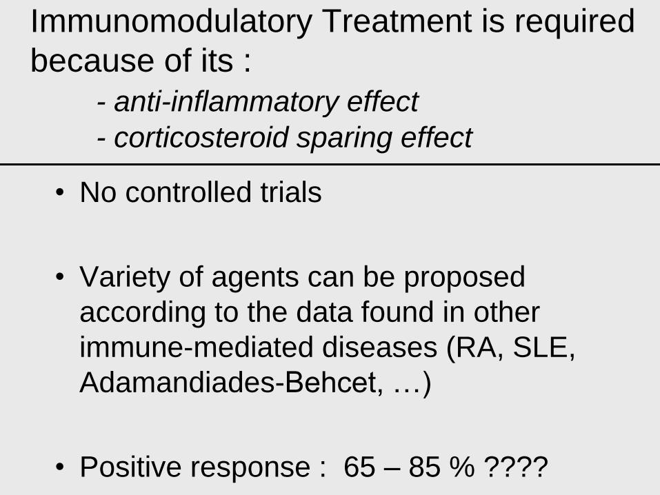
Immunomodulatory Treatment is required
because of its :
- anti-inflammatory effect
- corticosteroid sparing effect
• No controlled trials
• Variety of agents can be proposed
according to the data found in other
immune-mediated diseases (RA, SLE,
Adamandiades-Behcet, …)
• Positive response : 65 – 85 % ????
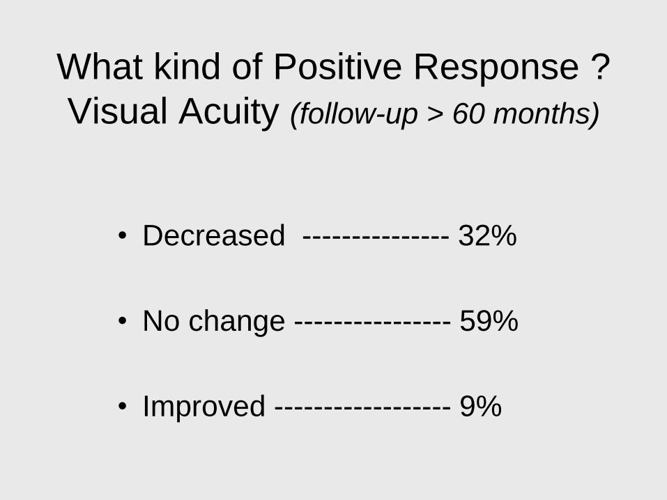
What kind of Positive Response ?
Visual Acuity (follow-up > 60 months)
• Decreased --------------- 32%
• No change ---------------- 59%
• Improved ------------------ 9%

Large variety of agents
• Cyclosporin monotherapy (high dosage with nephrotoxicity)
• Azathioprine
• Combination of low dose [CSA + azathioprine]
• Mycophenolate mofetil
• Combination of MMF + Cyclosporine
• Methotrexate
• Polyclonal IvIg
• Daclizumab
• TNF blockade (infliximab, adalimumab)
• Tacrolimus + mycophenolate mofetil
• INF alpha
• voclosporin
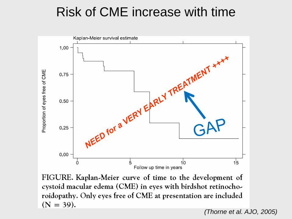
Risk of CME increase with time
(Thorne et al. AJO, 2005)

Gaps
• Randomized controlled trials
• Combined Scoring system :
(Disease Activity Score, Not only the VA)
– Visual acuity
– Clinical (vitreous haze)
– Visual fields (Humphrey, Goldmann)
– SD-OCT or better !
– Electrophysiology
– FA / ICG-A
• Utilize IMT as the first line treatment (as recommended by the AUS)
• Start VERY EARLY treatment based on novel indicators
Start earlier the
Immunomodulatory Therapy
• Future in Genetics, Biomarkers ?? : predict the onset of
BRC
• Tissue damage, criteria :
• Retinal vasculitis A29+ before the apparition of creamy spots
• Early detection of retinal deterioration (mfERGs, SD-OCT)
• Optic nerve deterioration (VF, color vision, contrast sensitivity)
• Early detection of choroidal disease (ICG-A)
• What Immunosuppressant should we used?

Monitoring : a set of criteria
• Do not rely only on VA
• Mandatory explorations at base line:
– VFs (Humphrey + Goldmann)
– Electrophysiology, ERG, mf-ERG
– FA and ICG-A
– High resolution SD-OCT, Swept S-OCT
GAP

Cone mediated 30Hz flicker ERGs
improvement on Rx
Conclusions :
The ERG data confirm that BCR frequently affects inner retinal function of cone and rod systems.
Clinical features were not reliable indicators of functional deterioration or recovery.
Objective electrophysiological assessment of retinal function demonstrated improvement following
treatment and provides a reliable method of monitoring treatment efficacy, enabling management
decisions to be taken with greater confidence and allowing early initiation or modification of
treatment.

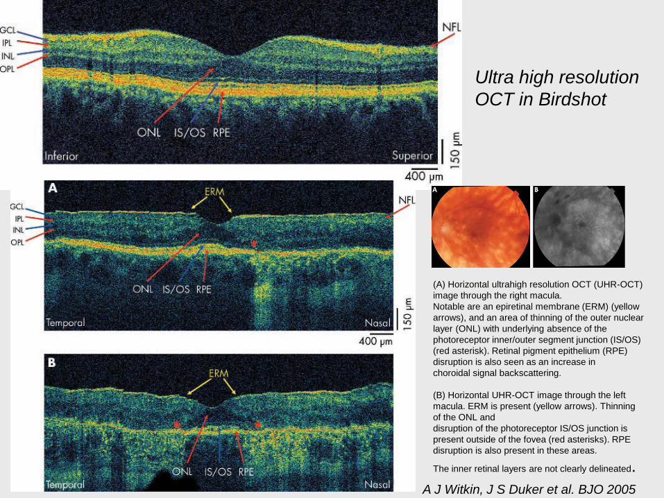
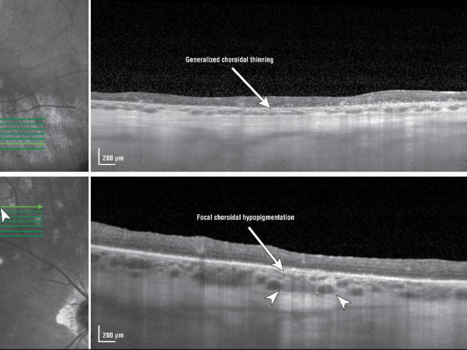
(A) Horizontal ultrahigh resolution OCT (UHR-OCT)
image through the right macula.
Notable are an epiretinal membrane (ERM) (yellow
arrows), and an area of thinning of the outer nuclear
layer (ONL) with underlying absence of the
photoreceptor inner/outer segment junction (IS/OS)
(red asterisk). Retinal pigment epithelium (RPE)
disruption is also seen as an increase in
choroidal signal backscattering.
(B) Horizontal UHR-OCT image through the left
macula. ERM is present (yellow arrows). Thinning
of the ONL and
disruption of the photoreceptor IS/OS junction is
present outside of the fovea (red asterisks). RPE
disruption is also present in these areas.
The inner retinal layers are not clearly delineated.
Figure 1 Ultrahigh
resolution optical
coherence tomography
image from a normal eye.
The
intraretinal layers are
labelled: NFL, nerve fibre
layer, GCL, ganglion cell
layer, IPL, inner plexiform
layer, INL, inner nuclear
layer, OPL, outer
plexiform layer, ONL,
outer nuclear layer,
IS/OS,
photoreceptor inner/outer
segment junction, RPE,
retinal pigment
epithelium.
A J Witkin, J S Duker et al. BJO 2005
Ultra high resolution
OCT in Birdshot

« in vivo histology »

- disruption of the
hypereflective line
representing the
junction between the
inner segments and
the outer segments
(IS/OS junction)
- distortion of the
normal lamination of
the inner retina,
- loss of the normal
hyporeflective space
corresponding to the
photoreceptor
outer segments
(PROS) in both
RE before
LE before
LE after
RE after
Prednisone + MMF
Birdshot
F. Forooghian et al. Ocul
Immunol Inflam 2010
FIGURE 2 Spectral-domain optical
coherence tomography
(SD-OCT). SD-OCT at baseline
showed a disruption of the
hypereflective line representing the
junction between the inner
segments and the outer segments
(IS/OS junction), a distortion
of the normal lamination of the
inner retina, as well as a loss of
the normal hyporeflective space
corresponding to the photoreceptor
outer segments (PROS) in both the
right eye (A) and
the left eye (B). After 4 months, SD-
OCT demonstrated restored
continuity of the IS/OS junction,
restoration of the lamination
of the inner retina, as well as the
presence of the normal PROS
hyporeflective space in the fovea in
the right eye (C) and the
left eye (D). Red arrows depict the
IS/OS junction, while yellow
arrows depict the PROS
hyporeflective space. All scans are
from the same cross section
through the center of the fovea.

BRC Keane 5

BRC Keane 4

Swept Source OCT (SS-OCT)

SS-OCT
Espace suprachoroïdien

Close Monitoring
• Combine several criteria for evaluating disease activity:
• Do Not only rely only on Visual Acuity +++
– Visual acuity
– Clinical (AC flare, vitreous haze)
– Laser Flare meter
– Visual fields (Humphrey, Goldmann)
– SD-OCT – Enhanced Depth Imaging OCT (EDI-OCT) – Swept-Source OCT (SS-OCT)
– Electrophysiology (30 hz flicker implicit times, mf ERG)
– FA / ICG-A

The Problem of Drug Delivery
LOCAL Rx = Bail out a leaky boat
SYSTEMIC Rx = Adverse effects
Lymphoid System
Effectors (cells) Effectors (cells)
inducing intraocular
inflammation
LOCAL Rx SYSTEMIC Rx
EYE
Combination ?
blood


Summary • Intraocular Rxs (injections, implants) are
effective
– good adjunct Rx (unilateral, acute attacks, flare ups)
– but cannot be considered as a long term maintenance Rx :
• transient action, difficult to adjust.
• side effects (glaucoma, cataract, retinal atrophy)
• Downstream (« bail out a leaky boat »)
• Evaluation of cancer risks with long term use of biologics
Birdshot RC -> Corticosteroids =
misleading security
• Corticosteroids Monotherapy may be used to
control recent active inflammation but are
insufficient to prevent the progressive retinal
alterations occurring during the chronic course
of the disease at acceptable dosage.
• Natural history of BSRC or the outcomes with
corticosteroid therapy alone, showed a
persistent worsening of vision with time despite
an apparent clinical remission.

CONCLUSION
Determinants of Remissions and Cure
• Early Diagnosis and Early Treatment – No intermittent Treatment
– Early IMT >>>> Early Corticosteroids
• Close monitoring (30hz flicker ERG implicit times, VF, OCTs, Angiograms).
• Do not rely only on clinical signs or judgment (Visual Acuity that can remain stable despite severe progressive retinal dysfunction)

Conclusion
• BRC : often considered as a chronically progressive
disease resistant to treatment
• Long-term preservation of visual function is attainable
• Visual prognosis of active BRC is poor without Rx
• Therefore, an aggressive approach should be
proposed early !
• IMT appears to be mandatory in most of the cases
• Further randomized multicenter studies are required