biosynthesis riboflavin: cloning, sequencing, mapping ... · biosynthesis of riboflavin 4047...
TRANSCRIPT
JOURNAL OF BACTERIOLOGY, July 1993, p. 4045-40510021-9193/93/134045-07$02.00/0Copyright C 1993, American Society for Microbiology
Vol. 175, No. 13
Biosynthesis of Riboflavin: Cloning, Sequencing, Mapping,and Expression of the Gene Coding for GTP
Cyclohydrolase II in Eschenichia coliGERALD RICHTER,' HARALD RITZ,' GERD KATZENMEIER,1 RAINER VOLK,'ANGELIKA KOHNLE,1 FRIEDRICH LOTTSPEICH,2 DOROTHEE ALLENDORF,1
AND ADELBERT BACHER'*Lehrstuhl fir Organische Chemie und Biochemie, Technische Universitat Munchen,Lichtenbergstrasse 4, D-8046 Garching, ' and Max-Planck-Institut fur Biochemie,
Am Klopferspitz, D-8033 Martinsriet4 2 Germany
Received 5 January 1993/Accepted 25 April 1993
GTP cyclohydrolase II catalyzes the first committed step in the biosynthesis of riboflavin. The gene codingfor this enzyme in Escherichia coli has been cloned by marker rescue. Sequencing indicated an open readingframe of 588 bp coding for a 21.8-kDa peptide of 196 amino acids. The gene was mapped to a position at 28.2min on the E. coli chromosome and is identical with ribA. GTP cyclohydrolase H was overexpressed in arecombinant strain carrying a plasmid with the cloned gene. The enzyme was purified to homogeneity fromthe recombinant strain. The N-terminal sequence determined by Edman degradation was identical to thepredicted sequence. The sequence is homologous to the 3' part of the central open reading frame in theriboflavin operon of Bacillus subtilis.
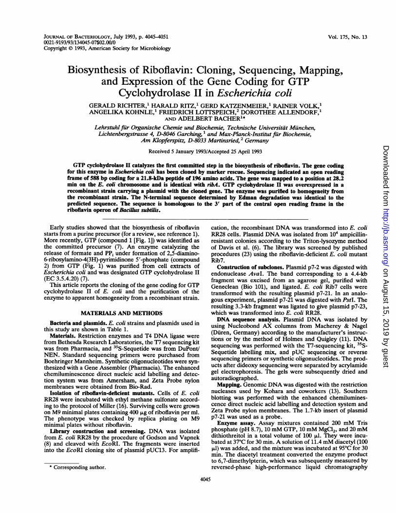
Early studies showed that the biosynthesis of riboflavinstarts from a purine precursor (for a review, see reference 1).More recently, GTP (compound 1 [Fig. 1]) was identified asthe committed precursor (7). An enzyme catalyzing therelease of formate and PP1 under formation of 2,5-diamino-6-ribosylamino-4(3H)-pyrimidinone 5'-phosphate (compound2) from GTP (Fig. 1) was purified from cell extracts ofEschetichia coli and was designated GTP cyclohydrolase II(EC 3.5.4.20) (7).
This article reports the cloning of the gene coding for GTPcyclohydrolase II of E. coli and the purification of theenzyme to apparent homogeneity from a recombinant strain.
MATERIALS AND METHODS
Bacteria and plasmids. E. coli strains and plasmids used inthis study are shown in Table 1.
Materials. Restriction enzymes and T4 DNA ligase werefrom Bethesda Research Laboratories, the T7 sequencing kitwas from Pharmacia, and 35S-Sequetide was from DuPont/NEN. Standard sequencing primers were purchased fromBoehringer Mannheim. Synthetic oligonucleotides were syn-thesized with a Gene Assembler (Pharmacia). The enhancedchemiluminescence direct nucleic acid labelling and detec-tion system was from Amersham, and Zeta Probe nylonmembranes were obtained from Bio-Rad.
Isolation of riboflavin-deficient mutants. Cells of E. coliRR28 were incubated with ethyl methane sulfonate accord-ing to the protocol of Miller (16). Surviving cells were grownon M9 minimal plates containing 400 ,ug of riboflavin per ml.The phenotype was checked by replica plating on M9minimal plates without riboflavin.
Library construction and screening. DNA was isolatedfrom E. coli RR28 by the procedure of Godson and Vapnek(8) and cleaved with EcoRI. The fragments were insertedinto the EcoRI cloning site of plasmid pUC13. For amplifi-
* Corresponding author.
cation, the recombinant DNA was transformed into E. coliRR28 cells. Plasmid DNA was isolated from 104 ampicillin-resistant colonies according to the Triton-lysozyme methodof Davis et al. (6). The library was screened by publishedprocedures (23) using the riboflavin-deficient E. coli mutantRib7.
Construction of subclones. Plasmid p7-2 was digested withendonuclease AvaL. The band corresponding to a 4.4-kbfragment was excised from an agarose gel, purified withGeneclean (Bio 101), and ligated. E. coli Rib7 cells weretransformed with the resulting plasmid p7-21. In an analo-gous experiment, plasmid p7-21 was digested with PstI. Theresulting 3.3-kb fragment was ligated to give plasmid p7-23,which was transformed into E. coli RR28.DNA sequence analysis. Plasmid DNA was isolated by
using Nucleobond AX columns from Macherey & Nagel(Duren, Germany) according to the manufacturer's instruc-tions or by the method of Holmes and Quigley (11). DNAsequencing was performed with the 17-sequencing kit, 35S-Sequetide labelling mix, and pUC sequencing or reversesequencing primers or synthetic oligonucleotides. The prod-ucts after dideoxy sequencing were separated by acrylamidegel electrophoresis. The gels were subsequently dried andautoradiographed.Mapping. Genomic DNA was digested with the restriction
nucleases used by Kohara and coworkers (13). Southernblotting was performed with the enhanced chemilumines-cence direct nucleic acid labelling and detection system andZeta Probe nylon membranes. The 1.7-kb insert of plasmidp7-21 was used as a probe.Enzyme assay. Assay mixtures contained 200 mM Tris
phosphate (pH 8.7), 10 mM GTP, 10 mM MgCl2, and 20 mMdithiothreitol in a total volume of 100 ,ul. They were incu-bated at 37°C for 30 min. A solution of 11.4 mM diacetyl (100,ul) was added, and the mixture was incubated at 95°C for 30min. The diacetyl treatment converted the enzyme productto 6,7-dimethylpterin, which was subsequently measured byreversed-phase high-performance liquid chromatography
4045
on August 15, 2019 by guest
http://jb.asm.org/
Dow
nloaded from
4046 RICHTER ET AL.
0
@@@OH2C o
1OH OH
CH20HC20
H-C-OH -
H-C-OHCH20®
4
0
H3C N N 0H2
H-C-OHH-C-OH 8H-C-OHCH20H
(X) H N<NH
HN N NH,®OH2C 2
OH OH
QJ CH,
HO-C-HCH,0®
H2NXH
H N 0CH,H5 c H, A
H-C-OHH-C-OH 6H-C-OHCH,0®
0
H2NtNHHNN N OCH2 H
H-C-OHH-C-OH 7H-C-OHCH2OH
0
H3C N NHOCH2
H-C -UHH O-OH 9H-C-OHCH2OH
FIG. 1. Biosynthesis of riboflavin (bacterial pathway). (A) GTPcyclohydrolase II; (B) 3,4-dihydroxy-2-butanone 4-phosphate syn-thase.
(HPLC) on a column of Lichrosorb RP18 (4 by 250 mm). Theeluent contained 100 mM ammonium formate and 40%methanol. The effluent was monitored fluorometrically (ex-citation, 365 nm; emission, 435 nm). One unit of enzymeactivity catalyzes the formation of 1 nmol of 2,5-diamino-6-ribosylamino-4(3H)-pyrimidinone 5'-phosphate per h at 37°C.
Protein purification. Mutant strain Rib7 harboring plasmidp7-21 was grown in shaking culture (Luria-Bertani mediumcontaining 150 mg of ampicillin per liter) at 37°C for 16 h.Frozen bacterial cells (31 g) were thawed in 62 ml of 200
mM Tris hydrochloride, pH 8, containing 2mM phenylmeth-ylsulfonyl fluoride. Lysozyme (31 mg) and DNase I (0.3 mg)were added, and the mixture was stirred at 37°C for 20 min.The suspension was centrifuged in an SS 34 rotor (Sorvall) at15,000 rpm at 4°C for 20 min.Ammonium sulfate (14.8 g) was added to the supernatant
(84 ml). The suspension was stirred for 30 min at 4°C. It wascentrifuged (Sorvall SS 34 rotor; 15,000 rpm, 20 min), andthe precipitate was discarded. Ammonium sulfate (17.0 g)was added to the supernatant (87 ml). The suspension wasstirred for 1 h at 4°C. The precipitate was harvested bycentrifugation and dissolved in 25 mM Tris hydrochloride,pH 8. The solution was dialyzed three times against 2 litersof 25 mM Tris hydrochloride, pH 8, containing 0.2 mMEDTA, 0.5 mM MgCl2, and 150 mM NaCl.The dialyzed solution was applied to a column of DEAE-
cellulose DE 52 (2.5 by 21 cm) which had been equilibratedwith the dialysis buffer. The flow rate was 30 ml/h. Thecolumn was developed with 300 ml of equilibration bufferfollowed by 25 mM Tris-HCl, pH 8, containing 0.2 mMEDTA, 0.5 mM MgCl2, and 200 mM NaCl. Fractions (440 to570 ml each) containing GTP cyclohydrolase II were col-lected and dialyzed against 2 liters of 25 mM Tris hydrochlo-ride, pH 8.The dialyzed protein solution was applied to a column of
hydroxyapatite (Bio-Rad; 2.2 by 3.5 cm) which had beenequilibrated with the dialysis buffer. The column was devel-oped with 150 ml of dialysis buffer followed by 25 mM Trishydrochloride, pH 8, containing 10 mM potassium phos-phate (flow rate, 32 ml/h). Fractions containing enzymeactivity were collected (280 to 320 ml) and concentrated to avolume of S ml by ultrafiltration (PM10 membrane; Amicon).The protein solution was applied to a Sephacryl S-200
column (2.2 by 90 cm) equilibrated with 25 mM Tris hydro-chloride, pH 8, containing 2.5 mM EDTA, 5 mM MgCl2, and100 mM NaCl. The column was developed with the samebuffer (flow rate, 12 ml/h). Fractions were combined (135 to186 ml) and concentrated by ultrafiltration.Molecular weight estimation. GTP cyclohydrolase II (10
pg) was applied to a Superdex 75 column. The column wasdeveloped with the buffer used for Sephacryl S-200 chroma-tography. The flow rate was 30 ml/h. The apparent molecularweight of the native enzyme was estimated by cochromatog-raphy with a mixture of standard proteins (bovine serumalbumin 166 kDa], ovalbumin [45 kDa], and lysozyme [14.5kDa]).
Protein sequencing. The N-terminal protein sequence ofthe purified protein was determined by automated Edmandegradation as described earlier (24).
TABLE 1. Bacterial strains and plasmids
Strain or Genotype or relevant Referenceplasmid characteristics or source
E. coliRR28 thi leu pro lac ara xyl enduA recA hsd r- 10
m pheS supE44Rib7 thi leu pro lac ara xyl endA recA hsd r- This study
m pheS supE44 ribBSV18 F- ibAl8::TnS thi hsdR 2
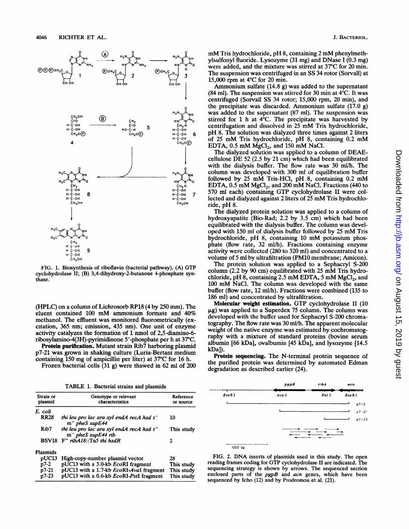
PlasmidspUC13 High-copy-number plasmid vector 28p7-2 pUC13 with a 3.0-kb EcoRI fragment This studyp7-21 pUC13 with a 1.7-kb EcoRI-AvaI fragment This studyp7-23 pUC13 with a 0.6-kb EcoRI-PstI fragment This study
pgpB
Ava I
ribA aen
Pst I EcoR I
p7-2
p7- 21
'p7-23
1000 bp
FIG. 2. DNA inserts of plasmids used in this study. The openreading frames coding for GTP cyclohydrolase II are indicated. Thesequencing strategy is shown by arrows. The sequenced sectionenclosed parts of the pgpB and acn genes, which have beensequenced by Icho (12) and by Prodromou et al. (21).
. - -mail$
J. BACTERIOL.
2
on August 15, 2019 by guest
http://jb.asm.org/
Dow
nloaded from
BIOSYNTHESIS OF RIBOFLAVIN 4047
CCCATGACTCCCGTTGCCCAGACCAGCAAGATAGCMTGGTTAACGTTCGCCGACGCGGC 60
CACAGCAAACCAACGGCCAGCAGTGCCCAACTGGCAGCAAACATCGTGTGACCGGAAGGA 120AAGGCAAACCCCGTCTCTTTCTGCCAGTGTGAACGCAAATATTGTGGGATATTTTTCTCT 180TCAGCCAACTGTTCTTTCACTAGATTTCCGCGTTCTGCTCGCTTTAAAGTGTAGAACTCA 240TCAACCGGAATATGATGTGTTTTTCCAGCCAGATAACAMAGGTCGTGGTTCCTGGACT 300
TTGTCTTTGATCCAGGATTTAACGCCTTGTCCCACAAGGATTGCGGCCGCCAGAATGGCA 360
AATAATACAAAGGCAGCCTTAATGCGAAMCGCAGACACCAGAGMACCAGCCGAATAW 420ATCAAATGTGTAATGACGCCCCAGGGCTGGGTGACAGTTTCAGTAACCCAAAMGCCGCT 480TTTAGTAGCCAACTTTGTTCTCCAGGTTGCCAACGCCAGCCAGAAATCCATACGGCTACT 540GGCATGACAAGCAATAGTGCAGCTCCCACTGCGGTACGTCTGGCMTCGAACGCATGGCC 600
pgpB 4=1TCTCCTTTTTGATAAGTCCCACAATCATACTGAAAACGCCAGTTCCAGGAAAAATTGAC 660AGATTTGTGCCATTCCGTGAACGATCGACGCGTCGTGATTAGGTGAACCCCTTCTCGTTA 720
S.D. M Q L K R 5TGGCAAAATAAGCCAATACAGAACCAGCATTATCTGGAGAATTTCATGCAGCTTAAACGT 780V A E A K L P T P W G D F L M V G F E E 25GTGGCAGAAGCCAAACTGCCAACCCCATGGGGCGATTTCCTGATGGTGGGATTTGAAGAA 840L A T G H D H V A L V Y G D I S G H T P 45
CTGGCAACCGGACACGATCATGTCGCGCTAGTCTATGGCGATATTTCCGGGCATACCCCG 900V L A R V H S E C L T G D A L F S L R C 65
GTACTTGCGCGCGTCCATTCCGAATGTCTGACCGGTGACGCCCTGTTCAGCTTGCGCTGC 960D C G F Q L E A A L T Q I A E E G R G I 85
GATTGTGGCTTCCAGCTCGAAGCGGCATTGACGCAAATTGCCGAGGAAGGCCGTGGTATT 1020L L Y H R Q E G R N I G L L N K I R A Y 105
TTGCTGTATCACCGTCAGGAAGGTCGTAACATTGGTCTGCTGAATAAAATCCGCGCTTAC 1080A L Q D Q G Y D T V E A N H Q L G F A A 125
GCACTGCAGGATCAAGGTTACGATACCGTAGAGGCTAACCACCAGTTAGGCTTCGCCGCT 1140D E R D F T L C A D H F K L L G V N E V 145GATGAGCGCGACTTCACTCTTTGCGCTGATATGTTCAAACTCCTTGGCGTCAATGAAGTC 1200R L L T N N P K K V E I L T E A G I N I 165
CGCTTGTTAACCAATAACCCGAAAAAAGTCGAAATTCTGACCGAAGCAGGGATTAATATT 1260V E R V P L I V G R N P N N E H Y L D T 185
GTTGAACGCGTACCATTGATTGTAGGTCGTAACCCCAATAACGAACATTATCTCGATACC 1320K A E K M G H L L N K 196AAAGCCGAGAAAATGGGCCATTTGCTGAACAAATAACCCTCTTGCATTGTGTAATTCATT 1380TGCTTGCCGGAAGCAAAATAACCGGCAAGCAAATAGTTGTTACTTCAACATATTACGAAT 1440
6*=acn
GACATAATGCAAAATGCCGTCGTTCTGGTAGTAGGTCAACTCCGTCGCGGTGTCGATACG 1500ACAACGGCAGGGTACGACTTCCTGGCTACCATC 1533FIG. 3. Nucleotide sequence of the gene coding for GTP cyclo-
hydrolase II and its flanking regions. The deduced amino acidsequence is shown, and N-terminal amino acids determined byEdman degradation are overlined. The putative ribosome bindingsite (Shine-Dalgarno [SD] sequence) is double underlined. Thegenes pgpB and acn are indicated.
1340
Nucleotide sequence accession number. The nucleotidesequence data reported in this article have been submitted toEMBL and have been assigned accession number X67876.
RESULTSCloning. Riboflavin-deficient mutants of E. coli were iso-
lated after mutagenesis of E. coli RR28 with ethyl methanesulfonate. The specific enzyme defects of these mutantswere not determined prior to the cloning experiments.A gene bank was generated by ligating EcoRI-restricted
genomic DNA from E. coli RR28 into plasmid pUC13. Thegene bank was used to transform the mutants, and coloniesgrowing without added riboflavin were selected. Cell ex-tracts of riboflavin-independent transformants were assayedfor GTP cyclohydrolase II activity. High enzyme levels inriboflavin-independent transformants of mutant Rib7 wereobserved. These strains were subsequently shown to containa plasmid, p7-2, carrying an insert of about 3 kb. Digestion ofthe plasmid with AvaI and ligation produced the plasmidp7-21 (Fig. 2), which retained the ability to complement theriboflavin deficiency of E. coli Rib7.DNA sequence analysis. Plasmid p7-21 contained an insert
of 1.7 kb, which was sequenced by a primer walk strategy assummarized in Fig. 2. A total of 1,533 bp on both DNAstrands was determined. The DNA segment contains anopen reading frame coding for 196 amino acids (Fig. 3).The predicted amino acid sequence was in line with the
result of Edman degradation of isolated GTP cyclohydrolaseII (see below). The predicted molecular weight of thepolypeptide is 21,836. The open reading frame is precededby a plausible ribosome binding site (Fig. 3).Mapping. In order to locate the GTP cyclohydrolase II
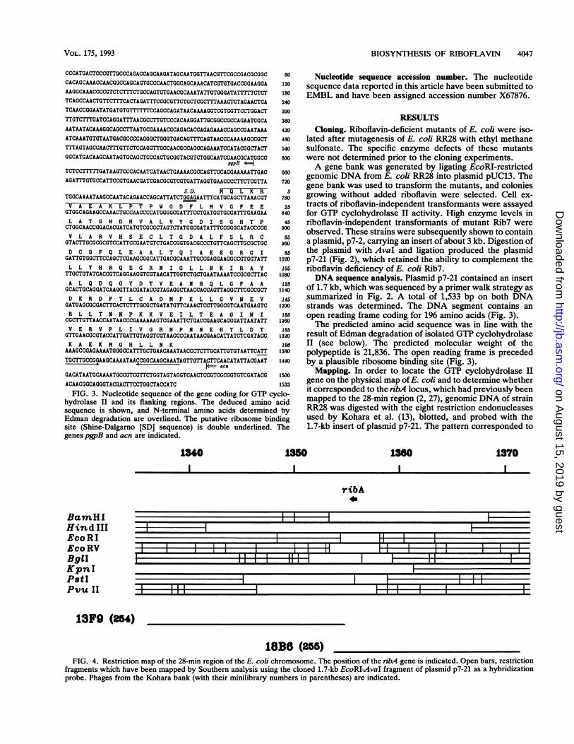
gene on the physical map ofE. coli and to determine whetherit corresponded to the ribA locus, which had previously beenmapped to the 28-min region (2, 27), genomic DNA of strainRR28 was digested with the eight restriction endonucleasesused by Kohara et al. (13), blotted, and probed with the1.7-kb insert of plasmid p7-21. The pattern corresponded to
1850 1360 1370
ribA
,, ,_
I l __ _ _ l _ _ 1 S - _ _ __ _ _ lS _ _ _ I __ l l l ll =P _ __ l l; 11 l ll l l I l
II I Il l l
l l l l l = w l l l [I l I I I _ [ I I [
BacmHIHind IIIEcoRIEco RVBgiIKpnIPatIPivu II
13F9 (24)
18B8 (255)FIG. 4. Restriction map of the 28-min region of the E. coli chromosome. The position of the nbA gene is indicated. Open bars, restriction
fragments which have been mapped by Southern analysis using the cloned 1.7-kb EcoRI-AvaI fragment of plasmid p7-21 as a hybridizationprobe. Phages from the Kohara bank (with their minilibrary numbers in parentheses) are indicated.
VOL. 175, 1993
on August 15, 2019 by guest
http://jb.asm.org/
Dow
nloaded from
4048 RICHTER ET AL.
TABLE 2. Purification of GTP cyclohydrolase II from E. coliRib7 carrying plasmid p7-21
Step Vol Activity Amt of protein Sp act(ml) (U) (mg) (U/mg)
Cell extract 61 54,000 1,300 42Ammonium sulfate 20 50,000 800 63DEAE-cellulose 150 41,000 170 240Hydroxyapatite 5 20,000 50 400Sephacryl S-200 6 31,000 14 2,250
the region at 28 min on the E. coli chromosome. Plaqueblotting of a subset of phages from the Kohara library withthe same probe gave a weak signal for phage 13F9 and astrong signal for phage 18B6 (Fig. 4). To check the identity ofthe rbA locus with the gene coding for GTP cyclohydrolaseII, we transformed the mutant strain BSV18 (ribA) (obtainedfrom B. Bachmann) with plasmids p7-2 and p7-21 as de-scribed in Materials and Methods. All transformants couldgrow on Luria-Bertani medium without added riboflavin. Itfollows that the gene coding for GTP cyclohydrolase II isidentical to rbA.Enzyme purification. E. coli strains carrying the plasmid
p7-21 show at least a 100-fold increased level of GTPcyclohydrolase II activity. The viability and growth rate ofthe host are not affected by the presence of the plasmid. InM9 minimal media from cell cultures of E. coli RR28containing p7-21, we detected 200 ,ug of riboflavin per literby reversed-phase HPLC. Since we found equal amounts ofthe vitamin in media from E. coli cells carrying the plasmidpUC13, the overproduction of GTP cyclohydrolase II doesnot show any influence on riboflavin production.The enzyme was purified to apparent homogeneity from
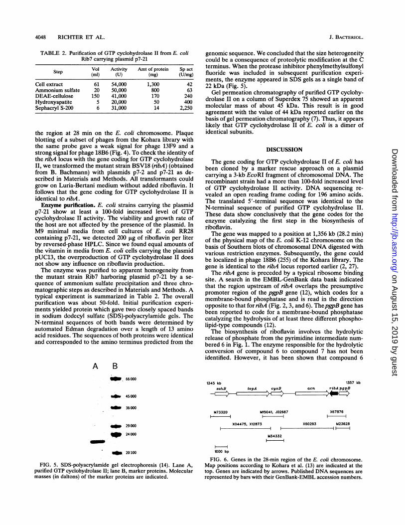
the mutant strain Rib7 harboring plasmid p7-21 by a se-quence of ammonium sulfate precipitation and three chro-matographic steps as described in Materials and Methods. Atypical experiment is summarized in Table 2. The overallpurification was about 50-fold. Initial purification experi-ments yielded protein which gave two closely spaced bandsin sodium dodecyl sulfate (SDS)-polyacrylamide gels. TheN-terminal sequences of both bands were determined byautomated Edman degradation over a length of 13 aminoacid residues. The sequences of both proteins were identicaland corresponded to the amino terminus predicted from the
A BI. 66 000
.*v 45000
1_ 36000
29 000
P 24000
_ 20100
FIG. 5. SDS-polyacrylamide gel electrophoresis (14). Lane A,purified GTP cyclohydrolase II; lane B, marker proteins. Molecularmasses (in daltons) of the marker proteins are indicated.
genomic sequence. We concluded that the size heterogeneitycould be a consequence of proteolytic modification at the Cterminus. When the protease inhibitor phenylmethylsulfonylfluoride was included in subsequent purification experi-ments, the enzyme appeared in SDS gels as a single band of22 kDa (Fig. 5).
Gel permeation chromatography of purified GTP cyclohy-drolase II on a column of Superdex 75 showed an apparentmolecUlar mass of about 45 kDa. This result is in goodagreement with the value of 44 kDa reported earlier on thebasis of gel permeation chromatography (7). Thus, it appearslikely that GTP cyclohydrolase II of E. coli is a dimer ofidentical subunits.
DISCUSSION
The gene coding for GTP cyclohydrolase II of E. coli hasbeen cloned by a marker rescue approach on a plasmidcarrying a 3-kb EcoRI fragment of chromosomal DNA. Therecombinant strain had a more than 100-fold increased levelof GTP cyclohydrolase II activity. DNA sequencing re-vealed an open reading frame coding for 196 amino acids.The translated 5'-terminal sequence was identical to theN-terminal sequence of purified GTP cyclohydrolase II.These data show conclusively that the gene codes for theenzyme catalyzing the first step in the biosynthesis ofriboflavin.The gene was mapped to a position at 1,356 kb (28.2 min)
of the physical map of the E. coli K-12 chromosome on thebasis of Southern blots of chromosomal DNA digested withvarious restriction enzymes. Subsequently, the gene couldbe localized in phage 18B6 (255) of the Kohara library. Thegene is identical to the ribA locus reported earlier (2, 27).The ribA gene is preceded by a typical ribosome binding
site. A search in the EMBL-GenBank data bank indicatedthat the region upstream of nbA overlaps the presumptivepromoter region of the pgpB gene (12), which codes for amembrane-bound phosphatase and is read in the directionopposite to that for nbA (Fig. 2, 3, and 6). ThepgpB gene hasbeen reported to code for a membrane-bound phosphatasecatalyzing the hydrolysis of at least three different phospho-lipid-type compounds (12).The biosynthesis of riboflavin involves the hydrolytic
release of phosphate from the pyrimidine intermediate num-bered 6 in Fig. 1. The enzyme responsible for the hydrolyticconversion of compound 6 to compound 7 has not beenidentified. However, it has been shown that compound 6
1345 kb 1357 kbsohB topA cysB acn ribA pgpBE~~E~z2~~ cz2 ~*EZ R--
M73320i-
M15041, J02687
X04475, X12873I-
M34332- H
1000 bp
X60293
X67876i
M23628- 1 -
FIG. 6. Genes in the 28-min region of the E. coli chromosome.Map positions according to Kohara et al. (13) are indicated at thetop. Genes are indicated by arrows. Published DNA sequences arerepresented by bars with their GenBank-EMBL accession numbers.
J. BACTERIOL.
on August 15, 2019 by guest
http://jb.asm.org/
Dow
nloaded from
BIOSYNTHESIS OF RIBOFLAVIN 4049
I M N -LS S|FGT|PfE R VNLLREGRGVMVL2 MSSTSLDE|FGTPVQRVERAI EAKNGLGVLLM3 MALSSAKEI IDDIRQIGIRMHILM4 MFHPISEELDAKK GEVIIVV
DDEDRENDDEDREND D EER E NDDEDREN
EGDM f P TMTVEQ MALT IE GD L IF S[ H LITIE A QMA L MIE G D LIA EI MVITIP[E AINFMIAE GD F VA L[ H A PEV IN FMA
1 RHGSG I VCLC I TEDRRKQLDLPMMVENNTSAYGTGFTV I EEAAEGVTTGVSAADRI TTVR
2 REIGISGtVCLCLTEERANWLDLPPMVKDNCSKNQTAFTVSI EAKEGVTTGVSAKIDRIVTITVIK3 THIG|R|G|L Icl|T|LS K ARCKTLEdPLIMVIQ GIN|NDNF STIP|F T]S EI A K YV TGIIS AIDRAKITV|L4 THIIRGL I CLTL I A DR
DLH PM SHHTAFTVSIDHR E- ISAVQ
1 AAIADGAKPSDLNRPGH|VFPLRAQAGVLTRGGHTEAT IDLMTLAFKPAL[E]LrTN]D[2 TATYFDIAIQPJ]EDLARP GHVFPLVAKTNGVLARRGHTEGT I DLMYLAN LVPIS[ ILLEILTINIPIDI3 A VAPNKSTD V PGH I FPLMAQDGGVL I RAGHTEAGCDV ARL GLEIE S VII.YEII LINIEIDI4 JLL D S K S VE]SDFOQR P G HI I SKK|G GVL|KRA|GH TE A|AV AE ACS P G AIG I E I MNEDS
1 GTMARAPECCIEFAN NMALVTIEDLVA[ QAHERKAS2 GAKL E FAR HGMPV LITIED IVDYRTG IDLRNEYKSGGREVSWS3 EGEMlRP L FAEKHGLKLGT I AD I EYRTQQESH I ER ISSEYEL N T[EY G I FTLVTYRDT
4 IGTM V K H Q L KMITI KDIQYRYN L T T LVIER E FK VY Y TN E V DGK EIH V AFF]5 MQLKRJV DFLMVGFEEL ATHDHVAL
3 K[1 I Q-AKAATLVRVHIVKDTLKDI LQVGLSQWS--ILEAAIRQT IS 1QESP4 MIGDVPF[EEE VLVRVHSECLTGDVFG[]H|RC C GPLEA A I AAE GRGVL L RQOEGRIG5 r I -SGHT LFSLRCD CGFQLEA A EIEGG RQ E G RN
3 FEK DMYA .P SPHSGIVQSRNIG LSI ADLGVKKI RLLS SNQGY4 1I GLI NKLKA YKLQEQ GYDTVEANEA[ LGLP[LRNYGIGA Q1 LRDL GVRNMK LLRN RK I
5 ILL RY QILGFIAADEDFTLC KLNEVI TNNPKV
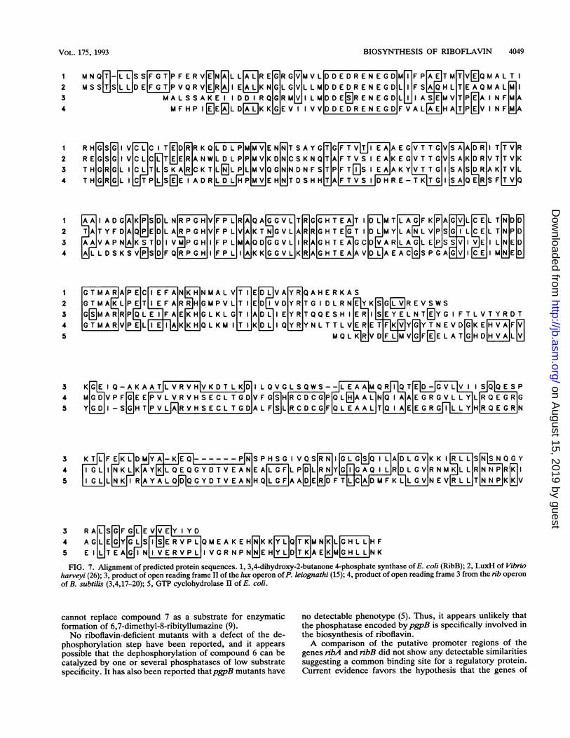
3 R ALGFGGEVEIYIYD4 AGLEGYGLSII ERVPLIQMEA K EHHKK[ YL]Q[TMN[K]L[GH L LIHF5 ElITEAJ IN ERVPLI VGRNPN[N]EHYLDTKAEKMGHLLNKFIG. 7. Alignment of predicted protein sequences. 1, 3,4-dihydroxy-2-butanone 4-phosphate synthase of E. coli (RibB); 2, LuxH of Vibrio
harveyi (26); 3, product of open reading frame II of the lux operon ofP. leiognathi (15); 4, product of open reading frame 3 from the rib operonof B. subtilis (3,4,17-20); 5, GTP cyclohydrolase II of E. coli.
cannot replace compound 7 as a substrate for enzymaticformation of 6,7-dimethyl-8-ribityllumazine (9).No riboflavin-deficient mutants with a defect of the de-
phosphorylation step have been reported, and it appearspossible that the dephosphorylation of compound 6 can becatalyzed by one or several phosphatases of low substratespecificity. It has also been reported thatpgpB mutants have
no detectable phenotype (5). Thus, it appears unlikely thatthe phosphatase encoded bypgpB is specifically involved inthe biosynthesis of riboflavin.A comparison of the putative promoter regions of the
genes nbA and ribB did not show any detectable similaritiessuggesting a common binding site for a regulatory protein.Current evidence favors the hypothesis that the genes of
VOL. 175, 1993
on August 15, 2019 by guest
http://jb.asm.org/
Dow
nloaded from
4050 RICHTER ET AL.
riboflavin biosynthesis are expressed constitutively in E. coli(27). Reporter gene insertion into rib genes of Salmonellatyphimurium also suggested constitutive expression in thisrelated organism (30). On the other hand, it should be notedthat the riboflavin pathway is tightly regulated in Bacillussubtilis (19).A recent data bank search also revealed that the 3' end of
ribA is flanked by the acn gene coding for aconitase (21).Since the termination of our mapping studies, the mapposition of the acn gene has been reported by others in fullagreement with the results of our experiments (21). Apresumptive terminator is located between the two genes(Fig. 3).
Contiguous sequence data are now available for a segmentextending from kb 1345 to 1357 of the E. coli chromosome.The genes located in this segment and the respective acces-sion numbers are given in Fig. 6.GTP cyclohydrolase II was purified to apparent homoge-
neity from cell extracts of a recombinant strain carrying theplasmid p7-21, in which it constituted about 2% of the totalcellular protein. In an earlier study, the enzyme had beenpurified about 2,200-fold from E. coli wild-type cells (7).An apparent molecular mass of 45 kDa has been estimated
for native GTP cyclohydrolase II of E. coli by gel filtration.This suggests tentatively that the protein is a dimer ofidentical subunits.
In B. subtilis, the enzymes involved in the biosynthesis ofriboflavin are encoded by an operon located at 2090. Theoperon has been sequenced and contains five open readingframes (3, 4, 20). Circumstantial evidence suggested that thecentral open reading frame encompassing a deduced proteinsequence of 398 amino acids should code for GTP cyclohy-drolase II (18). However, no conclusive evidence has beenpresented.Sequence comparison (Fig. 7) shows that the GTP cyclo-
hydrolase II of E. coli is homologous to the 3' part of openreading frame 3 of the B. subtilis operon. The homologyinvolves 54% identical residues. One gap of one amino acidhad to be introduced.The deduced peptide encoded by open reading frame 3 of
B. subtilis has about twice the size of the E. coli protein. Wehave shown recently that the 5' part of the B. subtilis gene ishomologous to the ribB gene coding for 3,4-dihydroxy-2-butanone 4-phosphate synthase in E. coli (23). This enzymecatalyzes the formation of the four-carbon precursor re-quired for the formation of the xylene ring of the vitamin(29). Thus, the B. subtilis protein represents a bifunctionalenzyme combining the activities of GTP cyclohydrolase IIand 3,4-dihydroxy-2-butanone 4-phosphate synthase.
Recently, several open reading frames have been se-quenced in the 3' region of the lux operon ofPhotobacteriumleiognathi (15) which codes for proteins involved in biolu-minescence. The 3' moiety of open reading frame II of the P.leiognathi operon shows homology to ribA of E. coli. More-over, open reading frame II shows homology to the openreading frame 3 ofB. subtilis over its entire length. It appearslikely that the P. leiognathi gene codes for a bifunctionalenzyme catalyzing the formation of compound 2 and ofcompound 5 from their respective substrates 1 and 4. Thereason for the expression of the rib genes in P. leiognathiunder the control of the lux regulon may be that thebioluminescence reaction requires flavin mononucleotide asa cofactor. Thus, activation of the bioluminescence systemmay be accompanied by an increased demand for riboflavin.
ACKNOWLEDGMENTSThis work was supported by the Deutsche Forschungsgemein-
schaft and the Fonds der Chemischen Industrie.We thank Y. Kohara for phages from his miniset library and B.
Bachmann and F. Gotz for E. coli strains. We also thank C. Kriegerfor help with the preparation of the manuscript.
REFERENCES1. Bacher, A. 1990. Biosynthesis of flavins, p. 215-259. In F.
Muller (ed.), Chemistry and biochemistry of flavoenzymes, vol.1. CRC Press, Inc., Boca Raton, Fla.
2. Bandrin, S. V., P. M. Rabinovich, and A. I. Stepanov. 1983.Three linkage groups of genes for riboflavin biosynthesis inEscherichia coli. Genetika 19:1419-1425.
3. Chikindas, M. L., E. V. Luk'yanov, P. M. Rabinovich, and A. I.Stepanov. 1987. Study of the region 210° of the Bacillus subtilischromosome with the aid of recombinant plasmids. Mol. Genet.Mikrobiol. Virusol. 2:20-24.
4. Chikindas, M. L., V. N. Mironov, E. V. Luk'yanov, Y. R.Borestskii, L. S. Arutyunova, P. M. Rabinovich, and A. I.Stepanov. 1987. Establishment of the boundaries of the ribofla-vin operon of Bacillus subtilis. Mol. Genet. Mikrobiol. Virusol.4:22-26.
5. Cronan, J. E., Jr., and C. 0. Rock. 1987. Biosynthesis ofmembrane lipids, p. 474-497. In F. C. Neidhardt, J. L. Ingra-ham, K. B. Low, B. Magasanik, M. Schaechter, and H. E.Umbarger (ed.), Eschenichia coli and Salmonella typhimurium:cellular and molecular biology. American Society for Microbi-ology, Washington, D.C.
6. Davis, L. G., M. D. Dibner, and J. F. Battey. 1986. Basicmethods in molecular biology. Elsevier, New York.
7. Foor, F., and G. M. Brown. 1975. Purification and properties ofguanosine triphosphate cyclohydrolase II from Escherichia coli.J. Biol. Chem. 250:3545-3551.
8. Godson, G. N., and D. Vapnek. 1973. A simple method ofpreparing large amounts of 4X 174RF1 supercoiled DNA.Biochim. Biophys. Acta 299:516-520.
9. Harzer, G., H. Rokos, M. K. Otto, A. Bacher, and S. Ghisla.1978. 6,7-Dimethyl-8-ribityllumazine 5'-phosphate is not a sub-strate for riboflavin synthase. Biochim. Biophys. Acta 540:48-54.
10. Hennecke, H., I. Gunther, and F. Binder. 1982. A novel cloningvector for the direct selection of recombinant DNA in Esche-richia coli. Gene 19:231-234.
11. Holmes, D. S., and M. Quigley. 1981. A rapid boiling method forthe preparation of bacterial plasmids. Anal. Biochem. 114:193-197.
12. Icho, T. 1988. Membrane-bound phosphatases in Escherichiacoli: sequence of thepgpB gene and dual subcellular localizationof the pgpB product. J. Bacteriol. 170:5117-5124.
13. Kohara, Y., K. Akiyama, and K. Isono. 1987. The physical mapof the whole Escherichia coli chromosome: application of a newstrategy for rapid analysis and sorting of a large genomic library.Cell 50:495-508.
14. Laemmli, U. K. 1970. Cleavage of structural proteins during theassembly of the head of bacteriophage T4. Nature (London)227:680-685.
15. Lee, C. Y., and E. A. Meighen. 1992. The lux genes in Photo-bacterium leiognathi are closely linked with genes correspond-ing in sequence to riboflavin synthesis genes. Biochem. Bio-phys. Res. Commun. 186:690-697.
16. Miller, J. H. 1972. Experiments in molecular genetics. ColdSpring Harbor Laboratory, Cold Spring Harbor, N.Y.
17. Mironov, V. N., M. L. Chikindas, A. S. Kraev, A. I. Stepanov,and K. G. Skryabin. 1989. Operon organization of genes ofriboflavin biosynthesis in Bacillus subtilis. Mol. Biol. 312:237-240.
18. Morozov, G. I., P. M. Rabinovich, S. V. Bandrin, and A. I.Stepanov. 1984. Structure of Bacillus subtilis riboflavin operon.Mol. Genet. Mikrobiol. Virusol. 7:42-46.
19. Perkins, J. B., and J. G. Pero. 1993. Biosynthesis of riboflavin,biotin, folic acid, and cobalamin, p. 319-334. In A. L. Sonen-shein, J. A. Hoch, and R. Losick (ed.), Bacillus subtilis and
J. BACTERIOL.
on August 15, 2019 by guest
http://jb.asm.org/
Dow
nloaded from
VOL. 175, 1993 BIOSYNTHESIS OF RIBOFLAVIN 4051
other gram-positive bacteria: biochemistry, physiology, andmolecular genetics. American Society for Microbiology, Wash-ington, D.C.
20. Perldns, J. B., J. G. Pero, and A. Sloma. 1991. Riboflavinoverproducing strains of bacteria. European patent application0405370.
21. Prodromou, C., P. J. Artymiuk, and J. R. Guest. 1992. Theaconitase of Eschenchia coli. Eur. J. Biochem. 204:599-609.
22. Read, S. M., and D. H. Northcote. 1981. Minimization ofvariation in the response to different proteins of the Coomassieblue G dye-binding assay for protein. Anal. Biochem. 116:53-64.
23. Richter, G., R. Volk, C. Krieger, H.-W. Lahm, U. Rothlisberger,and A. Bacher. 1992. Biosynthesis of riboflavin: cloning, se-quencing, and expression of the gene coding for 3,4-dihydroxy-2-butanone 4-phosphate synthase of Escherichia coli. J. Bacte-riol. 174:4050-4056.
24. Schott, K., J. Kellermann, F. Lottspeich, and A. Bacher. 1990.Riboflavin synthases of Bacillus subtilis. Purification and aminoacid sequence of the a subunit. J. Biol. Chem. 265:4204-4209.
25. Spackman, D. H., W. H. Stein, and S. Moore. 1958. Automaticrecording apparatus for use in the chromatography of aminoacids. Anal. Chem. 30:1190-1206.
26. Swartzmann, E., C. Miyamoto, A. Graham, and E. Meighen.1990. Delineation of the transcriptional boundaries of the laxoperon of Vibrio harveyi demonstrates the presence of two newlux genes. J. Biol. Chem. 265:3513-3517.
27. Teslyar, G. E., and G. M. Shavlovskii. 1983. Location of thegenes coding for riboflavin synthase and GTP-cyclohydrolaseon the chromosome of Escherichia coli K12. Tsitol. Genet.5:54-56.
28. Vleira, J., and J. Messing. 1982. The pUC plasmids, anM13mp7-derived system for insertion mutagenesis and sequenc-ing with synthetic universal primers. Gene 19:259-268.
29. Yolk, R., and A. Bacher. 1990. Studies on the four carbonprecursor in the biosynthesis of riboflavin. Purification andproperties of L-3,4-dihydroxy-2-butanone 4-phosphate syn-thase. J. Biol. Chem. 265:19479-19485.
30. Wang, A. 1991. Regulation of riboflavin biosynthesis in Salmo-nella typhimurium. Acta Microbiol. Sin. 3:315-317.
on August 15, 2019 by guest
http://jb.asm.org/
Dow
nloaded from