biomaterial topography alters healing in vivo and monocyte/macrophage activation in vitro
TRANSCRIPT
Biomaterial topography alters healing in vivo and monocyte/macrophage activation in vitro
Paige C. S. Bota,1*y Angela M. B. Collie,1*z Pauli Puolakkainen,2§ Robert B. Vernon,2
E. Helene Sage,2 Buddy D. Ratner,1 Patrick S. Stayton1
1Department of Bioengineering, University of Washington, Seattle, Washington 981952Hope Heart Program, Benaroya Research Institute at Virginia Mason, Seattle, Washington 98101
Received 20 March 2010; revised 22 April 2010; accepted 5 May 2010
Published online 19 August 2010 in Wiley Online Library (wileyonlinelibrary.com). DOI: 10.1002/jbm.a.32893
Abstract: The effect of biomaterial topography on healing
in vivo and monocyte/macrophage stimulation in vitro was
assessed. A series of expanded polytetrafluoroethylene
(ePTFE) materials were characterized by increasing average
intranodal distance of 1.2 lm (1.2-ePTFE), 3.0 lm (3.0-ePTFE),
and 4.4 lm (4.4-ePTFE), but presented consistent surface
chemistry with nonporous PTFE (np-PTFE). Subcutaneous im-
plantation of 4.4-ePTFE into mice resulted in a statistically thin-
ner capsule that appeared less organized and less dense than
the np-PTFE response. In vitro, isolated monocytes/macro-
phages cultured on np-PTFE produced low levels of interleukin
1-beta (IL-1b), 1.2-ePTFE and 3.0-ePTFE stimulated intermedi-
ate levels, and 4.4-ePTFE stimulated a 15-fold increase over np-
PTFE. Analysis of cDNA microarrays demonstrated that addi-
tional proinflammatory cytokines and chemokines, including
IL-1b, interleukin 6, tumor necrosis factor alpha, monocyte che-
motactic protein 1, and macrophage inflammatory protein 1-
beta, were expressed at higher levels by monocytes/macro-
phages cultured on 4.4-ePTFE at 4 and 24 h, respectively.
Expression ratios for several genes were quantified by RT-PCR
and were consistent with those from the cDNA array results.
These results demonstrate the effect of biomaterial topography
on early proinflammatory cytokine production and gene tran-
scription by monocytes/macrophages in vitro and decreased
fibrous capsule thickness in vivo. VC 2010 Wiley Periodicals, Inc.
J Biomed Mater Res Part A: 95A: 649–657, 2010.
Key Words: human monocytes/macrophages, topography,
surface dependent behavior, foreign body reaction,
microarray
INTRODUCTION
Monocytes are integrally involved in the host inflammatoryand foreign body response to biomaterials.1,2 Monocytes ini-tially move from the vasculature into the implant site wherethey mature to the macrophage phenotype and can furtherfuse into multinucleated foreign body giant cells at the bio-material interface. Adherent macrophages are powerfulinflammatory mediators and release a large number of mol-ecules, including proinflammatory cytokines that can con-tribute to cell activation, chronic inflammation, and fibrousencapsulation.2 However, macrophages are also involved inorchestrating wound healing and resolution, for which con-trolled macrophage activation is necessary.3 A better under-standing of how biomaterials influence key parameters ofthe inflammatory response is central to the development ofengineered constructs that integrate with host tissues topromote healing and regeneration of function.
There are numerous mechanical and surface propertiesof materials that can play important roles in macrophage
activation. Adherence of monocytes induces an activatedphenotype.4 Surface chemistry affects monocyte/macro-phage (MC/MO) secretion of cytokines such as interleukin1-beta (IL-1b), interleukin 6 (IL-6), and tumor necrosis fac-tor-alpha (TNF-a)5,6 and is the subject of proteomicstudies.7,8
Biomaterial topography affects cellular and healingresponses as well. Micro-range surface roughness modifiescultured cell response in vitro and biocompatibility and tis-sue attachment in vivo.9 Nanometer and micrometer surfacealterations induce changes in macrophage adhesion, orienta-tion, spreading, and cytoskeleton formation.10,11 Capsulethickness varies around filters made of the same materialbut with different pore sizes.12 Reduced fibrous-capsule for-mation, increased vascularity, and closer proximity of ves-sels to the implant surface have been demonstrated withpillar-textured implants and with porous polyvinyl alcoholimplants in comparison with the nontextured or smoothcontrol of the same material.13,14
*These authors contributed equally to this work.yPresent address: Edwards Lifesciences, Irvine, CA 92614zPresent address: Cleveland Clinic, Cleveland, OH 44195§Present address: Helsinki University Central Hospital, Helsinki, Finland and Turku University Central Hospital, Turku, Finland
Correspondence to: A. M. B. Collie, Cleveland Clinic, Pathology and Laboratory Medicine Institute, Mail Code L25, 9500 Euclid Avenue,
Cleveland, OH 44195; e-mail: [email protected]
Contract grant sponsor: National Science Foundation; contract grant number: NSF EEC-9529161
Contract grant sponsor: NIH; contract grant number: GM045873
VC 2010 WILEY PERIODICALS, INC. 649
Expanded PTFE (ePTFE) has been used historically inmedical device applications such as vascular grafts, to whichmacrophages have been shown to adhere within 24 h.15 Theinfluence of ePTFE materials on MC/MO activation and thesecretion of proinflammatory cytokines in vitro, as well asfibrous capsule formation in vivo, has been explored in com-parison with other surface chemistries.5,16–19 ExpandedPTFE materials with varied pore dimensions have displayeda relationship between pore type and healing.20–22 Previousstudies have demonstrated MC/MO activation and IL-1bproduction when these cells were seeded onto a uniaxially-expanded ePTFE material of a single expansion ratio andwere stimulated with LPS.5,16,19
To study the effect of topography, we investigated MC/MO activation in vitro and fibrous capsule formation in vivoin response to biaxially-expanded ePTFE materials in arange of pore sizes, and in comparison with nonporousPTFE, in the absence of additional LPS stimulation.
MATERIALS AND METHODS
PTFE materialsExpanded polytetrafluoroethylene (ePTFE) was obtained ina range of pore sizes with nominal filtration sizes of 0.2, 1,and 3 lm (Fluoropore filters, Millipore, Bedford, MA). Non-porous PTFE (np-PTFE, Berghof/America, Coral Springs, FL)was included for comparison. The PTFE was cut into diskswith diameters of 21 mm for in vitro studies and 8 mm forin vivo studies by a solvent-washed punch or scalpel, soni-cated three times in 100% ethanol for 20 min, and driedaseptically. The disks were tested with the Limulus amebo-cyte lysate assay (Associates of Cape Cod, Falmouth, MA)according to the protocol developed by the Food and DrugAdministration and found to be free of endotoxin with asensitivity of 0.03 EU/mL.
Scanning electron microscopyPTFE disks were mounted on studs with colloidal silverpaste and sputter coated with a Au/Pd source. The topogra-phy of the samples was analyzed using a JEOL JSM-6300Fscanning electron microscope with an accelerating voltageof 15 kV. Images were taken either flat or at a tilt of approx-imately 40� , recorded onto Polaroid film, and scanned into adigital format. Internodal, intranodal, and interfiber distan-ces were quantified with NIH Image.
Electron spectroscopy for chemical analysisPTFE disks were analyzed on a Surface Science Instruments(SSI) X-Probe ESCA instrument at the National ESCA andSurface Analysis Center for Biomedical Problems (NESAC/BIO, University of Washington). An aluminum Ka1,2 mono-chromatized X-ray source was used for the generation ofphotoelectrons from a sample surface. The samples wereanalyzed at a take-off angle of 55� with respect to the sur-face, allowing for analysis of the outermost 80 Å of the sam-ple. The elemental composition was calculated with the SSIdata analysis software using the F(1s) and C(1s) peak areasnormalized with a fluorine photoemission cross-section of4.43 and a carbon cross-section of 1.0.
Subcutaneous implantation of PTFE materials in micePTFE disks were implanted subcutaneously into the backsof five male Swiss Webster mice (B&K Universal, England).All work with mice was conducted according to the Univer-sity of Washington Animal Guide for the Care and Use ofLaboratory Animals. Each mouse received two disks, placedbilaterally. Four weeks after implantation, the disks and sur-rounding tissue were excised, fixed in formalin, sectioned,mounted on slides, and stained with hematoxylin and eosinfor general morphological analysis or Masson’s trichromefor analysis of collagen organization. Fibrous capsule thick-ness was measured with NIH Image and was confined tothe dermis above the implant which was not disrupted dur-ing sectioning.
Monocyte isolationPrimary human monocytes were isolated from peripheralblood drawn from anonymous donors who had signed aninformed consent form approved by the University of Wash-ington’s Human Subjects Review Committee. Blood was col-lected into 10 mL Vacutainer collection tubes containing so-dium heparin and 15 mL Vacutainer tubes withoutanticoagulant (Becton Dickinson, Franklin Lakes, NJ). Thetubes without anticoagulant were held at room temperaturefor 1 h and at 4�C for 1 h. Serum was removed by pipette,centrifuged at 2000 rpm for 10 minutes, and filteredthrough a 0.2-lm filter to produce autologous human se-rum. The whole blood/anticoagulant mixture in the heparintubes was diluted with an equal volume of Dulbecco’s phos-phate-buffered saline (DPBS, Gibco, Carlsbad, CA) containing1 mM EDTA (Gibco) and 60 U/mL heparin sodium salt(Sigma, St. Louis, MO), layered on top of a Histopaque 1077gradient (Sigma), and centrifuged. The interface layer con-taining peripheral blood mononuclear cells was separatedand washed. Monocytes were isolated by magnetic beadnegative selection using the Monocyte Isolation Kit II withMACS Column type LS (Miltenyi Biotec, Auburn, CA) accord-ing to the kit protocol. Cells were resuspended in RPMI1640 with glutamine containing 25 mM HEPES buffer, 1 mMsodium pyruvate, 1 mM nonessential amino acids, 100 U/mL penicillin, 100 mg/mL streptomycin (Gibco), and 15%filtered autologous human serum. Isolated monocytes wereincubated for one hour at 37�C prior to use.
Monocyte/macrophage culture on PTFE materialsand cytokine productionPTFE disks were placed in 12-well cell culture plates andsecured to the bottom of each well with glass rings that hadbeen stringently cleaned to remove endotoxin. The diskswere baked overnight at 180�C, autoclaved, and kept sterileuntil use. Negative controls of tissue culture polystyrene(TCPS) wells without materials were included in eachexperiment. Isolated monocytes from a single donor wereplated at a concentration of 2 � 106 cells/mL and volumeof 1 mL/well and were incubated for 2 h at 37�C and 5%CO2 to allow for adherence. Subsequently, the wells werewashed with prewarmed media. One milliliter of freshmedia was added per well, and the plates were incubated
650 BOTA ET AL. EFFECT OF BIOMATERIAL TOPOGRAPHY
an additional 2 or 22 h to the endpoints of 4 or 24 h fromthe original plating. Supernatants from MC/MO cultureswere removed 24 h after seeding, centrifuged to removecells and cell debris, aliquotted into sterile tubes, and frozenat �80�C. The supernatants were analyzed for IL-1b or ba-sic fibroblast growth factor (bFGF) by ELISA according tothe manufacturer’s instructions (R&D Systems, Minneapolis,MN).
The number of adherent and nonadherent cells in eachwell was determined by a modified lactate dehydrogenase(LDH) method (Roche, Indianapolis, IN). The cells werelysed by addition of 1 mL of cold 2% Triton X-100 in DPBSbuffer and vigorous pipetting. A standard curve was gener-ated with nonadherent monocytes. A 100-lL aliquot oflysate solution was added to an equal volume of LDH rea-gent prepared according to the manufacturer’s suggestion.The optical density was measured at 490 nm minus 650 nmand was compared to the standard curve.
RNA isolation and amplificationTotal RNA was isolated with the RNeasy Mini Kit accordingto the manufacturer’s instructions (Qiagen, Valencia, CA).Briefly, cells were lysed in the wells using the kit lysisbuffer. Cell lysates were centrifuged through the QIAshred-der homogenization column, and total RNA was isolated.The optional on-column DNase I digestion was completedwith the RNase-Free DNase Set (Qiagen). Total RNA wasstored at �80�C. Total RNA was amplified by use of theRiboAmp RNA Amplification Kit (Arcturus, Mountain View,CA) according to the manufacturer’s instructions. One roundof amplification was completed from 2 to 5 lg of total RNA,representing three to four cell culture wells. Amplified RNAconcentration was measured on a UV spectrophotometer.
cDNA labeling and array hybridizationLabeling and hybridization were completed by the GenomicsResource at the Fred Hutchinson Cancer Research Center(Seattle, WA). For each array sample, 5 lg of amplified RNAwas reverse-transcribed and labeled by an amino-allyl label-ing protocol as previously described, except that randomhexamers were used instead of oligo dT primers for thereverse transcription.23 Spotted human cDNA microarrayscontained over 17,600 spots representing named genes,ESTs, and control sequences. Certain genes were repre-sented on the array by multiple spots, with either identicalsequences or sequences from different portions of the gene.
Array analysisEach of the arrays from the independent donors was scannedand analyzed with the GenePix 4000 Microarray Scanner andGenePix Pro 3.0x software (Axon Instruments, Foster, CA).Raw data were acquired by determination of a diameter foreach spot on the array. Fluorescence intensity was measuredat 532 nm and 635 nm for each pixel within the spot and thelocal background. For each spot, median fluorescence inten-sity was calculated and corrected for the local backgroundfluorescence levels. These background-corrected fluores-cence intensities were then used to calculate an expression
ratio for each spot equal to (F635,Median � B635,Median)/(F532,Median – B532,Median), where FX,Median is the median fluo-rescence intensity at wavelength X for each spot and BX,Median
is the median fluorescence intensity of the local backgroundat wavelength X. The expression ratios were then normalizedsuch that the overall ratio for the entire chip was 1.0, withthe assumption that there was similar expression of mostgenes between the two samples, and corrects for differencesbetween the two dyes. This ratio was verified by the use ofseveral control spots on the chips. Normalized expressionratios were used in the following analysis. Ratios greaterthan 2.0 represent a higher expression in the 4.4-ePTFE sam-ple, whereas ratios less than 0.5 represent a higher expres-sion in the np-PTFE sample.
RT-PCR analysisAn aliquot of the total RNA was reserved for quantitativereverse transcription polymerase chain reaction (RT-PCR).Total RNA isolated from the first donor was used to verifythe array ratios for TNF-a, IL-1b, IL-6, interleukin 8 (IL-8),monocyte chemotactic protein 1 (MCP1), and interferon-gamma-inducible protein (IP10) by a two-step RT-PCR reac-tion. Primers were selected with Primer324 such that eitherthe left or right primer overlapped an exon/intron boundary(Table I). A reverse transcription (RT) reaction was per-formed on 0.3 or 0.5 lg of total RNA with Oligo dT20 pri-mers and C. therm polymerase (Roche). The reaction wasplaced at 60�C for 30 min, heated to 95�C for 5 min,diluted, and used in a quantitative PCR reaction with theQuantitect SYBR Green PCR kit (Qiagen) and the LightCyclerinstrument (Roche). The cycler program had an initial acti-vation step of 95�C for 15 min and a PCR cycle of 94�C for15 s, 55�C for 20 s, and 72�C for 15 s for 50 cycles. Sampleswere run in triplicate at one or two dilutions. A standardcurve consisting of known concentrations of plasmids(American Type Culture Collection, Manassas, VA) contain-ing full-length clones of the chosen gene was run simultane-ously (Table I). The LightCycler Data Analysis software wasused to determine the threshold cycle for each sample andstandard. The standard curve was subsequently used to
TABLE I. Primers for RT-PCR
Gene Accession Number Primer Sequence
TNF BC028148 F: tgtagcccatgttgtagcaaacR: cagcttgagggtttgctaca
IL-1b BC008678 F: cttcgacacatgggataacgR: gacatggagaacaccacttgt
IL-6 BC015511 F: tggattcaatgaggagacttgR: gcaggaactggatcaggac
IL-8 BC013615 F: gtttttgaagagggctgagaaR: tcttgtattgcatctggcaac
MCP1 BC009716 F: ctcagccagatgcaatcaaR: aatcctgaacccacttctgc
IP-10 BC010954 F: actctaagtggcattcaaggagtaR: catctcttctcacccttctttttc
F indicates forward primer, R indicates reverse primer. Accession
numbers are for plasmids used in development of the standard curve
for RT-PCR.
ORIGINAL ARTICLE
JOURNAL OF BIOMEDICAL MATERIALS RESEARCH A | NOV 2010 VOL 95A, ISSUE 2 651
determine the concentration of each sample, which was nor-malized based on total RNA concentration as quantified onan Agilent Bioanalyzer. Expression ratios and standard devi-ations for the ratios were calculated.
Statistical analysisStatistical significance for fibrous capsule thickness andMC/MO cytokine production was determined using theunpaired t-test in comparison with np-PTFE.
RESULTS
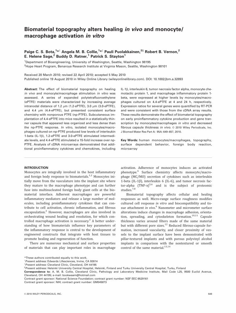
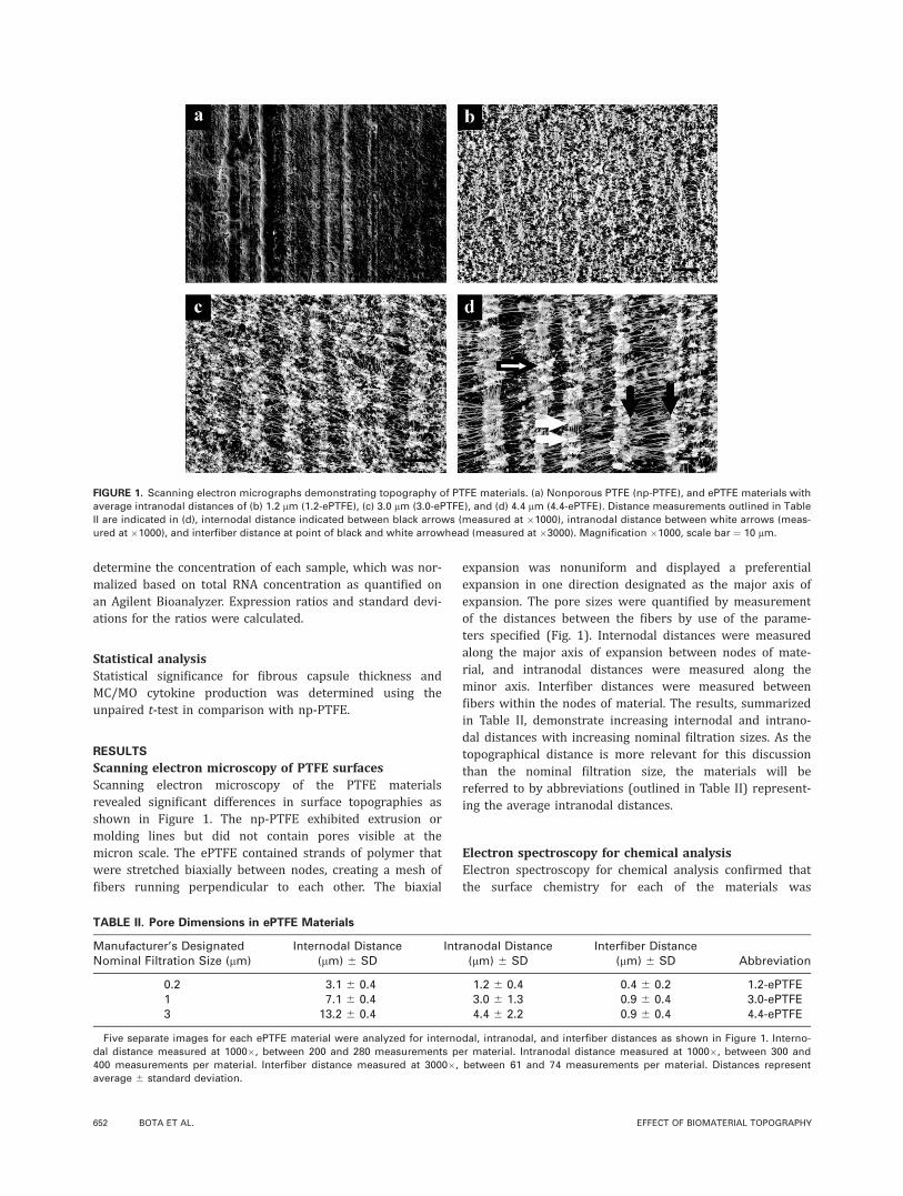
Scanning electron microscopy of PTFE surfacesScanning electron microscopy of the PTFE materialsrevealed significant differences in surface topographies asshown in Figure 1. The np-PTFE exhibited extrusion ormolding lines but did not contain pores visible at themicron scale. The ePTFE contained strands of polymer thatwere stretched biaxially between nodes, creating a mesh offibers running perpendicular to each other. The biaxial
expansion was nonuniform and displayed a preferentialexpansion in one direction designated as the major axis ofexpansion. The pore sizes were quantified by measurementof the distances between the fibers by use of the parame-ters specified (Fig. 1). Internodal distances were measuredalong the major axis of expansion between nodes of mate-rial, and intranodal distances were measured along theminor axis. Interfiber distances were measured betweenfibers within the nodes of material. The results, summarizedin Table II, demonstrate increasing internodal and intrano-dal distances with increasing nominal filtration sizes. As thetopographical distance is more relevant for this discussionthan the nominal filtration size, the materials will bereferred to by abbreviations (outlined in Table II) represent-ing the average intranodal distances.
Electron spectroscopy for chemical analysisElectron spectroscopy for chemical analysis confirmed thatthe surface chemistry for each of the materials was
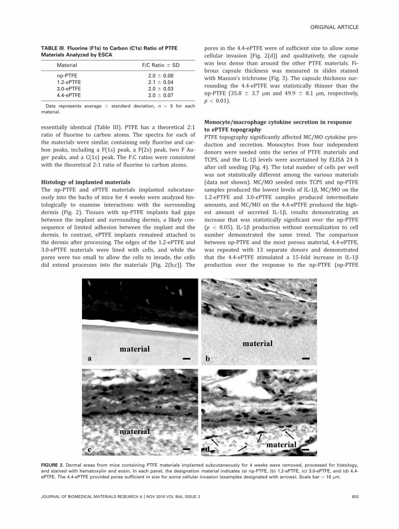
FIGURE 1. Scanning electron micrographs demonstrating topography of PTFE materials. (a) Nonporous PTFE (np-PTFE), and ePTFE materials with
average intranodal distances of (b) 1.2 lm (1.2-ePTFE), (c) 3.0 lm (3.0-ePTFE), and (d) 4.4 lm (4.4-ePTFE). Distance measurements outlined in Table
II are indicated in (d), internodal distance indicated between black arrows (measured at �1000), intranodal distance between white arrows (meas-
ured at �1000), and interfiber distance at point of black and white arrowhead (measured at �3000). Magnification �1000, scale bar ¼ 10 lm.
TABLE II. Pore Dimensions in ePTFE Materials
Manufacturer’s DesignatedNominal Filtration Size (lm)
Internodal Distance(lm) 6 SD
Intranodal Distance(lm) 6 SD
Interfiber Distance(lm) 6 SD Abbreviation
0.2 3.1 6 0.4 1.2 6 0.4 0.4 6 0.2 1.2-ePTFE1 7.1 6 0.4 3.0 6 1.3 0.9 6 0.4 3.0-ePTFE3 13.2 6 0.4 4.4 6 2.2 0.9 6 0.4 4.4-ePTFE
Five separate images for each ePTFE material were analyzed for internodal, intranodal, and interfiber distances as shown in Figure 1. Interno-
dal distance measured at 1000�, between 200 and 280 measurements per material. Intranodal distance measured at 1000�, between 300 and
400 measurements per material. Interfiber distance measured at 3000�, between 61 and 74 measurements per material. Distances represent
average 6 standard deviation.
652 BOTA ET AL. EFFECT OF BIOMATERIAL TOPOGRAPHY
essentially identical (Table III). PTFE has a theoretical 2:1ratio of fluorine to carbon atoms. The spectra for each ofthe materials were similar, containing only fluorine and car-bon peaks, including a F(1s) peak, a F(2s) peak, two F Au-ger peaks, and a C(1s) peak. The F:C ratios were consistentwith the theoretical 2:1 ratio of fluorine to carbon atoms.
Histology of implanted materialsThe np-PTFE and ePTFE materials implanted subcutane-ously into the backs of mice for 4 weeks were analyzed his-tologically to examine interactions with the surroundingdermis (Fig. 2). Tissues with np-PTFE implants had gapsbetween the implant and surrounding dermis, a likely con-sequence of limited adhesion between the implant and thedermis. In contrast, ePTFE implants remained attached tothe dermis after processing. The edges of the 1.2-ePTFE and3.0-ePTFE materials were lined with cells, and while thepores were too small to allow the cells to invade, the cellsdid extend processes into the materials [Fig. 2(b,c)]. The
pores in the 4.4-ePTFE were of sufficient size to allow somecellular invasion [Fig. 2(d)] and qualitatively, the capsulewas less dense than around the other PTFE materials. Fi-brous capsule thickness was measured in slides stainedwith Masson’s trichrome (Fig. 3). The capsule thickness sur-rounding the 4.4-ePTFE was statistically thinner than thenp-PTFE (35.8 6 3.7 lm and 49.9 6 8.1 lm, respectively,p < 0.01).
Monocyte/macrophage cytokine secretion in responseto ePTFE topographyPTFE topography significantly affected MC/MO cytokine pro-duction and secretion. Monocytes from four independentdonors were seeded onto the series of PTFE materials andTCPS, and the IL-1b levels were ascertained by ELISA 24 hafter cell seeding (Fig. 4). The total number of cells per wellwas not statistically different among the various materials(data not shown). MC/MO seeded onto TCPS and np-PTFEsamples produced the lowest levels of IL-1b, MC/MO on the1.2-ePTFE and 3.0-ePTFE samples produced intermediateamounts, and MC/MO on the 4.4-ePTFE produced the high-est amount of secreted IL-1b, results demonstrating anincrease that was statistically significant over the np-PTFE(p < 0.05). IL-1b production without normalization to cellnumber demonstrated the same trend. The comparisonbetween np-PTFE and the most porous material, 4.4-ePTFE,was repeated with 13 separate donors and demonstratedthat the 4.4-ePTFE stimulated a 15-fold increase in IL-1bproduction over the response to the np-PTFE (np-PTFE
TABLE III. Fluorine (F1s) to Carbon (C1s) Ratio of PTFE
Materials Analyzed by ESCA
Material F/C Ratio 6 SD
np-PTFE 2.0 6 0.081.2-ePTFE 2.1 6 0.043.0-ePTFE 2.0 6 0.034.4-ePTFE 2.0 6 0.07
Data represents average 6 standard deviation, n ¼ 5 for each
material.
FIGURE 2. Dermal areas from mice containing PTFE materials implanted subcutaneously for 4 weeks were removed, processed for histology,
and stained with hematoxylin and eosin. In each panel, the designation material indicates (a) np-PTFE, (b) 1.2-ePTFE, (c) 3.0-ePTFE, and (d) 4.4-
ePTFE. The 4.4-ePTFE provided pores sufficient in size for some cellular invasion (examples designated with arrows). Scale bar ¼ 10 lm.
ORIGINAL ARTICLE
JOURNAL OF BIOMEDICAL MATERIALS RESEARCH A | NOV 2010 VOL 95A, ISSUE 2 653
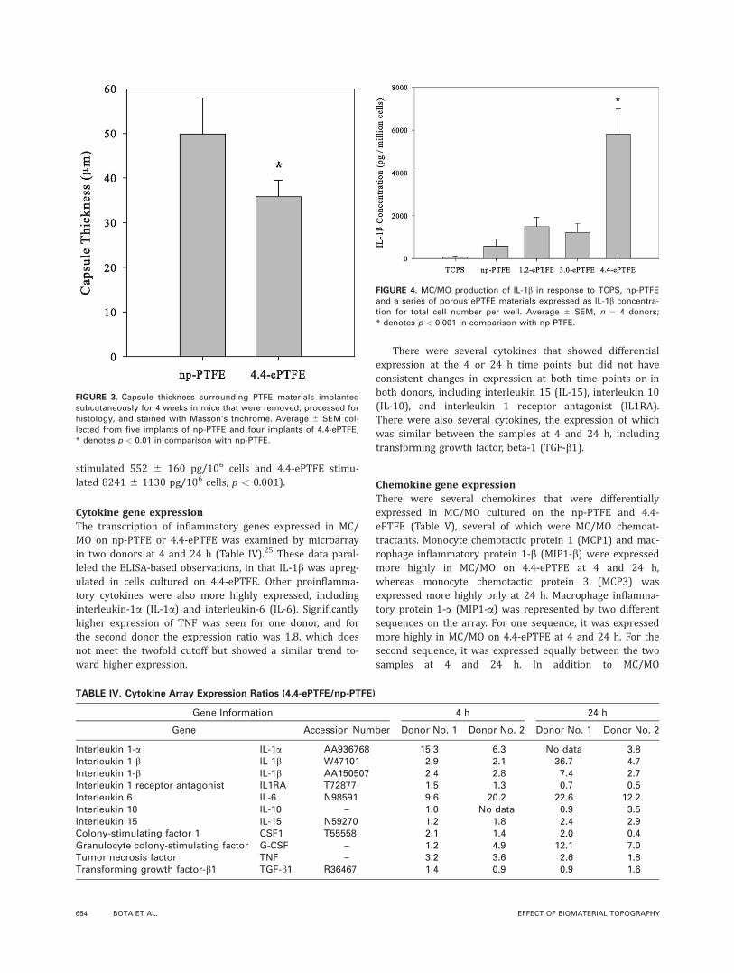
stimulated 552 6 160 pg/106 cells and 4.4-ePTFE stimu-lated 8241 6 1130 pg/106 cells, p < 0.001).
Cytokine gene expressionThe transcription of inflammatory genes expressed in MC/MO on np-PTFE or 4.4-ePTFE was examined by microarrayin two donors at 4 and 24 h (Table IV).25 These data paral-leled the ELISA-based observations, in that IL-1b was upreg-ulated in cells cultured on 4.4-ePTFE. Other proinflamma-tory cytokines were also more highly expressed, includinginterleukin-1a (IL-1a) and interleukin-6 (IL-6). Significantlyhigher expression of TNF was seen for one donor, and forthe second donor the expression ratio was 1.8, which doesnot meet the twofold cutoff but showed a similar trend to-ward higher expression.
There were several cytokines that showed differentialexpression at the 4 or 24 h time points but did not haveconsistent changes in expression at both time points or inboth donors, including interleukin 15 (IL-15), interleukin 10(IL-10), and interleukin 1 receptor antagonist (IL1RA).There were also several cytokines, the expression of whichwas similar between the samples at 4 and 24 h, includingtransforming growth factor, beta-1 (TGF-b1).
Chemokine gene expressionThere were several chemokines that were differentiallyexpressed in MC/MO cultured on the np-PTFE and 4.4-ePTFE (Table V), several of which were MC/MO chemoat-tractants. Monocyte chemotactic protein 1 (MCP1) and mac-rophage inflammatory protein 1-b (MIP1-b) were expressedmore highly in MC/MO on 4.4-ePTFE at 4 and 24 h,whereas monocyte chemotactic protein 3 (MCP3) wasexpressed more highly only at 24 h. Macrophage inflamma-tory protein 1-a (MIP1-a) was represented by two differentsequences on the array. For one sequence, it was expressedmore highly in MC/MO on 4.4-ePTFE at 4 and 24 h. For thesecond sequence, it was expressed equally between the twosamples at 4 and 24 h. In addition to MC/MO
FIGURE 3. Capsule thickness surrounding PTFE materials implanted
subcutaneously for 4 weeks in mice that were removed, processed for
histology, and stained with Masson’s trichrome. Average 6 SEM col-
lected from five implants of np-PTFE and four implants of 4.4-ePTFE,
* denotes p < 0.01 in comparison with np-PTFE.
FIGURE 4. MC/MO production of IL-1b in response to TCPS, np-PTFE
and a series of porous ePTFE materials expressed as IL-1b concentra-
tion for total cell number per well. Average 6 SEM, n ¼ 4 donors;
* denotes p < 0.001 in comparison with np-PTFE.
TABLE IV. Cytokine Array Expression Ratios (4.4-ePTFE/np-PTFE)
Gene Information 4 h 24 h
Gene Accession Number Donor No. 1 Donor No. 2 Donor No. 1 Donor No. 2
Interleukin 1-a IL-1a AA936768 15.3 6.3 No data 3.8Interleukin 1-b IL-1b W47101 2.9 2.1 36.7 4.7Interleukin 1-b IL-1b AA150507 2.4 2.8 7.4 2.7Interleukin 1 receptor antagonist IL1RA T72877 1.5 1.3 0.7 0.5Interleukin 6 IL-6 N98591 9.6 20.2 22.6 12.2Interleukin 10 IL-10 – 1.0 No data 0.9 3.5Interleukin 15 IL-15 N59270 1.2 1.8 2.4 2.9Colony-stimulating factor 1 CSF1 T55558 2.1 1.4 2.0 0.4Granulocyte colony-stimulating factor G-CSF – 1.2 4.9 12.1 7.0Tumor necrosis factor TNF – 3.2 3.6 2.6 1.8Transforming growth factor-b1 TGF-b1 R36467 1.4 0.9 0.9 1.6
654 BOTA ET AL. EFFECT OF BIOMATERIAL TOPOGRAPHY
chemoattractants, a neutrophil chemoattractant, GRO1 onco-gene (GRO1), was also expressed more highly in the 4.4-ePTFE sample at 4 and 24 h.26
Interestingly, interferon-gamma-inducible protein 10 (IP-10), which is a chemoattractant for monocytes and T cellsas well as an anti-angiogenic factor, had significantly higherexpression in the 4.4-ePTFE sample at 4 h but a trend wasseen toward higher expression in MC/MO on the np-PTFEat 24 h.27 Although several chemokines demonstrated differ-ential expression by the arrays, several did not show signifi-cant changes in expression between the samples at 4 and24 h, including IL-8, an inflammatory cytokine that is a che-moattractant for neutrophils, and Regulated Upon Activa-tion, Normally T-expressed, and Presumably Secreted(RANTES), a chemoattractant that acts on several cell typesincluding monocytes.26
Angiogenic factor expression and secretionPrevious studies demonstrated that increasing the porosityof ePTFE correlated with increased levels of post-implanta-tion vascularization in vivo.20,21 Accordingly, we examinedthe effect of np-PTFE and 4.4-ePTFE on the expression ofmRNAs for angiogenic factors known to be released by MC/MO (Table VI).28 These factors included vascular endothelialgrowth factor (VEGF), TGF-b1, platelet derived growth fac-tor (PDGF), and bFGF. VEGF, TGF- b1, and PDGF subunits Aand B were expressed similarly in both samples at 4 and24 h. However, bFGF was expressed similarly in both sam-ples at 4 h but at 24 h was greater in MC/MO cultured on
4.4-ePTFE compared to np-PTFE. Given this differentialresponse, levels of bFGF were examined by ELISA, but nodifferences were seen in the amount of bFGF secreted byMC/MO cultured for 24 h on 4.4-ePTFE compared to np-PTFE (data not shown).
RT-PCR verification of array resultsThe mRNA levels of several cytokine and chemokine geneswere verified by quantitative RT-PCR on total RNA from Do-nor 1 (Table VII). Trends from the array data were consist-ent with the RT-PCR verification for all genes analyzed.Although the RT-PCR expression ratios did not reach thesame levels observed on the expression array for IL-1b at4 h, IL-8 at 24 h, and TNF at 24 h, the trends were similar.The expression ratios for TNF showed high standard devia-tions, which could be due to the difficulty of quantifying thelow number of transcripts in the original sample.29–31
DISCUSSION
The ability to elicit desirable cell and tissue response to animplanted biomaterial by modification of the surface topog-raphy of the material would be a powerful tool in the devel-opment of biomaterials and engineered tissues. Althoughtheir surface chemistries were identical, the series of biax-ially expanded PTFE filters displayed complex topographiesthat were characterized by a mesh of fibers stretchedbetween nodes of material. Upon implantation, the capsulesurrounding the 4.4-ePTFE was qualitatively less organizedand less dense than the capsules surrounding the other
TABLE V. Chemokine Array Expression Ratios (4.4-ePTFE/np-PTFE)
Gene Information 4 h 24 h
Gene Systematic Nomenclature Accession Number Donor No. 1 Donor No. 2 Donor No. 1 Donor No. 2
MCP1 CCL2 AA425102 3.5 2.1 3.2 2.2MIP1-a CCL3 AA416584 1.7 1.0 1.0 1.0MIP1-a CCL3 AA677522 4.1 5.1 6.0 2.8MIP1-b CCL4 H62864 4.3 3.8 4.2 2.7RANTES CCL5 AA486072 1.1 0.9 1.1 0.9MCP3 CCL7 AA040170 1.6 1.3 2.9 2.1GRO1 CXCL1 W46900 4.7 2.1 12.1 3.6GRO1 CXCL1 W42723 6.6 2.1 14.4 3.6IL-8 CXCL8 AA102526 1.1 1.6 1.6 0.9MIG CXCL9 AA131406 1.8 1.3 0.9 0.8IP-10 CXCL10 AA878880 3.3 2.2 0.4 0.6
IL-8, interleukin 8; MCP1, monocyte chemotactic protein 1; MIP1-a, macrophage inflammatory protein 1-a; MIP1-b, macrophage inflammatory
protein 1-b; RANTES, regulated upon activation, normally T-expressed, and presumably secreted; MCP3, monocyte chemotactic protein; GRO1,
GRO1 oncogene; MIG, monokine induced by gamma interferon; IP-10, interferon-gamma-inducible protein 10.
TABLE VI. Array Expression Ratios for Angiogenic Factors (4.4-ePTFE/np-PTFE)
Gene Information 4 h 24 h
Gene Name Accession Number Donor No. 1 Donor No. 2 Donor No. 1 Donor No. 2
VEGF R45059 1.0 0.7 1.0 1.5PDGFA AA701502 0.9 1.0 1.5 1.3PDGFB T49540 0.8 1.5 1.1 1.2bFGF W51760 1.0 1.1 2.7 2.6
R38539 0.8 No data 5.3 4.1TGF-b1 R36467 1.4 0.9 0.9 1.6
ORIGINAL ARTICLE
JOURNAL OF BIOMEDICAL MATERIALS RESEARCH A | NOV 2010 VOL 95A, ISSUE 2 655
materials. In addition, the capsule surrounding the 4.4-ePTFE implant was statistically thinner than the capsulesurrounding the np-PTFE (p < 0.01). These results agreewith previous studies using similar materials and highlightthat material topography is a crucial variable for the hostresponse to biomaterials.20,21
Results from implantation experiments motivated amechanistic study to examine MC/MO activation related toPTFE membrane topography in a model in vitro system.MC/MO seeded onto the np-PTFE produced a relatively lowlevel of IL-1b. As the internodal and intranodal distancesincreased, the production of IL-1b by the MC/MO alsoincreased in a statistically significant manner, data demon-strating an increase in macrophage activation in response toePTFE topography.
A more global understanding of this MC/MO activationwas provided by examination of transcription-level differen-ces between MC/MO cultured on np-PTFE and 4.4-ePTFE atthe early time points of 4 and 24 h that define the initialcell response. The most striking finding was the significantlyhigher level of proinflammatory activation of MC/MO cul-tured on the 4.4-ePTFE material. In addition to higher levelsof inflammatory cytokines such as IL-1b and TNF, therewere significantly higher levels of mRNA expression on the4.4-ePTFE filter for chemokines involved in the recruitmentof monocytes and neutrophils. These results indicate that,compared to np-PTFE, the 4.4-ePTFE would have greaterrecruitment of inflammatory cells into the implant area.
Expanded PTFE with a larger pore size was not availablefor these studies. However, previous work with the largerpore PTFE materials had shown increased levels of vascula-rization surrounding the implant.20,21 Of the angiogenic fac-tors examined on the array, only bFGF showed higherexpression in MC/MO cultured on the 4.4-ePTFE filter at24 h but was shown to have similar amounts of secretedbFGF by MC/MO on the two materials.
In this study, we have examined the in vivo and in vitroeffects of PTFE materials that differ in topography but haveconsistent surface chemistry. We have shown a topographi-cal effect on early proinflammatory cytokine production andgene transcription in vitro and a decrease in fibrous capsulethickness and organization in vivo. It is still unclear to whatextent these in vitro, 24 h responses might contribute to theoverall foreign body reaction, but it is interesting that the
ePTFE surfaces that exhibited a more favorable in vivoresponse also exhibited a higher initial level of macrophageactivation across a broad range of molecular markers. Themechanism behind this reaction is the focus of future work.
ACKNOWLEDGMENTS
ESCA was conducted at the NIH NESAC/BIO Center (RR-01296), and SEMs were prepared by Stephanie Lara andThomasWight. P.C.S.B. was supported by a fellowship from theWhitaker Foundation. A.M.B.C. was supported by fellowshipsfrom the Medical Scientist Training Program (GM07266) andthe Engineered Biomaterials Training Program (GM065098).
REFERENCES1. Anderson JM. Biological responses to materials. Annu Rev Mater
Res 2001;31:81–110.
2. Ziats NP, Miller KM, Anderson JM. In vitro and in vivo interac-
tions of cells with biomaterials. Biomaterials 1988;9:5–13.
3. Brodbeck WG, Voskerician G, Ziats NP, Nakayama Y, Matsuda T,
Anderson JM. In vivo leukocyte cytokine mRNA responses to bio-
materials are dependent on surface chemistry. J Biomed Mater
Res A 2003;64:320–329.
4. Bauer GJ, Arbabi S, Garcia IA, deHingh I, Rosengart MR, Maier
RV. Adherence regulates macrophage signal transduction and
primes tumor necrosis factor production. Shock 2000;14:435–440.
5. Miller KM, Anderson JM. Human monocyte/macrophage activa-
tion and interleukin 1 generation by biomedical polymers.
J Biomed Mater Res 1988;22:713–731.
6. Yun JK, DeFife K, Colton E, Stack S, Azeez A, Cahalan L, Verho-
even M, Cahalan P, Anderson JM. Human monocyte/macrophage
adhesion and cytokine production on surface- modified poly(tetra-
fluoroethylene/hexafluoropropylene) polymers with and without
protein preadsorption. J Biomed Mater Res 1995;29:257–268.
7. Dinnes DL, Marcal H, Mahler SM, Santerre JP, Labow RS. Material
surfaces affect the protein expression patterns of human macro-
phages: A proteomics approach. J Biomed Mater Res A 2007;80:
895–908.
8. Jones JA, Chang DT, Meyerson H, Colton E, Kwon IK, Matsuda T,
Anderson JM. Proteomic analysis and quantification of cytokines
and chemokines from biomaterial surface-adherent macrophages
and foreign body giant cells. J Biomed Mater Res A 2007;83:
585–596.
9. von Recum AF, van Kooten TG. The influence of micro-topogra-
phy on cellular response and the implications for silicone
implants. J Biomater Sci Polym Ed 1995;7:181–198.
10. Wojciak-Stothard B, Curtis A, Monaghan W, MacDonald K, Wilkin-
son C. Guidance and activation of murine macrophages by nano-
metric scale topography. Exp Cell Res 1996;223:426–435.
11. Meyle J, Gultig K, Nisch W. Variation in contact guidance by
human cells on a microstructured surface. J Biomed Mater Res
1995;29:81–88.
TABLE VII. RT-PCR Expression Ratios (4.4-ePTFE/np-PTFE)
Gene
4 h 24 h
Donor No. 1RT-PCR Ratio
Donor No. 1Array Ratio
Donor No. 2Array Ratio
Donor No. 1RT-PCR Ratio
Donor No. 1Array Ratio
Donor No. 2Array Ratio
TNF 5.62 6 3.25 3.2 3.6 1.30 6 0.47 2.6 1.8IL-1b 1.81 6 0.43 2.9, 2.4 2.1, 2.8 354.6 6 37.95 36.7, 7.4 4.7, 2.7IL-6 4.32 6 2.36 9.6 20.2 3.24 6 1.42 22.6 12.2IL-8 0.97 6 0.20 1.1 1.6 3.22 6 0.10 1.6 0.9MCP1 3.94 6 0.78 3.5 2.1 2.07 6 0.01 3.2 2.2IP-10 4.61 6 0.31 3.3 2.2 0.45 6 0.26 0.4 0.6
RT-PCR expression ratios are the average of triplicate assays 6 the standard deviation.
656 BOTA ET AL. EFFECT OF BIOMATERIAL TOPOGRAPHY
12. Campbell CE, von Recum AF. Microtopography and soft tissue
response. J Invest Surg 1989;2:51–74.
13. Sharkawy AA, Klitzman B, Truskey GA, Reichert WM. Engineering
the tissue which encapsulates subcutaneous implants. I. Diffusion
properties. J Biomed Mater Res 1997;37:401–412.
14. Picha GJ, Drake RF. Pillared-surface microstructure and soft-tissue
implants: Effect of implant site and fixation. J Biomed Mater Res
1996;30:305–312.
15. Salthouse TN. Some aspects of macrophage behavior at the
implant interface. J Biomed Mater Res 1984;18:395–401.
16. Cardona MA, Simmons RL, Kaplan SS. TNF and IL-1 generation
by human monocytes in response to biomaterials. J Biomed
Mater Res 1992;26:851–859.
17. Bonfield TL, Anderson JM. Functional versus quantitative com-
parison of IL-1 beta from monocytes/macrophages on biomedical
polymers. J Biomed Mater Res 1993;27:1195–1199.
18. Miller KM, Rose-Caprara V, Anderson JM. Generation of IL-1-like ac-
tivity in response to biomedical polymer implants: a comparison of
in vitro and in vivo models. J BiomedMater Res 1989;23:1007–1026.
19. Bonfield TL, Colton E, Marchant RE, Anderson JM. Cytokine
and growth factor production by monocytes/macrophages on
protein preadsorbed polymers. J BiomedMater Res 1992;26:837–850.
20. Brauker JH, Carr-Brendel VE, Martinson LA, Crudele J, Johnston
WD, Johnson RC. Neovascularization of synthetic membranes
directed by membrane microarchitecture. J Biomed Mater Res
1995;29:1517–1524.
21. Sharkawy AA, Klitzman B, Truskey GA, Reichert WM. Engineering
the tissue which encapsulates subcutaneous implants. II. Plasma-
tissue exchange properties. J Biomed Mater Res 1998;40:586–597.
22. Salzmann DL, Kleinert LB, Berman SS, Williams SK. The effects
of porosity on endothelialization of ePTFE implanted in
subcutaneous and adipose tissue. J Biomed Mater Res 1997;34:
463–476.
23. Fazzio TG, Kooperberg C, Goldmark JP, Neal C, Basom R, Delrow
J, Tsukiyama T. Widespread collaboration of Isw2 and Sin3-Rpd3
chromatin remodeling complexes in transcriptional repression.
Mol Cell Biol 2001;21:6450–6460.
24. Rozen S, Skaletsky HJ. Primer3 on the WWW for general users
and for biologist programmers. In: Krawertz S, Misener S, editors.
Bioinformatics Methods and Protocols: Methods in Molecular
Biology. Totowa, NJ: Humana Press; 2000. p 365–386.
25. Middleton E, Elllis EF, Yunginger JW, Reed CE, Adkinson NF,
Busse WW. Allergy: Principles and Practice. St Louis: Mosby-Year
Book; 1998.
26. Luster AD. Chemokines–chemotactic cytokines that mediate
inflammation. N Engl J Med 1998;338:436–445.
27. Frangogiannis NG. Chemokines in the ischemic myocar-
dium: From inflammation to fibrosis. Inflamm Res 2004;53:
585–595.
28. Crowther M, Brown NJ, Bishop ET, Lewis CE. Microenvironmental
influence on macrophage regulation of angiogenesis in wounds
and malignant tumors. J Leukoc Biol 2001;70:478–490.
29. Overbergh L, Valckx D, Waer M, Mathieu C. Quantification of mu-
rine cytokine mRNAs using real time quantitative reverse tran-
scriptase PCR. Cytokine 1999;11:305–312.
30. Teo IA, Choi JW, Morlese J, Taylor G, Shaunak S. LightCycler
qPCR optimisation for low copy number target DNA. J Immunol
Methods 2002;270:119–133.
31. Broberg EK, Nygardas M, Salmi AA, Hukkanen V. Low copy num-
ber detection of herpes simplex virus type 1 mRNA and mouse
Th1 type cytokine mRNAs by Light Cycler quantitative real-time
PCR. J Virol Methods 2003;112:53–65.
ORIGINAL ARTICLE
JOURNAL OF BIOMEDICAL MATERIALS RESEARCH A | NOV 2010 VOL 95A, ISSUE 2 657